Article

Crystal Structural Analysis of Oryza sativa SGT1-TPR Domain

1
State Key Laboratory of Maize Bio-Breeding, China Agricultural University, Beijing 100083, China
2
Ministry of Agriculture Key Laboratory for Crop Pest Monitoring and Green Control, China Agricultural University, Beijing 100083, China
3
State Key Laboratory of Agrobiotechnology, China Agricultural University, Beijing 100083, China
*
Author to whom correspondence should be addressed.
Crystals 2025, 15(6), 543; https://doi.org/10.3390/cryst15060543
Submission received: 9 May 2025 / Revised: 31 May 2025 / Accepted: 3 June 2025 / Published: 6 June 2025
(This article belongs to the Section Biomolecular Crystals)

Abstract

:
SGT1 (the suppressor of the G2 allele of Skp1) functions as an adaptor protein that positively regulates plant defense and developmental processes. It comprises three functional domains: the tetratricopeptide repeat (TPR) domain, Chord SGT1 motif (CS), and SGT1-specific motif (SGS). In this study, we resolved the crystal structure of the Oryza sativa OsSGT1-TPR domain at 1.53 Å resolution. Structural analysis showed that the TPR domain adopts a homo-dimeric architecture stabilized by salt bridges (mediated by K52/R79/R109) and hydrophobic interactions (involving F17). Functional validation through gel filtration chromatography revealed that the disruption of the dimerization interface via F17A/K52A/R79A mutations caused complete dissociation into monomers, establishing the essential role of TPR-mediated oligomerization in maintaining the structural stability of full-length OsSGT1. Yeast two-hybrid assays showed that the dimerization disruption of SGT1 mutants retained the interaction with OsHSP81-2 (an HSP90 ortholog) and OsRAR1, indicating that SGT1 oligomerization serves primarily as a structural stabilizer rather than a prerequisite for partner interaction. Evolutionary analysis through the sequence alignment of plant SGT1 proteins revealed the conservation of the dimerization interface residues. This study provides structural insights into the conserved molecular features of SGT1 proteins and highlights the functional significance of their oligomerization state.

1. Introduction

Plant innate immunity relies on nucleotide-binding leucine-rich repeat (NLR) proteins, which detect pathogen effectors to activate effector-triggered immunity (ETI)—this refers to a robust defense response often accompanied by programmed cell death [1]. The HSP90-RAR1-SGT1 (HRS) chaperone complex has emerged as a central regulatory hub governing NLR protein stability and immune signal transduction [2,3]. SGT1 (a suppressor of the G2 allele of Skp1) functions through two distinct mechanisms: (1) acting as a co-chaperone maintaining NLR conformational stability, as validated by its role in the Tsw-mediated recognition of the Tomato spotted wilt virus NSm effector [4] and the phosphorylation-dependent potentiation of Arabidopsis RPS2 immunity [5] and (2) serving as an evolutionary battleground where pathogen effectors such as Ralstonia solanacearum RipAC and Puccinia striiformis PstSIE1 subvert immunity by competitively binding to SGT1 and disrupting HRS complex integrity [6,7]. Structural studies reveal that HSP90-SGT1-RAR1 forms symmetric complexes that facilitate NLR oligomerization [8], while the dynamic reorganization of SGT1 interactomes shifts from developmental regulation to immune signaling upon activation [9,10].
In rice, the multifunctional protein OsSGT1 plays a pivotal role in immune resistance. It participates in chemically induced resistance against Xanthomonas oryzae pv. Oryzae (Xoo) by regulating salicylic acid (SA) signaling and senescence-associated genes (SAGs) [11], while interacting with OsHsp90 through critical residues (e.g., Y173) to stabilize the chaperone complex [12]. The RNAi-mediated silencing of OsSGT1 severely impairs probenazole-induced resistance to Magnaporthe oryzae [13]. Conversely, the overexpression of OsSGT1-GFP enhances resistance to the specific Xoo strain DY89031 but may reduce resistance to another strain (PXO99), indicating pathogen-specific regulatory effects. Notably, OsSGT1 synergizes with OsRAR1 to regulate innate immunity, and the co-overexpression of both significantly enhances disease resistance [14]. Beyond pathogen defense, OsSGT1 facilitates melatonin-mediated stress adaptation, including drought tolerance and chromium detoxification, by modulating downstream signaling components [15]. Collectively, these findings underscore OsSGT1′s multifaceted role in plant immune networks, positioning it as a prime target for disease-resistant crop breeding [16].
Although SGT1 functions as a component of the HRS complex to maintain homeostasis between immunity and development, the structural basis underlying this regulation remains poorly characterized. To provide further insight into the SGT1 structure, we determined the crystal structure of the OsSGT1-TPR domain at a 1.53 Å resolution. Structural analyses revealed a conserved dimerization interface within the TPR domain that mediates OsSGT1 homodimerization. This plant-specific dimerization interface exhibits significant divergence from its yeast counterpart. The Y2H assay showed that dimerization-deficient mutants retain their binding capacity to both OsHSP81-2 and OsRAR1, suggesting that TPR-mediated dimerization primarily stabilizes the structural stability of OsSGT1 rather than directly mediating partner interactions.

2. Materials and Methods

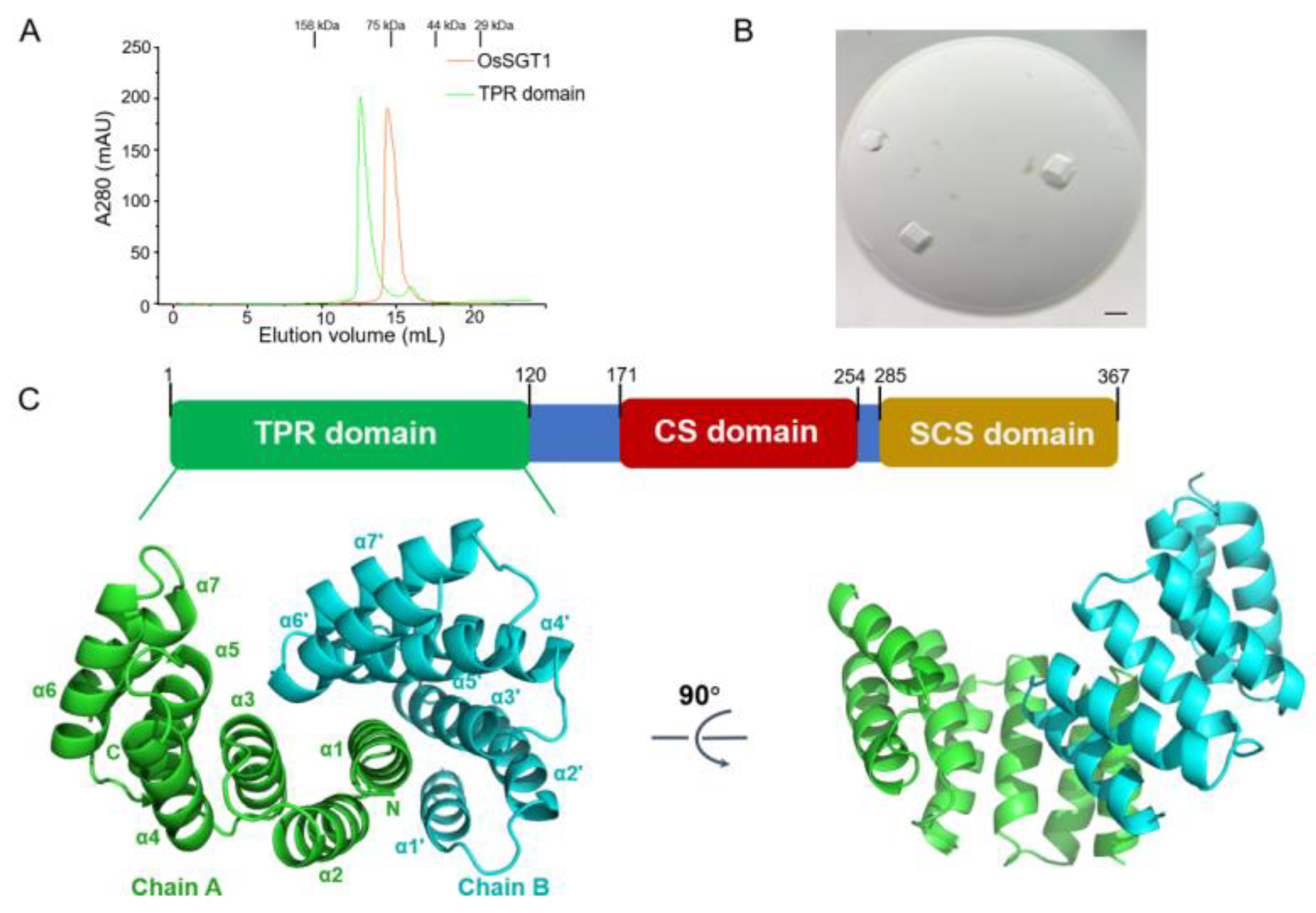
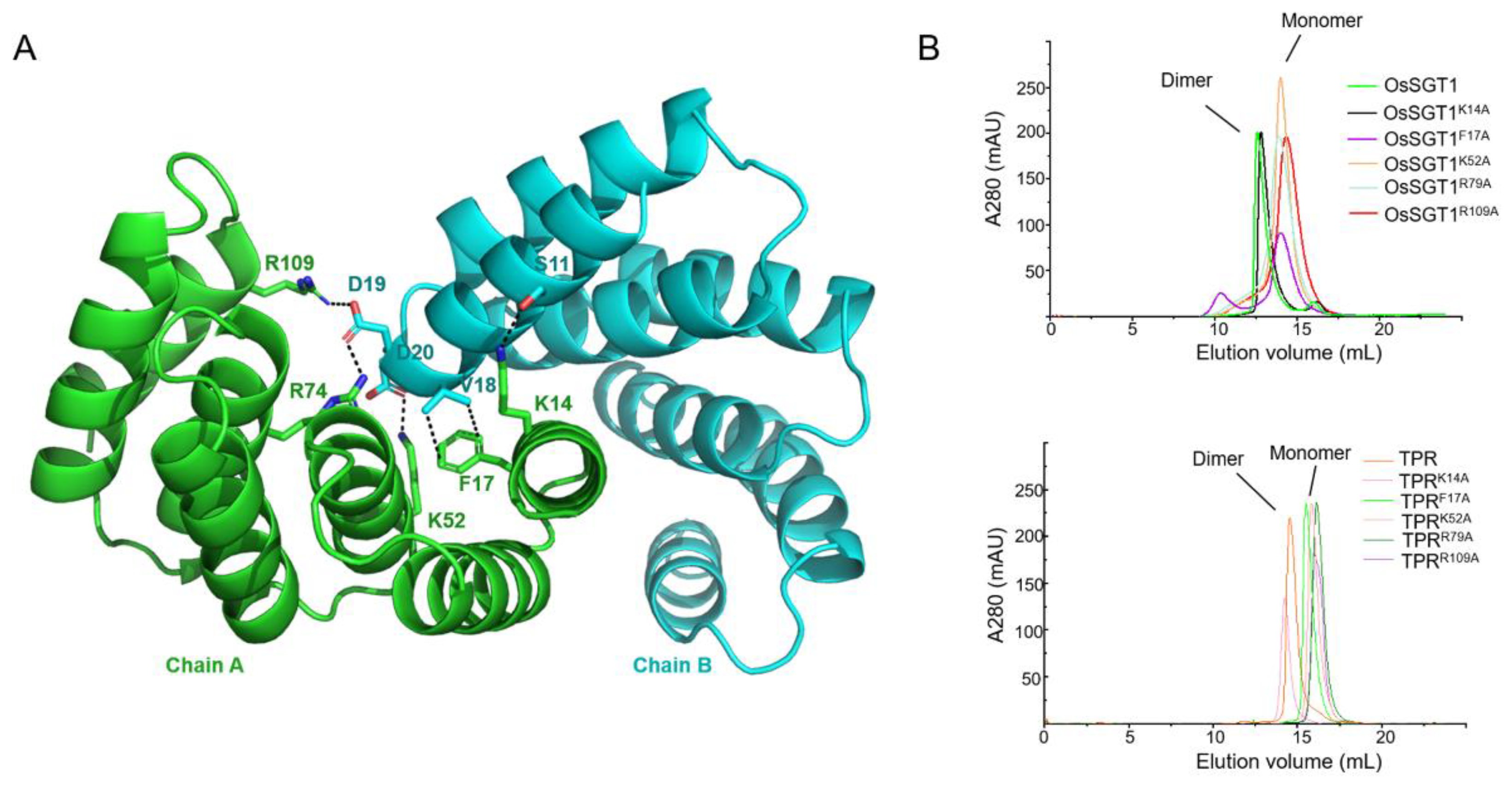
2.1. Protein Expression and Purification

The full-length OsSGT1 and TPR domain (encoding residues 1–120) was amplified by PCR using sticky-end cloning, which was ligated into the NcoI/XhoI-digested pETM13 expression vector to generate an N-terminal hexahistidine-tagged fusion construct [17]. Sequence-validated recombinant vectors were transformed into Escherichia coli BL21 (DE3) competent cells for heterologous expression. Transformed cultures were grown at 37 °C in Lysogeny Broth (LB) medium until the mid-log phase (OD600 = 0.6–0.8) was reached, followed by induction with 0.4 mM isopropyl β-D-1-thiogalactopyranoside (IPTG) at 18 °C for 16 h. Cells were harvested by centrifugation and resuspended in lysis buffer (20 mM Tris-HCl, 500 mM NaCl, pH 7.0) and lysed via ultrasonication at an amplitude of 40% (5 cycles: 3 s on and 3 s off). The supernatants of OsSGT1 and TPR domain proteins collected by centrifugation (12,000× g, 20 min, 4 °C) were applied to a Ni-NTA agarose column pre-equilibrated with lysis buffer. His-tagged proteins were eluted with a linear gradient of 50–500 mM imidazole in lysis buffer. Proteins were further purified by size-exclusion chromatography (SEC) on a Superdex 200 Increase column equilibrated with a gel-filtration buffer (20 mM Tris-HCl, 150 mM NaCl, pH 7.0). Purified OsSGT1 and TPR were concentrated to 13 mg/mL with Amicon Ultra-15 centrifugal devices (Millipore) and stored at −80 °C for crystallization trials.

2.2. Crystallization and X-Ray Diffraction Analysis

Initial crystallization screening was performed using commercially available sparse-matrix kits (Hampton Research, Aliso Viejo, CA, USA) via the sitting-drop vapor diffusion method at 18 °C with an Oryx4 robotic system (Douglas Instruments Limited, Berkshire, UK). Using the sitting-drop vapor diffusion method, 0.5 μL drops (composed of a 0.25 μL protein solution [13 mg/mL in the gel-filtration buffer] and 0.25 μL reservoir solution) were equilibrated over 35 μL of reservoir solution. Microcrystals of TPR proteins were initially observed in multiple conditions from the index screen. Crystal morphology optimization was achieved by iteratively adjusting precipitant concentrations (PEG 3350: 18–28% w/v) and additive screening (0.1–0.3 M MgCl2). The final crystallization condition contained 0.2 M of magnesium chloride hexahydrate, 0.1 M of Tris-HCl (pH 8.5), and 25% (w/v) PEG 3350, yielding diffraction-quality single crystals within 7 days. Crystals were briefly soaked in a reservoir solution containing 20% (v/v) glycerol as the cryoprotectant and flash-cooled in liquid nitrogen. X-ray diffraction data were collected at the Shanghai Synchrotron Radiation Facility (beamline BL18U1) (Shanghai Synchrotron Radiation Facility, Shanghai, China) using a wavelength of 0.9792 Å. A Pilatus3 6 M detector recorded diffraction images at 100 K. Crystals were diffracted to the 1.53 Å resolution and belonged to space group P3, with unit cell parameters (a = 93.493 Å, b = 93.493 Å, c = 32.941 Å). The dataset was indexed, integrated, and scaled using HKL-2000 _(version v722) [18]. Data quality was valued with phenix.xtriage (version 1.18.2-3874) [19].

2.3. Structure Determination and Refinement

The initial model of TPR was generated by a molecular replacement using 7OBE [20] as the search model by Phaser (version 1.18.2-3874) [21]. The structure was manually rebuilt using Coot (version 0.8.9.2) and refined with Phenix in an iterative manner [22,23]. Water molecules were built stepwise into the structure during the refinement. Structural figures were generated by PyMOL software (version 2.5). Refinement statistics are detailed in Table 1. The atomic coordinate of the structure was deposited in the PDB (accession code 9UIB).

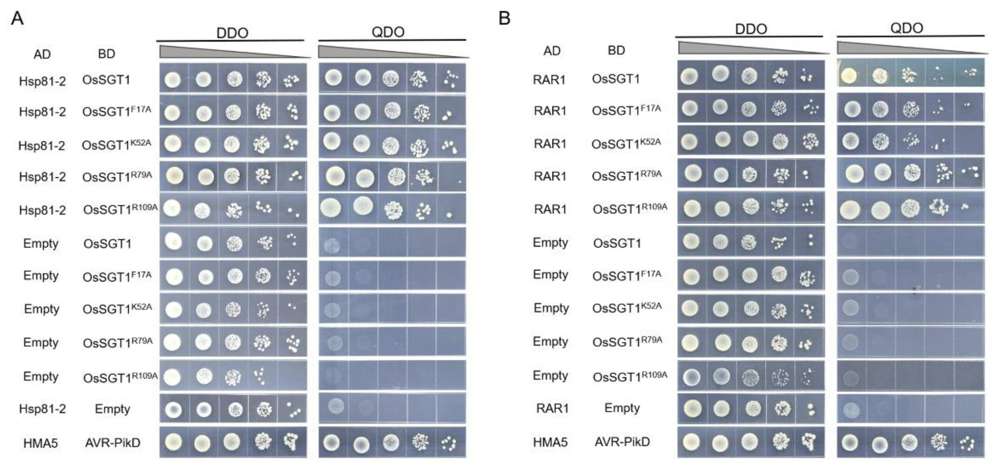
2.4. Yeast Two-Hybrid Assay

The coding sequences of OsSGT1 and OsTPR were inserted into the GAL4 DNA-Binding Domain (BD) vector pGBKT7 to generate bait proteins, while the coding sequences of OsHSP81-2 and OsRAR1 were cloned into the GAL4 Activation Domain (AD) vector pGADT7 to create prey proteins. The prey vectors were co-transformed with their corresponding bait vectors into the Saccharomyces cerevisiae strain AH109. Yeast cells expressing both plasmids were selected by growth on a synthetically defined (SD) medium lacking tryptophan and leucine (SD/-Trp/-Leu). Protein–protein interactions were verified by plating these transformants onto the SD medium with a further deficiency in histidine and adenine (SD/-Trp/-Leu/-His-Ade) at 30 °C for 3–5 days.

2.5. Circular Dichroism Spectroscopy

The purified wild-type and mutant proteins were analyzed by circular dichroism (CD) spectroscopy using a Chirascan-plus spectropolarimeter (Applied Photophysics Limited, Surrey, UK) at room temperature, with a protein concentration of approximately 0.2 mg/mL. CD spectra were collected in the wavelength range of 180–280 nm, and the final data were obtained by subtracting the blank spectrum of the corresponding buffer.

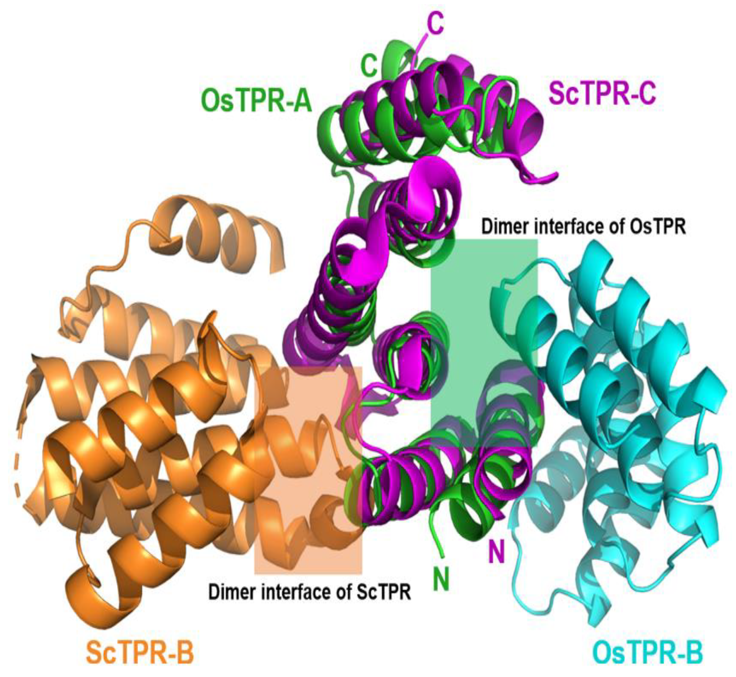
3. Results and Discussion

The recombinant protein TPR domain or full-length OsSGT1 proteins both formed homodimers, which were consistent with size-exclusion chromatography results (Figure 1A). We only achieved the crystals of the TPR domain and determined the crystal structure of the TPR domain at a 1.53 Å resolution, belonging to space group P3 (Figure 1B). All residues were matched into the electron density map, and detailed statistics on data collection and refinement are summarized in Table 1. The overall structure of the TPR domain (Protein Data Bank (PDB): 9UIB) adopts a canonical fold consisting of seven α-helices (α1–α7) interconnected by short loops (Figure 1C). Within the asymmetric unit, two TPR molecules were assembled into a homodimer via interactions of the loop region and α1 helix of one subunit with the concave surface of its adjacent counterpart (Figure 1C). This dimeric arrangement stabilizes the interface observed in the crystal lattice. Comparing this to the structure of the Saccharomyces cerevisiae SGT1-TPR domain, OsTPR was superimposed by the ScSGT1-TPR domain (PDB: 5AN3) with an RMSD of 2.6 Å for all main chain atoms, indicating conserved structural features (Figure 2). However, the dimerization interface of OsTPR was positioned opposite to that of ScTPR, indicating divergent dimerization mechanisms between plants and yeast (Figure 2).
The structural analysis of the TPR domain revealed that its dimerization was driven by salt bridges and hydrophobic interactions (Figure 3A). Specifically, the core residues K14, K52, R79, and R109 in chain A stabilized the interaction interface by forming hydrogen bonds and salt bridges with side chains of residues S11, D20, and D19 in adjacent subunits, respectively (Figure 3A). Furthermore, residue F17 participated in hydrophobic interactions with V18 to further stabilize the dimer interface. To validate the self-interaction interface, we compared the oligomerization states of wild-type and mutant proteins (the TPR domain or full-length OsSGT1) using size-exclusion chromatography. The gel filtration assay revealed that the wild-type TPR domain (−35 kDa) and full-length OsSGT1 (−84 kDa) eluted as stable dimers, while mutants (K52A, R79A, R109A, and F17A) migrated as monomers with apparent molecular masses of 16 kDa (isolated TPR mutants) and 42 kDa (full-length SGT1 mutants) (Figure 3B). These results demonstrate that the disruption of interfacial residues (K52A, R79A, R109A, or F17A), but not K14A, abolishes SGT1 dimerization in vitro, which is consistent with structural models of the TPR domain. Circular dichroism (CD) analysis in the far UV region of the spectra showed characteristic α-helical signatures (minima at −222 nm) for both wild-type and mutant TPR proteins, indicating that the mutations did not significantly perturb the overall structural stability (Figure S1). Sequence alignment confirmed that these critical residues (K52, R79, R109, F17A) are highly conserved across plant species (Figure 4), underscoring the functional importance of the TPR domain in mediating SGT1 oligomerization.
In plants, HSP90s, RAR1, and SGT1 proteins directly interact to form the ternary HRS complex, which stabilizes NLR proteins and regulates NLR-mediated resistance to diverse pathogens [10,24,25,26,27]. To further confirm whether the formation of this complex depends on SGT1 dimerization, we performed Y2H assays to analyze the interactions between OsSGT1 (wild-type or dimerization-disrupted mutants) and OsHSP81-2 or RAR1, respectively. When wild-type (WT) or mutant OsSGT1 proteins were used as bait, and OsHSP81-2 or RAR1 were used as prey, no obvious autoactivation was observed on TDO or QDO media (Figure 5). Both WT and mutants interacted with OsHSP81-2 and RAR1, indicating that SGT1 oligomerization is not required for HRS complex assembly (Figure 5).
The HRS chaperone complex is evolutionarily conserved across eukaryotes and plays diverse biological functions, including the assembly of kinetochore in eukaryotes and NLR-triggered plant immunity [3,25,28,29,30]. Previous structural and functional studies have shown that the TPR domain of ScSGT1 directly binds to the BTB domain of Skp1, thereby coupling ScSGT1 to both Hsp90-dependent signaling pathways and Skp1/Cullin/F-box (SCF) ubiquitin ligase-mediated protein degradation pathways [31,32]. Notably, the TPR domain is critical for the dimerization of full-length Sgt1 in yeast [32,33]. In this study, we provided the structural characterization of the rice OsSGT1-TPR domain and elucidated its role in mediating self-association in solution. Mutations targeting the oligomerization region abolished OsSGT1 dimerization. In addition, structural analysis revealed that the dimer interface is evolutionarily conserved in plants but different in yeast. The self-association interface of OsSGT1 overlaps spatially with the Skp1-binding interface of ScSGT1 (Figure S2). This structural divergence leads to the inability of OsSGT1 to bind Skp1 (Figure S3). However, dimerization-deficient mutants remain bound to OsHSP81-2 (HSP90 ortholog) and OsRAR1, indicating that TPR-mediated homodimerization is dispensable for partner recognition but contributes to scaffold complex stability. These findings indicate that the evolutionary conservation of this dimer interface in plant lineages reflects its critical role in stabilizing immune signaling complexes.

Supplementary Materials

The supporting information can be downloaded at: https://www.mdpi.com/article/10.3390/cryst15060543/s1, Figure S1: CD spectroscopy analysis of TPR domain and its mutant proteins; Figure S2: Structural comparison of OsTPR dimer with the ScTPR-Skp1 complex; Figure S3: Y2H validation of the interaction between TPR domain and OsSkp1.

Author Contributions

Conceptualization, J.L. and X.Z.; methodology, Y.C. and X.Z.; software, Y.C. and X.Z.; validation, Y.C., L.J., Y.Q. and Y.Y.; formal analysis, Y.C. T.L. and J.J.; investigation, Y.C.; resources, Y.C., Y.Q., C.Q., T.L. and J.J.; data curation, Y.C.; writing—original draft preparation, Y.C.; writing—review and editing, Y.C. and X.Z.; visualization, Y.C.; supervision, X.Z.; project administration, X.Z.; funding acquisition, J.L. and X.Z. All authors have read and agreed to the published version of the manuscript.

Funding

This research was funded by grants from the Natural Science Foundation of China and the Development Program of China for Young Scientists, grant number (Grant No. 32472507, 32102160 and 2022YFD1401400).

Data Availability Statement

All data supporting this study are included in the article and its Supplementary Materials. Additional datasets are available from the corresponding author upon reasonable request.

Acknowledgments

We thank the staff members at BL18U1 beamlines of the National Facility for Protein Science in Shanghai for their technical support in X-ray diffraction data collection and analysis.

Conflicts of Interest

The authors declare no conflicts of interest.

References

  1. Jones, J.D.G.; Dangl, J.L. The Plant Immune System. Nature 2006, 444, 323–329. [Google Scholar] [CrossRef] [PubMed]
  2. Chapman, A.V.E.; Elmore, J.M.; McReynolds, M.; Walley, J.W.; Wise, R.P. SGT1-Specific Domain Mutations Impair Interactions with the Barley MLA6 Immune Receptor in Association with Loss of NLR Protein. Mol. Plant-Microbe Interact. 2022, 35, 274–289. [Google Scholar] [CrossRef] [PubMed]
  3. Shirasu, K. The HSP90-SGT1 Chaperone Complex for NLR Immune Sensors. Annu. Rev. Plant Biol. 2009, 60, 139–164. [Google Scholar] [CrossRef] [PubMed]
  4. Chen, Z.; Wu, Q.; Tong, C.; Chen, H.; Miao, D.; Qian, X.; Zhao, X.; Jiang, L.; Tao, X. Characterization of the Roles of SGT1/RAR1, EDS1/NDR1, NPR1, and NRC/ADR1/NRG1 in Sw-5b-Mediated Resistance to Tomato Spotted Wilt Virus. Viruses 2021, 13, 1447. [Google Scholar] [CrossRef]
  5. Yu, G.; Xian, L.; Xue, H.; Yu, W.; Rufian, J.S.; Sang, Y.; Morcillo, R.J.L.; Wang, Y.; Macho, A.P. A Bacterial Effector Protein Prevents MAPK-Mediated Phosphorylation of SGT1 to Suppress Plant Immunity. PLoS Pathog. 2020, 16, e1008933. [Google Scholar] [CrossRef]
  6. Wang, Y.; Liu, C.; Du, Y.; Cai, K.; Wang, Y.; Guo, J.; Bai, X.; Kang, Z.; Guo, J. A Stripe Rust Fungal Effector PstSIE1 Targets TaSGT1 to Facilitate Pathogen Infection. Plant J. 2022, 112, 1413–1428. [Google Scholar] [CrossRef]
  7. Yu, G.; Xian, L.; Zhuang, H.; Macho, A.P. SGT1 Is Not Required for Plant LRR-RLK-Mediated Immunity. Mol. Plant Pathol. 2021, 22, 145–150. [Google Scholar] [CrossRef]
  8. Siligardi, G.; Zhang, M.; Prodromou, C. The Stoichiometric Interaction of the Hsp90-Sgt1-Rar1 Complex by CD and SRCD Spectroscopy. Front. Mol. Biosci. 2018, 4, 95. [Google Scholar] [CrossRef]
  9. Zhang, D.; Yang, X.; Wen, Z.; Li, Z.; Zhang, X.; Zhong, C.; She, J.; Zhang, Q.; Zhang, H.; Li, W.; et al. Proxitome Profiling Reveals a Conserved SGT1-NSL1 Signaling Module That Activates NLR-Mediated Immunity. Mol. Plant 2024, 17, 1369–1391. [Google Scholar] [CrossRef]
  10. Zhang, M.; Kadota, Y.; Prodromou, C.; Shirasu, K.; Pearl, L.H. Structural Basis for Assembly of Hsp90-Sgt1-CHORD Protein Complexes: Implications for Chaperoning of NLR Innate Immunity Receptors. Mol. Cell 2010, 39, 269–281. [Google Scholar] [CrossRef]
  11. Li, D.; Zhou, J.; Zheng, C.; Zheng, E.; Liang, W.; Tan, X.; Xu, R.; Yan, C.; Yang, Y.; Yi, K.; et al. OsTGAL1 Suppresses the Resistance of Rice to Bacterial Blight Disease by Regulating the Expression of Salicylic Acid Glucosyltransferase OsSGT1. Plant Cell Environ. 2022, 45, 1584–1602. [Google Scholar] [CrossRef] [PubMed]
  12. Yan, J.; Zhang, Y.; Ding, Y. Binding Mechanism between Hsp90 and Sgt1 Explored by Homology Modeling and Molecular Dynamics Simulations in Rice. J. Mol. Model. 2012, 18, 4665–4673. [Google Scholar] [CrossRef] [PubMed]
  13. Umemura, K.; Satou, J.; Iwata, M.; Uozumi, N.; Koga, J.; Kawano, T.; Koshiba, T.; Anzai, H.; Mitomi, M. Contribution of Salicylic Acid Glucosyltransferase, OsSGT1, to Chemically Induced Disease Resistance in Rice Plants. Plant J. 2009, 57, 463–472. [Google Scholar] [CrossRef] [PubMed]
  14. Wang, Y.; Gao, M.; Li, Q.; Wang, L.; Wang, J.; Jeon, J.-S.; Qu, N.; Zhang, Y.; He, Z. OsRAR1 and OsSGT1 Physically Interact and Function in Rice Basal Disease Resistance. Mol. Plant-Microbe Interact. 2008, 21, 294–303. [Google Scholar] [CrossRef]
  15. Li, R.; Zheng, W.; Yang, R.; Hu, Q.; Ma, L.; Zhang, H. OsSGT1 Promotes Melatonin-Ameliorated Seed Tolerance to Chromium Stress by Affecting the OsABI5–OsAPX1 Transcriptional Module in Rice. Plant J. 2022, 112, 151–171. [Google Scholar] [CrossRef]
  16. Kang, B.; Zhang, Z.; Wang, L.; Zheng, L.; Mao, W.; Li, M.; Wu, Y.; Wu, P.; Mo, X. OsCYP2, a Chaperone Involved in Degradation of Auxin-Responsive Proteins, Plays Crucial Roles in Rice Lateral Root Initiation. Plant J. 2013, 74, 86–97. [Google Scholar] [CrossRef]
  17. Zeng, G. Sticky-End PCR: New Method for Subcloning. BioTechniques 1998, 25, 206–208. [Google Scholar] [CrossRef]
  18. Otwinowski, Z.; Minor, W. [20] Processing of X-Ray Diffraction Data Collected in Oscillation Mode. Methods Enzymol. 1997, 276, 307–326. [Google Scholar]
  19. Xtriage and Fest: Automatic Assessment of X-Ray Data and Substructure Structure Factor Estimation. Available online: https://www.researchgate.net/publication/228661841_Xtriage_and_Fest_automatic_assessment_of_X-ray_data_and_substructure_structure_factor_estimation (accessed on 30 May 2025).
  20. von Horsten, S.; Essen, L.-O. Conformational Change of Tetratricopeptide Repeats Region Triggers Activation of Phytochrome-Associated Protein Phosphatase 5. Front. Plant Sci. 2021, 12, 733069. [Google Scholar] [CrossRef]
  21. McCoy, A.J.; Grosse-Kunstleve, R.W.; Adams, P.D.; Winn, M.D.; Storoni, L.C.; Read, R.J. Phaser Crystallographic Software. J. Appl. Crystallogr. 2007, 40, 658–674. [Google Scholar] [CrossRef]
  22. Emsley, P.; Cowtan, K. Coot: Model-Building Tools for Molecular Graphics. Acta Crystallogr. Sect. D-Struct. Biol. 2004, 60, 2126–2132. [Google Scholar] [CrossRef] [PubMed]
  23. Schomaker, V.; Trueblood, K.N. On the Rigid-Body Motion of Molecules in Crystals. Acta Crystallogr. Sect. B Struct. Sci. Cryst. Eng. Mater. 1968, 24, 63–76. [Google Scholar] [CrossRef]
  24. Cantu, D.; Yang, B.; Ruan, R.; Li, K.; Menzo, V.; Fu, D.; Chern, M.; Ronald, P.C.; Dubcovsky, J. Comparative Analysis of Protein-Protein Interactions in the Defense Response of Rice and Wheat. BMC Genom. 2013, 14, 166. [Google Scholar] [CrossRef]
  25. Tai, Y.-S. Interactome of Signaling Networks in Wheat: The Protein–Protein Interaction between TaRAR1 and TaSGT1. Mol. Biol. Rep. 2008, 35, 337–343. [Google Scholar] [CrossRef]
  26. Seo, Y.-S.; Lee, S.-K.; Song, M.-Y.; Suh, J.-P.; Hahn, T.-R.; Ronald, P.; Jeon, J.-S. The HSP90-SGT1-RAR1 Molecular Chaperone Complex: A Core Modulator in Plant Immunity. J. Plant Biol. 2008, 51, 1–10. [Google Scholar] [CrossRef]
  27. Shen, Q.; Schulze-Lefert, P. Rumble in the Nuclear Jungle: Compartmentalization, Trafficking, and Nuclear Action of Plant Immune Receptors. EMBO J. 2007, 26, 4293–4301. [Google Scholar] [CrossRef]
  28. Kitagawa, K.; Skowyra, D.; Elledge, S.J.; Harper, J.W.; Hieter, P. SGT1 Encodes an Essential Component of the Yeast Kinetochore Assembly Pathway and a Novel Subunit of the SCF Ubiquitin Ligase Complex. Mol. Cell 1999, 4, 21–33. [Google Scholar] [CrossRef]
  29. Botër, M.; Amigues, B.; Peart, J.; Breuer, C.; Kadota, Y.; Casais, C.; Moore, G.; Kleanthous, C.; Ochsenbein, F.; Shirasu, K.; et al. Structural and Functional Analysis of SGT1 Reveals That Its Interaction with HSP90 Is Required for the Accumulation of Rx, an R Protein Involved in Plant Immunity. Plant Cell 2007, 19, 3791–3804. [Google Scholar] [CrossRef]
  30. Kadota, Y.; Shirasu, K. The HSP90 Complex of Plants. Biochim. Biophys. Acta (BBA)-Mol. Cell Res. 2012, 1823, 689–697. [Google Scholar] [CrossRef]
  31. Zheng, N.; Schulman, B.A.; Song, L.; Miller, J.J.; Jeffrey, P.D.; Wang, P.; Chu, C.; Koepp, D.M.; Elledge, S.J.; Pagano, M.; et al. Structure of the Cul1–Rbx1–Skp1–F boxSkp2 SCF Ubiquitin Ligase Complex. Nature 2002, 416, 703–709. [Google Scholar] [CrossRef]
  32. Willhoft, O.; Kerr, R.; Patel, D.; Zhang, W.; Al-Jassar, C.; Daviter, T.; Millson, S.H.; Thalassinos, K.; Vaughan, C.K. The Crystal Structure of the Sgt1-Skp1 Complex: The Link between Hsp90 and Both SCF E3 Ubiquitin Ligases and Kinetochores. Sci. Rep. 2017, 7, 41626. [Google Scholar] [CrossRef] [PubMed]
  33. Nyarko, A.; Mosbahi, K.; Rowe, A.J.; Leech, A.; Boter, M.; Shirasu, K.; Kleanthous, C. TPR-Mediated Self-Association of Plant SGT1. Biochemistry 2007, 46, 11331–11341. [Google Scholar] [CrossRef]
Figure 1. The crystal structural characterization of the OsSGT1-TPR protein. (A) Size-exclusion chromatographic analysis revealed that both full-length OsSGT1 and its TPR domain form stable dimers in solution, as evidenced by elution volumes corresponding to their theoretical dimeric molecular weights. (B) Representative crystals of the recombinant OsSGT1-TPR domain were obtained under optimized crystallization conditions. The scale bar is 0.1 mm. (C) Schematic representations of OsSGT1 (up panel) and the dimeric TPR domain structure (lower panel), indicating conserved domains: the TPR, SGS, and CS domain in the classical architecture of OsSGT1. The overall structure of the TPR domain was shown in cartoon representations (90° rotation view). The overall dimeric structure of the TPR domain in the ribbon diagram presented two orthogonal views (0° and 90° rotation), with secondary structural elements clearly resolved.
Crystals 15 00543 g001
Figure 2. A comparative analysis of dimer interfaces in OsTPR and yeast ScTPR. The dimer interface of OsTPR (highlighted in green and blue) was demarcated by a green box, whereas the corresponding interface in ScTPR was enclosed in an orange box. This visual distinction emphasizes structural divergences between the two orthologs.
Crystals 15 00543 g002
Figure 3. The details of the OsTPR dimer interface. (A) Structural details of key residues mediating OsTPR dimer stabilization. Key hydrogen bonds and hydrophobic interactions are highlighted with dashed lines. (B) Size-exclusion chromatography profiles in vitro comparing the oligomerization states of wild-type and interface-disrupting mutants (the TPR domain and full-length OsSGT1), as evidenced by elution volumes corresponding to their theoretical molecular weights.
Crystals 15 00543 g003
Figure 4. The evolutionary conservation of SGT1 orthologs across plant species. The multiple sequence alignment of SGT1 proteins from six species: Oryza sativa OsSGT1 (AAF18438.1), Zea mays ZmSGT1 (AKD95366.1), Glycine max GmSGT1-1 (ACI31549.1), Nicotiana benthamiana NbSGT1 (AAW82048.1), Triticum aestivum TaSGT1 (ABQ23992.1), and Solanum tuberosum StSGT1 (AAU04979.1). Secondary structure elements (α-helices depicted as coils and β-strands as arrows) are labeled above the alignment. Residues critical for SGT1 dimerization are marked with blue triangles. Sequence alignment was performed using MEGA (version 4.21.4), with secondary structure predictions and visual annotations generated by ESPript 3.0.
Crystals 15 00543 g004
Figure 5. The yeast two-hybrid (Y2H) validation of OsSGT1 interactions with Hsp81-2 or RAR1. (A) Interaction analysis of OsSGT1 with Hsp81-2. (B) Interaction analysis of OsSGT1 with RAR1. Different combinations were co-transformed in the yeast AH109 strain, and transformants were selected on the double dropout medium (DDO; SD/-Leu/-Trp). Protein–protein interactions were assessed via plating onto a quadruple dropout medium (QDO; SD/-Leu/-Trp/-His/-Ade). Each test was repeated in triplicate, using independent biological replicates.
Crystals 15 00543 g005
Table 1. Data collection and refinement statistics.
BeamlineRRSF BL18U1
Wavelength0.9792
Resolution range22.46–1.53 (1.59–1.53) a
Space groupP 3
Unit cell93.493 93.493 32.941 90 90 120
Total reflections478,266
Unique reflections48,592 (4851)
Multiplicity18.9 (17.0)
Completeness (%)99.95 (100.00)
Mean I/sigma(I)48.41 (3.40)
Wilson B-factor20.8
Rmerge b0.075 (0.780)
Rmeas0.091 (0.748)
Rp.i.m0.021 (0.180)
CC1/20.998 (0.879)
Reflections used in refinement48,589 (4851)
Reflections used for Rfree2001 (198)
Rwork c0.176 (0.209)
Rfree0.190 (0.213)
Number of non-hydrogen atoms2039
Macromolecules1777
Solvent262
No. of protein residues231
R.m.s. deviations bond lengths (Å) (bonds)0.006
R.m.s. deviations bond angles (°) (angles)0.84
Ramachandran plot (%) d
Ramachandran favored97.36
Ramachandran allowed2.64
Ramachandran outliers0.00
Rotamer outliers0.00
Clashscore3.43
Average B-factor28.1
Macromolecules26.75
Solvent37.57
Number of TLS groups8
a: The numbers in parenthesis are for the highest resolution data shell. b: Rmerge =∑hklI (|Ii (hkl)– < I (hkl) > |)/∑hkliIi (hkl). c: Rwork = ∑hkl (||Fobs|–|Fcalc||)/∑Ihkl|Fobs|. d: As evaluated by MolProbity.
Disclaimer/Publisher’s Note: The statements, opinions and data contained in all publications are solely those of the individual author(s) and contributor(s) and not of MDPI and/or the editor(s). MDPI and/or the editor(s) disclaim responsibility for any injury to people or property resulting from any ideas, methods, instructions or products referred to in the content.

