Article

Synthesis, Crystal Structure, DFT Analysis and Docking Studies of a Novel Spiro Compound Effecting on EGR-1-Regulated Gene Expression

1
Department of Biological Sciences, Konkuk University, Seoul 05029, Republic of Korea
2
Cancer and Metabolism Institute, Konkuk University, Seoul 05029, Republic of Korea
3
Division of Bioscience and Biotechnology, BMIC, Konkuk University, Seoul 05029, Republic of Korea
4
Division of Chemistry and Bio-Environmental Sciences, Seoul Women’s University, Seoul 01797, Republic of Korea
5
Department of Applied Chemistry, Dongduk Women’s University, Seoul 02748, Republic of Korea
*
Authors to whom correspondence should be addressed.
Crystals 2025, 15(4), 338; https://doi.org/10.3390/cryst15040338
Submission received: 10 March 2025 / Revised: 21 March 2025 / Accepted: 23 March 2025 / Published: 2 April 2025
(This article belongs to the Topic Bioinformatics in Drug Design and Discovery—2nd Edition)

Abstract

:
The spiro compound, 5,5′-dimethoxy-1,3-bis(3-(trifluoromethyl)phenyl)-3,3a-dihydro-1H-spiro[cyclopenta[a]indene-2,2′-indene]-1′,8(3′H,8aH)-dione (4), was synthesized and identified by NMR spectroscopy, mass spectrometry, and X-ray crystallography. Compound 4, C36H26F6O4, was crystallized in the triclinic space group P-1with the cell parameters a = 8.8669(5) Å, b = 10.5298(8) Å, c = 17.0135(11) Å, α = 91.396(2)°, β = 90.490(2)°, γ = 109.235°, V = 1499.14(17) Å3, Z = 2. In an asymmetric unit, two molecules are packed by short contacts to form an inversion dimer. The molecules are linked into chains along the a- and b-axis directions by additional short contacts in the crystal. Compound 4 was synthesized by the dimerization of (E)-5-methoxy-2-(3-(trifluoromethyl)benzylidene)-2,3-dihydro-1H-inden-1-one (3). (E)-5-Methoxy-2-(3-methoxybenzylidene)-2,3-dihydro-1H-inden-1-one (5), one of the analogs of compound 3, was compared with compound 4 based on in vitro experiments, DFT calculations, and an in silico docking study. The HOMO/LUMO energy difference and binding energy difference between the two compounds are consistent with the results obtained from an in vitro assay where 4 showed a better effect than 5. To evaluate the biological activity of 4, we examined its inhibitory effects on Early Growth Respone-1 (EGR-1)-regulated gene expression in HaCaT keratinocytes. Treatment of cells with 4 reduced interleukin-4 (IL-4)-induced thymic stromal lymphopoietin (TSLP) mRNA levels, as revealed by reverse transcription-polymerase chain reaction and quantitative real-time PCR. Furthermore, the electrophoretic mobility shift assay demonstrated that 4 inhibited IL-4-induced DNA binding of EGR-1 to the promoter region of the TSLP gene.

1. Introduction

Early Growth Response 1 (EGR-1) is a transcription factor rapidly induced by various stimuli such as cytokines, growth factors, and DNA damage signals [1,2]. It plays a key mediator in multiple biological processes like proliferation, apoptosis, and inflammation [3,4,5,6]. In the skin, EGR-1 is highly expressed in damaged regions [7] and regulates genes involved in inflammation and immune responses, including thymic stromal lymphopoietin (TSLP) expression induced by interleukin (IL)-33 [8,9], psoriasin expression by IL-17 [10], and kallikrein-related peptidase 7 (KLK7) expression by IL-13 stimulation in keratinocytes [11,12]. Immune cell infiltration in atopic dermatitis-like skin lesions was attenuated in EGR-1-knockout mice, and TNFα-induced expression of inflammatory cytokines, including TSLP, was significantly reduced by the knockdown of EGR-1 expression in HaCaT keratinocytes [11]. Additionally, (E)-2-(2-(4-methoxystyryl)-4,6-dimethoxyphenyl)-3-hydroxy-6-nitro-4H-chromen-4-one (named AB1711), a small-molecule inhibitor targeting the EGR-1 zinc-finger DNA-binding domains, abrogated EGR-1-regulated inflammatory cytokine expression in keratinocytes and alleviated both skin inflammation and itching in DNCB-challenged mice [13]. EGR-1 also promotes the epidermal innervation of sensory neurons in house dust mite-exposed skin lesions [14], indicating its involvement in itch signaling in inflammatory skin conditions. These findings suggest that EGR-1 contributes to atopic dermatitis pathogenesis by driving inflammatory mediator production [15]. Atopic dermatitis is a chronic inflammatory skin disorder characterized by severe itching, redness, and recurrent eczematous lesions [16]. This condition is driven by a combination of genetic, immune, and environmental factors, leading to an increased susceptibility to irritants and allergens [17]. Currently, various systemic and topical medications have been developed for the treatment of atopic dermatitis [18]. In general, medications like immunosuppressants, including steroids and calcineurin inhibitors, and Janus kinase inhibitors are highly effective in managing inflammation; however, their potential for serious side effects limit their long-term uses [18]. Therefore, targeting EGR-1 may help control excessive inflammation and provide a therapeutic strategy for chronic skin inflammatory diseases. Our previous studies have revealed that benzylidene indenone derivatives, such as compound 5, exhibit inhibitory effects on early growth response-1 (EGR-1) expression [19]. All compounds used in previous studies had strong electron-donating groups on the benzylidene indenone. For comparative studies, compounds such as 3, which have strong electron-withdrawing groups on benzylidene indenone, were designed. However, once the compound 3 was synthesized, it underwent a spontaneous dimerization reaction and was converted into a spiro compound 4. Spiro compounds have at least two molecular rings sharing one common atom [20]. Compounds with spiro ring systems have unique, complicated molecular structure and three-dimensional molecular properties when compared to simple ring compounds [21]. The three-dimensional structures of several different types spiro compounds have been reported using X-ray diffraction experiments [22,23,24]. Recent reviews have reported that various spiro scaffolds possess diverse biological and pharmaceutical properties [25,26,27,28]. In addition, spiro compounds are widely used in organic light-emitting diodes (OLEDs) [29], optoelectronics [30], and pesticides [31]. Due to this wide range of applications, methods for synthesizing various spiro compounds have been reported [32,33,34,35,36]. The three-dimensional structure of spiro compound 4 was confirmed through X-ray diffraction experiments, and the inhibitory effects of this compound on EGR-1-regulated gene expression at the cellular level were confirmed. Additional DFT calculations and in silico docking experiments will be discussed to further elucidate the physiological efficacy of compound 4.

2. Materials and Methods

2.1. Synthetic Procedures

Synthesis of 5,5′-dimethoxy-1,3-bis(3-(trifluoromethyl)phenyl)-3,3a-dihydro-1H-spiro[cyclopenta[a]indene-2,2′-indene]-1′,8(3′H,8aH)-dione (4).
5-methoxy-2,3-dihydro-1H-inden-1-one (1, 1.5 mmol, 253 mg) was dissolved in 20 mL EtOH to give a clear solution. While stirring the above solution, 1 mL of 40% KOH aqueous solution was added and then 3-(trifluoromethyl)benzaldehyde (2, 1.6 mmol, 227 μL) was added. The reaction mixture turned into a yellow suspension, which was stirred at 50 °C for 7 h. After confirming that the reaction was complete by TLC, the reaction mixture was acidified by adding 3N HCl aqueous solution to form a white solid. The resulting solid was filtered and washed with cold methanol to provide the compound 4. The solid was recrystallized from methanol solution to obtain a pure crystalline compound 4.
1H NMR (700 MHz, DMSO-d6) δ 7.70 (d, J = 7.7 Hz, 1H), 7.66 (d, J = 8.6 Hz, 1H), 7.50–7.59 (m, 6H), 7.45 (dd, J = 7.8, 7.8 Hz, 1H), 7.41 (d, J = 8.5 Hz, 1H), 7.08 (dd, J = 8.6, 2.2 Hz, 1H), 6.72 (dd, J = 8.5, 1.9 Hz, 1H), 6.57 (d, J = 1.9 Hz, 1H), 6.38 (d, J = 2.2 Hz, 1H), 4.80 (dd, J = 10.7, 8.8 Hz, 1H), 4.20 (dd, J = 10.5, 8.8 Hz, 1H), 3.86 (d, J = 10.5 Hz, 1H), 3.69 (s, 3H), 3.64 (s, 3H), 3.58 (d, J = 10.7 Hz, 1H), 2.97 (d, J = 18.0, 1H), 2.83 (d, J = 18.0, 1H); 13C NMR (175 MHz, DMSO-d6) δ 203.9, 203.6, 165.2, 165.1, 158.1, 155.4, 138.7, 137.8, 132.5, 132.2, 129.6, 129.5, 129.2, 129.0 (q, J = 30 Hz), 128.8 (q, J = 32 Hz), 128.2, 126.1, 124.9 (q, J = 3.5 Hz), 124.7, 124.6 (q, J = 3.5 Hz), 124.2 (q, J = 3.5 Hz), 124.0 (q, J = 270 Hz), 123.95 (q, J = 270 Hz), 123.8 (q, 3.5 Hz), 115.6, 115.1, 109.4, 109.2, 69.5, 58.7, 55.6, 53.4, 52.8, 45.0, 29.1. Calculated for C36H27F6O4 (M + H)+: m/z 637.1814. Found: m/z 637.1892.

2.2. Crystal Structure Determination

The single crystal used for X-ray analysis is 0.226 × 0.204 × 0.056 mm3 in size. A Bruker PHOTON 100 CMOS diffractometer supported with graphite monochromated MoKα radiation [λ = 0.71073 Å at 223(2) K] was used to collect the data. The acquired data were processed using APEX2 suite [37] and SHELXT was used for structural elucidation [38]. Bruker SAINT was utilized for cell refinement and data reduction [37]. SHELXL-2014 was used as refinement and the OLEX2 software was used as a graphical interface [38,39]. Calculation of the intermolecular interactions and molecular graphics were generated using Mercury [40]. CCDC 2,428,625 contains the supplementary crystallographic data for this paper. The data can be obtained free of charge from The Cambridge Crystallographic Data Centre via www.ccdc.cam.ac.uk/structures (accessed on 10 March 2025). Table 1 summarizes details of the crystal data and structure refinement.

2.3. Nuclear Magnetic Resonance (NMR) Spectroscopy

Nuclear Magnetic Resonance (NMR) spectra were collected on an Agilent 700 MHz NMR spectrometer for 1H- and 13C-NMR [Figures S1 and S2], and on an Agilent 400 MHz NMR spectrometer for heteronuclear multiple quantum coherence (HMQC) [41] and heteronuclear multiple bonded connectivities (HMBC) [42] [Figures S3 and S4]. The sample was dissolved in deuterated DMSO-d6 solvent, and the detailed experimental procedure followed the methods published previously [43,44].

2.4. High-Resolution Mass Spectrometry (HR/MS)

The high-resolution mass spectrum was collected on an Agilent 6550 Q-TOF MS with a dual AJS ESI source (Agilent Technologies, PaloAlto, USA) with the help of KBSI Metropolitan Seoul Center. The analysis was performed within 10 ppm of mass tolerance. The source conditions were set to a Vcap of 4.0 kV in positive ESI mode and 3.5 kV in positive ESI mode with a nozzle voltage of 1 kV, a sheath gas temperature of 250 °C, and a sheath gas flow rate of 11 mL/min, which were optimized according to the manufacturer’s instructions. The mass spectrum was recorded in the range of 100–1700 m/z using a scan rate of 1 spectra/sec [45] [Figure S5].

2.5. In Silico Docking

In silico docking was performed using AutoDock Vina (Scripps Research Institute, La Jolla, San Diego, CA, USA) [46] and the protein–ligand complexes were generated by Chimera [47]. The three-dimensional (3D) images were visualized using PyMOL (The PyMOL Molecular Graphics System, Version 3.0 Schrödinger, LLC., Cambridge, MA, USA) and the binding conditions were analyzed using LigPlot (European Bioinformatics Institute, Hinxton, Cambridge, UK) [48].

2.6. Density Functional Theory (DFT) Calculations

DFT calculations were carried out using ORCA 6.0 [49]. To generate an input file for ORCA, the Avogadro 1.2 program was used [50]. The frontier molecule orbitals (FMO) including the highest occupied molecular orbital (HOMO) and the lowest unoccupied molecular orbital (LUMO) were visualized, and their energies were generated using the IboView program [51].

2.7. Cells and Cell Culture

Human keratinocyte HaCaT cells were obtained from the Cell Line Service (Eppelheim, Germany). The cells were cultured in Dulbecco’s modified Eagle’s medium containing 10% fetal bovine serum (HyClone, Logan, UT, USA) and penicillin-streptomycin (Sigma-Aldrich, Burlington, MA, USA) at 37 °C in 5% CO2 incubator.

2.8. TSLP Gene Promoter-Reporter Assay

The construct for the TSLP gene promoter-reporter was detailed in a previous study [52]. HaCaT cells were grown in 24-well plates and transfected with 0.2 μg of the TSLP gene promoter-reporter plasmid. After 48 h, the cells were exposed to IL-4 either alone or in combination with 20 μM compounds 5 or 4. Following a 12 h treatment, the cells were collected, and reporter luciferase activity was assessed using a luminometer (Centro LB960; Berthold Tech, Bad Wildbad, Germany). The activity of the TSLP gene promoter in cells treated with IL-4 alone was set to 100%, and the effect of compounds 5 and 4 on the IL-4-induced TSLP promoter activity was expressed as a % relative to the IL-4-treated cells [52].

2.9. Reverse Transcription-PCR (RT-PCR)

Total RNA was extracted from HaCaT cells using a TRIzol RNA Extraction Kit (Invitrogen, Carlsbad, CA, USA). Complementary DNA (cDNA) was synthesized with the help of an iScript cDNA Synthesis Kit (Bio-Rad, Hercules, CA, USA). A reverse transcription-PCR (RT-PCR) assay was conducted utilizing reverse transcriptase (Promega) along with primers designed for TSLP-specific mRNA as described previously [7]: forward primer, 5′ TAG CAA TCG GCC ACA TTG CCT-3′; reverse primer, 5′-GAA GCG ACG CCA CAA TCC TTG-3. Primers for GAPDH were employed as an internal control: forward primer, 5′-CCA AGG AGT AAG AAA CCC TGG AC-3′; reverse primer, 5′-GGG CCG AGT TGG GAT AGG G-3′. The thermal cycling and the separation of amplified PCR products by electrophoresis in a 2% agarose gel containing ethidium bromide were conducted as described previously [52].

2.10. Quantitative Real-Time PCR (qPCR)

The mRNA levels of the genes were quantified using an iCycler iQ system with an iQ SYBR Green Supermix kit (Bio-Rad, Hongkong, China). Validated Q-PCR primers and SYBR Green-based fluorescent probes specific for TSLP (id: qHsaCIP0030468) and GAPDH (id: qHsaCEP0041396) were obtained from Bio-Rad. The thermal cycling conditions used for PCR were as follows: denaturation at 95 °C for 2 min, followed by 40 cycles of denaturation at 95 °C for 10 s and 60 °C for 45 s. The relative mRNA levels of TSLP were normalized to those of GAPDH using the software provided by the manufacturer.

2.11. Electrophoretic Mobility Shift Assay (EMSA)

EMSA was conducted using a LightShift Chemiluminescence EMSA kit (Thermo Fisher Scientific, Waltham, MA, USA), following the prescribed instructions from the manufacturer. A biotin-labeled deoxyoligonucleotide probe corresponding to the EBS in the TSLP gene promoter (5′-CAA AAA GGA GGA AGG TGA GGG AA-biotin-3′) was prepared as described previously [52]. HaCaT keratinocytes were treated with 20 ng/mL IL-4 with or without 10 and 20 μM 4 for 2 h. Nuclear extracts (3 μg/sample) were prepared and incubated with 50 fmole biotin-labeled EBS probes. For the competition, 2.5 pmol of the unlabeled EBS probe was added. DNA–protein complexes were separated using non-denaturing 6% polyacrylamide gels, and the protein-DNA at the EBS was identified with streptoavidin-conjugated horseradish peroxidase. Following the guidelines provided by the manufacturer, the reactive bands were observed through.

3. Results and Discussion
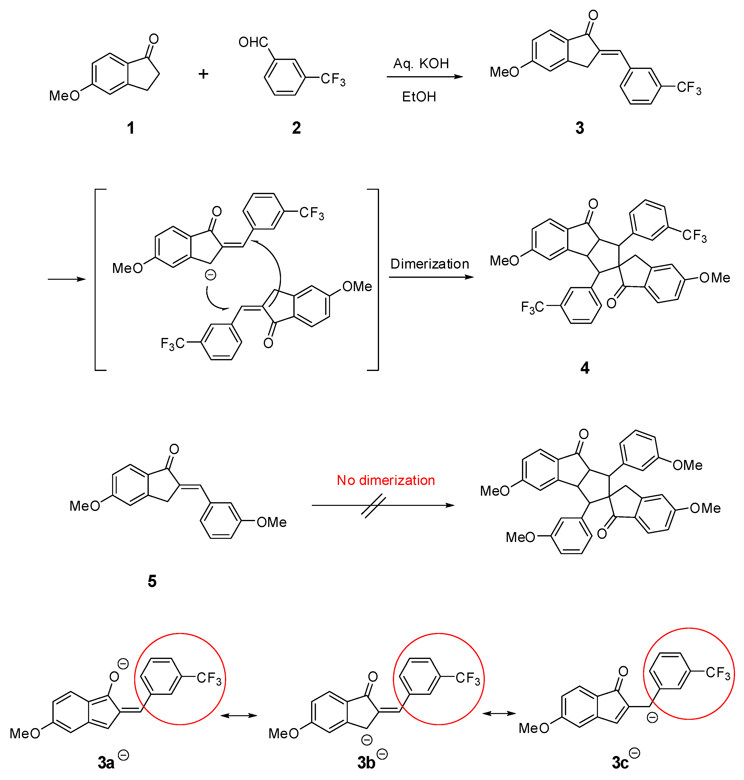
3.1. Synthesis

The Claisen–Schmidt condensation reaction between 5-methoy-1-indanone (1) and 3-(trifluoromethyl)benzaldehyde (2) in the presence of a basic aqueous KOH solution gave a benzylidene indenone product (3). When the benzylidene compound has a strong electron-withdrawing group on the phenyl ring (red circle in Scheme 1) bonded to the carbon–carbon double bond, such as compound 3, spiro compound 4 is formed through dimerization by an intermolecular spontaneous Michael reaction. The electron-withdrawing group enables the formation of resonance-stabilized anions such as anions 3a-3c under basic conditions. However, compound 5, which has an electron-donating group on the phenyl ring bonded to the carbon–carbon double bond, does not easily form anion and thus does not undergo dimerization [Scheme 1].

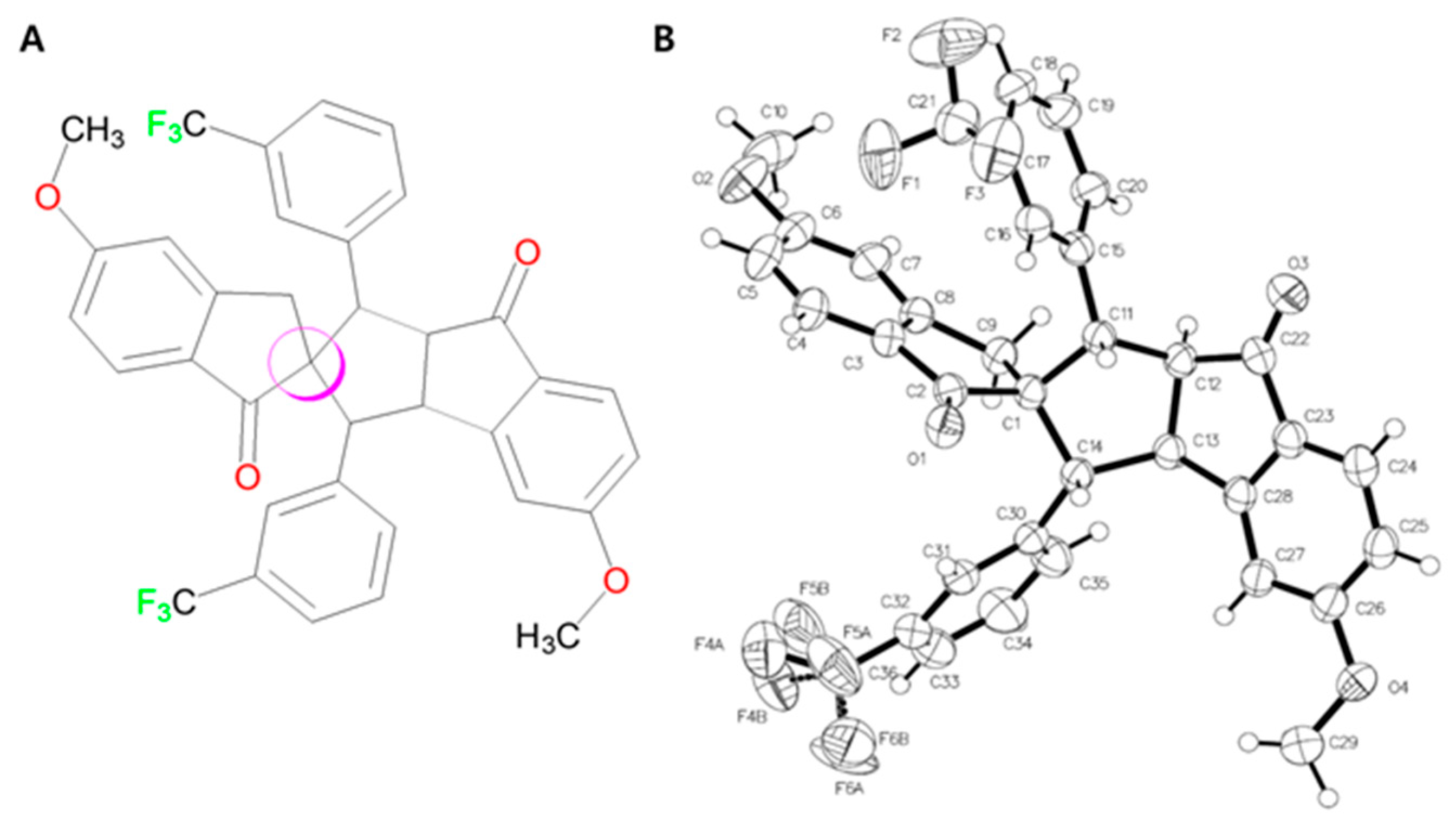
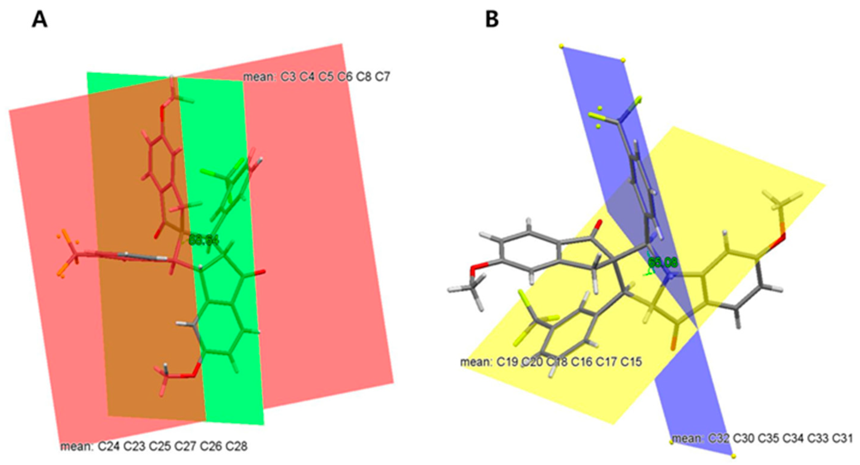
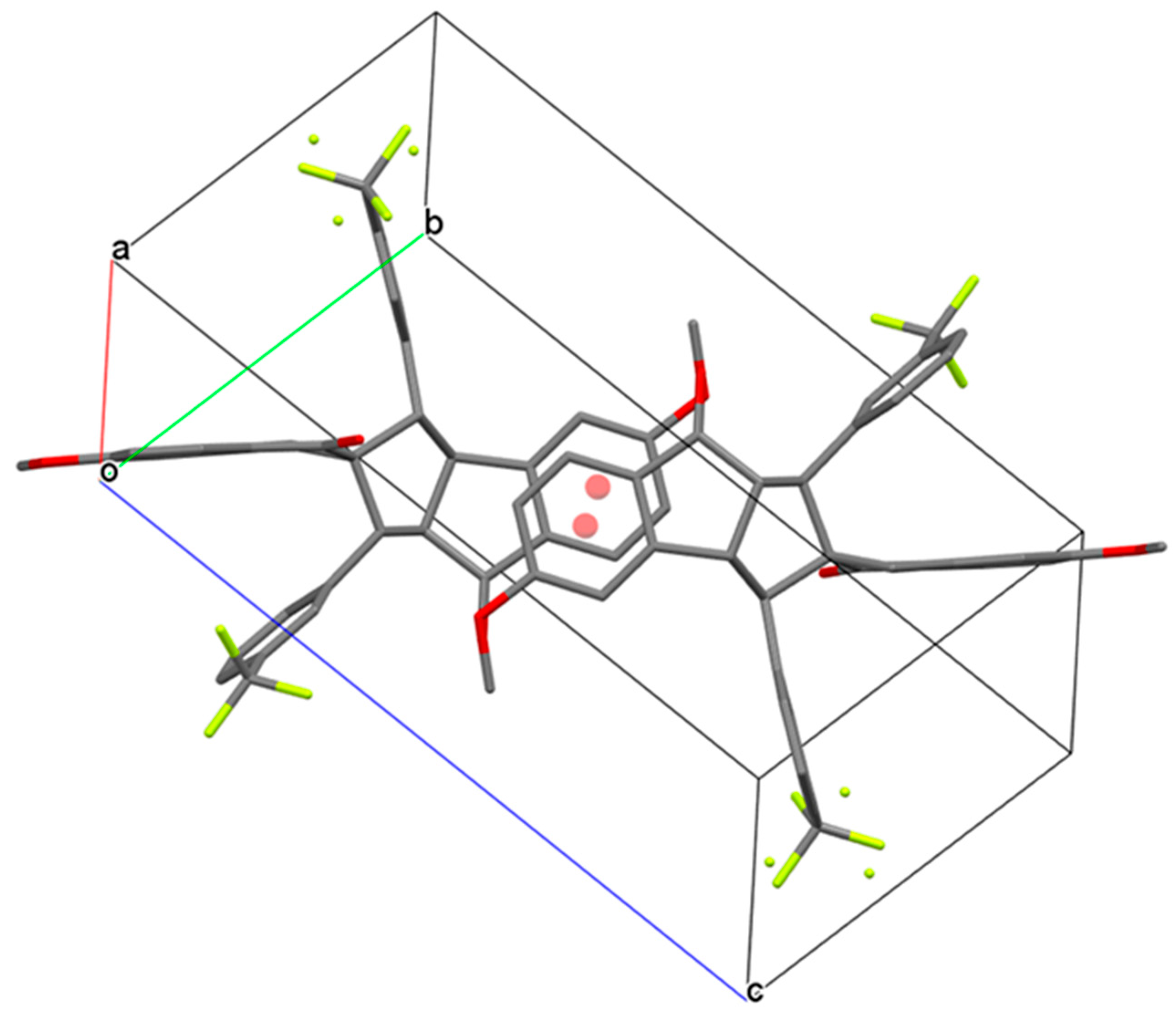
3.2. Crystal Structure of Spiro Compound 4

Figure 1A shows the chemical structure of the title compound. A spiro compound is defined as two molecular rings that share a common atom. The quaternary carbon in the pink circle forms four sp3 bonds with each of the four adjacent carbon atoms, connecting the two ring compounds. The three-dimensional molecular structure based on single-crystal X-ray studies is shown in Figure 1B with atomic labeling [Figure 1].
In the title molecule, the quaternary carbon C1 bonds with C2, C9 and C11, C14 to form two cyclopentane rings, where C1 is the common atom. The endocyclic angles ∠C2C1C9 (100.91°) and ∠C11C1C14 (103.97°) of cyclopentanes show lower values than the corresponding exocyclic angles ∠C2C1C11 (108.32°) and ∠C9C1C14 (116.35°). The benzene rings (C3–C8) and (C23–C28) contain a methoxy group. The methoxy group bonded to carbon C6 lies in the same plane of the benzene ring [dihedral angle of −0.15° for C7–C6–O2–C10], while the methoxy group bonded to carbon C26 is twisted in the benzene ring (dihedral angle of −12.18° for C27–C26–O4–C29) [Figure 2].
There are four benzene rings in the molecule, which are divided into two groups. The first group consists of two benzene rings C30–C35 and C15–C20 attached to C11 and C14 of cyclopentane, respectively, and they contain a CF3 group. The second group contains two benzene rings C3–C8 and C23–C28 fused to cyclopentane, which contains an OMe group. The planes of the two fused benzene rings C3–C8 and C23–C28 are almost perpendicular to each other, and the dihedral angle between them is 88.64° [Figure 3A]. However, the dihedral angle formed between the planes of the two benzene rings containing the CF3 group is 65.08° [Figure 3B].
The title compound 4 crystallized in the triclinic with space group P-1. In the unit cell, the dimer is formed by a combination of parallel displaced Pi–Pi stacking between the indane groups across the inversion center, further stabilized by these 2 C-H…O short contacts involving C25 and O1 [Figure 4, Table 2].
In the crystal, no classical hydrogen bonds are observed, the crystal packing is further stabilized by C-H…O interactions as listed in Table 2.

3.3. DFT Calculations of Compound 4

As mentioned above, compound 4 was synthesized during the spontaneous dimerization of compound 3, (Z)-5-methoxy-2-(3-(trifluoromethyl)benzylidene)-2,3-dihydro-1H-inden-1-one, which belongs to benzylidene indenone. Several benzylidene indenone derivatives were synthesized and their biological activities were reported in [19]. Of them, (E)-5-methoxy-2-(3-methoxybenzylidene)-2,3-dihydro-1H-inden-1-one (5) has a similar structure to compound 3 (Scheme 1). The methoxy group of 5 was replaced by a trifluoromethyl group in compound 3. In addition to the previous published results, the biological activities of two compounds, 4 and 5, were confirmed in this research. While the IL-4-induced TSLP gene promoter-reporter activity of 5 was 84.0% inhibiting effect compared to no treatment (100%), that of 4 was determined to be 55.3% inhibiting effect in this research. In vitro assay demonstrated that 4 showed a better effect than 5 [Figure 5].
Because Frontier Molecular Orbital (FMO), including HOMO, LUMO, and the energy gap between HOMO and LUMO, provides information about several molecular properties including the molecular chemical stability, molecular electrical transport properties, kinetic stability, polarizability, chemical hardness and softness, aromaticity, and electronegativity, the HOMO and LUMO energies of two molecules, 4 and 5, were obtained by DFT calculations. As listed in Table 3, the energy gap between HOMO and LUMO of 4 is smaller than that of 5, so that 4 is more reactive than 5. The graphical illustrations of the charge densities of HOMO of 4 generated using IboView demonstrated that its electron density is localized on two trifluoromethyl groups, as shown in Figure 6A, bottom. In the case of 5, its electron density spreads on the whole molecule except for cyclopentanone [Figure 6B, bottom]. The electron density of LUMO of 4 spreads on the whole molecule except for 5-methoxy-2,3-dihydro-1H-inden-1-one [Figure 6A, top]. Likewise, that of 5 spreads on the whole molecule except for the methoxy group of 3-methoxybenzylidene [Figure 6B, top].

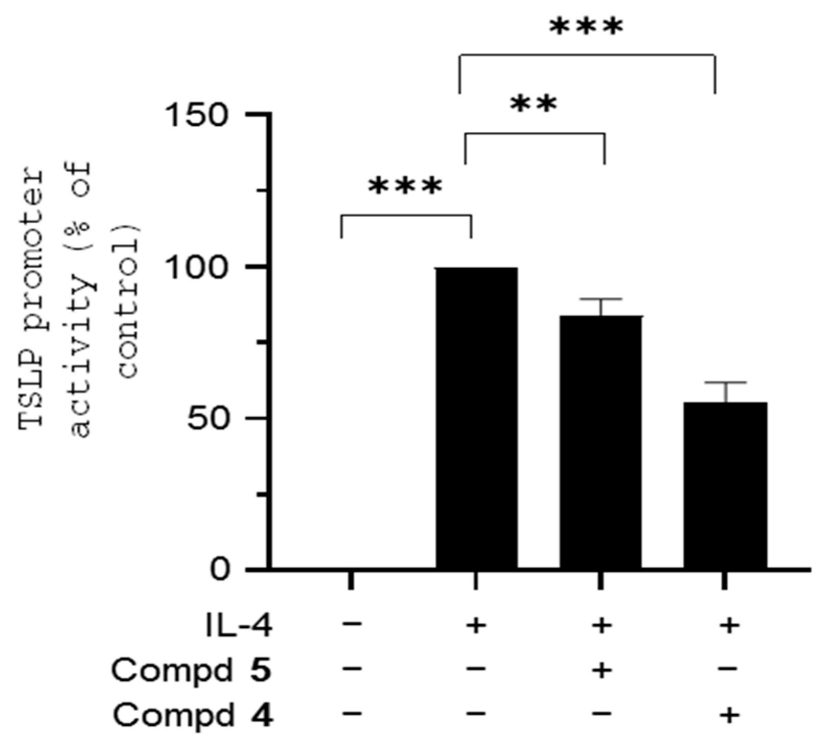
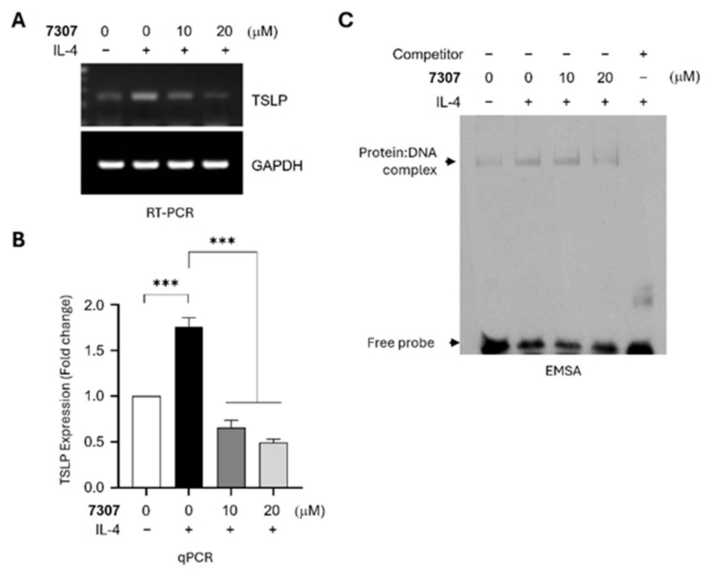
3.4. Effects on EGR-1-Regulated Gene Expression

To evaluate the biological activity of 4, we examined its inhibitory effects on EGR-1-regulated gene expression at the cellular level. Treatment of HaCaT keratinocytes with 4 reduced IL-4-induced TSLP mRNA levels, as demonstrated by reverse transcription-polymerase chain reaction (RT-PCR) [Figure 7A]. Quantitative real-time PCR (qPCR) analysis further confirmed that IL-4 increased TSLP mRNA expression 1.757-fold compared to the vehicle-treated control, while 4 reduced it to 0.657-fold and 0.499-fold at 10 and 20 μM, respectively [Figure 7B]. EGR-1 regulates TSLP expression by binding to the EGR-1-binding sequence (EBS) in the TSLP gene promoter [52]. To determine whether 4 inhibits EGR-1-mediated TSLP transcription by preventing its DNA binding activity, we performed an electrophoretic mobility shift assay (EMSA). Nuclear extracts from HaCaT cells treated with IL-4, with or without 4, were incubated with a biotinylated EBS within the TSLP gene promoter (5′-CAA AAA GGA GGA AGG TGA GGG AA-biotin-3′). Streptoavidin-conjugated horseradish peroxidase was used to detect the protein–DNA complex at the EBS. Treatment with 20 μM 4 substantially reduced the IL-4-induced DNA–protein complex formation [Figure 7C]. These findings suggest that 4 inhibits IL-4-induced TSLP gene expression by inhibiting the EGR-1 DNA binding to the TSLP gene promoter.

3.5. In Silico Docking

It has been reported that 1-(5-(4-methoxyphenyl)-4,5-dihydro-1H-pyrazol-3-yl)naphthalen-2-ol reduced the mRNA expression of EGR-1-regulated inflammatory genes such as TSLP [53]. Like this compound, the title molecule may influence EGR-1 and result in the TSLP expression. Therefore, in silico docking was performed between EGR-1 and 4. The procedures to determine the 3D structure of EGR-1 and its binding site followed the methods reported previously in [13]. The set-up of the GRID box for in silico docking followed the previous methods [46]. The 3D crystallographic structure of 4 obtained in this research was used for in silico docking as a ligand. Because AutoDock Vina generated nine protein–ligand complexes as a default, nine EGR-1–4 complexes were obtained from the current in silico docking process. The binding energy ranged from −8.2 kcal/mol to −6.9 kcal/mol. The complex with the lowest binding energy was chosen for the analysis. The binding condition was analyzed using LigPlot. Seven residues of EGR-1 including Arg357, His382, Phe377, Asn376, Lys366, Ile361, and Ser378 participate in the hydrophobic interaction with 4, and guanidine groups of Arg375 and Arg379 form two hydrogen bonds (H-bond) with each methoxy group of 4 at distances of 3.04 Å and 3.04 Å, respectively, as shown in [Figure S6]. The 3D image of the binding site of the EGR-1–4 complex was generated using PyMOL [Figure 8]. Likewise, docking of 5 into EGR-1 was attempted. Its 3D structure was adopted from the structure deposited in PubChem as CID 101866271. The binding energies of nine complexes ranged between −5.9 kcal/mol and −5.2 kcal/mol. The complex with the lowest binding energy was chosen for the analysis by LigPlot. Nine residues of EGR-1 including Ser378, Arg360, Lys366, Gln365, Glu354, His358, Phe349, Ile361, and Gly364 participate in the hydrophobic interactions with 5, and the guanidine group of Arg357 forms the methoxy group of 5 at a distance of 2.95 Å [Figure S7]. The 3D image of the binding site of the EGR-1–5 complex was generated using PyMOL [Figure S8]. The binding condition of 4 is similar to that of 5, but the binding energy of 4 is lower than that of 5. This is consistent with the results obtained from DFT calculations.

4. Conclusions

A Claisen–Schmidt condensation reaction between 5-methoy-1-indanone (1) and 3-(trifluoromethyl)benzaldehyde (2) gave a benzylidene indenone product (3). When it has a strong electron-withdrawing group, compound 3 dimerizes into spiro compound 4 through an intermolecular spontaneous Michael reaction. A novel spiro compound 4 was crystallized in the triclinic space group P-1 and revealed that two molecules are packed by short contacts to form an inversion dimer in the unit cell. In the crystal, the molecules are linked into chains along the a- and b-axis directions by additional intermolecular C-H···O interactions.
Since compound 4 was formed by the dimerization of compound 3, compound 5, one of the analogs of compound 3, was compared with 4 based on in vitro experiments, DFT calculations, and an in silico docking study. The energy gap between HOMO and LUMO of 4 was smaller than that of 5, so that 4 was expected to be more reactive than 5. The binding condition of 4 obtained from in silico docking was similar to that of 5, but the binding energy of 4 is lower than that of 5. These are consistent with the results obtained from in vitro assay where 4 showed a better effect than 5.
Spiro compound 4 demonstrated inhibitory effects on EGR-1-regulated gene expression in HaCaT keratinocytes. Compound 4 reduced IL-4-induced TSLP mRNA levels and the electrophoretic mobility shift assay showed that 4 inhibited IL-4-induced DNA binding of EGR-1 to the promoter region of the TSLP gene.

Supplementary Materials

The following supporting information can be downloaded at: https://www.mdpi.com/article/10.3390/cryst15040338/s1, CIF file for compound 4; Figure S1: 1H NMR (700 MHz, DMSO-d6) spectrum of compound 4 (top: aromatic region; bottom: aliphatic region); Figure S2: 13C-NMR (175 MHz, DMSO-d6) spectrum of compound 4; Figure S3: The HMQC (400 MHz, DMSO-d6) spectrum of compound 4; Figure S4: The HMBC (400 MHz, DMSO-d6) spectrum of compound 4; Figure S5: Mass spectrum of compound 4; Figure S6: The hydrophobic interactions (red half circle) and hydrogen-bond (green dot line) between EGR-1 and (4) analyzed using LigPlot; Figure S7: The hydrophobic interactions (red half circle) and hydrogen-bond (green dot line) between EGR-1 and (5) analyzed using LigPlot; Figure S8: The 3D image of the binding site of the EGR-1–5 complex was generated using PyMOL.

Author Contributions

Conceptualization, S.A. and D.K.; Methodology, H.L. and J.Y.; Software, H.-J.L.; Validation, Y.L.; Formal analysis, E.J.; Writing—original draft, S.Y.S. All authors have read and agreed to the published version of the manuscript.

Funding

S.Y. Shin was supported by the KU Research Professor Program of Konkuk University.

Data Availability Statement

Data are contained within the article and Supplementary Materials.

Conflicts of Interest

The authors declare no conflicts of interest.

References

  1. Wang, F. Chapter 1—Modeling Human Prostate Cancer in Genetically Engineered Mice. In Progress in Molecular Biology and Translational Science; Elsevier: Amsterdam, The Netherlands, 2011; Volume 100, pp. 1–49. [Google Scholar]
  2. Zou, K.; Zeng, Z. Role of early growth response 1 in inflammation-associated lung diseases. Am. J. Physiol. Lung Cell. Mol. Physiol. 2023, 325, L143–L154. [Google Scholar] [PubMed]
  3. Thiel, G.; Cibelli, G. Regulation of life and death by the zinc finger transcription factor Egr-1. J. Cell. Physiol. 2000, 193, 287–292. [Google Scholar]
  4. Pignatelli, M.; Luna-Medina, R.; Pérez-Rendón, A.; Santos, A.; Perez-Castillo, A. The transcription factor early growth response factor-1 (EGR-1) promotes apoptosis of neuroblastoma cells. Biochem. J. 2003, 373, 739–746. [Google Scholar]
  5. Khachigian, L.M. Early Growth Response-1, an Integrative Sensor in Cardiovascular and Inflammatory Disease. J. Am. Heart Assoc. 2021, 10, e023539. [Google Scholar]
  6. Wang, B.; Guo, H.; Yu, H.; Chen, Y.; Xu, H.; Zhao, G. The Role of the Transcription Factor EGR1 in Cancer. Front. Oncol. 2021, 11, 642547. [Google Scholar]
  7. Bryant, M.; Drew, G.M.; Houston, P.; Hissey, P.; Campbell, C.J.; Braddock, M. Tissue repair with a therapeutic transcription factor. Hum. Gene Ther. 2000, 11, 2143–2158. [Google Scholar]
  8. Ryu, W.I.; Lee, H.; Kim, J.H.; Bae, H.C.; Ryu, H.J.; Son, S.W. IL-33 induces Egr-1-dependent TSLP expression via the MAPK pathways in human keratinocytes. Exp. Dermatol. 2015, 24, 857–863. [Google Scholar]
  9. Lohoff, M.; Giaisi, M.; Köhler, R.; Casper, B.; Krammer, P.H.; Li-Weber, M. Early growth response protein-1 (Egr-1) is preferentially expressed in T helper type 2 (Th2) cells and is involved in acute transcription of the Th2 cytokine interleukin-4. J. Biol. Chem. 2010, 285, 1643–1652. [Google Scholar]
  10. Jeong, S.H.; Kim, H.J.; Jang, Y.; Ryu, W.I.; Lee, H.; Kim, J.H.; Bae, H.C.; Choi, J.E.; Kye, Y.C.; Son, S.W. Egr-1 is a key regulator of IL-17A-induced psoriasin upregulation in psoriasis. Exp. Dermatol. 2014, 23, 890–895. [Google Scholar]
  11. Yeo, H.; Ahn, S.S.; Lee, J.Y.; Shin, S.Y. EGR-1 acts as a transcriptional activator of KLK7 under IL-13 stimulation. Biochem. Biophys. Res. Commun. 2021, 534, 303–309. [Google Scholar]
  12. Matus, C.E.; Ehrenfeld, P.E.; Figueroa, C.D. The family of kallikrein-related peptidases and kinin peptides as modulators of epidermal homeostasis. Am. J. Physiol. Cell Physiol. 2022, 323, C1070–C1087. [Google Scholar] [CrossRef] [PubMed]
  13. Yeo, H.; Ahn, S.S.; Lee, J.Y.; Jung, E.; Jeong, M.; Kang, G.S.; Ahn, S.; Lee, Y.; Koh, D.; Lee, Y.H.; et al. Disrupting the DNA Binding of EGR-1 with a Small-Molecule Inhibitor Ameliorates 2,4-Dinitrochlorobenzene-Induced Skin Inflammation. J. Investig. Dermatol. 2021, 141, 1851–1855. [Google Scholar] [CrossRef] [PubMed]
  14. Yeo, H.; Ahn, S.S.; Ou, S.; Yun, S.J.; Lim, Y.; Koh, D.; Lee, Y.H.; Shin, S.Y. The EGR1-Artemin Axis in Keratinocytes Enhances the Innervation of Epidermal Sensory Neurons during Skin Inflammation Induced by House Dust Mite Extract from Dermatophagoidesfarinae. J. Investig. Dermatol. 2024, 144, 1817–1828.e17. [Google Scholar] [PubMed]
  15. Park, T.J.; Oh, H.; Kim, M.; Kim, J.; Kim, H.J.; Son, S.W. Urban particulate matters induce EGR-1 expression in keratinocytes which correlates with the severity of psoriasis. Mol. Cell. Toxicol. 2021, 17, 195–200. [Google Scholar] [CrossRef]
  16. Fania, L.; Moretta, G.; Antonelli, F.; Scala, E.; Abeni, D.; Albanesi, C.; Madonna, S. Multiple Roles for Cytokines in Atopic Dermatitis: From Pathogenic Mediators to Endotype-Specific Biomarkers to Therapeutic Targets. Int. J. Mol. Sci. 2022, 23, 2684. [Google Scholar] [CrossRef]
  17. Weidinger, S.; Novak, N. Atopic dermatitis. Lancet 2016, 387, 1109–1122. [Google Scholar] [CrossRef]
  18. Brunner, P.M.; Guttman-Yassky, E.; Leung, D.Y. The immunology of atopic dermatitis and its reversibility with broad-spectrum and targeted therapies. J. Allergy Clin. Immunol. 2017, 139, S65–S76. [Google Scholar] [CrossRef]
  19. Lee, Y.; Ahn, S.; Jung, E.; Koh, D.; Lim, T.; Lee, Y.H.; Shin, S.Y. Design, synthesis, and biological evaluation of (E)-2-benzylidene-1-indanones derivatized by bioisosteric replacement of aurones. Appl. Biol. Chem. 2024, 67, 114. [Google Scholar] [CrossRef]
  20. Moss, G.P. Extension and revision of the nomenclature for spiro compounds. Pure Appl. Chem. 1999, 71, 531–558. [Google Scholar] [CrossRef]
  21. Talele, T.T. Opportunities for tapping into three-dimensional chemical space through a quaternary carbon. J. Med. Chem. 2020, 63, 13291–13315. [Google Scholar] [CrossRef]
  22. Zeng, W.; Jiang, J. Synthesis and Crystal Structure of a New Hydrated Benzimidazolium Salt Containing Spiro Structure. Crystals 2017, 7, 303. [Google Scholar] [CrossRef]
  23. Romo, P.E.; Quiroga, J.; Cobo, J.; Glidewell, C. Synthesis and spectroscopic and structural characterization of spiro-[indoline-3,3′-indolizine]s formed by 1,3-dipolar cyclo-additions between isatins, pipecolic acid and an electron-deficient alkene. Acta Cryst. 2021, C77, 496–504. [Google Scholar]
  24. Bruña, S.; Cuadrado, I.; Perles, J. Unexpected formation of a silicon-centered spirocyclic oligosiloxane bearing eight pendant ferrocene units. Crystals 2022, 12, 1122. [Google Scholar] [CrossRef]
  25. Zheng, Y.; Tic, C.M.; Singh, S.B. The use of spirocyclic scaffolds in drug discovery. Bioorg. Med. Chem. Lett. 2014, 24, 3673–3682. [Google Scholar]
  26. Hiesinger, K.; Dar’in, D.; Proschak, E.; Krasavin, M. Spirocyclic scaffolds in medicinal chemistry. J. Med. Chem. 2021, 64, 150–183. [Google Scholar]
  27. Romero-Hernández, L.L.; Ahuja-Casarín, A.I.; Merino-Monti, P.; Montiel-Smith, S.; Vega-Baez, J.L.; Sandoval-Ramirez, J. Syntheses and medicinal chemistry of spiro heterocyclic steroids. Beilstein J. Org. Chem. 2024, 20, 1713–1745. [Google Scholar]
  28. Zhou, L.M.; Qu, R.Y.; Yang, G.F. An overview of spirooxindole as a promising scaffold for novel drug discovery. Expert Opin. Drug Discov. 2020, 15, 603–625. [Google Scholar]
  29. Qu, Y.-K.; Qi, Z.; Jian, F.; Liao, L.S.; Jiang, Z.Q. Spiro Compounds for organic light-emitting diodes. Acc. Mater. Res. 2021, 2, 1261–1271. [Google Scholar]
  30. Saragi, T.P.I.; Spehr, T.; Siebert, A.; Fuhrmann-Lieker, T.; Salbeck, J. Spiro compounds for organic optoelectronics. Chem. Rev. 2007, 107, 1011–1065. [Google Scholar]
  31. Yu, L.; Dai, A.; Zhang, W.; Liao, A.; Guo, S.; Wu, J. Spiro derivatives in the discovery of new pesticides: A Research review. J. Agric. Food Chem. 2022, 70, 10693–10707. [Google Scholar]
  32. Rios, R. Enantioselective methodologies for the synthesis of spiro compounds. Chem. Soc. Rev. 2012, 41, 1060–1074. [Google Scholar] [PubMed]
  33. Babar, K.; Zahoor, A.F.; Ahmad, S.; Akhtar, R. Recent synthetic strategies toward the synthesis of spirocyclic compounds comprising six-membered carbocyclic/heterocyclic ring systems. Mol. Divers. 2021, 25, 2487–2532. [Google Scholar] [PubMed]
  34. Patel, G.; Patel, A.R.; Kheti, S.; Sao, P.K.; Rathore, G.; Banerjee, S. Review on the Synthesis of Bio-Active Spiro-Fused Heterocyclic Molecules. Curr. Organocatal. 2023, 10, 180–208. [Google Scholar]
  35. Singh, R.; Bhardwaj, D.; Saini, M.R. Recent advancement in the synthesis of diverse spiro-indeno [1,2-b]quinoxalines: A review. RSC Adv. 2021, 11, 4760–4804. [Google Scholar]
  36. Gilles, L.; Antoniotti, S. Spirocyclic compounds in fragrance Chemistry: Synthesis and olfactory properties. ChemPlusChem 2022, 87, e202200227. [Google Scholar]
  37. Bruker. APEX2, SAINT and SADABS; Bruker AXS Inc.: Madison, WI, USA, 2012. [Google Scholar]
  38. Sheldrick, G.M. Crystal structure refinement with SHELXL. Acta Cryst. C 2015, 71, 3–8. [Google Scholar]
  39. Dolomanov, O.V.; Bourhis, L.J.; Gildea, R.J.; Howard, J.A.K.; Puschmann, H. OLEX2: A Complete Structure Solution, Re-finement and Analysis Program. J. Appl. Crystallogr. 2009, 42, 339–341. [Google Scholar]
  40. Macrae, C.F.; Sovago, I.; Cottrell, S.J.; Galek, P.T.A.; McCabe, P.; Pidcock, E.; Platings, M.; Shields, G.P.; Stevens, J.S.; Towler, M.; et al. Mercury 4.0: From visualization to analysis, design and prediction. J. Appl. Cryst. 2020, 53, 226–235. [Google Scholar]
  41. Bax, A. Two-dimensional heteronuclear relayed coherence transfer spectroscopy. J. Magn. Reson. 1983, 53, 149–153. [Google Scholar]
  42. Clore, G.M.; Gronenborn, A.M. Multidimensional heteronuclear nuclear magnetic resonance of proteins. Methods Enzymol. 1994, 239, 349–363. [Google Scholar]
  43. Reynolds, W.F.; Burns, D.C. Getting the Most out of HSQC and HMBC Spectra. In Annual Reports on NMR Spectroscopy; Elsevier: Amsterdam, The Netherlands, 2012; Volume 76, pp. 1–21. [Google Scholar]
  44. Charisiadis, P.; Venianakis, T.; Papaemmanouil, C.D.; Primikyri, A.; Tzakos, A.G.; Siskos, M.G.; Gerothanassis, I.P. On the Use of Strong Proton Donors as a Tool for Overcoming Line Broadening in NMR: A Comment. Magn. Reason. Chem. 2025, 63, 170–179. [Google Scholar] [CrossRef] [PubMed]
  45. Li, M.; Qin, Y.; Li, Z.; Lan, J.; Zhang, T.; Ding, Y. Comparative Pharmacokinetics of Cinobufacini Capsule and Injection by UPLC-MS/MS. Front. Pharmacol. 2022, 13, 944041. [Google Scholar] [CrossRef] [PubMed]
  46. Trott, O.; Olson, A.J. AutoDock Vina: Improving the speed and accuracy of docking with a new scoring function, efficient optimization, and multithreading. J. Comput. Chem. 2010, 31, 455–461. [Google Scholar] [CrossRef] [PubMed]
  47. Pettersen, E.F.; Goddard, T.D.; Huang, C.C.; Couch, G.S.; Greenblatt, D.M.; Meng, E.C.; Ferrin, T.E. UCSF Chimera—A visualization system for exploratory research and analysis. J. Comput. Chem. 2004, 25, 1605–1612. [Google Scholar] [CrossRef]
  48. Kramer, B.; Rarey, M.; Lengauer, T. Evaluation of the FLEXX incremental construction algorithm for protein–ligand docking. Proteins Struct. Funct. Genet. 1999, 37, 228–241. [Google Scholar] [CrossRef]
  49. Neese, F.; Wennmohs, F.; Becker, U.; Riplinger, C. The ORCA quantum chemistry program package. J. Chem. Phys. 2020, 152, 224108. [Google Scholar]
  50. Hanwell, M.D.; Curtis, D.E.; Lonie, D.C.; Vandermeersch, T.; Zurek, E.; Hutchison, G.R. Avogadro: An advanced semantic chemical editor, visualization, and analysis platform. J. Cheminform. 2012, 4, 17. [Google Scholar]
  51. Knizia, G.; Klein, J.E.M.N. Electron Flow in Reaction Mechanisms—Revealed from First Principles. Angew. Chem. Int. Ed. 2015, 54, 5518–5522. [Google Scholar] [CrossRef]
  52. Yeo, H.; Lee, Y.H.; Ahn, S.S.; Jung, E.; Lim, Y.; Shin, S.Y. Chrysin Inhibits TNF?-Induced TSLP Expression through Downregulation of EGR1 Expression in Keratinocytes. Int. J. Mol. Sci. 2021, 22, 4350. [Google Scholar] [CrossRef]
  53. Yoon, H.; Koh, D.; Lim, Y.; Lee, Y.H.; Lee, J.K.; Shin, S.Y. Pyrazolines inhibiting the activity of the early growth response-1 DNA-binding domain. Bioorg. Med. Chem. Lett. 2024, 113, 129952. [Google Scholar] [CrossRef]
Scheme 1. Synthetic scheme for the title compound 4.
Crystals 15 00338 sch001
Figure 1. Molecular structure of the spiro compound 4. The spiro region within the molecule is depicted as a pink circle (A). 3D structure with atomic labeling of the spiro compound 4 (B).
Crystals 15 00338 g001
Figure 2. Two cyclopentane rings sharing a common spiro atom C1. The endocycle angle and exocyclic angle between cyclopentane rings. The dihedral angle between benzene rings and OMe groups.
Crystals 15 00338 g002
Figure 3. Dihedral angle formed between the plane of fused benzene rings (A) and attached benzene rings (B).
Crystals 15 00338 g003
Figure 4. The dimer in the unit cell is formed by a combination of Pi–Pi stacking and is further stabilized by two C-H…O short contacts involving C25 and O1.
Crystals 15 00338 g004
Figure 5. Inhibitory effect of compound 4 and 5 on IL-4-induced TSLP gene promoter activity. ** p, < 0.01; *** p, < 0.001 (n = 3).
Crystals 15 00338 g005
Figure 6. The graphical illustrations of charge densities of HOMO and LUMO of (A) 4 and (B) 5 generated using IboView. The energy unit is Hartree (Eh).
Crystals 15 00338 g006
Figure 7. Inhibitory effect of 4 on IL-4-induced TSLP expression and EGR-1 DNA-binding activity. (A) HaCaT cells were treated with 20 ng/mL IL-4 with or without 4 for 8 h. Total RNA was isolated, and RT-PCR was performed to assess TSLP mRNA expression. GAPDH mRNA was used as an internal control. (B) HaCaT cells were treated as in (A) and then subjected to qPCR analysis. TSLP mRNA levels were quantified after normalization to GAPDH mRNA and expressed as fold change relative to vehicle-treated control. *** p < 0.001 (n = 3). (C) HaCaT cells were treated as in (A). Nuclear extracts were prepared and then EMSA analysis was carried out using a biotinylated EBS probe (50 fmole). Unlabeled EBS oligonucleotide (2500 fmole) was used as a competitor. Samples were separated by 6% non-denaturing PAGE, incubated with streptoavidin-conjugated horseradish peroxidase, and visualized using a Western blotting detection kit.
Crystals 15 00338 g007
Figure 8. The 3D image of the binding site of the EGR-1–4 complex was generated using PyMOL.
Crystals 15 00338 g008
Table 1. Crystal data and structure refinement for compound 4.
Empirical FormulaC36H26F6O4
Formula weight636.57
Temperature223(2) K
Wavelength0.71073 Å
Crystal systemTriclinic
Space groupP-1
Unit cell dimensionsa = 8.8669(5) Å
b = 10.5298(8) Å
c = 17.0135(11) Å
α = 91.396(2)°.
β = 90.490(2)°.
γ = 109.235(2)°.
Volume1499.14(17) Å3
Z2
Density (calculated)1.410 Mg/m3
Absorption coefficient0.116 mm−1
F(000)656
Crystal size0.226 × 0.204 × 0.056 mm3
Theta range for data collection2.049 to 28.2492°.
Index ranges−1–11 ≤ h ≤ 11, −14 ≤ k ≤ 14, −22 ≤ l ≤ 22
Reflections collected44768
Independent reflections7389 [R(int) = 0.0426]
Completeness to theta = 25.242°99.9%
Refinement methodFull-matrix least-squares on F2
Data/restraints/parameters7389/30/445
Goodness-of-fit on F21.036
Final R indices [I > 2sigma(I)]R1 = 0.0548, wR2 = 0.1309
R indices (all data)R1 = 0.0814, wR2 = 0.1481
Largest diff. peak and hole0.531 and −0.420 e·Å−3
Table 2. Intermolecular hydrogen bonds for the compound 4 [Å and °].
D-H…Ad(D-H)d(H…A)d(D…A)<(DHA)
C(12)-H(12)…O(3)#10.992.573.444(3)146.4
C(34)-H(34)…O(1)#20.942.533.413(3)157.4
C(25)-H(25)…O(1)#30.942.433.356(2)169.2
Symmetry transformations used to generate equivalent atoms: #1 − x + 1, −y + 2, −z + 1 #2 x − 1, y, z #3 − x + 1, −y + 1, −z + 1.
Table 3. The energy values of FMO of 4 and 5 as determined by DFT calculations. The energy unit is Hartree (Eh) and ΔE denotes the gap (ΔE) between HOMO and LUMO.
45
FMOEnergy/Hartree(Eh)Energy/Hartree(Eh)
3rd LUMO0.10220.1233
2nd LUMO0.10160.1163
1st LUMO0.09190.0648
1st HOMO−0.1635−0.3183
2nd HOMO−0.1994−0.3236
3rd HOMO−0.2541−0.3369
1st ΔE0.25540.3831
2nd ΔE0.30100.4399
3rd ΔE0.35630.4602
Disclaimer/Publisher’s Note: The statements, opinions and data contained in all publications are solely those of the individual author(s) and contributor(s) and not of MDPI and/or the editor(s). MDPI and/or the editor(s) disclaim responsibility for any injury to people or property resulting from any ideas, methods, instructions or products referred to in the content.

