Article

Comparative Study of Gemological and Spectroscopic Features and Coloration Mechanism of Three Types of Spodumene

1
School of Gemology, China University of Geosciences, Beijing 100083, China
2
Faculty of Innovation and Design, City University of Macau, Macau 999078, China
*
Author to whom correspondence should be addressed.
Crystals 2025, 15(2), 109; https://doi.org/10.3390/cryst15020109
Submission received: 5 December 2024 / Revised: 18 January 2025 / Accepted: 20 January 2025 / Published: 22 January 2025
(This article belongs to the Collection Topic Collection: Mineralogical Crystallography)

Abstract

:
Spodumene is a characteristic mineral in lithium-rich granitic pegmatites, serving both as a valuable mineral resource and an important gem material. This study incorporates three different color varieties of spodumene—pink to violet, yellow-green, and colorless—into a unified research framework. X-ray powder diffraction (XRD), electron probe microanalyzer (EPMA), Fourier transform infrared spectroscopy (FTIR), Raman spectroscopy, ultraviolet–visible spectroscopy (UV-Vis), and photoluminescence spectroscopy (PL) were employed to systematically analyze the chemical composition, crystal structure, and spectroscopic properties of spodumene. Furthermore, the coloration mechanism and fluorescence emission of the different color samples were investigated and analyzed. The results indicate that the presence and mixed valence states of the transition metals Fe and Mn primarily influence the color and photoluminescence of the three types of spodumene. Mn3+ is the primary color-causing element in pink to violet spodumene, while Fe3+ is the primary color-causing element in yellow-green spodumene. Photoluminescence in all three color varieties is dominated by Mn2+. These findings contribute to a deeper understanding of the color and luminescence mechanisms of spodumene, expanding its potential applications as both a gem material and a luminescent material.

1. Introduction

Spodumene, a lithium-rich mineral that is relatively easy to extract, is both a valuable resource and an important gemstone material. Due to its high lithium content and relatively high availability, it was one of the primary sources for lithium extraction in the past. Research on spodumene has primarily focused on industrial applications, particularly in mineral flotation technology, the production of lithium compounds, and semiconductor materials [1,2]. However, with the continuous development of the gemstone market, spodumene has gradually gained attention from high-end jewelry brands, and its unique colors offer more design possibilities. This shift has led to a growing body of research on gem-quality spodumene. Current research on gem-quality spodumene mainly focuses on the following aspects: its geological origins, crystal morphology, the application of heat treatment and irradiation techniques, coloration mechanisms, and luminescent properties.
Found primarily in granite pegmatites, the formation conditions and geological background of spodumene play a significant role in determining its quality. Studies have shown that the formation of spodumene is closely related to the enrichment of lithium in pegmatites, and other minerals in pegmatites, such as tourmaline and feldspar, can also influence the growth and quality of spodumene [3]. Spodumene exists in several crystal structures, including monoclinic α-spodumene, tetragonal β-spodumene, and hexagonal γ-spodumene [4]. Gem-quality spodumene is typically α-spodumene, which displays a wide range of colors, including pink to bluish-red, green, yellow, colorless, and blue, with lighter hues. These color variations also influence its market value.
Existing research indicates that the coloration of spodumene is closely related to the transition metal ions present in its crystal structure. Scholars such as Mattson and Rossman have pointed out that transition metal ions from the first period of the periodic table, particularly Ti, V, Cr, Mn, Fe, and Cu, are usually the main causes of the coloration in oxide and silicate gemstones. However, they did not specifically explain the exact colors caused by each metal ion [5]. Researchers have suggested that the elements Mn2+ and Cr3+ have significant effects on the coloration of purple spodumene (kunzite) and green spodumene (hiddenite) and further confirmed that these elements also contribute to its luminescent properties [6,7]. Schmitz proposed that the color of spodumene is closely related to the changes in the oxidation state of manganese ions, with hole color centers interacting with manganese ions through the capture of electrons or the release of holes. As manganese transitions from Mn3+ to Mn4+, the color of spodumene gradually shifts from purple to green [8]. Furthermore, Noithong and Czaja, among others, further pointed out that the coloration of purple spodumene is primarily attributed to Mn3+, with absorption peaks related to Mn3+ intensifying after irradiation. However, due to the instability of this oxidation state, redox reactions are likely to occur, leading to the fading of color [9,10,11]. Additionally, the color of spodumene can also undergo changes through heat treatment or various types of irradiations, such as γ-rays or X-rays. For instance, colorless or light purple spodumene can turn green after irradiation, but this color change is reversible. Heating to 200 °C or exposure to sunlight can restore the green spodumene to its original color [9,12,13]. Overall, current research on the coloration mechanism of spodumene mainly focuses on the color mechanisms and luminescent properties of purple spodumene (kunzite) and chromium-containing green spodumene (hiddenite). These studies have laid a solid foundation for understanding the role of different elements in the coloration mechanism of spodumene. However, except for purple spodumene, the coloration mechanisms of other colored spodumene have not been definitively concluded, and comparative studies on multiple colors have not been fully explored.
This study will incorporate spodumene of various colors (such as pink to violet, yellow-green, and colorless spodumene) into a unified research framework, conducting systematic gemological testing, spectroscopic analysis, and chemical composition analysis to explore the coloration mechanisms through comparative investigation. The study will employ various experimental techniques, including XRD, EPMA, FTIR, Raman spectroscopy, ultraviolet–visible spectroscopy (UV-Vis), and photoluminescence spectroscopy (PL), to systematically analyze the chemical composition, crystal structure, and spectroscopic properties of these different colored spodumene samples. This research will provide more detailed experimental evidence on the relationship between the formation of color in spodumene and its transition metal ions, thereby deepening the theoretical understanding of the coloration mechanism of spodumene. The results of this study will also provide scientific support for the market classification and identification of spodumene, enhancing its practical value in gemological research and applications.

2. Materials and Methods
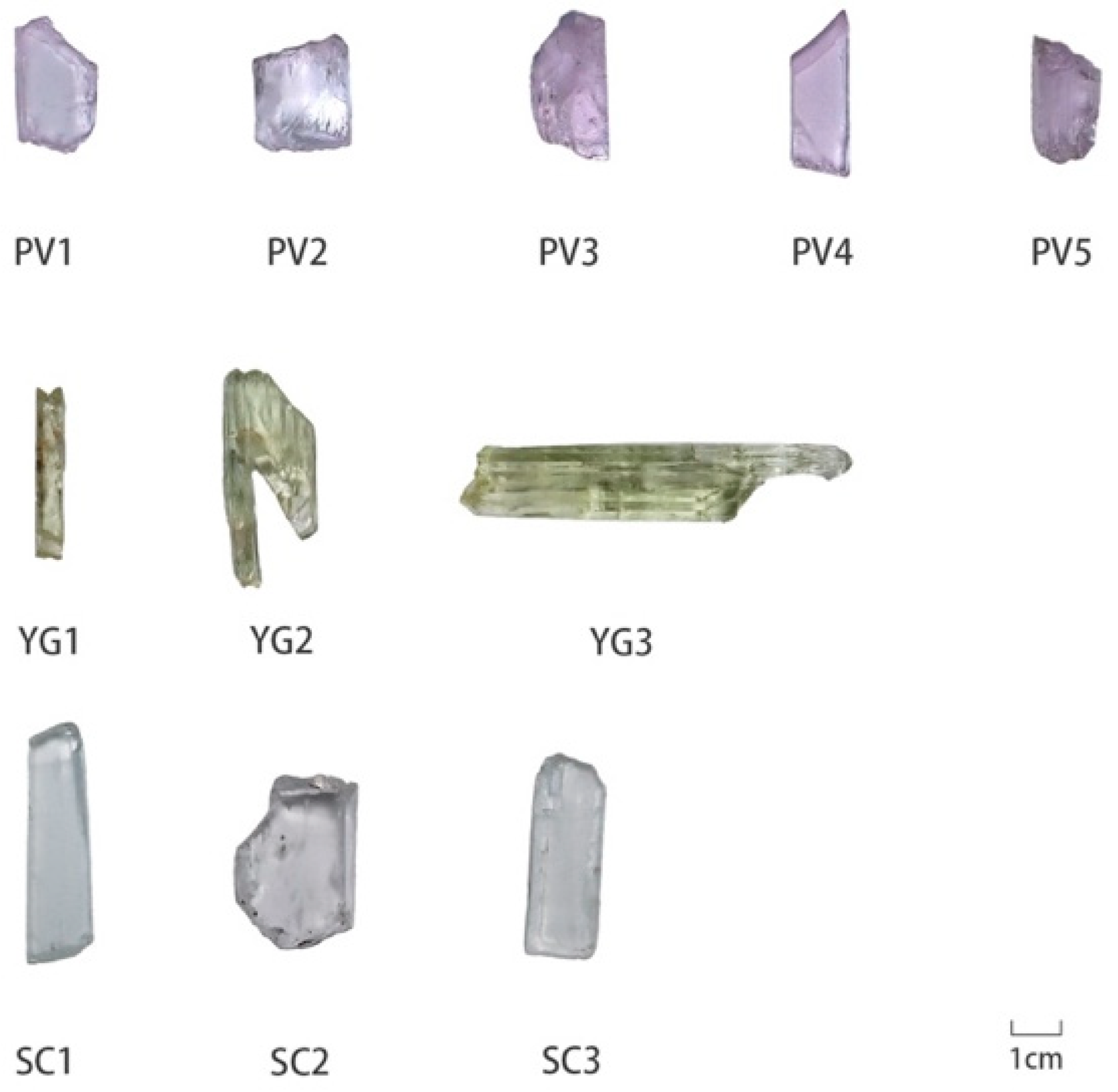
2.1. Materials

As shown in Figure 1, this study selected common market-available spodumene rough stones in pink to violet, yellow-green, and colorless varieties. Eleven samples were obtained from rough stone suppliers, all sourced from Pakistan. The sample numbers are as follows: Pink to violet spodumene (PV1–5), yellow-green spodumene (YG1–3), and colorless spodumene (SC1–3). As shown in Figure 1, these samples are mostly long prismatic or flattened; the crystals grow parallel to the C-axis, with at least one polished surface on each sample.

2.2. Methods

Conventional gemological tests were conducted, including measurements of refractive index, specific gravity, and microscopic features using a refractometer, the hydrostatic weighing method, and a gemological microscope, respectively. Testing was conducted at the Gem Testing Laboratory at the School of Gemmology, China University of Geosciences, Beijing (CUGB).
XRD was employed to study the crystal structure of spodumene, and Raman and infrared spectroscopy were used to compare the spectral characteristics of spodumene in four different colors.
The crystal structure of the sample was determined by the Smart Lab X-ray diffractometer (D8 Focus, XBruker, Germany) of the School of Materials, China University of Geosciences (Beijing, China). The main experimental conditions are as follows: a voltage of 45 kV; a slow scan scanning speed of 0.02°/s; a total effective scanning time of 5756 s; and a scanning range of 5°–120°. The scanning speed of a fast scan is 10°/min, with a scanning range of 5° to 80°.
A Vector 33 Fourier transform infrared spectrometer produced by Brucker in Germany (Bremen, Germany) was used to test the direct reflection method. The experimental conditions were as follows: a test range of 400–2000 cm−1, a resolution of 4 cm−1, and a sample scan time of 32 s.
Raman spectroscopy was performed at the China University of Geosciences Beijing (CUGB) Gemological Experimental Teaching Centre using a HORIBA LabRAM HREvolution Raman spectrometer. A microscope with ×50 objectives was used to focus on the samples. The laser beam was perpendicular to the surface of the sample. The Raman spectrometer was recorded in the 200–1000 cm−1 range and with 4 cm−1 resolution, with an excitation light source λ = 532 nm and 3 s exposure time.
EPMA was employed to determine the chemical composition of the three color varieties of spodumene, aiming to compare the differences between trace and major elements. The main elements of the samples were determined by a JXA-8230 electron microprobe (EPMA, Joel, Japan) from the Beida Microfabrication Laboratory. The main experimental conditions are as follows: an accelerating voltage of 15 kV, a beam current of 50.4 nA, and a beam diameter of 5 μm.
To analyze the luminescence of the three color varieties of spodumene, we used a fluorescence spectrometer. The FLS920 series of fully functional steady-state/transient fluorescence spectrometers produced by the Edinburgh Company (Edinburgh, UK) of China University of Geosciences (Beijing) was used. The experimental conditions were as follows: the excitation light source was a 450 W xenon lamp; the voltage was 450 V; the measuring wavelength range was 380–750 nm; and the scanning speed was 240 nm/min.

3. Results and Discussion

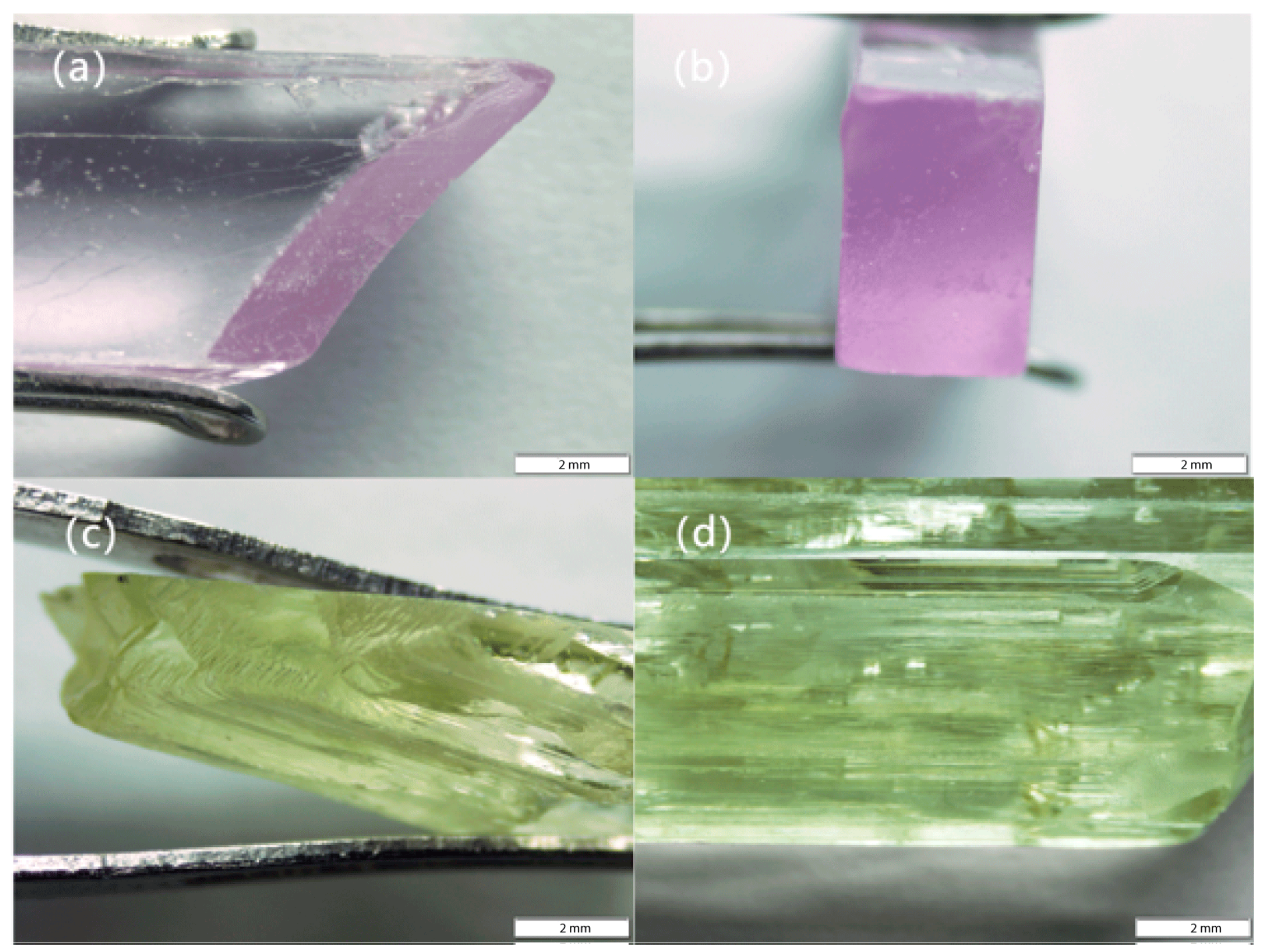
3.1. Conventional Gemological Features

The conventional gemological features of spodumene are summarized in Table 1. In appearance, spodumene typically forms in elongated prismatic or tabular shapes, with crystals growing parallel to the C-axis. The polished surface exhibits a vitreous luster, while the fracture surfaces display a greasy luster. The overall color of the samples is relatively uniform, with the Pink to violet spodumene displaying distinct pleochroism, changing from colorless to violet depending on the viewing angle. Most raw spodumene specimens exhibit good transparency, while some samples appear translucent due to internal cleavage development. The refractive index test results show a range between 1.66 and 1.67, with birefringence between 0.014 and 0.016. Specific gravity tests on 13 spodumene samples using the hydrostatic weighing method revealed a range between 3.15 and 3.20. As shown in Figure 2, under the microscope, the color of the samples is more intense perpendicular to the C-axis (Figure 2a,b). Further examination reveals a stepped fracture pattern (Figure 2c), and the unpolished surface exhibits distinct longitudinal striations (Figure 2d).

3.2. Crystal Structure and Chemical Composition Analysis

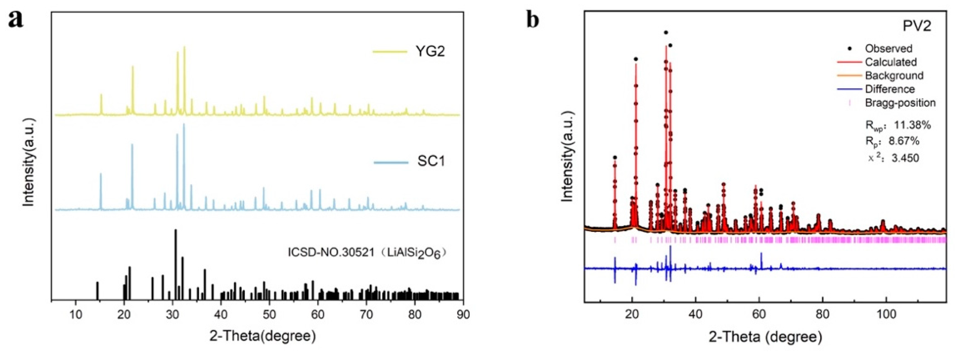
3.2.1. XRD Testing of Spodumene

One sample from each of the three colors of spodumene was selected for XRD crystal analysis. The XRD data of the YG2 and SC3 samples were processed using MDI Jade 6.5 software, and the resulting XRD spectra are shown in Figure 3a. The major diffraction peaks appear at 2θ = 14.661°, 21.289°, 30.649°, 32.156°, and 48.838°, with the strongest diffraction peak at 32.156°. These parameters are in excellent agreement with the data of α-spodumene (LiAlSi2O6, monoclinic system) from the ICSD database (ICSD #30521). The PV2 spodumene sample underwent structural refinement using the Rietveld method. The X-ray diffraction data of the PV2 sample were processed using the General Structure Analysis System (GSAS1.00) software, and the resulting XRD spectra are shown in Figure 3b. The main diffraction peaks in the spectrum appear at 2θ = 14.567°, 21.211°, 30.649°, 32.047°, and 48.838°, with the strongest diffraction peak at 30.649°. The crystal structure of spodumene can be further determined from the unit cell parameters in Table 2, a = 9.4756 Å, b = 8.4003 Å, c = 5.2260 Å, α = 90°, β = 110°, γ = 90°. These parameters are in excellent agreement with the data for α-spodumene (LiAlSi2O6, monoclinic system) from the ICSD database (ICSD #30521). The XRD crystal structure analysis confirmed that the primary crystalline phase of all three samples is α-spodumene and further verified the C2/c space group symmetry of the samples.
Under the C2/c space group symmetry, α-spodumene exhibits relatively high symmetry, and its crystal structure is composed of Li+, Al3+, and [SiO4] tetrahedra. The [SiO4] tetrahedra form continuous chains by sharing oxygen atoms, which are aligned along the crystal’s c-axis. The crystal structure is illustrated in Figure 4, where Al3+ typically occupies the M1 site, forming the six-fold coordinated light blue octahedral structure, while Li+ typically occupies the M2 site, forming the twisted green octahedral structure. The two M sites are connected to the [SiO4] tetrahedral chains via oxygen bridges, forming a relatively stable crystal structure. Furthermore, the metal cations at the M1 and M2 sites (Al and Li) can be substituted by transition metal elements, such as Mn, Fe, and others [14,15].

3.2.2. Electron Microprobe Analysis

In this study, spodumene samples PV2, YG2, and SC3, characterized by strong color and higher crystal clarity, were selected for electron probe thin section preparation. Four points were measured on each thin section, and the results are shown in Table S1 of Supporting Document S1. As shown in Table S2 of Supporting Document S1, the average SiO2 content of each sample ranges from 64.93 to 65.00 wt.%, which is the main component of spodumene. The average Al2O3 content ranges from 25.01 to 26.68 wt.%, Na2O content from 0.13 to 0.18 wt.%, FeO content from 0.02 to 0.70 wt.%, and MnO content from 0.06 to 0.12 wt.%.
Due to the limitations of EPMA technology, lithium, as a light element, cannot be directly measured. Therefore, the Li2O content was calculated stoichiometrically based on the average values of other oxide components in each sample. The specific calculation results are shown in Table S2, with Li2O contents of 8.87 wt.%, 9.8 wt.%, and 8.66 wt.% for PV2, YG2, and SC3 samples, respectively. The presence of Fe and Mn elements suggests the possibility of partial substitution at the Li+ or Al3+ sites [15].
The major oxides derived from the EPMA data indicate that the color of spodumene is primarily influenced by the transition metal elements Fe and Mn. In the yellow-green spodumene (YG2), the average FeO content is 0.7 wt.%, significantly higher than that of MnO, suggesting that Fe is the main factor responsible for the color of this sample. In the Pink to violet spodumene, the MnO content is higher than that of FeO, suggesting that the color is primarily influenced by Mn.

3.3. Spectral Characteristics and Chromogenic Mechanism Analysis

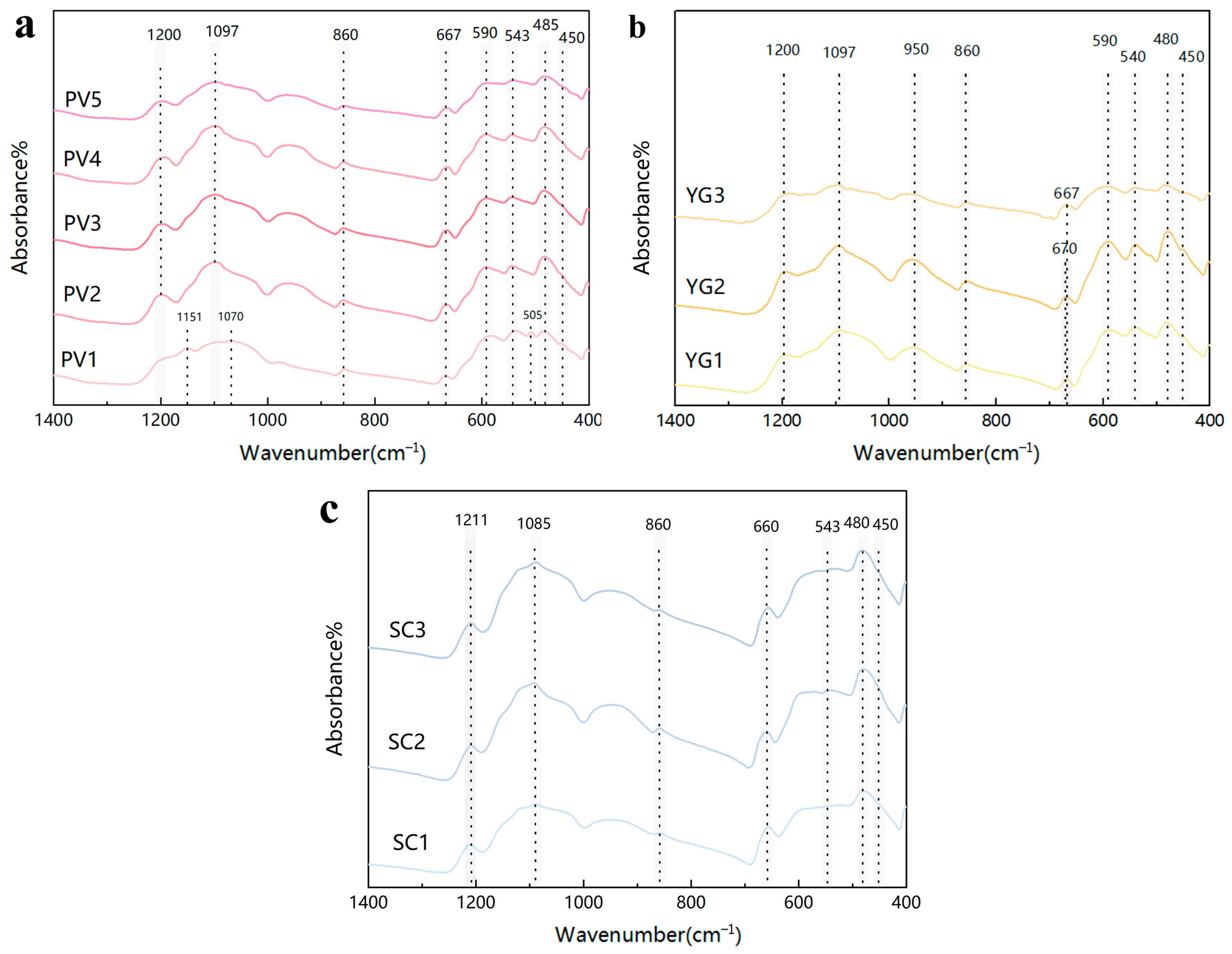
3.3.1. Infrared Spectra Analysis

Spodumene is a chain-type lithium silicate mineral, and its infrared spectral characteristics are primarily derived from the vibration modes of Si-O chain groups [16]. In this study, FTIR spectroscopy was conducted on pink to violet, yellow-green, and colorless spodumene samples. The experimental results, as shown in Figure 5, reveal that these samples exhibit several characteristic absorption peaks in the infrared spectrum, with the main peaks centered at 1200 cm−1, 1097 cm−1, 860 cm−1, 667 cm−1, 590 cm−1, 543 cm−1, 485 cm−1, and 450 cm−1. The analysis of these peaks will refer to the infrared spectral characteristics of spodumene with different crystal phases and pyroxene group minerals.
The peaks at 1200 cm−1, 1097 cm−1, and 860 cm−1 in pink to violet spodumene are suspected to correspond to Si-O stretching vibrations [17]. The absorption peaks and weak absorption bands observed in chain silicates in the range of 600–800 cm−1 are attributed to the bending vibrations of Si-O-Si in the silicate tetrahedra. Therefore, the absorption peak at 667 cm−1 is ascribed to Si-O-Si bending vibrations [18]. The peaks at 590 cm−1, 543 cm−1, 485 cm−1, and 450 cm−1 are likely related to Si-O stretching vibrations and M-O bending vibrations [16]. Overall, the main characteristic peaks in the infrared spectra of pink to violet, yellow-green, and colorless spodumene are quite consistent, though slight differences exist in certain peak positions, particularly the prominent peak at 950 cm−1 in yellow-green spodumene and the shift in the peak at 1211 cm−1 in colorless spodumene. These differences may be related to variations in trace elements within the samples or subtle changes in the crystal structure.
Based on the above spectroscopic data, the Infrared spectral features of spodumene were systematically summarized, and the related data are presented in Table 3.

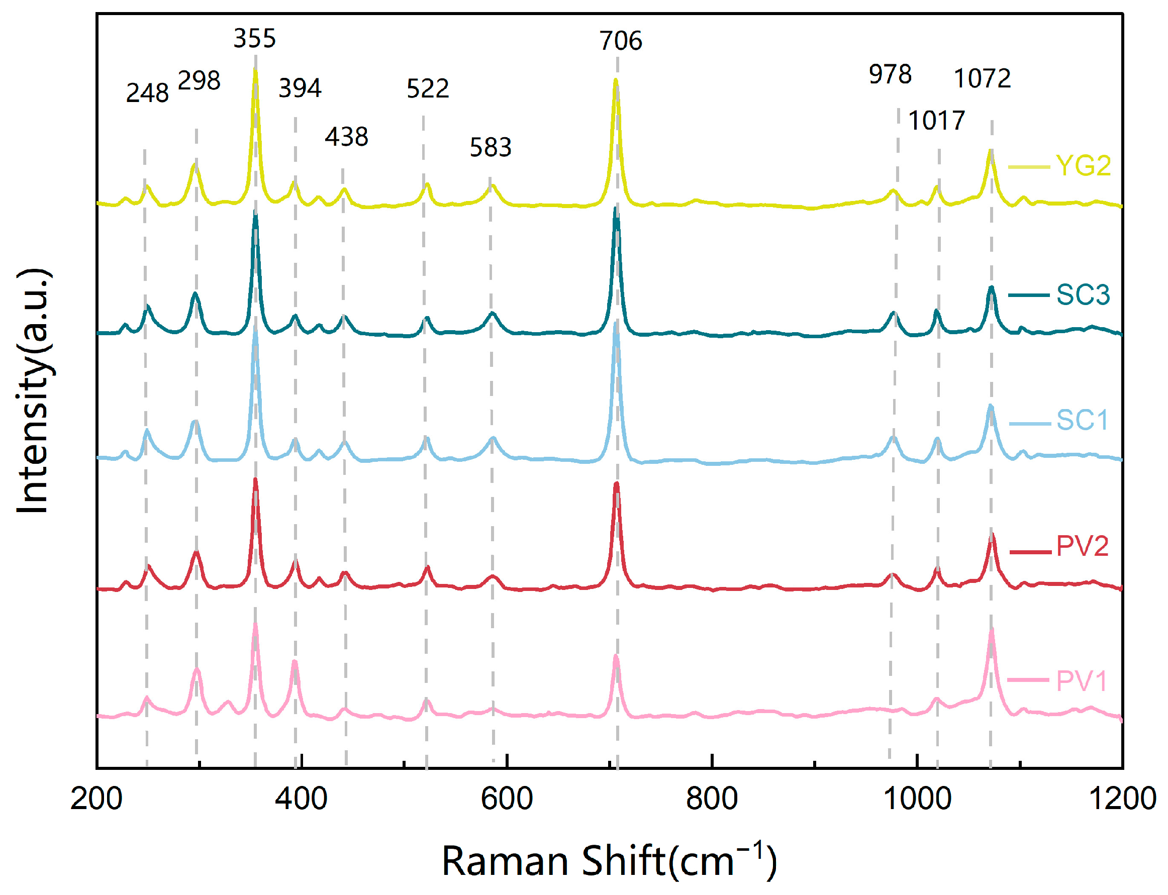
3.3.2. Raman Spectra Analysis

In this experiment, Raman spectroscopy was performed on spodumene samples of different colors, with 1 to 2 samples selected for each color. The results are shown in Figure 6. The results indicate that the spodumene samples of different colors exhibit high consistency in their crystal structures, and their Raman spectra closely match that of sample #X050155 in the RRUFF database. Based on previous studies on the Raman spectra of spodumene crystals, glasses, and single-chain silicates under different pressures, this paper analyzes and assigns the spectral peak positions of the experimental data.
In the range of 200 to 1200 cm−1, the spectrum displays 11 prominent absorption peaks, including 2 strong peaks, 2 moderate peaks, and 7 weaker peaks. The main characteristic peak values include 248 cm−1, 298 cm−1, 355 cm−1, 394 cm−1, 438 cm−1, 522 cm−1, 583 cm−1, 706 cm−1, 978 cm−1, 1017 cm−1, and 1072 cm−1, which correspond to different vibration modes in the crystal structure of spodumene. The high-frequency peaks at 1017 cm−1 and 1072 cm−1 are associated with the stretching vibration of Si-Onbr bonds (Si-O non-bridging oxygen bonds). The strong absorption peak at 706 cm−1 and the absorption peak at 978 cm−1 are related to the stretching vibrations of Si-Obr bonds (Si-O bridging oxygen bonds), characteristic of the pyroxene chain, with the 706 cm−1 peak being a signature absorption peak of the pyroxene structure [19,20]. The peaks at 522 cm−1 and 583 cm−1 reflect the bending vibrations of the O-Si-O bonds. Finally, the low-frequency peaks at 248 cm−1, 298 cm−1, 355 cm−1, 394 cm−1, and 438 cm−1 are related to the stretching or bending vibrations of M-O bonds. Since the major metal elements in spodumene are lithium (Li) and aluminum (Al), these peaks may correspond to the vibration modes of Li-O or Al-O bonds [20,21,22].
Based on the above spectroscopic data, the Raman spectral features of spodumene were systematically summarized, and the related data are presented in Table 4.

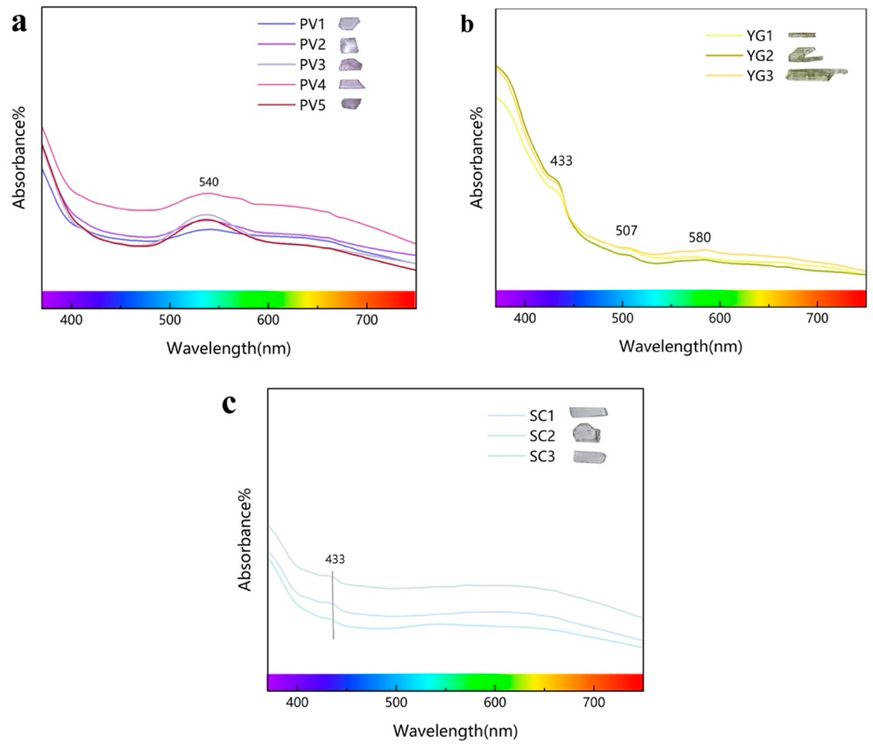
3.4. Ultraviolet–Visible Spectrum Analysis

Aside from pink to violet spodumene (Kunzite), the chromatic mechanisms of other color variations in spodumene remain insufficiently studied. Since spodumene is a silicate mineral of the pyroxene group, this study not only refers to existing research on the color origin of spodumene but also incorporates studies on the coloration mechanisms of other silicate gemstones and minerals. The color of spodumene samples is primarily determined by the presence and oxidation states of different transition metal elements, particularly Fe and Mn. Typically, Fe induces a green to yellow-green color in spodumene, while Mn may impart a pink to reddish-purple hue.
As shown in Figure 7, all five pink to violet spodumene samples exhibit a prominent broad peak at 540 nm, corresponding to absorption in the green light region, which imparts a reddish-purple appearance to the mineral. This absorption peak is typically associated with the 5E → 5T2 transition of Mn3+ ions in an octahedral coordination environment. Coupled with electron probe data showing a lower average Fe content (0.02 wt.%) compared to Mn (0.07 wt.%), it suggests that Mn3+ is the primary coloring agent in pink to violet spodumene [11,23,24]. Three yellow-green spodumene samples show a distinct absorption peak at 433 nm, located in the blue-violet region, which is typically associated with the 6A14E + 4A1 (4G) transition of Fe3+ ions [25,26,27]. Additionally, absorption peaks were also observed at 507 nm and 580 nm, corresponding to the blue-green and yellow light regions, respectively. These peaks contribute to the color of spodumene, but their absorption intensities are relatively weak, thus appearing as weaker absorption features in the spectrum. The absorption peaks resulting from the d-d transitions of Fe2+ typically appear near 530 nm and 570 nm. It is hypothesized that the absorption peaks at 507 nm and 580 nm may be related to the 5T25E transition of Fe2+.The electron probe data, showing a significantly higher average Fe content (0.70 wt.%) compared to Mn (0.12 wt.%), suggests that the overall color tone of yellow-green spodumene is primarily influenced by Fe [25,28]. The three colorless spodumene samples exhibit only a weak peak at 433 nm compared to the pink-to-violet and yellow-green samples. This peak is also primarily influenced by the 6A14E + 4A1 (4G) transition of Fe3+ ions, but it does not contribute significantly to the coloration of spodumene.
Based on the above spectroscopic data, the UV-Vis spectral features of spodumene were systematically summarized, and the related data are presented in Table 5.

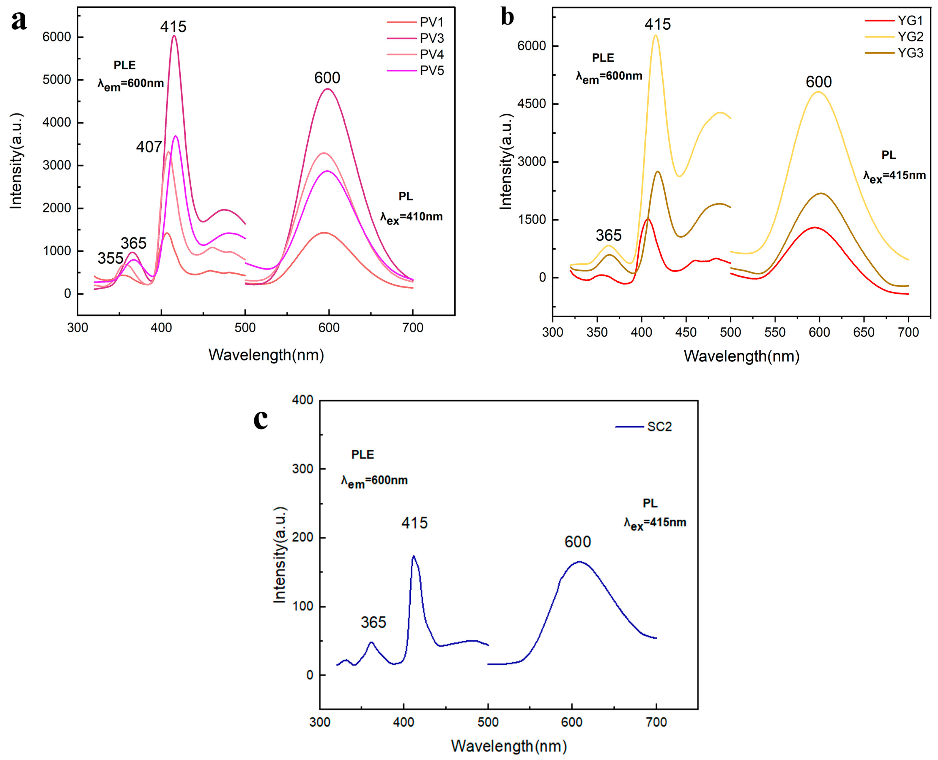
3.5. Luminescence Properties of Spodumene

Due to the presence of trace elements, spodumene typically exhibits varying degrees of fluorescence. Comparative photoluminescence (PL) spectroscopy was performed on spodumene samples of three different colors (pink to violet, yellow-green, and colorless) at an excitation wavelength of 415 nm. The experimental results, shown in Figure 8, indicate that all three color variations exhibit a broad emission peak around 600 nm, which can be attributed to the 4T1 (4G) → 6A1 (6S) transition of Mn2+ [29]. Photoluminescence excitation (PLE) spectra were measured for the three samples at an emission wavelength of 600 nm. The results show distinct excitation peaks around 365 nm and 415 nm in the blue-violet region. Some of the pink to violet spodumene samples exhibited slight blue shifts in their emission peaks, with shifts from 365 nm to 355 nm and from 415 nm to 407 nm. The excitation peak at 365 nm corresponds to the 6A14T2g (4D) transition of Mn2+, while the excitation peak at 415 nm corresponds to the 6A1 → 4E2g (4G) transition [30,31,32].
The experimental results indicate that the characteristic luminescence of Mn2+ ions is the primary luminescent mechanism in these colored spodumene samples. Compared to the pink-to-violet and yellow-green spodumene, the colorless spodumene exhibits weaker luminescence, which may be attributed to its lower Mn2+ content. Furthermore, the experiment revealed that the Fe content in the spodumene samples did not significantly suppress the luminescence of Mn2+. In the yellow-green spodumene sample (YG2), which contains higher Fe levels, the emission intensity of Mn2+ remains strong, suggesting that Fe is not a typical “luminescence killer” in spodumene [31].

4. Conclusions

In summary, this study systematically investigates three different colored spodumene samples, encompassing their gemological characteristics, crystal structures, and spectral properties, and preliminarily reveals the relationship between the formation mechanism of spodumene’s color and the trace elements Mn and Fe.
XRD analysis reveals that the three samples are primarily α-spodumene, with a crystal structure that matches the C2/c space group. FTIR spectroscopy reveals characteristic absorption peaks of spodumene at 1200 cm−1 and 1097 cm−1, corresponding to the Si-O stretching vibrations. Minor differences may exist between samples of different colors. Raman spectroscopy further confirms the crystal structure characteristics, with major absorption peaks, including the typical Si-Obr stretching vibrations peak at 706 cm−1, along with other vibration modes of Si-O and M-O bonds.
The study also reveals the significant impact of trace elements Fe and Mn, including their content and valence state mixture, on the color and luminescent properties of spodumene. By combining EPMA data, UV-Vis spectroscopy, and PL spectroscopy, the coloration and luminescence mechanisms of pink to violet spodumene are primarily influenced by the Mn element. In the UV-Vis spectrum, the pink to violet spodumene samples exhibit a distinct Mn3+ electronic transition absorption peak at 540 nm, which is the primary cause of its coloration. Its fluorescence characteristics are primarily dominated by the characteristic luminescence of Mn2+. For the yellow-green and colorless spodumene samples, their coloration and luminescence mechanisms result from the combined influence of Fe and Mn elements. Specifically, the yellow-green spodumene shows a distinct absorption peak at 433 nm in the UV-Vis spectrum, corresponding to the d-d transition of Fe3+. Additionally, weak absorption peaks at 507 nm and 580 nm are related to Fe2+ transitions. A weak absorption peak at 433 nm is also visible in the UV-Vis spectrum of colorless spodumene, but it does not result in a significant color change. The fluorescence characteristics of both samples are similarly dominated by the influence of Mn2+. Although Mn2+ ions are present in samples of different colors, they are not the primary coloring elements; they mainly contribute to the fluorescence of spodumene.
This study further enriches the research on the spectroscopic characteristics and coloration mechanisms of spodumene of different colors, identifying the influence of the transition metals Fe and Mn on the color and photoluminescence of spodumene. These results contribute to a deeper understanding of the coloration and luminescence of spodumene, expanding its potential applications as both a gemstone material and a luminescent material.

Supplementary Materials

The following supporting information can be downloaded at https://www.mdpi.com/article/10.3390/cryst15020109/s1. Supporting Document S1—EMPA.

Author Contributions

Z.Z., writing—original draft preparation and formal analysis; J.L., review and editing; K.H., review and editing; B.Y., investigations; Y.R., software. All authors have read and agreed to the published version of the manuscript.

Funding

This study was supported by the National Science and Technology Infrastructure—The National Infrastructure of Mineral, Rock and Fossil Resources for Science and Technology (http://www.nimrf.net.cn, accessed on 25 December 2021), and the Program of the Data Integration and Standardization in the Geological Science and Technology from MOST, China, grant number 2013FY110900-3.

Data Availability Statement

Data are contained within the article, further inquiries can be directed to the corresponding authors.

Conflicts of Interest

There are no conflicts to declare.

References

  1. Liu, Y.; Ma, B.; Lü, Y.; Wang, C.; Chen, Y. A review of lithium extraction from natural resources. Int. J. Miner. Metall. Mater. 2023, 30, 209–224. [Google Scholar] [CrossRef]
  2. Mohr, S.H.; Mudd, G.M.; Giurco, D. Lithium Resources and Production: Critical Assessment and Global Projections. Minerals 2012, 2, 65–84. [Google Scholar] [CrossRef]
  3. Sheikhi Gheshlaghi, R.; Ghorbani, M.; Sepahi, A.A.; Deevsalar, R.; Nakashima, K.; Shinjo, R. The origin of gem spodumene in the Hamadan Pegmatite, Alvand Plutonic Complex, western Iran. Can. Mineral. 2022, 60, 249–266. [Google Scholar] [CrossRef]
  4. Dessemond, C.; Soucy, G.; Harvey, J.-P.; Ouzilleau, P. Phase Transitions in the α–γ–β Spodumene Thermodynamic System and Impact of γ-Spodumene on the Efficiency of Lithium Extraction by Acid Leaching. Minerals 2020, 10, 519. [Google Scholar] [CrossRef]
  5. Mattson, S.M.; Rossman, G.R. Identifying characteristics of charge transfer transitions in minerals. Phys. Chem. Miner. 1987, 14, 94–99. [Google Scholar] [CrossRef]
  6. Walker, G.; El Jaer, A.; Sherlock, R.; Glynn, T.; Czaja, M.; Mazurak, Z. Luminescence spectroscopy of Cr3+ and Mn2+ in spodumene (LiAlSi2O6). J. Lumin. 1997, 72, 278–280. [Google Scholar] [CrossRef]
  7. Claffy, E.W. Composition, tenebrescence and luminescence of spodumene minerals. Am. Mineral. J. Earth Planet. Mater. 1953, 38, 919–931. [Google Scholar]
  8. Schmitz, B.; Lehmann, G. Color centers of manganese in natural spodumene LiAlSi2O6. Berichte Der Bunsenges. Für Phys. Chem. 1975, 79, 1044–1049. [Google Scholar] [CrossRef]
  9. Noithong, P.; Pakkong, P.; Naemchanthara, K. Color change of Spodumene gemstone by Electron beam irradiation. Adv. Mater. Res. 2013, 770, 370–373. [Google Scholar] [CrossRef]
  10. Czaja, M.; Lisiecki, R.; Kądziołka-Gaweł, M.; Winiarski, A. Some complementary data about the spectroscopic properties of manganese ions in spodumene crystals. Minerals 2020, 10, 554. [Google Scholar] [CrossRef]
  11. Rehman, H.U.; Martens, G.; Tsai, Y.L.; Chankhantha, C.; Kidkhunthod, P.; Shen, A.H. An X-ray Absorption Near-Edge Structure (XANES) Study on the Oxidation State of Chromophores in Natural Kunzite Samples from Nuristan, Afghanistan. Minerals 2020, 10, 463. [Google Scholar] [CrossRef]
  12. Ito, A.S.; Isotani, S. Optical Absorption and Electron Spin Resonance in Natural, Irradiated and Heated Spodumene; Sao Paulo Univ. (Brazil). Inst. de Fisica: Sao Paulo, Brazil, 1983. [Google Scholar]
  13. Ito, A.S.; Isotani, S. Heating effects on the optical absorption spectra of irradiated, natural spodumene. Radiat. Eff. Defects Solids 1991, 116, 307–314. [Google Scholar] [CrossRef]
  14. Graham, J. Some notes on α-spodumene, LiAlSi2O6. Am. Mineral. J. Earth Planet. Mater. 1975, 60, 919–923. [Google Scholar]
  15. Filip, J.; Novák, M.; Beran, A.; Zbořil, R. Crystal chemistry and OH defect concentrations in spodumene from different granitic pegmatites. Phys. Chem. Miner. 2006, 32, 733–746. [Google Scholar] [CrossRef]
  16. Wen, L.; Liang, W.; Zhang, Z. Mineral Infrared Spectroscopy; Chongqing University Press: Chongqing, China, 1989. (In Chinese) [Google Scholar]
  17. Kuang, G.; Chen, Z.B.; Guo, H.; Li, M.H. Lithium extraction mechanism from α-spodumene by fluorine chemical method. Adv. Mater. Res. 2012, 524, 2011–2016. [Google Scholar] [CrossRef]
  18. Bowey, J.; Hofmeister, A.; Keppel, E. Infrared spectra of pyroxenes (crystalline chain silicates) at room temperature. Mon. Not. R. Astron. Soc. 2020, 497, 3658–3673. [Google Scholar] [CrossRef]
  19. Sharma, S.K.; Simons, B.; Yoder, H. Raman study of anorthite, calcium Tschermak’s pyroxene, and gehlenite in crystalline and glassy states. Am. Mineral. 1983, 68, 1113–1125. [Google Scholar]
  20. Prencipe, M.; Mantovani, L.; Tribaudino, M.; Bersani, D.; Lottici, P.P. The Raman spectrum of diopside: A comparison between ab initio calculated and experimentally measured frequencies. Eur. J. Mineral. 2012, 24, 457–464. [Google Scholar] [CrossRef]
  21. Buzatu, A.; Buzgar, N. The Raman study of single-chain silicates. Analele Stiintifice Univ. AI Cuza Din Iasi. Sect. 2 Geol. 2010, 56, 107. [Google Scholar]
  22. Sharma, S.K.; Simons, B. Raman study of crystalline polymorphs and glasses of spodumene composition quenched from various pressures. Am. Mineral. 1981, 66, 118–126. [Google Scholar]
  23. Souza, S.; Ferraz, G.; Watanabe, S. Effects of Mn and Fe impurities on the TL and EPR properties of artificial spodumene polycrystals under irradiation. Nucl. Instrum. Methods Phys. Res. Sect. B Beam Interact. Mater. At. 2004, 218, 259–263. [Google Scholar] [CrossRef]
  24. Souza, S.; Watanabe, S.; Lima, A.; Lalic, M. Thermoluminescent mechanism in lilac spodumene. Acta Phys. Pol. A 2007, 112, 1001–1006. [Google Scholar] [CrossRef]
  25. Burns, R.G. Mineralogical Applications of Crystal Field Theory; Cambridge University Press: Cambridge, UK, 1993. [Google Scholar]
  26. Oliveira, R.A.P.; Mello, A.C.S.; Lima, H.R.B.R.; Campos, S.S.; Souza, S.O. Radiation detection using the color changes of lilac spodumene. In Proceedings of the International Nuclear Atlantic Conference (INAC 2009), Rio de Janeiro, RJ, Brazil, 27 September–2 October 2009. [Google Scholar]
  27. Farges, F.; Panczer, G.; Benbalagh, N.; Riondet, G. THE GRAND SAPPHIRE OF LOUIS XIV AND THE RUSPOLI SAPPHIRE: HISTORICAL AND GEMOLOGICAL DISCOVERIES. Gems Gemol. 2015, 51, 392–409. [Google Scholar] [CrossRef]
  28. Burns, R.G. Crystal field spectra and evidence of cation ordering in olivine minerals. Am. Mineral. J. Earth Planet. Mater. 1970, 55, 1608–1632. [Google Scholar]
  29. Chen, L.; Yang, W.; Fu, H.; Liu, W.; Shao, G.; Tang, B.; Zheng, J. Mn2+-doped Cs2NaInCl6 double perovskites and their photoluminescence properties. J. Mater. Sci. 2021, 56, 8048–8059. [Google Scholar] [CrossRef]
  30. Zhu, S.-Y.; Zhao, D.; Liu, W. A broad emission band of phosphor Cs2Zn3(P2O7)2:Mn2+ induced by multi-sites of Mn2+. Inorg. Chem. Commun. 2023, 150, 110397. [Google Scholar] [CrossRef]
  31. Czaja, M.; Lisiecki, R.; Kądziołka-Gaweł, M.; Winiarski, A.; Krzykawski, T. The afterglow effect of Mn-bearing natural LiAlSi2O6 spodumene crystals. Opt. Mater. 2019, 96, 109321. [Google Scholar] [CrossRef]
  32. Song, E.; Ding, S.; Wu, M.; Ye, S.; Xiao, F.; Zhou, S.; Zhang, Q. Anomalous NIR Luminescence in Mn2+-Doped Fluoride Perovskite Nanocrystals. Adv. Opt. Mater. 2014, 2, 670–678. [Google Scholar] [CrossRef]
Figure 1. Photos of spodumene samples.
Crystals 15 00109 g001
Figure 2. (a) Color of sample PV-4 parallel to the C-axis. (b) Color of sample PV-4 perpendicular to the C-axis. (c) Stepped fracture pattern of YG-2. (d) Distinct longitudinal striations on the surface of YG-3.
Crystals 15 00109 g002
Figure 3. (a) The XRD patterns of spodumene sample SC1, YG, LG1. (b) Structure refinement map of PV-2.
Crystals 15 00109 g003
Figure 4. Structure model of spodumene. (ac) show the crystal structure of spodumene in the direction of three crystal axes, a, b, and c, respectively. In the lower-left corner of the coordinate axis, the red arrow represents the a-crystal axis, the green arrow represents the b-crystal axis, and the blue arrow represents the c-crystal axis. In the crystal structure diagram, Oxygen atoms are in red. Al occupies the light green octahedron. Li occupies the light blue twisted tetrahedron. Si occupies the light blue tetrahedron.
Crystals 15 00109 g004
Figure 5. (a) Infrared spectra (reflection) of pink to violet samples. (b) Infrared spectra (transmission) of yellow-green samples. (c) Infrared spectra (transmission) of colorless samples.
Crystals 15 00109 g005
Figure 6. Raman spectra of the three color varieties of spodumene.
Crystals 15 00109 g006
Figure 7. (a) UV-Vis spectra of pink to violet samples; (b) UV-Vis spectra of yellow-green samples; (c) UV-Vis spectra of colorless samples.
Crystals 15 00109 g007
Figure 8. (a) Fluorescence spectra of pink to violet samples; (b) fluorescence spectra of yellow-green samples; (c) fluorescence spectra of colorless samples.
Crystals 15 00109 g008
Table 1. Gemological characteristics of the samples.
SampleColorTransparencyBirefringenceRefractive IndexWeight (ct)Specific Gravity
PV1pink to violetsemitransparent0.0151.662–1.6680.553.18
PV2pink to violetsemitransparent0.0141.668–1.6731.493.17
PV3pink to violetsemitransparent0.0151.662–1.6701.333.19
PV4pink to violettransparent0.0151.663–1.6710.893.18
PV5pink to violetsemitransparent0.0161.665–1.6731.083.18
YG1yellow-greensemitransparent0.0151.663–1.6680.313.22
YG2yellow-greentransparent0.0151.662–1.6700.973.16
YG3yellow-greensemitransparent0.0161.671–1.6762.273.20
SC1colorlesstransparent0.0151.663–1.6710.823.15
SC2colorlesstransparent0.0151.662–1.6701.213.18
SC3colorlesstransparent0.0141.663–1.6680.543.18
Table 2. Cell parameters of PV2.
abcalphabetagammaVolumeRwpRpχ2
Value9.4756898.4003115.22605590.000110.17690.000390.46211.38%8.67%3.450
Sigmas0.0000950.0000520.0000820.0000.0010.0000.008
Table 3. Assignment of infrared spectra of spodumene.
Si-O Stretching Vibrations and M-O Bending VibrationsSi-O-Si Bending VibrationsSi-O Stretching Vibrations
Infrared Characteristic peak/cm−1590 cm−1
543 cm−1
485 cm−1
450 cm−1
667 cm−11200 cm−1
1097 cm−1
860 cm−1
Table 4. Assignment of Raman spectra of spodumene.
M-O Bending VibrationO-Si-O Bending VibrationSi-Obr Stretching VibrationsSi-Onbr Asymmetric Stretching Vibration
Raman Characteristic peak/cm−1248 cm−1
298 cm−1
355 cm−1
394 cm−1
438 cm−1
522 cm−1
583 cm−1
706 cm−1
978 cm−1
1017 cm−1
1072 cm−1
Table 5. Assignment of ultraviolet–visible spectrum of spodumene.
SampleColorCharacteristic PeakAssignment
PV1–5pink to violet540 nmMn3+ (5E → 5T2)
YG1–3yellow-green433 nmFe3+ (6A14E + 4A1)
507 nmFe2+ (5T25E)
580 nm
SC1–3colorless433 nmFe3+ (6A14E + 4A1)
Disclaimer/Publisher’s Note: The statements, opinions and data contained in all publications are solely those of the individual author(s) and contributor(s) and not of MDPI and/or the editor(s). MDPI and/or the editor(s) disclaim responsibility for any injury to people or property resulting from any ideas, methods, instructions or products referred to in the content.

