Article

Mechanocatalytic Hydrogen Evolution on Centrosymmetric SnS Nanobelts: A Non-Piezoelectric Pathway

1
College of Chemistry and Chemical Engineering, Central South University, Changsha 410083, China
2
State Key Laboratory of Chemical Engineering, School of Chemical Engineering and Technology, Tianjin University, Tianjin 300072, China
3
Institute of Shaoxing, Tianjin University, Shaoxing 312300, China
4
Xiangjiang Laboratory, Changsha 410205, China
*
Authors to whom correspondence should be addressed.
Crystals 2025, 15(11), 940; https://doi.org/10.3390/cryst15110940 (registering DOI)
Submission received: 13 October 2025 / Revised: 26 October 2025 / Accepted: 30 October 2025 / Published: 31 October 2025
(This article belongs to the Special Issue Advances in Materials for Energy Conversion and Storage)

Abstract

Harnessing ubiquitous mechanical energy for chemical transformations is a grand challenge, primarily impeded by the crystallographic symmetry constraints of conventional piezocatalysts. Here, this long-standing paradigm is shattered by demonstrating potent mechanocatalytic activity in a centrosymmetric material. Synthesized via a facile hydrothermal method, unique SnS nanobelts exhibit a hydrogen evolution rate of 3889 µmol g−1 h−1 under mechanical vibration—achieved without any cocatalysts—a performance substantially surpassing that of most reported piezocatalysts and comparable to state-of-the-art photocatalytic systems. Moreover, the SnS nanobelts were also found to present good cyclic stability. This unprecedented activity was rationalized by the synergy between two effects: sonoluminescence, for which the material’s ideally suited band structure allows efficient photon capture, and flexoelectricity. Furthermore, direct electrical measurements confirmed that SnS generates a flexoelectric current under mechanical deformation, thereby driving the H2 evolution reaction. These findings not only expand the scope of potential mechanocatalysts by unlocking a vast and previously ignored territory of centrosymmetric materials but also offer valuable guidance and insights for designing high-efficiency, mechanically driven chemical reactions.

1. Introduction

Owing to its exceptional energy density and carbon-free combustion product (solely water), hydrogen (H2) is widely regarded as one of the most promising clean energy carriers for the future, serving as a cornerstone for establishing a hydrogen-based economy and achieving global carbon neutrality goals [1,2,3,4]. However, current industrial-scale H2 production is predominantly reliant on the steam reformation of fossil fuels (e.g., natural gas), a process that is highly energy-intensive and releases substantial quantities of greenhouse gases (CO2), running counter to the principles of sustainable development [4,5,6]. Therefore, developing an eco-friendly and sustainable H2 production pathway, particularly through water splitting, represents a core strategy for addressing the dual challenges of the global energy crisis and climate change and for building a future sustainable energy system [7,8].
Among various available strategies, photocatalytic hydrogen evolution reaction (HER) from water splitting has emerged as a promising technology that harnesses natural energy sources. Its practical application, however, is hampered by its dependence on intermittent light conditions [8,9]. While electrocatalysis offers a robust alternative for water splitting, it requires high-performance electrolyzers, and the prohibitive cost of electricity remains a significant barrier to widespread green hydrogen adoption [7,10]. A breakthrough came when Wang et al. discovered that mechanical energy could be converted into chemical energy via the piezoelectric effect, giving rise to the field of piezocatalysis [11,12,13]. This provided a compelling avenue for harvesting ambient mechanical energy, yet its application is fundamentally restricted to materials with non-centrosymmetric crystal structures.
In response, mechanocatalysis, which also utilizes mechanical energy to trigger chemical reactions through deformation-induced charge separation, breaks this structural constraint [14,15,16,17]. Nevertheless, the catalytic efficiency for HER using centrosymmetric mechanocatalysts has remained limited. For instance, Delogu et al. achieved a maximum H2 production rate of 258 µmol g−1 h−1 by milling quartz powder in a vibration mill [16]. Hitoki et al. realized overall water splitting by magnetically stirring suspensions of various transition metal tungstates and molybdates, among which NiWO4 exhibited the highest H2 yield of 540 µmol g−1 h−1 [18]. More recently, Zhang et al. achieved a rate of 322.7 µmol g−1 h−1 in pure water using F-doped HAP nanowires [19], while impressive efficiencies of 1156.3 and 1289.53 µmol g−1 h−1 have been reported by Du et al. and Mondal et al. for BaTi2O5 and SrTiO3, respectively [20,21]. These performances are still inferior to those of state-of-the-art piezocatalysts (typically 2000–4000 µmol g−1 h−1) [22,23,24,25,26].
This discrepancy is largely attributed to the fact that applying random external stress to a centrosymmetric catalyst induces the flexoelectric effect. Compared to piezoelectricity, the magnitude of the flexoelectric effect is generally smaller (about 10−9 C m−2), resulting in a weaker driving force for chemical reactions [27]. To address this, researchers have focused on morphology engineering; for example, Zhang et al. demonstrated that MAPbI3 nanowires possess a large strain gradient and a strong flexoelectric response, leading to significant H2 generation [19]. Similarly, another study by Wu et al. leveraged the interfacial strain gradient in high-entropy oxide nanocomposites to enhance flexoelectric polarization, achieving excellent H2 production rates under ultrasonic vibration [28]. Notably, many of the existing high-performance mechanocatalysts often involve complex compositions and contain heavy or precious metals (e.g., Cd, Pb, Au, and Pt), which leads to intricate synthesis protocols, high costs, and potential environmental/health risks, thereby limiting their practical application [29,30,31].
In recent years, transition metal chalcogenides, such as MoS2, CdS, and WS2, have garnered considerable interest for electro- and photocatalytic HERs due to their diverse morphologies, tunable electronic structures, and good chemical/structural stability [32,33,34]. As a binary transition metal chalcogenide, tin sulfide (SnS) exhibits an ideal visible-light-responsive band gap of ~1.2–1.5 eV and relatively high electrical conductivity [35]. Coupled with its earth-abundant and non-toxic nature, SnS emerges as a prime candidate material for HER. It is noteworthy that various SnS nanostructures, including nanoflowers, nanosheets, and nanowires, have been successfully synthesized, providing an excellent platform for mechanical energy harvesting and conversion [36,37,38]. To date, however, its applications have been primarily focused on solar cells, energy storage, and sensors, while its potential for catalytic hydrogen evolution, particularly via a mechanocatalytic route, remains unexplored [39,40,41,42,43].
Against this backdrop, we report the synthesis of a novel, centrosymmetric SnS nanobelt via a simple hydrothermal method and, for the first time, demonstrate its application in mechanocatalytic hydrogen evolution. Under optimized conditions, the non-piezoelectric SnS achieved a maximum H2 yield of 3889 µmol g−1 h−1, a rate that surpasses that of most previously reported piezocatalysts. Moreover, the potential catalytic mechanisms were investigated from the dual perspectives of the ultrasonic-cavitation-induced sonoluminescence effect and the flexoelectric effect.

2. Materials and Methods

2.1. Materials

Urea (CH4N2O), thioacetamide (C2H5NS), thiourea (CH4N2S), and Stannous chloride dihydrate (SnCl2·2H2O), were purchased from Sinopharm Chemical Reagent Co., Ltd. (Shanghai, China). All materials were analytical grade and used without any purification process. Deionized (DI) water (resistivity: 18.2 MΩ cm) used in experiment was obtained from a Milli-Q system (Molecular Corp., Louisville, CO, USA). Absolute ethanol used in the experiments were obtained from standard laboratory sources.

2.2. Synthesis of SnS Nanobelts

The SnS nanobelts were synthesized via a simple one-pot hydrothermal route, following a modified procedure previously reported by Tian et al. [44]. In a typical synthesis, 1.68 g (28 mmol) of CH4N2O and 1.50 g (20 mmol) of C2H5NS were dissolved in 80 mL of deionized water under magnetic stirring. After stirring for 30 min to form a clear solution, 0.27 g (1.2 mmol) of SnCl2·2H2O was added to the mixture. The resulting mixture was stirred for another 30 min until it became completely dissolved. Subsequently, the solution was transferred into a 100 mL Teflon-lined stainless-steel autoclave, which was then sealed and maintained at 160 °C for 12 h (Figure 1). After the autoclave cooled to room temperature naturally, the black precipitate was collected by centrifugation and washed several times with deionized water and ethanol. Finally, the product was obtained after being freeze-dried.

2.3. Electrochemical Measurements

All electrochemical measurements, including flexoelectric current response, and electrochemical impedance spectra (EIS) were measured on the electrochemical workstation (CHI 660C, CH Instrument, Austin, TX, USA) equipped with a three-electrode configuration. FTO electrodes were deposited with samples as the anode, a platinum foil as the counter electrode, and a saturated calomel electrode (SCE) as the reference electrode. The 0.5 M Na2SO4 aqueous solution was used as an electrolyte. All the samples were analyzed at room temperature without bias potential. The details of the preparation process for the working electrode are as follows: 5 mg catalysts were dispersed in 1 mL ethanol and 20 μL of 5 wt% Nafion to form a slurry mixture, and after that 100 μL slurry was fixed on FTO glass (1 cm × 3 cm) by drying at 50 °C. In a flexoelectric current measurement, a 40 W Supmile ultrasonic cleaner (KQ5200DE, Supmile, Kunshan, China) was employed as the vibration source. Notably, the flexoelectric current response was recorded continuously while the ultrasonic vibration was manually switched on and off at 20-s intervals (20 s on, 20 s off) to generate the transient signal. A detailed schematic of this experimental setup, along with a photograph of the working electrode, is provided in the Supporting Information (Figures S1 and S2).

3. Results

3.1. Structural, Morphological, and Compositional Characterization

The morphology and microstructure of the as-synthesized product were first investigated by scanning electron microscopy (SEM, Hitachi, Hitachi, Japan). The low-magnification SEM image (Figure 2a) reveals that the product consists of a large quantity of nanostructures with a uniform, well-defined belt-like morphology, with typical lengths of several round 10 μm. The high-magnification image (Figure 2b) further clarifies the structure of a typical individual nanobelt, showing a distinct diamond-shaped end and smooth surfaces. To determine the elemental composition and spatial distribution within the nanobelts, energy-dispersive X-ray spectroscopy (EDS, Hitachi, Hitachi, Japan) mapping was performed on the selected single nanobelt. As shown in Figure 2c,d, both tin (Sn, purple) and sulfur (S, cyan) elements are homogeneously distributed throughout the entire nanostructure. These results collectively confirm the successful synthesis of SnS nanobelts with high morphological purity and uniform elemental composition.
To ascertain the crystal structure, phase purity, and chemical states of the as-synthesized SnS nanobelts, a series of structural and spectroscopic analyses were conducted. As presented in the X-ray diffraction (XRD) pattern (Figure 3a), all the sharp and intense diffraction peaks can be unambiguously indexed to the orthorhombic phase of SnS (JCPDS No. 39-0354). The absence of any detectable peaks from impurities or other phases confirms the high phase purity of the sample. Notably, this structure belongs to the Pbnm (No. 62) space group, which is centrosymmetric. This crystallographic feature fundamentally precludes the possibility of piezoelectricity in the as-synthesized SnS. This structural identification was further corroborated by Raman spectroscopy (Figure 3b). The analysis revealed four prominent vibrational modes at 94, 162, 188, and 217 cm−1. These peaks are unambiguously assigned to the Ag (94, 188, 217 cm−1) and B3g (162 cm−1) modes, respectively, which is the characteristic vibrational fingerprint for the orthorhombic phase of SnS, consistent with previous reports [45,46]. The presence of these distinct modes also corroborates the anisotropic nature of the synthesized material.
The surface elemental composition and valence states were investigated by X-ray photoelectron spectroscopy (XPS) (Figure S3). The high-resolution Sn 3d spectrum (Figure 3c) is deconvoluted into two pairs of doublets. The primary doublet, with peaks at 485.1 eV (Sn 3d5/2) and 493.5 eV (Sn 3d3/2), is characteristic of Sn2+ in the SnS lattice. The weaker doublet at higher binding energies (486.3 eV and 494.7 eV) is attributed to a small amount of Sn4+, likely arising from slight surface oxidation. In the S 2p spectrum (Figure 3d), the peaks located at 161.2 eV (S 2p3/2) and 162.5 eV (S 2p1/2) are ascribed to the S2− state [47]. Collectively, these analyses provide compelling evidence that the product is phase-pure, well-crystallized orthorhombic SnS, where the constituent elements predominantly exist in their Sn2+ and S2− valence states.

3.2. Mechanocatalytic H2 Evolution Performance

The mechanocatalytic H2 evolution performance of the as-synthesized SnS nanobelts was first investigated under ultrasonic vibration. A series of control experiments were performed to establish the essential components for the reaction (Figure 4a). The complete system, comprising the SnS nanobelts, methanol (MOH) as a sacrificial agent, and ultrasonic irradiation, yielded an impressive H2 evolution rate. Conversely, control experiments revealed that H2 evolution was negligible without ultrasonic vibration, whereas it was only substantially reduced, not eliminated, in the absence of the SnS catalyst. These findings unequivocally confirm that the H2 production is a direct result of the intrinsic mechanocatalytic activity of the SnS nanobelts, which dominantly relies on the simultaneous presence of the catalyst and mechanical energy input.
To optimize the reaction conditions, the effect of various sacrificial agents was examined (Figure 4b). While negligible H2 was produced in pure water (blank), the addition of methanol (MOH), triethanolamine (TEOA), or ethylene glycol (EG) all significantly boosted the HER activity. Among them, MOH provided the highest efficiency, indicating its superior efficacy as a hole scavenger to suppress charge recombination. Moreover, the influence of ultrasonic parameters was also investigated. The H2 evolution rate was found to be highly dependent on both the ultrasonic power and frequency (Figure 4c). Specifically, a monotonic increase in H2 production was observed as the ultrasonic power was elevated from 360 W to 630 W. This trend is attributed to the fact that higher power generates more intense acoustic cavitation and imparts greater mechanical stress, which enhances both the photon flux from sonoluminescence and the magnitude of the flexoelectricity, thereby promoting more efficient charge carrier generation and separation [48]. Regarding the frequency, the presence of an optimal value strongly suggests a correlation with the natural resonant frequency of the SnS nanobelts. This phenomenon can be explained by the principles of mechanical resonance: when the external ultrasonic frequency matches the intrinsic resonant frequency of the nanostructures, they undergo maximum mechanical deformation. For a nanostructure like a nanobelt, this resonant frequency is governed by its specific dimensions (e.g., length and thickness) and intrinsic mechanical properties [49]. The maximized deformation at resonance generates the largest possible strain gradient, which in turn induces the strongest flexoelectric field, thereby maximizing charge carrier separation and boosting the H2 evolution rate. Therefore, the sharp peak in catalytic activity observed at a specific frequency (Figure 4c) serves as strong experimental evidence for this resonant coupling effect, which is critical for achieving peak catalytic efficiency. Under optimized conditions, the H2 evolution rate reaches 3889 µmol g−1 h−1, a performance that surpasses most reported piezocatalysts and is comparable to leading photocatalytic systems, as detailed in Table 1.
The long-term stability of the SnS nanobelts, a critical metric for practical applications, was subsequently evaluated through five consecutive 1-h catalytic runs (Figure 4d). The reactor was purged with argon before the start of each new cycle. Remarkably, the catalyst exhibited no discernible decay in its H2 production rate across all five cycles, maintaining its high initial activity. This exceptional durability demonstrates the profound structural and catalytic robustness of the SnS nanobelts under prolonged mechanical stress, underscoring their potential as a highly stable and efficient mechanocatalyst.
To elucidate the origin of this exceptional cycling stability, post-reaction analyses were performed on the SnS nanobelts after the five-cycle test. As illustrated in Figure 5a, the XRD pattern of the used catalyst is virtually identical to that of the pristine (fresh) sample, with all characteristic diffraction peaks of the orthorhombic SnS phase perfectly retained. No peak shifts or the emergence of new peaks corresponding to impurities or phase transformation were observed, indicating that the long-range crystalline integrity was well-preserved despite prolonged exposure to ultrasonic vibration.
The preservation of structural integrity was further corroborated by Raman spectroscopy (Figure 5b). The Raman spectrum of the used catalyst exhibits the same characteristic vibrational modes as the fresh sample, with no discernible changes in peak positions, shapes, or relative intensities. This confirms that the local atomic arrangement and chemical bonding within the SnS lattice were not altered during the demanding mechanocatalytic process. Taken together, these post-reaction characterizations provide compelling evidence for the outstanding structural robustness of the SnS nanobelts. This inherent ability to resist structural degradation under continuous mechanical stress is therefore identified as the key factor behind its sustained and stable catalytic performance.

3.3. Mechanism Study of Mechanocatalytic H2 Evolution

To elucidate the mechanism underlying the observed mechanocatalytic H2 evolution, we probed the electronic properties of the SnS nanobelts, testing the hypothesis of a process driven by sonoluminescence. This phenomenon, arising from ultrasonic cavitation, involves the generation of localized, intense light emission, which can in turn activate a semiconductor catalyst in a manner analogous to photocatalysis [64]. The viability of such a mechanism hinges on the material’s band structure being thermodynamically suitable for the H2 evolution reaction (HER).
The optical absorption properties of the SnS nanobelts were first examined using UV-vis-NIR diffuse reflectance spectroscopy (Figure 6a), revealing significant absorption across the visible and near-infrared regions. From the corresponding Tauc plot (Figure 6b), the optical band gap (Eg) was estimated by extrapolating the linear portion of the (αhν)2 versus photon energy (hν) curve. This analysis yielded a direct band gap of approximately 1.32 eV.
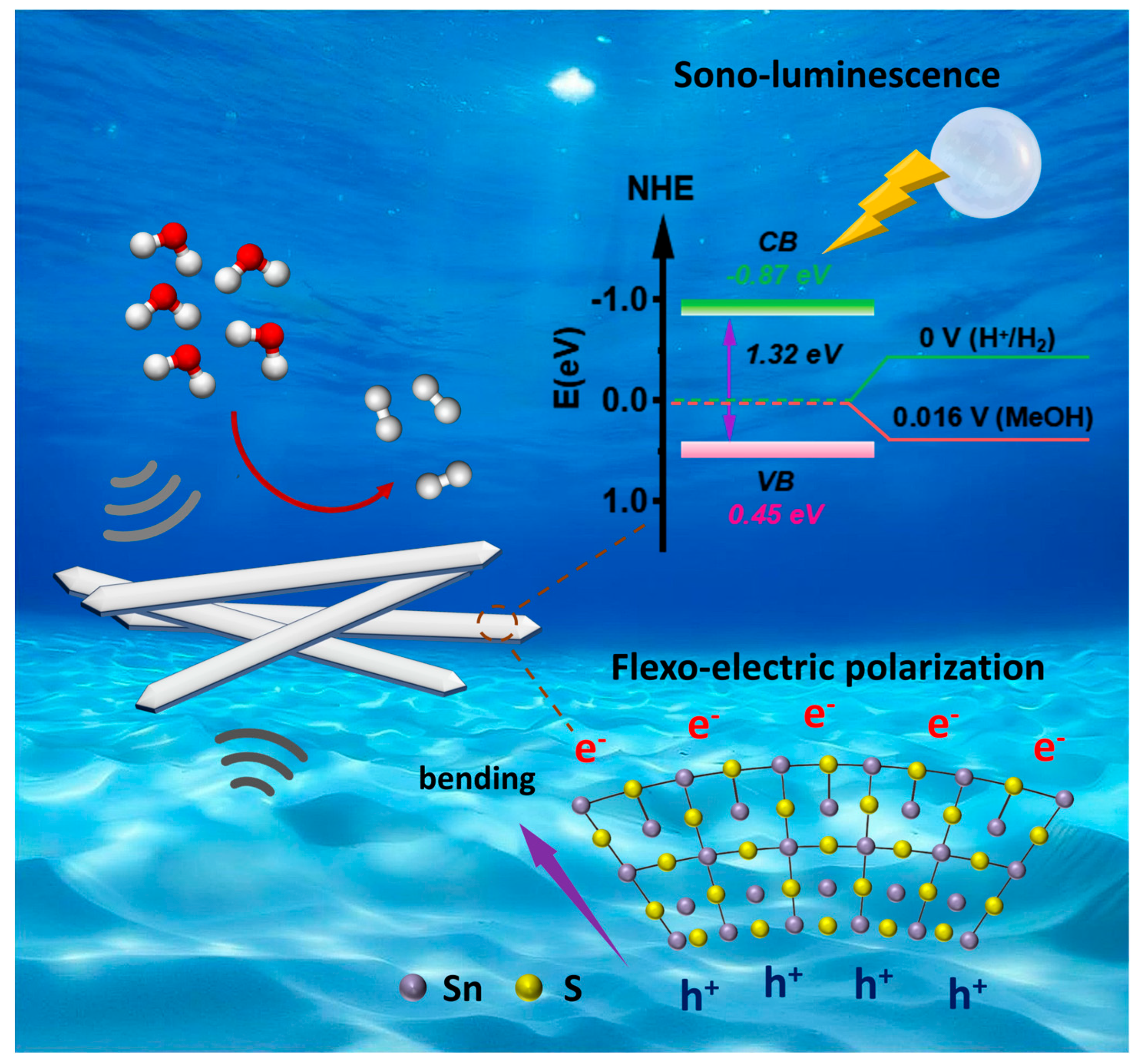
To determine the absolute energy band positions, the valence band maximum (VBM) was measured by valence band XPS (Figure 6c) and determined to be at +0.45 eV (vs. NHE). Subsequently, the conduction band minimum (CBM) was calculated using the equation ECBM = EVBM − Eg, positioning the CBM at −0.87 eV (vs. NHE). A schematic of the resultant band alignment is presented in Figure 6d. Crucially, the CBM of SnS (−0.87 eV) is substantially more negative than the redox potential of H2/H2O (0 eV vs. NHE), confirming that conduction band electrons possess the thermodynamic driving force to reduce H2O to H2. Concurrently, the VBM (+0.45 eV) is appropriately positioned for the efficient consumption of holes by the methanol sacrificial agent.
Synthesizing these findings, we propose a mechanism driven by sonoluminescence. Under ultrasonic irradiation, the violent collapse of cavitation bubbles generates transient, high-energy light emission with a typical wavelength range of around 200–700 nm [64]. This emitted light is absorbed by the SnS nanobelts, exciting electrons from the VBM to the CBM and creating electron-hole pairs. The photogenerated electrons at the CBM (−0.87 eV) subsequently drive the reduction of water, while the holes at the VBM (+0.45 eV) are effectively scavenged by methanol, thereby completing the catalytic cycle. This model successfully explains how the intrinsic electronic properties of SnS, which are suitable for photocatalysis, can be harnessed for efficient mechanocatalytic H2 production.
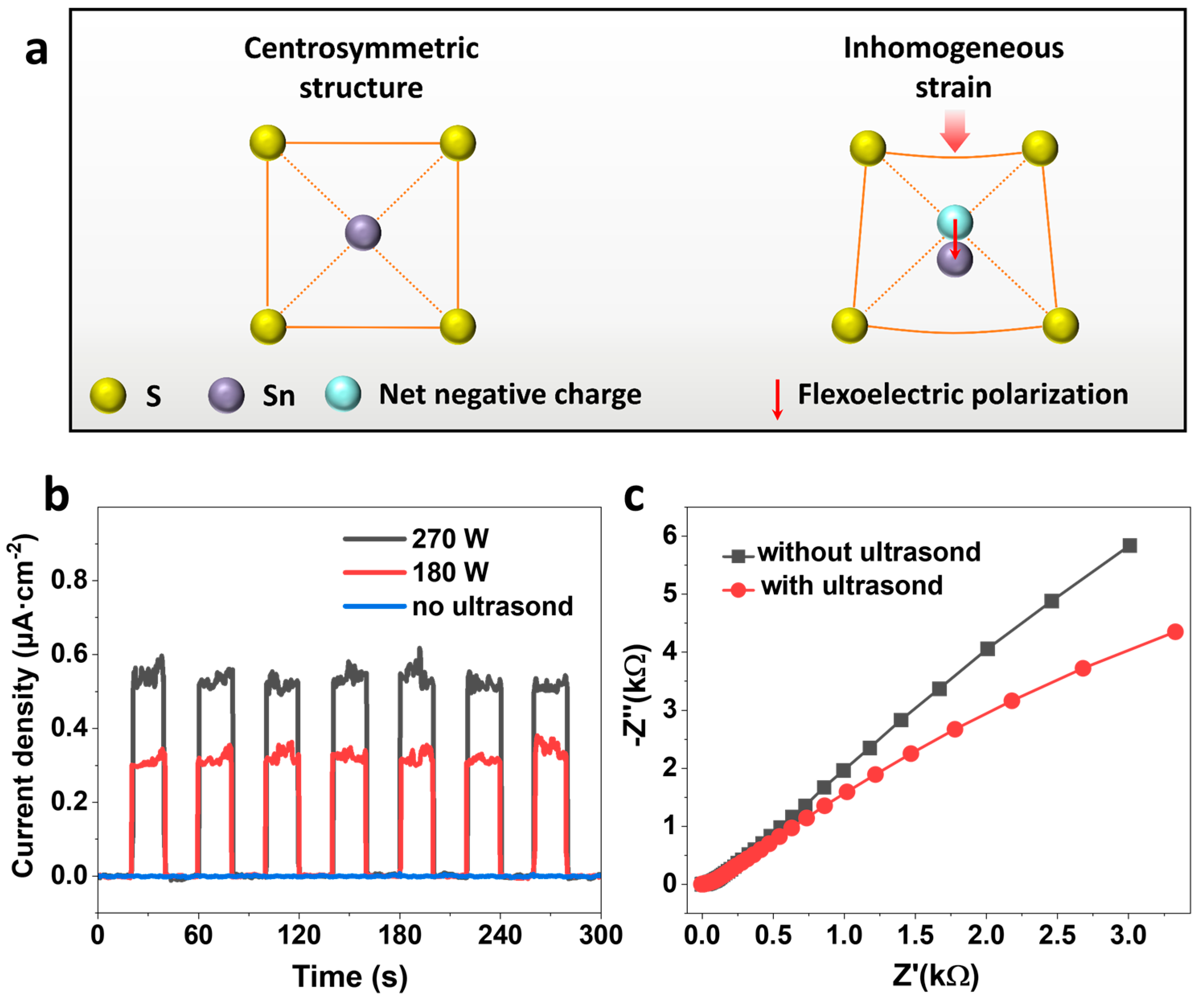
In addition to sonoluminescence, we explored an alternative, potentially synergistic mechanism rooted in the electromechanical properties of SnS: the flexoelectric effect [21]. Unlike piezoelectricity, which is confined to non-centrosymmetric materials, the flexoelectric effect—the coupling between strain gradients and electric polarization—is a universal phenomenon present in all dielectrics, even in centrosymmetric SnS. We hypothesized that the mechanical bending and deformation of the SnS nanobelts under ultrasonic vibration would generate a significant strain gradient, inducing a flexoelectric polarization field that could effectively separate charge carriers and drive catalysis (Figure 7a).
To experimentally validate this hypothesis, we employed the transient current response measurement, a direct and widely used technique to demonstrate mechano-electric conversion in catalytic systems. As shown in Figure 7b, the current from the SnS-coated electrode was negligible in the absence of ultrasound (“off” state). However, upon applying ultrasonic vibration (“on” state), a distinct and reproducible anodic current of ~0.13 µA was immediately generated, promptly returning to baseline once the vibration ceased. Furthermore, the magnitude of this current scaled with the ultrasonic input power. This hallmark “on/off” current response, consistent with leading-edge studies in flexocatalysis, provides unequivocal evidence that strain gradients within the SnS nanobelts generate a potent internal electric field that separates and mobilizes charge carriers [65,66,67]. The power-dependent nature of this current also directly accounts for the enhanced H2 production rates observed at higher ultrasonic powers (Figure 4c).
The impact of this induced polarization on charge transfer kinetics was further investigated using electrochemical impedance spectroscopy (EIS). The Nyquist plot for the SnS electrode (Figure 7c) displayed a large semicircular arc without ultrasound, indicating high charge transfer resistance (Rct). Remarkably, upon introducing ultrasonic vibration, the arc radius decreased obviously. This substantial reduction in Rct is a key indicator of accelerated reaction kinetics, signifying that the internal flexoelectric field not only separates charges but also critically facilitates their migration to the catalyst-electrolyte interface. This method of using EIS to confirm the kinetic benefits of an internal polarization field is a robust approach in catalysis research and has been specifically applied to validate the role of flexoelectricity [68]. By lowering the kinetic barrier for redox reactions, the flexoelectric effect is thus confirmed to play a pivotal role.
To clarify the physical origin of the flexoelectric effect, it is essential to explain how ultrasonic vibration induces an inhomogeneous strain, and consequently a strain gradient, within the SnS nanobelts. This mechanism can be understood through the following points: First, the ultrasonic cleaner generates high-frequency (e.g., >20 kHz) mechanical pressure waves that propagate through the aqueous solution and impinge on the SnS nanobelts. Due to their high aspect ratio and intrinsically low flexural rigidity, the nanobelts are highly susceptible to these mechanical perturbations. Instead of vibrating as rigid bodies, they undergo dynamic bending, flexing, and rippling modes in response to the acoustic waves [65,69]. Second, this non-uniform deformation is the direct source of the inhomogeneous strain. At any given moment, a bent section of a nanobelt will experience compressive strain on its inner curved surface and tensile strain on its outer surface. This spatial variation of strain across the thickness and along the length of the nanobelt constitutes a significant strain gradient (∇ ε ). Finally, several factors are responsible for ensuring this strain is inhomogeneous rather than uniform:
(i) Wave Interference and Standing Waves: The complex propagation of acoustic waves in the ultrasound cleaner can create interference patterns, leading to localized “hotspots” of vibration and deformation on the nanobelt surfaces, preventing uniform oscillation. (ii) Boundary Conditions and Defects: The random aggregation of nanobelts and any inherent structural defects or grain boundaries within the SnS nanobelts, act as stress concentration points, disrupting uniform deformation. (iii) Resonant Excitation: At certain frequencies, the ultrasound can match the natural resonant frequencies of the nanobelts, leading to a significant amplification of their bending amplitude, which in turn greatly enhances the magnitude of the strain gradient.
Therefore, it is this dynamically generated strain gradient that induces the flexoelectric polarization field in the centrosymmetric SnS lattice, providing the driving force for charge separation.
Collectively, we confirm that the flexoelectric effect plays a pivotal role in the mechanocatalysis of SnS. Ultrasonic agitation induces strain gradients, creating a strong internal polarization field that provides the primary driving force for separating electron-hole pairs and accelerates interfacial charge transfer. This mechanism, therefore, likely acts in concert with the sonoluminescence effect, with their combined action contributing to the exceptionally high efficiency and stability of the mechanocatalytic H2 evolution process (Figure 8).

4. Conclusions

This work demonstrates that centrosymmetric SnS nanobelts exhibit high mechanocatalytic activity for hydrogen evolution, challenging the prevailing notion that non-centrosymmetric piezoelectricity is required for efficient mechanically driven HER. Under ultrasonic irradiation and without any cocatalysts, the SnS nanobelts deliver a remarkable H2 production rate of 3889 µmol g−1 h−1—surpassing that of most reported piezocatalysts (typically 2000–4000 µmol g−1 h−1)—and maintain stable performance over five consecutive cycles, with XRD and Raman spectroscopy confirming structural integrity. The exceptional activity arises from a synergistic dual mechanism. First, the optical band gap (~1.32 eV) and band-edge positions (VBM ≈ +0.45 eV; CBM ≈ −0.87 eV vs. NHE) enable efficient utilization of cavitation-induced sonoluminescence to generate charge carriers, with methanol serving as an effective hole scavenger. Second, ultrasonic deformation creates strain gradients that induce a flexoelectric polarization field, as evidenced by on/off, power-dependent transient currents (up to 0.13 µA) and a marked reduction in charge-transfer resistance in EIS. This flexoelectric effect, arising from the nanobelt morphology rather than crystal symmetry breaking, effectively promotes carrier separation and interfacial transfer—providing the primary driving force for the observed catalytic activity.
The significance of these findings extends far beyond the specific SnS system. By demonstrating that centrosymmetric materials can achieve mechanocatalytic performance on par with or exceeding that of their piezoelectric counterparts, this work fundamentally expands the scope of material design such that it encompasses most of all the known crystal structures previously overlooked for mechanocatalysis. Moreover, the validated synergy between sonoluminescence and flexoelectricity establishes a generalizable design principle: materials with appropriate band structures and high aspect-ratio morphologies (conducive to large strain gradients) can be rationally engineered for efficient mechanical-to-chemical energy conversion, regardless of crystallographic symmetry. From a practical standpoint, the use of earth-abundant, non-toxic SnS synthesized through simple, scalable hydrothermal methods addresses critical issues of cost, environmental impact, and synthetic complexity that have plagued many existing high-performance mechanocatalysts containing heavy or precious metals (such as Cd, Pb, Au, and Pt). The combination of exceptional activity, excellent stability, facile synthesis, and environmental benignity positions SnS nanobelts as promising candidates for real-world sustainable hydrogen production.
In summary, this study broadens the scope of viable mechanocatalysts to centrosymmetric semiconductors accessible by facile synthesis routes, provides a comprehensive mechanistic roadmap for developing next-generation mechanically driven catalytic systems, and paves the way for harnessing a broader range of materials and ubiquitous mechanical energy sources for clean fuel production for achieving global carbon neutrality.

Supplementary Materials

The following supporting information can be downloaded at https://www.mdpi.com/article/10.3390/cryst15110940/s1. Figure S1: The Electrochemical workbench demonstration for flexoelectric current response measurement; Figure S2: Sample-coated FTO working electrode; Figure S3: XPS Survey spectrum of SnS.

Author Contributions

Conceptualization, X.S.; methodology, X.S. and Y.L.; validation, X.S., B.Z. and M.C.; formal analysis, X.S.; investigation, X.S.; resources, M.C.; data curation, X.S.; writing—original draft preparation, X.S.; writing—review and editing, X.C. and Q.L.; visualization, X.S.; supervision, X.C. and Q.L.; project administration, X.C.; funding acquisition, X.C. and Q.L. All authors have read and agreed to the published version of the manuscript.

Funding

This work was supported by the Innovation-Driven Project of Central South University (2023CXQD048).

Data Availability Statement

The data presented in this study are available on request from the corresponding author (due to privacy).

Acknowledgments

We are grateful for resources from the High-Performance Computing Centre of Central South University.

Conflicts of Interest

The authors declare no conflicts of interest.

References

  1. Dawson, J. Prospects for Hydrogen as an Energy Resource. Nature 1974, 249, 724–726. [Google Scholar] [CrossRef]
  2. De Kleijne, K.; Huijbregts, M.A.; Knobloch, F.; van Zelm, R.; Hilbers, J.P.; de Coninck, H.; Hanssen, S.V. Worldwide Greenhouse gas Emissions of Green Hydrogen Production and Transport. Nat. Energy 2024, 9, 1139–1152. [Google Scholar] [CrossRef]
  3. Megía, P.J.; Vizcaíno, A.J.; Calles, J.A.; Carrero, A. Hydrogen Production Technologies: From Fossil Fuels Toward Renewable Sources. A mini review. Energy Fuel 2021, 35, 16403–16415. [Google Scholar]
  4. Howarth, R.W.; Jacobson, M.Z. How Green is Blue Hydrogen? Energy Sci. Eng. 2021, 9, 1676–1687. [Google Scholar] [CrossRef]
  5. Chen, S.; Pei, C.; Gong, J. Insights into Interface Engineering in Steam Reforming Reactions for Hydrogen Production. Energy Environ. Sci. 2019, 12, 3473–3495. [Google Scholar] [CrossRef]
  6. Sun, X.; Li, R.; Zhang, B.; Wang, H.; Cheng, Y.; Guan, J.; Liu, Q.; Chen, X. Function–Structure–Synthesis: Machine Learning Enabled Closed-Loop Design of Biomass-Derived Porous Carbon Materials. ACS Sustain. Chem. Eng. 2025, 13, 7698–7709. [Google Scholar] [CrossRef]
  7. You, B.; Sun, Y. Innovative Strategies for Electrocatalytic Water Splitting. Acc. Chem. Res. 2018, 51, 1571–1580. [Google Scholar] [CrossRef]
  8. Nishioka, S.; Osterloh, F.E.; Wang, X.; Mallouk, T.E.; Maeda, K. Photocatalytic Water Splitting. Nat. Rev. Methods Primers 2023, 3, 42. [Google Scholar] [CrossRef]
  9. Rajaambal, S.; Sivaranjani, K.; Gopinath, C.S. Recent Developments in Solar H2 Generation from Water Splitting. J. Chem. Sci. 2015, 127, 33–47. [Google Scholar] [CrossRef]
  10. Qian, Q.; Zhu, Y.; Ahmad, N.; Feng, Y.; Zhang, H.; Cheng, M.; Liu, H.; Xiao, C.; Zhang, G.; Xie, Y. Recent Advancements in Electrochemical Hydrogen Production via Hybrid Water Splitting. Adv. Mater. 2024, 36, 2306108. [Google Scholar] [CrossRef]
  11. Wu, W.; Wang, L.; Li, Y.; Zhang, F.; Lin, L.; Niu, S.; Chenet, D.; Zhang, X.; Hao, Y.; Heinz, T.F. Piezoelectricity of Single-Atomic-Layer MoS2 for Energy Conversion and Piezotronics. Nature 2014, 514, 470–474. [Google Scholar] [CrossRef] [PubMed]
  12. Hong, K.-S.; Xu, H.; Konishi, H.; Li, X. Direct Water Splitting Through Vibrating Piezoelectric Microfibers in Water. J. Phys. Chem. Lett. 2010, 1, 997–1002. [Google Scholar] [CrossRef]
  13. Sun, X.; Cheng, Y.; Li, R.; Zhang, B.; Guan, J.; Xu, H.; Liu, Q.; Chen, X. Discovery of Piezoelectricity in Inorganic-Organic Hybrid ZnS (en)0.5 Nanosheets for Ultrahigh, Selective CO2-to-CO Conversion. Appl. Catal. B Environ. Energy 2025, 383, 126053. [Google Scholar] [CrossRef]
  14. Domen, K.; Ikeda, S.; Takata, T.; Tanaka, A.; Hara, M.; Kondo, J.N. Mechano-Catalytic Overall Water-Splitting into Hydrogen and Oxygen on Some Metal Oxides. Appl. Energy 2000, 67, 159–179. [Google Scholar]
  15. Ohta, T. Mechano-Catalytic Water-Splitting. Appl. Energy 2000, 67, 181–193. [Google Scholar] [CrossRef]
  16. Delogu, F. Hydrogen Generation by Mechanochemical Reaction of Quartz Powders in Water. Int. J. Hydrogen Energy 2011, 36, 15145–15152. [Google Scholar] [CrossRef]
  17. Yoshinari, S.; Miki, N.; Yuki, Y.; Ryota, G.; Takahiro, K.; Takahisa, M.; Tohru, T.; Miki, I.; Yuuichi, K.; Yasushi, S. Stainless-Steel-Mediated Quantitative Hydrogen Generation from Water under Ball Milling Conditions. ACS Sustain. Chem. Eng. 2015, 3, 683–689. [Google Scholar]
  18. Hitoki, G.; Takata, T.; Ikeda, S.; Hara, M.; Kondo, J.N.; Kakihana, M.; Domen, K. Mechano-Catalytic Overall Water Splitting on Some Mixed Oxides. Catal. Today 2000, 63, 175–181. [Google Scholar] [CrossRef]
  19. Zhang, Y.; Huang, J.; Jiang, L.; Qiang, J.; Zhang, Z.; Liu, Z.; Liu, Y.; Tian, T.; Wang, Z.; Fei, L. Boosting Hydrogen Evolution via Flexoelectric Catalysis in Gradient F-Doped Hydroxyapatite Nanowires. Chem. Sci. 2025, 16, 9905–9912. [Google Scholar] [CrossRef]
  20. Du, Y.; Sun, W.; Li, X.; Hao, C.; Wang, J.; Fan, Y.; Joseph, J.; Yang, C.; Gu, Q.; Liu, Y. Mechanocatalytic Hydrogen Generation in Centrosymmetric Barium Dititanate. Adv. Sci. 2024, 11, 2404483. [Google Scholar] [CrossRef]
  21. Mondal, S.; Das, R.C.; Du, Y.; Hou, Z.; Konstantinov, K.; Cheng, Z. Flexocatalytic Hydrogen Generation and Organics Degradation by Nano SrTiO3. Adv. Sci. 2025, 12, 2500034. [Google Scholar] [CrossRef]
  22. Wang, Y.C.; Wu, J.M. Effect of Controlled Oxygen Vacancy on H2-Production Through the Piezocatalysis and Piezophototronics of Ferroelectric R3C ZnSnO3 Nanowires. Adv. Funct. Mater. 2020, 30, 1907619. [Google Scholar] [CrossRef]
  23. He, J.; Yi, Z.; Chen, Q.; Li, Z.; Hu, J.; Zhu, M. Harvesting Mechanical Energy Induces Piezoelectric Polarization of MIL-100 (Fe) for Cocatalyst-Free Hydrogen Production. Chem. Commun. 2022, 58, 10723–10726. [Google Scholar] [CrossRef]
  24. Wang, M.; Zuo, Y.; Wang, J.; Wang, Y.; Shen, X.; Qiu, B.; Cai, L.; Zhou, F.; Lau, S.P.; Chai, Y. Remarkably Enhanced Hydrogen Generation of Organolead Halide Perovskites via Piezocatalysis and Photocatalysis. Adv. Energy Mater. 2019, 9, 1901801. [Google Scholar] [CrossRef]
  25. Yu, C.; He, J.; Tan, M.; Hou, Y.; Zeng, H.; Liu, C.; Meng, H.; Su, Y.; Qiao, L.; Lookman, T. Selective Enhancement of Photo-piezocatalytic Performance in BaTiO3 via Heterovalent ion Doping. Adv. Funct. Mater. 2022, 32, 2209365. [Google Scholar] [CrossRef]
  26. Tu, C.-Y.; Wu, J.M. Localized Surface Plasmon Resonance Coupling with Piezophototronic Effect for Enhancing Hydrogen Evolution Reaction with Au@MoS2 Nanoflowers. Nano Energy 2021, 87, 106131. [Google Scholar] [CrossRef]
  27. Wang, B.; Gu, Y.; Zhang, S.; Chen, L.-Q. Flexoelectricity in Solids: Progress, Challenges, and Perspectives. Prog. Mater. Sci. 2019, 106, 100570. [Google Scholar] [CrossRef]
  28. Wu, P.Y.; Le, K.T.; Lin, H.-Y.; Chen, Y.-C.; Wu, P.-H.; Wu, J.M. Flexoelectric Catalysts Based on Hierarchical Wrinkling Surface of Centrosymmetric High-Entropy Oxide. Acs Nano 2023, 17, 17417–17426. [Google Scholar] [PubMed]
  29. Shao, P.-W.; Lin, M.-C.; Zhuang, Q.; Huang, J.; Liu, S.; Chen, H.-W.; Liu, H.-L.; Lu, Y.-J.; Hsu, Y.-J.; Wu, J.-M. Flexo-Phototronic Effect in Centro-Symmetric BiVO4 Epitaxial Films. Appl. Catal. B Environ. 2022, 312, 121367. [Google Scholar] [CrossRef]
  30. Chen, Y.C.; Chen, P.H.; Liao, Y.S.; Chou, J.P.; Wu, J.M. Defect Engineering Centrosymmetric 2D Material Flexocatalysts. Small 2024, 20, 2401116. [Google Scholar] [CrossRef]
  31. Chen, Z.; Zhou, H.; Kong, F.; Wang, M. Piezocatalytic Oxidation of 5-hydroxymethylfurfural to 5-formyl-2-furancarboxylic acid over Pt decorated Hydroxyapatite. Appl. Catal. B Environ. 2022, 309, 121281. [Google Scholar] [CrossRef]
  32. Reddy, D.A.; Park, H.; Ma, R.; Kumar, D.P.; Lim, M.; Kim, T.K. Heterostructured WS2-MoS2 Ultrathin Nanosheets Integrated on CdS Nanorods to Promote Charge Separation and Migration and Improve Solar-Driven Photocatalytic Hydrogen Evolution. ChemSusChem 2017, 10, 1563–1570. [Google Scholar] [CrossRef]
  33. Xiang, Q.; Cheng, F.; Lang, D. Hierarchical Layered WS2/graphene-Modified CdS Nanorods for Efficient Photocatalytic Hydrogen Evolution. ChemSusChem 2016, 9, 996–1002. [Google Scholar] [CrossRef]
  34. Van Nguyen, T.; Tekalgne, M.; Nguyen, T.P.; Van Le, Q.; Ahn, S.H.; Kim, S.Y. Electrocatalysts Based on MoS2 and WS2 for Hydrogen Evolution Reaction: An overview. Battery Energy 2023, 2, 20220057. [Google Scholar] [CrossRef]
  35. Zhang, H.; Hu, C.; Wang, X.; Xi, Y.; Li, X. Synthesis and Photosensitivity of SnS Nanobelts. J. Alloys Compd. 2012, 513, 1–5. [Google Scholar] [CrossRef]
  36. Vaughn, D.D.; Hentz, O.D.; Chen, S.; Wang, D.; Schaak, R.E. Formation of SnS Nanoflowers for Lithium Ion Batteries. Chem. Commun. 2012, 48, 5608–5610. [Google Scholar] [CrossRef] [PubMed]
  37. Zhang, Y.; Lu, J.; Shen, S.; Xu, H.; Wang, Q. Ultralarge Single Crystal SnS Rectangular Nanosheets. Chem. Commun. 2011, 47, 5226–5228. [Google Scholar] [CrossRef] [PubMed]
  38. Xia, J.; Liu, L.; Jamil, S.; Xie, J.; Yan, H.; Yuan, Y.; Zhang, Y.; Nie, S.; Pan, J.; Wang, X. Free-Standing SnS/C nanofiber Anodes for Ultralong Cycle-Life Lithium-Ion Batteries and Sodium-Ion Batteries. Energy Storage Mater. 2019, 17, 1–11. [Google Scholar] [CrossRef]
  39. Sinsermsuksakul, P.; Sun, L.; Lee, S.W.; Park, H.H.; Kim, S.B.; Yang, C.; Gordon, R.G. Overcoming Efficiency Limitations of SnS-Based Solar Cells. Adv. Energy Mater. 2014, 4, 1400496. [Google Scholar]
  40. Dutta, P.K.; Sen, U.K.; Mitra, S. Excellent Electrochemical Performance of Tin Monosulphide (SnS) as a Sodium-Ion Battery Anode. RSC Adv. 2014, 4, 43155–43159. [Google Scholar] [CrossRef]
  41. Manh Hung, N.; Nguyen, C.V.; Arepalli, V.K.; Kim, J.; Duc Chinh, N.; Nguyen, T.D.; Seo, D.-B.; Kim, E.-T.; Kim, C.; Kim, D. Defect-induced Gas-Sensing Properties of a Flexible SnS Sensor Under UV Illumination at Room Temperature. Sensors 2020, 20, 5701. [Google Scholar] [CrossRef]
  42. Chia, X.; Lazar, P.; Sofer, Z.; Luxa, J.; Pumera, M. Layered SnS Versus SnS2: Valence and Structural Implications on Electrochemistry and Clean Energy Electrocatalysis. J. Phys. Chem. C 2016, 120, 24098–24111. [Google Scholar]
  43. Mishra, R.K.; Choi, G.J.; Verma, R.; Jin, S.H.; Bhardwaj, R.; Arya, S.; Singh, J.; Gwag, J.S. Revolutionizing Energy Evolution: SnS-Sn2S3 Layered Structures as Exceptional Electrocatalytic Materials for H2 and O2 Generation. Mater. Sci. Eng. B 2024, 303, 117292. [Google Scholar]
  44. Tian, W.; Li, N.; Chen, D.; Xu, Q.; Li, H.; Yan, C.; Lu, J. Vibration-Driven Reduction of CO2 to Acetate with 100% Selectivity by SnS Nanobelt Piezocatalysts. Angew. Chem. Int. Ed. 2023, 135, e202306964. [Google Scholar]
  45. Khan, H.; Mahmood, N.; Zavabeti, A.; Elbourne, A.; Rahman, M.A.; Zhang, B.Y.; Krishnamurthi, V.; Atkin, P.; Ghasemian, M.B.; Yang, J. Liquid Metal-Based Synthesis of High Performance Monolayer SnS Piezoelectric Nanogenerators. Nat. Commun. 2020, 11, 3449. [Google Scholar] [PubMed]
  46. Tian, Z.; Guo, C.; Zhao, M.; Li, R.; Xue, J. Two-Dimensional SnS: A Phosphorene Analogue with Strong in-Plane Electronic Anisotropy. ACS Nano 2017, 11, 2219–2226. [Google Scholar]
  47. Vrubel, H.; Merki, D.; Hu, X. Hydrogen Evolution Catalyzed by MoS3 and MoS2 Particles. Energy Environ. Sci. 2012, 5, 6136–6144. [Google Scholar]
  48. Sivakumar, V.; Mohan, R. Measurement and Mapping of Cavitation Energy in Leather and Material Processing Vessels Using an Ultrasonic Horn. Appl. Phys. A 2022, 128, 27. [Google Scholar]
  49. Ren, Z.; Chen, F.; Zhao, Q.; Zhao, G.; Li, H.; Sun, W.; Huang, H.; Ma, T. Efficient CO2 Reduction to Reveal the Piezocatalytic Mechanism: From Displacement Current to Active Sites. Appl. Catal. B Environ. 2023, 320, 122007. [Google Scholar] [CrossRef]
  50. Chen, B.; Zhang, M.; Yao, K.; Zhang, C.; Wu, Q.; Hu, T.; Shan, S. Dye-Sensitized NH2-UiO-66 Anchored with Copper Ions for Tandem Visible-Light-Driven Hydrogen Evolution. J. Environ. Chem. Eng. 2023, 11, 111349. [Google Scholar]
  51. Wang, C.; Cui, D.; Yang, X.; Zhang, T.; Sun, Z.; Li, Q.; Li, F. H3PMo12O40@ ZIF-67-Derived CoMoC/ZnIn2S4 Schottky Heterojunction for Enhanced Photocatalytic Hydrogen Evolution. Int. J. Hydrogen Energy 2024, 77, 666–676. [Google Scholar]
  52. Zheng, L.-L.; Zhang, L.-S.; Chen, Y.; Tian, L.; Jiang, X.-H.; Chen, L.-S.; Xing, Q.-J.; Liu, X.-Z.; Wu, D.-S.; Zou, J.-P. A New Strategy for the Fabrication of Covalent Organic Framework-Metal-Organic Framework Hybrids via In-Situ Functionalization of Ligands for Improved Hydrogen Evolution Reaction Activity. Chin. J. Catal. 2022, 43, 811–819. [Google Scholar] [CrossRef]
  53. Musa, E.N.; Yadav, A.K.; Srichareonkul, M.; Thampetraruk, D.; Frechette, E.; Thiele, H.C.; Stylianou, K.C. What Up with MOFs in Photocatalysis: Exploring the Influence of Experimental Conditions on the Reproducibility of Hydrogen Evolution Rates. ACS Appl. Mater. Interfaces 2024, 16, 70675–70684. [Google Scholar] [CrossRef]
  54. Wang, S.; Ai, Z.; Niu, X.; Yang, W.; Kang, R.; Lin, Z.; Waseem, A.; Jiao, L.; Jiang, H.L. Linker Engineering of Sandwich-Structured Metal–Organic Framework Composites for Optimized Photocatalytic H2 Production. Adv. Mater. 2023, 35, 2302512. [Google Scholar] [CrossRef]
  55. Ding, M.; Li, M.; Wang, J.; Jin, Z. Visible-Light-Induced Photocatalytic Hydrogen Evolution Performance of Graphdiyne-Alkyne Phosphating Mo–Metal-Organic Frameworks Heterojunction. Solar RRL 2024, 8, 2400041. [Google Scholar] [CrossRef]
  56. Ranjan, A.; Hsiao, K.-Y.; Lin, C.-Y.; Tseng, Y.-H.; Lu, M.-Y. Enhanced Piezocatalytic Activity in Bi1/2Na1/2TiO3 for Water Splitting by Oxygen Vacancy Engineering. ACS Appl. Mater. Interfaces 2022, 14, 35635–35644. [Google Scholar] [PubMed]
  57. Long, Y.; Xu, H.; He, J.; Li, C.; Zhu, M. Piezoelectric Polarization of BiOCl via Capturing Mechanical Energy for Catalytic H2 Evolution. Surf. Interfaces 2022, 31, 102056. [Google Scholar] [CrossRef]
  58. Jiang, Y.; Xie, J.; Lu, Z.; Hu, J.; Hao, A.; Cao, Y. Insight into the Effect of OH Modification on the Piezo-Photocatalytic Hydrogen Production Activity of SrTiO3. J. Colloid Interface Sci. 2022, 612, 111–120. [Google Scholar] [CrossRef]
  59. Yu, C.; Tan, M.; Li, Y.; Liu, C.; Yin, R.; Meng, H.; Su, Y.; Qiao, L.; Bai, Y. Ultrahigh Piezocatalytic Capability in Eco-Friendly BaTiO3 Nanosheets Promoted by 2D Morphology Engineering. J. Colloid Interface Sci. 2021, 596, 288–296. [Google Scholar] [CrossRef]
  60. Xu, X.; Xiao, L.; Wu, Z.; Jia, Y.; Ye, X.; Wang, F.; Yuan, B.; Yu, Y.; Huang, H.; Zou, G. Harvesting Vibration Energy to Piezo-catalytically Generate Hydrogen Through Bi2WO6 Layered-Perovskite. Nano Energy 2020, 78, 105351. [Google Scholar]
  61. Zhang, M.; Zhao, S.; Zhao, Z.; Li, S.; Wang, F. Piezocatalytic Effect Induced Hydrogen Production from Water Over Non-Noble Metal Ni Deposited Ultralong GaN Nanowires. ACS Appl. Mater. Interfaces 2021, 13, 10916–10924. [Google Scholar] [CrossRef]
  62. Su, R.; Wang, Z.; Zhu, L.; Pan, Y.; Zhang, D.; Wen, H.; Luo, Z.D.; Li, L.; Li, F.T.; Wu, M. Strain-Engineered Nano-Ferroelectrics for High-Efficiency Piezocatalytic Overall Water Splitting. Angew. Chem. Int. Ed. 2021, 60, 16019–16026. [Google Scholar] [CrossRef] [PubMed]
  63. Jiang, Y.; Zhou, S.; Mofarah, S.S.; Niu, R.; Sun, Y.; Rawal, A.; Ma, H.; Xue, K.; Fang, X.; Toe, C.Y. Efficient and Stable Piezo-Photocatalytic Splitting of Water and Seawater by Interfacial Engineering of Na0.5Bi0.5TiO3/Na0.5Bi4.5Ti4O15 Self-Generated Heterojunctions. Nano Energy 2023, 116, 108830. [Google Scholar] [CrossRef]
  64. Qiu, P.; Park, B.; Choi, J.; Thokchom, B.; Pandit, A.B.; Khim, J. A Review on Heterogeneous Sonocatalyst for Treatment of Organic Pollutants in Aqueous Phase Based on Catalytic Mechanism. Ultrason. Sonochem. 2018, 45, 29–49. [Google Scholar] [CrossRef]
  65. Wu, T.; Liu, K.; Liu, S.; Feng, X.; Wang, X.; Wang, L.; Qin, Y.; Wang, Z.L. Highly Efficient Flexocatalysis of Two-Dimensional Semiconductors. Adv. Mater. 2023, 35, 2208121. [Google Scholar]
  66. Liu, Z.; Wen, X.; Wang, Y.; Jia, Y.; Wang, F.; Yuan, G.; Wang, Y. Robust Flexo-Catalysis in Centrosymmetric Nanoparticles. Adv. Mater. Technol. 2022, 7, 2101484. [Google Scholar] [CrossRef]
  67. Du, Y.; Zhang, S.; Cheng, Z. Flexocatalysis of Nanoscale Titanium Dioxide. Nano Energy 2024, 127, 109731. [Google Scholar] [CrossRef]
  68. Zhang, K.; Sun, X.; Hu, H.; Qin, A.; Huang, H.; Yao, Y.; Zhang, Y.; Ma, T. Harvesting Vibration Energy for Efficient Cocatalyst-Free Sonocatalytic H2 Production over Magnetically Separable Ultra-Low-Cost Fe3O4. Materials 2024, 17, 1463. [Google Scholar] [CrossRef]
  69. Wang, S.; Wang, X.; Tong, W.; Li, X.; Zhang, Y. Microstructure Designed Flexoelectric Materials and Tip Force for Multifunctional Applications. Nano Energy 2025, 133, 110442. [Google Scholar]
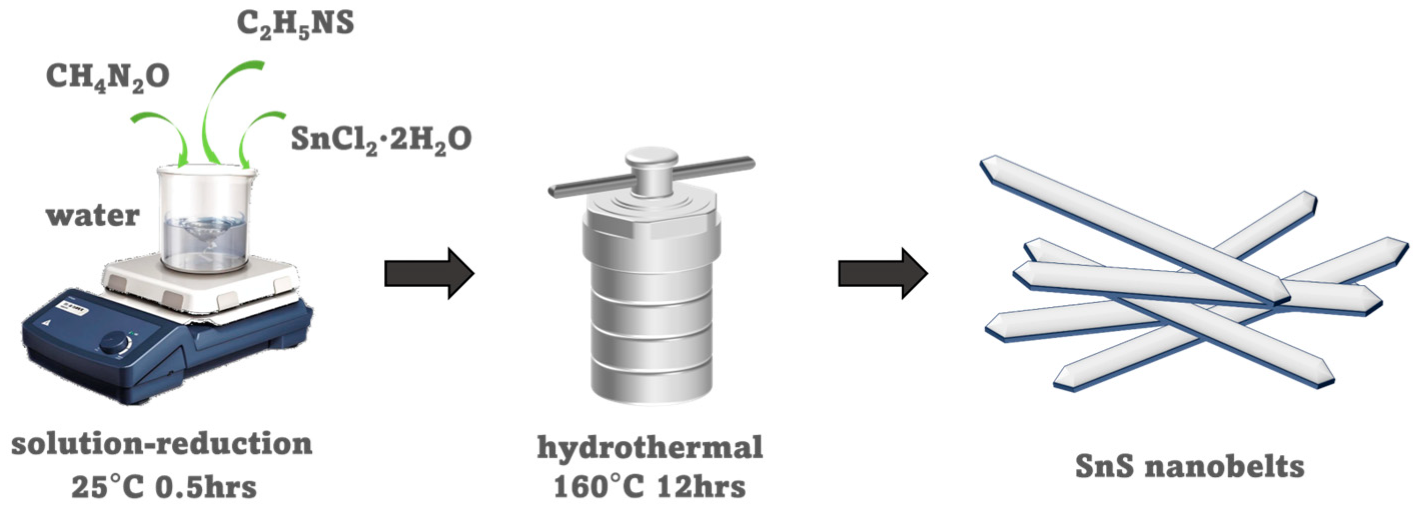
Figure 1. Illustration of the synthesis procedure for SnS nanobelts.
Crystals 15 00940 g001
Figure 2. Morphological and compositional characterization of the as-synthesized SnS nanobelts. (a) SEM image, (b) Magnified view of selected area of SnS single nanobelt, and (c,d) Corresponding EDS elemental mapping of Sn and S for the single nanobelt shown in (b), demonstrating a uniform distribution.
Crystals 15 00940 g002
Figure 3. Structural and chemical characterization of the as-synthesized SnS nanobelts. (a) XRD pattern, with the standard pattern for orthorhombic SnS (JCPDS No. 39-0354) shown as red bars. (b) Raman spectrum (magenta); Raman-active modes are labeled (Ag and B3g; blue). (c) High-resolution XPS of Sn 3d: deconvolution into Sn4+ (red) and Sn2+ (purple) doublets; the summed fit is shown as the shaded envelope. (d) High-resolution XPS of S 2p: S2− 2p3/2 (light blue) and 2p1/2 (teal) components; the summed fit is shown as the shaded envelope.
Crystals 15 00940 g003
Figure 4. Mechanocatalytic H2 evolution performance and key influencing factors for SnS nanobelts. (a) Time-course of H2 production under different control conditions: SnS + vibration + methanol (MOH) (red bars), vibration + MOH (no catalyst) (green bars), and SnS + MOH (no vibration) (blue bars). (b) Comparison of H2 evolution using different sacrificial agents: methanol (MOH; gray line), triethanolamine (TEOA; red line), and ethylene glycol (EG; blue line), compared to pure water without a sacrificial agent (green line). (c) H2 production rates (blue bars) under various ultrasonic power and frequency settings. (d) Cycling stability test of SnS nanobelts for H2 evolution over five consecutive runs (1h per run), showing the first (red), second (orange), third (green), fourth (blue), and fifth (purple) runs. Error bars represent the standard deviation of three independent measurements.
Crystals 15 00940 g004
Figure 5. Structural stability analysis of the SnS nanobelts. Comparison of (a) XRD patterns and (b) Raman spectra of the SnS catalyst before and after the mechanocatalytic stability test.
Crystals 15 00940 g005
Figure 6. Electronic band structure analysis and proposed mechanocatalytic mechanism for SnS nanobelts. (a) UV-vis-NIR diffuse reflectance spectrum. (b) Tauc plot derived from (a) for the estimation of the optical band gap (Eg). (c) Valence band XPS spectrum. (d) Schematic illustration of the band structure alignment of SnS nanobelts relative to the redox potential of H2/H2O, for the proposed sonoluminescence-induced catalytic process.
Crystals 15 00940 g006
Figure 7. Investigation of the flexoelectric effect as a driving force for mechanocatalysis in SnS nanobelts. (a) Schematic illustration of the emergence of flexoelectric polarization in the centrosymmetric SnS lattice under an inhomogeneous strain, leading to charge separation. (b) Transient flexoelectric current response under intermittent (on/off) ultrasonic vibration at varying input powers. (c) Electrochemical impedance spectroscopy (EIS) Nyquist plots recorded with and without ultrasonic vibration.
Crystals 15 00940 g007
Figure 8. Schematic illustration of the proposed dual mechanism for mechanocatalytic H2 evolution on SnS nanobelts. The diagram depicts two synergistic pathways: Mechanical bending induces flexoelectric polarization, which separates mobile electrons (e, red) and holes (h+, blue) within the SnS lattice. Sonoluminescence generates photons (yellow lightning bolt) that excite electrons from the valence band (VB, pink) to the conduction band (CB, green), facilitating the reduction of water to H2. For atomic visualization, tin (Sn), sulfur (S), oxygen (O), and hydrogen (H) atoms are represented by purple, yellow, red, and white spheres, respectively.
Crystals 15 00940 g008
Table 1. A summary of catalytic H2 evolution performance by using different kinds of photo/piezocatalysts.
Type of ReactionCatalystsDriving SourceReaction SystemCatalytic Activity
PhotoatalysisEY-6Cu-NU-66 [50]300 W Xe lampTEOA (aq)H2: 3579.8 μmol g−1 h−1
PhotoatalysisCoMoC/ZnIn2S4 [51]300 W Xe lampTEOA (aq)H2: 2232 μmol g−1 h−1
PhotoatalysisB-CTF-C-Ti-MOF [52]300 W Xe lampTEOA (aq)H2: 1975 μmol g−1 h−1
PhotoatalysisMIL-125-NH2/Ni2P [53]300 W Xe lampTEOA (aq)H2: 4327 μmol g−1 h−1
PhotoatalysisUiO-66-NH2@
Pt@UiO-66-H [54]
300 W Xe lampTEOA (aq)H2: 2708.2 μmol g−1 h−1
PhotoatalysisPMF/G-25 [55]Mutichannel reaction systemTEOA (aq)H2: 1688.5 μmol g−1 h−1
PiezocatalysisBi0.5Na0.5TiO3 [56]100 W, 40 kHz vibrationMOH (aq)H2: 506.7 μmol g−1 h−1
PiezocatalysisBiOCl nanosheets [57]120 W, 40 kHz vibrationMOH (aq)H2: 975.4 μmol g−1 h−1
PiezocatalysisAu/MoS2
Nanoflowers [26]
vibrationMOH (aq)H2: 2981 μmol g−1 h−1
PiezocatalysisOH-SrTiO3 [58]300 W, 40 kHz vibrationMOH (aq)H2: 321.5 μmol g−1 h−1
PiezocatalysisMIL-100(Fe) [23]40 W, 120 kHz vibrationMOH (aq)H2: 2800 μmol g−1 h−1
PiezocatalysisBTO
Nanosheets [59]
40 W, 100 kHz vibrationTEOA (aq)H2: 92 μmol g−1 h−1
PiezocatalysisBi2WO6 nanosheets [60]40 kHz vibrationTEOA (aq)H2: 191.3 μmol g−1 h−1
PiezocatalysisGaN
nanowires [61]
110 W, 40 kHz vibrationTEOA (aq)H2: 88.3 μmol g−1 h−1
PiezocatalysisLi-doped BTO
Nanosheets [25]
40 kHz vibrationTEOA (aq)H2: 3700 μmol g−1 h−1
PiezocatalysisBTO NPs [62]40 kHz vibrationH2OH2: 159 μmol g−1 h−1
PiezocatalysisNa0.5Bi0.5TiO3/
Na0.5Bi4.5Ti4O15 [63]
400 W, 40 kHz vibrationH2OH2: 128 μmol g−1 h−1
Mechano-
catalysis
SnS (This work)630 W, 80 kHz vibrationMOH (aq)CO: 3889 μmol g−1 h−1
Disclaimer/Publisher’s Note: The statements, opinions and data contained in all publications are solely those of the individual author(s) and contributor(s) and not of MDPI and/or the editor(s). MDPI and/or the editor(s) disclaim responsibility for any injury to people or property resulting from any ideas, methods, instructions or products referred to in the content.