Article

Synthesis, Structure, and Investigation of Terbium(III) Luminescent Metal-Organic Framework Based on (N-Morpholyl)-Functionalized 1,10-Phenanthroline

by
Anna A. Ovchinnikova
1,2,
Pavel A. Demakov
1,*,
Alexey A. Ryadun
1,
Alexander M. Agafontsev
2,3,
Vladimir P. Fedin
1 and
Danil N. Dybtsev
1,*
1
Nikolaev Institute of Inorganic Chemistry, Siberian Branch of the Russian Academy of Sciences, 3rd Lavrentiev Avenue, 630090 Novosibirsk, Russia
2
Department of Natural Sciences, Novosibirsk State University, 2nd Pirogov Street, 630090 Novosibirsk, Russia
3
Vorozhtsov Institute of Organic Chemistry, Siberian Branch of the Russian Academy of Sciences, 9th Lavrentiev Avenue, 630090 Novosibirsk, Russia
*
Authors to whom correspondence should be addressed.
Crystals 2025, 15(10), 906; https://doi.org/10.3390/cryst15100906
Submission received: 16 September 2025 / Revised: 14 October 2025 / Accepted: 15 October 2025 / Published: 18 October 2025
(This article belongs to the Section Hybrid and Composite Crystalline Materials)

Abstract

4,7-di(N-morpholyl)-1,10-phenanthroline (morphen) was introduced for the first time as a ligand for the construction of metal–organic frameworks. The obtained MOF compound has the crystallographic formula {[Tb2(morphen)2Br2(chdc)2]}n (1; chdc2− = trans-1,4-cyclohexanedicarboxylate) and is based on binuclear {Tb2(N^N)2Br2(OOCR)4} carboxylate blocks, interlinked by ditopicchdc linkers into a layered coordination network with sql topology. Purity and integrity of the as-synthesized 1 were confirmed by common characterization techniques, such as PXRD, CHN, IR, and TGA. Compound 1 was found to be hydrolytically stable and possessing typical green emission for Tb(III) complexes. Exploiting its high stability, luminescent 1@PVA films were successfully prepared from 1 and polyvinyl alcohol (PVA) through the water solution drying approach.

1. Introduction

Metal–organic frameworks (MOFs) have emerged as a versatile class of materials with applications in gas storage and separation [1,2], catalysis [3], luminescence and sensing [4,5] due to their tunable structures and functional properties [6,7,8]. Among them, lanthanide(III)-based MOFs are particularly attractive for their unique photophysical characteristics, including narrow-banded emissions, long luminescence lifetimes, and high color purity, making them promising candidates for optical sensors and light-emitting devices [9,10,11,12]. Even though bare Ln3+ cations are known to be poor luminophores because of their weak optical absorption resulting from forbidden f-f electron transitions, π-conjugated light-harvesting ligands (photosensitizers) can successfully resolve such issue. The luminescence efficiency of the corresponding MOFs is affected by both ligand-to-metal energy transfer processes and non-radiative quenching, which, in turn, depend on the local metal ion coordination environment, nature of organic linkers, topology, crystal packing, etc., with all these factors being poorly predictable [13,14,15,16].
1,10-Phenanthroline (phen) is a widely used chelator capable of forming stable complexes with metal ions of diverse nature [17,18,19], including lanthanides(III) [20,21,22,23]. Its extended conjugated π-electron system and suitable electronic energy levels (ca. 21,800 cm−1 as the energy of the T1 level [24]) allow phen to function as an effective sensitizer (“antenna”) for Ln3+ luminescence. Functionalization of phen molecules modifies the electronic structure, which tunes their coordination chemistry and important photophysical features, such as triplet level energy, optical absorbance, and self-emission, providing multiple opportunities for the design of highly emissive functional complexes [25,26,27,28,29]. Electron-donor substituents typically increase the efficiency of emission for the free (uncoordinated) phenanthrolines [19]. As well, such substituents enhance the emission of Eu(III)- and, to a lesser extent, Tb(III)-based phen complexes due to shifting down the T1 triplet level of the ligand closer to the energies of Ln3+ emissive levels [30]. The strongest donors, such as mesomeric (+M) -NR2 and -OR substituents, are weakly presented in the phenanthroline chemistry, with quite a small number of their-derived Ln(III) complexes reported [31,32,33]. At the same time, the only reported Ln(III) complex based on phenanthroline with double NR2-functionalization in the 4 and 7 positions possesses outstanding emission with a quantum yield up to 80% [34]. Other metal complexes based on 4,7-di(-NR2)-substituted phenanthrolines have also been reported as highly efficient phosphors [35] or TADF [36,37] luminophores with promising applications as multicolored dual emitters [38,39], LEDs [35,40], and luminescent sensors [41,42]. In this regard, an incorporation of Ln(III) complexes with morpholine-grafted phen ligands into a MOF architecture seems to be a fruitful direction towards new luminescence materials. Moreover, the oxygen n-donor atom of the morpholine pendant is able to bind oxophilic metal cations, providing an opportunity for their luminescence detection through a modification of the luminescence properties of the corresponding Ln(III) complexes.
Despite interesting opportunities, preparation and investigation of coordination complexes with morpholine-grafted phen ligands remains underexplored. To the best of our knowledge, there is only one example of a structurally characterized compound based on 4,7-di(N-morpholino)-1,10-phenanthroline (morhpen) ligand–[Re(morphen)Cl(CO)3], demonstrating moderate luminescence quantum yields of ca. 4.30% [38]. Previously, our group obtained several families of porous Ln(III) trans-1,4-cyclohexanedicarboxylates [43,44], built from binuclear carboxylate blocks with the formulae {Ln2(N^N)2(X)2(OOCR)4}, where X = nitrate, chloride, or additional carboxylate, and N^N is an N-donor chelator (2,2′-bipyridine, 1,10-phenanthroline (phen), or its derivatives). A decoration of such structures with phen antennas allows obtaining highly efficient luminophores with quantum yields up to 84%, multicolored mixed-metal compositions, and luminescent sensing properties [45,46]. Using 4,7-dimethyl-1,10-phenanthroline in a similar layered structure of terbium(III) MOF gave a highly sensitive luminescent sensor for nitroaromatic compounds [47]. Following our previous efforts, we decided to include a significantly larger N-morpholyl substituent-decorated phen antenna into this chemistry. In the current work, we report the microwave synthesis of the morphen lingand as well as the crystallization, structural characterization, and luminescent properties of a new terbium(III)-based MOF, {[Tb2(morphen)2Br2(chdc)2]}n (1), using trans-1,4-cyclohexanedicarboxylic acid (H2chdc) as an optically transparent linker. The title compound consists of binuclear terbium(III)-carboxylate units and appears as a rare example of Br coordination to the oxophilic Ln3+ ion in the carboxylate complexes. The blocks are arranged into an sql-type layered structure through the bridging by ditopic carboxylate ligands. Compound 1 possesses green emission, as expected for Tb(III) complexes with such type ligands, and was found to be stable in water. Such high stability allowed a preparation of flexible thin films of microcrystalline 1 with polyvinyl alcohol (PVA) with selective Fe(III) detection and promising applications in luminescence sensor devices.

2. Materials and Methods

2.1. Materials

All reagents were commercially available and used without further purification. Distilled water was employed in all synthetic experiments. Meldrum’s acid was synthesized according to the previously published procedure [48].

2.2. Instruments

FT-IR spectra (in KBr pellets) were obtained using a Scimitar FTS 2000 spectrometer (Bruker, Billerica, MA, USA), covering the range of 4000–400 cm−1. Elemental (CHN) analysis was carried out using a VarioMICROcube analyzer (ElementarAnalysensysteme GmbH, Hanau, Germany). Powder X-ray diffraction (PXRD) measurements were conducted at room temperature on a XRD-7000 diffractometer (Cu-Kα radiation, λ = 1.54178 Å; Shimadzu, Kyoto, Japan). Thermogravimetric analysis was conducted using a Netzsch TG 209 F1 Iris instrument (Netzsch, Selb, Germany) under Ar flow (30 cm3·min−1) and a 10 K∙min−1 heating rate. Solid-state diffuse reflectance spectra (DRS) were recorded using a Shimadzu UV-3101 spectrometer (Shimadzu, Tokyo, Japan). Samples for DRS were prepared by their grinding with BaSO4 (ca. 1:1) used as a transparent shaping matrix. The initial dependencies of the reflection of samples (R) on the wavelength were recalculated to the Kubelka-Munk function (M) by the equation M = (1 − R)2/2R. Digital photographs were taken using a Hamamatsu C11924-211 (Hamamatsu Photonics, Hamamatsu, Japan) wide-banded diode UV-LED module with the maximum λex = 365 nm. Photoluminescence spectra and excited-state lifetimes were obtained using a HORIBA Fluorolog 3 spectrofluorometer (Horiba, Kyoto, Japan) equipped with a 450 W power ozone-free Xe-lamp, cooled photon detector R928/1860 PFR technologies with refrigerated chamber PC177CE-010, and double grating monochromators. The spectra were corrected for source intensity and detector spectral response by standard correction curves. A set of NanoLED pulsed nanosecond lasers was used for the time-resolved measurements, and a Quanta-φ integrating sphere (Horiba, Kyoto, Japan) was used for the determination of absolute quantum yields (QYs). The reaction between 4,7-dichloro-1,10-phenanthroline (O3) and morpholine was carried out in a Single-Mode Microwave reactor Monowave 300 Anton Paar MAS 24 (Anton Paar GmbH, Graz, Austria) using a special 10 mL sealed reaction vessel G10. NMR spectra (Figures S1–S5) were acquired at 20–23 °C for solutions (C = 20–40 mg/mL) on a Avance 400 spectrometer (400 MHz for 1H, 100 MHz for 13C; Bruker, Billerica, MA, USA). The chemical shifts were calculated relative to the solvent signals used as the internal standard: δC = 76.90 ppm and δH = 7.24 ppm for CDCl3; δC = 39.50 ppm and δH = 2.50 ppm for dmso-d6.

2.3. Synthetic Methods

Synthesis of 4,7-Di(N-morpholino)-1,10-phenanthroline (morphen; Scheme 1). 2.89 g (33.2 mmol) of morpholine and 662 mg (2.66 mmol) of 4,7-dichloro-1,10-phenanthroline (O3, see ESI file) were mixed in a special glass vial G10, and the mixture was heated in a microwave reactor for 1 h at 150 °C. The “Standard” method was used, with the function “Heat as fast as possible” and a maximum energy of 100 W. Then, an excess of morpholine was distilled off in vacuo. The residue was dissolved in 20 mL of chloroform and extracted thrice with 50 mL of water. The organic layer was separated and dried using anhydrous Na2SO4, then the solvent was evaporated in vacuo and the residue recrystallized from toluene to yield 661 mg (71%) of a light yellow solid morphen. 1H NMR (dmso-d6, δ, ppm; Figure S4): 8.83 (d, J = 5.2 Hz, 2H, H(1,1′)), 7.98 (s, 2H, H(6,6′)), 7.19 (d, J = 5.2 Hz, 2H, H(2,2′)), 3.85—3.91 (m, 8H, H(8,8′)), 3.15—3.21 (m, 8H, H(7.7′)). 13C NMR (dmso-d6, δ, ppm; Figure S5): 156.25 (C(3,3′)), 149.94 (C(1,1′)), 147.07 (C(5,5′)), 122.03 (C(4,4′)), 120.84 (C(6,6′)), 111.07 (C(2,2′)), 66.13 (C(7,7′)), 52.22 (C(8,8′)).
Synthesis of TbBr3·6H2O. 2.00 g (2.67 mmol) of Tb4O7 and 5.0 mL of water were mixed in a glass vial and intensively stirred. Then, 10.0 mL (88 mmol) of concentrated hydrogen bromide (HBr, 48 wt.% solution in water) were slowly added dropwise at continuous stirring to avoid foaming of the reaction mixture. During several hours of stirring, Tb4O7 was completely dissolved and reacted. After the reaction, the solution was transferred to an evaporation cup and fully evaporated at 150 °C to evaporate an excess of HBr. After completing drying, the solid residue was dissolved in water; its pH value was found to be 5–6 using universal indicator paper, confirming full deletion of an HBr excess. Finally, a solution had been slowly dried in an evaporating cup for ca. 18 h at 90 °C in a fume hood until crystallization of TbBr6H2O and its careful drying were achieved.
Synthesis of {[Tb2(morphen)2Br2(chdc)2]}n (1). 40.5 mg (0.080 mmol) of TbBr3·6H2O, 13.7 mg (0.080 mmol) of H2chdc and 14.0 mg (0.040 mmol) of morphen were mixed in a glass vial and dissolved in 2.0 mL of N,N-dimethylformamide (DMF). The obtained solution was heated at 110 °C for 48 h. The formed white precipitate was filtered, and the solid was washed with DMF and air-dried. Yield: 31.7 mg (50.4%). Elemental analysis: Found (%): C, 43.5; H, 4.4; N, 8.0. Calculated (%) for [Tb2(C20H22N4O2)2Br2(C8H10O4)2]·0.5C3H7NO·H2O (Tb2C57.5H69.5N8.5O13.5Br2): C, 43.9; H, 4.5; N, 7.6. IR spectrum characteristic bands (KBr, cm−1): 3430 (w., br., νO–H); 3077 (w., νCsp2–H); 2940 and 2855 (m., νCsp3–H); 1682 (w., νCOamide); 1586 (s., νCOOas); 1416 (s., νCOOs); 1150 and 1050 (m., νC–N).
Synthesis of 1@PVA films. A ca. 3.2 wt.% polyvinyl alcohol (PVA) solution was obtained by heating a mixture of PVA (1.0 g) and distilled water (30 mL) at 90 °C for 2 h. Then powder of 1 was added to the solution, and the mixture was kept under alternating stirring and hitting in the ultrasound bath at room temperature for 1 h until a homogeneous suspension was obtained. 1@PVA film was made through covering of the Petri dish bottom by ca. 3 mL of the suspension and subsequently drying it at room temperature for ca. 72 h.
Cation detection experiments. Drops of 0.01M ethanol solutions of different metal nitrates Mg(NO3)2, Fe(NO3)3, Cu(NO3)2, Zn(NO3)2, Al(NO3)3, Ga(NO3)3, Cd(NO3)2, NaNO3, Cr(NO3)3, Ni(NO3)2, La(NO3)3, and Co(NO3)2 were placed on 1@PVA samples. After fast drying of the ethanol solvent (achieved in ca. 2 min), digital photographs of the samples have been made. A treatment by Fe3+ salt, which showed a pronounced quenching response, has been repeated using a new film sample with sequential luminescent spectroscopy measurements before and after Fe3+ dropping-then-drying.

2.4. Single-Crystal X-Ray Diffraction Details

Diffraction data were collected on a Bruker D8 Venture diffractometer (Billerica, MA, USA) equipped with a PHOTON III area detector and an Incoatec IuS3.0 microfocus X-ray tube with a HELIOS multilayer mirror monochromator (λ(MoKα) = 0.71073 Å) at 150 K. Bruker AXS, Inc. APEX3, Version 3.0 [49], SAINT, Version 8.40a [50], and SADABS, Version 2016-2 [51], [49] were used for the integration, absorption correction, and determination of unit cell parameters. Dual space algorithm (SHELXT [52]) was used for structure solution, and the full-matrix least squares technique (SHELXL [53]) was used for structure refinement. Anisotropic approximation was applied for all atoms, except hydrogens. Positions of hydrogen atoms of organic ligands were calculated geometrically and refined in the riding model. Details for single crystal structure determination experiments and structure refinements are summarized in Table A1. The supplementary crystallographic data for this paper (CCDC 2475501) can be accessed free of charge from the Cambridge Crystallographic Data Centre at: https://www.ccdc.cam.ac.uk/structures/ (Accessed on 14 October 2025). An analysis of coordination polyhedra (see Table A2) has been performed in the Shape 2.1 [54] program.

3. Results and Discussion

3.1. Synthesis and Similar Compounds

4,7-dichloro-1,10-phenanthroline, acting as a convenient precursor for the synthesis of diverse phenanthroline derivatives substituted in the 4 and 7 positions, has been synthesized in a typical four-step procedure. Its synthetic way has been adapted from the literature report [48]. Synthesis of the ligand morphen and rare examples of its complexes have been previously described in the literature [38,55]. In the originally reported method, morphen had been prepared by refluxing 4,7-dichloro-1,10-phenanthroline with morpholine for 19 h. We decided to carry out this reaction using microwave irradiation, since microwave radiation assistance is known to accelerate many chemical reactions [56,57]. It was found herein that carrying out the reaction in a microwave system at 150 °C allows one to achieve complete conversion of 4,7-dichloro-1,10-phenanthroline in one hour with a yield comparable to the original method. Apparently, 4,7-di(N-morpholino)-1,10-phenanthroline (morphen) can act as an efficient light-harvesting antenna ligand for luminescent complexes, so a preparation and characterization of corresponding Ln(III) MOFs as well as functional materials was carried out in this work.
The new compound {[Tb2(morphen)2Br2(chdc)2]}n (1) possesses a generally similar coordination layer structure to the prototypic MOF [Tb2(phen)2(NO3)2(chdc)2] [45], yet with some unexpected structural features. Compound 1 was synthesized from a system containing N,N-dimethylformamide (DMF) as the main solvent through the reaction between terbium(III) bromide hexahydrate, 4,7-di(N-morpholyl)-1,10-phenanthroline (morphen), and trans-1,4-cyclohexanedicarboxylic acid (H2chdc) at a molar ratio of about 1:2:2. Interestingly, the large Br atom occupies similar positions as terminal k2-nitrates. At the same time, 1 appears to be a rare example of direct bromide coordination to the oxophilic Ln3+ cations in carboxylate complexes, since only 12 structures of such Br-coordinated Ln-carboxylate complexes have been reported [58,59,60,61,62].

3.2. Crystal Structure Description

Compound 1 crystallizes in monoclinic symmetry with the P21/c space group (Table A1). 1 contains one independent terbium atom. Tb(III) is coordinated by two O atoms from two μ:k1,k1-carboxylate groups, three O atoms from two μ:k1,k2-carboxylate groups, a bromide anion, and two nitrogen atoms from di(N-morpholyl)-substituted phenanthroline (Figure 1a). Therefore, a metal coordination number (CN) is 8. The lengths of Tb–O(COO) bonds lie in the range 2.310(8)–2.507(7) Å. The Tb–N bond lengths are 2.511(8) Å and 2.547(9) Å. The length of the Tb–Br bond is 2.8235(16) Å. According to the analysis performed in Shape 2.1 [47], the Tb atom in 1 adopts a triangular dodecahedron (TDD-8) environment, which is related to the lowest deviation criterion (2.148, see Table A2). Two closest Tb(III) ions are bound into binuclear carboxylate blocks {Tb2(morphen)2(Br)2(μ:κ11-OOCR)2(μ:κ12-OOCR)2} with Tb…Tb intermetal distances to be 3.88 Å. These secondary building units are bridged by trans-1,4-cyclohexanedicarboxylates into distorted tetragonal coordination layers (Figure 1b) (sql topology). The layers are densely packed within a 3D MOF structure. According to the PLATON [63] calculations, the structure of 1 does not contain any significant voids (Figure 1c). Despite the lack of porosity, no hydrogen bonds or any other significant interlayer contacts are observed in the structure, which can suggest its high layer-to-layer mobility and possible “opening” of voids when large guest molecules are included.

3.3. Characterization and Stability

The phase purity of the as-synthesized compound 1 was confirmed by powder X-ray diffraction (Figure 2a). Its chemical composition was verified by the elemental CHN analysis (see the experimental). The infrared spectrum of 1 (Figure 2b) contains characteristic absorption bands of C(sp3)–H stretching vibrations (2855–2940 cm−1), antisymmetric (1586 cm−1) and symmetric (1416 cm−1) stretching vibrations of the coordinated COO group. Weak C(sp2)–H stretching bands around 3070 cm−1, corresponding to the phenanthroline rings, and two narrow bands in 1050–1150 cm−1, corresponding to C–N and C–O vibrations in the morpholine ring, are also present, all of these bands confirming the presence of the main necessary molecular constituents in the synthesized compound. A wide O–H stretching band around 3430 cm−1 and a weak C=Oamide stretching vibrations band (1682 cm−1) show the presence of a minor amount of H2O and DMF in the 1 sample, both confirmed by CHN and TGA data (see below).
The as-synthesized crystals of 1 are stable in air after filtration during prolonged storage and also resistant to hydrolysis, which has been confirmed by PXRD for the sample after 24 h stirring in water (Figure 2c). According to thermogravimetric analysis (TGA, Figure 2d), 1 loses ca. 2% of its weight in the temperature range up to 80 °C, which is apparently related to the presence of some occluded solvent in the sample and fits to 1 additional water molecule and 0.5 additional DMF per formula unit, confirmed by CHN and IR data (see the experimental section). Then, slow weight loss occurs in the 80–370 °C range, followed by the framework decomposition at T ≈ 372 °C. Such high thermal and hydrolytic stability of 1 encourages an investigation of its physical properties as well as a preparation of functional optical devices.

3.4. Luminescent Properties

Solid-state excitation and emission spectra (Figure 3a) were recorded for compound 1. The excitation spectrum contains a wide band in the UV region (efficient light absorption in the 300–395 nm area), apparently corresponding to the absorption of the π-conjugated organic moiety (4,7-di(N-morpholyl)-1,10-phenanthroline antenna). Similarly, the diffusion reflectance spectrum (DRS) of crystalline powder 1 features a strong increase in the adsorption below λ ≤ 400 nm related to the adsorption of the morphen ligand (Figure S6). An efficient energy transfer from the aromatic photosensitizer to the metal cation is manifested by strong Tb3+-centered emission, while the emission of the organic morphen ligand is undetected. The luminescence spectrum of 1 contains characteristic bands, corresponding to the series of 5D47FJ (J = 6, 5, 4, 3) [17] transitions at λmax = 480 nm, 545 nm, 588 nm, and 618 nm, respectively. A photoluminescence quantum yield (QY) for 1ex = 375 nm) was determined to be 0.21 ± 0.09%. Even though the QY is rather modest for Tb(III) complexes, the apparent visible luminescence of 1 is strong enough, allowing an investigation of luminescent applications with no considerable limitations. The reason for the low emission quantum yield of 1 cannot be clearly determined at the moment. As mentioned in the introduction, no morphen-based complexes have been reported for any lanthanide, along with the only example of Eu complex with NR2-derived phenanthroline substituted in the 4 and 7 positions [34]. That europium complex with 4,7-di(carbazol-9-yl)-1,10-phenanthroline possessed up to 80% QY values [34], which is apparently attributed to its strikingly different electronic structure. An impact of a heavy Br atom into a deactivation of the excited states [64] can also be suggested as a possible reason for the low quantum yield of 1. However, there are no examples of the determined quantum yields for carboxylate complexes with additionally coordinated bromide or iodide, while for carboxylates containing coordinated lighter halogen Cl there are reported examples of both low [65,66] and high [66,67] QY values, giving no ability to make any meaningful conclusion in this regard. The excited state lifetime (τl) for compound 1 was determined as (0.623 ± 0.021) ms, which is typical for Tb(III) carboxylate complexes [68,69,70] and clearly confirms its common phosphorescence mechanism.

3.5. Film Preparation and Ion Sensing Applications

A high hydrolytic stability allowed combining a crystalline 1 MOF sample with a water-soluble polymer, polyvinyl alcohol (PVA), acting as a shaping matrix. PVA is a non-toxic and easily processable polymeric filler widely used in drug capsules and food processing. Several examples of the preparation of MOF/PVA composites and mixed-matrix membranes have been reported [71,72,73,74,75,76]. In this regard, PVA was chosen as a convenient polymeric filler for a preparation of flexible luminescent films suitable for diverse applications. Thus, 1@PVA films were prepared by dispersing crystals of 1 in a water solution of PVA with a subsequent continuous hitting in the ultrasound bath and stirring to obtain a highly homogeneous suspension. The dispersion had been carefully distributed on the bottom of the Petri dish and left to slowly dry at room temperature. Digital photographs of the obtained films (see Figure 3b) confirmed the preservation of the MOF green emission and high homogeneity of its emission throughout all the film surface. The emission spectrum of the 1@PVA film with a non-delayed recording contains a number of characteristic Tb3+-related long-lived phosphorescence bands similar to the initial 1 as well as a new wide band in the blue region, apparently attributed to the short-lived fluorescence emission of the PVA matrix [77]. Recording the emission with a 0.2 ms delay (to exclude the fluorescence impact) gave a clear Tb3+-centered emission spectrum similar to the solid MOF. The (x,y) points in the CIE 1931 chromaticity diagram calculated from these spectra (Figure S7) and their derived characteristic color wavelengths (Table S1) quantitatively confirm the prevailing blue fluorescence component in the emission of 1@PVA, while the phosphorescence of the film recorded with a 0.2 ms delay is much closer to the initial MOF emission color. Excitation spectra of 1@PVA, both with and without delayed recording, contain a new additional wide band with ca. 280 nm maximum, also observable in its DRS spectrum (Figure S6), suggesting an involvement of PVA in photoexcitation energy transfer to the MOF microparticles. The excited state lifetime for Tb3+ phosphorescence is more than two times longer than for 1@PVA compared to the initial solid MOF, which is apparently attributed to the diluting of highly emissive MOF moieties in the less photoactive and quasi-amorphous polymeric filler. All of these data confirm a successful integration of the luminescent MOF 1 and polymeric PVA filler within the shaped sample for promising luminescence and sensing applications.
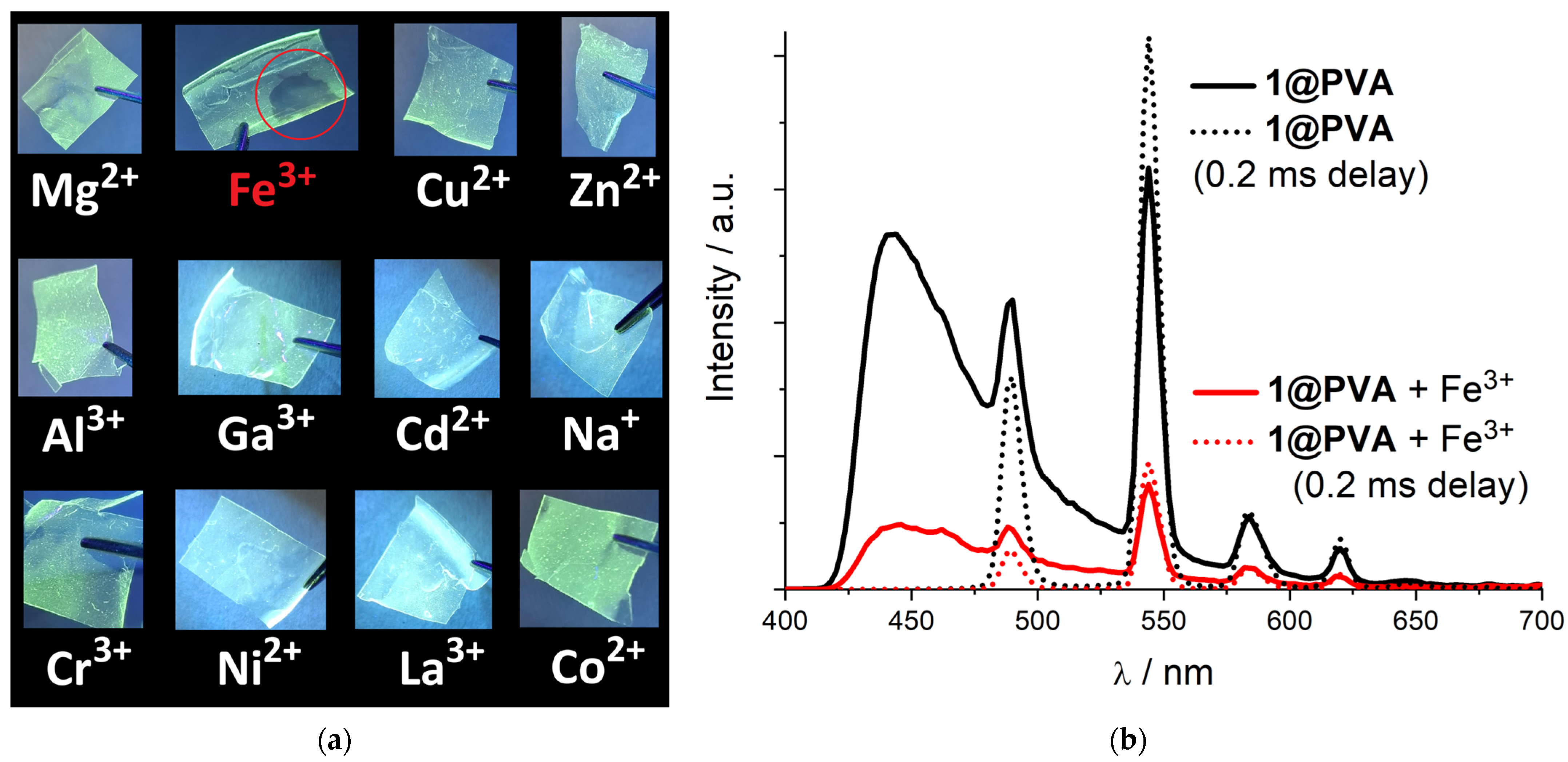
As mentioned in the introduction section, uncoordinated O-atoms of the morpholine pendant moieties may interact with various metal cations, thus changing the electronic structure of the phenanthroline core and its luminescence behavior. The luminescence responses of 1@PVA films were systematically tested in the presence of various metal cations with different charges and electronic configurations. When a drop of metal nitrate solution in ethanol was applied to the 1@PVA film, most of the samples showed no change in the luminescence brightness or color (Figure 4a). The only notable exception is Fe(III), which invokes a clearly visible decrease in the luminescence intensity. Subsequently recorded excitation (Figure S8) and emission (Figure 4b) spectra for the same film before and after Fe(III) treatment confirm more than a fivefold decrease in the 1@PVA luminescence. Among possible reasons for the luminescence quenching could be (i) partial absorption of the excitation wavelength (λex = 375 nm) by the iron(III) cations as well as (ii) energy or electron transfer from the photoexcited state of the phen ligand to the electrophilic Fe(III) cation following its reduction to the Fe(II) state and non-radiation energy dissipation. Indeed, among all the metal cations under investigation (Na+, Mg2+, Co2+, Ni2+, Cu2+, Zn2+, Cd2+, Al3+, Cr3+, Fe3+, Ga3+, La3+), only Fe(III) possesses considerable oxidative redox activity (E°(Fe3+/Fe2+) = 0.77 V); therefore, an electron transfer from π*(phen) to Fe3+ with subsequent non-radiative relaxation should not be excluded from the luminescence quenching mechanism. Even though the nature of the ion-selective luminescence detection of Fe(III) is debatable at the moment, the unique application potential of polymer films containing MOF based on morpholine-modified phenanthroline complexes is successfully demonstrated.

4. Conclusions

To summarize, 4,7-di(N-morpholyl)-1,10-phenanthroline (morphen) ligand has been introduced for the first time into the chemistry of metal–organic frameworks. Its efficient antenna-like behavior for Tb(III)-centered luminescence of the MOF was shown. Exploiting the high thermal and hydrolytic stability of the synthesized MOF, flexible films were successfully fabricated using polyvinyl alcohol (PVA), demonstrating remarkable ion-selective Fe(III) photoluminescent detection.

Supplementary Materials

The following supporting information can be downloaded at: https://www.mdpi.com/article/10.3390/cryst15100906/s1, Scheme S1. Synthetic scheme for 4,7-dichloro-1,10-phenanthroline (O3) precursor; Figure S1. 1H NMR spectrum of 1,2-bis-[(2,2-dimethyl-4,6-dioxo-1,3-dioxan-5-ylidenemethyl)amino]benzene (O1) recorded in CDCl3; Figure S2. 1H NMR spectrum of 4,7-dihydroxy-1,10-phenanthroline (O2) recorded in DMSO-d6; Figure S3. 1H NMR spectrum of 4,7-dichloro-1,10-phenanthroline (O3) recorded in CDCl3; Figure S4. 1H NMR spectrum of 4,7-di(N-morpholyl)-1,10-phenanthroline (morphen) in dmso-d6; Figure S5. 13C J-mod NMR spectrum of 4,7-dimorpholyl-1,10-phenanthroline (mprphen) in dmso-d6; Figure S6. Diffuse reflectance spectra (DRS) for 1 and 1@PVA in initial reflectance R-λ (a) and their-derived Kubelka-Munk function KM-λ (b) dependencies; Figure S7. CIE 1931 chromaticity diagram for 1 and 1@PVA, (x,y) coordinates for the latter are calculated for emission spectra with both delayed and non-delayed recording; Figure S8. Excitation spectra for 1@PVA before and after Fe3+ treatment at λem = 545 nm; Table S1. Characteristic emission wavelengths and color purities calculated from CIE 1931 (x,y) coordinates.

Author Contributions

Conceptualization, P.A.D. and D.N.D.; methodology, P.A.D., A.M.A. and D.N.D.; validation, P.A.D., A.M.A. and V.P.F.; formal analysis, A.A.R.; investigation, A.A.O. (synthesis, characterization, graphing), A.M.A. (synthesis, characterization, graphing), A.A.R. (luminescence measurements), P.A.D. (single-crystal XRD, graphing); resources, D.N.D. and V.P.F.; data curation, P.A.D., A.M.A. and V.P.F.; writing—original draft preparation, A.A.O., P.A.D. and A.M.A.; writing—review and editing, D.N.D. and V.P.F.; visualization, A.A.O., P.A.D. and A.M.A.; supervision, P.A.D.; project administration, D.N.D.; funding acquisition, D.N.D. All authors have read and agreed to the published version of the manuscript.

Funding

The research was supported by the Russian Science Foundation, project no. 23-13-00310.

Data Availability Statement

The CCDC 2475501 entry contains the supplementary crystallographic data for this paper. These data can be obtained free of charge from The Cambridge Crystallographic Data Center at https://www.ccdc.cam.ac.uk/structures/ (accessed on 14 October 2025).

Conflicts of Interest

There are no conflicts of interest to declare.

Appendix A. Crystal Data

Table A1. Single-crystal X-ray diffraction and structure refinement details.
Parameter1
Chemical formulaC56H64Br2N8O12Tb2
Mr, g/mol1518.81
Crystal systemMonoclinic
Space groupP21/c
a, Å13.546(3)
b, Å16.975(5)
c, Å13.121(3)
Temperature, K150
α, °90
β, °112.510(6)
γ, °90
V, Å32787.2(12)
Z2
D(calc.), g·cm−31.810
μ, mm−14.02
F(000)1504
Crystal size, mm0.06 × 0.02 × 0.02
θ range for data collection, °2.02 ≤ θ ≤ 25.24
Index ranges−15 ≤ h ≤ 14; −20 ≤ k ≤ 20; −16 ≤ l ≤ 16
No. of reflections: measured/independent/
observed[I > 2σ(I)]
4797/4797/3058
Rint0.123
Goodness-of-fit on F20.944
Final R indices [I > 2σ(I)]R1 = 0.0673; wR2 = 0.1503
Final R indices [all data]R1 = 0.1016; wR2 = 0.1663
Largest diff. peak, hole, e/Å32.17, −1.32
Table A2. Fitting criteria of Tb(III) polyhedra in compound 1 with according to the SHAPE 2.1 [47] calculations.
Polyhedron1
Heptagonal pyramid (HPY-8)21.622
Hexagonal bipyramid (HBPY-8)16.485
Square antiprism (SAPR-8)2.898
Triangulardodecahedron (TDD-8)2.148
Johnson gyrobifastigium J26 (JGBF-8)14.773
Johnson elongated triangular bipyramid J14 (JETBPY-8)26.205
Biaugmented trigonal prism J50 (JBTPR-8)3.254
Biaugmented trigonal prism (BTPR-8)2.386
Snub disphenoid J84 (JSD-8)4.481
Triakistetrahedron (TT-8)12.822
Elongated trigonal bipyramid (ETBPY-8)24.261

References

  1. Furukawa, H.; Cordova, K.E.; O’Keeffe, M.; Yaghi, O.M. The chemistry and applications of metal-organic frameworks. Science 2013, 341, 1230444. [Google Scholar] [CrossRef]
  2. Zhao, X.; Wang, Y.; Li, D.-S.; Bu, X.; Feng, P. Metal–organic frameworks for separation. Adv. Mater. 2018, 30, 1705189. [Google Scholar] [CrossRef]
  3. Ranocchiari, M.; Van Bokhoven, J.A. Catalysis by metal–organic frameworks: Fundamentals and opportunities. Phys. Chem. Chem. Phys. 2011, 13, 6388–6396. [Google Scholar] [CrossRef]
  4. Hu, Z.; Deibert, B.J.; Li, J. Luminescent metal–organic frameworks for chemical sensing and explosive detection. Chem. Soc. Rev. 2014, 43, 5815–5840. [Google Scholar] [CrossRef] [PubMed]
  5. Della Rocca, J.; Liu, D.; Lin, W. Nanoscale metal–organic frameworks for biomedical imaging and drug delivery. Acc. Chem. Res. 2011, 44, 957–968. [Google Scholar] [CrossRef]
  6. Wang, L.; Han, Y.; Feng, X.; Zhou, J.; Qi, P.; Wang, B. Metal–organic frameworks for energy storage: Batteries and supercapacitors. Coord. Chem. Rev. 2016, 307, 301–381. [Google Scholar] [CrossRef]
  7. Wang, C.; Liu, X.; Demir, N.K.; Chen, J.P.; Li, K. Applications of water stable metal–organic frameworks. Chem. Soc. Rev. 2016, 45, 5107–5134. [Google Scholar] [CrossRef]
  8. Shekhah, O.; Liu, J.; Fischer, R.A.; Wöll, C. MOF thin films: Existing and future applications. Chem. Soc. Rev. 2011, 40, 1081–1106. [Google Scholar] [CrossRef] [PubMed]
  9. Patra, K.; Pal, H. Lanthanide-based metal–organic frameworks (Ln-MOFs): Synthesis, properties and applications. RSC Sustain. 2025, 3, 629–660. [Google Scholar] [CrossRef]
  10. Wang, X.; Jiang, Y.; Tissot, A.; Serre, C. Luminescent sensing platforms based on lanthanide metal-organic frameworks: Current strategies and perspectives. Coord. Chem. Rev. 2023, 497, 215454. [Google Scholar] [CrossRef]
  11. Saraci, F.; Quezada-Novoa, V.; Donnarumma, P.; Howarth, A.J. Rare-earth metal–organic frameworks: From structure to applications. Chem. Soc. Rev. 2020, 49, 7949–7977. [Google Scholar] [CrossRef] [PubMed]
  12. Zhang, Y.; Wei, P.; Li, Z.; Sun, Y.; Liu, Y.; Huang, S. Advancements in rare earth metal-organic frameworks: Harnessing the power of photonics and beyond. Coord. Chem. Rev. 2024, 514, 215905. [Google Scholar] [CrossRef]
  13. Allendorf, M.D.; Bauer, C.A.; Bhakta, R.K.; Houk, R.J.T. Luminescent metal–organic frameworks. Chem. Soc. Rev. 2009, 38, 1330–1352. [Google Scholar] [CrossRef]
  14. Cui, Y.; Yue, Y.; Qian, G.; Chen, B. Luminescent Functional Metal–Organic Frameworks. Chem. Rev. 2012, 112, 1126–1162. [Google Scholar] [CrossRef] [PubMed]
  15. Utochnikova, V.V. The use of luminescent spectroscopy to obtain information about the composition and the structure of lanthanide coordination compounds. Coord. Chem. Rev. 2019, 398, 113006. [Google Scholar] [CrossRef]
  16. Utochnikova, V.V.; Kuzmina, N.P. Photoluminescence of lanthanide aromatic carboxylates. Russ. J. Coord. Chem. 2016, 42, 679–694. [Google Scholar] [CrossRef]
  17. Rothfuss, H.; Knöfel, N.D.; Tzvetkova, P.; Michenfelder, N.C.; Baraban, S.; Unterreiner, A.N.; Roesky, P.W.; Barner-Kowollik, C. Phenanthroline—A Versatile Ligand for Advanced Functional Polymeric Materials. Chem. Eur. J. 2018, 24, 17475–17486. [Google Scholar] [CrossRef]
  18. Accorsi, G.; Listorti, A.; Yoosaf, K.; Armaroli, N. 1, 10-Phenanthrolines: Versatile building blocks for luminescent molecules, materials and metal complexes. Chem. Soc. Rev. 2009, 38, 1690–1700. [Google Scholar] [CrossRef]
  19. Takeda, H.; Irimajiri, M.; Mizutani, T.; Nozawa, S.; Matsuura, Y.; Kurosu, M.; Ishitani, O. Photocatalytic CO2 Reduction Using Mixed Catalytic Systems Comprising an Iron Cation with Bulky Phenanthroline Ligands. Inorg. Chem. 2024, 63, 7343–7355. [Google Scholar] [CrossRef]
  20. Hendrich, J.M.; White, F.D.; Sykora, R.E. Lanthanide dicyanoaurate coordination polymers containing 1, 10-phenanthroline: Synthesis, structure, and luminescence. Inorg. Chim. Acta 2021, 527, 120562. [Google Scholar] [CrossRef]
  21. Song, H.; Liu, G.; Fan, C.; Pu, S. A novel fluorescent sensor for Al3+ and Zn2+ based on a new europium complex with a 1, 10-phenanthroline ligand. J. Rare Earths 2021, 39, 460–468. [Google Scholar] [CrossRef]
  22. Zahariev, T.; Shandurkov, D.; Gutzov, S.; Trendafilova, N.; Enseling, D.; Jüstel, T.; Georgieva, I. Phenanthroline chromophore as efficient antenna for Tb3+ green luminescence: A theoretical study. Dyes Pigm. 2021, 185, 108890. [Google Scholar] [CrossRef]
  23. Bolot’ko, A.E.; Shmelev, M.A.; Chistyakov, A.S.; Voronina, J.K.; Varaksina, E.A.; Gogoleva, N.V.; Taydakov, I.V.; Sidorov, A.A.; Eremenko, I.L. Luminescenceenhancementbymixingcarboxylatebenzoate–pentafluorobenzoateligandsinpolynuclear {Eu2Zn2} and {Tb2Zn2} complexes. Dalton Trans. 2025, 54, 5708–5720. [Google Scholar] [CrossRef]
  24. Kot, K.; Oczko, G.; Puchalska, M.; Starynowicz, P. Structural and spectroscopic studies of heavy lanthanide complexes with o-phenanthroline and isothiocyanate. Polyhedron 2019, 173, 114119. [Google Scholar] [CrossRef]
  25. Qiao, Y.; Li, Z.-M.; Wang, X.-B.; Guan, W.-S.; Liu, L.-H.; Liu, B.; Wang, J.-K.; Che, G.-B.; Liu, C.-B.; Lin, X. Thermal behaviors and adsorption properties of two Europium (III) complexes based on 2-(4-carboxyphenyl) imidazo [4, 5-f]-1, 10-phenanthroline. Inorg. Chim. Acta 2018, 471, 397–403. [Google Scholar] [CrossRef]
  26. D’Vries, R.F.; Gomez, G.E.; Hodak, J.H.; Soler-Illia, G.J.A.A.; Ellena, J. Tuning the structure, dimensionality and luminescent properties of lanthanide metal–organic frameworks under ancillary ligand influence. Dalton Trans. 2016, 45, 646–656. [Google Scholar] [CrossRef]
  27. Wang, S.-J.; Li, Q.; Xiu, G.-L.; You, L.-X.; Fu Ding, F.; Van Deun, R.; Dragutan, I.; Dragutan, V.; Sun, Y.-G. New Ln-MOFs based on mixed organic ligands: Synthesis, structure and efficient luminescence sensing of the Hg2+ ions in aqueous solutions. Dalton Trans. 2021, 50, 15612–15619. [Google Scholar] [CrossRef]
  28. Luo, A.-Y.; Lan, B.-L.; Shao, B.; Lu, X.-M.; Lan, Y.-F.; Liao, Y.-Z.; Zhang, Z. A 2D mixed-lanthanide metal–organic framework as dual-emitting luminescent sensor for ratiometric detection of tetracycline and nitrophenols. J. Mol. Struct. 2024, 1295, 136734. [Google Scholar] [CrossRef]
  29. Bryleva, Y.A.; Mikheylis, A.V.; Agafontsev, A.M.; Glinskaya, L.A.; Tkachev, A.V. Antenna effect of 1,10-phenanthroline derivative bearing (−)-borneol moieties in luminescent lanthanide(III) complexes. J. Lumin. 2025, 281, 121144. [Google Scholar] [CrossRef]
  30. Pettinari, C.; Drozdov, A.; Belousov, Y. Coordination Compounds of Lanthanides as Materials for Luminescent Turn Off Sensors. In Rare Earth Elements—Emerging Advances, Technology Utilization, and Resource Procurement; IntechOpen: London, UK, 2022; Chapter 1; pp. 1–31. [Google Scholar] [CrossRef]
  31. Zarubin, D.N.; Bushkov, N.S.; Lavrov, H.V.; Dolgushin, F.M.; Ustynyuk, N.A.; Ustynyuk, Y.A. 4,7-Di-n-butoxy-1,10-phenanthroline-2,9-dicarboxamide: A Tetradentate Ligand Featuring Excellent Solubility in Nonpolar Media. INEOS Open 2019, 2, 130–133. [Google Scholar] [CrossRef]
  32. Borisova, N.E.; Kostin, A.A.; Reshetova, M.D.; Lyssenko, K.A.; Belova, E.V.; Myasoedov, B.F. The structurally rigid tetradentate N,N′,O,O′-ligands based on phenanthroline for binding of f-elements: The substituents vs. structures of the complexes. Inorg. Chim. Acta 2018, 478, 148–154. [Google Scholar] [CrossRef]
  33. Evsiunina, M.V.; Khult, E.K.; Matveev, P.I.; Kalle, P.; Lemport, P.S.; Petrov, V.S.; Aksenova, S.A.; Nelyubina, Y.V.; Koshelev, D.S.; Utochnikova, V.V.; et al. Unravelling the mechanism of f-element extraction by phenanthroline-diamides: A case of 4,7-substituted 1,10-phenanthroline-2,9-diamides. Sep. Purif. Technol. 2024, 339, 126621. [Google Scholar] [CrossRef]
  34. Zucchi, G.; Murugesan, V.; Tondelier, D.; Aldakov, D.; Jeon, T.; Yang, F.; Thuéry, P.; Ephritikhine, M.; Geffroy, B. Solution, Solid State, and Film Properties of a Structurally Characterized Highly Luminescent Molecular Europium Plastic Material Excitable with Visible Light. Inorg. Chem. 2011, 50, 4851–4856. [Google Scholar] [CrossRef] [PubMed]
  35. Li, H.-B.; Cao, H.-T.; Zhu, D.-X.; Su, Z.-M.; Liao, Y. Synthesis, structure and photophysical properties of cationic Ir(III) complexes with functionalized 1,10-phenanthroline ancillary ligands. J. Organomet. Chem. 2012, 713, 20–26. [Google Scholar] [CrossRef]
  36. Wang, X.; Li, W.; Li, X.; Hou, C.; Wei, S.; Lei, W.; Chen, X.-L. TADF-emitting copper(I) and silver(I) complexes featuring intra-ligand charge transfer based on a donor–acceptor–donor ligand. New J. Chem. 2025, 49, 755–760. [Google Scholar] [CrossRef]
  37. Wu, K.; Zhang, T.; Zhan, L.; Zhong, C.; Gong, S.; Lu, Z.-H.; Yang, C. Tailoring Optoelectronic Properties of Phenanthroline-Based Thermally Activated Delayed Fluorescence Emitters through Isomer Engineering. Adv. Opt. Mater. 2016, 4, 1558–1566. [Google Scholar] [CrossRef]
  38. Palion-Gazda, J.; Choroba, K.; Penkala, M.; Rawicka, P.; Machura, B. Further Insights into the Impact of Ligand-Localized Excited States on the Photophysics of Phenanthroline-Based Rhenium(I) Tricarbonyl Complexes. Inorg. Chem. 2024, 63, 1356–1366. [Google Scholar] [CrossRef]
  39. Palion-Gazda, J.; Machura, B.; Szłapa-Kula, A.; Maroń, A.M.; Nycz, J.E.; Ledwon, P.; Schab-Balcerzak, E.; Siwy, M.; Grzelak, J.; Maćkowski, S. Effect of carbazole and pyrrolidine functionalization of phenanthroline ligand on ground- and excited-state properties of rhenium(I) complexes. Interplay between 3MLCT and 3IL/3ILCT. Dyes Pigm. 2022, 200, 110113. [Google Scholar] [CrossRef]
  40. Maroń, A.M.; Szlapa-Kula, A.; Matussek, M.; Kruszynski, R.; Siwy, M.; Janeczek, H.; Grzelak, J.; Maćkowski, S.; Schab-Balcerzak, E.; Machura, B. Photoluminescence enhancement of Re(I) carbonyl complexes bearing D–A and D–π–A ligands. Dalton Trans. 2020, 49, 4441–4453. [Google Scholar] [CrossRef]
  41. Sen, B.; Patra, S.K.; Khatua, S. Ruthenium(II) Polypyridine Complex-Based Aggregation-Induced Emission Luminogen for Rapid and Selective Detection of Phosgene in Solution and in the Gas Phase. Inorg. Chem. 2021, 60, 19175–19188. [Google Scholar] [CrossRef]
  42. Patra, S.K.; Rabha, M.; Sen, B.; Aguan, K.; Khatua, S. An aggregation induced emission active bis-heterolepticruthenium(II) complex for luminescence light-up detection of pyrophosphate ions. Dalton Trans. 2023, 52, 2592–2602. [Google Scholar] [CrossRef]
  43. Demakov, P.A.; Ryadun, A.A.; Dorovatovskii, P.V.; Lazarenko, V.A.; Samsonenko, D.G.; Brylev, K.A.; Fedin, V.P.; Dybtsev, D.N. Intense multi-colored luminescence in a series of rare-earth metal–organic frameworks with aliphatic linkers. Dalton Trans. 2021, 50, 11899–11908. [Google Scholar] [CrossRef] [PubMed]
  44. Demakov, P.A.; Vasileva, A.A.; Lazarenko, V.A.; Ryadun, A.A.; Fedin, V.P. Crystal Structures, Thermal and Luminescent Properties of Gadolinium(III) Trans-1,4-cyclohexanedicarboxylate Metal-Organic Frameworks. Crystals 2021, 11, 1375. [Google Scholar] [CrossRef]
  45. Demakov, P.A.; Vasileva, A.A.; Volynkin, S.S.; Ryadun, A.A.; Samsonenko, D.G.; Fedin, V.P.; Dybtsev, D.N. Cinnamal Sensing and Luminescence Color Tuning in a Series of Rare-Earth Metal−Organic Frameworks with Trans-1,4-cyclohexanedicarboxylate. Molecules 2021, 26, 5145. [Google Scholar] [CrossRef]
  46. Vasileva, A.A.; Demakov, P.A.; Guselnikova, T.Y.; Ryadun, A.A.; Fedin, V.P.; Dybtsev, D.N. Solvatomorphic phase transitions and tunable luminescence emission in lanthanide metal–organic frameworks. Dalton Trans. 2025, 54, 641–648. [Google Scholar] [CrossRef]
  47. Ovchinnikova, A.A.; Demakov, P.A.; Ryadun, A.A.; Fedin, V.P.; Dybtsev, D.N. Structures and Luminescent Sensing Properties of Terbium Metal–Organic Frameworks with Methyl-Decorated Phenanthroline Ligand. Crystals 2024, 14, 1026. [Google Scholar] [CrossRef]
  48. Graf, G.I.; Hastreiter, D.; da Silva, L.E.; Rebelo, R.A.; Montalban, A.G.; McKillop, A. The synthesis of aromatic diazatricycles from phenylenediamine-bis(methylene Meldrum’s acid) derivatives. Tetrahedron 2002, 58, 9095–9100. [Google Scholar] [CrossRef]
  49. Bruker AXS, Inc. APEX3, Version 3.0. Bruker Advanced X-ray Solutions. Bruker AXS Inc.: Madison, WI, USA, 2012.
  50. SAINT, Version 8.40a. Bruker Advanced X-ray Solutions. Bruker AXS Inc.: Madison, WI, USA, 2012.
  51. SADABS, Version 2016-2. Bruker Advanced X-ray Solutions. Bruker AXS Inc.: Madison, WI, USA, 2012.
  52. Sheldrick, G.M. SHELXT—Integrated space-group and crystal structure determination. Acta Cryst 2015, 71, 3–8. [Google Scholar] [CrossRef] [PubMed]
  53. Sheldrick, G.M. Crystal structure refinement with SHELXL. Acta Cryst. 2015, 71, 3–8. [Google Scholar] [CrossRef]
  54. Llunell, M.; Casanova, D.; Cirera, J.; Alemany, P.; Alvarez, S. SHAPE, version 2.1; Universitat de Barcelona: Barcelona, Spain, 2013.
  55. Schmittel, M.; Ammon, H.; Wöhrle, C. Tris(1,10-phenanthroline)iron(II) Complexes—Broad Variation of the Redox Potential by 4,7-Substitution at the Phenanthroline Ligands. Chem. Ber. 1995, 128, 845–850. [Google Scholar] [CrossRef]
  56. De la Hoz, A.; Díaz-Ortiza, Á.; Moreno, A. Microwaves in organic synthesis. Thermal and non-thermal microwave effects. Chem. Soc. Rev. 2005, 34, 164–178. [Google Scholar] [CrossRef]
  57. Tiwari, G.; Khanna, A.; Mishraa, V.K.; Sagar, R. Recent developments on microwave-assisted organic synthesis of nitrogen- and oxygen-containing preferred heterocyclic scaffolds. RSC Adv. 2023, 13, 32858–32892. [Google Scholar] [CrossRef] [PubMed]
  58. Gao, M.-J.; Wang, Y.-L.; Cao, H.-Y.; Liu, Q.-Y.; Chen, L.-L. Ionothermal Syntheses, Crystal Structures and Luminescence of Two Lanthanide-Carboxylate Frameworks based on the 1, 4-Naphthalenedicarboxylate and Oxalate Mixed Ligands. Z. Anorg. Allg. Chem. 2014, 640, 2472–2476. [Google Scholar] [CrossRef]
  59. Wang, R.; Selby, H.D.; Liu, H.; Carducci, M.D.; Jin, T.; Zheng, Z.; Anthis, J.W.; Staples, R.J. Halide-Templated Assembly of Polynuclear Lanthanide-Hydroxo Complexes. Inorg. Chem. 2002, 41, 278–286. [Google Scholar] [CrossRef]
  60. Zhong, S.; Yin, Q.; Diao, Y.; Yang, F.; He, X.; Liu, S.; Wang, Y. Optimization of synthesis conditions, characterization and magnetic properties of lanthanide metal organic frameworks from Brønsted acidic ionic liquid. J. Mol. Struct. 2023, 1278, 134974. [Google Scholar] [CrossRef]
  61. Chen, W.-X.; Ren, Y.-P.; Long, L.-S.; Huang, R.-B.; Zheng, L.-S. Ionothermal synthesis of 3d–4f and 4f layered anionic metal–organic frameworks. CrystEngComm 2009, 11, 1522–1525. [Google Scholar] [CrossRef]
  62. Huang, W.; Zhang, Z.; Wu, Y.; Chen, W.; Rotsch, D.A.; Messerle, L.; Zheng, Z. A systematic study of halide-template effects in the assembly of lanthanide hydroxide cluster complexes with histidine. Inorg. Chem. Front. 2021, 8, 26–34. [Google Scholar] [CrossRef]
  63. Spek, A.L. Single-crystal structure validation with the program PLATON. J. Appl. Crystallogr. 2003, 36, 7–13. [Google Scholar] [CrossRef]
  64. Ibrayev, N.; Seliverstova, E.; Valiev, R.; Aymagambetova, A.; Sundholm, D. The effect of heavy atoms on the deactivation of electronic excited states of dye molecules near the surface of metal nanoparticles. Phys. Chem. Chem. Phys. 2024, 26, 25986–25993. [Google Scholar] [CrossRef]
  65. Puntus, L.N.; Lyssenko, K.A.; Pekareva, I.S.; Bünzli, J.-C.G. Intermolecular interactions as actors in energy-transfer processes in lanthanide complexes with 2,2′-bipyridine. Intermolecular Interactions as Actors in Energy-Transfer Processes in Lanthanide Complexes with 2,2′-Bipyridine. J. Phys. Chem. B 2009, 113, 9265–9277. [Google Scholar] [CrossRef] [PubMed]
  66. Matthes, P.R.; Nitsch, J.; Kuzmanoski, A.; Feldmann, C.; Steffen, A.; Marder, T.B.; Müller-Buschbaum, K. The series of rare earth complexes [Ln2Cl6(μ-4,4′-bipy)(py)6], Ln = Y, Pr, Nd, Sm-Yb: A molecular model system for luminescence properties in MOFs based on LnCl3 and 4,4′-bipyridine. Chem Eur. J 2013, 19, 17369–17378. [Google Scholar] [CrossRef]
  67. Bardonov, D.A.; Komarov, P.D.; Ovchinnikova, V.I.; Puntus, L.N.; Minyaev, M.E.; Nifant’ev, I.E.; Lyssenko, K.A.; Korshunov, V.M.; Taidakov, I.V.; Roitershtein, D.M. Accessing mononuclear triphenylcyclopentadienyl lanthanide complexes by using tridentate nitrogen ligands: Synthesis, structure, luminescence, and catalysis. Organometallics 2021, 40, 1235–1243. [Google Scholar] [CrossRef]
  68. Sheichenko, E.D.; Yapryntsev, A.D.; Gogoleva, N.V.; Kiskin, M.A.; Volykhov, A.A.; Breslavskaya, N.N.; Ananyev, I.V.; Efimov, N.N.; Baranchikov, A.E.; Ivanov, V.K. Layered Rare-Earth Hydroxides Intercalated with Metal Complexes: Copper Malonates Make a Difference. Inorg. Chem. 2025, 64, 16838–16855. [Google Scholar] [CrossRef]
  69. Petrova, A.S.; Butorlin, O.S.; Toikka, Y.N.; Kolesnikov, I.E.; Orlov, S.N.; Ryazantsev, M.N.; Bogachev, N.A.; Skripkin, M.Y.; Mereshchenko, A.S. The Structure and Optical Properties of Luminescent Terbium Terephthalate Metal–Organic Frameworks Doped with Yttrium, Gadolinium, and Lanthanum Ions. Crystals 2024, 14, 966. [Google Scholar] [CrossRef]
  70. Gogoleva, N.V.; Shmelev, M.A.; Chistyakov, A.S.; Razgonyaeva, G.A.; Korshunov, V.M.; Tsorieva, A.V.; Taydakov, I.V.; Sidorov, A.A.; Eremenko, I.L. Synthesis, structure, and photoluminescent properties of a mixed carboxylate pentafluorobenzoate–phenylacetate complex of terbium. Mend. Commun. 2024, 34, 484–487. [Google Scholar] [CrossRef]
  71. Liu, B.; Zhang, S.; Li, M.; Wang, Y.; Mei, D. Metal–Organic Framework/Polyvinyl Alcohol Composite Films for Multiple Applications Prepared by Different Methods. Membranes 2023, 13, 755. [Google Scholar] [CrossRef] [PubMed]
  72. Fan, L.; Guo, Z.; Zhang, Y.; Wu, X.; Zhao, C.; Sun, X.; Yang, G.; Feng, Y.; Zhang, N. Stable artificial solid electrolyte interphase films for lithium metal anode via metal–organic frameworks cemented by polyvinyl alcohol. J. Mater. Chem. A 2020, 8, 251–258. [Google Scholar] [CrossRef]
  73. Wen, J.; Guo, Y.; Li, X.; Wang, B.; Wang, H.; Gao, X.; Niu, B.; Li, W. Photocatalytic Ag-MOF confers efficient antimicrobial activity to modified polyvinyl alcohol films. Food Biosci. 2024, 61, 104959. [Google Scholar] [CrossRef]
  74. Noori, S.M.A.; Khezerlou, A.; Hashemi, M.; Alizadeh-Sani, M.; Firoozy, S.; Khodaian, F.; Adibi, S.; Naghashpour, M.; Tavassoli, M. Polyvinyl Alcohol/Chitosan Nanofiber-Based Films Incorporated with Barberry Anthocyanin-Loaded CO-MOF as Multifunctional Performance for Red Meat Sample Packaging. Food Bioprocess Technol. 2025, 18, 1498–1513. [Google Scholar] [CrossRef]
  75. Khan, N.A.; Niazi, M.B.K.; Sher, F.; Jahan, Z.; Noor, T.; Azhar, O.; Rashid, T.; Iqbal, N. Metal Organic Frameworks Derived Sustainable Polyvinyl Alcohol/Starch Nanocomposite Films as Robust Materials for Packaging Applications. Polymers 2021, 13, 2307. [Google Scholar] [CrossRef]
  76. Nigiz, F.U.; Ünügül, T. Polyvinyl Alcohol/Zr-based Metal Organic Framework Mixed-matrix Membranes Synthesis and Application for Hydrogen Separation. J. Inorg. Organomet. Polym. 2024, 34, 4463–4476. [Google Scholar] [CrossRef]
  77. Wang, J.; Xu, L.; Zhong, S.; Yang, Y.; Feng, G.; Meng, Q.; Gao, Y.; Cui, X. Clustering-triggered emission of poly(vinyl) alcohol. Polym. Chem. 2021, 12, 7048–7055. [Google Scholar] [CrossRef]
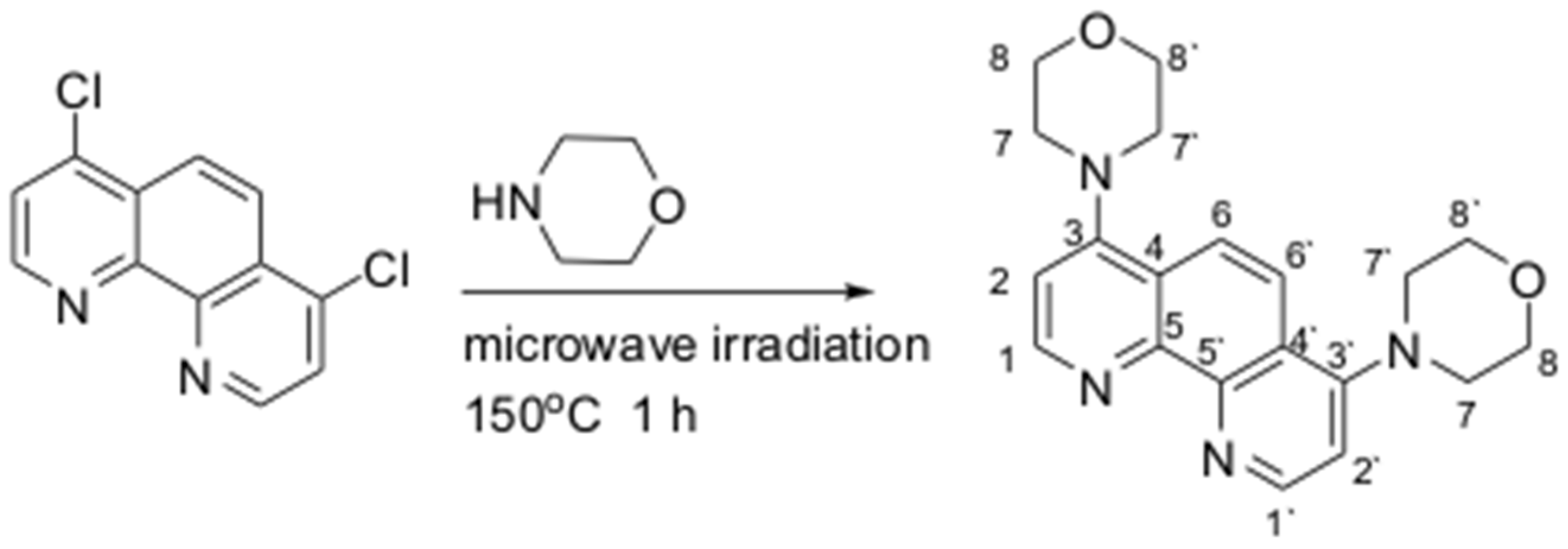
Scheme 1. Synthetic scheme for morphen (the numbering scheme is given for NMR interpretation only).
Crystals 15 00906 sch001
Figure 1. Closest environment of Tb(III) in 1 (a). Tb atoms are shown in green, O in red, N in blue, and Br in maroon. Two neighboring layers, viewed along the [1 0 0] normal axis (b) and along the c crystallographic axis (c). Atoms of independent layers are shown in different colors. Hydrogen atoms are not shown.
Crystals 15 00906 g001
Figure 2. PXRD pattern (a) and IR spectrum (b) for the as-synthesized 1; PXRD pattern for 1 after 24 h stirring in water (c) and TG plot for 1 (d).
Crystals 15 00906 g002
Figure 3. Excitation and emission spectra of compound 1 (a). Inset: digital photograph of the luminescent sample under 365 nm diode light. Excitation and emission spectra of 1@PVA film with delayed and non-delayed registration (b). Inset: Digital photographs of the as-prepared 1@PVA film (bottom left) and its emission under 365 nm diode light and different distances from the source (15 cm–top left, 10 cm–bottom right, 5 cm–top right).
Crystals 15 00906 g003
Figure 4. Digital photographs of the 1@PVA films treated by metal salts under λex = 365 nm diode light (a). A quenched spot on Fe3+-treated film is shown in red circle. Emission spectra of 1@PVA film treated by Fe3+ with delayed and non-delayed registration under λex = 375 nm (b).
Crystals 15 00906 g004
Disclaimer/Publisher’s Note: The statements, opinions and data contained in all publications are solely those of the individual author(s) and contributor(s) and not of MDPI and/or the editor(s). MDPI and/or the editor(s) disclaim responsibility for any injury to people or property resulting from any ideas, methods, instructions or products referred to in the content.