Article

Insights into Crystallization of Neuronal Nicotinic α4β2 Receptor in Polarized Lipid Matrices

by
Juan C. Villalobos-Santos
1,2,
Mallerie Carrasquillo-Rivera
1,2,
Josué A. Rodríguez-Cordero
1,
Orestes Quesada
2,3 and
José Antonio Lasalde-Dominicci
1,2,4,*
1
Department of Biology, Rio Piedras Campus, University of Puerto Rico, San Juan 00931, Puerto Rico
2
Molecular Sciences Research Center, San Juan 00931, Puerto Rico
3
Department of Physical Sciences, Rio Piedras Campus, University of Puerto Rico, San Juan 00931, Puerto Rico
4
Department of Chemistry, Rio Piedras Campus, University of Puerto Rico, San Juan 00931, Puerto Rico
*
Author to whom correspondence should be addressed.
Crystals 2024, 14(10), 889; https://doi.org/10.3390/cryst14100889
Submission received: 1 September 2024 / Revised: 5 October 2024 / Accepted: 6 October 2024 / Published: 12 October 2024
(This article belongs to the Section Biomolecular Crystals)

Abstract

:
Obtaining high-resolution 3D structures of membrane proteins through X-ray crystallography remains a longstanding bottleneck in the field of structural biology. This challenge has led to the optimization of purification methods to acquire high-yielding, pure proteins suitable for crystallization. In this study, we performed crystallization screenings of purified human α4β2 nAChR using a polarized in meso method. After reconstituting the detergent-solubilized α4β2 nAChR into the LCP matrix, the samples were incubated in a polarized lipid matrix using the RMP@LMx device developed in our laboratory. The results showed that under these conditions, the α4β2-nAChR-LFC 16 complex gave a mobile fraction >0.8, suggesting that its diffusion in the polarized lipid matrix is favorable for crystal nucleation. Voltages above 70 mV restricted crystal formation due to sample dehydration. Furthermore, a lipid analysis using UPLC-ESI MS/MS revealed a profile necessary for preserving protein integrity and promoting diffusion across the LCP. We harvested a single crystal and subjected it to X-ray diffraction, resulting in reflections comparable to previous studies of the muscle-type nAChR from Torpedo californica. X-ray diffraction of a single crystal gave distinct low-resolution diffractions of protein nature. These findings lay the groundwork for further optimization of membrane protein crystallization in polarized in meso phases.

1. Introduction

Nicotinic acetylcholine receptors (nAChRs) are pentameric ligand-gated ion channels that have been targeted in structural studies since their discovery. They play a key role in neurotransmission modulation, leading to an increased effort toward elucidating their atomic structure in order to understand their function [1,2]. Neuronal nAChRs exist as homomeric and heteromeric configurations, such as the α7 and α4β2 subtypes, respectively, which are associated with many neurological processes, including the regulation of the anti-inflammatory response in HIV patients [3]. Among these subtypes, the human α4β2 nAChR is actively expressed in cholinergic neurons and is widely distributed throughout the brain, making it the focus of pharmaceutical research that seeks to develop therapeutic compounds for smoking cessation and other neurodegenerative diseases [4,5,6,7,8,9]. To study receptor–ligand interactions at an atomic level, particularly with molecules like nicotine, a high-resolution, three-dimensional (3D) structure of the receptor and its bound ligands is required.
In the past decade, there has been an increasing number of attempts to elucidate the 3D structure of membrane proteins including nAChRs, with overall success among cryo-electron microscopy (cryo-EM) and X-ray crystallography studies [10,11,12,13,14,15,16]. The highest resolutions achieved for the α4β2 nAChR have been obtained through cryo-EM technology, owing the latest high-resolution structure (2.35 Ǻ) to the study of calcium potentiation in variable stoichiometries ((α4)3(β2)2 and (α4)2(β2)3) of the receptor [17]. However, no further advancements in the crystallization of this receptor and its stoichiometries have been made since it was initially crystallized [18]. There is a longstanding bottleneck in the preparation of milligram amounts of pure nAChRs suitable for crystallization. Since nAChRs are integral membrane proteins, their structural integrity is intrinsically associated with their immediate lipid environment. Our research has demonstrated that many detergents commonly used in the solubilization of these receptors actually denature the proteins, rendering them unresponsive to known agonists under controlled conditions [19]. However, detergents analogous to phospholipids, such as LysoFos Choline 16 (LFC-16), have been shown to effectively preserve the function of reconstituted muscle-type nAChRs, as measured by macroscopic electrophysiology in Xenopus oocytes. Furthermore, lipidomic analysis of the same samples has indicated that common detergents used in the solubilization of membrane proteins cause delipidation, directly affecting their structural stability and functionality [14]. In turn, phospholipid analog detergents help to preserve the integrity of the lipid composition of purified nAChRs [20].
The first and only atomic structure of the α4β2-subtype nicotinic acetylcholine receptor solved by X-ray crystallography was obtained from a single crystal with a final resolution of approximately 3.94 Ǻ, using the vapor diffusion method [18]. However, the aqueous solutions used in this method are not optimal for membrane proteins due to their dependence on their native lipid environment [1,18,20]. One method that has proved suitable for obtaining membrane protein crystals is the lipidic cubic phase (LCP) or in meso method [18,19,21]. Initially, its success was attributed to the crystallization of the intermediate states of the bacteriorhodopsin photocycle [22,23], but it gained widespread popularity after the crystallization of the human β2-adrenergic receptor, and it is still widely used [24,25,26]. In the present study, we used the LCP technique to incorporate the α4β2-nAChR–detergent complex (α4β2-nAChR-DC) into a lipidic matrix, allowing the complexes to freely diffuse and reconstitute. This lipid environment is presumably more stable than detergent micelles, providing a medium that can promote crystal nucleation.
The aim of this study was to perform crystallization screenings with α4β2 nAChRs solubilized with the phospholipid analog detergent LFC-16, reconstituted in a monoolein matrix, to obtain crystals suitable for synchrotron X-ray diffraction. Furthermore, we implemented a novel incubation method with a device designed in our laboratory, the RMP@LMx V1.5 (UPR Molecular Science Research Center, San Juan, Puerto Rico) (US patent 11,717,819), in the attempt to crystallize the receptor. Moreover, we investigated the resulting micelle post-solubilization to identify lipids associated with the α4β2-nAChR-LFC-16 complex to confirm that the samples were not delipidated, a factor known to negatively affect the receptor’s functionality.
The RMP@LMx V1.5 (US patent 11,717,819) device generates lipidic cubic phase (LCP) crystals in an electrically polarized environment. The biophysical principle of this device is that a membrane protein sits in a voltage gradient across a membrane, and specific localized domains in the protein can display voltage-dependent conformations. Based on this principle, this method should yield a more homogeneous population of membrane protein conformations, which is favorable for crystal nucleation and growth. Our central hypothesis is that the resting membrane potentials (RMPs) can be used to restrict the conformation freedom of voltage-dependent MPs to facilitate crystallization.

2. Materials and Methods

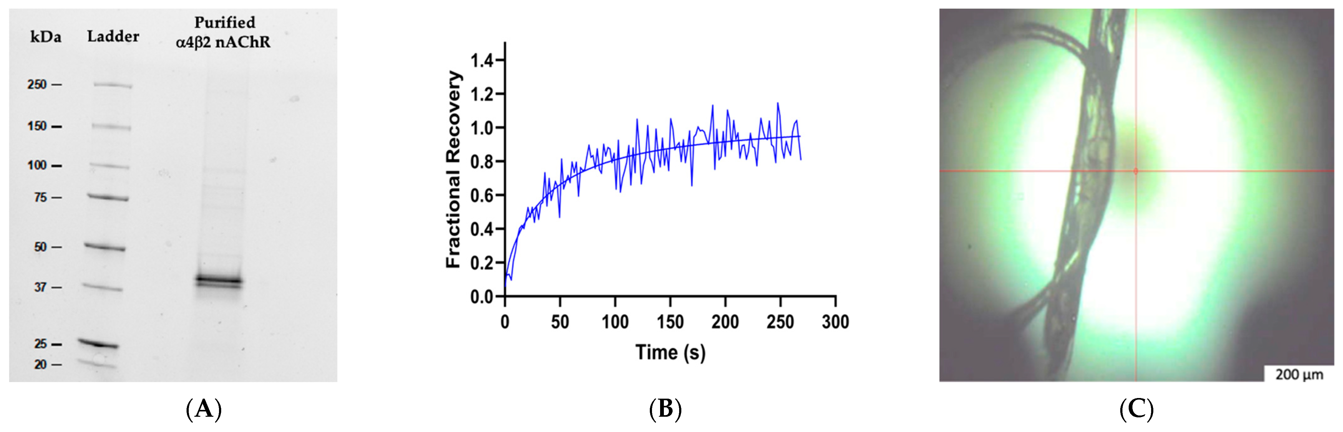
2.1. Expression and Purification of Human α4β2 nAChR

All reagents used in the experiments are from Sigma-Aldrich unless otherwise stated. pEZT-BM vectors (Addgene, Watertown, MA, USA) of each subunit, α4 and β2, were optimized for large-scale expression of the assembled receptor using the BacMam system. The expression of the nicotinic acetylcholine receptor (nAChR) followed the protocol developed by Morales-Pérez [18,27]. Briefly, suspension cultures of HEK GnTI-cells (ATCC, CRL 3022) were cultivated in FreeStyle 293 (Gibco, Carlsbad, CA, USA) media supplemented with 3 mM sodium butyrate and 0.1 mM nicotine to enhance protein overexpression. The cells were transduced with previously amplified baculoviruses encoding each subunit at a multiplicity of infection (MOI) of 0.25:0.5 of α4 and β2, respectively, and then incubated at 30 °C, 8% CO2, and 95% humidity for 72 h. Afterward, the cells were harvested by centrifugation and resuspended in 20 mM Tris, pH 7.4, 150 mM NaCl (TBS buffer), 1 mM nicotine, and 1 mM of phenyl methane sulfonyl fluoride (PMSF). The resuspended cells were lysed using the Emulsiflex C5 (Avestin, Ottawa, Canada) and subjected to a two-step differential centrifugation to isolate the membranes containing the nAChRs. The initial centrifugation settings were 9800× g for 15 min to remove cellular debris, followed by 186,000× g for 2 h at 4 °C to collect the membranes. The pelleted membranes were mechanically homogenized and incubated for 1 h at 4 °C in a solubilization buffer containing 1 mM nicotine, 40 mM LFC-16, and 0.2 mM cholesteryl hemisuccinate (CHS) (pH 7.4). After solubilization, the samples were centrifuged at 186,000× g for 1 h at 4 °C and purified via affinity chromatography using a Strep Trap HP column (GE Healthcare, Chicago, IL, USA). The binding buffer consisted of 150 mM NaCl, 20 mM Tris (pH 7.4), 1 mM PMSF, 1 mM LFC-16, 1 mM nicotine, 0.2 mM CHS, and 1 mM tris (2-carboxyethyl) phosphine (TCEP). The same binding buffer was used to prepare the elution buffer by adding 5 mM desthiobiotin.

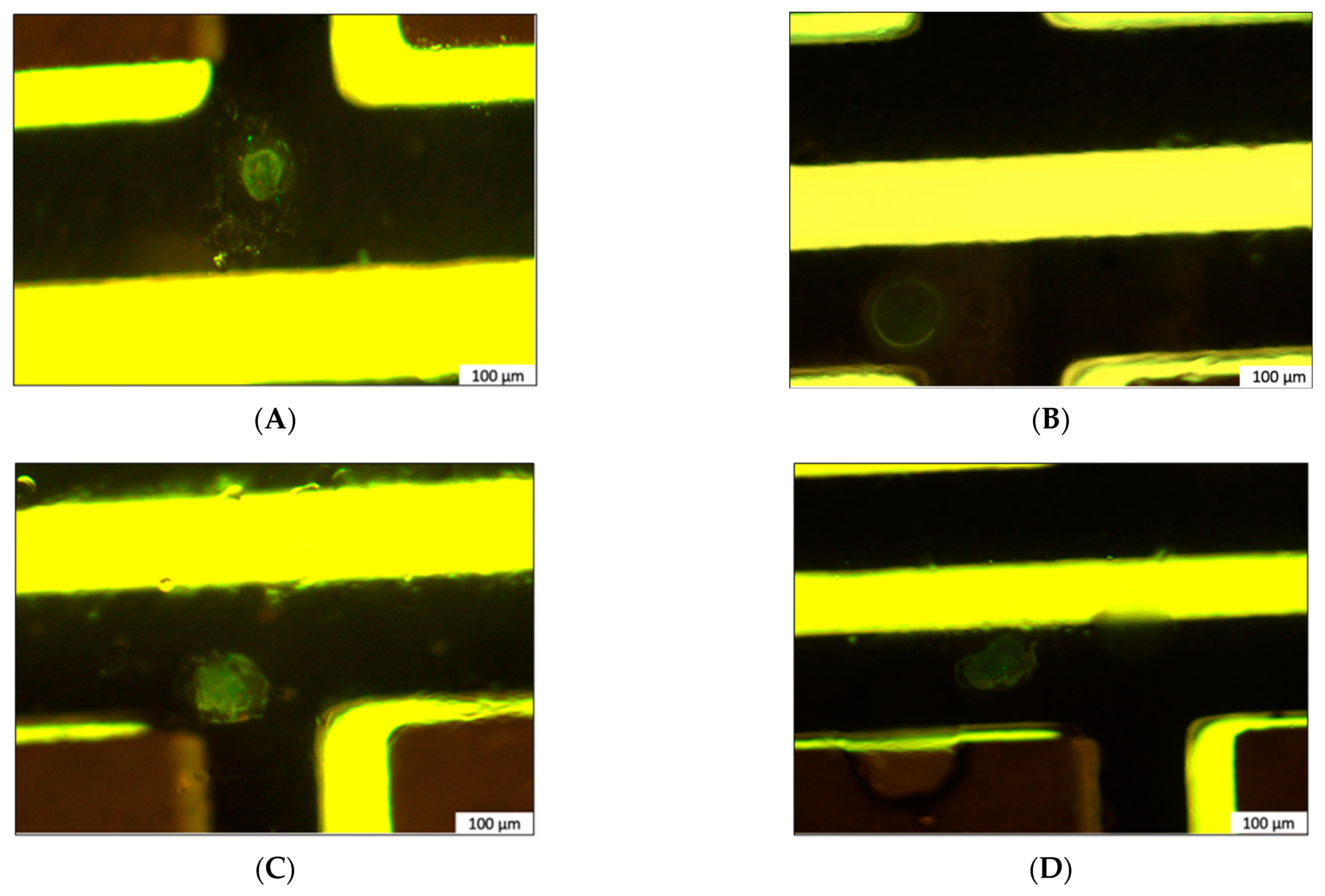
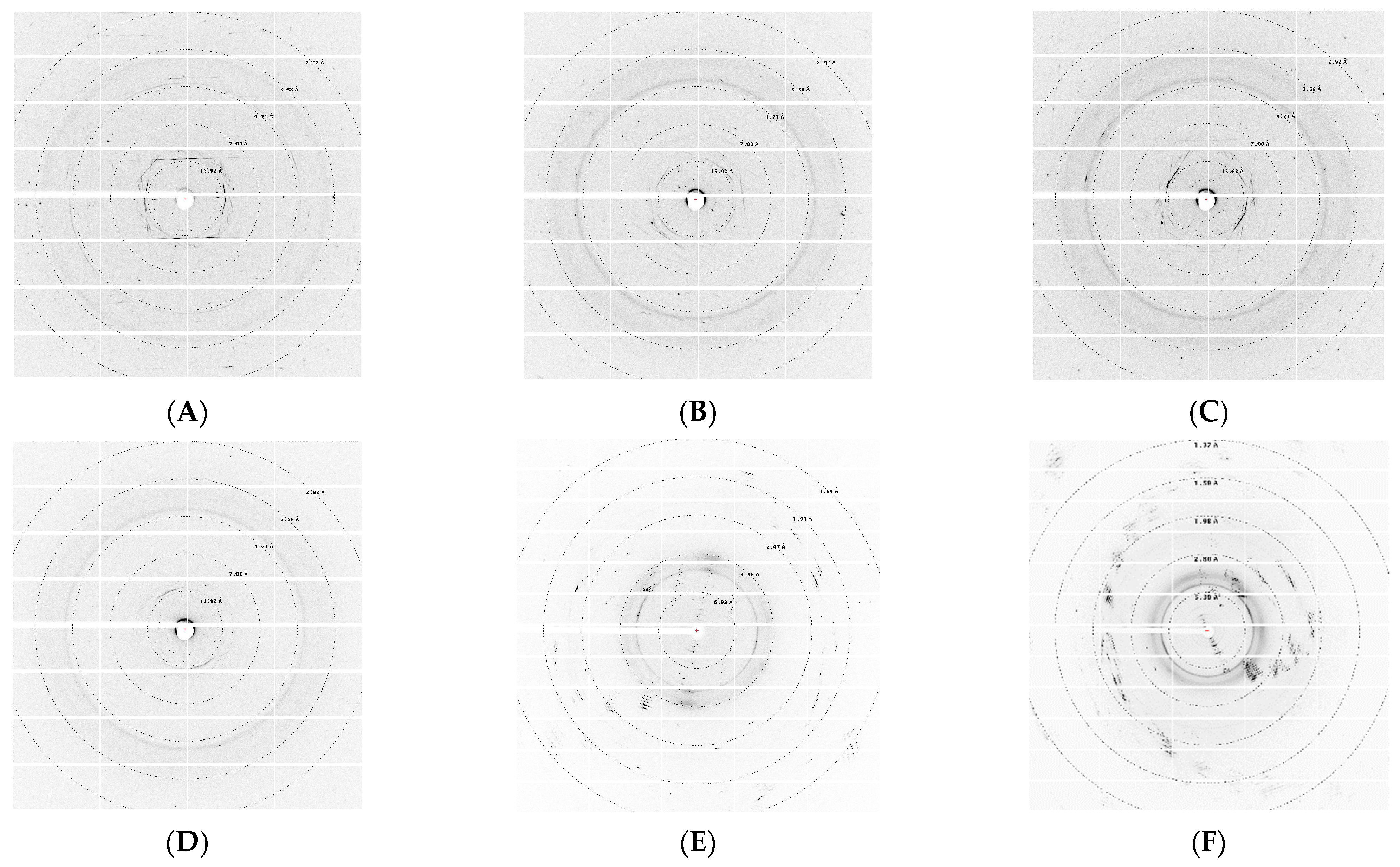
2.2. In Meso Crystallization with the RPM@LMx Device and X-ray Diffraction

Crystallization screenings followed the protocol reported by Liu and Cherezov [28], with some modifications incorporating the RPM@LMx (US patent 11,717,819) device designed in our laboratory. To concentrate the α4β2 nAChR–detergent complexes (DC), we centrifuged the samples using Amicon centrifugal filter units (Millipore, Burlington, NJ, USA). The protein fractions were concentrated from 1.1 to 3.1 mg/mL and reconstituted into the lipid cubic phase (LCP) monoolein matrix. In brief, the monoolein was melted at 40 °C and loaded from the reservoir using a 250 μL gastight syringe with a coupler. An additional 100 μL gastight syringe was used to load the α4β2 nAChR-DC samples. The syringes were connected through the coupler to mix the monoolein and the protein at a 40/60 (v/v) ratio, respectively. The contents of each syringe were mixed gently until the lipid mesophase became transparent and without white aggregates.
The RMP@LMx device is composed of a voltage supply circuit containing 24 slots, which can modulate variable voltages. Each of the voltage supply circuit slots can hold an individual crystallization sample board, capable of holding up to 36 LCP samples of approximately 50 to 500 nL for crystallization screenings. The device is configured through a computer to apply variable voltages to each crystallization sample board. After the receptor–detergent complexes were incorporated into the lipid mesophase, the mixture was transferred into a 10 μL gastight syringe attached to a PB600 dispenser (Hamilton, Reno, NV, USA) and dispensed in 0.2 μL boluses on the crystallization sample board. Additionally, we added 1 μL of precipitant solution composed of Pi-PEG #30 (50 mM ADA pH 6.8, 12.5% w/v PEG 1500, and 10% w/v PEG 1000) to each sample before sealing with a coverslip to avoid sample dehydration. The sample boards were then connected to the voltage supply circuit of our high-throughput crystallographic screening device (RMP@LMx, UPR Molecular Science Research Center, San Juan, Puerto Rico). The crystallization screenings were monitored while incubating on the sample boards of the RMP@LMx device at 20 °C and exposed to varying voltages ranging from 70 to 120 mV, where each voltage supply circuit increased in 10 mV intervals. All samples were incubated for 1–2 months inside the same Faraday cage to isolate them from electromagnetic contaminants. More than 300 samples per voltage variation were screened, of which 36 samples were collected from the voltage supply circuit configured at 70 mV. The samples were collected from the plates with MiTeGen loops (MiTeGen, Ithaca, NY, USA), cryo-protected, and flash-frozen before being shipped for X-ray diffraction data collection at the Argonne National Laboratory’s Advanced Photon Source in Chicago, IL, USA.

2.3. Lipidic Cubic Phase Fluorescence Recovery after Photobleaching (LCP–FRAP)

We performed the FRAP experiments according to Cherezov’s method, as modified by Padilla-Morales [20]. The purified α4β2-nAChR-LFC-16 complex was reconstituted in the LCP matrix and incubated as described in Section 2.2. Because the modified construct of the α4β2 nAChR intrinsically expresses green fluorescent protein (GFP), no additional fluorophores were incorporated into the samples. We conducted the FRAP assays at room temperature during a 30-day period at intervals of 5 days using a Zeiss Axio Observer LSM 800 confocal microscope (Zeiss, Oberkochen, Germany) at a 20× total magnification. Five pre-bleach images established the baseline fluorescence with a laser bleaching intensity of 6.97% of the total power, followed by a scanning sequence of 500 images. Consequently, each sample was integrated within three 30.0 μm regions of interest (ROIs). The average integrated intensity of the bleached 30.0 μm ROI was used to correct for photobleaching from irradiation during image acquisition. The equations used to calculate fractional fluorescence recovery are described by Quesada et al. [29].

2.4. Analysis of Phospholipid Molecular Species by Ultra-Performance Liquid Chromatography (UPLC) Coupled to Electrospray Ionization Tandem Mass Spectrometry (ESI-MS/MS)

The lipids were extracted using the Bligh and Dyer method with the addition of butylated hydroxytoluene (BHT; 2.9 × 10⁻⁵ M). To hydrolyze the phospholipids, the samples were refluxed for 3.5 h with MeOH/HCl and dried under nitrogen [30]. For lipid analysis in UPLC-ESI-MS/MS, the samples were sent to CD BioGlyco Company (Shirley, NY, USA). These were analyzed with Vanquish ultra-high performance liquid phase (UHPLC) coupled with a Thermo Orbitrap Fusion high-resolution mass spectrometer (Thermo Fisher Scientific, Waltham, MA, USA). The chromatography was performed using a Thermo-C30 (2.6 µm, 2.1 mm × 100 mm) column at 40 °C. For the mass spectrometry, mobile phase A was a 10 mM ammonium acetate solution in 60:40 of acetonitrile and water. In mobile phase B, the 10 mM ammonium acetate solution was prepared in 10:90 of acetonitrile to isopropanol. The elution gradient was 0–2 min, 30% B; 2–2.1 min, 43% B; 2.1–12 min, 55% B; 12–15 min, 65% B; 15–19.1 min, 100% B; and 19.1–20 min, 30% B. The injection volume was 5 µL with a flow rate of 0.26 mL/min. Prior to injection in the spectrometer, the samples were reconstituted in 100 µL of isopropanol, acetonitrile, and water (65:30:5). LipidSearch Software (V4.2.28) was used to identify lipids on the raw mass spectra and for peak alignment and filtering. Total peak normalization was applied to correct the values for magnitude comparison.

3. Results

3.1. Lipid Analysis of Purified Human α4β2 nAChR-DCs

The UPLC ESI-MS/MS analysis of purified α4β2 nAChR provides an insight into the lipidic profile present within the LFC-16 micelles. As expected, phospholipids were abundant, comprising a total of five species present (LPC, PC, LPMt, PEt, and PG), with lysophosphatidylcholine (LPC) being the most abundant (Table 1). Notably, we observed an abundance of LPC 16:0 of molecular weight (MW) 496.3398 m/z, indicating that the micelle is primarily composed of the LFC-16 detergent (MW 495.63). Furthermore, these five species displayed acyl-chain lengths ranging from 16:0 to 18:0 carbons, which is consistent with previously published data showing high phospholipid levels in the plasma membrane of HEK cells, among other cell types [31]. Lipids different from the detergent, found in larger quantities, might be closely associated with the α4β2 nAChR transmembrane domain (TD) and could play an important role in maintaining its integrity.

3.2. α4β2 nAChR-DC Diffusion in LCP

We successfully expressed the human α4β2 nAChR using the BacMam system on HEK GnTI-cells, as outlined in Morales-Pérez et al. [27]. The membranes were isolated through cell lysis and differential centrifugation, followed by solubilization. The solubilized α4β2-nAChR-LFC-16 complex was then readily purified by affinity chromatography through its strep tag (Figure 1A). Measuring the fluorescence fractional recovery has proved to be effective at demonstrating the nAChR’s ability to freely diffuse across the LCP matrix [20]. We employed FRAP to evaluate the stability of α4β2-nAChR complexes following solubilization with the LFC-16 detergent. Our results strongly suggest that the α4β2 nAChR-DCs were actively diffusing, with a fractional recovery above 80% throughout the entire 30-day incubation period (Figure 1B).

3.3. Polarized In Meso Crystallization Screening of Human α4β2 nAChR

The α4β2 nAChR allowed us to track the protein within the lipid matrix, revealing a diffusion pattern toward nucleation spots that could eventually result in crystal formation. In samples incubated in higher voltages (>70 mV), dehydration ensued, causing sample loss. Conversely, other samples solubilized using alternative phospholipid analog detergents failed to yield protein crystals. Furthermore, we conducted additional crystallization screenings using n-dodecyl-β-D-maltoside (DDM), a common detergent used for membrane protein solubilization known to hinder protein function. No nucleation spots were observed in these samples, as monitored through GFP fluorescence.
The samples subjected to higher voltages underwent dehydration after a two-week incubation period, whereas those exposed to lower voltages remained in their same gel-like state. The evaporation of the precipitant solution in the former might be attributable to an increased temperature caused by the constant voltage exposure. From these first crystallization screenings, a single crystal was harvested from a sample of LCP containing the α4β2-nAChR-LFC-16 complex at 2.0 mg/m, within the 70 mV slot of the RMP@LMx V.1.5 device (US patent 11,717,819). The crystal was promptly stored in liquid nitrogen for further diffraction analysis at the Argonne National Laboratory—Advanced Photon Source (APS) (Figure 2).

3.4. Synchrotron X-ray Diffraction

The crystallization conditions that previously yielded diffraction patterns in Tc nAChR studies also resulted in a crystal that enabled the first diffraction patterns of the α4β2-nAChR-LFC-16 complex using LCP crystallization setups, as shown in Figure 3. We see that low-resolution protein diffractions, no higher than 10 Ǻ, resulted from these samples. These patterns suggest a consistent degree of organization across all data sets. We repeatedly obtained “crystal-like” samples that lacked diffraction but were detectable by GFP, indicating that the α4β2-nAChR-LFC-16 complex was in fact diffusing toward nucleation under these conditions. Aside from the crystallization solution Pi-PEG #30, which yielded the samples we diffracted in this study, other conditions were tested between screenings. Among these variations, one particular condition resulted in several “crystal-like” formations, including GFP fluorescent crystals. The crystallization condition comprised 0.1 M sodium cacodylate, 18% (w/v) PEG 8000, 0.2 M calcium acetate, and 0.5 mM DTT; its diffraction resulted in patterns similar to those obtained in previous experiments.

4. Discussion

Membrane protein solubilization occurs by the incorporation of detergent micelles into the lipid bilayer, resulting in a detergent complex of annular and non-annular lipids crucial to maintaining target protein integrity [32]. The annular lipids are those peripheral to the receptor and usually diffuse across the membrane, potentially further diffusing into the solution during the solubilization process. The α4β2-nAChR-LFC-16 complex underwent affinity purification with a buffer exchange step to remove most impurities and excess detergent, ensuring that the micelles contained only the receptor and its associated lipids. Membrane proteins often exhibit poor stability in aqueous crystallization setups due to their dependence on lipids, gradually reshaping structural biology by replacing X-ray crystallography with cryo-EM [33,34,35,36]. In contrast to aqueous crystallization conditions, this study used preparations of purified α4β2 nAChR integrated into an LCP matrix and exposed them to polarization using the RMP@LMx V.1.5 device. The incorporation of these purified α4β2 nAChR-DC samples into LCP matrices helped stabilize hydrophobic domains while diffusing toward nucleation and possibly preserved the remaining non-annular lipids that are crucial to its function [29,30]. Furthermore, the acyl-chain length of the lipids present in the sample approximated those isolated from native muscle-type nAChR-DCs extracted from the electric organ of Torpedo californica (Tc) [27]. Our findings show that α4β2 nAChR-DCs are mostly composed of the detergent LFC-16, which contains structural characteristics similar to LPC lipids, the most abundant group of phospholipids in HEK cell membranes [37]. This lipid profile resembles that observed in functional muscle-type nAChRs, which was expected since they share similar TD characteristics with neuronal nAChRs, and the results suggest that the micelles do not delipidate after solubilization [18,30].
Studies on muscle-type nAChRs from our laboratory have demonstrated the negative effect that common detergents used for solubilization have on the integrity of nAChRs and their capacity to diffuse within the LCP, triggering the nucleation event by increasing supersaturation [29]. The human neuronal α4β2-nAChR-LFC-16 complex can diffuse across the LCP matrix (mobile fraction > 0.80), showing nucleation at approximately 2–4 weeks of incubation in a polarized environment. Samples exposed to a voltage of 70 mV did not undergo dehydration, as opposed to those exposed to higher voltages. This could be due to an increase in localized temperature, leading to the evaporation of the precipitant solution [38]. Although the diffractions obtained from our crystals lacked sufficient information regarding the unit cell, we managed to isolate a single, tubular crystal that provided diffraction data with patterns that were strongly indicative of the presence of proteins within the crystal.
Protein crystallization poses a major challenge because of the unpredictable aggregation behavior of proteins. Generally, nucleation requires an interface formation and is facilitated in an intermediate dense liquid phase where aggregates can freely diffuse [39]. In the LCP method, the ideal transition state for membrane protein crystallization is the lamellar phase, which best resembles a lipid bilayer [40]. The results from our LCP-FRAP experiments demonstrate that the α4β2-nAChR-LFC-16 complex can diffuse through the lipidic matrices and potentially aggregate into precrystalline clusters. Previous studies indicate that exogenous factors, such as exposure to an internal electric field, positively impact crystal nucleation [41,42]. By using direct currents, the nucleation rate of microcrystals decreases, which in turn increases the probability of obtaining single, larger, and high-quality crystals that are suitable for structural studies [41,42]. Furthermore, protein alignment has been shown to be influenced by fatty acids as they not only stabilize the protein but also promote aggregation under an electric field when cations are present [43]. It is well known that lipid bilayers favor helical packings that constitute tubular crystalline structures and are highly impacted by steric confinement of proteins [44,45].
As a control experiment, we also prepared bacteriorhodopsin crystals from Halobacterium salinarum under the same conditions by employing the described standard protocol using the RMP@LMx V.1.5 device at 70 mV. We collected data from 10 crystals at APS ID 23 and solved these structures with resolutions in the range of 2.15–3.70 Å using molecular replacement. The structural data showed minor changes in the bacteriorhodopsin structure obtained using the RMP@LMX V1.5 device as compared to the structure reported in LCP [46]. The data were consistent with the fact that the bacteriorhodopsin has no net charge at pH 5.5. We performed a crystallographic screening to induce charged residues in bacteriorhodopsin at different pH levels (4.0, 4.5, 5.0, 6.0, 6.5, 7.0, 7.5, and 8.0), but no crystal growth was observed under these conditions.
In summary, in a polarized in meso crystallization, the α4β2 nAChR produced tubular crystalline structures, whereas bacteriorhodopsin produced high-quality 3D crystals. It is important to highlight that we observed no crystal formation for the α4β2 nAChR in non-polarized in meso crystallization (LCP). These results suggest that the diffractions obtained in the polarized in meso matrix for the α4β2 nAChR are intrinsic to its structure.

5. Conclusions

Our current understanding of membrane protein structure and crystallization methodologies has been limited by the need to maintain these proteins in their native lipid environment. Despite challenges with the solubilization and purification of the receptors, our laboratory has successfully identified detergents capable of extracting key lipid species that preserve their integrity, positively impacting the receptor’s diffusion through LCP matrices. In the present study, we showed that crystallization of the α4β2 nAChR in a polarized in meso environment potentially favors nucleation. While most crystals lacked reflections, the isolation of a single crystal and “crystal-like” samples showed low-resolution protein diffractions. These patterns offer promising insights into improving this crystallization technique. Thus, our findings lay the groundwork for further optimization of membrane protein crystallization in polarized in meso phases.

6. Patents

Four US patents have been awarded to the RMP@LMx methods and device: US 10,155,221, US 10,358,475, US 11,440,001, and US 11,717,819, with the latter being issued on 8 August 2023. US Continuation Patent Application 18/225,981 has been allowed, and US Provisional Patent Application 63/700,640 has been filed.

Author Contributions

Conceptualization, J.C.V.-S., O.Q. and J.A.L.-D.; methodology, J.C.V.-S., J.A.R.-C., M.C.-R., O.Q. and J.A.L.-D.; data curation, J.C.V.-S. and O.Q.; writing—original draft preparation, J.C.V.-S.; writing—review and editing, J.C.V.-S., M.C.-R., O.Q. and J.A.L.-D. All authors have read and agreed to the published version of the manuscript.

Funding

This work was supported by the Research Initiative for Scientific Enhancement (RISE) program [5R25GM061151-22]; the Neuroscience Graduate, Resilience, Affirmation and Diversity (NeuroGRAD) program [1R25NS127776-01]; the National Institutes of Health (NIH) [1R01GM098343]; Centers of Biomedical Research Excellence (COBRE) [1P20GM103642], and Bridge to the Doctorate-Louis Stokes Alliance for Minority Participation (LSAMP) [1612393].

Data Availability Statement

The raw data supporting the conclusions of this article will be made available by the authors on request.

Acknowledgments

We would like to thank CD BioGlyco for generating the lipid profile of our samples and the administrative staff at the Argonne National Laboratory—Advanced Photon Source (APS) for assisting us in generating the diffraction patterns at beamline 23-ID-D. We also extend our gratitude to those involved in the editorial process of this article, including Cristina Martínez-Benito and colleagues from the BIOL6996 course.

Conflicts of Interest

The authors declare no conflicts of interest.

References

  1. Delgado-Vélez, M.; Quesada, O.; Villalobos-Santos, J.C.; Maldonado-Hernández, R.; Asmar-Rovira, G.; Stevens, R.C.; Lasalde-Dominicci, J.A. Pursuing high-resolution structures of nicotinic acetylcholine receptors: Lessons learned from five decades. Molecules 2021, 26, 5753. [Google Scholar] [CrossRef] [PubMed]
  2. Changeux, J.P. Discovery of the First Neurotransmitter Receptor: The Acetylcholine Nicotinic Receptor. Biomolecules 2020, 10, 547. [Google Scholar] [CrossRef]
  3. Delgado-Vélez, M.; Báez-Pagán, C.A.; Gerena, Y.; Quesada, O.; Santiago-Pérez, L.I.; Capó-Vélez, C.M.; Wojna, V.; Meléndez, L.; León-Rivera, R.; Silva, W.; et al. The α 7-nicotinic receptor is upregulated in immune cells from HIV-seropositive women: Consequences to the cholinergic anti-in flammatory response. Clin. Transl. Immunol. 2015, 4, e53. [Google Scholar] [CrossRef] [PubMed]
  4. Hurst, R.; Rollema, H.; Bertrand, D. Nicotinic acetylcholine receptors: From basic science to therapeutics. Pharmacol. Ther. 2013, 137, 22–54. [Google Scholar] [CrossRef]
  5. Polosa, R.B.N.L. Treatment of nicotine addiction: Present therapeutic options and pipeline developments. Trends Pharmacol Sci. 2011, 32, 281–289. [Google Scholar] [CrossRef]
  6. Quik, M.; O’Leary, K.; Tanner, C.M. Nicotine and Parkinson’s disease; implications for therapy. Physiol. Behav. 2008, 23, 1641–1652. [Google Scholar] [CrossRef] [PubMed]
  7. Cooper, S.Y.; Akers, A.T.; Journigan, V.B.; Henderson, B.J. Novel putative positive modulators of α4β2 nachrs potentiate nicotine reward-related behavior. Molecules 2021, 26, 4793. [Google Scholar] [CrossRef]
  8. Buisson, B.; Bertrand, D. Open-channel blockers at the human α4β2 neuronal nicotinic acetylcholine receptor. Mol. Pharmacol. 1998, 53, 555–563. [Google Scholar] [CrossRef]
  9. Vallés, A.S.; Barrantes, F.J. Nicotinic acetylcholine receptor dysfunction in addiction and in some neurodegenerative and neuropsychiatric diseases. Cells 2023, 12, 2051. [Google Scholar] [CrossRef]
  10. Walsh, R.M., Jr.; Roh, S.H.; Gharpure, A.; Morales-Perez, C.L.; Teng, J.; Hibbs, R.E. Structural principles of distinct assemblies of the human α 4 β 2 nicotinic receptor. Nature 2018, 557, 261–265. [Google Scholar] [CrossRef]
  11. Gharpure, A.; Teng, J.; Zhuang, Y.; Noviello, C.M.; Walsh, R.M., Jr.; Cabuco, R.; Howard, R.J.; Zaveri, N.T.; Lindahl, E.; Hibbs, R.E. Agonist Selectivity and Ion Permeation in the α3β4 Ganglionic Nicotinic Receptor. Neuron 2019, 104, 501–511.e6. [Google Scholar] [CrossRef] [PubMed]
  12. Rahman, M.M.; Teng, J.; Worrell, B.T.; Noviello, C.M.; Lee, M.; Karlin, A.; Stowell, M.H.B.; Hibbs, R.E. Structure of the Native Muscle-type Nicotinic Receptor and Inhibition by Snake Venom Toxins. Neuron 2020, 106, 952–962.e5. [Google Scholar] [CrossRef]
  13. Rahman, M.; Basta, T.; Teng, J.; Lee, M.; Worrell, B.T.; Stowell, M.H.B.; Hibbs, R.E. Structural mechanism of muscle nicotinic receptor desensitization and block by curare. Nat. Struct. Mol. Biol. 2022, 4, 386–394. [Google Scholar] [CrossRef] [PubMed]
  14. Noviello, C.M.; Gharpure, A.; Mukhtasimova, N.; Cabuco, R.; Baxter, L.; Borek, D.; Sine, S.M.; Hibbs, R.E. Structure and gating mechanism of the α7 nicotinic acetylcholine receptor. Cell 2021, 184, 2121–2134. [Google Scholar] [CrossRef]
  15. Zhang, D.; Liu, Y.; Zaidi, S.A.; Xu, L.; Zhan, Y.; Chen, A.; Guo, J.; Huang, X.; Roth, B.L.; Katritch, V.; et al. Structural insight into angiotensin receptor signaling modulation by balanced and biased agonists. EMBO J. 2023, 42, e112940. [Google Scholar] [CrossRef] [PubMed]
  16. Yang, Y. Structural insight into apelin receptor-G protein stoichiometry. Nat. Struct. Mol. Biol. 2022, 29, 688–697. [Google Scholar]
  17. Mazzaferro, S.; Kang, G.; Natarajan, K.; Hibbs, R.E.; Sine, S.M. Structural bases for Stoichiometry-Selective Calcium Potentiation of a Neuronal Nicotinic Receptor. Br. J. Pharmacol. 2024, 181, 1973–1992. [Google Scholar] [CrossRef]
  18. Morales-Perez, C.L.; Noviello, C.M.; Hibbs, R.E. X-ray structure of the human α4β2 nicotinic receptor. Nature 2016, 538, 411–415. [Google Scholar] [CrossRef]
  19. Padilla-Morales, L.F.; Colón-Sáez, J.O.; González-Nieves, J.E.; Quesada-González, O.; Lasalde-Dominicci, J.A. Functionality and stability data of detergent purified nAChR from Torpedo using lipidic matrixes and macroscopic electrophysiology. Data Brief 2016, 6, 433–437. [Google Scholar] [CrossRef]
  20. Padilla-Morales, L.F.; Colón-Sáez, J.O.; González-Nieves, J.E.; Quesada-González, O.; Lasalde-Dominicci, J.A. Assessment of the Functionality and Stability of Detergent Purified nAChR from Torpedo using Lipidic Matrixes and Macroscopic Electrophysiology. Biochim. Biophys. Acta (BBA)-Biomembr. 2017, 1858, 47–56. [Google Scholar] [CrossRef]
  21. Caffrey, M.; Cherezov, V. Crystallizing Membrane Proteins Using Lipidic Mesophases. Nat. Protoc. 2010, 4, 706–731. [Google Scholar] [CrossRef] [PubMed]
  22. Luecke, H.; Schobert, B.; Richter, H.T.; Cartailler, J.P.; Lanyi, J.K. Structure of bacteriorhodopsin at 1.55 Å resolution. J. Mol. Biol. 1999, 291, 899–911. [Google Scholar] [CrossRef] [PubMed]
  23. Caffrey, M. A comprehensive review of the lipid cubic phase or in meso method for crystallizing membrane and soluble proteins and complexes. Acta Crystallogr. Sect. FStructural Biol. Commun. 2015, 71, 3–18. [Google Scholar] [CrossRef] [PubMed]
  24. Cherezov, V.; Rosenbaum, D.M.; Hanson, M.A.; Rasmussen, S.G.; Thian, F.S.; Kobilka, T.S.; Choi, H.J.; Kuhn, P.; Weis, W.I.; Kobilka, B.K.; et al. High-Resolution Crystal Structure of an Engineered Human β2-adrenergic G protein–coupled receptor. Science 2007, 318, 1258–1265. [Google Scholar] [CrossRef]
  25. Martynowycz, M.W.; Shiriaeva, A.; Ge, X.; Hattne, J.; Nannenga, B.L.; Cherezov, V.; Gonen, T. MicroED structure of the human adenosine receptor determined from a single nanocrystal in LCP. Proc. Natl. Acad. Sci. USA 2021, 118, e2106041118. [Google Scholar] [CrossRef]
  26. Michaelian, N.; Sadybekov, A.; Besserer-Offroy, É.; Han, G.W.; Krishnamurthy, H.; Zamlynny, B.A.; Fradera, X.; Siliphaivanh, P.; Presland, J.; Spencer, K.B.; et al. Structural insights on ligand recognition at the human leukotriene B4 receptor 1. Nat. Commun. 2021, 12, 2971. [Google Scholar] [CrossRef]
  27. Morales-Perez, C.L.; Noviello, C.M.; Hibbs, R.E. Manipulation of Subunit Stoichiometry in Heteromeric Membrane Proteins Resource Manipulation of Subunit Stoichiometry in Heteromeric Membrane Proteins. Struct. Des. 2016, 24, 797–805. [Google Scholar] [CrossRef]
  28. Liu, W.; Cherezov, V. Crystallization of membrane proteins in lipidic mesophases. J. Vis. Exp. 2011, 49. [Google Scholar] [CrossRef]
  29. Quesada, O.; González-Freire, J.E.; Colón, J.; Maldonado-Hernández, R.; González-Freire, C.; Acevedo-Cintrón, J.; Rosado-Millán, I.D.; Lasalde-Dominicci, J.A. Assessment of Purity, Functionality, Stability, and Lipid Composition of Cyclofos-nAChR-Detergent Complexes from Torpedo californica Using Lipid Matrix and Macroscopic Electrophysiology. J. Mem. Biol. 2023, 256, 271–285. [Google Scholar] [CrossRef]
  30. Quesada, O.; Freire, C.G.; Ferrer, M.C.; -Sáez, J.O.C.; Fernández-García, E.; Mercado, J.; Dávila, A.; Morales, R.; Lasalde-Dominicci, J.A. Uncovering the lipidic basis for the preparation of functional nicotinic acetylcholine receptor detergent complexes for structural studies. Sci. Rep. 2016, 6, 32766. [Google Scholar] [CrossRef]
  31. Symons, J.L.; Cho, K.J.; Chang, J.T.; Du, G.; Waxham, M.N.; Hancock, J.F.; Levental, I.; Levental, K.R. Lipidomic atlas of mammalian cell membranes reveals hierarchical variation induced by culture conditions, subcellular membranes, and cell lineages. Soft Matter 2021, 17, 288–297. [Google Scholar] [CrossRef] [PubMed]
  32. Palsdottir, H.; Hunte, C. Lipids in membrane protein structures. Biochim. Biophys. Acta 2004, 1666, 2–18. [Google Scholar] [CrossRef] [PubMed]
  33. McPherson, A.; Gavira, J.A. Introduction to protein crystallization. Acta Cryst. 2013, F70, 2–20. [Google Scholar]
  34. Shoemaker, S.C.; Ando, N. X-rays in the Cryo-EM era: Structural Biology’s dynamic future. Biochemistry 2019, 57, 277–285. [Google Scholar] [CrossRef]
  35. Barrantes, F.J. Structure and function meet at the nicotinic acetylcholine receptor-lipid interface. Pharmacol. Res. 2023, 190, 106729. [Google Scholar] [CrossRef] [PubMed]
  36. Barrantes, F.J. Modulation of a rapid neurotransmitter receptor-ion channel by membrane lipids. Front. Cell Dev. Biol. 2024, 11, 1328875. [Google Scholar] [CrossRef]
  37. Dawaliby, R.; Trubbia, C.; Delporte, C.; Noyon, C.; Ruysschaert, J.M.; Antwerpen, P.V.; Govaerts, C. Phosphatidylethanolamine is a key regulator of membrane fluidity in eukaryotic cells. J. Biol. Chem. 2016, 291, 3658–3668. [Google Scholar] [CrossRef]
  38. Hejazi, S.; Pahlavandanzadeh, H.; Elliott, J.A.W. Thermodynamic investigation of the effect of electric field on solid-liquid equilibrium. J. Phys. Chem. B 2021, 125, 1271–1281. [Google Scholar] [CrossRef]
  39. Nanev, C.N. Recent Insights into the Crystallization Process; Protein Crystal Nucleation and Growth Peculiarities; Processes in the Presence of Electric Fields. Crystals 2017, 7, 310. [Google Scholar] [CrossRef]
  40. Van’t Hag, L.; Knoblich, K.; Seabrook, S.A.; Kirby, N.M.; Mudie, S.T.; Lau, D.; Li, X.; Gras, S.L.; Mulet, X.; Call, M.E.; et al. Exploring the in meso crystallization mechanism by characterizing the lipid mesophase microenvironment during the growth of single transmembrane alpha-helical peptide crystals. Phil. Trans. R. Soc. A 2016, 374, 20150125. [Google Scholar] [CrossRef]
  41. Al-Haq, M.I.; Lebrasseur, E.; Tsuchiya, H.; Torri, T. Protein crystallization under an electric field. Cryst. Rev. 2007, 13, 29–64. [Google Scholar] [CrossRef]
  42. Ogata, M.; Aoki, D.; Suzuki, M.; Tanaka, D.; Wakamatsu, T. The role of an applied electric field in protein crystallization at low temperature. Jpn. J. Appl. Phys. 2019, 58, 110903. [Google Scholar] [CrossRef]
  43. Kan, K. The effect of fatty acids, ionic strength, and electric fields on the microscopic dynamics of BSA aggregates. Front. Phys. 2023, 11, 1282099. [Google Scholar]
  44. Yager, P.; Price, R.R.; Schnur, J.M.; Schoen, P.E.; Singh, A.; Rhodes, D.G. The mechanism of formation of lipid tubules from liposomes. Chem. Phys. Lipids 1988, 46, 171–179. [Google Scholar] [CrossRef]
  45. Stachowiak, J.C.; Hayden, C.C.; Sasaki, D.Y. Steric confinement of proteins on lipid membranes can drive curvature and tubulation. Proc. Natl. Acad. Sci. USA 2010, 107, 7781–7786. [Google Scholar] [CrossRef]
  46. Pebay-Peyroula, E.; Rummel, G.; Rosenbusch, J.P.; Landau, E.M. X-ray structure of bacteriorhodopsin at 2.5 angstroms from microcrystals grown in lipidic cubic phases. Science 1997, 277, 1676–1681. [Google Scholar] [CrossRef]
Figure 1. Diffusion across the lipid matrix and single crystal obtained from LCP crystallization of the human α4β2 nAChR using the RMP@LMx V.1.5 device. (A) SDS-PAGE of the purified sample used to produce the crystal. (B) LCP-FRAP of purified α4β2 nAChR-DCs. (C) Micrograph of a single crystal of the human α4β2 nAChR taken at the time of X-ray diffraction.
Crystals 14 00889 g001
Figure 2. LCP samples containing the purified α4β2 nAChR-DCs during incubation in the RMP@LMx V.1.5 device. (AD) Fluorescent microscopy images of “nucleation spots” characterized by the detection of GFP fluorescence belonging to the α4β2 nAChR-DCs. Every sample was dispensed in the capacitor area of the crystallization sample board to maximize its exposure to the determined voltage. All samples in this figure represent crystallization screenings with a constant voltage of 70 mV, ensuring minimal evaporation of the precipitants. At 0 mV (non-polarized control), no crystal formation was observed.
Crystals 14 00889 g002
Figure 3. Synchrotron X-ray diffraction patterns of the human α4β2 nAChR from a single crystal. (AF) Reflections resulting from the diffraction of a single crystal and another “crystal-like” sample harvested from the purified α4β2 nAChR solubilized using the detergent LFC-16. Here, diffractions were observed from both protein nature and a small molecule from one of the crystallization components of the reservoir solution. The diffracted samples were grown from a purified sample of α4β2 nAChR at 2.0 mg/mL, within the 70 mV sample board slot of the RMP@LMx V.1.5 device. The reservoir solution used was Pi-PEG #30 (50 mM ADA pH 6.8, 12.5% w/v PEG 1500, and 10% w/v PEG 1000). These diffractions were obtained at beamline 23-ID-D at Argonne National Laboratory—Advanced Photon Source. All diffraction patterns were managed through the Adxv software (Current version 1.9.15) (https://www.scripps.edu/tainer/arvai/adxv.html, accessed on 30 June 2019).
Crystals 14 00889 g003
Table 1. Most abundant lipid species in α4β2 nAChR-DCs detected with UPLC ESI-MS/MS. Among these species, LPC 16:0 (496.3398 m/z) is found in larger quantities than any other, suggesting that the micelle is composed mostly of the detergent LFC-16.
Molecular Speciesm/z (M + H)+Mean Peak Area
LPC 16:0496.339817,941,873.71
Cer m22:0338.341712,200,000.00
TG P-23:6/21:1721.57664,509,407.999
LPC 17:0554.34633,323,174.888
LPMt 16:0423.25171,824,723.222
LPC (PC 16:0)518.32171,595,348.877
DAP O-11:0/O-5:0391.2843922,029.400
DAP O-7:0/O-7:0363.2529769,424.966
SM d34:1703.5749480,092.577
LPC 18:0524.3711266,859.633
Disclaimer/Publisher’s Note: The statements, opinions and data contained in all publications are solely those of the individual author(s) and contributor(s) and not of MDPI and/or the editor(s). MDPI and/or the editor(s) disclaim responsibility for any injury to people or property resulting from any ideas, methods, instructions or products referred to in the content.

