Article

SiO2/SiC Nanowire Surfaces as a Candidate Biomaterial for Bone Regeneration

1
Centro Universitario di Odontoiatria, Dipartimento di Medicina e Chirurgia, Università di Parma, Via Gramsci 14, 43123 Parma, Italy
2
IMEM-CNR, Parco Area delle Scienze 37A, 43124 Parma, Italy
3
CNR-SCITEC, Via A. Corti 12, 20133 Milano, Italy
4
Lintes Research Laboratory, Via Isola 2, 64010 Colonnella, Italy
*
Author to whom correspondence should be addressed.
Crystals 2023, 13(8), 1280; https://doi.org/10.3390/cryst13081280
Submission received: 29 July 2023 / Revised: 12 August 2023 / Accepted: 14 August 2023 / Published: 19 August 2023
(This article belongs to the Special Issue Advances of Silicon Carbide Crystals)

Abstract

:
Tissue engineering (TE) and nanomedicine require devices with hydrophilic surfaces to better interact with the biological environment. This work presents a study on the wettability of cubic silicon-carbide-based (SiC) surfaces. We developed four cubic silicon-carbide-based epitaxial layers and three nanowire (NW) substrates. Sample morphologies were analyzed, and their wettabilities were quantified before and after a hydrogen plasma treatment to remove impurities due to growth residues and enhance hydrophilicity. Moreover, sample biocompatibility has been assessed with regard to L929 cells. Our results showed that core–shell nanowires (SiO2/SiC NWs), with and without hydrogen plasma treatment, are the most suitable candidate material for biological applications due to their high wettability that is not influenced by specific treatments. Biological tests underlined the non-toxicity of the developed biomaterials with regard to murine fibroblasts, and the proliferation assay highlighted the efficacy of all the surfaces with regard to murine osteoblasts. In conclusion, SiO2/SiC NWs offer a suitable substrate to develop platforms and membranes useful for biomedical applications in tissue engineering due to their peculiar characteristics.

1. Introduction

Biomaterials are increasingly used to develop scaffolds to replace damaged tissues and organs [1,2]. They are commonly characterized by a structured and modified biological material such as collagen, chitosan, or decellularized bone, whereas biomimetic materials consist of synthetic polymers, metal, or ceramics with surface or bulk modifications able to make the material biocompatible and suitable for tissue implant or tissue engineering (TE) [3,4,5,6]. The hierarchical organization of tissue (from macro- to nano-structures) is the key factor in synthesizing functionally active biomaterials: the first step in the development of a scaffold is to identify the specific cues and characteristics it must possess to resemble as better as possible the extracellular matrix of the tissue involved in the regeneration [7]. Moreover, to design the ideal scaffold, it is necessary to accurately define different parameters, such as mechanical or physical properties (i.e., elastic module, wettability), and its three-dimensional structure in the microenvironment, which heavily influences the functional macroscopic properties of the graft when it has been inserted in the human body [8]. In this framework, the ideal scaffold should possess a number of requirements to be suitable for TE: biodegradability, bioactivity, mechanical properties, porosity, topography, chemistry, and wettability [3]. Wettability is the characteristic that mainly defines the behavior of the material during the first contact with biological fluids [9]. Biomaterial surfaces can be either hydrophilic or hydrophobic depending on their degree of affinity to water, which is highly influenced by the substrate shape/topography [10]. This property arose a huge interest in widespread sectors (i.e., biology, medicine, nanoelectronics, engineering, etc.) [11,12,13]. Hydrophilic surfaces are characterized by their good affinity for water; they can prevent the formation of moisture, as the water sprayed on them forms a thin film instead of droplets, especially if the surface is rough [14]. These kinds of surfaces have been widely used in the last few years for different biomedical applications (i.e., catheters, guidewires and other vascular accessories, endoscopy, and respirators) due to their high biocompatibility and resistance to corrosion [15]. Even the biological performances of bone substitutes as orthopedic or dental implants are heavily influenced by the wettability of the surface, which seems to be the key to better osteointegration and long-term success [16,17,18,19].
Among the numerous techniques to ameliorate the wettability of biomaterials if it is initially poor (e.g., thermal treatment, surface roughness, etc.), many materials, especially metals, can be treated with hydrogen plasma treatment. Indeed, it has been seen that the hydrogen species generated by hydrogen plasma treatment allows one to reach an effective surface passivation on semiconductor devices [20,21,22]. Moreover, it has been seen that the improvement obtained in the passivation layer due to the diffusion of hydrogen to the amorphous–crystalline silicon interface can also circumvent some limits regarding epitaxial growth as it occurs for defects and structural disorders [23]. Lastly, literature data highlight the possible use of hydrogen plasma treatment as a cleaning procedure for the surface, as plasma is normally generated by an activated gas when exposed to the appropriate pressure and energy [24]. The resulting effect on the surface is mainly related to the temperature, the duration of the treatment, its energy, and the chemical features of the treated surface [24]. The use of hydrogen plasma treatment as a cleaning procedure also paves the way for the possibility of facilitating surface functionalization, for example, in the presence of silanol functional groups using an alternative cleansing method to the classic RCA [25].
Silicon carbide (SiC) is a wide bandgap semiconductor commonly used for electronic applications in hostile environments (high temperature, high power, presence of strong radiation, and chemicals) [26]. The high bonding energy between silicon and carbon (Si-C) gives it a high Young’s modulus, which results in high mechanical strength and chemical inertness. SiC presents different polytypes, but the most used in the semiconductor field are the cubic (3C or β), while the hexagonal polytypes (4H and 6H) are still very expensive to be produced [27]. Precisely because of this, the following experiments will be conducted with cubic polytype 3C-SiC samples, which can be grown in the forms of epitaxial layers and nanowires on silicon substrates, and are used in the majority of high-power devices [27]. Nevertheless, it has been shown that SiC possesses great features to also be used in a biological environment, operating, for example, with biosensors, membranes, scaffolds, or devices already in use at their prototype level. In this context, the interesting properties and performances showed by nanomaterials, if compared to the equivalent bulk, led to the rapid development of new materials and growth methods; the peculiarities of nanowires (NWs) are radically different from those of the massive form, for example, the already mentioned high surface-to-volume ratio, due to the quantum confinement effect that renders nanowires optimal candidates for the more varied applications (e.g., nano-sensors, nano-probes) for biomedical applications [28,29]. Indeed, nanostructures based on SiC, as well as SiC NWs, open promising near-future perspectives for the design and fabrication of nano-scaled devices as membranes, sponges, or three-dimensional scaffolds for TE [30,31,32,33]. Moreover, single-crystal silicon carbide presents a high Young’s modulus (370 GPa), excellent hardness (9 on the Mhos scale), a low friction coefficient (0.17), and high resistance to acid and basic chemical attack, wear, and corrosion that suggests a material resistance in harsh environments such as body fluids [34,35,36,37]. These features, together with a low thermal expansion coefficient, low weight, and transparency to visible light, elevate SiC as an optimal biomaterial to be used in a wide variety of cutting-edge applications, varying from smart medical implants to environmental biosensors. Today, the main interests are focusing on nanoelectronic devices (e.g., nano field-effect transistors), nano-electromechanical systems able to operate even in harsh environments, and nano-sensors exploiting the SiC NWs as biocompatible nano-probes for biological systems due to its bio- and hemo-compatibility; different devices have already been realized for biomedical use and are at the prototype level [38,39,40,41]. Also, silicon-carbide-based nanowires have been tested in vitro for different applications in tissue regeneration with osteoblastic cells, epithelial cells, and platelet activation, showing a good effect on cellular behavior and favoring their proliferation [16,38,42]. Due to the possible use in a bio-based microenvironment, the wettability of silicon carbide seems to be a key point to be understood in order to ameliorate the tissue-biomaterial contact and the cellular behavior [17,33]. Silicon-carbide-derived biomaterials biocompatibility most likely depends on surface morphology, which heavily affects wettability and biomaterial–proteins interface [38,39,40]. Tests of the adaptability and cell multiplication were carried out with A549 cells on SiC epilayers, demonstrating good multiplication and coverage. Even more on SK-N-MC cell cultures with results similar to those presented in the literature on in vitro biocompatibility [43].
The focus of this work is to obtain a better understanding of the influence of morphology and surface treatments (hydrogen plasma treatment) on the wettability of SiC-based epitaxial layers and nanowires surfaces. Specifically, it refers to the study of the wettability of silicon-carbide-based epitaxial layers in the absence or in the presence of a hydrogen plasma treatment (PT), compared with the same surfaces under the form of nanowires to verify if there is a more suitable surface to be candidate for three-dimensional scaffold production.

2. Materials and Methods

2.1. Sample Preparation

The analyzed materials are schematized in Figure 1. Briefly, cubic silicon carbide epitaxial layers (SiC/Si) with correspective cubic silicon carbide nanowires (SiC/Si NWs) (Figure 1a–d), silicon oxide epitaxial layers (SiO2/Si) and nanowires (SiO2/Si NWs) (Figure 1b–e), and heterostructures of silicon oxide/silicon carbide (SiO2/SiC/Si) in both flat and core–shell nanowire forms (SiO2/SiC/Si NWs) (Figure 1c–f), were produced for this study.

2.1.1. Epitaxial Layers

The epitaxial layers of SiC/Si (Figure 1a) were grown by vapor-phase epitaxy (VPE) with propane and silane as precursors diluted to 3% in hydrogen at two different growth temperatures: lower temperature of 1200 °C (SiC/Si LT) and higher temperature of 1380 °C (SiC/Si HT) [44,45]. The SiO2/Si epitaxial layers (Figure 1b) were obtained by oxidation of the silicon surface at a temperature of 1000 °C in an oxygen atmosphere, while the heterostructures SiO2/SiC/Si (Figure 1c) were obtained in two steps: epitaxial silicon carbide has been deposited on silicon as described above (1200 °C), while SiO2 over that was grown using CVD method from SiO powder in oxygen flow at the temperature of 1100 °C for 8 h.

2.1.2. Nanowires

Cubic silicon carbide nanowires (SiC/Si NWs) were grown on silicon substrates using the VPE method (Figure 1d). They were obtained in a VPE apparatus with iron as a catalyst, deposited on silicon by sputtering with a thickness of 2 nm. During the synthesis, the reactor was kept at atmospheric pressure with silane and propane diluted to 3% in hydrogen as precursors, and H2 was used as carrier gas, as detailed by Attolini et al. 2014 [46]. Core–shell nanowires (SiC/SiO2/Si NWs, Figure 1f) were grown using Fe (iron III nitrate) as catalysts in a 0.05 M ethyl alcohol solution; some drops of this solution were deposited on silicon surfaces to cover it completely, then dried. The as-prepared substrates were inserted into the growth chamber at a temperature of 1100 °C, with CO as precursor gas, for about 30 min [47]. Silicon oxide nanowires (SiO2/Si NWs, Figure 1e) were grown on silicon through the same procedure used for the core–shell nanowires, but without the CO precursor gas, at 1100 °C for 30 min, using Si and SiO2 powders as precursors.

2.2. Morphological Analysis

The surface morphology of the epitaxial layers was observed through atomic force microscopy (AFM, Digital Instruments nanoscope), while for nanowires, a dual beam Zeiss Auriga Compact system equipped with a GEMINI field-effect scanning electron microscopy (SEM) column (Carl Zeiss, Jena, Germany) was used. SEM analysis was performed at 5 keV.

2.3. Plasma Treatment

A low-pressure plasma-enhanced (LPPE) treatment in hydrogen, as detailed in Ghezzi et al. 2019 [16], has been performed on the samples with specific conditions: (i) epitaxial layers, working pressure of 3 × 10⁻² mbar, hydrogen flow of 30 sccm, energy (RF ICP) of 900 watts CW, and duration of treatment of 4 min was used; and (ii) nanowires, working energy of 700 watts for 2 min of treatment [48].

2.4. Contact Angle Measurements

A self-made apparatus (Lintes Laboratories) consisting of a sample holder has been used to investigate the wettability of the samples through contact angle (C.A.−Ɵ) measurements, as previously described in Ghezzi et al. 2019 [16]. All the measurements refer to samples before and after undergoing hydrogen plasma treatment.

2.5. Biological Assays

2.5.1. Cell Culture

L929 cell line (murine fibroblasts) was obtained from the American Type Culture Collection (LGC Standards S.r.l., Sesto S. Giovanni, MI, Italy). Cells were cultured in complete DMEM (Thermo Fisher Scientific, Carlsbad, CA, USA) combined with 10% fetal bovine serum (FBS, Thermo Fisher Scientific), 1% antibiotics (Penstrep, Thermo Fisher Scientific), and 1% glutamine (Thermo Fisher Scientific). Moreover, to preliminary assess osteoblastic proliferation onto the biomaterials, MC3T3-E1 pre-osteoblastic cells obtained from the American Type Culture Collection (LGC Standards S.r.l., Sesto S. Giovanni, MI, Italy) were used. Osteoblasts were cultured in complete Alpha-MEM (Thermo Fisher Scientific) combined with 10% fetal bovine serum (FBS, Thermo Fisher Scientific), 1% glutamine (Thermo Fisher Scientific), and 1% antibiotics (Penstrep, Thermo Fisher Scientific). Cells were seeded at a final concentration of 5000 cells/cm2 on the substrates (size 0.3 cm × 0.3 cm), which were previously sterilized in absolute ethanol (Sigma-Aldrich) for 15 min.

2.5.2. Indirect-Contact Cytotoxicity Assay

The release of cytotoxic compounds in the culturing medium has been assessed following the ISO 10993-5 guidelines for the toxicity study of porous materials through an indirect-contact cytotoxicity test. To this purpose, all the flat, previously sterilized surfaces have been submerged with 1 mL of complete medium in a sterile environment and stored at 37 °C and 5% CO2 in a humidified atmosphere for 1, 3, 7, and 10 days. Subsequently, 5.000 cells were seeded in 96-well plates in complete DMEM, and the plates were maintained for 24 h at 37 °C. At selected time points, the conditioned medium was collected from the materials and added to the pristine medium at percentages of 50%, 70%, and 100% of the total final volume. Culturing medium was then added to the cells that were cultured for a further 24 h. At the end of the incubation time, a CellTiter-Glo (Promega, Madison, WI, USA) chemiluminescent proliferation assay was performed to assess cell viability, following the manufacturer’s instructions. Samples luminescence was measured with a luminometer with double injectors (GLOMAX 20/20, Promega). Indirect-contact cytotoxicity assay has been performed only on the flat surfaces, as the biological tests for all the selected NWs have already been developed in our previous data [49].

2.5.3. Osteoblastic Proliferation Assay

The preliminary evaluation of osteoblastic proliferation onto the epitaxial layers has been performed through the chemiluminescence assay CellTiter-GLO (Promega). Briefly, after 24 and 48 h of culture directly onto the selected materials, the medium was discharged, and cells were washed in PBS. Subsequently, a mixture of 50:50 culturing medium and CellTiterGLO lysis buffer was applied to all the samples that were immediately shaken for 2 min in the dark. Sample luminescence was measured with a luminometer with double injectors (GLOMAX 20/20, Promega).

3. Results

3.1. Morphological Analysis and Contact Angle Measurements of Epitaxial Layers

To verify the differences in epitaxial layers obtained with different procedures, the surface morphologies and the contact angles were analyzed, and the obtained data are presented in Figure 2 and Figure 3.
Silicon substrates with native SiO2 on the surface have been used to obtain the reference value on which the different analyzed structures were deposited (Figure 2a and Figure 3a).
AFM analysis performed on the epitaxial layers obtained at low temperatures (SiC/Si LT—Figure 2b) showed a rough surface (2.5 nm), thus not refuting the results obtained by Bosi et al. underlining the presence of a cubical structure with good crystalline quality [44]. However, they presented a little misorientation of the mosaic domains due to the presence of the lattice defects with respect to the (001) direction, as confirmed by the previous results of Bosi et al. with the TEM analysis showing stacking faults and twins [44]. The epitaxial layers grown at high temperatures (SiC/Si HT—Figure 2c) showed a mirror-like smooth 3C-SiC surface, confirming the (001) orientation known by the literature with the typical antiphase domains [44,45]. Compared to the LT, there the sample showed a very low misorientation of the mosaic domains with respect to the (001) direction and few crystalline defects (stacking faults and twins) [44,45]. From the wettability measurements, both SiC/Si HT and SiC/Si LT epitaxial layers contact angles (Figure 3b,d) were greater (68° and 60°, respectively) if compared to the native silicon surface (control, Figure 3a—42°). HT and LT surface wettabilities after plasma treatment had a peculiar reshaping, plunging to 21° and 23°, respectively (Figure 3c,e). Surface analysis of silicon oxide layers grown directly on the silicon (SiO2/Si) showed a very flat sample, with a roughness of 0.9 nm (Figure 2d) and a contact angle that, from a starting value of 74.5°, became 21° after the hydrogen plasma treatment (Figure 3f,g). Finally, considering the SiO2/SiC/Si epitaxial structure, where silicon dioxide was grown on the first SiC layer, it is clear that the morphological appearance depicts grains on the surface (Figure 2e) that tends to alter the roughness of the material (decreased to 3.2 nm). This aspect is reflected in an initial contact angle of 57.5°, which becomes lower (14°) after the treatment (Figure 3h,i).

3.2. Morphological Analysis and Contact Angle Measurements of Nanowires

The surface properties of nanowires grown on silicon and presenting the same composition sequences of the epitaxial layers, as previously shown in Figure 1, have been analyzed and compared.
Nanostructure morphologies were observed through SEM and appeared quite similar for all the samples. The SiC NWs obtained using iron as a catalyst and grown on silicon substrates are cubic monocrystalline, growing along the <111> direction. Some defects (stacking faults) are present on (111) planes and perpendicular to the growth axis. Core–shell SiC/SiO2 NWs showed through TEM characterization a cubic crystalline SiC core with a growth direction along <111> axis, coated with an amorphous SiO2 shell, as previously described by Dhanabalan et al. 2014 [47]. Along its length, the core has areas with very low defect density, with stacking faults present only in a few parts, as normally occurs in the synthesis process [47]. A deepener detail of the nanowires obtained with cathodoluminescence has already been investigated in some previous work by our group [50,51,52]. Here, the different nanowires have been tested for their hydrophilicity, and the values θ of contact angles are given in Figure 4a. Figure 4b–l show SEM microphotographs corresponding to the NWs samples and the corresponding contact angle images. The appearance of the surfaces is slightly different among the samples, even if the nanowires have similar sizes and appear packed, tangled together, and homogeneously distributed in all the conditions.
Finally, SiO2/SiC/Si core–shell NWs (Figure 4h) and pure SiO2 nanowires (Figure 4e) sample morphologies were observed. SiO2/SiC/Si core–shell and pure SiO2 nanowires underlined their similar morphologies, only with a little difference for SiO2 that seemed less rigid and more sinuous with softer shapes. These samples present excellent wettability, as confirmed by the contact angle measurements, with a low value for SiO2 and zero on core–shell NWs (Figure 4i,l). Noteworthy, on all the analyzed samples, the value of the contact angle was reached in 2 s and was then stable during that time. This effect might be due to the water that immediately penetrates the NWs pattern, even if the volume of the drop is higher than the estimated free space between the nanowires.

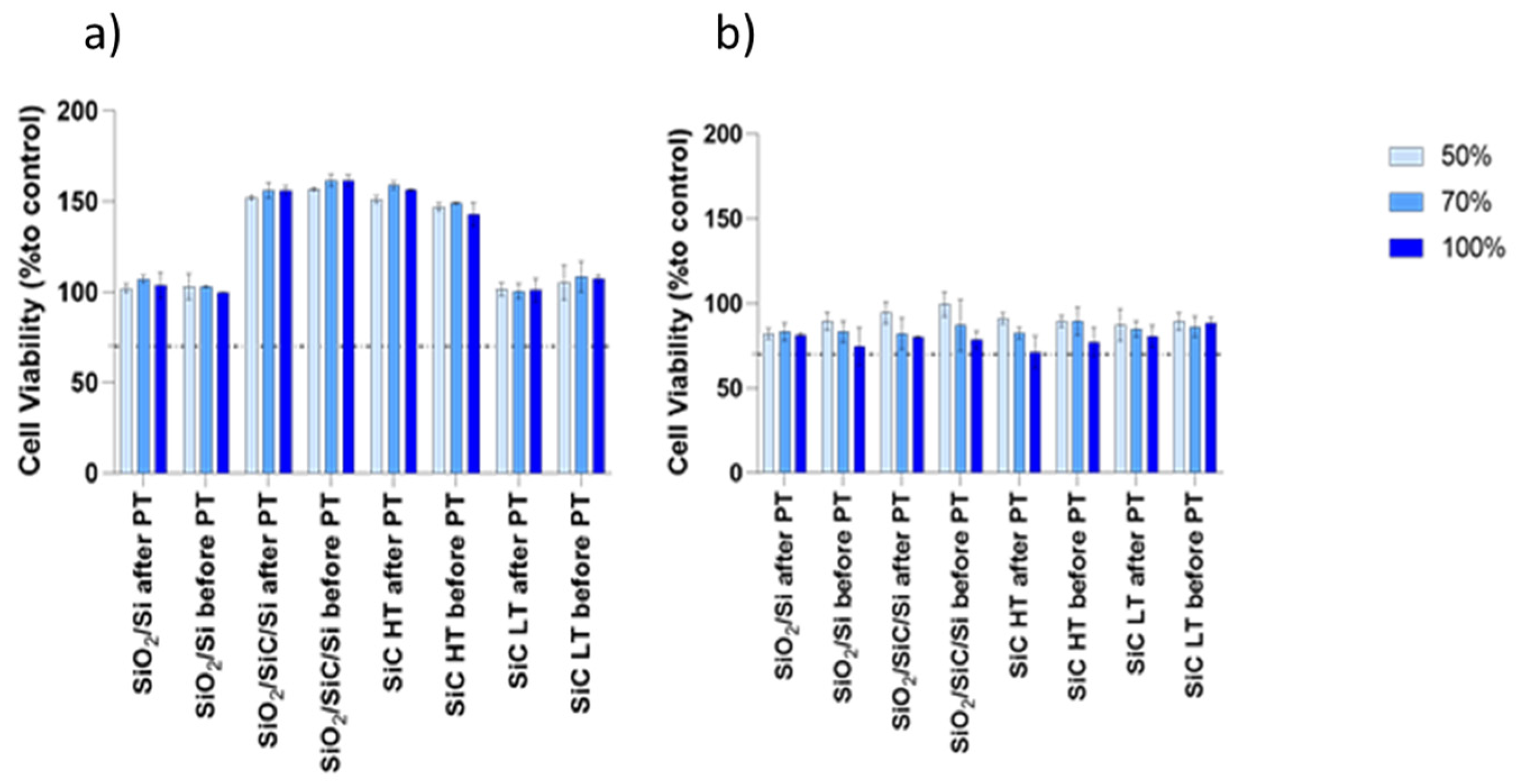
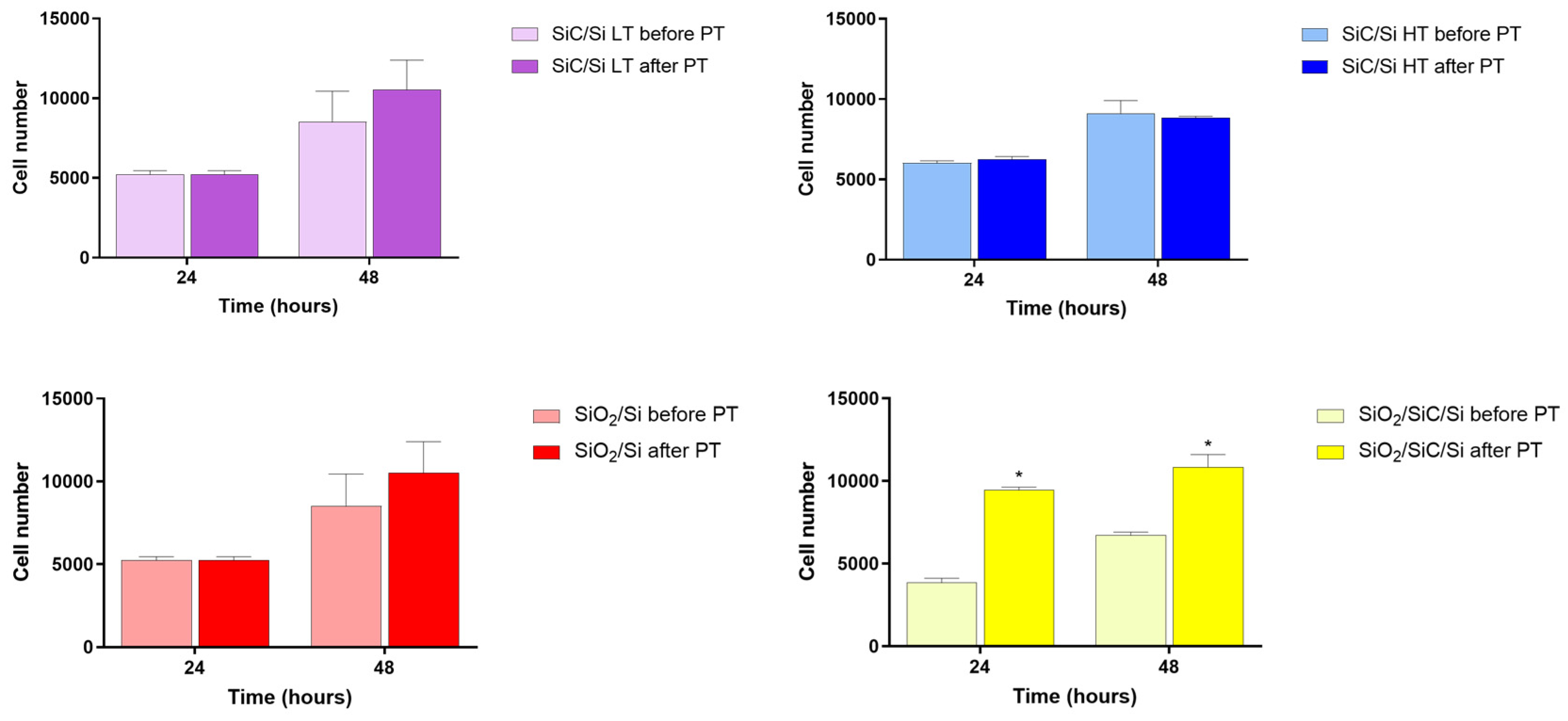
3.3. Biological Assays

In vitro results obtained with the L929 cell line confirmed the non-cytotoxic behavior of the analyzed surfaces at both the selected time points. Samples were submerged for 1 and 10 days in the culture medium, and the conditioned medium obtained was used to culture L929 cells. As shown in Figure 5, the percentage of viable cells on all the samples and during the whole experiment was higher than 70%, selected value for the assessment of non-cytotoxicity as per ISO guidelines 10993-5. Noteworthy, the viability of cells cultured in the medium conditioned for 1 day was higher when compared to the 10 days. Nonetheless, SiO2/SiC/Si and SiC HT samples showed higher values of viability, even if all the results were in the range of biocompatibility, without a clear change in the presence or in the absence of PT.
Lastly, osteoblastic cells MC3T3-E1 have been seeded onto the samples to preliminary assess their behavior with regards to SiC-based epitaxial layers. After 24 and 48 h, their proliferation has been analyzed through a chemiluminescent assay, as shown in Figure 6.
The proliferation of osteoblasts onto the epitaxial layers is not impaired by any of the surfaces, and the hydrogen plasma treatment seems to slightly increase cell number, especially at 48 h of culture, even if the p-values were not statistically significant for most of the samples, with the exclusion of SiO2/SiC/Si. Indeed, SiO2/SiC/Si showed a replicable behavior at both time points; the number of cells grown onto the treated surface was much higher if compared to the same sample before the hydrogen plasma treatment, probably an indication that the treatment is effective on the specific surface.

4. Discussion

In the analyzed epitaxial layers samples, we confirmed the presence of a cubical structure with good crystalline quality on SiC/Si LT, while the high-temperature SiC/Si appeared with a mirror-like smooth topography, typical of the antiphase domains. After PT, both their wettabilities appeared to be increased compared to the native silicon sample; this behavior might be attributed to the different roughness, as rougher surfaces generally present a smaller contact angle due to their enhanced hydrophilicity [17]. It is known that one of the factors influencing the contact angle value is the presence of impurities on the surface; in our case, residues as impurities due to the thermal decomposition of the precursors during growth can be adsorbed during the cooling phase in the reactor before the samples are removed. The residues from the decomposition of silane and propane can be mainly C-based [48]. The plasma treatment has a known cleaning action, and after this process, the surfaces become even more hydrophilic as the impurities due to residues of the gaseous phase that are deposited on the surfaces during the heating phase have been eliminated during the treatment. The same thing also appears in silicon oxide layers grown directly on the silicon (SiO2/Si), which showed a very flat sample with a contact angle that changes from 74.5° (before PT) to 21° (after PT). As it is evident, the as-grown SiO2/Si presents a higher contact angle if compared to the native SiO2; one of the causes of such a result might be recognized in the different water/oxide interaction, probably linked to the oxide thickness and to the formation of silanols on the SiO2 layer, a phenomenon already described in the literature, which should be further investigated [53,54]. The results relative to the SiO2/SiC/Si epitaxial structure underlined a drastic decrease in C.A., probably due to the presence of an amorphous SiO2 layer on the top of the sample. SEM observations of nanostructures topography showed a very similar structure for all of the samples, remembering an “open mesh” where nanowires tangled together on the silicon substrate. The nanowire samples have been tested for their hydrophilicity (Figure 4a). Interestingly, SiC NWs and SiO2 NWs in the absence of plasma treatment showed nonoptimal hydrophilicities (C.A. = 97.95° and C.A. = 14.9°, respectively), contrary to the core/shell NWs that presented zero-degree contact angles before the hydrogen plasma treatment. Nevertheless, all the samples exposed to hydrogen PT increased their wettabilities, presenting contact angles of about 0° (core/shell and SiO2 NWS C.A. = 0°, SiC NWs C.A. = 7.7°). After plasma treatment in hydrogen, the measured contact angle reaches zero. This can be ascribed to the presence of carbon in the outer part of the shell, which has probably been eliminated from the hydrogen present in the treatment. Even if SEM microphotographs showed a similar appearance of the surfaces, there is a diffused and not well-defined whitish stain attributable to residues of the catalyst on SiC NWs (Figure 4b), which is probably responsible for the difference in the contact angle between those and the SiO2 and SiO2/SiC samples; this hypothesis is corroborated by the measurement of contact angle after plasma treatment that is much lower (7.7°) due to the cleaning of the surface. Notably, the wettability of SiO2/SiC NWs is 0° even before the PT, thus indicating that the core–shell structure of silicon-carbide-based NWs might be the best one to be used with the as-grown characteristics, avoiding the use of specific techniques to ameliorate the behavior in biological conditions and facilitate protein interaction (i.e., thermal treatments, hydrogen plasma treatments, UVC, etc.). This aspect might be the key to the development of an innovative biomaterial with the topography of a nanostructured surface (surface topography is known to heavily influence the hydrophilicity) and a high wettability, which is fundamental in the early phase of biomaterial insertion in the human body and its interaction with the biological environment. As we already demonstrated, plasma proteins such as fibronectin can enhance the response of cells with regard to the biomaterial and facilitate osteoblastic differentiation and matrix deposition [17,55,56]. Moreover, all the samples showed good biocompatibility as per ISO guidelines for the cytotoxicity of porous biomaterials, thus encouraging the use of such silicon derivatives. Also, the preliminary assay for the proliferation of osteoblasts showed a slight increase after the use of hydrogen plasma treatment, which has been found as statistically significant only for the SiO2/SiC/Si surface. The amelioration in cell–surface interaction is the key to an appropriate cellular response leading to biomaterial osseointegration and bone regeneration. For sure, the next question we will need to answer will be the biological effects of the presented materials to assess their validation as cytocompatible three-dimensional scaffolds.

5. Conclusions

This study aimed to observe the change in wettability of epitaxial layers and nanowires of SiC, SiO2, and SiC/SiO2. Our results underline that both epitaxial layers and NWs samples after H2 plasma treatment presented a noteworthy decrease in the contact angle value, thus confirming the efficacy of hydrogen plasma effects on surface hydrophilicity and paving the way to the possible use of such a technique to adapt organic and inorganic materials as scaffolding biomaterials with applications in the field of tissue regeneration. Notably, the SiO2/SiC NW’s surface wettability has not been affected by the PT because of its as-grown super-hydrophilic behavior, and cell proliferation has been supported in all the selected cases. This observation can pave the way for the use of such core/shell nanowire-based biomaterials for applications such as the creation of scaffolds or membranes for bone regeneration due to their peculiar hydrophilicity, which is a fundamental aspect to be considered for cellular interactions. Moreover, it is important to underline that such results may have potential implications in a large number of biomedical implementations other than tissue engineering, such as for diagnostics, biosensor development, or drug delivery systems (for both prolonged release of drugs or targeted cancer therapy) [57]. The great advancements in the use of silicon-carbide-based biomaterials in biomedicine are fundamental as their hydrophilicity, besides all the other characteristics already discussed, is optimum for their use in contact with biological fluids and blood, leading to the overcome of many limitations of the actual surfaces. Furthermore, the possible use of silicon-based sensors for environmental surveys might be farsighted as these specific surfaces are resistant and durable to weather and hostile environments, as well as their possible implementation in the use of electronics.

Author Contributions

Conceptualization, B.G., M.B., M.N. and G.A.; methodology, P.L., P.M.R., C.C. and G.A.; investigation, B.G., M.N. and M.B.; resources, G.A., G.M.M. and S.L.; data curation, B.G., G.A. and S.L.; writing—original draft preparation, B.G.; writing—review and editing, G.A., S.L., P.M.R., C.C. and G.M.M.; supervision, G.M.M.; project administration, G.A.; funding acquisition, G.A., M.B. and G.M.M. All authors have read and agreed to the published version of the manuscript.

Funding

This research received no external funding.

Informed Consent Statement

Not applicable.

Data Availability Statement

The data presented in this study are available on request from the corresponding author.

Acknowledgments

The authors acknowledge the assistance of Ponraj J.S. (Department of Photonics, Tyndall National Institute, University of Cork, Cork, Ireland) and Rimoldi T. (Department of Mathematical, Physical and Computer Sciences, University of Parma, 43124 Parma, Italy) for their help in the analysis development and data analysis.

Conflicts of Interest

The authors declare no conflict of interest.

References

  1. Burg, K.J.; Holder, W.D., Jr.; Culberson, C.R.; Beiler, R.J.; Greene, K.G.; Loebsack, A.B.; Roland, W.D.; Eiselt, P.; Mooney, D.J.; Halberstadt, C.R. Comparative study of seeding methods for three-dimensional polymeric scaffolds. J. Biomed. Mater. Res. 2000, 51, 642–649. [Google Scholar] [PubMed]
  2. Gugliandolo, A.; Fonticoli, L.; Trubiani, O.; Rajan, T.S.; Marconi, G.D.; Bramanti, P.; Mazzon, E.; Pizzicannella, J.; Diomede, F. Oral Bone Tissue Regeneration: Mesenchymal Stem Cells, Secretome, and Biomaterials. Int. J. Mol. Sci. 2021, 22, 5236. [Google Scholar] [CrossRef] [PubMed]
  3. Bakhshandeh, B.; Zarrintaj, P.; Oftadeh, M.O.; Keramati, F.; Fouladiha, H.; Sohrabi-Jahromi, S.; Ziraksaz, Z. Tissue engineering; strategies, tissues, and biomaterials. Biotechnol. Genet. Eng. Rev. 2017, 33, 144–172. [Google Scholar] [PubMed]
  4. Girón, J.; Kerstner, E.; Medeiros, T.; Oliveira, L.; Machado, G.M.; Malfatti, C.F.; Pranke, P. Biomaterials for bone regeneration: An orthopedic and dentistry overview. Braz. J. Med. Biol. Res. 2021, 54, e11055. [Google Scholar]
  5. Battafarano, G.; Rossi, M.; De Martino, V.; Marampon, F.; Borro, L.; Secinaro, A.; Del Fattore, A. Strategies for Bone Regeneration: From Graft to Tissue Engineering. Int. J. Mol. Sci. 2021, 22, 1128. [Google Scholar] [CrossRef] [PubMed]
  6. Dec, P.; Modrzejewski, A.; Pawlik, A. Existing and Novel Biomaterials for Bone Tissue Engineering. Int. J. Mol. Sci. 2022, 24, 529. [Google Scholar]
  7. Carotenuto, F.; Politi, S.; Ul Haq, A.; De Matteis, F.; Tamburri, E.; Terranova, M.L.; Teodori, L.; Pasquo, A.; Di Nardo, P. From Soft to Hard Biomimetic Materials: Tuning Micro/Nano-Architecture of Scaffolds for Tissue Regeneration. Micromachines 2022, 13, 780. [Google Scholar]
  8. Teng, C.; Tong, Z.; He, Q.; Zhu, H.; Wang, L.; Zhang, X.; Wei, W. Mesenchymal Stem Cells–Hydrogel Microspheres System for Bone Regeneration in Calvarial Defects. Gels 2022, 8, 275. [Google Scholar] [CrossRef]
  9. Sun, L.; Guo, J.; Chen, H.; Zhang, D.; Shang, L.; Zhang, B.; Zhao, Y. Tailoring Materials with Specific Wettability in Biomedical Engineering. Adv. Sci. 2021, 8, 2100126. [Google Scholar]
  10. Lagonegro, P.; Trevisi, G.; Nasi, L.; Parisi, L.; Manfredi, E.; Lumetti, S.; Rossi, F.; Macaluso, G.M.; Salviati, G.; Galli, C. Osteoblasts preferentially adhere to peaks on micro-structured titanium. Dent. Mater. J. 2018, 37, 278–285. [Google Scholar] [CrossRef]
  11. Kamath, S.; Bhattacharyya, D.; Padukudru, C.; Timmons, R.B.; Tang, L. Surface chemistry influences implant-mediated host tissue responses. J. Biomed. Mater. Res. A 2008, 86, 617–626. [Google Scholar] [CrossRef]
  12. Nath, N.; Hyun, J.; Ma, H.; Chilkoti, A. Surface engineering strategies for control of protein and cell interactions. Surf. Sci. 2004, 570, 98–110. [Google Scholar]
  13. Wang, Y.X.; Robertson, J.L.; Spillman, W.B., Jr.; Claus, R.O. Effects of the chemical structure and the surface properties of polymeric biomaterials on their biocompatibility. Pharm. Res. 2004, 21, 1362–1373. [Google Scholar] [CrossRef] [PubMed]
  14. Ahmad, D.; van den Boogaert, I.; Miller, J.; Presswell, R.; Hussam, J. Hydrophilic and hydrophobic materials and their applications. Energy Sources Part A Recovery Util. Environ. Eff. 2018, 40, 2686–2725. [Google Scholar]
  15. Drelich, J.; Chibowski, E.; Meng, D.D.; Terpilowski, K. Hydrophilic and superhydrophilic surfaces and materials. Soft Matter 2011, 7, 9804–9828. [Google Scholar]
  16. Ghezzi, B.; Lagonegro, P.; Pece, R.; Parisi, L.; Bianchi, M.; Tatti, R.; Verucchi, R.; Attolini, G.; Quaretti, M.; Macaluso, G.M. Osteoblast adhesion and response mediated by terminal-SH group charge surface of SiOxCy nanowires. J. Mater. Sci. Mater. Med. 2019, 30, 43. [Google Scholar]
  17. Parisi, L.; Ghezzi, B.; Bianchi, M.G.; Toffoli, A.; Rossi, F.; Bussolati, O.; Macaluso, G.M. Titanium dental implants hydrophilicity promotes preferential serum fibronectin over albumin competitive adsorption modulating early cell response. Mater. Sci. Eng. C 2020, 117, 111307. [Google Scholar]
  18. Toffoli, A.; Parisi, L.; Bianchi, M.G.; Lumetti, S.; Bussolati, O.; Macaluso, G.M. Thermal treatment to increase titanium wettability induces selective proteins adsorption from blood serum thus affecting osteoblasts adhesion. Mater. Sci. Eng. C 2020, 107, 110250. [Google Scholar]
  19. Fernández-Rodríguez, M.; Sánchez Treviño, A.Y.; De Luna-Bertos, E.; Ramos Torrecillas, J.; García-Martínez, O.; Ruiz, C.; Rodríguez-Valverde, M.A.; Cabrerizo-Vílchez, M. Wettability and osteoblastic cell adhesion on ultrapolished commercially pure titanium surfaces: The role of the oxidation and pollution states. J. Adhes. Sci. Technol. 2014, 28, 1207–1218. [Google Scholar] [CrossRef]
  20. Kim, B.; Kim, M.; Yoo, S.; Nam, S.K. Atomistic insights on hydrogen plasma treatment for stabilizing High-k/Si interface. Appl. Surf. Sci. 2022, 593, 153297. [Google Scholar] [CrossRef]
  21. Zhang, H.; Kumagai, A.; Xu, G.; Ishibashi, K. Low-Temperature Atomic Hydrogen Treatment of SiO2/Si Structures. Jpn. J. Appl. Phys. 2003, 42, 6252. [Google Scholar] [CrossRef]
  22. Miyagawa, S.; Gotoh, K.; Ogura, S.; Wilde, M.; Kurokawa, Y.; Fukutani, K.; Usami, N. Effect of hydrogen plasma treatment on the passivation performance of TiOx on crystalline silicon prepared by atomic layer deposition. J. Vac. Sci. Technol. A 2020, 38, 022410. [Google Scholar] [CrossRef]
  23. Mews, M.; Conrad, E.; Kirner, S.; Mingirulli, N.; Korte, L. Hydrogen Plasma Treatments of Amorphous/Crystalline Silicon Heterojunctions. Energy Procedia 2014, 55, 827–833. [Google Scholar]
  24. Gupta, V.; Madaan, N.; Jensen, D.S.; Kunzler, S.C.; Linford, M.R. Hydrogen Plasma Treatment of Silicon Dioxide for Improved Silane Deposition. Langmuir 2013, 29, 3604–3609. [Google Scholar] [CrossRef]
  25. Parikh, A.N.; Liedberg, B.; Atre, S.V.; Ho, M.; Allara, D.L. Correlation of Molecular Organization and Substrate Wettability in the Self-Assembly of n-Alkylsiloxane Monolayers. J. Phys. Chem. 1995, 99, 9996–10008. [Google Scholar] [CrossRef]
  26. Yonenaga, I. Thermo-mechanical stability of wide-bandgap semiconductors: High temperature hardness of SiC, AlN, GaN, ZnO and ZnSe. Phys. B Condens. Matter 2001, 308–310, 1150–1152. [Google Scholar] [CrossRef]
  27. La Via, F.; Severino, A.; Anzalone, R.; Bongiorno, C.; Litrico, G.; Mauceri, M.; Schoeler, M.; Schuh, P.; Wellmann, P. From thin film to bulk 3C-SiC growth: Understanding the mechanism of defects reduction. Mater. Sci. Semicond. Process. 2018, 78, 57–68. [Google Scholar]
  28. Viguier, B.; Zor, K.; Kasotakis, E.; Mitraki, A.; Clausen, C.H.; Svendsen, W.E.; Castillo-Leon, J. Development of an electrochemical metal-ion biosensor using self-assembled peptide nanofibrils. ACS Appl. Mater. Interfaces 2011, 3, 1594–1600. [Google Scholar] [CrossRef]
  29. Tian, B.; Lieber, C.M. Design, synthesis, and characterization of novel nanowire structures for photovoltaics and intracellular probes. Pure Appl. Chem. 2011, 83, 2153–2169. [Google Scholar] [PubMed]
  30. Santavirta, S.; Takagi, M.; Nordsletten, L.; Anttila, A.; Lappalainen, R.; Konttinen, Y.T. Biocompatibility of silicon carbide in colony formation test in vitro. A promising new ceramic THR implant coating material. Arch. Orthop. Trauma Surg. 1998, 118, 89–91. [Google Scholar] [CrossRef] [PubMed]
  31. Hashiguchi, K.; Hashimoto, K. Mechanical and Histological Investigations on Pressureless Sintered SiC Dental Implants. Okajimas Folia Anat. Jpn. 1999, 75, 281–296. [Google Scholar] [CrossRef]
  32. Godignon, P. SiC materials and technologies for sensors development. Mater. Sci. Forum 2005, 483–485, 1009–1014. [Google Scholar] [CrossRef]
  33. Yakimova, R.; Petoral, R.M.; Yazdi, G.R.; Vahlberg, C.; Lloyd Spetz, A.; Uvdal, K. Surface functionalization and biomedical applications based on SiC. J. Phys. D Appl. Phys. 2007, 40, 6435–6442. [Google Scholar] [CrossRef]
  34. Huczko, A.; Dąbrowska, A.; Savchyn, V.; Popov, A.I.; Karbovnyk, I. Silicon carbide nanowires: Synthesis and cathodoluminescence. Phys. Status Solidi B 2009, 246, 2806–2808. [Google Scholar] [CrossRef]
  35. Zhao, Q.; Ju, B.; Zhao, K.; Zhan, J.; Liu, M.; Zhang, N.; Qian, J.; Xiu, Z.; Kang, P.; Yang, W. Effect of Reinforcement Size on Mechanical Behavior of SiC-Nanowires-Reinforced 6061Al Composites. Materials 2022, 15, 8484. [Google Scholar] [CrossRef]
  36. Monnink, S.H.; Van Boven, A.J.; Peels, H.O.; Tigchelaar, I.; de Kam, P.J.; Crijns, H.J.; van Oeveren, W. Silicon-carbide coated coronary stents have low platelet and leukocyte adhesion during platelet activation. J. Investig. Med. 1999, 47, 304–310. [Google Scholar]
  37. Li, X.; Wang, X.; Bondokov, R.; Morris, J.; An, Y.H.; Sudarshan, T.S. Micro/nanoscale mechanical and tribological characterization of SiC for orthopedic applications. J. Biomed. Mater. Res. B Appl. Biomater. 2005, 72, 353–361. [Google Scholar] [CrossRef]
  38. Lagonegro, P.; Rossi, F.; Galli, C.; Smerieri, A.; Alinovi, R.; Pinelli, S.; Rimoldi, T.; Attolini, G.; Macaluso, G.; Macaluso, C.; et al. A cytotoxicity study of silicon oxycarbide nanowires as cell scaffold for biomedical applications. Mater. Sci. Eng. C 2017, 73, 465–471. [Google Scholar] [CrossRef]
  39. Oliveros, A.; Guiseppi-Elie, A.; Saddow, S.E. Silicon carbide: A versatile material for biosensor applications. Biomed. Microdevices 2013, 15, 353–368. [Google Scholar] [CrossRef]
  40. Saddow, S.E. Silicon Carbide Materials for Biomedical Applications. In Silicon Carbide Biotechnology; Springer: Berlin/Heidelberg, Germany, 2012. [Google Scholar]
  41. Adamski, R.; Siuta, D. Mechanical, Structural, and Biological Properties of Chitosan/Hydroxyapatite/Silica Composites for Bone Tissue Engineering. Molecules 2021, 26, 1976. [Google Scholar] [CrossRef]
  42. Cacchioli, A.; Ravanetti, F.; Alinovi, R.; Pinelli, S.; Rossi, F.; Negri, M.; Bedogni, E.; Campanini, M.; Galetti, M.; Goldoni, M.; et al. Cytocompatibility and cellular internalization mechanisms of SiC/SiO2 nanowires. Nano Lett. 2014, 14, 4368–4375. [Google Scholar] [CrossRef] [PubMed]
  43. Coletti, C.; Jaroszeski, M.J.; Pallaoro, A.; Hoff, A.M.; Iannotta, S.; Saddow, S.E. Biocompatibility and wettability of crystalline SiC and Si surfaces. In Proceedings of the 2007 29th Annual International Conference of the IEEE Engineering in Medicine and Biology Society, Lyon, France, 22–26 August 2007; Volume 2007, pp. 5850–5853. [Google Scholar]
  44. Bosi, M.; Watts, B.E.; Attolini, G.; Ferrari, C.; Frigeri, C.; Salviati, G.; Poggi, A.; Mancarella, F.; Roncaglia, A.; Martínez, O.; et al. Growth and Characterization of 3C-SiC Films for Micro Electro Mechanical Systems (MEMS) Applications. Cryst. Growth Des. 2009, 9, 4852–4859. [Google Scholar] [CrossRef]
  45. Bosi, M.; Attolini, G.; Negri, M.; Frigeri, C.; Buffagni, E.; Ferrari, C.; Rimoldi, T.; Cristofolini, L.; Aversa, L.; Tatti, R.; et al. Optimization of a buffer layer for cubic silicon carbide growth on silicon substrates. J. Cryst. Growth 2013, 383, 84–94. [Google Scholar] [CrossRef]
  46. Attolini, G.; Rossi, F.; Negri, M.; Dhanabalan, S.C.; Bosi, M.; Boschi, F.; Lagonegro, P.; Lupo, P.; Salviati, G. Growth of SiC NWs by vapor phase technique using Fe as catalyst. Mater. Lett. 2014, 124, 169–172. [Google Scholar] [CrossRef]
  47. Dhanabalan, S.C.; Negri, M.; Rossi, F.; Attolini, G.; Fabbri, F.; Campanini, M.; Bosi, M.; Salviati, G. Effects of growth parameters on SiC/SiO2 core/shell nanowires radial structure. Mater. Sci. Forum 2013, 740–742, 494–497. [Google Scholar] [CrossRef]
  48. Attolini, G.; Rotonda, P.M.; Cornelissen, C.; Mazzera, M.; Bosi, M. Plasma Treatment of 3C-SiC Surfaces. Mater. Sci. Forum 2013, 740–742, 287–290. [Google Scholar] [CrossRef]
  49. Ghezzi, B.; Lagonegro, P.; Attolini, G.; Rotonda, P.M.; Cornelissen, C.; Ponraj, J.S.; Parisi, L.; Passeri, G.; Rossi, F.; Macaluso, G.M. Hydrogen plasma treatment confers enhanced bioactivity to silicon carbide-based nanowires promoting osteoblast adhesion. Mater. Sci. Eng. C 2021, 121, 111772. [Google Scholar] [CrossRef]
  50. Rossi, F.; Fabbri, F.; Attolini, G.; Bosi, M.; Watts, B.E.; Salviati, G. TEM and SEM-CL Studies of SiC Nanowires. Mater. Sci. Forum 2010, 645–648, 387–390. [Google Scholar] [CrossRef]
  51. Fabbri, F.; Rossi, F.; Attolini, G.; Salviati, G.; Dierre, B.; Sekiguchi, T.; Fukata, N. Luminescence properties of SiC/SiO2 core–shell nanowires with different radial structure. Mater. Lett. 2012, 71, 137–140. [Google Scholar] [CrossRef]
  52. Negri, M.; Rossi, F.; Attolini, G.; Fabbri, F.; Dhanabalan, S.C.; Boschi, F.; Bosi, M.; Nardi, M.V.; Salviati, G. Cubic Silicon Carbide Nanowires. In Exotic Properties of Carbon Nanomatter: Advances in Physics and Chemistry; Putz, M.V., Ori, O., Eds.; Springer: Dordrecht, The Netherlands, 2015. [Google Scholar]
  53. Thomas, R.R.; Kaufman, F.B.; Kirleis, J.T.; Beisky, R.A. Wettabilily of Polished Silicon Oxide Surfaces. J. Electrochem. Soc. 1996, 143, 643–648. [Google Scholar] [CrossRef]
  54. Martinez, N.Y.; Reidy, R.F. Wettability of silicon, silicon dioxide, and organosilicate glass. Master’s Thesis, University of North Texas Libraries, UNT Digital Library, Denton, TX, USA, 2009. [Google Scholar]
  55. Galli, C.; Parisi, L.; Piergianni, M.; Smerieri, A.; Passeri, G.; Guizzardi, S.; Costa, F.; Lumetti, S.; Manfredi, E.; Macaluso, G.M. Improved scaffold biocompatibility through anti-Fibronectin aptamer functionalization. Acta Biomater. 2016, 42, 147–156. [Google Scholar] [CrossRef] [PubMed]
  56. Parisi, L.; Galli, C.; Bianchera, A.; Lagonegro, P.; Elviri, L.; Smerieri, A.; Lumetti, S.; Manfredi, E.; Bettini, R.; Macaluso, G.M. Anti-fibronectin aptamers improve the colonization of chitosan films modified with D-(+) Raffinose by murine osteoblastic cells. J. Mater. Sci. Mater. Med. 2017, 28, 136. [Google Scholar] [CrossRef] [PubMed]
  57. Gatou, M.-A.; Vagena, I.-A.; Pippa, N.; Gazouli, M.; Pavlatou, E.A.; Lagopati, N. The Use of Crystalline Carbon-Based Nanomaterials (CBNs) in Various Biomedical Applications. Crystals 2023, 13, 1236. [Google Scholar] [CrossRef]
Figure 1. Schematic description of the different epitaxial layers deposited on silicon substrates (ac) and the corresponding nanowires (df).
Crystals 13 01280 g001
Figure 2. The panel shows AFM images of surface morphologies: (a) control silicon substrate, (b) SiC/Si LT, (c) SiC/Si HT, (d) SiO2/Si, (e) SiO2/SiC/Si, and (f) AFM roughness quantification of epitaxial layers. **** p < 0.0001 SiC/Si LT vs. SiC/Si HT, SiO2/Si, SiO2/SiC/Si, and Si; ° p < 0.0001 SiO2/Si vs. SiC/Si LT, SiC/Si HT, SiO2/SiC/Si, and Si.
Crystals 13 01280 g002
Figure 3. Wettability measurements showing the detected contact angles in the absence (a,d,f,h) or in the presence (c,e,g,i) of hydrogen plasma treatment of all the analyzed samples. In detail, (a) control silicon substrate; (b,c) SiC/Si LT flat surface; (d,e) SiC/Si HT flat surface; (f,g) SiO2/Si flat surface; (h,i) SiO2/SiC/Si flat surface; (l) wettability measurements in the presence (after) or in the absence (before) of hydrogen plasma treatment. § p < 0.0001 after PT vs. before PT.
Crystals 13 01280 g003
Figure 4. Panels showing the morphologies of nanowires and their wettabilities before and after PT. (a) Contact angles of analyzed nanostructures before and after plasma treatment § p < 0.0001 after PT vs. before PT; (b) SEM microphotographs of SiC NWs and (c,d) contact angle measurements before and after plasma treatment of SiC NWs; (e) SEM microphotographs of SiO2 NWs and (f) and (g) contact angle measurements before and after plasma treatment of SiO2 NWs; (h) SEM microphotographs of SiO2/SiC NWs and (i,l) contact angle measurements before and after plasma treatment of SiO2/SiC NWs.
Crystals 13 01280 g004
Figure 5. Indirect-contact cytotoxicity assay. L929 cells viability after 24 h of culture in conditioned medium at 1 day (a) and 10 days (b).
Crystals 13 01280 g005
Figure 6. Cell proliferation assay. MC3T3-E1 behavior after 24 and 48 h of proliferation in direct contact with the biomaterials. * p = 0.0489 SiO2/SiC/Si before PT vs. SiO2/SiC/Si after PT at 24 h and p = 0.0433 SiO2/SiC/Si before PT vs. SiO2/SiC/Si after PT at 48 h.
Crystals 13 01280 g006
Disclaimer/Publisher’s Note: The statements, opinions and data contained in all publications are solely those of the individual author(s) and contributor(s) and not of MDPI and/or the editor(s). MDPI and/or the editor(s) disclaim responsibility for any injury to people or property resulting from any ideas, methods, instructions or products referred to in the content.

