Review

Mimicking Natural-Colored Photonic Structures with Cellulose-Based Materials

by
Ana Rita Quelhas
1,2 and
Ana Catarina Trindade
3,4,*
1
CICS-UBI—Health Sciences Research Centre, University of Beira Interior, 6200-506 Covilhã, Portugal
2
Department of Chemistry, University of Beira Interior, 6201-001 Covilhã, Portugal
3
CENIMAT|i3N, Department of Materials Science, School of Science and Technology, NOVA University Lisbon, 2829-516 Caparica, Portugal
4
Atlântica—Instituto Universitário, Fábrica da Pólvora, 2730-036 Barcarena, Portugal
*
Author to whom correspondence should be addressed.
Crystals 2023, 13(7), 1010; https://doi.org/10.3390/cryst13071010
Submission received: 8 June 2023 / Revised: 16 June 2023 / Accepted: 20 June 2023 / Published: 25 June 2023

Abstract

:
Structural coloration has become a fascinating field of research, inspiring scientists and engineers to explore the vibrant colors observed in nature and develop bio-inspired photonic structures for various applications. Cellulose-based materials derived from plant fibers offer a promising platform for mimicking natural photonic structures. Their abundance, renewability, and versatility in form and structure make them ideal for engineering specific optical properties. Self-assembly techniques enable the creation of ordered, periodic structures at the nanoscale by manipulating the interactions between cellulose fibers through chemical modification or physical manipulation. Alternatively, additive manufacturing techniques like 3D printing and nanoimprint lithography can directly fabricate desired structures. By em-ulating natural photonic structures, cellulose-based materials hold immense potential for applications such as colorimetric sensors, optoelectronic devices, camouflage, and decorative materials. However, further research is needed to fully com-prehend and control their optical properties, as well as develop cost-effective and scalable manufacturing processes. This article presents a comprehensive review of the fundaments behind natural structural colors exhibited by living organisms and their bio-inspired artificial counterparts. Emphasis is placed on understanding the underlying mechanisms, strategies for tunability, and potential applications of these photonic nanostructures, with special focus on the utilization of cellulose nanocrystals (CNCs) for fabricating photonic materials with visible structural color. The challenges and future prospects of these materials are also discussed, highlighting the potential for advancements to unlock the full potential of cellulose-based materials with structural color.

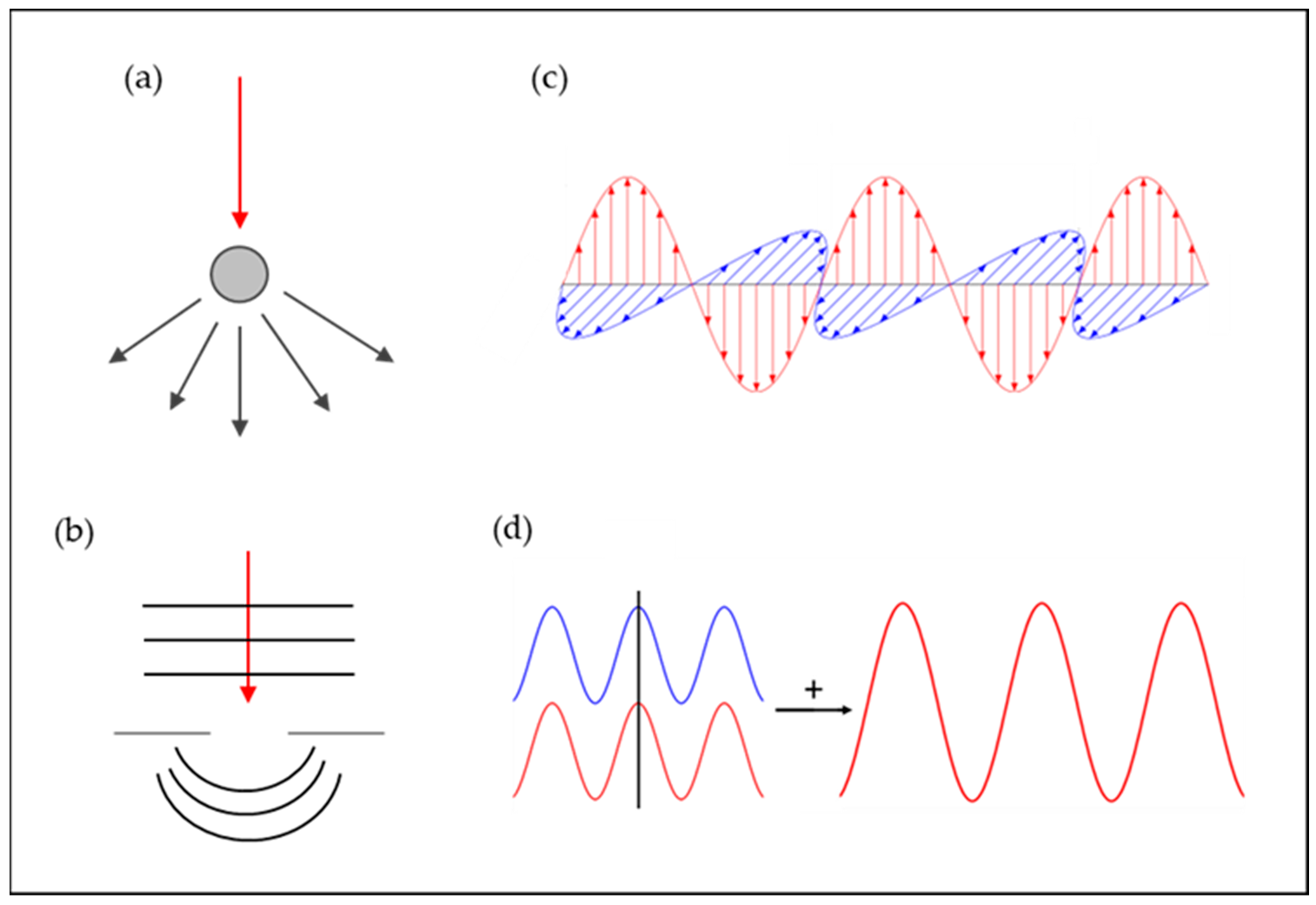
1. Soft Matter and Structural Color: Short Introduction

Materials science researchers have long looked to nature for inspiration due to the extraordinary multifunctional molecules and materials developed through evolution and natural selection. Natural creatures have evolved to establish distinctive functions, such as directional water collection on spider silk, antifogging properties of mosquito compound eyes, water capture and wing-locking devices of beetles, multicolor of butterfly wings, and superhydrophobicity and low adhesion of lotus leaves, among other examples [1].
Biological photonic structures, honed through countless generations of evolution, offer valuable insights for the development of innovative artificial photonic materials with multiple functions. Nature’s captivating and vibrant colors are not achieved through the application of pigments but rather by manipulating transparent materials at the nanoscale. For instance, plants can achieve stunning hues by arranging cellulose nano-fibers in their cell walls. This periodic arrangement enables various plant species to exhibit remarkably vivid colors spanning the entire visible spectrum, ranging from rich purples to vibrant reds.
In recent years, the study of structural colors has gained significant attention in various research fields due to the complex interactions between light and sophisticated microstructures found in nature [2]. This mechanism is responsible for some of the most stunning displays of color in nature, which can be observed in butterflies, moths, beetles, birds, fishes, plants, and fruits. Examples of such colors are, for instance, the Morpho butterfly’s wings have lamellar structures in their ridges, which reflect light strongly at a particular wavelength, resulting in its distinctive coloration [3].
The concept of “soft matter” was initially introduced by Pierre-Gilles de Gennes during his Nobel acceptance speech in 1991. It refers to a category of materials that lie between aqueous substances and ideal solids. These materials encompass a range of substances such as colloids, foams, liquid crystals (LCs), gels, polymers, and active matter [4,5]. In nature, soft matter materials serve as the fundamental building blocks of living systems. They possess the remarkable ability to self-assemble into functional structures and exhibit exceptional responsiveness to various environmental stimuli [6,7,8,9].
The interactions between the constituents of soft matter are relatively weak, resulting in a delicate balance between entropic and enthalpic contributions to the overall free energy. This delicate balance facilitates the spontaneous formation of microstructures at multiple length scales. The emergence of these structures is influenced by both the inherent properties of nanomaterials and the engineering of building blocks within a given spatial region. For example, chameleons demonstrate a rapid and reversible change in color patterns by actively arranging non-close-packed guanine nanocrystals within their skin in response to external stimuli. Over the years, significant advancements have been made in the development of soft materials with diverse and complex configurations, vibrant patterns, metastable states, and macroscopic softness. These materials have provided valuable insights and inspiration for addressing contemporary challenges across various fields, particularly in advanced optical and photonic technologies. They have propelled the progress of soft matter photonics.
When it comes to creating synthetic photonic crystal-based materials, the majority of studies have centered around the use of synthetic opals like SiO2 and polystyrene, as well as inverse opals, to achieve structural color [10]. However, it is not limited to synthetic materials for the production of photonic crystals. Biorenewable materials, such as cellulose and chitin nanocrystals, have shown promise in forming colored and iridescent materials.

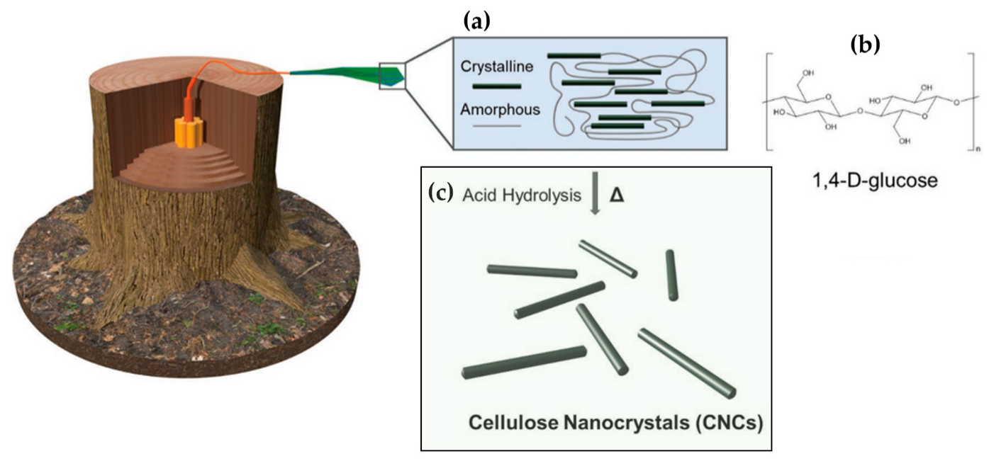
2. Cellulose and Cellulose Nanocrystals: Short Overview

The depletion of petroleum resources and its environmental impact, including global warming, has created a growing interest in developing sustainable materials. In this context, the field of materials science has been progressing towards prioritizing the replacement of highly polluting substances with alternatives that have a reduced environmental footprint. Bio-based materials have numerous advantages, including renewability, biodegradability, and environmental friendliness, making them a viable solution to environmental challenges [11,12] Cellulose, arguably the most abundant biopolymers available on our plant, is a renewable organic compound, fascinating and nearly limitless natural polymer that has been widely used in everyday products and applications. It is also considered an alternative to petroleum-based polymers due to its availability, low cost, low density, non-toxicity, low abrasiveness, biocompatibility, and biodegradability. Cellulose is used in various fields, such as automotive and construction industries, electronic components, sports, and leisure [12].
Cellulose is a robust and fibrous polysaccharide that is not soluble in water and can be found in the cell walls of various organisms, such as plants, bacteria, algae, and certain sea animals like tunicates [13]. Cellulose consists of a linear chain with hundreds of thousands of repeating unit β-1,4-linked D-glucose units. The basic building block is often considered to be a glucose dimer called cellobiose (Figure 1), arranged in an alternating pattern, with each unit inverted at a 180° angle, creating a main chain polymer through 1,4 glycosidic bonds that are formed by a condensation reaction. Usually, approximately 36 separate cellulose molecules come together to form larger entities known as elementary fibrils or protofibrils. These elementary fibrils or protofibrils then undergo further assembly to create microfibrils. It is the arrangement of these microfibrils that gives rise to the well-known cellulose fibers [14]. Cellulose contains multiple hydroxyl groups, which facilitate the formation of numerous hydrogen bonds between hydrogen and oxygen molecules, both within and between cellulose chains. These hydrogen bonds play a crucial role in holding the cellulose chains firmly together in the crystalline regions of the fibrils [15]. These microfibrils self-assemble into macrofibers and fibers, forming hierarchical structures, grouped into both crystalline and disordered regions, as illustrated in Figure 1b [16]. The average degree of polymerization ( D P ¯ ) differs depending on the source of cellulose. For instance, wood-derived cellulose has a D P ¯ of 10,000, while wool-derived cellulose has a D P ¯ of 15,000 [13].
The development of nanotechnology has stimulated interest in nanocellulose, as well as the attention given to bio-based materials that utilize cellulose. By reducing the size of cellulose to the nanoscale, nanocellulose has widened the scope of potential applications for this material. Cellulose nanocrystals, CNCs, (also referred to as nanocrystalline cellulose (NCC), cellulose nano whiskers (CNWs), or cellulose crystallites) are nontoxic, sustainable nanomaterials obtained from wood biomass via strong acid hydrolysis (sulfuric or hydrochloric). This process was first proposed by Rånby over half a century ago [19,20] and involves the selective chemical removal of the amorphous regions of the cellulose chain, thereby preserving the crystalline regions. Since the early introduction of these steps, the process has evolved and been applied to various wood and non-wood species [21,22,23,24,25,26,27,28,29].
Different types of nanocellulose structures can be obtained depending on the preparation method, shape, dimension, and function. The raw fibers must first undergo a specific pre-treatment process to remove the lignin and hemicellulose present in their raw state and isolate purified cellulose [30]. The pre-treatment process employed in this procedure should be tailored to the cellulose source and the desired morphology. Pulping, bleaching, oxidation, and enzymatic processes are some of the most commonly used pre-treatments. After the initial purification, the obtained fibers can undergo a specific process to obtain either cellulose microfibrils (CMFs) or cellulose nanocrystals (CNCs) [13]. Rojas and colleagues obtained Eucalyptus cellulose micro/nanofibers through three different processes: refining, sonication, and acidic hydrolysis of the cellulosic pulp [31]. They demonstrated that mechanical and chemical processes can be used to isolate the micro/nanofibers.
The dimensions of cellulose nanocrystals (CNCs), such as length and width, vary depending on the source of the cellulose microfibrils and the acidic hydrolysis conditions, such as time and temperature [10]. Nanocellulose has several advantages, such as high stiffness combined with low weight, an impressive surface area-to-volume ratio, a high aspect ratio, excellent mechanical properties (nano-strength), biodegradability, and the possibility of use as reinforcement [12]. The use of cellulose at the nanoscale allows the production of a new type of cellulose-based building block called nanocellulose, which enables the creation of multifunctional polymer nanocomposites [12]. Nanocellulose possesses several advantages, including a high aspect ratio, low density of 1.6 g·cm−3, and reactive hydroxyl side groups on its surface that facilitate the attachment of functional groups to achieve different surface properties. The CNCs exceptional mechanical properties, make them suitable for use in various fields, such as materials science, electronics, and medicine [11,32].

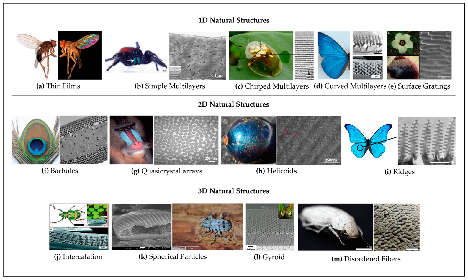
3. Photonic Structures in Nature

Structural coloration, observed in a wide range of organisms including animals, plants, and fruits, serves vital ecological functions. It plays a key role in attracting pollinators, signaling ripeness, deterring herbivores, gaining a competitive advantage, and manipulating light. This remarkable adaptation enhances reproductive success, species survival, and ecological interactions within diverse ecosystems. The phenomenon of structural coloration in natural systema has garnered significant attention from researchers and engineers in recent times, due to the captivating display of vibrant colors observed and the potential applications of bio-inspired functional photonic structures and materials. Numerous studies have been conducted to uncover and replicate the physical mechanisms responsible for the natural occurrence of structural colors in plants, fruits and animals [33,34,35,36] and revealed the self-assembling structural color in Nature [34,35] (see Figure 2).
Structural coloration in plants serves important functions related to their ecological interactions, and demonstrates their intricate interaction with their environment. From attracting pollinators to deterring herbivores and manipulating light, these visual cues contribute to the plant’s reproductive success, competitive advantage, and overall survival in their environment. The presence of vibrant and visually striking hues plays a crucial role in attracting pollinators, ensuring successful reproduction. Flowers employ structural colors, such as vivid petals and intricate patterns, as visual cues to signal the availability of nectar or pollen rewards, thus enticing pollinators like bees, butterflies, and birds. In 2009, Whitney et al. made a groundbreaking discovery regarding iridescence in Hibiscus trionum and revealed that the iridescence exhibited by this plant is a result of regular nanoscale patterns, such as striations or wrinkles, that are formed on the cuticle covering the flat epidermis of the petal’s surface [37,38]. These patterns act as diffraction gratings (see Figure 2e), leading to diffractive optical effects [37,39,40]. A similar iridescence phenomenon was observed in the H. trionum tulip species, where periodic striations are present on top of the purple-pigmented epidermis of the petal [41]. Conversely, the SEM image of the side of the tulip petal reveals an unorganized structure and lack of iridescence [42].
Whitney et al. emphasized the significance of these patterns in petals for biological purposes. They discovered that the iridescent signals produced by the H. trionum flower, through its diffraction gratings, allow it to interact with its main pollinators, particularly bumblebeesn [37]. Remarkably, the researchers successfully trained bumblebees to distinguish between replicas of iridescent H. trionum petals and identical non-iridescent replicas with smooth surfaces [37]. This is in accordance with the demonstration of Kevan and Lane that the microtextures present on the surface of flower petals serve as tactile cues for bees during pollination. They found that honeybees can differentiate between petals with distinct textures and detect variations in textures within petals of the same species [43].
The optical properties of the flower petals depend on the shape of the epithelial cells. Kourounioti et al. discovered that in order to generate iridescence through diffraction gratings, the epithelial cells must be planar and exhibit regular striations within the petal cuticle, with appropriate spacing between them. These striations or wrinkles can be either parallel or perpendicular to the long axis of the cells. Such variations in orientation can be observed within the same plant species and across different plant species. For example, the striations in H. trionum and Kalanchoe blossfeldiana were found to be parallel, while those in Yunnan rhododendron, Ursinia calendulifolia, and daisy were found to be perpendicular to the long axis of the cells [37,42,44].
Additionally, structural coloration in fruits serves as a signaling mechanism for ripeness. As fruits mature, they develop distinct hues that catch the attention of fruit-eating animals, indicating their readiness for consumption. This coloration facilitates seed dispersal, as animals consume the fruit and spread the enclosed seeds to different locations, contributing to the plant’s reproductive success. As structural colors do not fade, this bright coloration on fruits is maintained after the fruit is picked or has fallen from the plant, increasing its probability of being further dispersed [45,46,47].
Animals possess structural coloration for various purposes, including communication, camouflage, mate attraction, warning signals, and thermoregulation. Structural coloration serves as visual signals for species recognition, social interactions, and reproductive success. It aids in camouflage by blending with the environment or enhancing hunting abilities. In mate attraction, vibrant colors and patterns play a role in sexual selection. Some animals use bright colors as warning signals to deter predators, while structural coloration also assists in thermoregulation. These adaptations contribute to the survival, reproduction, and ecological interactions of animals. Similarly to plants and fruits, the mechanism behind structural colors in animals is based on diffraction and specular reflection and on nano and microscopic-scale patterns [48,49,50,51,52,53]. The majority of the incoming light travels through the biopolymeric formations largely unhindered; nonetheless, a specific set of wavelengths (those with specific ratios to the size of the structure’s periodic patterns) are selectively bounced back and generate angle-dependent iridescent shades found in various insects, birds, and marine creatures. Common examples of structural colors that appears in animals are peacock feathers [54,55,56,57], colorful birds [58,59,60,61], butterfly wings [62,63,64] and beetle exoskeletons [52,65,66,67,68,69].
Figure 2. Examples of natural materials that exhibits structural coloration. (Copyright (2022) Wiley. Adapted with permission from [35]). Summary of examples of natural material structures. 1D forms of structural color: (a) thin iridescence films in tropical wasp wings (Adapted with permission. [70] Copyright 2011, National Academy of Sciences, USA); (b) multilayers structure that appears in Phidippus johnsoni jumping spiders (Adapted with permission. [71] Copyright 2011, Elsevier); (c) chirped multilayers in Chrysina aurigans beetles (Adapted under the terms of the CC-BY Creative Commons Attribution 4.0 International license (https://creativecommons.org/licenses/by/4.0 accessed on 1 June 2023). [72] Copyright 2020); (d) sculpted and curved multilayers or bowls in Papilio Palinurus (Adapted under the terms of the CC-BY Creative Commons Attribution 4.0 International license (https://creativecommons.org/licenses/by/4.0 accessed on 1 June 2023)). [66] Copyright 2014); and (e) surface gratings in Hibiscus trionum flower (From [37]. Reprinted with permission from AAAS). Examples of 2D forms of structural color includes: (f) barbule nanostructure photonic crystals in Pavo cristatus (Adapted from [54] under the terms of CC BY 4.0 Creative Commons Attribution 4.0 International license (https://creativecommons.org/licenses/by/4.0 accessed on 1 June 2023)); (g) the Mandrillus sphinx face displaying quasi-ordered crystal arrays, balancing some long-range order with short-range disorder (Adapted with permission. [73] Copyright 2004, Company of Biologists); (h) the helicoid of Polia condensata fruit (adapted with permission from [47]); and (i) the ridge of the Morpho butterfly (Adapted under the terms of the CC-BY Creative Commons Attribution 4.0 International license (https://creativecommons.org/licenses/by/4.0 accessed on 1 June 2023). [72] Copyright 2020). 3D forms of structural color examples: (j) intercalation as in Trigonophorus rothschildi (Adapted with permission. [74] Copyright 2012, American Physical Society); (k) particle arrangements that appears in Pseudomyagrus waterhousei (Reprinted with permission from [75] Copyright 2011 by the American Physical Society); (l) the gyroid in Parides sesostris (Adapted with permission from [76]. Copyright 2018 American Chemical Society); and (m) disordered fibers in Cyphochilus beetles (Adapted under the terms of the CC-BY Creative Commons Attribution 4.0 International license (https://creativecommons.org/licenses/by/4.0 accessed on 1 June 2023). [77] Copyright 2014).
Crystals 13 01010 g002
These natural structures, such as the iridescent colors found in flowers and animals, are, as known, often created through mechanisms of scatter, diffraction, polarization and interference of light interacting with periodic micro- and nano-scale structures in the materials (Figure 3).

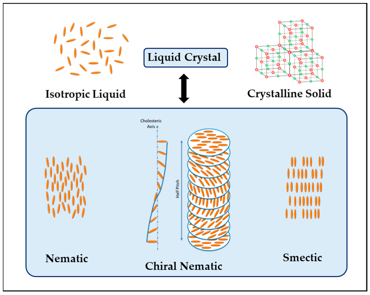
4. Liquid Crystalline Phases in Cellulose: Short Overview

Recently, cellulose has been found to be an ideal environmentally friendly material to mimic periodic micro and nanostructures that produce iridescence and structural coloration [82]. The extraction of CNCs using acidic hydrolysis generates a suspension of nanorods [83] and, under appropriate conditions and above a critical concentration, CNCs self-assemble in the suspension to form a chiral nematic liquid crystalline phase due to their rod-like shape (see Figure 4) [84]. Beck-Candanedo et al. mentioned Rånby and Ribi in the production of stable suspensions of colloidal-sized cellulose crystals by sulfuric acidic hydrolysis of wood and cotton cellulose in 1949 [85]. To date, CNCs have been extracted from a large variety of natural sources [11], spanning plants [19], bacteria [86,87,88], and tunicates [89,90,91].
Liquid crystals (LCs) are a distinct state of matter that exhibits optical characteristics similar to those of crystals, such as birefringence, while retaining the mechanical properties of liquids. There are various types of LC structures, including the nematic (N), smectic (Sm), and cholesteric (Ch, N*) (Figure 4), which have been widely studied and documented in literature [92].
The nematic structure is characterized by long-range orientational order of the molecules along a preferred direction or “director” (n), without any positional order, unlike layered structures [93]. Cholesteric or chiral nematic structures are formed by pseudo-layers of molecules aligned with the director (n), with the orientation of each layer rotated by a fixed angle around the cholesteric perpendicular axis, as depicted in Figure 4 [94]. The pitch (P) of a chiral nematic structure, defined as the distance over which the director makes one complete rotation of 360°, typically falls within the range of 0.4–0.8 μm, corresponding to visible light wavelengths. If the pitch of the chiral nematic structure is comparable to the wavelength of visible light, it reflects circularly polarized light, resulting in an iridescent appearance that changes with the viewing angle [15].
The de Vries equation describes the dependence of the reflected wavelength on the angle of incidence:
λ = n P sinθ
where θ represents the angle of light incidence; λ the reflected wavelength by the sample; n the average refractive index of the sample (cholesteric or chiral nematic) phase; and P the value of helical pitch [95]. The pitch, defined by the distance required for the director to complete a full turn is a function of both the temperature and concentration [96].
Chiral nematic liquid crystals, with a pitch comparable to the wavelength of visible light, exhibit the ability to reflect circularly polarized light of the same handedness as their chiral nematic phase [94]. As a result, when viewed from different angles, these materials display an iridescent effect due to the wavelength of the selectively reflected light varying. The iridescent appearance of chiral nematic liquid crystals is attributed to the change in the reflected wavelength based on the angle at which light is incident upon them. The chiral nematic helix possesses a specific internal handedness, and just like the pitch of the helix influences light reflection, the handedness also impacts the optical properties of these materials. Consequently, one can anticipate that circularly polarized light will be differentially affected by the handedness of the helix [97,98]. The formation of iridescent liquid and solid phases by cellulose derivatives has been well-established for a considerable time. The CNCs chiral nematic structure reflects left-handed circularly polarized (LCP) light due to the rotation of the director in each successive layer. On the other hand, cholesteric structures with a right-handed helix reflect right circularly polarized (RCP) light, while those with a left-handed helix reflect LCP light [99]. Godinho et al. presented a photonic structure based on cellulose that reflects both RCP and LCP light and can be adjusted by changing the temperature and applying an external electric field [100].
The presence of asymmetric carbon atoms in the anhydroglucose units of cellulose-based liquid crystals accounts for their optical activity in chiral nematic systems. Various cellulose derivatives with substituents attached to the hydroxyl groups of cellulose also exhibit liquid-crystalline phases, for instance: cellulose phenylcarbamate (CPC), ethyl cellulose (EC), and hydroxypropylcellulose (HPC). The existence of the liquid-crystalline phase is influenced by several key factors, including the type of substituent, molecular weight, and average degree of substitution (DS, which represents the average number of hydroxyl substituents per anhydroglucose unit). Substituents capable of further chemical functionalization, as seen in HPC and CPC, allow for continuous growth of side chains. The molar substitution denotes the average number of substituents per anhydroglucose units, encompassing all substitutions rather than solely those replacing hydroxyl groups on the cellulosic main chain. Cellulosic materials with high DS values (ranging from 2 to 3), dissolved in suitable solvents at sufficiently high concentrations, form lyotropic liquid-crystalline phases [101,102,103].
This manuscript focus on the development of structural coloration in aqueous solutions of cellulose-bases materials and their consequent films and so only liquid crystalline cholesteric case is referred. However, interesting studies on the properties of cellulose in different solutions and host matrices and their anisotropy can be also essential to many applications. One example is the work of Fujisawa et al. [104] related to the study of nanocellulose-doped starch–polyurethane nanocomposite biodegradable films via direct contact measurement of thermal properties. For the first time, thermal diffusivity was investigated in this polymer nanocomposite.

5. Photonic Structures in Cellulose-Based Materials

Natural structural colors serve as fascinating examples of nature’s ability to achieve functionality by shaping and molding inherently non-functional compounds into precisely defined structures [36,48]. Similar principles of creating functionality through structural design have become fundamental in the field of nanomaterials and nanotechnology over the past 40 years [105]. The resulting materials, often referred to as metamaterials, consist of conventional substances such as metals, semiconductors, ceramics, or polymers, yet they exhibit unconventional properties due to their nanostructures. For instance, by synthesizing semiconductors in the form of nanometer-sized spheres or rods, their electronic and optical properties can be dramatically altered. Similarly, transforming a transparent ceramic into a colored coating can be achieved by structuring it with a three-dimensional periodic architecture featuring a lattice spacing of a few hundred nanometers. Alternatively, a polymer film can be converted into a super-hydrophobic, self-cleaning surface by introducing hierarchically organized nano-to-micrometer-sized protrusions and filaments.
In nature, the most brilliant example of blue coloration stems from a helical arrangement of cellulose fibers in tropical fruits such as Pollia condensata (Figure 2h) and Margaritaria nobilis [47,106,107,108]. Mimicking these natural-colored photonic structures and getting inspiration for material design and sustainable processing using natural materials is an interesting area of research [109] and cellulosic materials have gained considerable prominence due to their renewable nature, compatibility with living organisms, and ability to degrade naturally, making them an eco-conscious alternative [110].
Revol et al. made a surprising finding when they stumbled upon the fact that a solid film could retain the chiral nematic liquid crystalline arrangement observed in a suspension of cellulose nanocrystals [23,111]. The researchers accomplished this by evaporating the water from a cellulose nanocrystal suspension with a concentration of approximately 3.5% by weight, resulting in a film with preserved chiral nematic order (Figure 5). Upon examination using polarized optical microscopy (POM), they observed that the films exhibited birefringence, and their structures were susceptible to disruption by shear forces. Films made from CNCs have unique optical properties, including iridescence and the ability to selectively reflect left circularly polarized light while transmitting right circularly polarized light [100,112,113]. Similar to a suspension of cellulose nanocrystals, the chiral nematic orientation within the solid films yielded a positive signal when subjected to CD spectroscopy [114]. The helical twist of the cellulose nanocrystals consistently displayed a left-handed nature, as confirmed by the positive CD signal for transmitted light and the generation of left-handed circularly polarized light upon reflection.
Films formed through the self-assembly of cellulose nanocrystals often exhibit captivating colors due to the helical arrangements of cholesteric liquid crystals. The orientation and pitch of these structures determine the photonic bandgap, with the ability to modify the pitch allowing for alterations in perceived color. When the helix’s pitch approximated the wavelengths of visible light, the CNC films exhibited striking iridescent structural colors. These chiral nematic CNC films can be considered as one-dimensional photonic crystals and this manipulation relies on Bragg-like reflections that generate various colors [82,115,116,117,118].
The precise optical properties that can be developed in CNC films depends on many parameters, which leads to the development of a variety of tunable photonic CNC materials and technologies. The effects of water evaporation (evaporation at different relative humidity) and initial CNC concentration have been investigated by several groups [18,82,113] and it was possible to obtain CNC films covering most the visible spectrum [119]. Later, Tran et al. [113,120] modified the evaporation time of CNC suspensions and obtained CNC films in the suspensions´ slow evaporation resulted in blue-shifted films. Utilizing differential evaporation, CNC films with gradients could be designed. They also discovered that the application of a cellulose acetate mask on top of a drying CNC suspension led to patterns with higher resolutions [113]. They also showed that the obtained colored patterns could be tuned, from red to blue, depending on the stage of self-assembly when the masks were applied.
Despite their capacity to generate films with vibrant colors, cellulose nanocrystals (CNCs) possess certain constraints that result in limited productivity. The process of self-assembly is highly susceptible to disruptions and may necessitate an extended period of several days for the complete evaporation of water. Chen et al. [121] developed a protocol consisted in a preliminary treatment of CNC suspensions through ultrasonication. They discovered that the duration of ultrasonication, volume of the suspension, and the application of vacuum were determinant in the preparation of iridescent CNC films. The resulting films exhibit striking and vibrant colors, when compared with those obtained with slower water evaporation techniques. Another influent parameter that was investigated was the surface upon which the CNC suspension is cast on [122,123,124]. Several surfaces were tested (including aluminum, silicon wafers, mica and polystyrene) and different optical properties were obtained, meaning that substrate surface properties, such as wettability and hydrophobicity, influence the self-assembly behavior of the CNCs.
Not only altering CNC suspensions can have impact on the characteristics of chiral nematic CNC films. Also, external factors, including temperature and additives, can have influence on the properties of CNC films. The introduction of energy through methods such as heating or sonication gave rise to CNC films with an enhanced helical pitch, allowing the production of films with adjustable chiral photonic properties [125,126,127,128,129].
As previously mentioned, cellulose possesses attractive qualities for optical and photonic applications, thanks to its refractive index, transparency, dielectric properties, and birefringence [130,131,132]. These combined characteristics enable the development of relevant technologies in the field of photonics. The crystal structure of cellulose plays a crucial role in modulating its optical properties, resulting in vibrant colors and establishing it as a valuable contender for sustainable bio-based optical materials. The self-assembly of cellulose nanocrystals offers a promising and cost-effective approach to producing optical materials on a large scale [74]. Given that sustainability and the circular economy are crucial concerns today, several scientists are exploring the potential use of cellulose derived from biomass and waste materials in photonic and its capacity to form chiral nematic structures upon drying, which exhibit fascinating optical and photonic properties [133,134,135,136,137]. Recent investigations in this field have primarily concentrated on understanding the self-assembly dynamics of helicoidal structures and optimizing them to achieve desired polarization responses.
Cellulose nanocrystals in water suspensions behave as lyotropic liquid crystals forming a chiral nematic phase above a critical concentration. It is well known that such an organization can be retained in solid films and give rise to an intense colored appearance. In several studies, researchers have characterized their optical response via optical and scanning electron microscopy, imaging scatterometry, and angle-resolved reflectance measurements [138,139,140]. Wilts et al. go further by showing that the experimental results can be well explained by computational modeling using the finite-difference time-domain method [140]. They performed 3-D finite-difference time-domain (FDTD) calculations, using a commercial-grade Maxwell equation solver, Lumerical FDTD Solutions 8.16, to simulate the polarization-dependent light scattering from a liquid-crystalline, helicoidal stack of cellulose in the wavelength range 350–700 nm. They examined the variation in reflectance with changing angles of a cellulose film formed through self-assembly and their findings demonstrate that the significant disparity in circular polarization is maintained across a wide range of incident light angles. This reflectance behavior can be effectively explained through the use of finite-difference time-domain modeling. Previous research has indicated that the color characteristics of these films can be manipulated by adjusting the self-assembly conditions of CNCs. Together, these studies contribute to a more holistic understanding of the angle-dependent color appearance in helicoidal layers, which holds potential for the development of sustainable colored materials, such as responsive dyes or food colorants.

6. Cellulose-Based Composite Materials with Structural Color

As mentioned before, the self-assembly of CNCs into a chiral nematic structure is tolerant to additives, which has allowed incorporation of additives and enabled the creation of a range of interesting materials, such as thin-films [141,142], hydrogels [143,144,145,146,147,148], and organosilicas [149,150,151] (Figure 6). Incorporating additives provides a means to fine-tune the optical and mechanical properties of the resulting CNC-based materials. For instance, pure CNC thin films are known for their toughness but lack flexibility, besides by introducing hydroxypropyl cellulose (HPC) or chitosan/chitin into the matrix, the cellulose-based composite material’s flexibility can be significantly improved [152,153,154,155,156,157,158]. Previous research in this field has also explored the incorporation of inorganic materials like metallic nanoparticles [159,160,161,162], infiltration of proteins or amino acids [163,164,165,166] or the addition of organic units through careful surface modification [153,167], resulting in materials that exhibit unique chiroptical properties and are capable of changing color under applied pressure [152,154,155,168,169,170,171,172,173].
The optical properties of films are also dependent on surface roughness, as no surface of a biobased material is completely flat and, as shown previously, its surface roughness will directly impact its interaction with light [174]. In fact, surface patterning and successful production of highly precise structures in a predeterminate configuration can be employed to create light interactive-structures, where nanoscale and microscale patterns are generated to control diffraction, scattering, or light outcoupling. Various lithographic techniques, involving similar steps but differ in processing and curing specifics, can be applied. Initially, the material to be modified is uniformly distributed across a surface, followed by the application of a mask or mold, and finally, through chemical and/or physical treatments, the modified surface is obtained [175,176]. Several protocols process of obtaining modified surfaces on structural colored cellulosic-based materials using photolithography [174,177], soft lithography [178,179,180], and nanoimprint lithography [181,182,183,184] were documented.
Wolfberger et al. [177] described an easy and versatile efficient patterning method for cellulose thin films by means of photolithography and enzymatic digestion. Depending on the conditions of development, either negative and positive type cellulose structures can be obtained, offering lateral resolutions down to the single-digit micro meter range by means of contact photolithography. These photochemically structured cellulose thin films are successfully implemented as dielectric layers in prototype organic thin film transistors.
The research findings of Mihi et al. [178] introduced a groundbreaking approach to fabricating photonic crystals and plasmonic structures using a derivative of cellulose through the nanostructuring method known as soft lithography. Through the periodic nanostructuring of the cellulose film, its transparency is effectively eliminated, leading to the emergence of vibrant colors in its reflective properties, contingent upon the specific pattern employed during the molding process. By leveraging this innovative technique, which is both highly scalable and cost-effective, as an alternative to the conventional self-assembly of cellulose nanocrystals, a superior nanostructure is rapidly and reproducibly generated on the polymer substrate [185]. This process offers a wide spectrum of iridescent colors solely reliant on the size and morphology of the resultant structures. The resulting photonic crystals can be nanoimprinted onto diverse substrates to confer photonic capabilities on surfaces lacking this characteristic, such as paper. This technology exhibits immense potential as photonic ink and finds practical applications in domains like anti-counterfeiting technology, packaging, decorative paper, labels, and sensors, among others [174]. When these structures are coated with a thin layer of metal, they acquire plasmonic properties while retaining their flexibility, thereby intensifying the colors displayed. Additionally, the biodegradability and water solubility of the cellulose derivative can be adjusted based on the specific type employed. These plasmonic structures are ideal for disposable sensors, enabling Raman emission, or enhancing the light emitted by a dye [178].
Figure 6. (a) CNC/HPC mixed in different weight ratios demonstrating the ability to tune the reflected color across the visible spectrum. Insets are of the chiral nematic structure at low and high amounts of CNC, scale bar = 3.5 cm. (Adapted with permission from [155]. Copyright 2020 American Chemical Society). (b) Photographs of solid CNC films containing different amounts of glycerol (G) (Reprinted with permission from [186]. Copyright 2018 American Chemical Society). (c) Photographs showing the structural colors of CNC/GlU (glucose) films (6 cm in diameter) with different weight percentage (wt%) compositions (CNC/GLU = 100/0, 66/34, 55/45, 52/48, 46/54 or 39/61) with UV–vis transmission spectra. (Adapted with permission under a Creative Commons Attribution 4.0 International License from [187]). (d) Structural color generated by micropatterned cellulose film. (Adapted with permission from [131], under available under Creative Commons license).
Crystals 13 01010 g006
In industrial settings, thermal imprinting has been implemented using roll-to-roll processing, which can also be utilized in thermal nanoimprinting lithography. Mäkelä et al. conducted experiments using a laboratory-scale roll-to-roll imprinting system to create cellulose acetate films, CNF films, and TEMPO-CNF films with pillar structures imprinted using a Ni-mold [181,182,184]. The formation of these structures was heavily influenced by the temperature, speed, and pressure applied during the process. The resulting films exhibited varying levels of surface roughness, leading to different levels of transparency. When white light passed through the microstructures, diffraction colors such as blue, red, and green were observed, showcasing the potential for applications in optics and electronics [181,182,183].
Considering the cost of production and application in lithography, high-throughput techniques like soft lithography and roll-to-roll lithography tend to be more cost-effective, while photo and e-beam lithography are often more expensive due to their limited scalability [188]. However, it should be noted that different surface patterning methods present varying restrictions on the achievable feature size of the fabricated surface structures.

7. Light Responsive and Color-Stimuli-Responsive Cellulose-Based Materials

Stimuli-responsive displays exhibit a change in their properties (such as shape, wettability, adhesion, optical, electrical, thermal, and mechanical properties) when exposed to external stimuli [189]. These displays hold significant potential for a wide range of applications, including on-demand drug delivery, tissue regeneration/repair, biosensing, smart coatings, artificial muscle drug delivery, diagnostics, biosensors, and textiles [189,190]. Within the field of stimuli-responsive materials, there is a growing interest in biocompatible and biodegradable materials based on lignin [191], hemicelluloses [192], and cellulose [193,194,195]. Among these stimuli-responsive materials, cellulose has been the most extensively studied for light stimulation and photo-responsive applications (Figure 7).
Considering the limited light absorption of cellulose in the visible spectrum, cellulose-based materials with light-responsive properties can be created by modifying cellulose with a light-responsive molecule or by incorporating light-responsive polymers, leveraging supramolecular interactions. Light-responsive cellulose materials have been developed through derivatization or graft copolymerization of a chromophore (such as spiropyran or coumarin) onto the cellulose backbone (or its derivatives) [196,197]. These light-responsive cellulose materials exhibit optically active sites that find applications in photo recording devices, liquid crystal displays, and other light-sensitive applications [198].
It is possible to produce dynamic optical or phototunable materials that undergo color changes when exposed to a specific light wavelength, by modifying cellulose and cellulose derivatives [176,199]. Optical materials that respond to multiple wavelengths can be achieved via electrostatic interactions between light-responsive cellulosic materials and carbon nanodots [200]. Ai et al. combined photo-responsive cellulose with carbon nanodots and were able to produce photo-triggering and reversible chromic materials, that responded to both UV and visible light. Light-responsive cellulosic materials containing imidazole salt groups and spiropyran groups were immobilized on the surfaces of negatively charged carbon nanodots. In this system, the carbon nanodots absorbed UV light (365 nm) and emitted blue fluorescence. With increasing UV irradiation time, the intensity at 460 nm (blue fluorescence) decreased and the intensity at 658 nm (red fluorescence) increased. The material eventually became fully red as a result of this photoinduced fluorochromic phenomenon. Finally, by changing the concentration of carbon nanodots in the ink, different colors and shades were obtained, imparting high versatility to these materials [200,201].
It is also possible to adjust the mechanical and optical properties of thin, transparent cellulosic films by altering the precursor liquid crystalline properties and adding CNCs. As described by Fernandes et al. [99] cellulosic films can be produced with customized mechanical and structural color characteristics, resembling the natural structures found in flower petals.
Zhang et al. proposed a printable structural color ink composed of cholesteric cellulose liquid crystals together with gelatin and a thermal-responsive hydrogel [151]. The ink exhibits vivid structural colors and printability due to its constituents. Based on this, Zhang and co-workers print a series of graphics and three-dimensional (3D) objects with vivid color appearances. The HPC cholesteric mesophase contributes to structural coloration and the incorporation of gelatin improves the printability of the ink. Combined with in situ photo-crosslinking of this polymer, the cholesteric mesophase can be retained in the gel state, resulting in vivid metallic structural colors of the HPG hydrogel. Due to the thermal responsiveness of both HPC and PACA polymer, these printed objects show visible color changes around body temperature (Figure 7c). Zhang et al. developed a 3D printing setup and fabricate a series of graphics and 3D photonic objects, even with multiple colors. Since it is able to be processed as liquid at increased shear rates and relax back to a self-supporting gel state with the reduced shear, it is therefore versatile for large-scale production with well-established industrial processing techniques. The main constituents of the HGP ink are HPC and gelatin, which are edible, cost-effective, and biocompatible. Further modifying the formulation of the ink can be studied, such as replacing the responsive hydrogels with natural derived alternatives and this system can be extended to daily-life commodities including colorant-free decorations in food industry, drugs, and cosmetics, as well as wearable biosensors or customized bionic skins. These performances, together with the biocompatibility of the constituents, indicate that the present ink represents a leap forward to the next-generation environmentally friendly 3D photonic printing and would unlock a wide range of real-life applications.

8. Tapping the Engineering and Industrial Potential

As described in previous paragraphs, structural coloration refers to the phenomenon where colors are produced by the physical structure of an object rather than by pigments or dyes. Examples of natural beauty, such as the brilliant-blue feathers of a kingfisher, iridescent hues of butterfly wings, and metallic shimmering chitin covers of beetles, are based on structural color. Mimicking them is both a desire and a huge challenge for chemical manufacturers, as the structures are complex and the use of new technologies in coloration have been explored for industrial purposes, mimicking the color brilliance of nature in a more efficient and economical way. Due to its versatility, structural color can be used in many ways, especially to ensure long-lasting colors for applications ranging from coatings to cosmetics.
One of the main applications of structural coloration is related with anti-counterfeiting measures. Structural coloration can be used to create unique and intricate patterns that are difficult to replicate, making it an effective tool for anti-counterfeiting measures. These patterns have been laboratory and prototype tested and can be incorporated into currency, identification cards, and valuable products to ensure their authenticity. References [201,202,203,204,205,206,207,208,209,210] provide some examples of prototypes and demonstrations of cellulose-based materials with structural colors in anti-counterfeiting applications and provide more information on the fabrication methods, properties, and potential applications of these materials in the industry.
Structural coloration can also be utilized in displays and signage to create vibrant and eye-catching visual effects. By manipulating the structure and arrangement of microscopic elements, such as nanostructures or photonic crystals, it is possible to produce colors that are more vibrant and stable than those achieved with traditional pigments or dyes. Some examples and laboratorial prototypes of cellulose-based structural colored displays can be found in references [18,139,211,212,213,214].
Color is a vital element in decoration and architecture, serving as a powerful tool to create atmosphere, express emotions, and shape perception. It enables individuals to showcase their personal style and personality while defining the ambiance of a space. Colors have the ability to influence mood, energy levels, and productivity, with each hue evoking different emotions and setting specific atmospheres. The strategic use of color can visually alter the size and proportion of a room and direct attention to focal points, establishing a visual hierarchy. In essence, color plays a vital role in decoration and architecture and the incorporation of structural coloration into architectural elements, interior design, and decorative applications have been tried and performed worldwide. By integrating materials with specific structures, it is possible to create surfaces that exhibit different colors depending on the angle of observation, resulting in visually striking and dynamic environments [215]. Some examples demonstrate how structural coloration can be incorporated into architectural and design elements all over the world, adding a dynamic and visually captivating dimension to the built environment, are: the Lotus Temple, New Delhi, India (the temple’s exterior is composed of white marble panels that feature a series of intricate, petal-like shapes, resulting in an iridescent effect that changes with the position of the sun); Elbphilharmonie, Hamburg, Germany (the Elbphilharmonie concert hall boasts a glass facade that showcases a striking interplay of colors; the surface of the glass panels is textured with small, geometric shapes that reflect and refract light, and the building’s appearance changes depending on the angle of view and the lighting conditions); Beijing National Aquatics Center (Water Cube), Beijing, China (an iconic structure from the 2008 Olympic Games, features a unique façade made of lightweight ethylene tetrafluoroethylene cushions; these cushions have a pattern of air-filled cells that scatter and reflect light and, as a result, the building’s surface exhibits a shimmering effect with ever-changing hues); Barcelona Pavilion, Barcelona, Spain (the pavilion’s interior walls feature polished and veined marble panels, which have a unique structural arrangement, that creates a subtle iridescent effect, adding depth and visual interest to the space); Opéra Bastille, Paris, France (has a distinctive façade composed of large glass blocks with an irregular surface texture, causing light to refract and create a play of colors; so, depending on the angle and intensity of the sunlight, the building appears to change colors, ranging from blue to green to golden hues). Taking into account the innumerous advantages, also related with the increasing concerns on environmental impact, several groups of scientists are studying the use of cellulose-based materials in decoration and architecture. Examples of references that explore the use of cellulose-based materials with structural coloration in architectural design, decoration, and coatings can be found in detail in references [151,216,217,218]. These references provide insights into the design principles, fabrication methods, and potential applications of these materials in creating visually appealing and sustainable architectural elements. Please note that while these references discuss the application of cellulose-based materials with structural coloration in decoration and architecture, it’s important to conduct further research and explore specific case studies for detailed examples and practical implementations in this field.
The use of structural coloration in coatings and paints offers a wide range of possibilities for achieving novel and aesthetically pleasing color effects in various industries, to achieve unique and desirable color effects. By incorporating nanostructures or microstructures into the coatings, it is possible to produce coatings that reflect specific wavelengths of light, resulting in colors that are highly vibrant, iridescent, and durable and resistant to fading or discoloration over time. Structural coloration can be employed in automotive and industrial finishes to create coatings that offer enhanced color options and improved durability. By incorporating nanostructures into the paint, it is possible to achieve unique color effects that change depending on the viewing angle. This creates an iridescent or pearlescent appearance, giving the vehicle a distinct and eye-catching look. Manufacturers can achieve metallic or pearlescent effects without the use of metallic pigments, resulting in coatings that are more environmentally friendly and resistant to wear. One example is Lexus Structural Blue with a unique paint technology developed by Lexus, a luxury vehicle division of the Japanese automaker Toyota. Introduced in 2018, Lexus Structural Blue possesses an innovative paint color that uses a complex manufacturing process to create a vibrant and captivating blue shade inspired by the iridescent coloration of Morpho butterfly wings. The process of applying the paint is intricate and time-consuming, adding to the exclusivity of the color. It’s worth noting that Lexus Structural Blue is a highly specialized paint option and is usually available on select high-end Lexus models as part of an optional package. This example represents the commitment to innovation and craftsmanship, offering a striking and visually captivating option for those who appreciate unique automotive finishes. Some works related with coatings and paints based on cellulosic materials would be an important achievement to the industry, and some laboratorial studies showed already that this could be a short-term achievement [120,151,219,220,221,222,223].
Another application is in the cosmetics industry. Structural coloration can be used in makeup products, such as lipsticks or nail polishes, to create striking and long-lasting colors. The reflective properties of structural colors can enhance the visual appeal and provide a different look compared to traditional pigment-based cosmetics. Few popular brands that are selling cosmetics that uses technology based on structural coloration are: Fenty Beauty (the brand offers a wide range of innovative cosmetics, including prismatic highlighters that provide a multidimensional glow); Urban Decay (is known for its highly pigmented and innovative eyeshadows; they have a line of iridescent eyeshadows that feature colorful reflections); MAC Cosmetics (is a leading brand in the cosmetics industry and offers a variety of products with structural color; MAC Cosmetics have holographic lipsticks, iridescent eyeshadows, and prismatic highlighters in their lineup); Huda Beauty (is known for its creative approach to cosmetics and have a line of holographic lipsticks and prismatic highlighters that provide an intense glow); ColourPop (is an affordable cosmetics brand that offers a variety of products with structural color; the brand has duochrome eyeshadows and highlighters that provide a shimmering glow). Additionally, the cosmetics industry is constantly evolving, so new brands and products may emerge in the market over time, some of them inspired by laboratory-scale studies with cellulose-based materials with structural color [224].
Another industry that sees the benefits of utilizing/exploring structural color is the textile industry, as structural coloration can be used to create fabrics with unique and iridescent color effects. By incorporating nanostructures or microstructures into textile fibers, it is possible to produce fabrics that exhibit different colors depending on the viewing angle. There are already textile brands that utilize structural color technology in their products. Here are a few examples: The Unseen (a British fashion and materials exploration company that specializes in creating color-changing and reactive textiles; they use structural color technology to develop fabrics that can change color based on environmental factors such as temperature, light, or air quality); The North Face (an outdoor clothing brand, has collaborated with The Unseen to create a jacket called “The Black Series” that incorporates structural color technology, which changes color in response to fluctuations in temperature, creating a visually dynamic and unique garment); Pangaia (a sustainable fashion brand that focuses on creating innovative and eco-friendly textiles; Pangaia have developed a material called “Flowerdown”, which uses structural coloration inspired by butterfly wings to produce vibrant and iridescent colors); Loomia (is a technology and materials company that integrates smart fabrics into various industries, including fashion; they have developed a fabric called “Loomia Colors”, which uses embedded electronics to control the appearance and color of the textile, creating dynamic and customizable visual effects, which can be particularly attractive for high-end fashion and specialty textile products). As the field of textile innovation continues to grow, it’s possible that more brands will incorporate this technology into their designs in more sustainable textiles and particularly with cellulosic materials as anticipated by several researchers in the area [225,226,227,228].
These are just a few examples of how structure coloration can be used in diverse industrial applications. As research in the field continues, more innovative uses and practical applications of cellulose-based materials with structural coloration are likely to emerge.

9. Conclusions and Future Directions

In conclusion, the study of structural color in cellulose-based materials offers exciting opportunities for the development of functional photonic structures inspired by nature. Through the exploration of natural photonic structures and the utilization of cellulose and cellulose nanocrystals, researchers have made significant progress in understanding the underlying principles and engineering artificial counterparts. The versatile nature of cellulose-based materials, coupled with the ability to create ordered structures at the nanoscale, opens up a wide range of possibilities for applications in various fields.
From the mimicking of natural photonic structures to the development of cellulose-based composite materials with structural color, the potential applications are vast. These materials hold promise in colorimetric sensors, optoelectronic devices, and even in areas such as camouflage and decorative materials. Additionally, the advent of light-responsive and color-stimuli-responsive cellulose-based materials introduces new avenues for dynamic control and adaptability.
However, there are still challenges to address. Further research is needed to fully comprehend and fine-tune the optical properties of cellulose-based materials, as well as to develop cost-effective and scalable manufacturing processes. Additionally, exploring the integration of other functional materials and advancing the understanding of liquid crystalline phases in cellulose can enhance the design and performance of these materials.
As we look to the future, the continued collaboration between researchers from diverse fields, including materials science, chemistry, and biology, will be crucial in unlocking the full potential of cellulose-based materials with structural color. By harnessing the wealth of knowledge from nature and employing innovative strategies, we can create functional photonic structures that not only replicate the beauty of natural colors but also offer practical solutions to a range of technological challenges.
In summary, the exploration of structural color in cellulose-based materials presents an exciting avenue for research and innovation. By drawing inspiration from nature and leveraging the unique properties of cellulose, we can pave the way for a new generation of functional materials with remarkable optical properties and diverse applications. More, unlocking a wide range of real-life new applications, the utilization of 3D printing with cellulosic hydrogels marks a substantial progress towards next-generation environmentally friendly 3D photonic printing.

Author Contributions

Conceptualization A.C.T.; writing—original draft preparation A.C.T. and A.R.Q.; writing—review and editing A.C.T. All authors have read and agreed to the published version of the manuscript.

Funding

This work was financed by national funds from FCT—Fundação para a Ciência e a Tecnologia, I.P., in the scope of the projects LA/P/0037/2020, UIDP/50025/2020 and UIDB/50025/2020 of the Associate Laboratory Institute of Nanostructures, Nanomodelling and Nanofabrication–i3N. Authors are grateful for the financial support from Fundação para a Ciência e a Tecnologia (FCT), Portugal, through project 2022.01619.PTDC and project 2022.04191.PTDC.

Conflicts of Interest

The authors declare no conflict of interest.

References

  1. Niu, S.; Li, B.; Mu, Z.; Yang, M. Excellent Structure-Based Multifunction of Morpho Butterfly Wings: A Review. J. Bionic. Eng. 2015, 12, 170–189. [Google Scholar] [CrossRef]
  2. Kinoshita, S. Front Matter. In Structural Colors in the Realm of Nature; World Scientific: Singapore, 2008; Volume 136, pp. i–xiii. [Google Scholar]
  3. Tadepalli, S.; Slocik, J.M.; Gupta, M.K.; Naik, R.R.; Singamaneni, S. Bio-Optics and Bio-Inspired Optical Materials. Chem. Rev. 2017, 117, 12705–12763. [Google Scholar] [CrossRef]
  4. de Gennes, P.-G.; Prost, J. The Physics of Liquid Crystals; International Series of Monographs on Physics; Oxford University Press: Oxford, UK, 1993; Volume 83. [Google Scholar]
  5. Kléman, M.; Lavrentovich, O.D. Soft Matter Physics: An Introduction; Springer: New York, NY, USA, 2003. [Google Scholar]
  6. Yang, D.K.; Wu, S.T. Fundamentals of Liquid Crystal Devices, 2nd ed.; John Wiley & Sons: Hoboken, NJ, USA, 2014. [Google Scholar]
  7. Liu, P.; Wang, J.; Qi, H.; Koddenberg, T.; Xu, D.; Liu, S.; Zhang, K. Biomimetic Confined Self-Assembly of Chitin Nanocrystals. Nano Today 2022, 43, 101420. [Google Scholar] [CrossRef]
  8. Bisoyi, H.K.; Li, Q. Liquid Crystals: Versatile Self-Organized Smart Soft Materials. Chem. Rev. 2022, 122, 4887–4926. [Google Scholar] [CrossRef]
  9. Fernández-Rico, C.; Chiappini, M.; Yanagishima, T.; de Sousa, H.; Aarts, D.G.A.L.; Dijkstra, M.; Dullens, R.P.A. Shaping Colloidal Bananas to Reveal Biaxial, Splay-Bend Nematic, and Smectic Phases. Science 2020, 369, 950–955. [Google Scholar] [CrossRef]
  10. Dumanli, A.G.; Savin, T. Recent Advances in the Biomimicry of Structural Colours. Chem. Soc. Rev. 2016, 45, 6698–6724. [Google Scholar] [CrossRef] [Green Version]
  11. George, J.; S N, S. Cellulose Nanocrystals: Synthesis, Functional Properties, and Applications. Nanotechnol. Sci. Appl. 2015, 8, 45–54. [Google Scholar] [CrossRef] [Green Version]
  12. Borges, J.P.; Canejo, J.P.; Fernandes, S.N.; Brogueira, P.; Godinho, M.H. Cellulose-Based Liquid Crystalline Composite Systems. In Nanocellulose Polymer Nanocomposites; John Wiley & Sons, Inc.: Hoboken, NJ, USA, 2014; pp. 215–235. [Google Scholar]
  13. Siqueira, G.; Bras, J.; Dufresne, A. Cellulosic Bionanocomposites: A Review of Preparation, Properties and Applications. Polymers 2010, 2, 728–765. [Google Scholar] [CrossRef] [Green Version]
  14. Frey-Wyssling, A. The Fine Structure of Cellulose Microfibrils. Science 1954, 119, 80–82. [Google Scholar] [CrossRef]
  15. Hamad, W. On the Development and Applications of Cellulosic Nanofibrillar and Nanocrystalline Materials. Can. J. Chem. Eng. 2008, 84, 513–519. [Google Scholar] [CrossRef]
  16. Kaushik, M.; Fraschini, C.; Chauve, G.; Putaux, J.-L.; Moores, A. Transmission Electron Microscopy for the Characterization of Cellulose Nanocrystals. In The Transmission Electron. Microscope—Theory and Applications; InTech: Gurugram, India, 2015. [Google Scholar]
  17. Available online: https://Commons.Wikimedia.Org/Wiki/File:Cellulose_Sessel.Svg (accessed on 1 June 2023).
  18. Tran, A.; Boott, C.E.; MacLachlan, M.J. Understanding the Self-Assembly of Cellulose Nanocrystals—Toward Chiral Photonic Materials. Adv. Mater. 2020, 32, 1905876. [Google Scholar] [CrossRef]
  19. Rånby, B.G. The Colloidal Properties of Cellulose Micelles. Discuss. Faraday Soc. 1951, 11, 158–164. [Google Scholar] [CrossRef]
  20. Rånby, B.G. The Cellular Micelles. TAPPI J. 1952, 35, 53–58. [Google Scholar]
  21. Revol, J.-F.; Godbout, L.; Gray, D.G. Solidifi Ed Liquid Crystals of Cellulose with Optically Variable Properties. U.S. Patent No 5,629,055, 13 May 1997. [Google Scholar]
  22. Dong, X.M.; Revol, J.-F.; Gray, D.G. Effects of Microcrystallite Preparation Conditions on the Formation of Colloid Crystals of Cellulose. Cellulose 1998, 5, 19–32. [Google Scholar] [CrossRef]
  23. Revol, J.-F.; Bradford, H.; Giasson, J.; Marchessault, R.H.; Gray, D.G. Helicoidal Self-Ordering of Cellulose Microfibrils in Aqueous Suspension. Int. J. Biol. Macromol. 1992, 14, 170–172. [Google Scholar] [CrossRef]
  24. Sassi, J.; Chanzy, H. Ultrastructure Aspects of the Acetylation of Cellulose. Cellulose 1995, 2, 111–127. [Google Scholar] [CrossRef]
  25. Guo, J.-X.; Gray, D.G. Lyotropic Cellulosic Liquid Crystals. In Cellulosic Polymers, Blends and Composites; Gilbert, R.D., Ed.; Hanser: Cincinnati, OH, USA, 1994; pp. 25–45. [Google Scholar]
  26. Cavaille, J.; Chanzy, H.; Fleury, E.; Sassi, J. Surface-Modifi Ed Cellulose Microfi Brils, Method for Making the Same, and Use Thereof as a Filler in Composite Materials. U.S. Patent No 6,117,545, 12 September 2000. [Google Scholar]
  27. Heux, L.; Chauve, G.; Bonini, C. Nonflocculating and Chiral-Nematic Self-Ordering of Cellulose Microcrystals Suspensions in Nonpolar Solvents. Langmuir 2000, 16, 8210–8212. [Google Scholar] [CrossRef]
  28. Araki, J.; Wada, M.; Kuga, S. Steric Stabilization of a Cellulose Microcrystal Suspension by Poly(Ethylene Glycol) Grafting. Langmuir 2001, 17, 21–27. [Google Scholar] [CrossRef]
  29. Hanna, M.; Biby, G.; Miladinov, V. Production of Microcrystalline Cellulose by Reactive Extraction. U.S. Patent No 6,228,213, 8 May 2001. [Google Scholar]
  30. Kargarzadeh, H.; Ioelovich, M.; Ahmad, I.; Thomas, S.; Dufresne, A. Methods for Extraction of Nanocellulose from Various Sources. In Handbook of Nanocellulose and Cellulose Nanocomposites; Wiley-VCH Verlag GmbH & Co. KGaA: Weinheim, Germany, 2017; pp. 1–49. [Google Scholar]
  31. Tonoli, G.H.D.; Teixeira, E.M.; Corrêa, A.C.; Marconcini, J.M.; Caixeta, L.A.; Pereira-da-Silva, M.A.; Mattoso, L.H.C. Cellulose Micro/Nanofibres from Eucalyptus Kraft Pulp: Preparation and Properties. Carbohydr. Polym. 2012, 89, 80–88. [Google Scholar] [CrossRef]
  32. Shishehbor, M.; Zavattieri, P.D. Effects of Interface Properties on the Mechanical Properties of Bio-Inspired Cellulose Nanocrystal (CNC)-Based Materials. J. Mech. Phys. Solids 2019, 124, 871–896. [Google Scholar] [CrossRef]
  33. Fu, Y.; Tippets, C.A.; Donev, E.U.; Lopez, R. Structural Colors: From Natural to Artificial Systems. WIREs Nanomed. Nanobiotechnol. 2016, 8, 758–775. [Google Scholar] [CrossRef]
  34. Burg, S.L.; Parnell, A.J. Self-Assembling Structural Colour in Nature. J. Phys. Condens. Matter 2018, 30, 413001. [Google Scholar] [CrossRef]
  35. Datta, B.; Spero, E.F.; Martin-Martinez, F.J.; Ortiz, C. Socially-Directed Development of Materials for Structural Color. Adv. Mater. 2022, 34, 2100939. [Google Scholar] [CrossRef]
  36. Tan, A.; Ahmad, Z.; Vukusic, P.; Cabral, J.T. Multifaceted Structurally Coloured Materials: Diffraction and Total Internal Reflection (TIR) from Nanoscale Surface Wrinkling. Molecules 2023, 28, 1710. [Google Scholar] [CrossRef]
  37. Whitney, H.M.; Kolle, M.; Andrew, P.; Chittka, L.; Steiner, U.; Glover, B.J. Floral Iridescence, Produced by Diffractive Optics, Acts As a Cue for Animal Pollinators. Science 2009, 323, 130–133. [Google Scholar] [CrossRef] [Green Version]
  38. Barthlott, W.; Mail, M.; Bhushan, B.; Koch, K. Plant Surfaces: Structures and Functions for Biomimetic Innovations. Nanomicro Lett. 2017, 9, 23. [Google Scholar] [CrossRef] [Green Version]
  39. de Premorel, G.; Giurfa, M.; Andraud, C.; Gomez, D. Higher Iridescent-to-Pigment Optical Effect in Flowers Facilitates Learning, Memory and Generalization in Foraging Bumblebees. Proc. R. Soc. B Biol. Sci. 2017, 284, 20171097. [Google Scholar] [CrossRef]
  40. Gegear, R.J.; Burns, J.G. The Birds, the Bees, and the Virtual Flowers: Can Pollinator Behavior Drive Ecological Speciation in Flowering Plants? Am. Nat. 2007, 170, 551–566. [Google Scholar] [CrossRef]
  41. Vignolini, S.; Moyroud, E.; Glover, B.J.; Steiner, U. Analysing Photonic Structures in Plants. J. R. Soc. Interface 2013, 10, 20130394. [Google Scholar] [CrossRef] [Green Version]
  42. Antoniou Kourounioti, R.L.; Band, L.R.; Fozard, J.A.; Hampstead, A.; Lovrics, A.; Moyroud, E.; Vignolini, S.; King, J.R.; Jensen, O.E.; Glover, B.J. Buckling as an Origin of Ordered Cuticular Patterns in Flower Petals. J. R. Soc. Interface 2013, 10, 20120847. [Google Scholar] [CrossRef]
  43. Kevan, P.G.; Lane, M.A. Flower Petal Microtexture Is a Tactile Cue for Bees. Proc. Natl. Acad. Sci. USA 1985, 82, 4750–4752. [Google Scholar] [CrossRef] [Green Version]
  44. Huang, X.; Hai, Y.; Xie, W.-H. Anisotropic Cell Growth-Regulated Surface Micropatterns in Flower Petals. Theor. Appl. Mech. Lett. 2017, 7, 169–174. [Google Scholar] [CrossRef]
  45. Vukusic, P. Evolutionary Photonics with a Twist. Science 2009, 325, 398–399. [Google Scholar] [CrossRef]
  46. Chang, Y.; Middleton, R.; Ogawa, Y.; Gregory, T.; Steiner, L.M.; Kovalev, A.; Karanja, R.H.N.; Rudall, P.J.; Glover, B.J.; Gorb, S.N. Cell Wall Composition Determines Handedness Reversal in Helicoidal Cellulose Architectures of Pollia condensata Fruits. Proc. Natl. Acad. Sci. USA 2021, 118. [Google Scholar] [CrossRef] [PubMed]
  47. Vignolini, S.; Rudall, P.J.; Rowland, A.V.; Reed, A.; Moyroud, E.; Faden, R.B.; Baumberg, J.J.; Glover, B.J.; Steiner, U. Pointillist Structural Color in Pollia Fruit. Proc. Natl. Acad. Sci. USA 2012, 109, 15712–15715. [Google Scholar] [CrossRef] [PubMed] [Green Version]
  48. Tan, Y.; Hu, B.; Song, J.; Chu, Z.; Wu, W. Bioinspired Multiscale Wrinkling Patterns on Curved Substrates: An Overview. Nanomicro Lett. 2020, 12, 101. [Google Scholar] [CrossRef] [PubMed] [Green Version]
  49. Mason, C.W. Structural Colors in Insects. I. J. Phys. Chem. 1926, 30, 383–395. [Google Scholar] [CrossRef]
  50. Mason, C.W. Structural Colors in Insects. II. J. Phys. Chem. 1927, 31, 321–354. [Google Scholar] [CrossRef]
  51. Jacucci, G.; Vignolini, S.; Schertel, L. The Limitations of Extending Nature’s Color Palette in Correlated, Disordered Systems. Proc. Natl. Acad. Sci. USA 2020, 117, 23345–23349. [Google Scholar] [CrossRef]
  52. Roberts, N.W.; Marshall, N.J.; Cronin, T.W. High Levels of Reflectivity and Pointillist Structural Color in Fish, Cephalopods, and Beetles. Proc. Natl. Acad. Sci. USA 2012, 109. [Google Scholar] [CrossRef] [Green Version]
  53. Fan, X.; Zheng, X.; An, T.; Li, X.; Leung, N.; Zhu, B.; Sui, T.; Shi, N.; Fan, T.; Zhao, Q. Light Diffraction by Sarcomeres Produces Iridescence in Transmission in the Transparent Ghost Catfish. Proc. Natl. Acad. Sci. USA 2023, 120, e2219300120. [Google Scholar] [CrossRef]
  54. Medina, J.M.; Díaz, J.A.; Vukusic, P. Classification of Peacock Feather Reflectance Using Principal Component Analysis Similarity Factors from Multispectral Imaging Data. Opt. Express 2015, 23, 10198. [Google Scholar] [CrossRef]
  55. Okazaki, T. Ultraviolet Reflectance Structures of Peacock Feathers. Zoolog. Sci. 2018, 35, 421–426. [Google Scholar] [CrossRef]
  56. Eliason, C.M.; Shawkey, M.D. Rapid, Reversible Response of Iridescent Feather Color to Ambient Humidity. Opt. Express 2010, 18, 21284. [Google Scholar] [CrossRef]
  57. Wang, Y.; Ren, Y.; Wang, Z.; Xu, Q.; Zhang, L. Study on the Microstructure and Its Coloration Mechanism of Peacock Feather by the FDTD Method. J. Phys. Conf. Ser. 2020, 1549, 032036. [Google Scholar] [CrossRef]
  58. Okazaki, T. Structural Color Expression Due to Specular Reflection from Bird Feathers. FORMA 2022, 37, 5–12. [Google Scholar] [CrossRef]
  59. Liao, S.-F.; Yao, C.-Y.; Lee, C.-C. Measuring and Modeling the Inconspicuous Iridescence of Formosan Blue Magpie’s Feather (Urocissacaerulea). Appl. Opt. 2015, 54, 4979. [Google Scholar] [CrossRef] [PubMed]
  60. Jeon, D.-J.; Ji, S.; Lee, E.; Kang, J.; Kim, J.; D’Alba, L.; Manceau, M.; Shawkey, M.D.; Yeo, J.-S. How Keratin Cortex Thickness Affects Iridescent Feather Colours. R. Soc. Open. Sci. 2023, 10. [Google Scholar] [CrossRef]
  61. Doucet, S.M.; Shawkey, M.D.; Hill, G.E.; Montgomerie, R. Iridescent Plumage in Satin Bowerbirds: Structure, Mechanisms and Nanostructural Predictors of Individual Variation in Colour. J. Exp. Biol. 2006, 209, 380–390. [Google Scholar] [CrossRef] [Green Version]
  62. Kertész, K.; Bálint, Z.; Piszter, G.; Horváth, Z.E.; Biró, L.P. Multi-Instrumental Techniques for Evaluating Butterfly Structural Colors: A Case Study on Polyommatus Bellargus (Rottemburg, 1775) (Lepidoptera: Lycaenidae: Polyommatinae). Arthropod. Struct. Dev. 2021, 61, 101010. [Google Scholar] [CrossRef]
  63. Ghiradella, H. Light and Color on the Wing: Structural Colors in Butterflies and Moths. Appl. Opt. 1991, 30, 3492. [Google Scholar] [CrossRef]
  64. Kinoshita, S.; Yoshioka, S.; Kawagoe, K. Mechanisms of Structural Colour in the Morpho Butterfly: Cooperation of Regularity and Irregularity in an Iridescent Scale. Proc. R. Soc. Lond. B Biol. Sci. 2002, 269, 1417–1421. [Google Scholar] [CrossRef] [PubMed] [Green Version]
  65. Noh, M.Y.; Muthukrishnan, S.; Kramer, K.J.; Arakane, Y. Cuticle Formation and Pigmentation in Beetles. Curr. Opin. Insect Sci. 2016, 17, 1–9. [Google Scholar] [CrossRef]
  66. Barrows, F.P.; Bartl, M.H. Photonic Structures in Biology: A Possible Blueprint for Nanotechnology. Nanomater. Nanotechnol. 2014, 4, 1. [Google Scholar] [CrossRef]
  67. Scalet, J.M.; Sprouse, P.A.; Schroeder, J.D.; Dittmer, N.; Kramer, K.J.; Kanost, M.R.; Gehrke, S.H. Temporal Changes in the Physical and Mechanical Properties of Beetle Elytra during Maturation. Acta Biomater. 2022, 151, 457–467. [Google Scholar] [CrossRef]
  68. Vincent, J.F.V.; Wegst, U.G.K. Design and Mechanical Properties of Insect Cuticle. Arthropod. Struct. Dev. 2004, 33, 187–199. [Google Scholar] [CrossRef]
  69. Hernández-Jiménez, M.; Azofeifa, D.E.; Libby, E.; Barboza-Aguilar, C.; Solís, Á.; Arce-Marenco, L.; García-Aguilar, I.; Hernández, A.; Vargas, W.E. Qualitative Correlation between Structural Chirality through the Cuticle of Chrysina Aurigans Scarabs and Left-Handed Circular Polarization of the Reflected Light. Opt. Mater. Express 2014, 4, 2632. [Google Scholar] [CrossRef]
  70. Shevtsova, E.; Hansson, C.; Janzen, D.H.; Kjærandsen, J. Stable Structural Color Patterns Displayed on Transparent Insect Wings. Proc. Natl. Acad. Sci. USA 2011, 108, 668–673. [Google Scholar] [CrossRef] [PubMed] [Green Version]
  71. Ingram, A.L.; Deparis, O.; Boulenguez, J.; Kennaway, G.; Berthier, S.; Parker, A.R. Structural Origin of the Green Iridescence on the Chelicerae of the Red-Backed Jumping Spider, Phidippus Johnsoni (Salticidae: Araneae). Arthropod. Struct. Dev. 2011, 40, 21–25. [Google Scholar] [CrossRef] [PubMed]
  72. Barry, M.A.; Berthier, V.; Wilts, B.D.; Cambourieux, M.-C.; Bennet, P.; Pollès, R.; Teytaud, O.; Centeno, E.; Biais, N.; Moreau, A. Evolutionary Algorithms Converge towards Evolved Biological Photonic Structures. Sci. Rep. 2020, 10, 12024. [Google Scholar] [CrossRef]
  73. Prum, R.O.; Torres, R.H. Structural Colouration of Mammalian Skin: Convergent Evolution of Coherently Scattering Dermal Collagen Arrays. J. Exp. Biol. 2004, 207, 2157–2172. [Google Scholar] [CrossRef] [PubMed] [Green Version]
  74. Sun, J.; Bhushan, B. Structure and Mechanical Properties of Beetle Wings: A Review. RSC Adv. 2012, 2, 12606. [Google Scholar] [CrossRef]
  75. Simonis, P.; Vigneron, J.P. Structural Color Produced by a Three-Dimensional Photonic Polycrystal in the Scales of a Longhorn Beetle: Pseudomyagrus Waterhousei (Coleoptera: Cerambicidae). Phys. Rev. E 2011, 83, 011908. [Google Scholar] [CrossRef] [PubMed]
  76. Lin, E.-L.; Hsu, W.-L.; Chiang, Y.-W. Trapping Structural Coloration by a Bioinspired Gyroid Microstructure in Solid State. ACS Nano 2018, 12, 485–493. [Google Scholar] [CrossRef] [PubMed]
  77. Burresi, M.; Cortese, L.; Pattelli, L.; Kolle, M.; Vukusic, P.; Wiersma, D.S.; Steiner, U.; Vignolini, S. Bright-White Beetle Scales Optimise Multiple Scattering of Light. Sci. Rep. 2014, 4, 6075. [Google Scholar] [CrossRef] [Green Version]
  78. Available online: https://en.wikipedia.org/wiki/Compton_scattering (accessed on 26 May 2023).
  79. Available online: https://bmet.fandom.com/wiki/Diffraction (accessed on 26 May 2023).
  80. Available online: https://en.wikipedia.org/wiki/Polarization_%28physics%29 (accessed on 26 May 2023).
  81. Available online: https://www.e-education.psu.edu/mcl-optpro/book/export/html/858 (accessed on 26 May 2023).
  82. Moud, A.A.; Moud, A.A. Flow and assembly of cellulose nanocrystals (CNC): A bottom-up perspective—A review. Int. J. Biol. Macromol. 2023, 232, 123391. [Google Scholar] [CrossRef]
  83. Reid, M.S.; Villalobos, M.; Cranston, E.D. Benchmarking Cellulose Nanocrystals: From the Laboratory to Industrial Production. Langmuir 2017, 33, 1583–1598. [Google Scholar] [CrossRef]
  84. Gil, U. Mechanical Properties of Cellulose Nanocrystal Thin Films; McMaster University: Hamilton, ON, USA, 2017. [Google Scholar]
  85. Beck-Candanedo, S.; Roman, M.; Gray, D.G. Effect of Reaction Conditions on the Properties and Behavior of Wood Cellulose Nanocrystal Suspensions. Biomacromolecules 2005, 6, 1048–1054. [Google Scholar] [CrossRef]
  86. Brown, A.J. XLIII.—On an Acetic Ferment Which Forms Cellulose. J. Chem. Soc. Trans. 1886, 49, 432–439. [Google Scholar] [CrossRef] [Green Version]
  87. Yan, H.; Chen, X.; Song, H.; Li, J.; Feng, Y.; Shi, Z.; Wang, X.; Lin, Q. Synthesis of Bacterial Cellulose and Bacterial Cellulose Nanocrystals for Their Applications in the Stabilization of Olive Oil Pickering Emulsion. Food Hydrocoll. 2017, 72, 127–135. [Google Scholar] [CrossRef]
  88. Salari, M.; Sowti Khiabani, M.; Rezaei Mokarram, R.; Ghanbarzadeh, B.; Samadi Kafil, H. Preparation and Characterization of Cellulose Nanocrystals from Bacterial Cellulose Produced in Sugar Beet Molasses and Cheese Whey Media. Int. J. Biol. Macromol. 2019, 122, 280–288. [Google Scholar] [CrossRef] [PubMed]
  89. Belton, P.S.; Tanner, S.F.; Cartier, N.; Chanzy, H. High-Resolution Solid-State Carbon-13 Nuclear Magnetic Resonance Spectroscopy of Tunicin, an Animal Cellulose. Macromolecules 1989, 22, 1615–1617. [Google Scholar] [CrossRef]
  90. Dunlop, M.J.; Clemons, C.; Reiner, R.; Sabo, R.; Agarwal, U.P.; Bissessur, R.; Sojoudiasli, H.; Carreau, P.J.; Acharya, B. Towards the Scalable Isolation of Cellulose Nanocrystals from Tunicates. Sci. Rep. 2020, 10, 19090. [Google Scholar] [CrossRef] [PubMed]
  91. Chanthathamrongsiri, N.; Petchsomrit, A.; Leelakanok, N.; Siranonthana, N.; Sirirak, T. The Comparison of the Properties of Nanocellulose Isolated from Colonial and Solitary Marine Tunicates. Heliyon 2021, 7, e07819. [Google Scholar] [CrossRef]
  92. Martins, A.F. Os Cristais Líquidos. Independent 1991, 7, 253. [Google Scholar]
  93. Collings, P.J.; Hird, M. Introduction to Liquid Crystals Chemistry and Physics; CRC Press: Boca Raton, FL, USA, 2017; ISBN 9781315272801. [Google Scholar]
  94. Hamad, W.Y. Cellulose Nanocrystals: Properties, Production and Applications; Wiley: Hoboken, NJ, USA, 2017. [Google Scholar]
  95. de Vries, H. Rotatory Power and Other Optical Properties of Certain Liquid Crystals. Acta Crystallogr. 1951, 4, 219–226. [Google Scholar] [CrossRef] [Green Version]
  96. Almeida, A.P.C.; Canejo, J.P.; Fernandes, S.N.; Echeverria, C.; Almeida, P.L.; Godinho, M.H. Cellulose-Based Biomimetics and Their Applications. Adv. Mater. 2018, 30, 1703655. [Google Scholar] [CrossRef]
  97. Rojas, O.J. Cellulose Chemistry and Properties: Fibers, Nanocelluloses and Advanced Materials; Springer: Berlin/Heidelberg, Germany, 2016. [Google Scholar]
  98. Stein, P.; Finkelmann, H. Chirality in Liquid Crystal Elastomers. In Chirality in Liquid Crystals; Springer: Berlin/Heidelberg, Germany, 2000; pp. 433–446. [Google Scholar]
  99. Fernandes, S.N.; Geng, Y.; Vignolini, S.; Glover, B.J.; Trindade, A.C.; Canejo, J.P.; Almeida, P.L.; Brogueira, P.; Godinho, M.H. Structural Color and Iridescence in Transparent Sheared Cellulosic Films. Macromol. Chem. Phys. 2013, 214, 25–32. [Google Scholar] [CrossRef] [Green Version]
  100. Fernandes, S.N.; Almeida, P.L.; Monge, N.; Aguirre, L.E.; Reis, D.; de Oliveira, C.L.P.; Neto, A.M.F.; Pieranski, P.; Godinho, M.H. Mind the Microgap in Iridescent Cellulose Nanocrystal Films. Adv. Mater. 2017, 29, 1603560. [Google Scholar] [CrossRef] [Green Version]
  101. Flory, P.J. Statistical Thermodynamics of Semi-Flexible Chain Molecules. Proc. R. Soc. Lond. A Math. Phys. Sci. 1956, 234, 60–73. [Google Scholar] [CrossRef]
  102. Gray, D.G. Cellulose Nanocrystal Research; A Personal Perspective. Carbohydr. Polym. 2020, 250, 116888. [Google Scholar] [CrossRef]
  103. Gray, D.G. Chemical Characteristics of Cellulosic Liquid Crystals. Faraday Discuss. Chem. Soc. 1985, 79, 257. [Google Scholar] [CrossRef]
  104. Fujisawa, H.; Ryu, M.; Lundgaard, S.; Linklater, D.P.; Ivanova, E.P.; Nishijima, Y.; Juodkazis, S.; Morikawa, J. Direct Measurement of Temperature Diffusivity of Nanocellulose-Doped Biodegradable Composite Films. Micromachines 2020, 11, 738. [Google Scholar] [CrossRef]
  105. Malshe, A.; Bapat, S.; Rajurkar, K.; Melkote, S. Biological Strategies from Natural Structures for Resilience in Manufacturing. CIRP J. Manuf. Sci. Technol. 2021, 34, 146–156. [Google Scholar] [CrossRef]
  106. McDougal, A.; Miller, B.; Singh, M.; Kolle, M. Biological Growth and Synthetic Fabrication of Structurally Colored Materials. J. Opt. 2019, 21, 073001. [Google Scholar] [CrossRef]
  107. Shatkin, J.A.; Wegner, T.H.; Bilek, E.M.; Cowie, J. Market Projections of Cellulose Nanomaterial-Enabled Products—Part 1: Applications. TAPPI J. 2014, 13, 9–16. [Google Scholar] [CrossRef]
  108. Vignolini, S.; Gregory, T.; Kolle, M.; Lethbridge, A.; Moyroud, E.; Steiner, U.; Glover, B.J.; Vukusic, P.; Rudall, P.J. Structural Colour from Helicoidal Cell-Wall Architecture in Fruits of Margaritaria nobilis. J. R. Soc. Interface 2016, 13, 20160645. [Google Scholar] [CrossRef] [Green Version]
  109. Parker, R.M.; Guidetti, G.; Williams, C.A.; Zhao, T.; Narkevicius, A.; Vignolini, S.; Frka-Petesic, B. The Self-Assembly of Cellulose Nanocrystals: Hierarchical Design of Visual Appearance. Adv. Mater. 2018, 30, 1704477. [Google Scholar] [CrossRef] [Green Version]
  110. Tan, K.; Heo, S.; Foo, M.; Chew, I.M.; Yoo, C. An Insight into Nanocellulose as Soft Condensed Matter: Challenge and Future Prospective toward Environmental Sustainability. Sci. Total Environ. 2019, 650, 1309–1326. [Google Scholar] [CrossRef] [PubMed]
  111. Revol, J.-F.; Godbout, L.; Gray, D.G. Solid Self-Assembled Films of Cellulose with Chiral Nematic Order and Optically Variable Properties. J. Pulp Pap. Sci. 1998, 24, 146–149. [Google Scholar]
  112. Wang, C.; Tang, C.; Wang, Y.; Shen, Y.; Qi, W.; Zhang, T.; Su, R.; He, Z. Chiral Photonic Materials Self-Assembled by Cellulose Nanocrystals. Curr. Opin. Solid State Mater. Sci. 2022, 26, 101017. [Google Scholar] [CrossRef]
  113. Tran, A.; Hamad, W.Y.; MacLachlan, M.J. Tactoid Annealing Improves Order in Self-Assembled Cellulose Nanocrystal Films with Chiral Nematic Structures. Langmuir 2018, 34, 646–652. [Google Scholar] [CrossRef]
  114. Dionne, G.F.; Allen, G.A.; Haddad, P.R.; Ross, C.A.; Lax, B. Circular Polarization and Nonreciprocal Propagation in Magnetic Media. Linc. Lab. J. 2005, 15, 323–340. [Google Scholar]
  115. Frka-Petesic, B.; Vignolini, S. So Much More than Paper. Nat. Photonics 2019, 13, 365–367. [Google Scholar] [CrossRef] [PubMed]
  116. Liang, H.-L.; Bay, M.M.; Vadrucci, R.; Barty-King, C.H.; Peng, J.; Baumberg, J.J.; De Volder, M.F.L.; Vignolini, S. Roll-to-Roll Fabrication of Touch-Responsive Cellulose Photonic Laminates. Nat. Commun. 2018, 9, 4632. [Google Scholar] [CrossRef] [PubMed] [Green Version]
  117. Nasseri, R.; Deutschman, C.P.; Han, L.; Pope, M.A.; Tam, K.C. Cellulose Nanocrystals in Smart and Stimuli-Responsive Materials: A Review. Mater. Today Adv. 2020, 5, 100055. [Google Scholar] [CrossRef]
  118. Lagerwall, J.P.F.; Schütz, C.; Salajkova, M.; Noh, J.; Hyun Park, J.; Scalia, G.; Bergström, L. Cellulose Nanocrystal-Based Materials: From Liquid Crystal Self-Assembly and Glass Formation to Multifunctional Thin Films. NPG Asia Mater. 2014, 6, e80. [Google Scholar] [CrossRef] [Green Version]
  119. Dumanli, A.G.; Kamita, G.; Landman, J.; van der Kooij, H.; Glover, B.J.; Baumberg, J.J.; Steiner, U.; Vignolini, S. Controlled, Bio-inspired Self-Assembly of Cellulose-Based Chiral Reflectors. Adv. Opt. Mater. 2014, 2, 646–650. [Google Scholar] [CrossRef]
  120. Tran, A.; Hamad, W.Y.; MacLachlan, M.J. Fabrication of Cellulose Nanocrystal Films through Differential Evaporation for Patterned Coatings. ACS Appl. Nano Mater. 2018, 1, 3098–3104. [Google Scholar] [CrossRef]
  121. Chen, Q.; Liu, P.; Nan, F.; Zhou, L.; Zhang, J. Tuning the Iridescence of Chiral Nematic Cellulose Nanocrystal Films with a Vacuum-Assisted Self-Assembly Technique. Biomacromolecules 2014, 15, 4343–4350. [Google Scholar] [CrossRef]
  122. Nguyen, T.-D.; Hamad, W.Y.; MacLachlan, M.J. Tuning the Iridescence of Chiral Nematic Cellulose Nanocrystals and Mesoporous Silica Films by Substrate Variation. Chem. Commun. 2013, 49, 11296. [Google Scholar] [CrossRef]
  123. O’Keeffe, O.; Wang, P.-X.; Hamad, W.Y.; MacLachlan, M.J. Boundary Geometry Effects on the Coalescence of Liquid Crystalline Tactoids and Formation of Topological Defects. J. Phys. Chem. Lett. 2019, 10, 278–282. [Google Scholar] [CrossRef] [PubMed]
  124. Tardy, B.L.; Mattos, B.D.; Greca, L.G.; Kämäräinen, T.; Klockars, K.W.; Rojas, O.J. Tessellation of Chiral-Nematic Cellulose Nanocrystal Films by Microtemplating. Adv. Funct. Mater. 2019, 29, 1808518. [Google Scholar] [CrossRef] [Green Version]
  125. Beck, S.; Bouchard, J.; Chauve, G.; Berry, R. Controlled Production of Patterns in Iridescent Solid Films of Cellulose Nanocrystals. Cellulose 2013, 20, 1401–1411. [Google Scholar] [CrossRef]
  126. Dumanli, A.G.; van der Kooij, H.M.; Kamita, G.; Reisner, E.; Baumberg, J.J.; Steiner, U.; Vignolini, S. Digital Color in Cellulose Nanocrystal Films. ACS Appl. Mater. Interfaces 2014, 6, 12302–12306. [Google Scholar] [CrossRef]
  127. Liu, D.; Wang, S.; Ma, Z.; Tian, D.; Gu, M.; Lin, F. Structure–Color Mechanism of Iridescent Cellulose Nanocrystal Films. RSC Adv. 2014, 4, 39322–39331. [Google Scholar] [CrossRef]
  128. Jativa, F.; Schütz, C.; Bergström, L.; Zhang, X.; Wicklein, B. Confined Self-Assembly of Cellulose Nanocrystals in a Shrinking Droplet. Soft Matter 2015, 11, 5374–5380. [Google Scholar] [CrossRef] [PubMed] [Green Version]
  129. Beck, S.; Bouchard, J.; Berry, R. Controlling the Reflection Wavelength of Iridescent Solid Films of Nanocrystalline Cellulose. Biomacromolecules 2011, 12, 167–172. [Google Scholar] [CrossRef]
  130. De La Cruz, J.A.; Liu, Q.; Senyuk, B.; Frazier, A.W.; Peddireddy, K.; Smalyukh, I.I. Cellulose-Based Reflective Liquid Crystal Films as Optical Filters and Solar Gain Regulators. ACS Photonics 2018, 5, 2468–2477. [Google Scholar] [CrossRef]
  131. Caligiuri, V.; Tedeschi, G.; Palei, M.; Miscuglio, M.; Martin-Garcia, B.; Guzman-Puyol, S.; Hedayati, M.K.; Kristensen, A.; Athanassiou, A.; Cingolani, R. Biodegradable and Insoluble Cellulose Photonic Crystals and Metasurfaces. ACS Nano 2020, 14, 9502–9511. [Google Scholar] [CrossRef] [PubMed]
  132. Davis, C.S.; Grolman, D.L.; Karim, A.; Gilman, J.W. What Do We Still Need to Understand to Commercialize Cellulose Nanomaterials. Green Mater. 2015, 3, 53–58. [Google Scholar] [CrossRef] [Green Version]
  133. Pan, J.; Hamad, W.; Straus, S.K. Parameters Affecting the Chiral Nematic Phase of Nanocrystalline Cellulose Films. Macromolecules 2010, 43, 3851–3858. [Google Scholar] [CrossRef]
  134. Meda, R.S.; Jain, S.; Singh, S.; Verma, C.; Nandi, U.; Maji, P.K. Novel Lagenaria Siceraria Peel Waste Based Cellulose Nanocrystals: Isolation and Rationalizing H-Bonding Interactions. Ind. Crops Prod. 2022, 186, 115197. [Google Scholar] [CrossRef]
  135. Bhardwaj, S.; Singh, S.; Meda, R.S.; Jain, S.; Maji, P.K. Structural and Morphological Exploration of Cellulose Nanocrystals Extracted from Lignocellulosic Waste Biomass of Brassica Nigra (Mustard Straw). Biomass Convers. Biorefin. 2023. [Google Scholar] [CrossRef]
  136. Rani, A.; Kumari, A.; Thakur, M.; Mandhan, K.; Chandel, M.; Sharma, A. Bionanocomposite Synthesized from Nanocellulose Obtained from Agricultural Biomass as Raw Material. In Biorenewable Nanocomposite Materials, Vol. 1: Electrocatalysts and Energy Storage; American Chemical Society: Washington, DC, USA, 2022; pp. 47–74. [Google Scholar]
  137. Raza, M.; Abu-Jdayil, B.; Banat, F.; Al-Marzouqi, A.H. Isolation and Characterization of Cellulose Nanocrystals from Date Palm Waste. ACS Omega 2022, 7, 25366–25379. [Google Scholar] [CrossRef] [PubMed]
  138. Gray, D. Recent Advances in Chiral Nematic Structure and Iridescent Color of Cellulose Nanocrystal Films. Nanomaterials 2016, 6, 213. [Google Scholar] [CrossRef] [PubMed] [Green Version]
  139. Silva, P.E.S.; Chagas, R.; Fernandes, S.N.; Pieranski, P.; Selinger, R.L.B.; Godinho, M.H. Travelling Colourful Patterns in Self-Organized Cellulose-Based Liquid Crystalline Structures. Commun. Mater. 2021, 2, 79. [Google Scholar] [CrossRef]
  140. Wilts, B.D.; Dumanli, A.G.; Middleton, R.; Vukusic, P.; Vignolini, S. Invited Article: Chiral Optics of Helicoidal Cellulose Nanocrystal Films. APL Photonics 2017, 2, 040801. [Google Scholar] [CrossRef] [Green Version]
  141. Trindade, A.C.; Carreto, M.; Helgesen, G.; Knudsen, K.D.; Puchtler, F.; Breu, J.; Fernandes, S.; Godinho, M.H.; Fossum, J.O. Photonic Composite Materials from Cellulose Nanorods and Clay Nanolayers. Eur. Phys. J. Spec. Top. 2020, 229, 2741–2755. [Google Scholar] [CrossRef]
  142. Kumar, A.; Cruz, C.; Figueirinhas, J.L.; Sebastião, P.J.; Trindade, A.C.; Fernandes, S.N.; Godinho, M.H.; Fossum, J.O. Water Dynamics in Composite Aqueous Suspensions of Cellulose Nanocrystals and a Clay Mineral Studied through Magnetic Resonance Relaxometry. J. Phys. Chem. B 2021, 125, 12787–12796. [Google Scholar] [CrossRef] [PubMed]
  143. Kelly, J.A.; Shukaliak, A.M.; Cheung, C.C.Y.; Shopsowitz, K.E.; Hamad, W.Y.; MacLachlan, M.J. Responsive Photonic Hydrogels Based on Nanocrystalline Cellulose. Angew. Chem. Int. Ed. 2013, 52, 8912–8916. [Google Scholar] [CrossRef]
  144. Yurtsever, A.; Wang, P.-X.; Priante, F.; Morais Jaques, Y.; Miyazawa, K.; MacLachlan, M.J.; Foster, A.S.; Fukuma, T. Molecular Insights on the Crystalline Cellulose-Water Interfaces via Three-Dimensional Atomic Force Microscopy. Sci. Adv. 2022, 8. [Google Scholar] [CrossRef] [PubMed]
  145. Yan, D.; Lu, W.; Qiu, L.; Meng, Z.; Qiao, Y. Thermal and Stress Tension Dual-Responsive Photonic Crystal Nanocomposite Hydrogels. RSC Adv. 2019, 9, 21202–21205. [Google Scholar] [CrossRef] [Green Version]
  146. Chakrabarty, A.; Teramoto, Y. Recent Advances in Nanocellulose Composites with Polymers: A Guide for Choosing Partners and How to Incorporate Them. Polymers 2018, 10, 517. [Google Scholar] [CrossRef] [PubMed] [Green Version]
  147. Zhou, S.; Han, C.; Ni, Z.; Yang, C.; Ni, Y.; Lv, Y. Gelatin-Oxidized Nanocellulose Hydrogels Suitable for Extrusion-Based 3D Bioprinting. Processes 2022, 10, 2216. [Google Scholar] [CrossRef]
  148. Han, C.; Wang, X.; Ni, Z.; Ni, Y.; Huan, W.; Lv, Y.; Bai, S. Effects of Nanocellulose on Alginate/Gelatin Bio-Inks for Extrusion-Based 3D Printing. Bioresources 2020, 15, 7357–7373. [Google Scholar] [CrossRef]
  149. Szymkowiak, J.K.; Walters, C.M.; Hamad, W.Y.; MacLachlan, M.J. Tuning the Properties of Chiral Nematic Mesoporous (Organo)Silica Through Thiol-Ene Click Chemistry. Eur. J. Inorg. Chem. 2022, 2022, e202200218. [Google Scholar] [CrossRef]
  150. Terpstra, A.S.; Arnett, L.P.; Manning, A.P.; Michal, C.A.; Hamad, W.Y.; MacLachlan, M.J. Iridescent Chiral Nematic Mesoporous Organosilicas with Alkylene Spacers. Adv. Opt. Mater. 2018, 6, 1800163. [Google Scholar] [CrossRef]
  151. Zhang, Z.; Wang, C.; Wang, Q.; Zhao, Y.; Shang, L. Cholesteric Cellulose Liquid Crystal Ink for Three-Dimensional Structural Coloration. Proc. Natl. Acad. Sci. USA 2022, 119. [Google Scholar] [CrossRef]
  152. Giese, M.; Khan, M.K.; Hamad, W.Y.; MacLachlan, M.J. Imprinting of Photonic Patterns with Thermosetting Amino-Formaldehyde-Cellulose Composites. ACS Macro. Lett. 2013, 2, 818–821. [Google Scholar] [CrossRef] [PubMed]
  153. Lizundia, E.; Nguyen, T.-D.; Vilas, J.L.; Hamad, W.Y.; MacLachlan, M.J. Chiroptical, Morphological and Conducting Properties of Chiral Nematic Mesoporous Cellulose/Polypyrrole Composite Films. J. Mater. Chem. A Mater. 2017, 5, 19184–19194. [Google Scholar] [CrossRef]
  154. Sun, C.; Zhu, D.; Jia, H.; Lei, K.; Zheng, Z.; Wang, X. Humidity and Heat Dual Response Cellulose Nanocrystals/Poly(N-Isopropylacrylamide) Composite Films with Cyclic Performance. ACS Appl. Mater. Interfaces 2019, 11, 39192–39200. [Google Scholar] [CrossRef] [PubMed]
  155. Walters, C.M.; Boott, C.E.; Nguyen, T.-D.; Hamad, W.Y.; MacLachlan, M.J. Iridescent Cellulose Nanocrystal Films Modified with Hydroxypropyl Cellulose. Biomacromolecules 2020, 21, 1295–1302. [Google Scholar] [CrossRef]
  156. Chen, J.; Ling, Z.; Wang, X.; Ping, X.; Xie, Y.; Ma, H.; Guo, J.; Yong, Q. All Bio-Based Chiral Nematic Cellulose Nanocrystals Films under Supramolecular Tuning by Chitosan/Deacetylated Chitin Nanofibers for Reversible Multi-Response and Sensor Application. Chem. Eng. J. 2023, 466, 143148. [Google Scholar] [CrossRef]
  157. Chen, J.; Zhu, Z.; Chen, J.; Luo, Y.; Li, L.; Liu, K.; Ding, S.; Li, H.; Liu, M.; Zhou, C. Photocurable Liquid Crystal Hydrogels with Different Chargeability and Tunable Viscoelasticity Based on Chitin Whiskers. Carbohydr. Polym. 2023, 301, 120299. [Google Scholar] [CrossRef]
  158. Basta, A.A.H.; Lotfy, V.; Micky, J.; Salem, A.M. Selective Route for Enhancing Liquid Crystal-Based Hydroxylpropyl Cellulose by Esterification. Pigment. Resin. Technol. 2023, 52, 285–298. [Google Scholar] [CrossRef]
  159. Querejeta-Fernández, A.; Chauve, G.; Methot, M.; Bouchard, J.; Kumacheva, E. Chiral Plasmonic Films Formed by Gold Nanorods and Cellulose Nanocrystals. J. Am. Chem. Soc. 2014, 136, 4788–4793. [Google Scholar] [CrossRef]
  160. Xia, K.; Zheng, X.; Wang, Y.; Zhong, W.; Dong, Z.; Ye, Z.; Zhang, Z. Biomimetic Chiral Photonic Materials with Tunable Metallic Colorations Prepared from Chiral Melanin-like Nanorods for UV Shielding, Humidity Sensing, and Cosmetics. Langmuir 2022, 38, 8114–8124. [Google Scholar] [CrossRef]
  161. Qi, F.; Jeong, K.-J.; Gong, J.; Tang, Z. Modulation of Nano-Superstructures and Their Optical Properties. Acc. Chem. Res. 2022, 55, 2425–2438. [Google Scholar] [CrossRef]
  162. Schlesinger, M.; Giese, M.; Blusch, L.K.; Hamad, W.Y.; MacLachlan, M.J. Chiral Nematic Cellulose–Gold Nanoparticle Composites from Mesoporous Photonic Cellulose. Chem. Commun. 2015, 51, 530–533. [Google Scholar] [CrossRef] [PubMed] [Green Version]
  163. Bast, L.K.; Klockars, K.W.; Greca, L.G.; Rojas, O.J.; Tardy, B.L.; Bruns, N. Infiltration of Proteins in Cholesteric Cellulose Structures. Biomacromolecules 2021, 22, 2067–2080. [Google Scholar] [CrossRef] [PubMed]
  164. Mehranfar, A.; Khavani, M.; Mofrad, M.R.K. Adsorption Process of Various Antimicrobial Peptides on Different Surfaces of Cellulose. ACS Appl. Bio Mater. 2023, 6, 1041–1053. [Google Scholar] [CrossRef] [PubMed]
  165. Mohammadi, P.; Gandier, J.; Nonappa; Wagermaier, W.; Miserez, A.; Penttilä, M. Bioinspired Functionally Graded Composite Assembled Using Cellulose Nanocrystals and Genetically Engineered Proteins with Controlled Biomineralization. Adv. Mater. 2021, 33, 2102658. [Google Scholar] [CrossRef] [PubMed]
  166. Xiao, X.; Chen, J.; Ling, Z.; Guo, J.; Huang, J.; Ma, J.; Jin, Z. Chiral Nematic Cellulose Nanocrystal Films Cooperated with Amino Acids for Tunable Optical Properties. Polymers 2021, 13, 4389. [Google Scholar] [CrossRef]
  167. Aalbers, G.J.W.; Boott, C.E.; D’Acierno, F.; Lewis, L.; Ho, J.; Michal, C.A.; Hamad, W.Y.; MacLachlan, M.J. Post-Modification of Cellulose Nanocrystal Aerogels with Thiol–Ene Click Chemistry. Biomacromolecules 2019, 20, 2779–2785. [Google Scholar] [CrossRef]
  168. Xu, Y.-T.; Walters, C.M.; D’Acierno, F.; Hamad, W.Y.; Michal, C.A.; MacLachlan, M.J. Cellulose Nanocrystal Chiral Nematic Composites with Wet Mechanical Adaptability. Chem. Mater. 2022, 34, 4311–4319. [Google Scholar] [CrossRef]
  169. Andrew, L.J.; Walters, C.M.; Hamad, W.Y.; MacLachlan, M.J. Coassembly of Cellulose Nanocrystals and Neutral Polymers in Iridescent Chiral Nematic Films. Biomacromolecules 2023, 24, 896–908. [Google Scholar] [CrossRef]
  170. Zhao, G.; Zhang, Y.; Zhai, S.; Sugiyama, J.; Pan, M.; Shi, J.; Lu, H. Dual Response of Photonic Films with Chiral Nematic Cellulose Nanocrystals: Humidity and Formaldehyde. ACS Appl. Mater. Interfaces 2020, 12, 17833–17844. [Google Scholar] [CrossRef]
  171. Peng, N.; Huang, D.; Gong, C.; Wang, Y.; Zhou, J.; Chang, C. Controlled Arrangement of Nanocellulose in Polymeric Matrix: From Reinforcement to Functionality. ACS Nano 2020, 14, 16169–16179. [Google Scholar] [CrossRef]
  172. Ling, Z.; Chen, J.; Wang, X.; Shao, L.; Wang, C.; Chen, S.; Guo, J.; Yong, Q. Nature-Inspired Construction of Iridescent CNC/Nano-Lignin Films for UV Resistance and Ultra-Fast Humidity Response. Carbohydr. Polym. 2022, 296, 119920. [Google Scholar] [CrossRef]
  173. Duan, R.; Lu, M.; Tang, R.; Guo, Y.; Zhao, D. Structural Color Controllable Humidity Response Chiral Nematic Cellulose Nanocrystalline Film. Biosensors 2022, 12, 707. [Google Scholar] [CrossRef]
  174. Kaschuk, J.J.; Al Haj, Y.; Rojas, O.J.; Miettunen, K.; Abitbol, T.; Vapaavuori, J. Plant-Based Structures as an Opportunity to Engineer Optical Functions in Next-Generation Light Management. Adv. Mater. 2022, 34, 2104473. [Google Scholar] [CrossRef]
  175. Tran, K.T.M.; Nguyen, T.D. Lithography-Based Methods to Manufacture Biomaterials at Small Scales. J. Sci. Adv. Mater. Devices 2017, 2, 1–14. [Google Scholar] [CrossRef]
  176. Kasani, S.; Curtin, K.; Wu, N. A Review of 2D and 3D Plasmonic Nanostructure Array Patterns: Fabrication, Light Management and Sensing Applications. Nanophotonics 2019, 8, 2065–2089. [Google Scholar] [CrossRef]
  177. Wolfberger, A.; Petritz, A.; Fian, A.; Herka, J.; Schmidt, V.; Stadlober, B.; Kargl, R.; Spirk, S.; Griesser, T. Photolithographic Patterning of Cellulose: A Versatile Dual-Tone Photoresist for Advanced Applications. Cellulose 2015, 22, 717–727. [Google Scholar] [CrossRef] [Green Version]
  178. Espinha, A.; Dore, C.; Matricardi, C.; Alonso, M.I.; Goñi, A.R.; Mihi, A. Hydroxypropyl Cellulose Photonic Architectures by Soft Nanoimprinting Lithography. Nat. Photonics 2018, 12, 343–348. [Google Scholar] [CrossRef] [PubMed]
  179. Chu, G.; Camposeo, A.; Vilensky, R.; Vasilyev, G.; Martin, P.; Pisignano, D.; Zussman, E. Printing Flowers? Custom-Tailored Photonic Cellulose Films with Engineered Surface Topography. Matter 2019, 1, 988–1000. [Google Scholar] [CrossRef] [Green Version]
  180. Chu, G.; Qu, D.; Camposeo, A.; Pisignano, D.; Zussman, E. When Nanocellulose Meets Diffraction Grating: Freestanding Photonic Paper with Programmable Optical Coupling. Mater. Horiz. 2020, 7, 511–519. [Google Scholar] [CrossRef] [PubMed] [Green Version]
  181. Khakalo, A.; Mäkelä, T.; Johansson, L.-S.; Orelma, H.; Tammelin, T. High-Throughput Tailoring of Nanocellulose Films: From Complex Bio-Based Materials to Defined Multifunctional Architectures. ACS Appl. Bio Mater. 2020, 3, 7428–7438. [Google Scholar] [CrossRef] [PubMed]
  182. Mäkelä, T.; Hokkanen, A.; Sneck, A.; Ruotsalainen, T.; Khakalo, A.; Tammelin, T. Vapour-Assisted Roll-to-Roll Nanoimprinting of Micropillars on Nanocellulose Films. Microelectron. Eng. 2020, 225, 111258. [Google Scholar] [CrossRef]
  183. Mäkelä, T.; Kainlauri, M.; Willberg-Keyriläinen, P.; Tammelin, T.; Forsström, U. Fabrication of Micropillars on Nanocellulose Films Using a Roll-to-Roll Nanoimprinting Method. Microelectron. Eng. 2016, 163, 1–6. [Google Scholar] [CrossRef]
  184. Mäkelä, T.; Haatainen, T.; Ahopelto, J. Roll-to-Roll Printed Gratings in Cellulose Acetate Web Using Novel Nanoimprinting Device. Microelectron. Eng. 2011, 88, 2045–2047. [Google Scholar] [CrossRef]
  185. Daqiqeh Rezaei, S.; Dong, Z.; You En Chan, J.; Trisno, J.; Ng, R.J.H.; Ruan, Q.; Qiu, C.-W.; Mortensen, N.A.; Yang, J.K.W. Nanophotonic Structural Colors. ACS Photonics 2021, 8, 18–33. [Google Scholar] [CrossRef]
  186. He, Y.-D.; Zhang, Z.-L.; Xue, J.; Wang, X.-H.; Song, F.; Wang, X.-L.; Zhu, L.-L.; Wang, Y.-Z. Biomimetic Optical Cellulose Nanocrystal Films with Controllable Iridescent Color and Environmental Stimuli-Responsive Chromism. ACS Appl. Mater. Interfaces 2018, 10, 5805–5811. [Google Scholar] [CrossRef] [PubMed]
  187. Anusuyadevi, P.R.; Shanker, R.; Cui, Y.; Riazanova, A.V.; Järn, M.; Jonsson, M.P.; Svagan, A.J. Photoresponsive and Polarization-Sensitive Structural Colors from Cellulose/Liquid Crystal Nanophotonic Structures. Adv. Mater. 2021, 33, 2101519. [Google Scholar] [CrossRef]
  188. Zhu, S.; Tang, Y.; Lin, C.; Liu, X.Y.; Lin, Y. Recent Advances in Patterning Natural Polymers: From Nanofabrication Techniques to Applications. Small Methods 2021, 5, 2001060. [Google Scholar] [CrossRef] [PubMed]
  189. Stuart, M.A.C.; Huck, W.T.S.; Genzer, J.; Müller, M.; Ober, C.; Stamm, M.; Sukhorukov, G.B.; Szleifer, I.; Tsukruk, V.V.; Urban, M. Emerging Applications of Stimuli-Responsive Polymer Materials. Nat. Mater. 2010, 9, 101–113. [Google Scholar] [CrossRef]
  190. Li, Z.; Yang, X.; Li, W.; Liu, H. Stimuli-Responsive Cellulose Paper Materials. Carbohydr. Polym. 2019, 210, 350–363. [Google Scholar] [CrossRef]
  191. Moreno, A.; Sipponen, M.H. Lignin-Based Smart Materials: A Roadmap to Processing and Synthesis for Current and Future Applications. Mater. Horiz. 2020, 7, 2237–2257. [Google Scholar] [CrossRef]
  192. Cao, X.; Peng, X.; Zhong, L.; Sun, R. Multiresponsive Hydrogels Based on Xylan-Type Hemicelluloses and Photoisomerized Azobenzene Copolymer as Drug Delivery Carrier. J. Agric. Food Chem. 2014, 62, 10000–10007. [Google Scholar] [CrossRef] [PubMed]
  193. Peng, Z.; Lin, Q.; Tai, Y.-A.A.; Wang, Y. Applications of Cellulose Nanomaterials in Stimuli-Responsive Optics. J. Agric. Food Chem. 2020, 68, 12940–12955. [Google Scholar] [CrossRef] [PubMed]
  194. Xu, C.; Huang, C.; Huang, H. Recent Advances in Structural Color Display of Cellulose Nanocrystal Materials. Appl. Mater. Today 2021, 22, 100912. [Google Scholar] [CrossRef]
  195. Wang, Q.; Ji, C.; Sun, J.; Zhu, Q.; Liu, J. Structure and Properties of Polylactic Acid Biocomposite Films Reinforced with Cellulose Nanofibrils. Molecules 2020, 25, 3306. [Google Scholar] [CrossRef]
  196. Pinto, L.F.V.; Kundu, S.; Brogueira, P.; Cruz, C.; Fernandes, S.N.; Aluculesei, A.; Godinho, M.H. Cellulose-Based Liquid Crystalline Photoresponsive Films with Tunable Surface Wettability. Langmuir 2011, 27, 6330–6337. [Google Scholar] [CrossRef]
  197. Huang, Y.; Kang, H.; Li, G.; Wang, C.; Huang, Y.; Liu, R. Synthesis and Photosensitivity of Azobenzene Functionalized Hydroxypropylcellulose. RSC Adv. 2013, 3, 15909. [Google Scholar] [CrossRef]
  198. Filpponen, I.; Sadeghifar, H.; Argyropoulos, D.S. Photoresponsive Cellulose Nanocrystals. Nanomater. Nanotechnol. 2011, 1, 7. [Google Scholar] [CrossRef]
  199. Pagliaro, M.; Ciriminna, R.; Morozova, S.M. Sustainable optics? A critical insight into biopolymer-enabled optics. Tetrahedron Green Chem. 2023, 1, 100005. [Google Scholar] [CrossRef]
  200. Ai, L.; Liu, H.; Liu, R.; Song, H.; Song, Z.; Nie, M.; Waterhouse, G.I.N.; Lu, S. Dual Sensitivity of Spiropyran-Functionalized Carbon Dots for Full Color Conversions. Sci. China Chem. 2022, 65, 2274–2282. [Google Scholar] [CrossRef]
  201. Jin, K.; Ji, X.; Yang, T.; Zhang, J.; Tian, W.; Yu, J.; Zhang, X.; Chen, Z.; Zhang, J. Facile Access to Photo-Switchable, Dynamic-Optical, Multi-Colored and Solid-State Materials from Carbon Dots and Cellulose for Photo-Rewritable Paper and Advanced Anti-Counterfeiting. Chem. Eng. J. 2021, 406, 126794. [Google Scholar] [CrossRef]
  202. Chu, L.; Zhang, X.; Niu, W.; Wu, S.; Ma, W.; Tang, B.; Zhang, S. Hollow Silica Opals/Cellulose Acetate Nanocomposite Films with Structural Colors for Anti-Counterfeiting of Banknotes. J. Mater. Chem. C Mater. 2019, 7, 7411–7417. [Google Scholar] [CrossRef]
  203. Ma, W.; Kou, Y.; Zhao, P.; Zhang, S. Bioinspired Structural Color Patterns Derived from 1D Photonic Crystals with High Saturation and Brightness for Double Anti-Counterfeiting Decoration. ACS Appl. Polym. Mater. 2020, 2, 1605–1613. [Google Scholar] [CrossRef]
  204. Liu, S.-Y.; Gong, Y.-B.; Ma, S.; Wang, Y.-H.; Gan, L.; Huang, J. Antistatic Structural Color and Photoluminescent Membranes from Co-Assembling Cellulose Nanocrystals and Carbon Nanomaterials for Anti-Counterfeiting. Chin. J. Polym. Sci. 2020, 38, 1061–1071. [Google Scholar] [CrossRef]
  205. Choi, J.; Hua, M.; Lee, S.Y.; Jo, W.; Lo, C.; Kim, S.; Kim, H.; He, X. Hydrocipher: Bioinspired Dynamic Structural Color-Based Cryptographic Surface. Adv. Opt. Mater. 2020, 8, 1901259. [Google Scholar] [CrossRef]
  206. Lv, H.; Wang, S.; Wang, Z.; Meng, W.; Han, X.; Pu, J. Fluorescent Cellulose-Based Hydrogel with Carboxymethyl Cellulose and Carbon Quantum Dots for Information Storage and Fluorescent Anti-Counterfeiting. Cellulose 2022, 29, 6193–6204. [Google Scholar] [CrossRef]
  207. Li, D.; Yuan, J.; Cheng, Q.; Wei, P.; Cheng, G.J.; Chang, C. Additive Printing of Recyclable Anti-Counterfeiting Patterns with Sol–Gel Cellulose Nanocrystal Inks. Nanoscale 2021, 13, 11808–11816. [Google Scholar] [CrossRef]
  208. Chang, T.; Wang, B.; Yuan, D.; Wang, Y.; Smalyukh, I.; Zhou, G.; Zhang, Z. Cellulose Nanocrystal Chiral Photonic Micro-Flakes for Multilevel Anti-Counterfeiting and Identification. Chem. Eng. J. 2022, 446, 136630. [Google Scholar] [CrossRef]
  209. Nawaz, H.; Chen, S.; Li, X.; Zhang, X.; Zhang, X.; Wang, J.-Q.; Xu, F. Cellulose-Based Environment-Friendly Smart Materials for Colorimetric and Fluorescent Detection of Cu2+/Fe3+ Ions and Their Anti-Counterfeiting Applications. Chem. Eng. J. 2022, 438, 135595. [Google Scholar] [CrossRef]
  210. Hong, W.; Yuan, Z.; Chen, X. Structural Color Materials for Optical Anticounterfeiting. Small 2020, 16, 1907626. [Google Scholar] [CrossRef]
  211. Tittl, A. Tunable Structural Colors on Display. Light Sci. Appl. 2022, 11, 155. [Google Scholar] [CrossRef]
  212. Available online: htttps://Www.Photonics.Com/Articles/Tunable_Approach_to_Structural_Color_Powers/A67188 (accessed on 15 May 2023).
  213. Espinha, A.; Guidetti, G.; Serrano, M.C.; Frka-Petesic, B.; Dumanli, A.G.; Hamad, W.Y.; Blanco, Á.; López, C.; Vignolini, S. Shape Memory Cellulose-Based Photonic Reflectors. ACS Appl. Mater. Interfaces 2016, 8, 31935–31940. [Google Scholar] [CrossRef] [PubMed] [Green Version]
  214. Duan, C.; Cheng, Z.; Wang, B.; Zeng, J.; Xu, J.; Li, J.; Gao, W.; Chen, K. Chiral Photonic Liquid Crystal Films Derived from Cellulose Nanocrystals. Small 2021, 17, 2007306. [Google Scholar] [CrossRef]
  215. Ghiradella, H. Coloration. In Encyclopedia of Insects; Elsevier: Amsterdam, The Netherlands, 2009; pp. 213–220. [Google Scholar]
  216. Eremeeva, E.; Sergeeva, E.; Neterebskaia, V.; Morozova, S.; Kolchanov, D.; Morozov, M.; Chernyshov, I.; Milichko, V.; Vinogradov, A. Printing of Colorful Cellulose Nanocrystalline Patterns Visible in Linearly Polarized Light. ACS Appl. Mater. Interfaces 2020, 12, 45145–45154. [Google Scholar] [CrossRef] [PubMed]
  217. Umani, V. Colors in Architecture: Matter and Communication Tool. In Colour and Colorimetry Multidisciplinary Contributions Vol. XVII; AIC Association Internationale de la Couleur: Florence, Italy, 2022. [Google Scholar]
  218. Körner, A.; Born, L.; Bucklin, O.; Suzuki, S.; Vasey, L.; Gresser, G.T.; Menges, A.; Knippers, J. Integrative Design and Fabrication Methodology for Bio-Inspired Folding Mechanisms for Architectural Applications. Comput. Aided Des. 2021, 133, 102988. [Google Scholar] [CrossRef]
  219. An, B.; Xu, M.; Sun, J.; Sun, W.; Miao, Y.; Ma, C.; Luo, S.; Li, J.; Li, W.; Liu, S. Cellulose Nanocrystals-Based Bio-Composite Optical Materials for Reversible Colorimetric Responsive Films and Coatings. Int. J. Biol. Macromol. 2023, 233, 123600. [Google Scholar] [CrossRef]
  220. Droguet, B.E.; Liang, H.-L.; Frka-Petesic, B.; Parker, R.M.; De Volder, M.F.L.; Baumberg, J.J.; Vignolini, S. Large-Scale Fabrication of Structurally Coloured Cellulose Nanocrystal Films and Effect Pigments. Nat. Mater. 2022, 21, 352–358. [Google Scholar] [CrossRef]
  221. Wei, J.; Zhang, G.; Dong, J.; Wang, H.; Guo, Y.; Zhuo, X.; Li, C.; Liang, H.; Gu, S.; Li, C. Facile, Scalable Spray-Coating of Stable Emulsion for Transparent Self-Cleaning Surface of Cellulose-Based Materials. ACS Sustain. Chem. Eng. 2018, 6, 11335–11344. [Google Scholar] [CrossRef]
  222. Teisala, H.; Tuominen, M.; Kuusipalo, J. Superhydrophobic Coatings on Cellulose-Based Materials: Fabrication, Properties, and Applications. Adv. Mater. Interfaces 2014, 1, 1300026. [Google Scholar] [CrossRef]
  223. Chen, Z.; Xiao, P.; Zhang, J.; Tian, W.; Jia, R.; Nawaz, H.; Jin, K.; Zhang, J. A Facile Strategy to Fabricate Cellulose-Based, Flame-Retardant, Transparent and Anti-Dripping Protective Coatings. Chem. Eng. J. 2020, 379, 122270. [Google Scholar] [CrossRef]
  224. de Amorim, J.D.P.; de Souza, K.C.; Duarte, C.R.; da Silva Duarte, I.; de Assis Sales Ribeiro, F.; Silva, G.S.; de Farias, P.M.A.; Stingl, A.; Costa, A.F.S.; Vinhas, G.M. Plant and Bacterial Nanocellulose: Production, Properties and Applications in Medicine, Food, Cosmetics, Electronics and Engineering. A Review. Environ. Chem. Lett. 2020, 18, 851–869. [Google Scholar] [CrossRef]
  225. Yıldırım, F.F.; Yavas, A.; Avinc, O. Bacteria Working to Create Sustainable Textile Materials and Textile Colorants Leading to Sustainable Textile Design. In Sustainability in the Textile and Apparel Industries: Sustainable Textiles, Clothing Design and Repurposing; Springer: Berlin/Heidelberg, Germany, 2020; pp. 109–126. [Google Scholar]
  226. Benkhaya, S.; M’ rabet, S.; El Harfi, A. A Review on Classifications, Recent Synthesis and Applications of Textile Dyes. Inorg. Chem. Commun. 2020, 115, 107891. [Google Scholar] [CrossRef]
  227. Yavuz, G.; Zille, A.; Seventekin, N.; Souto, A.P. Structural Coloration of Chitosan Coated Cellulose Fabrics by Electrostatic Self-Assembled Poly (Styrene-Methyl Methacrylate-Acrylic Acid) Photonic Crystals. Carbohydr. Polym. 2018, 193, 343–352. [Google Scholar] [CrossRef] [PubMed]
  228. Nie, X.; Wu, S.; Lv, P.; Ke, H.; Huang, F.; Wei, Q. Chameleon-Inspired Iridescent Structural Color Textiles with Reversible Multiple Stimulus-Responsive Functions. Chem. Eng. J. 2022, 433, 134410. [Google Scholar] [CrossRef]
Figure 1. Preparation of CNCs from wood source. (a) Chemical structure representation of cellulose repetitive unit [17]. (b) Cellulose chain with both amorphous and crystalline regions. (c) Wood pulp can be hydrolyzed with sulfuric acid to selectively remove amorphous regions from cellulose chain, leaving behind crystalline cellulose nanorods. Typical dimensions of CNCs range from 50 to 1160 nm in length and 3 to 50 nm in diameter, depending on the cellulose source and acid hydrolysis conditions. (Adapted with permission of John Wiley & Sons Ltd. from [18]).
Crystals 13 01010 g001
Figure 3. Illustration of examples of optical mechanisms of color: (a) scatter (adapted from [78]); (b) diffraction (adapted from [79]); (c) polarization (adapted from [80]); and (d) interference (adapted from [81]).
Crystals 13 01010 g003
Figure 4. Schematic representation of the arrangement of rod-like mesogens in liquid crystalline phases depending on its alignment: nematic, chiral nematic, or smectic phases.
Crystals 13 01010 g004
Figure 5. Schematic of the self-assembly of a CNC suspension upon evaporation to form a structurally colored film. (a) Phase diagram showing the transition from isotropic to cholesteric phase (blue dots) upon increasing CNC concentration and the corresponding equilibrium pitch (red diamonds). (b) Atomic force microscopy image of individual cellulose nanocrystals. (c) Polarized optical microscopy image of a typical fingerprint pattern of the cholesteric phase. (d) Photograph of a typical CNC film and (e) the corresponding SEM cross-section showing the characteristic left-handed helical structure. ((ad) Adapted under the terms of the CC-BY Creative Commons Attribution 4.0 International license (https://creativecommons.org/licenses/by/4.0 accessed on 1 June 2023). [109] Copyright 2018 and with Permission by John Wiley and Sons from [18]. Copyright Clearance Center; (d,e) adapted with permission from [113]. Copyright 2018 American Chemical Society).
Crystals 13 01010 g005
Figure 7. (a) Photograph demonstrating the flexibility of a CNC/polymer composite. (b) Pressure sensitive chiroptical properties of a melamine-urea-formaldehyde/CNC composite thin film. (Reprinted with permission from [152]. Copyright 2013 American Chemical Society). (c) Thermal-responsiveness of 3D printed HPC-gelatin-poly(acrylamide-coacrylic acid) objects. Color variations of a 3D printed object under different temperatures. And Overall appearance of the object at 20 °C (Left) and 40 °C (Right). (Adapted from [151] Copyright © 2023 the Author(s). Published by PNAS. This article is distributed underCreative Commons Attribution-NonCommercial-NoDerivatives License 4.0 (CC BY-NC-ND)).
Crystals 13 01010 g007
Disclaimer/Publisher’s Note: The statements, opinions and data contained in all publications are solely those of the individual author(s) and contributor(s) and not of MDPI and/or the editor(s). MDPI and/or the editor(s) disclaim responsibility for any injury to people or property resulting from any ideas, methods, instructions or products referred to in the content.

