Review

Research on Mesoscale Nucleation and Growth Processes in Solution Crystallization: A Review

1
State Key Laboratory of Chemical Engineering, School of Chemical Engineering and Technology, Tianjin University, Tianjin 300072, China
2
Haihe Laboratory of Sustainable Chemical Transformations, Tianjin 300072, China
3
Institute of Shaoxing, Tianjin University, Shaoxing 312300, China
4
Zhejiang Huakang Pharmaceutical Co., Ltd., Quzhou 324302, China
5
School of Chemistry, Biology and Materials Science, East China University of Technology, Nanchang 330013, China
*
Authors to whom correspondence should be addressed.
These authors contributed equally to this work.
Crystals 2022, 12(9), 1234; https://doi.org/10.3390/cryst12091234
Submission received: 23 July 2022 / Revised: 15 August 2022 / Accepted: 29 August 2022 / Published: 1 September 2022
(This article belongs to the Special Issue Crystallization Process and Simulation Calculation)

Abstract

:
In recent studies, the existence of mesoscale precursors has been confirmed in crystallization. Different from the classical crystallization theory, which only considers the sequential attachment of basic monomers (atoms, ions, or molecules), the nonclassical crystallization process involving precursors such as prenucleation clusters, nanoparticles, and mesocrystals is more complicated. The mesoscale structure is important for the quantitative description and directional regulation of the solution crystallization process. It is necessary to explore the mechanism by the mesoscale scientific research methods on the base of traditional chemical engineering and process system engineering research methods. Therefore, the paper reviews several representative nonclassical nucleation and growth theories, mainly including two-step nucleation theory, prenucleation clusters theory, particle agglomeration theory, amorphous precursor growth theory, particle attachment growth theory and mesocrystal growth theory. Then, the mesoscale structure and its spatiotemporal dynamic behavior are discussed, and the application of the EMMS model in the nucleation and growth process is analyzed. Finally, we put forward our views on the prospect of the paradigms and theoretical innovations of using mesoscale methods in crystal nucleation and growth.

1. Introduction

Crystallization has been widely used as an important separation and purification method in chemical, food and pharmaceutical engineering [1,2,3]. Solution crystallization is the most widely used crystallization method due to its high efficiency and low pollution [4]. Solution crystallization is the transformation of a substance from liquid state to crystalline state and its process undergoes two steps, which are nucleation and crystal growth. Nucleation is the generation of a number of crystal nuclei in the supersaturated solution and crystal growth is the further growth of crystal nuclei. Nucleation and growth of crystals are the most important scientific questions in the crystallization field, which have been attracting the extensive attention of researchers [5].
There are few theories describing the nucleation and growth processes of crystals. However, most of the existing theories are based on simple assumptions, which lead to significant limitations in the application of their models [6,7]. Under the classical nucleation and growth theory, solute molecules in supersaturated solutions form clusters through motion and collisions, and nuclei are formed when the clusters exceed a certain critical size. The crystal nuclei grow by adding monomers such as molecules, atoms and ions to the surface one by one, and finally form a crystal with an ordered structure. Both system free-energy changes and reaction kinetics lead to diversification of crystallization pathways [8,9]. With the further study on crystallization and the development of process analytical technology (PAT), it is possible to directly observe the nucleation and growth process of crystals at the molecular and nanoscale. According to experimental observations, the classical crystallization theory cannot apply in all systems. Then, nonclassical nucleation theories, such as two-step nucleation theory, prenucleation clusters theory, and particle agglomeration theory have been proposed. The most typical difference between these theories and classical nucleation theories is that molecules undergo intermediate states such as prenucleation clusters, metastable crystalline states and amorphous states before nucleation. In addition, nonclassical growth theories have also been proposed, which explain that the growth units from molecules, atoms and ions to precursors such as oligomers, solute-rich droplets, mesocrystals and nanoparticles [10]. Nonclassical crystallization pathways have been observed in the nucleation and growth of protein crystals, inorganic nanomaterials and organic crystals [11,12]. In a word, the exact mechanism of crystal nucleation and growth is still unclear, and there are still lots of difficulties to explore in the boundary between micro- and macro-scales.
In the process of crystal nucleation and growth, the precursors between molecules and crystals are typical mesoscale structures. Mesoscale is a relative concept, which refers to a complex structural scale between the individual microscale and the whole scale composed of them [13]. Mesoscale is universal in different levels, but its specific forms are different. The physical mechanisms in different mesoscale structures have a commonality that the competition and coordination among the dominant mechanisms tend to minimize the energy consumed in the system. The commonality is the core of mesoscience, based on the principle of the energy minimization multi-scale (EMMS) [14]. The EMMS principle of mesoscience provides an important research idea and method for the study of crystal nucleation and growth. The traditional crystallization theory mainly focuses on the crystal nucleation and growth at the macroscopic and microscopic scales, but the mesoscale structure is less involved. Therefore, it is the focus of subsequent research work to fully consider the mesoscale structure of the nucleation and growth processes, and to establish the stability conditions of the mesoscale structure to achieve the quantitative description of its structure and properties [15].
In this paper, we review the basic principles and concepts of classical nucleation and growth theory and summarize the recent progress of mesoscale structure research in nonclassical nucleation and growth theory. In addition, we also analyze the application of the EMMS model in the nucleation and growth process and describe the prospect of the mesoscale research paradigm and theoretical development of crystal nucleation and growth in solution crystallization.

2. Mesoscale Structure during Nucleation

The spatio-temporal dynamic structure of nucleation precursors is a typical mesoscale problem, and it is also a key factor that could affect the nucleation process in solution crystallization. Various nucleation theories mainly involve nucleation precursors with different mesoscale structures, such as ordered clusters, disordered clusters, prenucleation clusters, and nanoparticles.

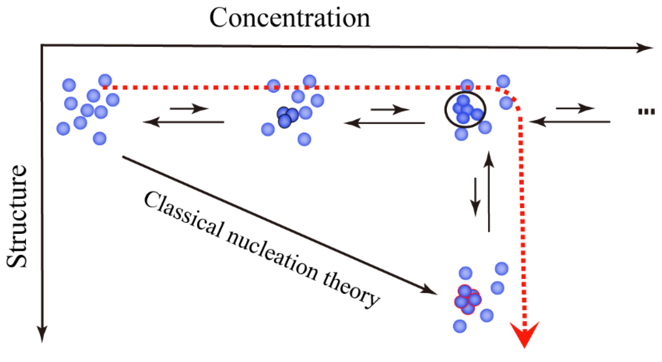
2.1. Ordered Clusters in Classical Nucleation Theory

The classical nucleation theory (CNT) stemmed from the job of Gibbs [16] in 1877, then Farkas, Volmer and Weber [17] improved it by quantitative analysis and modification, and it was formally proposed by Becker and Döring [18] in 1935. CNT is based on the process of condensation of supersaturated vapor into liquid and can be applied to liquid–solid equilibrium systems, such as solution crystallization. CNT considers that when primary monomers such as molecules, atoms or ions collide with each other, they will follow the dynamic equilibrium of multi-stage adsorption and desorption to form ordered clusters [19,20]. The ordered clusters can grow into crystal embryos, which can establish thermodynamic equilibrium with the solution during the growth process and form crystal nuclei. The crystal nuclei may continue to grow into crystals, and the specific process is shown in Figure 1a.
The structure and behavior of critical ordered clusters are the highlight of research in CNT. The prediction method of critical size has also been reported in many papers. Joseph L. Katz [21] used CNT and Reiss theory to predict critical ordered clusters size of nonane, which consisted of eight and nine molecules. Joel D [22] investigated the nucleation process of AgBr in water using molecular dynamics methods and found that for Ag6Br6 and Ag9Br9, the critical ordered clusters consisted of 5–6 monomers. However, the critical ordered clusters of Ag18Br18 consisted of three monomers because nucleation occurred immediately. Adamski [23] further found that critical ordered clusters formed by 10−15 g barium salts consisted of one million monomers. Moreover, CNT with critical ordered clusters has been confirmed by relevant experiments. S. T [24] observed the structure of ordered clusters in apoferritin crystallization by atomic force microscopy (AFM) (Figure 2). As shown in Figure 2, the number of molecules constituting these clusters was different and they were all monolayers. These ordered clusters would dissolve or continue to grow after the remaining adsorbed for 2 to 30 min, which demonstrated the formation of critical ordered clusters during nucleation.
In CNT, the molecular self-assembly process from the molecular scale to the crystal scale is relatively ordered and involves fewer mesoscale structures. CNT has been successfully used to qualitatively analyzed the crystallization process and has achieved relatively accurate results in crystal engineering design and control. However, CNT cannot be applicable to nucleation processes with mesoscale structures such as prenucleation clusters.

2.2. Disordered Clusters in Two-Step Nucleation Theory

As shown in Figure 1b, the two-step nucleation theory indicates that solute molecules first aggregate into dense disordered nucleation clusters in supersaturated solutions. When dense disordered clusters reach a certain level, their interior will reorganize into an ordered crystal nucleus and eventually form crystals [25,26]. The two-step nucleation theory was originally proposed to explain the process of protein nucleation, but now, this theory has also been applied to small molecule materials and colloids [27,28].
Disordered clusters are mesoscale structures in the two-step nucleation theory. Since disordered clusters are microstructures with spatio-temporal dynamic changes, it is very difficult to observe and analyze them with instruments. Therefore, simulation methods are often used for this research. The mechanism of two-step nucleation was initially proposed by Wolde and Frenkel [29], who used the Monte Carlo method to study uniform nucleation in the Lennard–Jones system. Their simulations showed simultaneous changes in density and structural order parameters away from the liquid–liquid critical point, but high-density fluctuations were observed at the critical point, which made a significant change in the crystallization nucleation process. Disordered droplets were first formed, and then crystal nuclei were generated inside droplets larger than a certain critical size. Nathan Duffl [30] proposed the Potts lattice gas model to study the nucleation in solution, and the simulation results showed that the nucleation pathway would change from one-step to two-step nucleation as the temperature approaches the melting temperature, which conformed with the two-step nucleation mechanism. Myerson [31] treated glycine solution with near-infrared laser and obtained different crystal forms of glycine by using linear polarization and circular polarization pulses, respectively. The result suggested that molecules underwent a high-density intermediate state before aggregating to form a nucleus, confirming the two-step nucleation mechanism of glycine. They later confirmed the two-step nucleation process of glycine using small-angle X-ray scattering (SAXS) [32].
The most important mechanism in the two-step nucleation theory was proposed by Vekilov et al. [33]. They demonstrated that in supersaturated solutions, dense disordered clusters were formed first, and then crystal nuclei were formed from the dense disordered clusters, as shown in Figure 3. The second step determined the crystal nucleation rate. Two-step nucleation theory successfully explains some difficult questions of crystal nucleation: the actual nucleation rate is much lower than that predicted by CNT, and what is the effect of heterogeneous matrices on polymorphic selection?
In general, the two-step nucleation theory first considers mesoscale disordered clusters, and preliminarily describes the mesoscale structure and behavior from the molecular scale to the crystal scale, which is a supplement to the relatively ordered CNT. Moreover, the two-step nucleation theory can perfectly explain the nucleation process of proteins, some organic small molecules, inorganic molecular systems, colloidal systems, and biological minerals [34,35,36,37,38].

2.3. Prenucleation Clusters in Prenucleation Clusters Theory

In recent years, much evidence has shown that inorganic systems, such as calcium carbonate and calcium phosphate, have stable aggregates in unsaturated and supersaturated solutions, also known as prenucleation clusters [39,40,41]. The discovery of these prenucleation clusters contradicts the hypothesis of ordered clusters formed by monomer aggregation in CNT, and thus the mechanism of prenucleation clusters is a nonclassical nucleation mechanism. Except in inorganic systems, studies have also reported the existence of prenucleation clusters in some amino acid systems [42]. As shown in Figure 1d, prenucleation clusters theory suggests that stable prenucleation clusters do not form crystal nuclei through the gradual accumulation of monomers but form larger amorphous solid through collisional agglomeration between prenucleation clusters. Subsequently, the internal molecular structure of the amorphous solid reorganizes to crystal nuclei, which grow into macroscopic crystals.
Prenucleation clusters are mesoscale structures in the nucleation process described by prenucleation clusters theory. Prenucleation clusters are thermodynamically stable clusters composed of atoms, molecules or ions, and there is no phase boundary between them and the surrounding solution. Meanwhile, prenucleation clusters are molecular precursors for solution nucleation and can participate in the phase separation process. Compared with the disordered cluster structure described by the two-step nucleation theory, the key difference of this mesoscale structure is the formation of stable and ordered solute self-associations [43,44].
Zhang [45] proposed that at high magnesium concentrations, amorphous calcium carbonate coexisted with stable prenucleation clusters. These prenucleation clusters consisting of ions and water molecules played an important role in the crystallization of calcium carbonate. Habraken [46] used cryo-TEM and in-situ AFM to detect calcium phosphate prenucleation clusters, which were confirmed to be calcium triphosphate clusters [Ca2(HPO4)3]−2, and the prenucleation clusters aggregated and absorbed additional calcium ions to form amorphous calcium phosphate. Thi Thanh [47] demonstrated that prenucleation clusters were formed during the crystallization process of L-glutamic acid by TEM and SAED. As shown in Figure 4, when the crystalline system passed through the metastable region, prenucleation clusters formed by spontaneous aggregation from solution. The prenucleation clusters may grow further into larger amorphous solid through agglomeration or Ostwald ripening mechanisms. After this stage, the amorphous solid may undergo shrinkage and reorganization, which led to compaction into nuclei.
Overall, prenucleation clusters theory further considers prenucleation clusters generated by self-association of solute molecules, and the nucleation pathway for collisional aggregation of the aggregate cluster is formed. The theory further complements the mesoscale structure and behavior from the molecular scale to the crystal scale.

2.4. Particles in Particle Agglomeration Theory

Particle agglomeration nucleation is another nonclassical nucleation theory. When the supersaturation of the system is low and the number of nanoparticles generated before nucleation is large, the agglomeration nucleation of nanoparticles often occurs. The specific process is shown in Figure 1c. The substance first forms fine nanoparticles, and then nanoparticles aggregate to form larger and a stable crystal nucleus [48,49].
In recent years, particle agglomeration theory has gradually attracted the attention of researchers. However, due to the limitations of conventional characterization methods, there is rare understanding of the theory. The main challenge in studying agglomeration nucleation processes is that few PAT are available to monitor the process in real time. Fortunately, researchers have made many efforts to solve this challenge in terms of both computation and experiment. As shown in Figure 5, Baumgartner [50] used Cryo-TEM to observe the presence of particle agglomeration consisting of Fe2+ and Fe3+ with the size of about 2 nm before magnetite nucleation. This is in contrast to the formation of the amorphous phase through the attachment of prenucleation clusters in calcium carbonate and phosphate. Mirabello [51] demonstrated that the generation of magnetite was a nanoparticle self-assembly process. The crystal nuclei of magnetite were produced by the agglomeration of metastable primary particles and dehydration. Theoretical calculations showed that the agglomerated nucleation of the initial particles had a lower energy barrier than CNT. The Pt 3D structure reconstructed by Jungwon using electron tomography showed that the metal nuclei consisted of multiple crystalline domains, which resulted from the agglomeration of smaller nanoparticles during nucleation [52]. In addition, PATs have detected in real time the aggregation of nanoparticles in prenucleation solutions. Polte [53] used in-situ small-angle X-ray scattering to monitor the formation of numerous particles with the size of about 0.8 nm in solution during the growth of gold nanocrystals. Subsequently, the number of nanoparticles decreased rapidly, and the size of the particles gradually became larger, which confirmed that the nucleation process of gold nanocrystals was formed by particle agglomeration.
Compared with the prenucleation cluster theory, the particle agglomeration nucleation theory considers the existence of precursors such as nanoparticles and colloidal particles for the first time, and more specifically considers the nucleation modes at the mesoscale, such as directional attachment, self-assembly, and polymerization from a kinetic perspective, which further enriches the mesoscale structure and behavior.

3. Mesoscale Structure during Growth

With the development of microscopic research methods, researchers have gone deeper into the crystal growth theory. The growth unit in the crystal growth process has expanded from atomic, molecular and ions to mesoscale intermediate states such as amorphous precursors and nanoparticles. The intermediate states are essential for crystal growth.

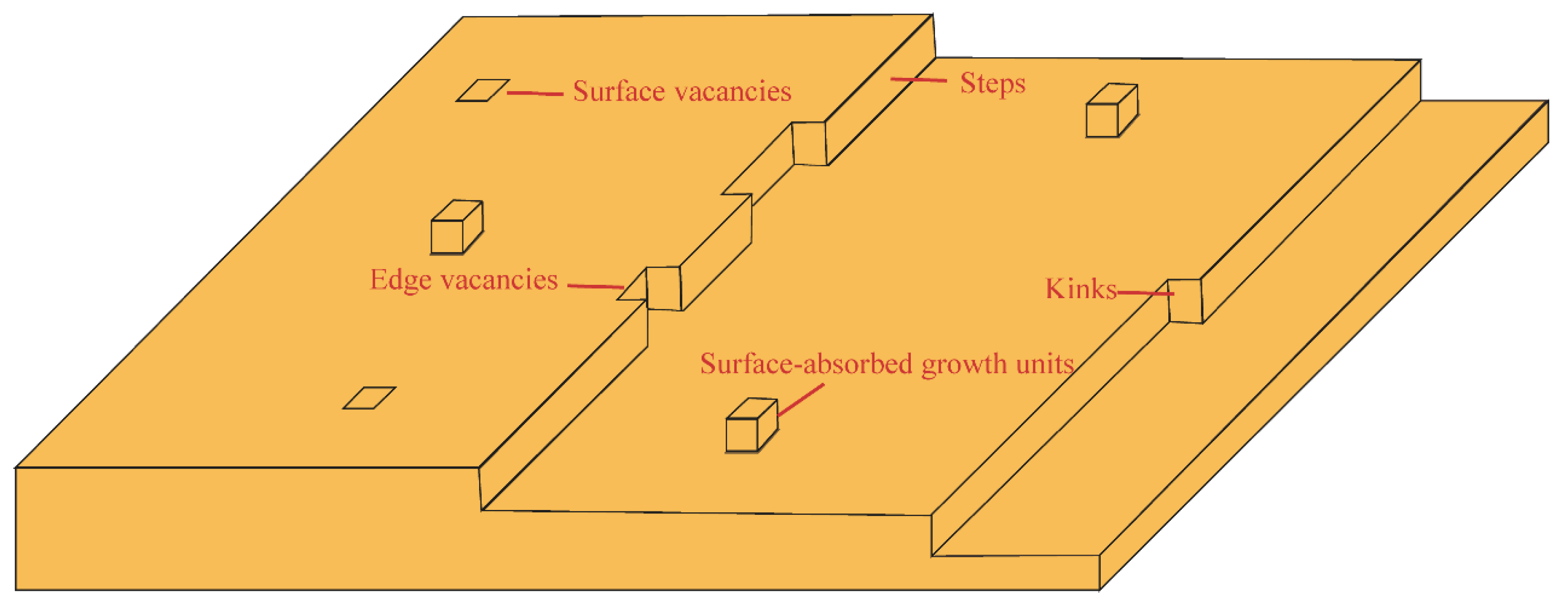
3.1. Units in Classical Growth Theory

Since the early 20th century, researchers have tried various experimental methods and numerical analysis methods to reveal the crystal growth mechanism at the atomic, molecular and ionic levels, and summarized many classical growth theories, such as diffusion control growth theory, two-dimensional nucleation growth theory, and dislocation growth theory. The classical crystal growth theory considers that the basic unit of crystal growth is atoms, molecules or ions, and during the growth process the units are transferred and attached from solution to solid crystal surface one by one.
The diffusion control growth theory means that the interface structure is a rough interface, which is equivalent to accepting molecules deposited from the liquid phase, and the advancement of the interface is mainly due to the random and continuous attachment of molecules on the interface [54]. Molecules diffused from the liquid phase are easily attached to the crystal, and the crystal grows much easier than the smooth surface. The rate at which solute molecules are transported from the supersaturated solution to the crystal surface determines the crystal growth rate [55]. Wang investigated the effect of protein on calcium phosphate growth based on diffusion-controlled growth theory. The addition of protein increased the viscosity of the solution and increased the diffusion resistance of calcium and phosphorus ions, leading to a decline in growth rate of calcium phosphate [56].
Two-dimensional nucleation growth theory means that after the nucleus is formed, the atoms in solution are attracted to a certain lattice surface of the nucleus at first and aggregate with each other to form a stable atomic layer with sufficient area to form a two-dimensional crystal island raised on the lattice surface, which is called a two-dimensional nucleus. Then the atoms in solution can spontaneously accumulate outwards at the concave corners of the platform’s periphery until a complete layer is formed. The above process is repeated continuously on the new lattice plane, so that it grows into a complete crystal layer by layer, as shown in Figure 6 In the 1920s, Volmer [57] first proposed the two-dimensional nucleation growth theory. Burton, Cabrera and Frank [58] elaborated on the mechanism of two-dimensional nucleation, concluding that the formation of a nucleus exceeding the critical radius required large activation energy, so it is unfeasible to grow through two-dimensional nucleation at low supersaturation. Mallink [59] used atomic force microscopy (AFM) to study the growth mechanisms of more than 30 types of thaumatin, peroxidases, egg lysozyme and chromosomal mosaic viruses. The results showed that the crystal growth was consistent with two-dimensional nucleation growth under the condition of low or high supersaturation. This result is contrary to the conclusion that two-dimensional nucleation at low supersaturation was difficult to achieve. Furthermore, Nozawa [60] demonstrated by single-particle resolution that the dominant growth mechanism in colloidal crystallization was the two-dimensional nucleation growth mechanism.
In the actual crystal growth, some researchers have observed that crystals can often grow far below the critical supersaturation required for two-dimensional nucleation growth, and this growth mechanism is dislocation growth theory. The screw dislocation mechanism points out that the crystal is not complete, and there are screw dislocation outcrops on the crystal surface, which can be used as a step source for crystal growth. During the crystal growth process, the growth steps spirally diffuse around the screw dislocation lines and never disappear [61]. Therefore, crystal growth no longer requires two-dimensional nucleation and can grow at low supersaturation. Chernov [62] combined the basic parameters of the actual step motion and the bulk diffusion model into the basic idea of screw dislocation growth, which better generalized the screw dislocation growth theory. At first, the contribution of defects other than screw dislocations to crystal growth was not considered in crystal growth until Bauser [63] observed that edge dislocations can also provide a step source for crystal growth. Min [64] analyzed the distortion of the lattice planes caused by different types of dislocations and the molecular configuration adjacent to the surface outcrop, and proposed that if the surface intersects with the dislocation line and was not in the crystal band with the dislocation’s Berger vector as the axis, the never-disappearing step will exist at the outcrop of the surface dislocation, regardless of the orientation of the dislocation line. Therefore, whether it is the screw dislocation, the edge dislocation, or the mixed dislocation, their contributions to crystal growth are exactly the same, and they all provide a never-disappearing step for crystal growth, which makes the dislocation growth theory continuously improve.
In the classical growth theory, crystal growth is the process in which particles such as atoms, molecules, and ions in supersaturated solution deposit and grow on the formed crystal nucleus, which involves fewer mesoscale structures. With further exploration, many researchers have found that the growth of crystals with special morphologies involves mesoscale structures such as clusters and nanoparticles, which cannot be explained by classical crystal growth theories.

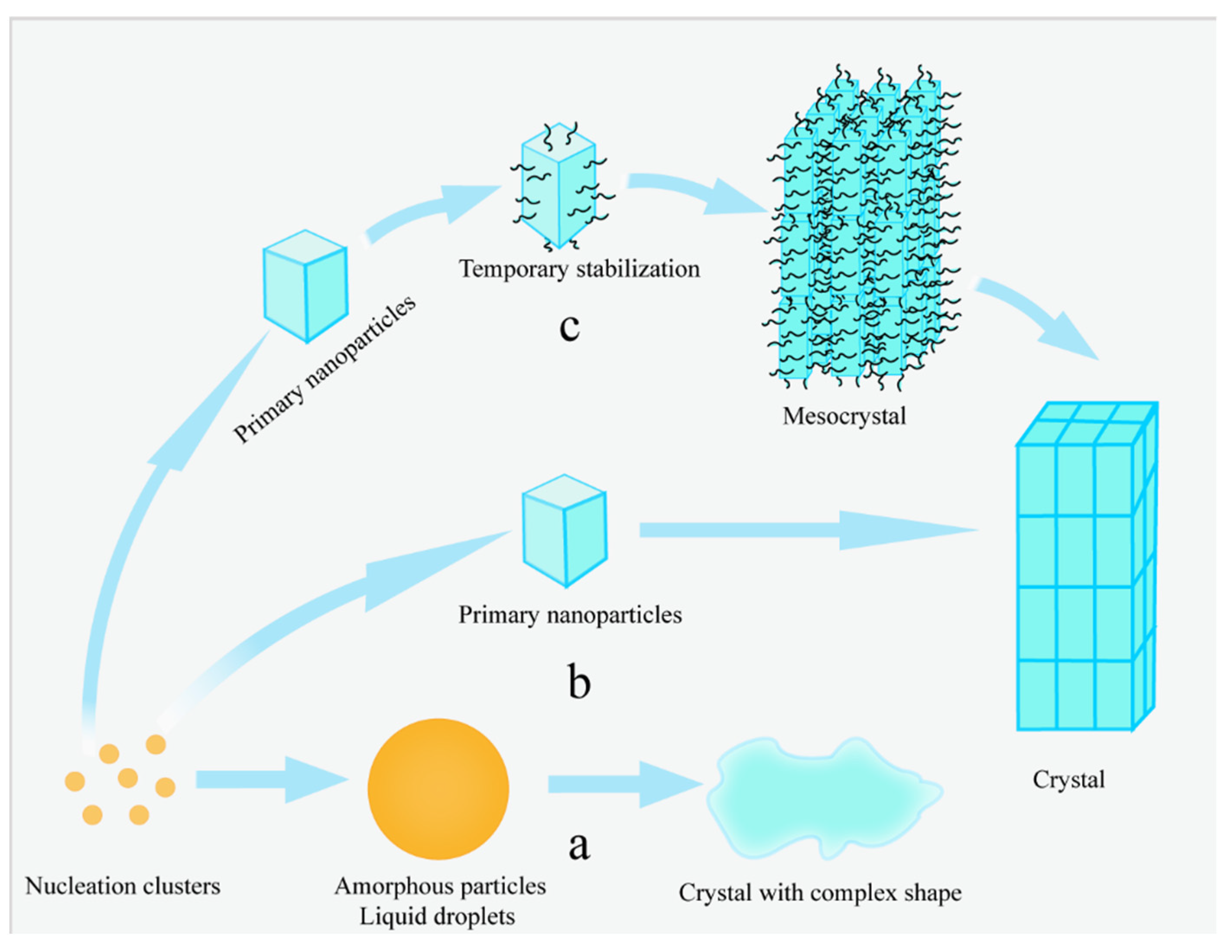
3.2. Amorphous Precursors in Amorphous Precursor Growth Theory

Since amorphous precursors have good plasticity and can form crystals with various morphologies, the formation and transformation of amorphous precursors are often the first step in the nonclassical crystallization process, as shown in Figure 7a. Amorphous particles and liquid droplets are common amorphous precursors with mesoscale structures whose size and structure are between those of solutes and crystals. According to Ostwald ripening principle, the more unstable the formed substance and the lower the degree of modification, the lower the activation energy barrier required. Therefore, in highly supersaturated solutions, amorphous precursors are preferentially formed, and the higher the degree of supersaturation, the more amorphous clusters are formed.
Amorphous precursors have transient characteristics and instability, making them difficult to detect [65]. However, by applying corresponding analytical methods, the changes of ions and supersaturation in the solution can be monitored in situ, and the re-dissolution and mesoscopic transformation of amorphous precursors can be distinguished. In addition, amorphous precursors can also be characterized by transmission electron microscopy, scanning electron microscopy, selected-area electron diffraction, and X-ray diffraction. Yang [66] observed the nonclassical formation process of nickel nanocrystals using high-resolution TEM. At the beginning of the reaction, amorphous particles were formed due to the induction of surface interactions, and crystalline domains appeared and gradually grew into nanocrystals. This amorphous-precursor-mediated nonclassical growth mechanism had significant meaning for the formation of nanocrystals. Vuk [67] demonstrated that the growth patterns on different crystal planes were different during the growth of hydroxyapatite (HAp). Figure 8 showed that the crystal growth on the (100) crystal plane was the classical growth process, in which atoms and ions were continuously accumulated to form crystals. The crystal growth on the (001) crystal plane was the nonclassical growth process, and the spherical amorphous nanoparticles were continuously accumulated to form single crystals.
Droplets are disordered and liquid amorphous precursors, around 100 nm in size. It appears as small droplets under the optical microscope, and then grows into crystals through aggregation and fusion. Liquid precursors do not have a specific ionic composition but can be neutralized by inclusion of ternary counterions and additives themselves. The composition of the liquid precursors is intimately associated with the reaction conditions, such as the concentration of the reagents, the reaction temperature, the order of adding the reagents and the way of mixing. For example, CaCO3 liquid precursors present two forms of over-carbonated and under-carbonated, which means that they need to absorb specific ions from the external environment to complete the crystallization. DiMasi [68] discovered that carbonate transport was the decisive step in under-carbonated CaCO3 liquid precursor transformation.

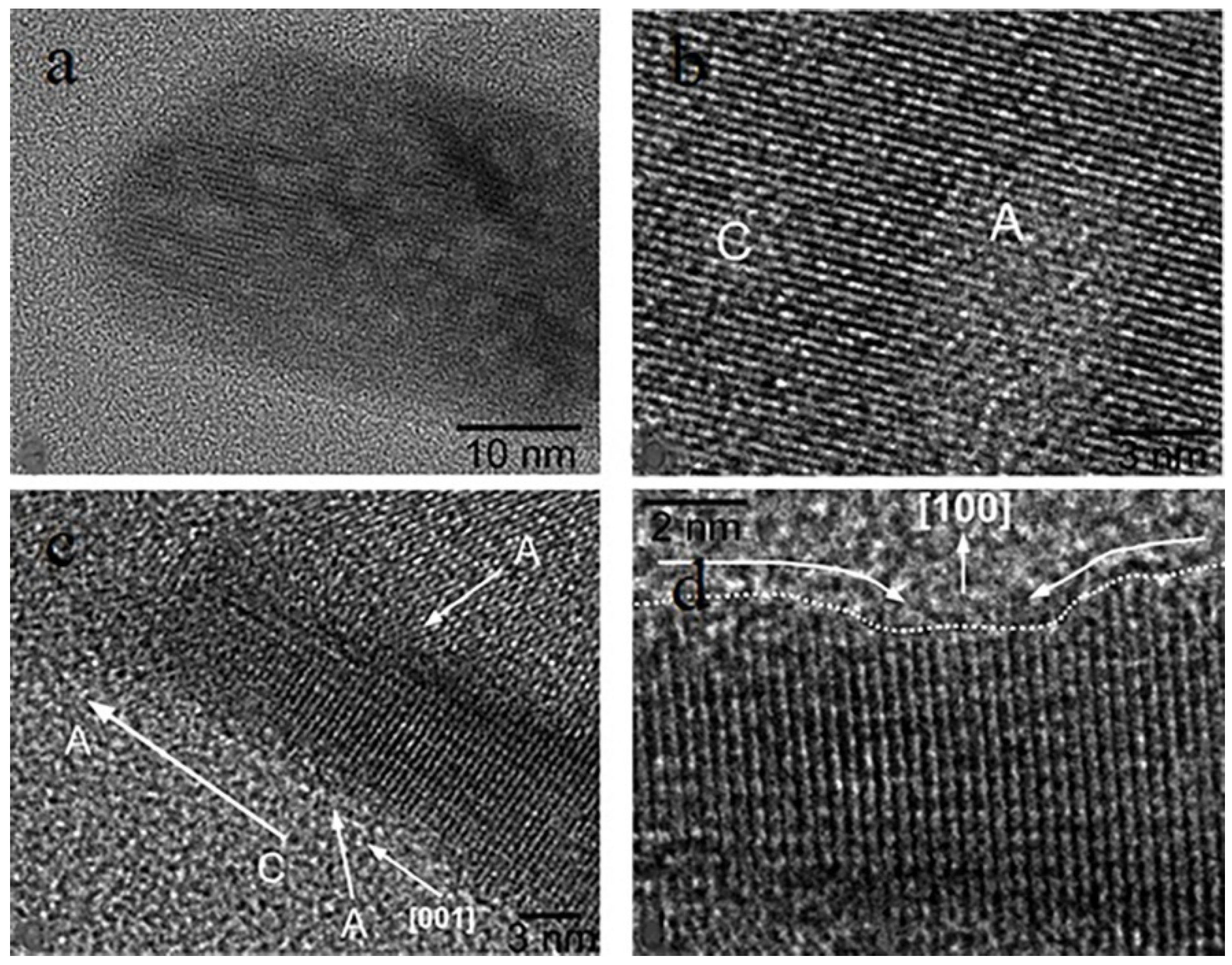
3.3. Nanoparticles in Particle Attachment Growth Theory

The particle attachment growth theory is a well-recognized nonclassical growth theory. Different from the classical growth theory, the particle attached growth theory extends the traditional crystal growth unit from atoms, ions or molecules to clusters and nanoparticles with mesoscale structures. It is pointed out that the crystal growth is the alignment of specific crystal planes through lattice matching between nanoparticles to obtain a highly ordered assembled superstructure, and finally fuse to grow into complete crystals. The particle attachment growth theory mainly includes oriented attachment and random attachment.
Oriented attachment growth is a process of self-assembly growth between adjacent nanoparticles. As shown in Figure 7b, during the crystal growth process, nanoparticles are continuously contacted, separated and repeatedly aligned before attachment to match the lattice orientation, and then align along in the same direction to form a larger crystal. Nanoparticles tend to attach through the crystal face with the highest surface energy or surface area, minimizing the total energy between the two particles. Penn and Banfield [69,70] used high-resolution TEM to observe the assembly and growth of smaller TiO2 nanoparticles in the process of hydrothermal synthesis of TiO2 nanocrystals, revealing this nonclassical oriented attachment growth mechanism for the first time. Subsequently, numerous studies have found that inorganic crystals such as Co3O4 [71], CaCO3 [72], ZnS [73,74] generally have an oriented attachment growth during the growth process. Cho [75] considered that the electric dipole moments in the {100}, {110}, and {111} orientations of octahedral PbSe nanocrystals could facilitate the occurrence of oriented attachment growth. Pacholski [76] reported that hemispherical ZnO nanoparticles formed high-quality single-crystal ZnO nanorods by the oriented attachment growth mechanism.
In recent years, the development of in-situ TEM has further revealed the mechanism of the oriented attachment growth process. Cao [77] investigated the attachment growth process of ZnO nanoparticles in ethanol solution by a series of methods such as transmission electron microscopy. As shown in Figure 9, ZnO nuclei were formed first, and the nuclei gradually grew into ZnO nanoparticles, and then the surface of the nanoparticles became rough, and the nanoparticles with rough surfaces underwent oriented attachment to form complete ZnO crystals. Zhu [78] used in-situ liquid cell TEM to observe that ligand interactions on the surface of gold nanoparticles induce the growth of nanoparticles by oriented attachment. The organic ligands on the Au surface after the addition of citric acid could guide the rotation of the particles, and fusion occurred only when the {111} facets of the two particles were completely aligned. First-principles calculations confirmed that the preferential attachment of nanoparticles at the {111} facets was due to the lower ligand binding energy of these facets. Oriented attachment growth can control the size, shape and microstructure of nanoparticles and prepare artificial nanomaterials with anisotropic shapes, which will gain a lot of attention in the coming years.
Random attachment growth is another way of particle attachment growth. In this case, nanoparticles with crystalline structures first form polycrystals by random attachment growth, and then achieve crystals reorientation through rotation and alignment, and finally form single crystals through internal structural reorganization or external forces. The random attachment growth theory further reveals the crystal growth mechanism and enriches the crystal growth theory. Hu [79] synthesized CdS colloidal spheres by the controllable solvothermal method and investigated their growth mechanism. The TEM results showed that small nanocrystalline nuclei were first formed, and the nanocrystalline nuclei continued to aggregate to nanoparticles, and finally these nanoparticles grew by random attachment to form CdS colloidal spheres. Liu [80] also observed the random attachment growth process during the assembly of vaterite nanocrystals. As shown in Figure 10, during the attachment growth of nanocrystals to the main crystal, the lattices between their interfaces were not matched, and the surface layer of vaterite nanoparticles with random orientations was formed around the main crystal through random attachment growth. Polycrystalline vaterite rapidly transformed into single crystals under pressure. There are still many questions in the study of random attachment growth theory. The current research mainly focuses on the experimental observation stage, and the intrinsic mechanism of random attachment growth and its difference from oriented attachment growth are the future research directions.
The major difference between particle attachment growth theory and classical growth theory is that the basic unit of crystal growth is nanoparticles with mesoscale structure, and nanoparticles grow into macroscopical crystals through oriented attachment or random attachment on the mesoscale. As an effective complement to the classical crystal growth theory, the particle attachment growth theory provides more possibilities for the functional development of novel nanomaterials.

3.4. Mesocrystals in Mesocrystal Growth Theory

Mesocrystals are the mesoscale intermediate between dispersed particle and single crystal with a size between 1–1000 nm [81,82]. The mesocrystal growth process is described as shown in Figure 7c: the crystal nuclei grow into primary nanocrystals through the Ostwald ripening process, and then primary nanocrystals are assembled into mesocrystals in three-dimensional directions through ordered arrangement and lattice interconnection, and finally mesocrystals evolve into a single crystal through the fusion and recrystallization process between the internal crystal planes [83]. Mesocrystal growth is also considered as a nonclassical growth process.
Mesocrystals are assembled from many primary nanocrystals, which provides more ideas for the design of nanostructures. Mesocrystal growth theory can not only be used to guide the preparation of single crystals with complex structures, but also superstructures with nanoparticles as basic units. O’Brien [84] considered that mesocrystals were in the metastable state rather than in the thermodynamically stable state and could transform to other crystal forms under certain conditions. The growth rate and equilibrium morphology of mesocrystals depend on the stability of the nanocrystals and the long-range forces on the vectors between the assembled units, and the nanocrystals must have sufficient time for ordered self-assembly rather than random binding. As shown in Figure 11, Zhang [85] obtained the CuO mesocrystals structure by oriented attachment growth of CuO nanoparticles in three-dimensional directions. The entire structure consisted of several hundred primary CuO nanoparticles whose orientations were crystallographically consistent. As the basic units that make up mesocrystals are aligned in the same direction, mesoscopic crystals tend to have similar electron diffraction patterns to single crystals. Chen [86] successfully prepared α-calcium sulfate hemihydrate spherical mesocrystals in an ethylene glycol-water system and analyzed the growth mechanism by ultramicroscopic technique. The results showed that α-calcium sulfate hemihydrate spherical mesocrystals were solid-state mesocrystals composed of nanorods oriented along the longitudinal axis, and the nanorods were formed by the continuous arrangement of irregular subunits guided by EDTA. Meanwhile, the mesocrystal growth mechanism could also be applied to the design of complex functional materials.
Mesocrystals are widely used in the fields of photocatalysis, optoelectronics, lithium-ion batteries and biological materials due to their unique structural characteristics [87,88,89]. Moreover, mesocrystal growth is not limited to biomineralized systems such as sea urchins and pearl oysters but has also been observed in other reactions such as kinetic metastable systems or without crystallization additives. Tang [87] invented a method to prepare TiO2 mesocrystals by controlling the hydrolysis of TiCl3 in polyethylene glycol. The ultrafine TiO2 nanocrystals formed by the reaction were stabilized by polyethylene glycol molecules and formed nanoparticle aggregates through oriented attachment. When TiO2 nanocrystals in the solution were completely attached to the aggregates, spindle-shaped TiO2 mesocrystals were formed. The synthesized TiO2 mesocrystals were characterized by high crystallinity and high porosity, which enabled them to exhibit stronger visible light activity for photocatalytic NO removal. Ding [88] used a solvothermal method to prepare Li2FeSiO4 mesocrystals, a cathode material for lithium-ion batteries with excellent properties. Li2FeSiO4 mesocrystals consisted of nanoparticles along [001] zone axis, with the oriented single-crystal structure and highly exposed (001) planes. Li2FeSiO4 mesocrystals showed near-theoretical discharge capacity, excellent rate capacity, and favorable cycling stability. Sascha [89] proposed that the size of nickel hexacyanoferrate nanoparticles could be regulated by controlling the citrate concentration and total supersaturation, enabling them to orientate-attach to form nickel hexacyanoferrate colloidal mesocrystals. As shown in Figure 12, nickel hexacyanoferrate nanoparticles with cubic structures as building units were continuously assembled in an orderly manner along the same crystallographic direction to form highly catalytic active colloidal mesocrystals, which could efficiently treat caffeine in wastewater.
At present, the mesocrystal growth theory has been confirmed in many systems, such as BaSO4, CaCO3, zeolite, TiO2 [90,91,92]. However, the mechanism of mesocrystal formation and growth is still not fully understood. This is because mesocrystals as intermediate products have a short lifetime and are not easy to detect, and there is no ideal method for in-situ observation in solution. For example, atomic force microscopy (AFM) can only work on a certain surface. If the surface does not exist in solution, it cannot be used for the study of mesocrystals. Transmission electron microscopy can only observe the structure of larger mesocrystals, but the formation process of mesocrystals cannot be observed. Continuing in-depth research on mesocrystal growth has important theoretical and applied value for the preparation of nanostructured crystals and the development of materials with novel properties.

4. EMMS Model and Crystal Nucleation and Growth

As we study complex systems in science and engineering, we generally need to understand them from the macroscopic scale, and then gradually study the various mechanisms at the microscopic scale, and gradually try to establish the relationship between the two scales. However, it is very difficult to establish this correlation directly. Complex systems mostly exhibit different layers, and each layer has multi-scale structures [93]. Although the multi-scale structures at different levels are different, they all have a common property that the behavior of the system at the boundary scale of each level is relatively simple and easy to characterize and analyze. However, at the scales between them, the behavior of the system is mostly very complex, and these scales are called mesoscales. Within the mesoscale, there exists a characteristic structure, namely the mesoscale structure. Since the mesoscale structure is spatiotemporally dynamic in space and time, which makes it difficult to correlate the microscale with the macroscale, it can only be analyzed by means of averaging. Research has gradually shown that there may be a universal dominant principle at the mesoscale that the coordination of different control mechanisms in competition. For this reason, the interdisciplinary concept of mesoscience is proposed to address various issues [94].
Since the late 20th century, researchers have studied mesoscale problems in reactors [95]. Starting from the phenomenon of particle agglomeration in the gas–solid system, it is believed that the formation of mesoscale agglomeration originated from the coordination of the respective motion trends of gas and particles in the competition, thus establishing the stability condition of the mesoscale agglomeration structure and proposing the theory of energy minimization multi-scale (EMMS) [96]. The EMMS model solved the problem of quantitative simulation of heterogeneous gas–solid systems, and significantly improved the prediction performance of gas–solid two-phase computational fluid dynamics and the ability to solve practical problems. Later, the EMMS principle was extended and applied to other multiphase systems [97].
The EMMS model contains eight variables to describe the gas–solid two-phase flow system, and these eight variables are constrained by six dynamic equations [98].
Dilute-phase force balance equation: the effective load-bearing capacity of the dilute phase particles per unit volume is equal to the drag force of the dilute phase particles by the surrounding dilute phase gas.
3 4 C D f 1 ε f d p ρ g U s f 2 1 ε f ρ p ρ g g = 0
where C D f is the drag coefficient of the dilute phase, ε f is the void fraction of the dilute phase, d p is the particle diameter, ρ g is the density of the gas phase, U s f is the apparent slip velocity of the dilute phase, and ρ p is the density of the particle.
Dense-phase force balance equation: the effective load-bearing capacity of dense phase particles per unit volume is equal to the drag force of dense phase particles by the surrounding dense phase gas and the drag force of agglomerates by the surrounding dilute phase gas.
3 4 C D c 1 ε c d p ρ g U s c 2 + 3 4 C D i 1 d c l ρ g U s i 2 1 ε c ρ p ρ g g = 0
where C D c is the drag coefficient of the dense phase, C D i is the interphase drag coefficient, ε c is the void fraction of the dense phase, d c l is the diameter of the particle agglomeration, U s c is the apparent slip velocity of the dense phase, and U s i is the interphase apparent slip velocity.
Interphase pressure drop balance equation: The fluid maintains the pressure drop balance of the system itself through the drag force acting on the dense two-phase particles, and the dense phase pressure drop is equal to the dilute phase pressure drop and the interphase pressure drop caused by the surrounding agglomerates.
C D f 1 ε f d p ρ g U s f 2 + 1 1 f C D i f d c l ρ g U s i 2 = C D c 1 ε c d p ρ g U s c 2
where f is the dense phase fraction.
Fluid continuity equation: The flow rate of the fluid flowing through the entire section is equal to the sum of the fluid flow rates in the dense and dense phases.
U g = 1 f U f + f U c
where U g is the superficial gas velocity of the gas phase, U f is the superficial gas velocity of the dilute phase, and U c   is the superficial gas velocity of the dense phase.
Particle continuity equation: The particle phase flow through the entire section is equal to the sum of the particle mass flow in the dilute and dense phases.
U p = 1 f U p f + f U p c
where U p is the superficial gas velocity of the particle phase, U p f is the superficial gas velocity of the particles in the dilute phase, and U p c   is the superficial gas velocity of the particles in the dense phase.
Agglomerate size equation: This correlation assumes that the agglomerate size is inversely proportional to the system input energy.
d c l = d p · U p g 1 ε m a x U m f + ε m f 1 ε m f U p g N s t · ρ p ρ p ρ g U m f + ε m f 1 ε m f U p g
where U m f is the superficial gas velocity of minimum fluidization, ε m f is the minimum fluidized void fraction, ε m a x is the maximum non-uniform void fraction, and N s t   is the suspended pumping energy consumption per unit mass.
Stability conditions: The stability condition is expressed by the compromise of two dominant mechanisms in competition.
N s t = W s t 1 ε g ρ p = ρ p ρ g ρ p · g · U g f 1 f ε f ε g 1 ε g · U f m i n
where ε g   is the void fraction of the gas phase.
Due to the nonlinear nature of the equations, it remains a challenge to directly find the analytical solution.
The characteristic of the EMMS model is the description of the non-uniform structure. The multi-scale structure is firstly found in a complex system, and the complex system is regarded as being composed of internal multi-scale structures and their interactions. Then the characteristic equations of the system are constructed, the main motion modes in the system are analyzed and the equations are constructed by the conservation equations. Finally, the stability of the whole system is achieved by the mutual coordination between control mechanisms of different parts to solve the closed equation system [99]. The EMMS model has been developed into a universal EMMS principle for multiple systems, which mainly considers the influence of the mesoscale structure between the whole system and its constituent units on the system behavior, and thus builds a theoretical framework for the development of mesoscale science. The core idea is the mesoscale structure and the compromise in competition of multiple mechanisms [100]. When the system involves two dominant mechanisms, A and B, with different motion trends, the system will present a relatively uniform structure under a single dominant mechanism (A or B). When both A and B cannot dominate and must compromise with each other, a dynamic inhomogeneous structure will appear in the system [101,102,103]. Additionally, it has been proved that the EMMS model can be applied to the mesoscale behavior of gas–solid, liquid–solid, and other multiphase systems [104,105]. In recent years, the EMMS principle has been further extended to the mesoscale study of proteins, heterogeneous catalysis systems and crystal growth, and some progress has been made [106,107,108].
Although there are few practical applications of EMMS principle in the crystallization process, the intermediates in the nonclassical crystallization process are typical mesoscale structures, so the EMMS principle can also be used in the crystallization process theoretically. For example, in the crystallization of calcium carbonate, the diffusion and reaction of chemicals both dominate the structure of products. When diffusion controls the crystallization process, particles with a branched structure are produced. When crystallization is controlled by reaction, spherical particles are formed. When the compromise between diffusion and reaction dominates the process, snowflake-like particles are formed [109]. The traditional crystallization research paradigms are based on the crystallization theory. By considering the mass and heat transfer during the crystallization process, the removal of the crystallization heat and the sufficient mixing of the system are realized. However, because of the inability to maintain the consistency of the process between the crystallization theory and the complex system, the crystallization theory cannot guide the actual production process accurately. Additionally, the traditional crystallization research paradigm only focuses on the unit scale and system scale at each level and lacks the understanding of the mesoscale structure. Therefore, the theory of crystal nucleation and growth can be supplemented by the EMMS method of global distribution, local simulation, and detailed evolution. The precursor and solution system are disassembled into precursor dense phase, precursor dilute phase and interaction phase. The surface energy and volume energy of the three subsystems are calculated according to the stability conditions, so as to obtain the critical sizes of various precursors. For the nonclassical crystallization process, the existence of the precursor dense phase is essentially considered. It is possible to infer other nucleation and growth pathways and process mechanisms by considering the compromise and competition between the precursor dilute phase and the interaction phase. According to the EMMS principle, the dominant roles of ordering and disordering in the mesoscale to reach compromise in competition during nucleation should be studied. Additionally, the competition and compromise between the two dominant mechanisms of diffusion and reaction during crystal growth should also be studied [110]. The EMMS model is the mathematical modeling method at the mesoscale, which cannot be used in classical nucleation and growth processes. However, EMMS models can be applied to a variety of nonclassical nucleation and growth processes with mesoscale structures, such as two-step nucleation theory, prenucleation clusters theory, particle agglomeration theory, amorphous precursor growth theory, particle attachment growth theory and mesocrystal growth theory. The mathematical models at mesoscale have been preliminarily established in the existing nonclassical crystallization theories, but these models are still not perfect. The mathematical modeling method of EMMS at mesoscale can further modify and supplement the existing theories and models of nucleation and growth, and it provides new ideas for researchers to use the EMMS theory to study the crystallization process furtherly.

5. Conclusions and Outlook

An increasing number of studies have confirmed nonclassical crystallization pathways on the mesoscale. Precursors with mesoscale structures during nucleation and growth can deviate the crystallization behavior from the predictions of classical nucleation theoretical models. Therefore, it is urgent to modify and refine crystal nucleation and growth theories. In this paper, a series of nonclassical crystallization phenomena discovered in recent years and the growth paths based on experiments and simulation calculations are reviewed. The two-step nucleation theory, prenucleation clusters theory, particle agglomeration theory, amorphous precursor growth theory, particle attachment growth theory and mesocrystal growth theory involving mesoscale precursors are summarized, and the research paradigm of the EMMS model based on mesoscale science to guide the solution crystallization process is presented.
With the rapid development of PAT, computational simulation and other technologies, the research on nonclassical crystallization theory has been effectively promoted. In-situ monitoring of mesoscale crystallization processes can be achieved by in-situ liquid TEM, and the formation of various intermediate structures between microscale and macroscale, including prenucleation clusters, nanoparticles, and mesocrystals, has changed the classical crystallization point. Nonclassical crystallization is not a process of basic monomer attachment, but a process of precursor assembly and aggregation at the mesoscale. According to the idea of constructing a mathematical model based on the EMMS principle, virtual process engineering combined with mesoscale science, supercomputing and machine learning may become a brand-new crystallization process research model. On the basis of the traditional crystal research and the development of the model, virtual process engineering can combine basic disciplines with traditional chemical theory, mesoscale science, algorithm software, supercomputing and virtual reality. The physicochemical changes in the whole crystallization process can be reproduced accurately and completely [111,112]. Meanwhile, the algorithm software is used to simulate the mesoscale crystallization process. Based on supercomputing, the visualization of the whole process design and amplification are realized. Additionally, the crystallization theoretical system with multi-level, multi-scale properties and mesoscale complexity will be formed, which has broad application value in the fields of crystal engineering, crystallization separation and material sciences.

Author Contributions

Conceptualization, X.W. and K.L.; methodology, X.W. and K.L.; validation, X.W.; formal analysis, K.L.; investigation, X.Q.; resources, M.L.; data curation, Y.L.; writing—original draft preparation, X.W.; writing—review and editing, K.L. and M.C.; visualization, M.C. and J.O.; supervision, Y.A., W.Y., M.C., J.O. and J.G.; project administration, Y.A., W.Y., M.C., J.O. and J.G.; funding acquisition, Y.A., W.Y., M.C., J.O. and J.G. All authors have read and agreed to the published version of the manuscript.

Funding

This work was financially supported by the National Science Foundation of China 22108195, Key R&D Program of Zhejiang (2022C01208), the key project of State Key Laboratory of Chemical Engineering (SKL-ChE-20Z03), the Chemistry and Chemical Engineering Guangdong Laboratory (Grant No. 1912014) and the financial support of Haihe Laboratory of Sustainable Chemical Transformations, Academic and Technical Leader Training Program for Major Disciplines in Jiangxi Province (20212BCJ23001).

Institutional Review Board Statement

Not applicable.

Informed Consent Statement

Not applicable.

Data Availability Statement

Not applicable.

Acknowledgments

Kangli Li thanks the Shaoxing People’s Government for a supporting of her post-doctoral research. This work was financially supported by the Institute of Shaoxing.

Conflicts of Interest

The authors declare no conflict of interest.

References

  1. Kato, S.; Furukawa, S.; Aoki, D.; Goseki, R.; Oikawa, K.; Tsuchiya, K.; Shimada, N.; Maruyama, A.; Numata, K.; Otsuka, H. Crystallization-induced mechanofluorescence for visualization of polymer crystallization. Nat. Commun. 2021, 12, 126. [Google Scholar] [CrossRef] [PubMed]
  2. Zhang, J.; Fang, Y.; Zhao, W.; Han, R.; Wen, J.; Liu, S. Molten-Salt-Assisted CsPbI3 Perovskite Crystallization for Nearly 20%-Efficiency Solar Cells. Adv. Mater. 2021, 33, 2103770. [Google Scholar] [CrossRef] [PubMed]
  3. Mu, X.; Wu, F.; Tang, Y.; Wang, R.; Li, Y.; Li, K.; Li, C.; Lu, Y.; Zhou, X.; Li, Z. Boost photothermal theranostics via self-assembly-induced crystallization (SAIC). Aggregate 2022, e170, 2692–4560. [Google Scholar] [CrossRef]
  4. Cui, W.; He, Z.; Zhang, Y.; Fan, Q.; Feng, N. Naringenin Cocrystals Prepared by Solution Crystallization Method for Improving Bioavailability and Anti-hyperlipidemia Effects. AAPS PharmSciTech 2019, 20, 115. [Google Scholar] [CrossRef] [PubMed]
  5. Wei, Z.; Zhang, Q.; Li, X. Crystallization Kinetics of α-Hemihydrate Gypsum Prepared by Hydrothermal Method in Atmospheric Salt Solution Medium. Crystals 2021, 11, 843. [Google Scholar] [CrossRef]
  6. Frenkel, J. A general theory of heterophase fluctuations and pretransition phenomena. J. Chem. Phys. 1939, 7, 538–547. [Google Scholar] [CrossRef]
  7. Volmer, M. Nucleus formation in supersaturated systems. Z. Phys. Chem. 1926, 119, 277–301. [Google Scholar] [CrossRef]
  8. Santra, M.; Singh, R.S.; Bagchi, B. Polymorph selection during crystallization of a model colloidal fluid with a free energy landscape containing a metastable solid. Phys. Rev. E 2018, 98, 032606. [Google Scholar] [CrossRef]
  9. Janković, B.; Marinović-Cincović, M.; Dramićanin, M. Kinetic study of isothermal crystallization process of Gd2Ti2O7 pre-cursor’s powder prepared through the Pechini synthetic approach. J. Phys. Chem. Sol. 2015, 85, 160–172. [Google Scholar] [CrossRef]
  10. Chang, H.; Bootharaju, M.S.; Lee, S.; Kim, J.H.; Kim, B.H.; Hyeon, T. To inorganic nanoparticles via nanoclusters: Nonclassical nucleation and growth pathway. Bull. Korean Chem. Soc. 2021, 42, 1386–1399. [Google Scholar] [CrossRef]
  11. Houben, L.; Weissman, H.; Wolf, S.G.; Rybtchinski, B. A mechanism of ferritin crystallization revealed by cryo-STEM tomography. Nature 2020, 579, 540–543. [Google Scholar] [CrossRef] [PubMed]
  12. Wiedenbeck, E.; Kovermann, M.; Gebauer, D.; Cölfen, H. Liquid metastable precursors of ibuprofen as aqueous nucleation intermediates. Angew. Chem. Int. Ed. 2019, 58, 19103–19109. [Google Scholar] [CrossRef] [PubMed]
  13. Li, J.; Kwauk, M. Exploring complex systems in chemical engineering—the multi-scale methodology. Chem. Eng. Sci. 2003, 58, 521–535. [Google Scholar] [CrossRef]
  14. Huang, W.; Li, J.; Edwards, P.P. Mesoscience: Exploring the common principle at mesoscales. Natl. Sci. Rev. 2018, 5, 27–32. [Google Scholar] [CrossRef]
  15. Grossmann, L.; King, B.T.; Reichlmaier, S.; Hartmann, N.; Rosen, J.; Heckl, W.M.; Björk, J.; Lackinger, M. On-surface photopolymerization of two-dimensional polymers ordered on the mesoscale. Nat. Chem. 2021, 13, 730–736. [Google Scholar] [CrossRef]
  16. Gibbs, J.W. On the equilibrium of heterogeneous substances. Am. J. Sci. 1878, 3, 441–458. [Google Scholar] [CrossRef]
  17. Farkas, L. Keimbildungsgeschwindigkeit in übersättigten Dämpfen. Z. Phys. Chem. 1927, 125, 236–242. [Google Scholar] [CrossRef]
  18. Becker, R.; Döring, W. Kinetische behandlung der keimbildung in übersättigten dämpfen. Ann. Phys. 1935, 416, 719–752. [Google Scholar] [CrossRef]
  19. Li, C.; Liu, Z.; Goonetilleke, E.C.; Huang, X. Temperature-dependent kinetic pathways of heterogeneous ice nucleation competing between classical and non-classical nucleation. Nat. Commun. 2021, 12, 4954. [Google Scholar] [CrossRef]
  20. Banner, D.J.; Firlar, E.; Rehak, P.; Phakatkar, A.H.; Foroozan, T.; Osborn, J.K.; Sorokina, L.V.; Narayanan, S.; Tahseen, T.; Baggia, Y. In Situ Liquid-Cell TEM Observation of Multiphase Classical and Nonclassical Nucleation of Calcium Oxalate. Adv. Funct. Mater. 2021, 31, 2007736. [Google Scholar] [CrossRef]
  21. Katz, J.L. The critical supersaturations predicted by nucleation theory. J. Stat. Phys. 1970, 2, 137–146. [Google Scholar] [CrossRef]
  22. Shore, J.D.; Perchak, D.; Shnidman, Y. Simulations of the nucleation of AgBr from solution. J. Chem. Phys. 2000, 113, 6276–6284. [Google Scholar] [CrossRef]
  23. Adamski, T. Commination of crystal nucleation by a precipitation method. Nature 1963, 197, 894. [Google Scholar] [CrossRef]
  24. Yau, S.-T.; Vekilov, P.G. Direct observation of nucleus structure and nucleation pathways in apoferritin crystallization. J. Am. Chem. Soc. 2001, 123, 1080–1089. [Google Scholar] [CrossRef]
  25. Erdemir, D.; Lee, A.Y.; Myerson, A.S. Nucleation of crystals from solution: Classical and two-step models. Acc. Chem. Res. 2009, 42, 621–629. [Google Scholar] [CrossRef] [PubMed]
  26. Davey, R.J.; Schroeder, S.L.; Ter Horst, J.H. Nucleation of organic crystals—a molecular perspective. Angew. Chem. Int. Ed. 2013, 52, 2166–2179. [Google Scholar] [CrossRef] [PubMed]
  27. Chen, P.-Z.; Niu, L.-Y.; Zhang, H.; Chen, Y.-Z.; Yang, Q.-Z. Exploration of the two-step crystallization of organic micro/nano crystalline materials by fluorescence spectroscopy. Mater. Chem. Front. 2018, 2, 1323–1327. [Google Scholar] [CrossRef]
  28. Fang, H.; Hagan, M.F.; Rogers, W.B. Two-step crystallization and solid–solid transitions in binary colloidal mixtures. Proc. Natl. Acad. Sci. USA 2020, 117, 27927–27933. [Google Scholar] [CrossRef] [PubMed]
  29. ten Wolde, P.R.; Frenkel, D. Enhancement of protein crystal nucleation by critical density fluctuations. Science 1997, 277, 1975–1978. [Google Scholar] [CrossRef]
  30. Duff, N.; Peters, B. Nucleation in a Potts lattice gas model of crystallization from solution. J. Chem. Phys. 2009, 131, 184101. [Google Scholar] [CrossRef]
  31. Sun, X.; Garetz, B.A.; Myerson, A.S. Polarization switching of crystal structure in the nonphotochemical laser-induced nucleation of supersaturated aqueous l-histidine. Cryst. Growth Des. 2008, 8, 1720–1722. [Google Scholar] [CrossRef]
  32. Chattopadhyay, S.; Erdemir, D.; Evans, J.M.; Ilavsky, J.; Amenitsch, H.; Segre, C.U.; Myerson, A.S. SAXS study of the nucleation of glycine crystals from a supersaturated solution. Cryst. Growth Des. 2005, 5, 523–527. [Google Scholar] [CrossRef]
  33. Vekilov, P.G. The two-step mechanism of nucleation of crystals in solution. Nanoscale 2010, 2, 2346–2357. [Google Scholar] [CrossRef]
  34. Harano, K.; Homma, T.; Niimi, Y.; Koshino, M.; Suenaga, K.; Leibler, L.; Nakamura, E. Heterogeneous nucleation of organic crystals mediated by single-molecule templates. Nat. Mater. 2012, 11, 877–881. [Google Scholar] [CrossRef]
  35. Leunissen, M.E.; Christova, C.G.; Hynninen, A.P.; Royall, C.P.; Campbell, A.I.; Imhof, A.; Van Blaaderen, A. Ionic colloidal crystals of oppositely charged particles. Nature 2005, 437, 235–240. [Google Scholar] [CrossRef]
  36. Savage, J.R.; Dinsmore, A.D. Experimental evidence for two-step nucleation in colloidal crystallization. Phys. Rev. Lett. 2009, 102, 198302. [Google Scholar] [CrossRef]
  37. Pouget, E.M.; Bomans, P.H.; Goos, J.A.; Frederik, P.M.; de With, G.; Sommerdijk, N.A. The initial stages of template-controlled CaCO3 formation revealed by cryo-TEM. Science 2009, 323, 1455–1458. [Google Scholar] [CrossRef]
  38. Gower, L.B. Biomimetic model systems for investigating the amorphous precursor pathway and its role in biomineralization. Chem. Rev. 2008, 108, 4551–4627. [Google Scholar] [CrossRef]
  39. Betts, F.; Posner, A. An X-ray radial distribution study of amorphous calcium phosphate. Mater. Res. Bull. 1974, 9, 353–360. [Google Scholar] [CrossRef]
  40. Gebauer, D.; Kellermeier, M.; Gale, J.D.; Bergström, L.; Cölfen, H. Pre-nucleation clusters as solute precursors in crystallisation. Chem. Soc. Rev. 2014, 43, 2348–2371. [Google Scholar] [CrossRef] [Green Version]
  41. Gebauer, D.; Cölfen, H. Prenucleation clusters and non-classical nucleation. Nano Today 2011, 6, 564–584. [Google Scholar] [CrossRef]
  42. Kellermeier, M.; Rosenberg, R.; Moise, A.; Anders, U.; Przybylski, M.; Cölfen, H. Amino acids form prenucleation clusters: ESI-MS as a fast detection method in comparison to analytical ultracentrifugation. Faraday Discuss. 2012, 159, 23–45. [Google Scholar] [CrossRef]
  43. Lu, J.; Li, Z.; Jiang, X. Polymorphism of pharmaceutical molecules: Perspectives on nucleation. Front. Chem. Eng. Chin. 2010, 4, 37–44. [Google Scholar] [CrossRef]
  44. Lin, T.-J.; Chiu, C.-C. Structures and infrared spectra of calcium phosphate clusters by ab initio methods with implicit solvation models. Phys. Chem. Chem. Phys. 2018, 20, 345–356. [Google Scholar] [CrossRef]
  45. Zhang, J.; Zhou, X.; Dong, C.; Sun, Y.; Yu, J. Investigation of amorphous calcium carbonate’s formation under high concentration of magnesium: The prenucleation cluster pathway. J. Cryst. Growth 2018, 494, 8–16. [Google Scholar] [CrossRef]
  46. Habraken, W.J.; Tao, J.; Brylka, L.J.; Friedrich, H.; Bertinetti, L.; Schenk, A.S.; Verch, A.; Dmitrovic, V.; Bomans, P.H.; Frederik, P.M. Ion-association complexes unite classical and non-classical theories for the biomimetic nucleation of calcium phosphate. Nat. Commun. 2013, 4, 1507. [Google Scholar] [CrossRef]
  47. Trinh, T.T.H.; Khuu, C.Q.; Wolf, S.E.; Nguyen, A.-T. The multiple stages towards crystal formation of L-glutamic acid. J. Cryst. Growth 2020, 544, 125727. [Google Scholar] [CrossRef]
  48. Wang, F.; Richards, V.N.; Shields, S.P.; Buhro, W.E. Kinetics and mechanisms of aggregative nanocrystal growth. Chem. Mater. 2014, 26, 5–21. [Google Scholar] [CrossRef]
  49. De Yoreo, J.J.; Gilbert, P.U.; Sommerdijk, N.A.; Penn, R.L.; Whitelam, S.; Joester, D.; Zhang, H.; Rimer, J.D.; Navrotsky, A.; Banfield, J.F. Crystallization by particle attachment in synthetic, biogenic, and geologic environments. Science 2015, 349, 6760. [Google Scholar] [CrossRef]
  50. Baumgartner, J.; Dey, A.; Bomans, P.H.; Le Coadou, C.; Fratzl, P.; Sommerdijk, N.A.; Faivre, D. Nucleation and growth of magnetite from solution. Nat. Mater. 2013, 12, 310–314. [Google Scholar] [CrossRef] [PubMed]
  51. Mirabello, G.; Ianiro, A.; Bomans, P.H.; Yoda, T.; Arakaki, A.; Friedrich, H.; de With, G.; Sommerdijk, N.A. Crystallization by particle attachment is a colloidal assembly process. Nat. Mater. 2020, 19, 391–396. [Google Scholar] [CrossRef]
  52. Park, J.; Elmlund, H.; Ercius, P.; Yuk, J.M.; Limmer, D.T.; Chen, Q.; Kim, K.; Han, S.H.; Weitz, D.A.; Zettl, A. 3D structure of individual nanocrystals in solution by electron microscopy. Science 2015, 349, 290–295. [Google Scholar] [CrossRef]
  53. Polte, J.; Erler, R.; Thunemann, A.F.; Sokolov, S.; Ahner, T.T.; Rademann, K.; Emmerling, F.; Kraehnert, R. Nucleation and growth of gold nanoparticles studied via in situ small angle X-ray scattering at millisecond time resolution. ACS Nano 2010, 4, 1076–1082. [Google Scholar] [CrossRef]
  54. Crespo, D.; Pradell, T.; Clavaguera-Mora, M.; Clavaguera, N. Microstructural evaluation of primary crystallization with diffusion-controlled grain growth. Phys. Rev. B 1997, 55, 3435. [Google Scholar] [CrossRef]
  55. Brüning, K.; Schneider, K.; Roth, S.V.; Heinrich, G. Kinetics of strain-induced crystallization in natural rubber: A diffusion-controlled rate law. Polymer 2015, 72, 52–58. [Google Scholar] [CrossRef]
  56. Wang, K.; Leng, Y.; Lu, X.; Ren, F.; Ge, X.; Ding, Y. Theoretical analysis of protein effects on calcium phosphate precipitation in simulated body fluid. CrystEngComm 2012, 14, 5870–5878. [Google Scholar] [CrossRef]
  57. Volmer, M. Zum problem des kristallwachstums. Z. Phys. Chem. 1922, 102, 267–275. [Google Scholar] [CrossRef]
  58. Burton, W.-K.; Cabrera, N.; Frank, F. The growth of crystals and the equilibrium structure of their surfaces. Philos. Trans. R. Soc. 1951, 243, 299–358. [Google Scholar]
  59. Malkin, A.; Kuznetsov, Y.G.; Land, T.; DeYoreo, J.; McPherson, A. Mechanisms of growth for protein and virus crystals. Nat. Struct. Biol. 1995, 2, 956–959. [Google Scholar] [CrossRef]
  60. Nozawa, J.; Uda, S.; Guo, S.; Hu, S.; Toyotama, A.; Yamanaka, J.; Okada, J.; Koizumi, H. Two-dimensional nucleation on the terrace of colloidal crystals with added polymers. Langmuir 2017, 33, 3262–3269. [Google Scholar] [CrossRef]
  61. Weng, J.; Huang, Y.; Hao, D.; Ji, Y. Recent advances of pharmaceutical crystallization theories. Chin. J. Chem. Eng. 2020, 28, 935–948. [Google Scholar] [CrossRef]
  62. Chernov, A. Present-day understanding of crystal growth from aqueous solutions. Prog. Cryst. Growth Charact. Mater. 1993, 26, 121–151. [Google Scholar] [CrossRef]
  63. Kalt, H.; Rühle, W.; Reimann, K.; Rinker, M.; Bauser, E. Alloy-disorder-induced intervalley coupling. Phys. Rev. B 1991, 43, 12364. [Google Scholar] [CrossRef]
  64. Ming, N.-B. Defect mechanisms of crystal growth and their kinetics. J. Cryst. Growth 1993, 128, 104–112. [Google Scholar] [CrossRef]
  65. Rodriguez-Navarro, C.; Burgos-Cara, A.; Lorenzo, F.D.; Ruiz-Agudo, E.; Elert, K. Nonclassical crystallization of calcium hydroxide via amorphous precursors and the role of additives. Cryst. Growth Des. 2020, 20, 4418–4432. [Google Scholar] [CrossRef]
  66. Yang, J.; Koo, J.; Kim, S.; Jeon, S.; Choi, B.K.; Kwon, S.; Kim, J.; Kim, B.H.; Lee, W.C.; Lee, W.B. Amorphous-phase-mediated crystallization of Ni nanocrystals revealed by high-resolution liquid-phase electron microscopy. J. Am. Chem. Soc. 2019, 141, 763–768. [Google Scholar] [CrossRef] [PubMed]
  67. Uskoković, V. Disordering the disorder as the route to a higher order: Incoherent crystallization of calcium phosphate through amorphous precursors. Cryst. Growth Des. 2019, 19, 4340–4357. [Google Scholar] [CrossRef]
  68. DiMasi, E.; Kwak, S.-Y.; Amos, F.F.; Olszta, M.J.; Lush, D.; Gower, L.B. Complementary control by additives of the kinetics of amorphous CaCO3 mineralization at an organic interface: In-situ synchrotron X-ray observations. Phys. Rev. Lett. 2006, 97, 045503. [Google Scholar] [CrossRef]
  69. Penn, R.L.; Banfield, J.F. Imperfect oriented attachment: Dislocation generation in defect-free nanocrystals. Science 1998, 281, 969–971. [Google Scholar] [CrossRef]
  70. Penn, R.L.; Banfield, J.F. Morphology development and crystal growth in nanocrystalline aggregates under hydrothermal conditions: Insights from titania. Geochim. Cosmochim. Acta 1999, 63, 1549–1557. [Google Scholar] [CrossRef]
  71. Tsukiyama, K.; Takasaki, M.; Oaki, Y.; Imai, H. Evolution of Co3O4 nanocubes through stepwise oriented attachment. Langmuir 2019, 35, 8025–8030. [Google Scholar] [CrossRef] [PubMed]
  72. Gehrke, N.; Cölfen, H.; Pinna, N.; Antonietti, M.; Nassif, N. Superstructures of calcium carbonate crystals by oriented attachment. Cryst. Growth Des. 2005, 5, 1317–1319. [Google Scholar] [CrossRef]
  73. Zhang, J.; Lin, Z.; Lan, Y.; Ren, G.; Chen, D.; Huang, F.; Hong, M. A multistep oriented attachment kinetics: Coarsening of ZnS nanoparticle in concentrated NaOH. J. Am. Chem. Soc. 2006, 128, 12981–12987. [Google Scholar] [CrossRef] [PubMed]
  74. Kurz, W.; Rappaz, M.; Trivedi, R. Progress in modelling solidification microstructures in metals and alloys. Part II: Dendrites from 2001 to 2018. Int. Mater. Rev. 2021, 66, 30–76. [Google Scholar] [CrossRef]
  75. Cho, K.-S.; Talapin, D.V.; Gaschler, W.; Murray, C.B. Designing PbSe nanowires and nanorings through oriented attachment of nanoparticles. J. Am. Chem. Soc. 2005, 127, 7140–7147. [Google Scholar] [CrossRef]
  76. Pacholski, C.; Kornowski, A.; Weller, H. Self-assembly of ZnO: From nanodots to nanorods. Angew. Chem. Int. Ed. 2002, 41, 1188–1191. [Google Scholar] [CrossRef]
  77. Cao, D.; Gong, S.; Shu, X.; Zhu, D.; Liang, S. Preparation of ZnO nanoparticles with high dispersibility based on oriented attachment (OA) process. Nanoscale Res. Lett. 2019, 14, 210. [Google Scholar] [CrossRef]
  78. Zhu, C.; Liang, S.; Song, E.; Zhou, Y.; Wang, W.; Shan, F.; Shi, Y.; Hao, C.; Yin, K.; Zhang, T. In-situ liquid cell transmission electron microscopy investigation on oriented attachment of gold nanoparticles. Nat. Commun. 2018, 9, 421. [Google Scholar] [CrossRef]
  79. Li, X.H.; Li, J.X.; Li, G.D.; Liu, D.P.; Chen, J.S. Controlled synthesis, growth mechanism, and properties of monodisperse CdS colloidal spheres. Chem. Eur. J. 2007, 13, 8754–8761. [Google Scholar] [CrossRef]
  80. Liu, Z.; Pan, H.; Zhu, G.; Li, Y.; Tao, J.; Jin, B.; Tang, R. Realignment of nanocrystal aggregates into single crystals as a result of inherent surface stress. Angew. Chem. 2016, 128, 13028–13032. [Google Scholar] [CrossRef]
  81. Ye, Y.; Chen, C.; Li, W.; Guo, X.; Yang, H.; Guan, H.; Xi, G. Highly sensitive W18O49 mesocrystal Raman scattering substrate with large-area signal uniformity. Anal. Chem. 2021, 93, 3138–3145. [Google Scholar] [CrossRef]
  82. Li, L.; Liu, C.Y. Organic small molecule-assisted synthesis of high active TiO2 rod-like mesocrystals. CrystEngComm 2010, 12, 2073–2078. [Google Scholar] [CrossRef]
  83. Zhou, L.; O’Brien, P. Mesocrystals: A new class of solid materials. Small 2008, 4, 1566–1574. [Google Scholar] [CrossRef]
  84. Zhou, L.; O’Brien, P. Mesocrystals—Properties and Applications. J. Phys. Chem. Lett. 2012, 3, 620–628. [Google Scholar] [CrossRef]
  85. Zhang, Z.; Sun, H.; Shao, X.; Li, D.; Yu, H.; Han, M. Three-Dimensionally Oriented Aggregation of a Few Hundred Nanoparticles into Monocrystalline Architectures. Adv. Mate. 2005, 17, 42–47. [Google Scholar] [CrossRef]
  86. Chen, Q.; Wu, L.; Zeng, Y.; Jia, C.; Lin, J.; Yates, M.Z.; Guan, B. Formation of spherical calcium sulfate mesocrystals: Orientation controlled by subunit growth. CrystEngComm 2019, 21, 5973–5979. [Google Scholar] [CrossRef]
  87. Tan, B.; Zhang, X.; Li, Y.; Chen, H.; Ye, X.; Wang, Y.; Ye, J. Anatase TiO2 Mesocrystals: Green Synthesis, In Situ Conversion to Porous Single Crystals, and Self-Doping Ti3+ for Enhanced Visible Light Driven Photocatalytic Removal of NO. Chem. Eur. J. 2017, 23, 5478–5487. [Google Scholar] [CrossRef]
  88. Ding, Z.; Zhang, D.; Feng, Y.; Zhang, F.; Chen, L.; Du, Y.; Ivey, D.G.; Wei, W. Tuning anisotropic ion transport in mesocrystalline lithium orthosilicate nanostructures with preferentially exposed facets. NPG Asia Mater. 2018, 10, 606–617. [Google Scholar] [CrossRef]
  89. Keßler, S.; Reinalter, E.R.; Ni, B.; Cölfen, H. Rational Design of Environmentally Compatible Nickel Hexacyanoferrate Mesocrystals as Catalysts. J. Phys. Chem. C 2021, 125, 26503–26511. [Google Scholar] [CrossRef]
  90. Ruiz-Agudo, C.; Ruiz-Agudo, E.; Putnis, C.V.; Putnis, A. Mechanistic principles of barite formation: From nanoparticles to micron-sized crystals. Cryst. Growth Des. 2015, 15, 3724–3733. [Google Scholar] [CrossRef]
  91. Wang, T.; Cölfen, H.; Antonietti, M. Nonclassical crystallization: Mesocrystals and morphology change of CaCO3 crystals in the presence of a polyelectrolyte additive. J. Am. Chem. Soc. 2005, 127, 3246–3247. [Google Scholar] [CrossRef] [PubMed] [Green Version]
  92. Lin, F.; Ye, Z.; Kong, L.; Liu, P.; Zhang, Y.; Zhang, H.; Tang, Y. Facile Morphology and Porosity Regulation of Zeolite ZSM-5 Mesocrystals with Synergistically Enhanced Catalytic Activity and Shape Selectivity. Nanomaterials 2022, 12, 1601. [Google Scholar] [CrossRef] [PubMed]
  93. Estrin, Y.; Beygelzimer, Y.; Kulagin, R.; Gumbsch, P.; Fratzl, P.; Zhu, Y.; Hahn, H. Architecturing materials at mesoscale: Some current trends. Mater. Lett. 2021, 9, 399–421. [Google Scholar] [CrossRef]
  94. Guo, L.; Wu, J.; Li, J. Complexity at Mesoscales: A common challenge in developing artificial intelligence. Engineering 2019, 5, 924–929. [Google Scholar] [CrossRef]
  95. Li, J. Particle-Fluid Two-Phase Flow: The Energy-Minimization Multi-Scale Method; Metallurgical Industry Press: Beijing, China, 1994. [Google Scholar]
  96. Ge, W.; Chen, F.; Gao, J.; Gao, S.; Huang, J.; Liu, X.; Ren, Y.; Sun, Q.; Wang, L.; Wang, W. Analytical multi-scale method for multi-phase complex systems in process engineering—Bridging reductionism and holism. Chem. Eng. Sci. 2007, 62, 3346–3377. [Google Scholar] [CrossRef]
  97. Li, J.; Ge, W.; Wang, W.; Yang, N.; Liu, X.; Wang, L.; He, X.; Wang, X.; Wang, J.; Kwauk, M. From Multiscale Modeling to Meso-science; Springer: Berlin/Heidelberg, Germany, 2013. [Google Scholar]
  98. Lu, B.; Niu, Y.; Chen, F.; Ahmad, N.; Wang, W.; Li, J. Energy-minimization multiscale based mesoscale modeling and applications in gas-fluidized catalytic reactors. Rev. Chem. Eng. 2019, 35, 879–915. [Google Scholar] [CrossRef]
  99. Wang, H.; Lu, Y. Mesoscale-Structure-Dependent EMMS Drag Model for an SCW Fluidized Bed: Formulation of Conservation Equations Based on Structures in Subphases. Ind. Eng. Chem. Res. 2021, 60, 18136–18153. [Google Scholar] [CrossRef]
  100. Li, J.; Ge, W.; Wang, W.; Yang, N.; Huang, W. Focusing on mesoscales: From the energy-minimization multiscale model to mesoscience. Curr. Opin. Chem. Eng. 2016, 13, 10–23. [Google Scholar] [CrossRef]
  101. Li, J.; Huang, W.; Chen, J.; Ge, W.; Hou, C. Mesoscience based on the EMMS principle of compromise in competition. Chem. Eng. J. 2018, 333, 327–335. [Google Scholar] [CrossRef]
  102. Wang, W.; Lu, B.; Geng, J.; Li, F. Mesoscale drag modeling: A critical review. Curr. Opin. Chem. Eng. 2020, 29, 96–103. [Google Scholar] [CrossRef]
  103. Geng, J.; Tian, Y.; Wang, W. Exploring a unified EMMS drag model for gas-solid fluidization. Chem. Eng. Sci. 2022, 251, 117444. [Google Scholar] [CrossRef]
  104. Xu, T.; Jiang, X.; Yang, N.; Zhu, J. CFD simulation of internal-loop airlift reactor using EMMS drag model. Particuology 2015, 19, 124–132. [Google Scholar] [CrossRef]
  105. Wang, S.; Zhao, Y.; Li, X.; Liu, L.; Wei, L.; Liu, Y.; Gao, J. Study of hydrodynamic characteristics of particles in liquid–solid fluidized bed with modified drag model based on EMMS. Adv. Powder Technol. 2014, 25, 1103–1110. [Google Scholar] [CrossRef]
  106. Wu, N.; Ji, X.; Li, L.; Zhu, J.; Lu, X. Mesoscience in supported nano-metal catalysts based on molecular thermodynamic modeling: A mini review and perspective. Chem. Eng. Sci. 2021, 229, 116164. [Google Scholar] [CrossRef]
  107. Yang, T.; Han, Y.; Li, J. Manipulating silver dendritic structures via diffusion and reaction. Chem. Eng. Sci. 2015, 138, 457–464. [Google Scholar] [CrossRef]
  108. Han, M.; Xu, J.; Ren, Y.; Li, J. Simulations of flow induced structural transition of the β-switch region of glycoprotein Ibα. Biophys. Chem. 2016, 209, 9–20. [Google Scholar] [CrossRef]
  109. Wang, H.; Han, Y.; Li, J. Dominant role of compromise between diffusion and reaction in the formation of snow-shaped vaterite. Cryst. Growth Des. 2013, 13, 1820–1825. [Google Scholar] [CrossRef]
  110. Li, J.; Huang, W. From multiscale to mesoscience: Addressing mesoscales in mesoregimes of different levels. Annu. Rev. Chem. Biomol. Eng. 2018, 9, 41–60. [Google Scholar] [CrossRef]
  111. Ge, W.; Wang, W.; Yang, N.; Li, J.; Kwauk, M.; Chen, F.; Zhou, G. Meso-scale oriented simulation towards virtual process engineering (VPE)—The EMMS paradigm. Chem. Eng. Sci. 2011, 66, 4426–4458. [Google Scholar] [CrossRef]
  112. Ge, W.; Guo, L.; Liu, X.; Meng, F.; Xu, J.; Huang, W.L.; Li, J. Mesoscience-based virtual process engineering. Comput. Chem. Eng. 2019, 126, 68–82. [Google Scholar] [CrossRef]
Figure 1. Schematic diagram of (a) classical nucleation theory, (b) two-step nucleation theory, (c). particle agglomeration theory, (d) prenucleation clusters theory.
Crystals 12 01234 g001
Figure 2. AFM image of clusters evolution in protein solution with C = 0.23 mg mL−1 and σ = 2.3. (a) 0 s, (b) 384 s, (c) 640 s, (d) 1102 s. C as the actual protein concentration and σ as the supersaturation of the solution with respect to the crystalline phase. The figure is reproduced with copyright permission of ref. [24].
Crystals 12 01234 g002
Figure 3. Microscopic views on the (Concentration, Structure) plane during the two-step nucleation process and classical nucleation process. The figure is reproduced with copyright permission of ref. [33].
Crystals 12 01234 g003
Figure 4. (ad) show the TEM images of the evolution of the amorphous intermediate in the initial stage of L-glutamic acid phase separation. Red arrow in (a) indicates smaller units with a diameter of 3 nm to 5 nm in diameter. Yellow arrow in (d) indicates compaction process. The figure is reproduced with copyright permission of ref. [47].
Crystals 12 01234 g004
Figure 5. TEM images of magnetite particles aggregated to form nuclei: (a) 2 min, (b) 6 min, and (c) 82 min. Arrows in (b) indicate early formed crystalline magnetite nanoparticles. The figure is reproduced with copyright permission of ref. [50].
Crystals 12 01234 g005
Figure 6. Schematic diagram of two-dimensional nucleation growth theory.
Crystals 12 01234 g006
Figure 7. Schematic diagram of (a) amorphous precursor growth theory, (b) oriented attachment growth theory, (c) mesocrystal growth theory.
Crystals 12 01234 g007
Figure 8. TEM images: (a) amorphous spherical units attached to grow rod-shaped HAp nanoparticles; (b) the interface between amorphous spheroid unit and nanoparticle; (c) [001] crystallographic direction and the amorphous coating around the crystalline particle interior; and (d) steps formed at the growth front of an HAp nanoparticle in the (a). Arrows in (c) indicate the growth direction. The figure is reproduced with copyright permission of ref. [67].
Crystals 12 01234 g008
Figure 9. TEM images of ZnO nanoparticles at various growth stages. (a,d) Primary nucleation. (b,e) flake-like aggregates. (c,f) Well-crystallized particles. The figure is reproduced with copyright permission of ref. [77].
Crystals 12 01234 g009
Figure 10. The process of random attachment of nanocrystals to the surface of vaterite. (I) Individual nanoparticle; (II) initial attachment of a nanocrystal without lattice matching; (III) later stage of attachment of a nanocrystal with the spindle vaterite surface without lattice matching; (IV) nonoriented surface layer; (V) oriented bulk. The figure is reproduced with copyright permission of ref. [80].
Crystals 12 01234 g010
Figure 11. (a) An overview TEM image, (b) A HRTEM mage of CuO mesocrystals, (c) Schematic illustration of CuO mesocrystal built from aggregated nanoparticles. Arrows in (a) indicate ellipsoidal particles lying on their sides. The figure is reproduced with copyright permission of ref. [85].
Crystals 12 01234 g011
Figure 12. (a) Schematic illustration of the time-dependent alignment of nickel hexacyanoferrate nanoparticles. SEM image of the nickel hexacyanoferrate mesocrystals formation. (b) 1 h, (c) 2 h, (d) 3 h, (e) 4 h. The figure is reproduced with copyright permission of ref. [89].
Crystals 12 01234 g012
Publisher’s Note: MDPI stays neutral with regard to jurisdictional claims in published maps and institutional affiliations.

