Article

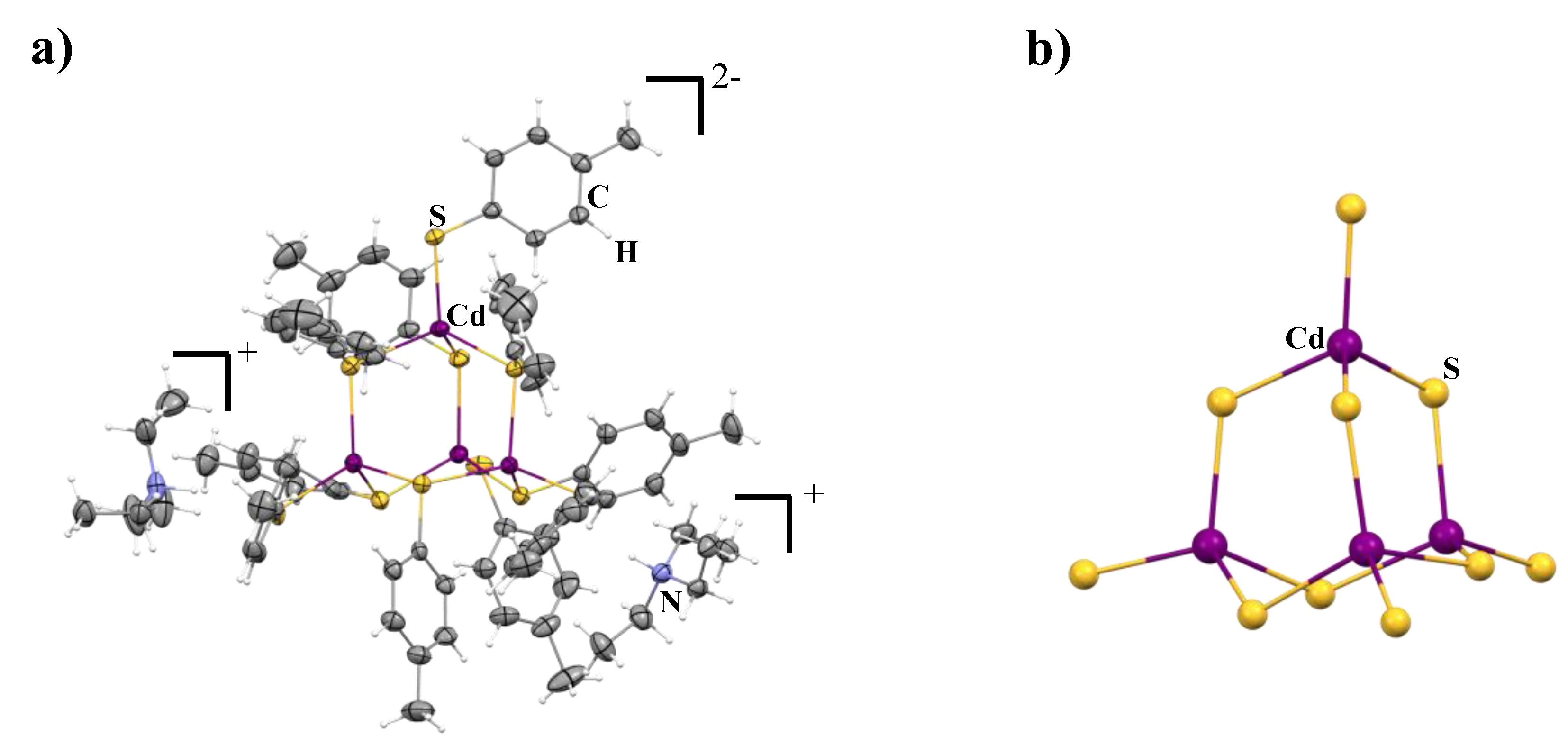
Synthesis, Structure, and Optical Properties of a Molecular Cluster Cd4(p-MBT)10

Interdisciplinary Materials Research Center, School of Materials Science and Engineering, Tongji University, Shanghai 201804, China
*
Authors to whom correspondence should be addressed.
Crystals 2022, 12(9), 1236; https://doi.org/10.3390/cryst12091236
Submission received: 25 July 2022 / Revised: 21 August 2022 / Accepted: 29 August 2022 / Published: 1 September 2022
(This article belongs to the Special Issue Coordination Environment for Metal Centers in Crystals)

Abstract

:
The creation of atomically precise nanoclusters has become an important research direction in nanoscience, because such nanomaterials can demonstrate unique chemo-physical properties that are significantly different from their corresponding bulk materials. The cause of such disparities lies in their different construction pattern for the atomic structures, in which the bulk materials display a highly symmetric, extended atomic lattice, while the ultrasmall nanoclusters feature low symmetric molecular structures. In this work, we report a new [HNEt3]2[Cd4(SC7H7)10] (denoted as Cd4(p-MBT)10, p-MBT = p-methylbenzene thiolate) nanocluster obtained through a one-pot synthetic pathway, and its atomic structure was revealed by single crystal X-ray diffraction technique. It shows that the molecular structure for Cd4(p-MBT)10 demonstrates the embryonic features of the corresponding bulk CdS. That is, the whole structure is built from four [CdS4] units which are connected to each other by shared corner S atoms. Due to the molecular nature, the structure of Cd4(p-MBT)10 is distorted, which yields two enantiomeric isomers with chiral Cd-S frameworks that co-crystallize into a non-chiral space group. In addition, the electronic structure was characterized by photoluminescence spectroscopy and calculated by density functional theory.

1. Introduction

Atomically precise nanoclusters (NCs) have become an emerging research direction in both fundamental and applied sciences [1,2,3]. The significance of the nanoclusters in nanoscience mainly lies in two aspects: (1) their unique position in the gap between small molecules and large nanoparticles (NPs), and (2) the nature of their almost perfect monodispersity at the atomic level. On account of their sub-nano dimensions (usually < 2 nm), [4] NCs can be considered as an isolatable intermediate phase between macroscopic nanoparticles and small molecules. In this respect, it provides deep insights into the kinetic control in the design and synthesis of desired NPs [5,6,7,8]. Meanwhile, the NCs can behave like both molecules and larger NPs, exhibiting their own distinct physiochemical properties due to the strong quantum confinement [9,10,11,12,13]. Therefore, NCs afford new platforms to explore untapped functional materials for a variety of interesting applications. In addition, because of their atomically homogenous structure, it makes the acquisition of single crystals of NCs possible and ultimately, the atomically precise structure becomes accessible through single crystal X-ray diffraction [14,15], which can offer important structural information such as the framework of the metals [16], the binding mode of organic ligands to the surface [17], the origin of chirality, etc. to probe the relationship of structure and property in depth [18,19].
In reality, the NCs based on noble metals, especially Au and Ag [1], dominate the explored NCs because of their relatively high stability and controllable synthetic processes. Therefore, such NCs have been used as models for delving into fundamental understandings of synthetic strategies [2], X-ray structures [15], ligand-induced structure transformations [20], chirality [21,22], isomerization and structure-related optical and catalytic properties [16,23,24]. In addition, based on the model structure of homometallic NCs, bi-/multi-metallic NCs have been further developed, greatly enriching the scope of the NC family [25,26] Those achievements are undoubtedly instructive in the later attempts of tactically designing new NCs, of which the structures and properties are no longer random, but predictable. However, the semiconducting nanoclusters, such as chalcogenides (e.g., CdS, CdSe) lag far behind the development of metallic nanoclusters. This is because difficulties in synthesis impede the effective acquisition of atomically precise nanoclusters, and the structure characterization for such small nanomaterials requires high-quality single crystals of the as-prepared nanoclusters [27].
In this work, we reported a seed structure of [HNEt3]2[Cd4(p-MBT)10] (thereafter denoted as Cd4(p-MBT)10) by employing a relatively rigid ligand p-MBT (p-MBT = p-methylbenzene thiolate) through a one-pot synthetic pathway in high yield. Such a molecular nanocluster has already demonstrated an embryonic structure of bulk CdS. Moreover, the molecular nature makes Cd4(p-MBT)10 show a structural distortion for the Cd-S framework, which generates chirality in the structure.

2. Materials and Methods

2.1. Materials and Measurements

All of the manipulations were carried out in a dry, oxygen-free argon atmosphere using standard Schlenk and glove box techniques. Cadmium perchlorate hydrate (Cd(ClO4)2·xH2O) and tetramethylammonium chloride ((CH3)4N+Cl) were purchased from Sigma-Aldrich, St. Louis, MO, USA. p-methylbenzene thiolate (CH3C6H4SH) was purchased from Acros, Chaoyang District, Beijing, China. Triethylamine (N(CH2CH3)3) and methanol (CH3OH) were purchased from General-reagent, Xuhui District, Shanghai, China. The photoluminescence excitation and photoluminescence spectra were detected and recorded by Fluor Spectro photometer (Hitachi, FL4700, Chiyoda Ward, Tokyo, Japan) at room temperature in methanol. The UV-Vis spectrum was recorded by UV-Vis spectrophotometer (INESA, L6S, Xuhui District, Shanghai, China) with Beer–Lambert Law at room temperature in methanol.

2.2. Synthesis of Cd4(p-MBT)10

Firstly, 100.0 mg Cd(ClO4)2·xH2O (0.32 mmol) was loaded in a 50 mL tube Schlenk flask and then 3 mL methanol was added, which resulted in a colorless, transparent solution. Next, 119 mg p- methylbenzene thiolate (p-MBT) (0.96 mmol) was loaded in a 50 mL tube Schlenk flask and then 3 mL of methanol was added into the flask, which resulted in a colorless, transparent solution. After that, 0.15 mL of triethylamine (TEA) (0.96 mmol) was injected into the p-MBT/methanol solution to produce deprotonated p-MBT. The resultant solution appears in a colorless, transparent form. Following this, 35 mg tetramethylammonium chloride (TMAC) (0.32 mmol) was loaded in a 50 mL tube Schlenk flask and then 3 mL of methanol was added, which resulted in a colorless, transparent solution. The above deprotonated p-MBT solution was transported to the Cd(ClO4)2·xH2O/methanol solution using a 5 mL syringe. As the solution was added, a white precipitate first appeared, then the solution became clear. The solution was then stirred for about 1 h, after which TMAC/methanol was added into the solution using a 5 mL syringe. White precipitation gradually occurred after mixing the above solutions, which could not be further dissolved. Subsequently, the filtrate was filtered off, and the filtrate was slowly volatilized (about 3 h) to obtain colorless crystals.

2.3. X-ray Crystallographic Procedures

Data collection of [(HNEt3)]2[Cd4(p-MBT)10] was performed on a Bruker D8 VENTURE X-ray diffractometer with PHOTON 100 CMOS detector equipped with a Mo-target. The Incoatec microfocus source IµS X-ray tube (λ = 0.71073 Å) at T = 193(2) K. Data reduction and integration were performed with the Bruker software package SAINT (version 8.38A) [28]. Data were corrected for absorption effects using the empirical methods as implemented in SADABS (version 2016/2) [29]. The structure was solved by SHELXT [30] and refined by full-matrix least-squares procedures using the Bruker SHELXTL (version 2018/3) software package through the OLEX2 graphical interface [31]. All non-hydrogen atoms were refined anisotropically. The H-atoms were also included at calculated positions and refined as riders, with Uiso(H) = 1.2 Ueq(C). The anisotropic displacement parameters in the direction of the bonds were restrained to be equal with a standard uncertainty of 0.004 Å2. They were also restrained to have the same Uij components, with a standard uncertainty of 0.01 Å2. Further crystal and data collection details are listed in Table 1 (Vide Infra).

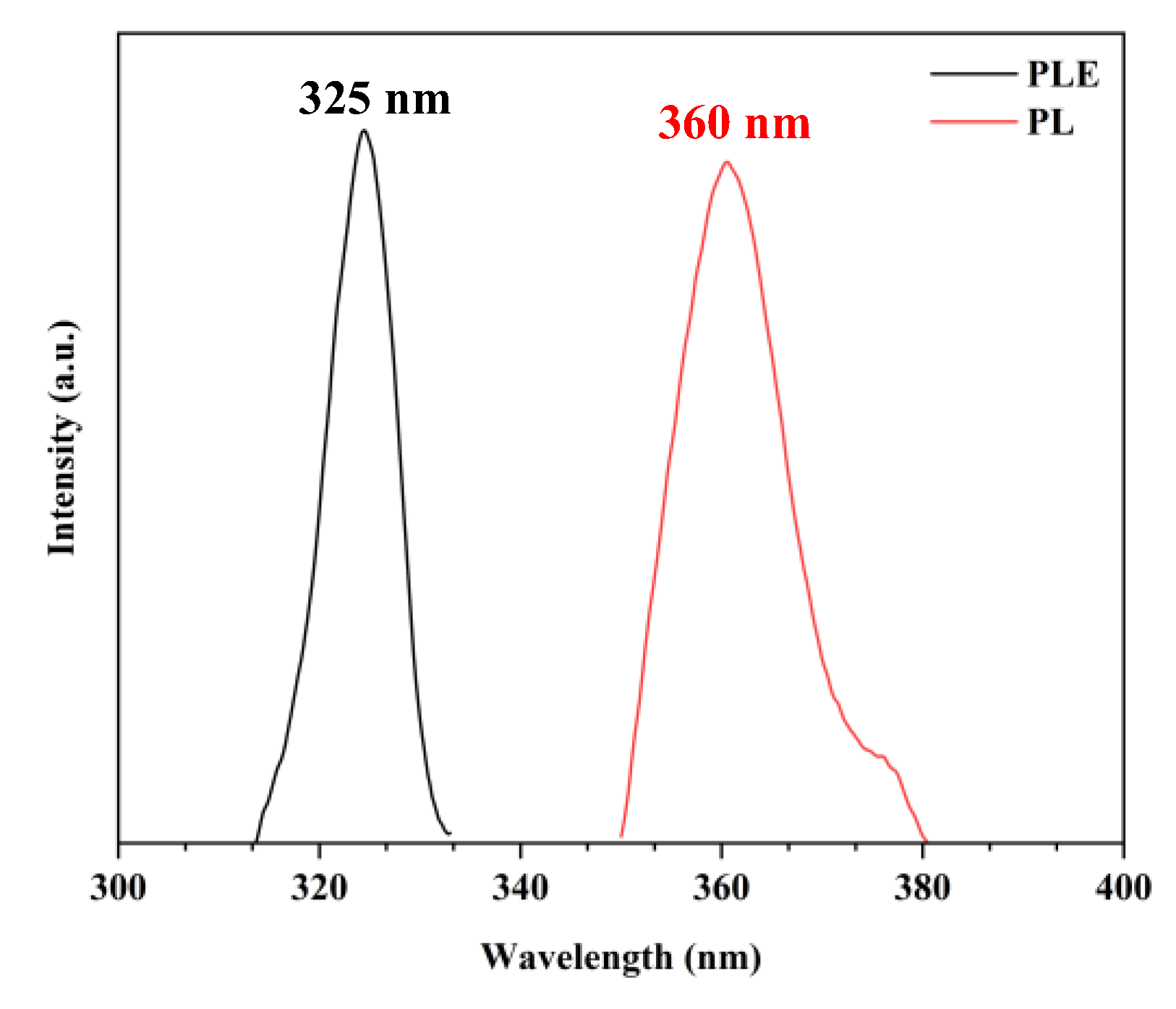
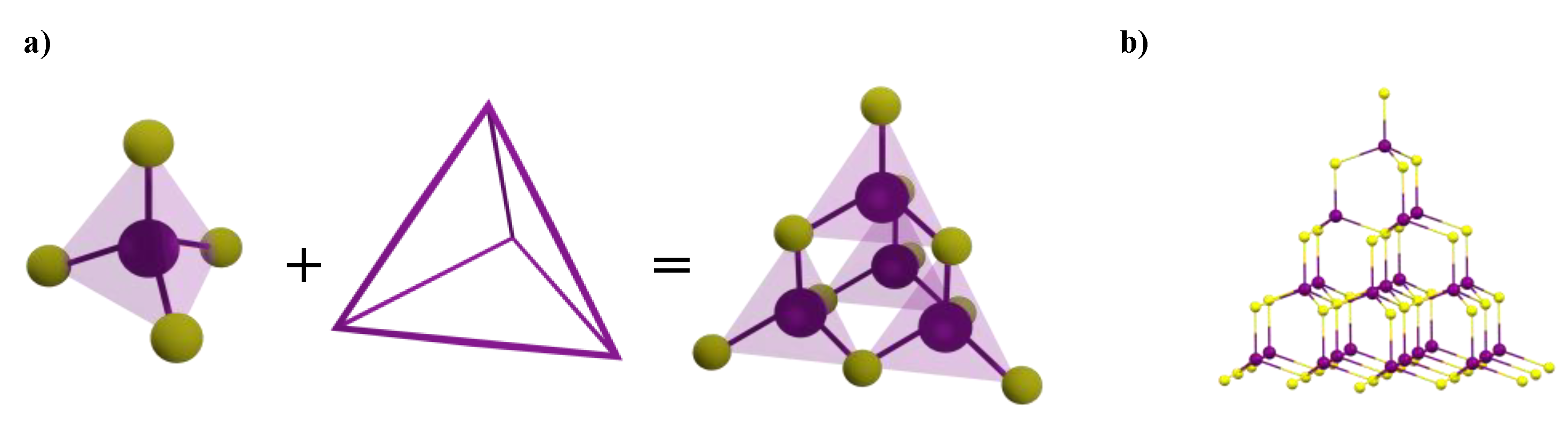
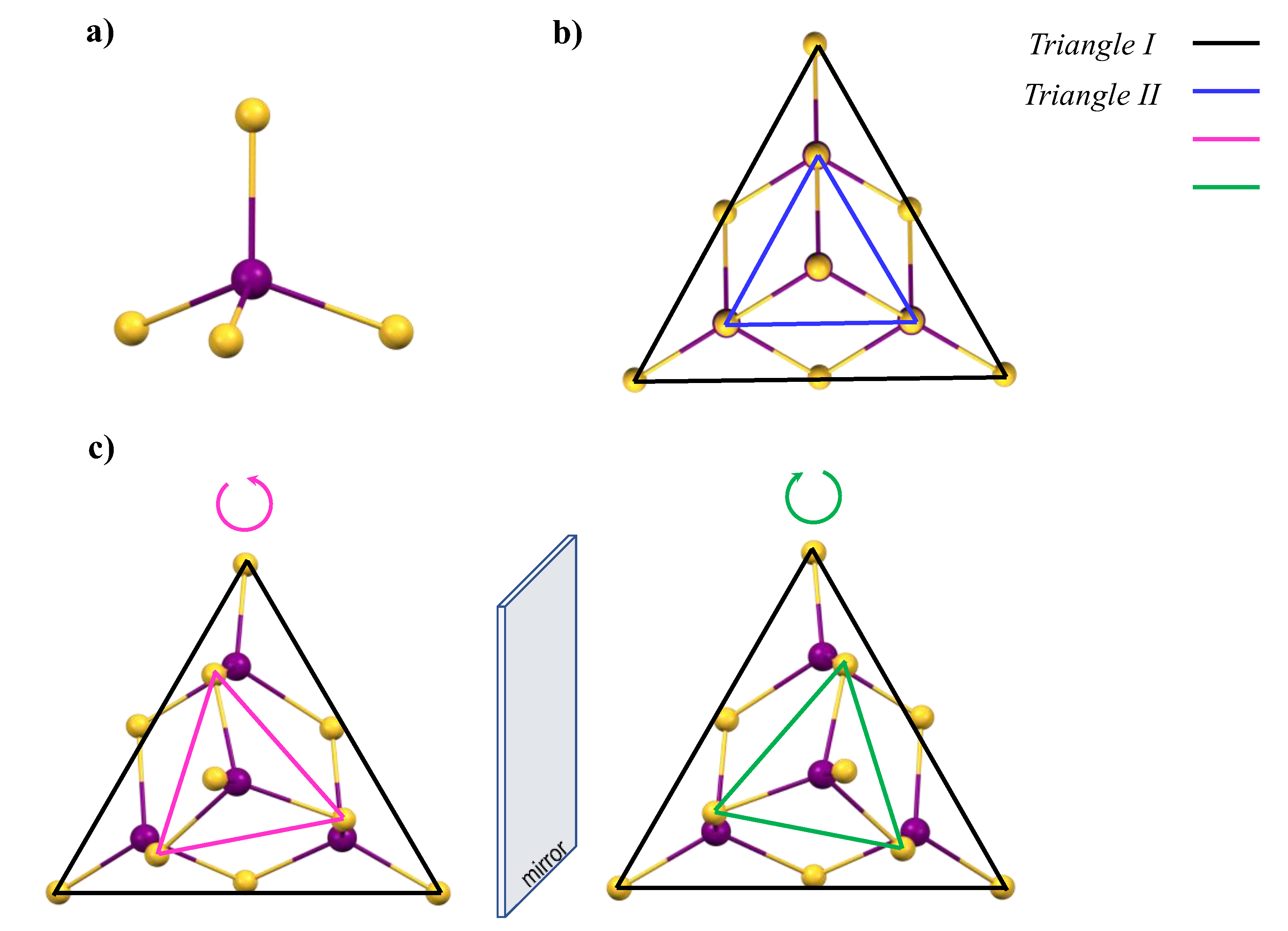
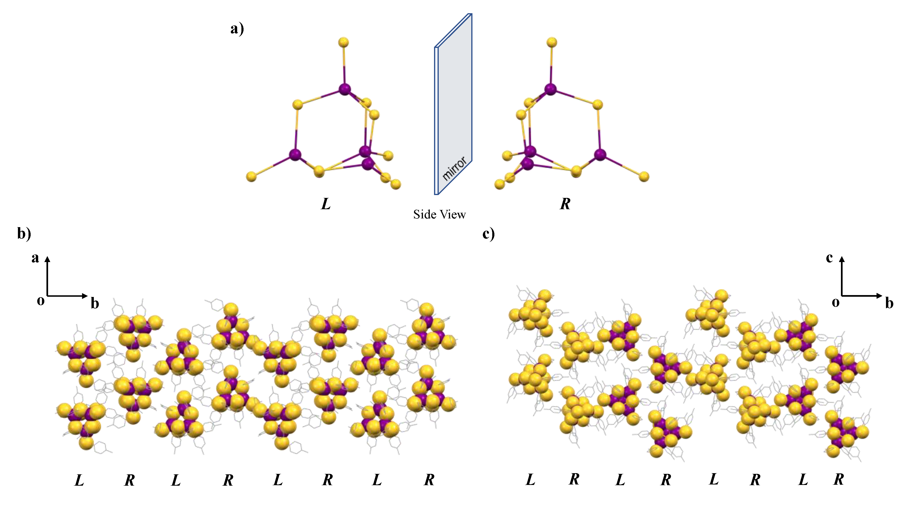
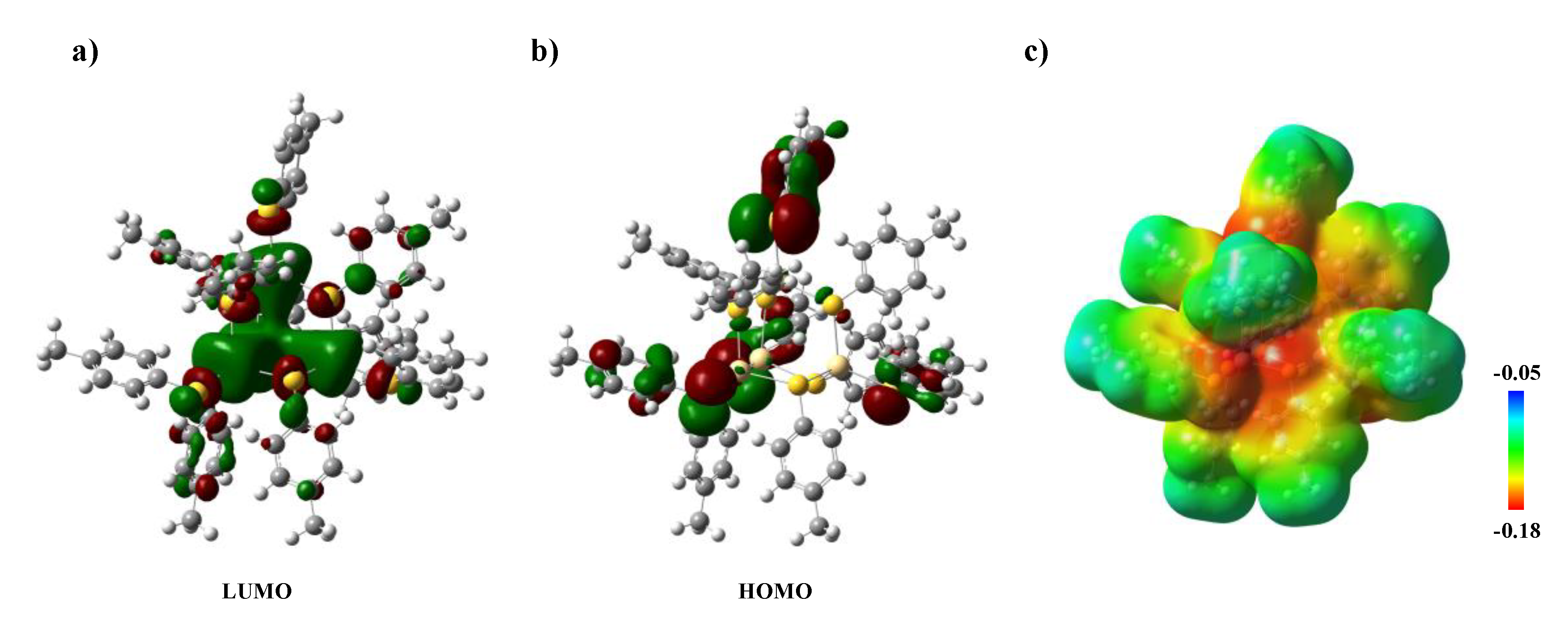
3. Results and Discussion

In this report, we have developed a robust synthetic route to access a new seed structure Cd4(p-MBT)10 for bulk CdS. Here, we employed p-MBT (Scheme 1) as the surfactant ligand, as its relatively rigid structure can help avoid severe structural disorder in crystallization. In addition, p-MBT has an –SH group where the S atom can participate in the construction of the Cd-S framework. Experimentally, the Cd4(p-MBT)10 cluster can be effectively prepared in a high yield through the following reaction (Equation (1)).
4Cd(ClO4)2 + 10HSC7H7 + 10NEt3 → [HNEt3]2[Cd4(SC7H7)10] + 8(HNEt3)(ClO4)
This reaction can be performed in solution in which methanol is used as the solvent to readily dissolve all the starting reagents, thus ensuring an efficient and homogeneous reaction process. Cd(ClO4)2 was employed as the Cd source owing to its high reactivity, and NEt3 acts as the Lewis base to facilitate the deprotonation of the neutral p-MBT. All the starting materials were mixed in a stoichiometric ratio in methanol, and the resultant colorless, transparent solution was stirred for 1 h. After that, tetramethylammonium chloride (TMAC) was added into the solution and a white precipitations were formed. The final Cd4(p-MBT)10 was crystallized from the filtrate, and it is interesting to note that the TMAC did not participate in the structure [32,33]. We also attempted to conduct a similar reaction process without TMAC, but it ultimately failed to crystallize the Cd4(p-MBT)10 product. It should be addressed here that although ammonium salts are widely used in the preparation of nanoclusters [8,14], the mechanism by which it participates is still not clearly understood. The as-synthesized Cd4(p-MBT)10 appears as a white crystalline powder and is stable in the open air, which endows the nanoclusters with great feasibility to be thoroughly analyzed by a variety of different techniques. The solubility test shows that the cluster is soluble in polar solvents, such as alcohols, acetone, or acetonitrile, but has limited solubility in non-polar solvents such as hexanes.
The solid-state structure of the Cd4(p-MBT)10 cluster was investigated by single crystal X-ray diffraction technique (SI, Figure S1). It is revealed that the Cd4(p-MBT)10 clusters crystalize into the space group of P21/n. In a unit cell, there are four clusters that are correlated through either the 21-screw axis or the n glide plane, resulting in two pairs of enantiomeric isomers. A full cluster is composed of two cationic [HNEt3]+ and one anionic [Cd4(p-MBT)10]2− units (Figure 1), which interact via the directional S···H−N contact (2.237 Å and 2.182 Å) based on the single crystal X-ray diffraction analysis (SI, Figure S2). It may be inferred that in addition to the electrostatic interactions, hydrogen bonding could also come into play among the ionic units, since the distances are comparable with other reported structures with clear hydrogen bonding interactions (SI, Figure S2 and Table S2) [34,35,36]. Based on single crystal X-ray diffraction analysis, the electrostatic effect or H-bonding could be the dominating attractive force on their own, or they both take effect in a synergistic way. Similarly, this type of hydrogen bond has also appeared in the structure of Fe2S6(TACN)2) [34]. At the same time, after DFT calculation, it is confirmed that the role of S···H−N interaction may play an important role in stabilizing the structure [37]. In a Cd4(p-MBT)10 unit, the 10 p-MBT ligands covalently bond to the Cd2+ centers through the S atoms after the deportation of the –SH group. Specifically, out of the ten ligands, four p-MBTs are simply donated to a single metal center, while the rest bridge the two neighboring Cd2+ cations, eventually constructing a 3-dimensional Cd4(p-MBT)10 structure, in which all Cd metals display a distorted tetrahedral coordination environment. It is interesting to note that such a tetrahedrally-coordinating pattern for Cd2+ is different from that in a conventional Cd-containing coordination compound, where the octahedron coordination environment is commonly seen because of the d-orbitals splitting into the t2g and eg energy levels for the Cd atom (SI, Figure S3) [38]. In fact, the Cd4S10 core in Cd4(p-MBT)10 cluster shows a great similarity to the basic unit of the atomic lattice for bulk CdS (Figure 2). Therefore, it is of interest to have a detailed structural comparison with the respective molecular and bulk counterparts, as the structural resemblance to the bulk Cd-S framework could offer Cd4(p-MBT)10 cluster semiconducting properties, and its molecule nature with ultrasmall size may bring about novel properties originated from the intrinsic quantum confinement as well. Bond distance inspection reveals that in the Cd4(p-MBT)10 cluster, the averaged Cd-S bond lengths are 2.475(7) Å and 2.558(6) Å (SI, Table S1) when the S atoms are involved in single-bond donating and µ2-bridging modes, respectively, which are comparable to those (Table 2) in other reported molecular structures [32,39,40,41,42]. Actually, such a Cd4S10 configuration is not rare and similar structures have been reported before (SI, Figure S4). It should be addressed here that in comparison to the two major polymorphs of bulk CdS, namely the hexagonal wurtzite and the cubic zinc blende, Cd4(p-MBT)10 displays more polarization in respect to the Cd-S bond distances. In the wurtzite form, the Cd-S bond distance is around 2.518 Å and 2.532 Å (Cd-S along the c axis, (SI, Figure S5) and the zinc blende CdS demonstrates an equal length of 2.520 Å for all Cd-S bonds [43,44].
Confining the epitaxial growth of the Cd-S lattice into the molecular level will result in structural distortion for the corresponding unit, and this can be used as an important means to introduce chirality into the new cluster. Bulk CdS has a highly symmetric atomic lattice that crystallizes in the achiral space groups of P63mc and F-43m. Picking out the Cd4S10 unit from the extended Cd-S framework shows that such a motif can be considered as the pyramidal construction of the four ideal CdS4 tetrahedrons (Figure 3a), which are linked by sharing their vertex S atoms. Looking straight down from the top S atom (top view of Cd4S10 unit (bulk materials), Figure 3b) clearly outlines its overall triagonal geometry by connecting the three most outside S atoms. Due to the high symmetry of the structure, the inner triangle built from the three µ2-S atoms is just a proportional scaling down from triangle I, whose shape and relative orientation remain the same. It is, however, not true in our newly obtained Cd4(p-MBT)10 cluster, which has a low symmetric, distorted Cd4S10 core structure (Figure 3c). Although the outer triangle I in Cd4(p-MBT)10 is still somewhat similar to that in bulk CdS, the respective triangle II is obviously rotating either clockwise or counterclockwise relative to the triangle I, generating distinct chiral features for the structure. Meanwhile, the two enantiomers with opposite chirality are arranged in the lattice in an alternating manner, through b-axis (Figure 4b,c). This case suggests that if one can effectively confine the nanocrystal growth at the molecular level, it will be possible to create an intrinsically chiral nanocluster without employing chiral surfactant ligands [45,46], even though the two enantiomeric isomers are likely to co-crystalize out to form a racemic mixture. Usually, such a packing pattern can be effectively directed by certain weak intermolecular interactions, such as hydrogen bonding, C−H···π, or π···π interactions, etc [47,48]. Structure inspection reveals that in the packed Cd4(p-MBT)10 assembly, the C−H···π intermolecular interactions are the dominating driving forces that dictate the construction of the final 3D network and more detailed discussion can be found in Supplementary Information (SI, Figures S6–S9).
Density functional theory (DFT) calculations were carried out at the B3LYP/def2-TZVP level to gain more insight into the electronic structure of the Cd4(p-MBT)10 nanocluster (SI, Table S4) [49]. To this end, we focused on the anionic core, the Cd4(p-MBT)102− unit. The electrostatic potential (ESP) map (Figure 5c) shows the electronic density is mainly concentrated on the S atoms of the p-MBT ligands in their ground state, and the four µ2-S atoms are slightly less negative compared with the µ-S ones. This points out their different binding abilities during the formation of the Cd4S10 core, which is consistent with the crystallographic analysis. Moreover, all the p-methylphenyl groups are slightly negatively charged, indicating a long-range molecular assembly might occur through intermolecular interactions between adjacent Cd4(p-MBT)10 nanoclusters. Notably, the LUMO and HOMO of Cd4(p-MBT)10 (Figure 5a,b) suggests a ligand-to-metal charge transfer (LMCT) might occur during excitation between the Cd2+ ions and the p-MBT ligands with µ-S atoms, during which process the p-methylphenyl groups act as an electron reservoir to be involved in the HOMO-LUMO transition. In this regard, tailoring the phenyl rings by substituting different groups, such as methyl and ethyl at different positions may be an effective lever to engineer its electronic structure, which in turn tunes the overall optical properties of the nanocluster. Moreover, the HOMO-LUMO energy difference is well aligned with its absorption behavior (SI, Figure S10). We also tested the photoluminescent property for the newly obtained Cd4(p-MBT)10 (Figure 6). The results show that there are obvious photoexcitation and emission peaks located at 325 nm and 360 nm, respectively (Stokes shift = 35 nm). The near ultraviolet PL (photoluminescence) peak has a great agreement with its intrinsic ultrasmall size. The results are also in line with the previously reported structures. For example, Xiong and co-workers have previously synthesized a [Cd10S4(SPh)16]4− tetra-anionic nanocluster, which displays PLE (Photoluminescence excitation) and PL peaks at 350 nm and 430 nm, respectively [50]. It should be noted again that such a small nanocluster still has a molecular nature with strong quantum confinement. With the number of atoms increasing through crystal growth, the PLE and PL peaks will be gradually red-shifted, accompanied by enlargement of the Stokes shift. For example, [Cd10S4(SPh)16]4− shows PLE and PL at 350 nm and 430 nm (Stokes shift = 80 nm) [50], while a larger Cd32S14(SC6H6)36·DMF4 nanocluster demonstrates those at 384 nm and 500 nm (Stokes shift = 116 nm) [51]. Going beyond the scope of the atomically precise nanoclusters, monodispersed nanoparticles also obey such trends; for instance, ~14 nm and ~21 nm CdS nanoparticles have a PLE/PL of 445 nm/536 nm and 480 nm/589 nm, respectively (see SI Table S3 for more details) [52,53]. Therefore, it seems such a Cd4(p-MBT)10 nanocluster acquired in this work not only represents the very basic unit for bulk CdS, but also hits the borderline of the optical spectrum for this system.

4. Conclusions

In summary, a molecular structure Cd4(p-MBT)10 has been successfully synthesized in a high yield through a facile one-pot synthetic approach and its atomic structure was well-established by single crystal X-ray diffraction technique. It is revealed that the Cd4(p-MBT)10 cluster possesses embryonic atomic configuration for bulk CdS, but the Cd4S10 core structure in cluster is slightly distorted because of its molecular nature, eventually bringing about two enantiomers that packed together in an alternating manner. In addition, the electronic structure of the title cluster has been investigated by photoluminescence spectroscopy, and it shows a relatively large Stokes shift which is mainly caused by the LMCT process, which was confirmed by DFT calculation. The Cd4(p-MBT)10 cluster reported in the current work demonstrates structural similarities to bulk CdS, which may imply that it can serve as a seed structure for growing larger CdS nanocrystals.

Supplementary Materials

The following supporting information can be downloaded at: https://www.mdpi.com/article/10.3390/cryst12091236/s1, Figure S1: Solid-state structure of [(HNEt3)]2[Cd4(p-MBT)10] drawn with thermal ellipsoids at the 40% probability level. Hydrogen atoms are represented by spheres of arbitrary radius. Only cadmium and sulfur atoms are labeled; Table S1: Selected bond distances (Å) and angles (deg.) in the structure of [(HNEt3)]2[Cd4(p-MBT)10], Black and blue are results obtained from single crystal structure and theoretical calculation, respectively; Figure S2: Two kinds of S···H−N interaction in [(HNEt3)]2[Cd4(p-MBT)10]; Table S2: N−H···S interaction comparison of the [(HNEt3)2][Cd4(p-MBT)10] cluster with the related compound; Figure S3: Total structure of [Cd1.5(bbta)1.5(NCS)3]n polymer. Such a structure shows the octahedron coordination environment for Cd2+. All hydrogen atoms are omitted for clarity; Figure S4: Total structure of Cd4(SPh)102−. This structure shows the Cd4S10 configuration obtained from the most used thiophenol ligand. All hydrogen atoms are omitted for clarity; Figure S5: (a) Two views of bulk CdS wurtzite structure. (b) Viewed along c axis. Cd–S bond distance on this plane is equal. (c) Cd–S bond distance along the c axis on this plane is not equal to those in other directions; Figure S6: C−H···π interaction between p-MBT and HNEt3+: 2.590–2.674 Å.; Figure S7: C−H···π interaction between the neighboring p-MBT ligands.; Figure S8: C−H···π and hydrogen bonding interactions between p-MBT ligand and HNEt3+: 2.708/2.749 Å and H-bonding: 2.237 Å.; Figure S9: Single crystal packing structure of [(HNEt3)2][Cd4(p-MBT)10] through different interactions.; Table S3: Photoluminescence excitation and photoluminescence comparison of the Cd4(p-MBT)10 cluster with the related compound; Figure S10: The UV-Vis spectrum of [(HNEt3)]2[Cd4(p-MBT)10]; Table S4: Cartesian coordinates of the Cd4(p-MBT)102–. Refs. [35,36,37,38,39,43,49,50,51,52,53] have been cited in supplementary materials.

Author Contributions

C.X. conducted synthesis and obtained suitable single crystals for X-ray diffraction analysis; Z.Z. performed theoretical calculations. H.H. conceived this project, supervised and guided the design, analysis and interpretation, and wrote the manuscript. All authors have read and agreed to the published version of the manuscript.

Funding

This work is supported by the Fundamental Research Funds for the Central Universities (22120220118 and 22120220111) and National Natural Science Foundation of China (22101205). The authors also thank Prof. Zhen Fan from Tongji University for measuring absorption and photoluminescence spectroscopy.

Data Availability Statement

Data in this study is available upon request.

Conflicts of Interest

The authors declare no conflict of interest.

References

  1. Jin, R.; Zeng, C.; Zhou, M.; Chen, Y. Atomically Precise Colloidal Metal Nanoclusters and Nanoparticles: Fundamentals and Opportunities. Chem. Rev. 2016, 116, 10346–10413. [Google Scholar] [CrossRef] [PubMed]
  2. Cook, A.W.; Hayton, T.W. Case Studies in Nanocluster Synthesis and Characterization: Challenges and Opportunities. Acc. Chem. Res. 2018, 51, 2456–2464. [Google Scholar] [CrossRef]
  3. Singh, V.; Priyanka; More, P.V.; Hemmer, E.; Mishra, Y.K.; Khanna, P.K. Magic-Sized CdSe Nanoclusters: A Review on Synthesis, Properties and White Light Potential. Mater. Adv. 2021, 2, 1204–1228. [Google Scholar] [CrossRef]
  4. Qian, H.; Zhu, M.; Wu, Z.; Jin, R. Quantum Sized Gold Nanoclusters with Atomic Precision. Acc. Chem. Res. 2012, 45, 1470–1479. [Google Scholar] [CrossRef] [PubMed]
  5. Wei, X.; Xu, C.; Li, H.; Kang, X.; Zhu, M. Fabrication of a Family of Atomically Precise Silver Nanoclusters via Dual-Level Kinetic Control. Chem. Sci. 2022, 13, 5531–5538. [Google Scholar] [CrossRef] [PubMed]
  6. Li, Q.; Huang, B.; Yang, S.; Zhang, H.; Chai, J.; Pei, Y.; Zhu, M. Unraveling the Nucleation Process from a Au(I)-SR Complex to Transition-Size Nanoclusters. J. Am. Chem. Soc. 2021, 143, 15224–15232. [Google Scholar] [CrossRef]
  7. Touchton, A.J.; Wu, G.; Hayton, T.W. Understanding the Early Stages of Nickel Sulfide Nanocluster Growth: Isolation of Ni3, Ni4, Ni5, and Ni8 Intermediates. Small 2020, 17, 2003133. [Google Scholar] [CrossRef]
  8. Zhu, M.; Lanni, E.; Garg, N.; Bier, A.M.E.; Jin, R. Kinetically Controlled, High-Yield Synthesis of Au25 Clusters. J. Am. Chem. Soc. 2008, 130, 1138–1139. [Google Scholar] [CrossRef]
  9. Zhu, C.; Xin, J.; Li, J.; Li, H.; Kang, X.; Pei, Y.; Zhu, M. Fluorescence or Phosphorescence? The Metallic Composition of the Nanocluster Kernel Does Matter. Angew. Chem. Int. Ed. 2022, 31, e202205947. [Google Scholar] [CrossRef]
  10. Wan, X.; Wang, J.; Wang, Q.-M. Ligand-Protected Au55 with a Novel Structure and Remarkable CO2 Electroreduction Performance. Angew. Chem. Int. Ed. 2021, 133, 20916–20921. [Google Scholar] [CrossRef]
  11. Li, Y.; Song, Y.; Zhang, X.; Liu, T.; Xu, T.; Wang, H.; Jiang, D.-E.; Jin, R. Atomically Precise Au42 Nanorods with Longitudinal Excitons for an Intense Photothermal Effect. J. Am. Chem. Soc. 2022, 144, 12381–12389. [Google Scholar] [CrossRef] [PubMed]
  12. Wu, W.-H.; Zeng, H.-M.; Yu, Z.-N.; Wang, C.; Jiang, Z.-G.; Zhan, C.-H. Unusual Structural Transformation and Luminescence Response of Magic-Size Silver(I) Chalcogenide Clusters via Ligand-Exchange. Chem. Commun. 2021, 57, 13337–13340. [Google Scholar] [CrossRef]
  13. Jiang, Z.-G.; Wu, W.-H.; Jin, B.-X.; Zeng, H.-M.; Jin, Z.-G.; Zhan, C.-H. A Chloride-Doped Silver-Sulfide Cluster [Ag148S26Cl30(C@CBut)60]6+: Hierarchical Assembly, Enhanced Luminescence and Cytotoxicity to Cancer Cells. Nanoscale 2021, 14, 1971–1977. [Google Scholar] [CrossRef]
  14. Zhu, M.; Aikens, C.M.; Hollander, F.J.; Schatz, G.C.; Jin, R. Correlating the Crystal Structure of a Thiol-Protected Au25 Cluster and Optical Properties. J. Am. Chem. Soc. 2008, 130, 5883–5885. [Google Scholar] [CrossRef] [PubMed]
  15. Jadzinsky, P.D.; Calero, G.; Ackerson, C.J.; Bushnell, D.A.; Kornberg, R.D. Structure of a Thiol Monolayer-Protected Gold Nanoparticle at 1.1 A Resolution. Science 2007, 318, 430–433. [Google Scholar] [CrossRef] [PubMed]
  16. Chen, Y.; Liu, C.; Tang, Q.; Zeng, C.; Higaki, T.; Das, A.; Jiang, D.-E.; Rosi, N.L.; Jin, R. Isomerism in Au28(SR)20 Nanocluster and Stable Structures. J. Am. Chem. Soc. 2016, 138, 1482–1485. [Google Scholar] [CrossRef]
  17. Gary, D.C.; Flowers, S.E.; Kaminsky, W.; Petrone, A.; Li, X.; Cossairt, B.M. Single-Crystal and Electronic Structure of a 1.3 nm Indium Phosphide Nanocluster. J. Am. Chem. Soc. 2016, 138, 1510–1513. [Google Scholar] [CrossRef]
  18. Li, Q.; Zhou, M.; So, W.Y.; Huang, J.; Li, M.; Kauffman, D.R.; Cotlet, M.; Higaki, T.; Peteanu, L.A.; Shao, Z.; et al. A Mono-Cuboctahedral Series of Gold Nanoclusters: Photoluminescence Origin, Large Enhancement, Wide Tunability, and Structure–Property Correlation. J. Am. Chem. Soc. 2019, 141, 5314–5325. [Google Scholar] [CrossRef]
  19. Liang, X.-Q.; Li, Y.-Z.; Wang, Z.; Zhang, S.-S.; Liu, Y.-C.; Cao, Z.-Z.; Feng, L.; Gao, Z.-Y.; Xue, Q.-W.; Tung, C.-H.; et al. Revealing the Chirality Origin and Homochirality Crystallization of Ag14 Nanocluster at the Molecular Level. Nat. Commun. 2021, 12, 1–10. [Google Scholar] [CrossRef]
  20. Zeng, Y.; Havenridge, S.; Gharib, M.; Baksi, A.; Weerawardene, K.L.D.M.; Ziefuß, A.R.; Strelow, C.; Rehbock, C.; Mews, A.; Barcikowski, S.; et al. Impact of Ligands on Structural and Optical Properties of Ag29 Nanoclusters. J. Am. Chem. Soc. 2021, 143, 9405–9414. [Google Scholar] [CrossRef]
  21. Shen, H.; Xu, Z.; Wang, L.; Han, Y.; Liu, X.; Malola, S.; Teo, B.K.; Häkkinen, H.; Zheng, N. Tertiary Chiral Nanostructures from C−H···F Directed Assembly of Chiroptical Superatoms. Angew. Chem. Int. Ed. 2021, 133, 22585–22590. [Google Scholar] [CrossRef]
  22. Li, Y.; Higaki, T.; Du, X.; Jin, R. Chirality and Surface Bonding Correlation in Atomically Precise Metal Nanoclusters. Adv. Mater. 2020, 32, 1905488. [Google Scholar] [CrossRef] [PubMed]
  23. Kang, X.; Zhu, M. Tailoring the Photoluminescence of Atomically Precise Nanoclusters. Chem. Soc. Rev. 2019, 48, 2422–2457. [Google Scholar] [CrossRef]
  24. Du, X.; Jin, R. Atomically Precise Metal Nanoclusters for Catalysis. ACS Nano 2019, 13, 7383–7387. [Google Scholar] [CrossRef] [PubMed]
  25. Ghosh, A.; Mohammed, O.F.; Bakr, O.M. Atomic-Level Doping of Metal Clusters. Acc. Chem. Res. 2018, 51, 3094–3103. [Google Scholar] [CrossRef]
  26. Kang, X.; Li, Y.; Zhu, M.; Jin, R. Atomically Precise Alloy Nanoclusters: Syntheses, Structures, and Properties. Chem. Soc. Rev. 2020, 49, 6443–6514. [Google Scholar] [CrossRef]
  27. Beecher, A.N.; Yang, X.; Palmer, J.H.; LaGrassa, A.L.; Juhas, P.; Billinge, S.J.L.; Owen, J.S. Atomic Structures and Gram Scale Synthesis of Three Tetrahedral Quantum Dots. J. Am. Chem. Soc. 2014, 136, 10645–10653. [Google Scholar] [CrossRef]
  28. SAINT. Part of Bruker APEX3 Software Package (version 2016.9-0); Bruker AXS: Billerica, MA, USA, 2016. [Google Scholar]
  29. SADABS. Part of Bruker APEX3 Software Package (version 2016.9-0); Bruker AXS: Billerica, MA, USA, 2016. [Google Scholar]
  30. Sheldrick, G.M. SHELXT—Integrated Space-Group and Crystal-Structure Determination. Acta Crystallogr. Sect. A Found. Adv. 2015, 71, 3–8. [Google Scholar] [CrossRef]
  31. Sheldrick, G.M. Crystal Structure Refinement with SHELXL. Acta Crystallogr. Sect. C Struct. Chem. 2015, C71, 3–8. [Google Scholar] [CrossRef]
  32. Hencher, J.; Khan, M.A.; Said, F.F.; Tuck, D.G. The Direct Electrochemical Synthesis and Crystal Structure of Salts of [M4(SC6H5)10]2− Anions (M = Zn, Cd). Polyhedron 1985, 4, 1263–1267. [Google Scholar] [CrossRef]
  33. Sugiuchi, M.; Shichibu, Y.; Konishi, K. An Inherently Chiral Au24 Framework with Double-Helical Hexagold Strands. Angew. Chem. Int. Ed. 2018, 57, 7855–7859. [Google Scholar] [CrossRef] [PubMed]
  34. Moreland, A.C.; Rauchfuss, T.B. Synthesis, Structure, and Reactivity of a Remarkable Iron Sulfide: (TACN)2Fe2S6. J. Am. Chem. Soc. 1998, 120, 9376–9377. [Google Scholar] [CrossRef]
  35. Baba, T.; Okamura, T.; Yamamoto, H.; Yamamoto, T.; Ueyama, N. Zinc, Cadmium, and Mercury 1,2-Benzenedithiolates with Intramolecular NH···S Hydrogen Bonds. Inorg. Chem. 2008, 47, 2837–2848. [Google Scholar] [CrossRef] [PubMed]
  36. Kato, M.; Kojima, K.; Okamura, T.; Yamamoto, H.; Yamamura, T.; Ueyama, N. Relation between Intramolecular NH···S Hydrogen Bonds and Coordination Number in Mercury(II) Complexes with Carbamoylbenzenethiol Derivatives. Inorg. Chem. 2005, 44, 4037–4044. [Google Scholar] [CrossRef]
  37. François, S.; Rohmer, M.-M.; Bénard, M.; Moreland, A.C.; Rauchfuss, T.B. The N−H···S Hydrogen Bond in (TACN)2Fe2S6 (TACN = Triazacyclononane) and in Model Systems Involving the Persulfido Moiety: An ab Initio and DFT Study. J. Am. Chem. Soc. 2000, 122, 12743–12750. [Google Scholar] [CrossRef]
  38. Zhou, J.; Peng, Y.; Zhang, Y.; Li, B.; Zhang, Y. Synthesis, Crystal Structure and Luminescent Properties of a Novel Cadmium Coordination Polymer with Unprecedented One-Dimensional Hetero-Triple-Stranded Chain. Inorg. Chem. Commun. 2004, 7, 1181–1183. [Google Scholar] [CrossRef]
  39. Zhang, X.; Tian, Y.; Jin, F.; Wu, J.; Xie, Y.; Tao, X.; Jiang, M. Self-Assembly of an Organic Chromophore with Cd−S Nanoclusters: Supramolecular Structures and Enhanced Emissions. Cryst. Growth Des. 2005, 5, 565–570. [Google Scholar] [CrossRef]
  40. Jiang, J.-B.; Bian, G.-Q.; Zhang, Y.-P.; Luo, W.; Zhu, Q.-Y.; Dai, J. Anion–Cation Charge-Transfer Properties and Spectral Studies of [M(phen)3][Cd4(SPh)10] (M = Ru, Fe, and Ni). Dalton Trans. 2011, 40, 9551–9556. [Google Scholar] [CrossRef] [PubMed]
  41. Tang, X.-Y.; Yuan, R.-X.; Chen, J.-X.; Zhao, W.; Zheng, A.-X.; Yu, M.; Li, H.-X.; Ren, Z.-G.; Lang, J.-P. Group 12 Metal Zwitterionic Thiolate Compounds: Preparation and Structural Characterization. Dalton Trans. 2012, 41, 6162–6172. [Google Scholar] [CrossRef]
  42. Xu, C.; Shi, H.-T.; Xin, Z.-F.; Jia, A.-Q.; Zhang, Q.-F. Ligand-Participated Formation of Covalent Superlattices Containing [Cd4(μ-SPh)6] Cores with Adamantane-Like Stereochemistry. J. Clust. Sci. 2014, 25, 1353–1361. [Google Scholar] [CrossRef]
  43. Xu, Y.-N.; Ching, W.Y. Electronic, Optical, and Structural Properties of Some Wurtzite Crystals. Phys. Rev. B 1993, 48, 4335–4351. [Google Scholar] [CrossRef] [PubMed]
  44. Ulrich, F.; Zachariasen, W. XIV. Über die Kristallstruktur des «- und ß-CdS, sowie des Wurtzits. Z. Krist. - Cryst. Mater. 1925, 62, 260–273. [Google Scholar] [CrossRef]
  45. Han, H.; Yao, Y.; Bhargava, A.; Wei, Z.; Tang, Z.; Suntivitch, J.; Voznyy, O.; Robinson, R.D. Tertiary Hierarchical Complexity in Assemblies of Sulfur-Bridged Metal Chiral Clusters. J. Am. Chem. Soc. 2020, 142, 14495–14503. [Google Scholar] [CrossRef] [PubMed]
  46. Zeng, C.; Chen, Y.; Kirschbaum, K.; Lambright, K.J.; Jin, R. Emergence of Hierarchical Structural Complexities in Nanoparticles and Their Assembly. Science 2016, 354, 1580–1584. [Google Scholar] [CrossRef]
  47. Ashfaq, M.; Ali, A.; Tahir, M.N.; Khalid, M.; Assiri, M.A.; Imran, M.; Munawar, K.S.; Habiba, U. Synthetic Approach to Achieve Halo Imine Units: Solid-State Assembly, DFT Based Electronic and Non Linear Optical Behavior. Chem. Phys. Lett. 2022, 803, 139843–139851. [Google Scholar] [CrossRef]
  48. Haroon, M.; Akhtar, T.; Yousuf, M.; Tahir, M.N.; Rasheed, L.; Zahra, S.S.; Haq, I.U.; Ashfaq, M. Synthesis, Crystal Structure, Hirshfeld Surface Investigation and Comparative DFT Studies of Ethyl 2-[2-(2-nitrobenzylidene)hydrazinyl] thiazole-4-carboxylate. BMC Chem. 2022, 16, 1–17. [Google Scholar] [CrossRef] [PubMed]
  49. Frisch, M.J.; Trucks, G.W.; Schlegel, H.B.; Scuseria, G.E.; Robb, M.A.; Cheeseman, J.R.; Scalmani, G.; Barone, V.; Mennucci, B.; Petersson, G.A.; et al. Gaussian 09, Revision D.01; Gaussian, Inc.: Wallingford, CT, USA, 2013. [Google Scholar]
  50. Adams, R.D.; Zhang, B.; Murphy, C.J.; Yeung, L.K. Halide Enhancement of the Luminescence of Cd10S4 Thiolate Clusters. Chem. Commun. 1999, 383–384. [Google Scholar] [CrossRef]
  51. Herron, N.; Calabrese, J.C.; Farneth, W.E.; Wang, Y. Crystal Structure and Optical Properties of Cd32S14(SC6H5)36·DMF4, a Cluster with a 15 Angstrom CdS Core. Science 1993, 259, 1426–1428. [Google Scholar] [CrossRef]
  52. Xue, X.; Wang, X.S.; Wang, L.Z.; Xiong, R.G.; Abrahams, B.F.; You, X.Z.; Xue, Z.L.; Che, C.M. Hydrothermal Preparation of Novel Cd(II) Coordination Polymers Employing 5-(4-Pyridyl)tetrazolate as a Bridging Ligand. Inorg. Chem. 2002, 41, 6544–6546. [Google Scholar] [CrossRef]
  53. Thomas, B.; Jose, E.T.; Chacko, J.K.; Divya, K.V. Green Light Emitting Cadmium Sulfide Nanoparticles with Coral Surface Morphology. J. Clust. Sci. 2021, 1–13. [Google Scholar] [CrossRef]
Scheme 1. Schematic structure of the p-MBT ligand.
Crystals 12 01236 sch001
Figure 1. (a) Crystal structure of the [(HNEt3)]2[Cd4(p-MBT)10] cluster drawn with thermal ellipsoids at the 40% probability level. Hydrogen atoms are represented by spheres of arbitrary radius. (b) Inner core structure of the Cd-S framework drawn with ball and stick model.
Crystals 12 01236 g001
Figure 2. (a) Schematic configuration of Cd4S10 smallest unit. (b) Structure of CdS bulk materials [44].
Crystals 12 01236 g002
Figure 3. (a) Ideal CdS4 tetrahedrons. (b) Top view of Cd4S10 unit (bulk materials) [44]. (c) Top view of two Cd4S10 enantiomers in Cd4(p-MBT)10 (Left is counterclockwise and Right is clockwise).
Crystals 12 01236 g003
Figure 4. Packing mode of Cd4(p-MBT)10 enantiomers. (a) Two Cd4S10 core enantiomers (side view). (b) Packing mode viewed along c axis. (c) Packing mode viewed along a axis. All hydrogen atoms are omitted for clarity.
Crystals 12 01236 g004
Figure 5. (a) The electron densities of the lowest unoccupied molecular orbital (LUMO). (b) The electron densities of the highest occupied molecular orbital (HOMO). (c) Electrostatic potential (ESP) maps.
Crystals 12 01236 g005
Figure 6. Photoluminescence excitation (PLE) and Photoluminescence (PL) spectra.
Crystals 12 01236 g006
Table 1. Crystal data and structure refinement parameters for [(HNEt3)]2[Cd4(p-MBT)10].
Compound[(HNEt3)]2[Cd4(p-MBT)10]
Empirical formulaC82H102Cd4N2S10
Formula weight1885.85
Temperature (K)193.00
Wavelength (Ǻ)0.71073
Crystal systemMonoclinic
Space groupP21/n
a (Å)13.4250(4)
b (Å)42.2528(11)
c (Å)15.5838(5)
α (°)90
β (°)98.8170(10)
γ (°)90
V3)8735.4(4)
Z4
ρcalcd (g·cm−3)1.434
µ (mm−1)1.240
F(000)3840
Crystal size (mm)0.2 × 0.1 × 0.05
θ range for data collection (°)2.378–33.621
Reflections collected252515
Independent reflections42272
[Rint = 0.0779]
Transmission factors (min/max)1/0.917
Data/restraints/params.42272/497/992
R1, a wR2 b(I > 2σ(I))0.0550/0.0953
R1, a wR2 b (all data)0.0849/0.1047
Quality-of-fit c1.081
Largest diff. peak and hole (ē·Å−3)0.827 and −0.625
aR1 = Σ||Fo| − |Fc||/Σ|Fo|. b wR2 = [Σ[w(Fo2Fc2)2/Σ[w(Fo2)2]]. c Quality-of-fit = [Σ[w(Fo2Fc2)2]/(NobsNparams)]½, based on all data.
Table 2. Bond distance comparison of the Cd4(p-MBT)10 cluster with the related structures.
Compound.Sp. Gr.M − S a (Å)M − S b (Å)
[(HNEt3)]2[Cd4(p-MBT)10]P21/n2.475(7)2.558(6)
[A]2[Cd4(SPh)10] [39]P-12.460(3)2.555(3)
[Fe(phen)3][Cd4(SPh)10] [40]P-12.467(2)2.560(2)
[Cd4(Tab)10](PF6)8 [41]P-12.4542(8)2.5470(8)
[(NMe4)]2(Cd4(SPh)9Cl) [42]P-12.465(2)2.5505(2)
[(HNEt3)]2[Zn4(SPh)10] [32]P-12.286(6)2.370(1)
α -CdS (wurtzite) [43]P63mc2.5182.532
β -CdS (zinc blende) [44]F-43m2.520
Note: a = sulfur atoms display a donating mode. b = sulfur atoms display a bridging mode; M = Cd or Zn; A = trans-4-(4-dimethylanilinostyryl)-N-methyl-pyridinium.
Publisher’s Note: MDPI stays neutral with regard to jurisdictional claims in published maps and institutional affiliations.

