
Sustainable Synthesis and Characterization of Zinc Oxide Nanoparticles Using Raphanus sativus Extract and Its Biomedical Applications

 ,
,  , , ,
, , ,  , and
, and
Abstract
:1. Introduction
2. Resources and Techniques
2.1. Chemical Used
2.2. Phytochemical Analysis
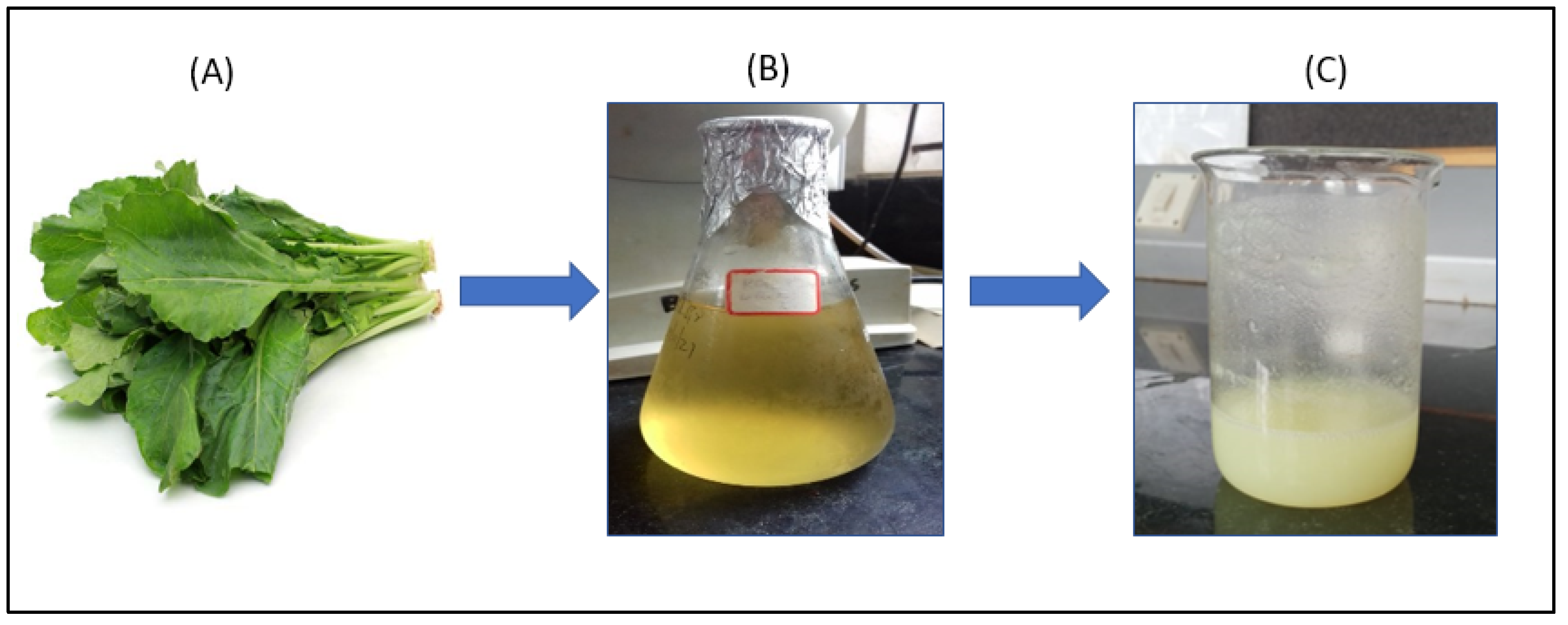
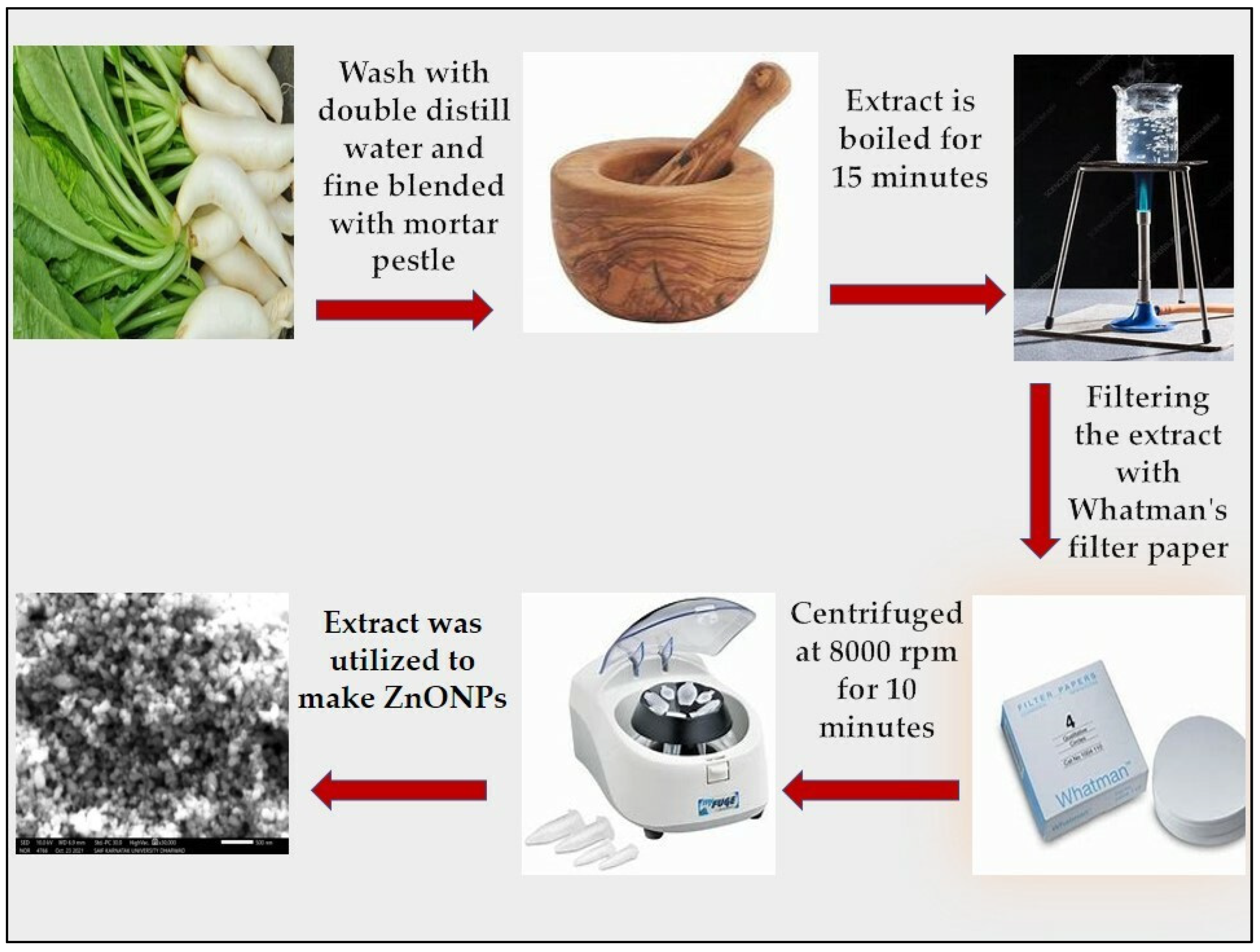
2.3. Nanoparticles Synthesis
2.4. Characterization of ZnO Nanoparticles
2.4.1. UV-Visible Spectroscopy
2.4.2. SEM with EDS
2.4.3. FTIR
2.4.4. XRD
2.4.5. Effect of pH and Temperature
2.5. Antibacterial Activity
2.5.1. MTT Assay for Cytotoxicity
2.5.2. Programmed Cell Death Assay by Flow Cytometer
2.5.3. Cell Cycle Analysis
3. Results and Discussion
3.1. Phytochemical Analysis
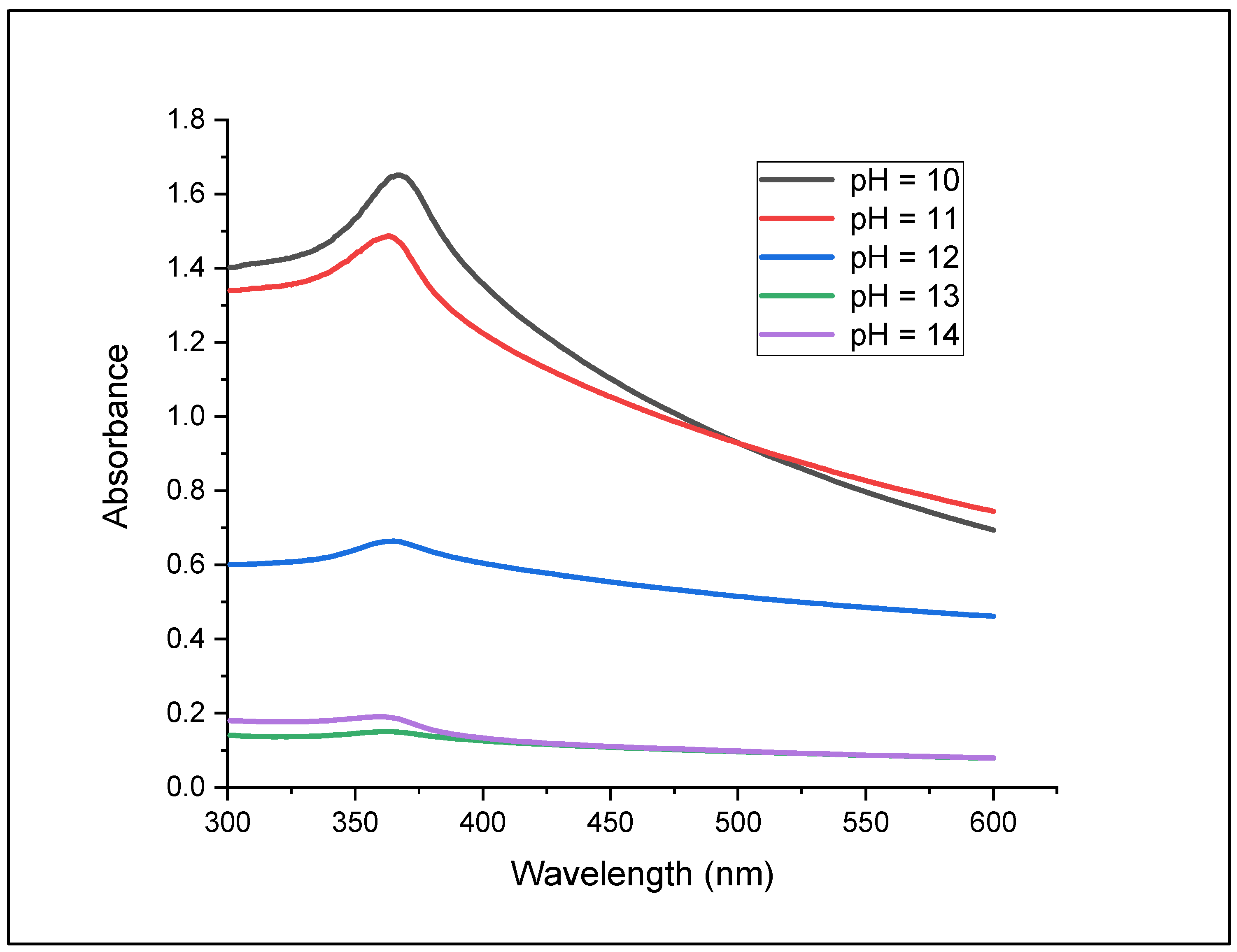
3.2. Effect of pH
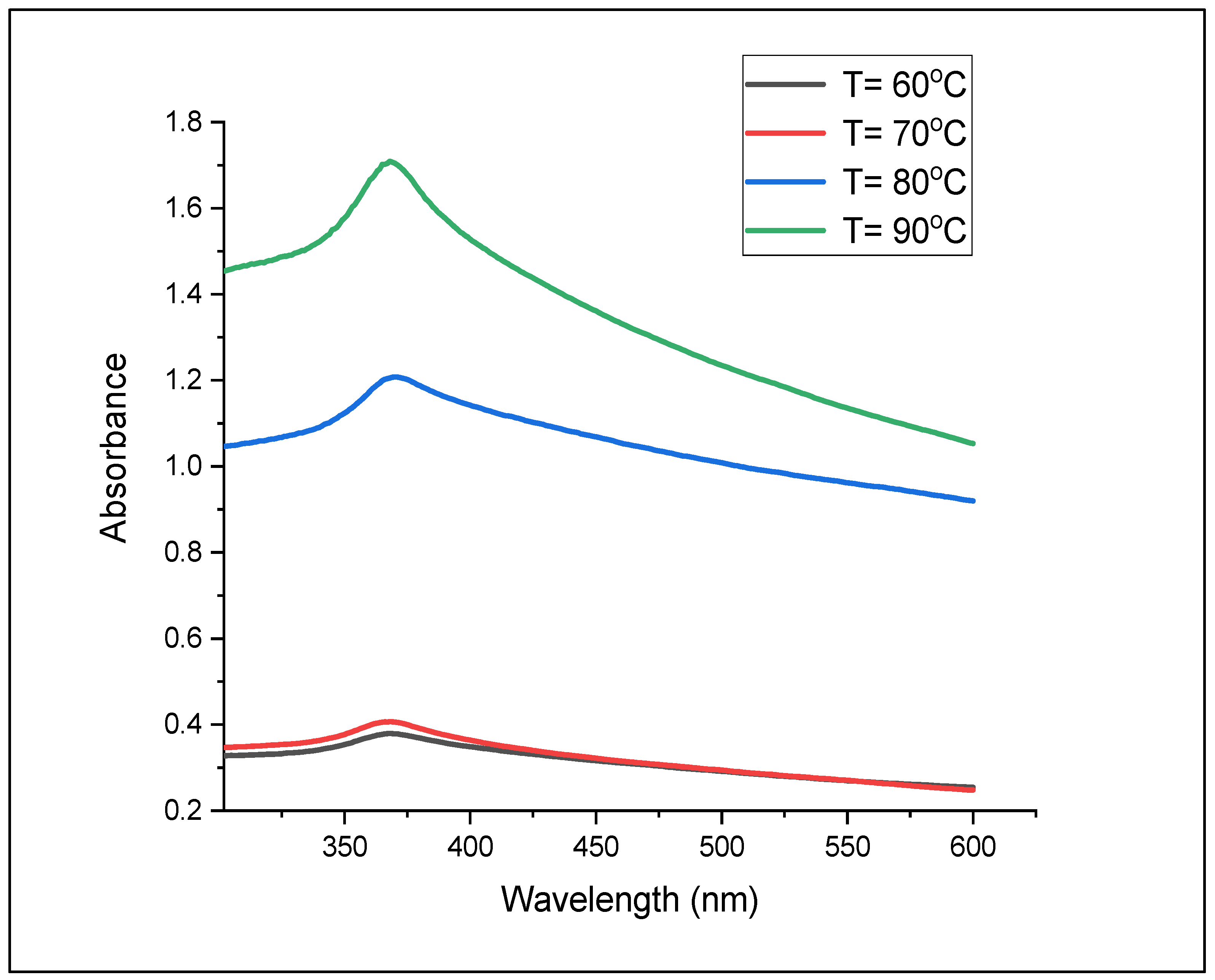
3.3. Effect of Temperature
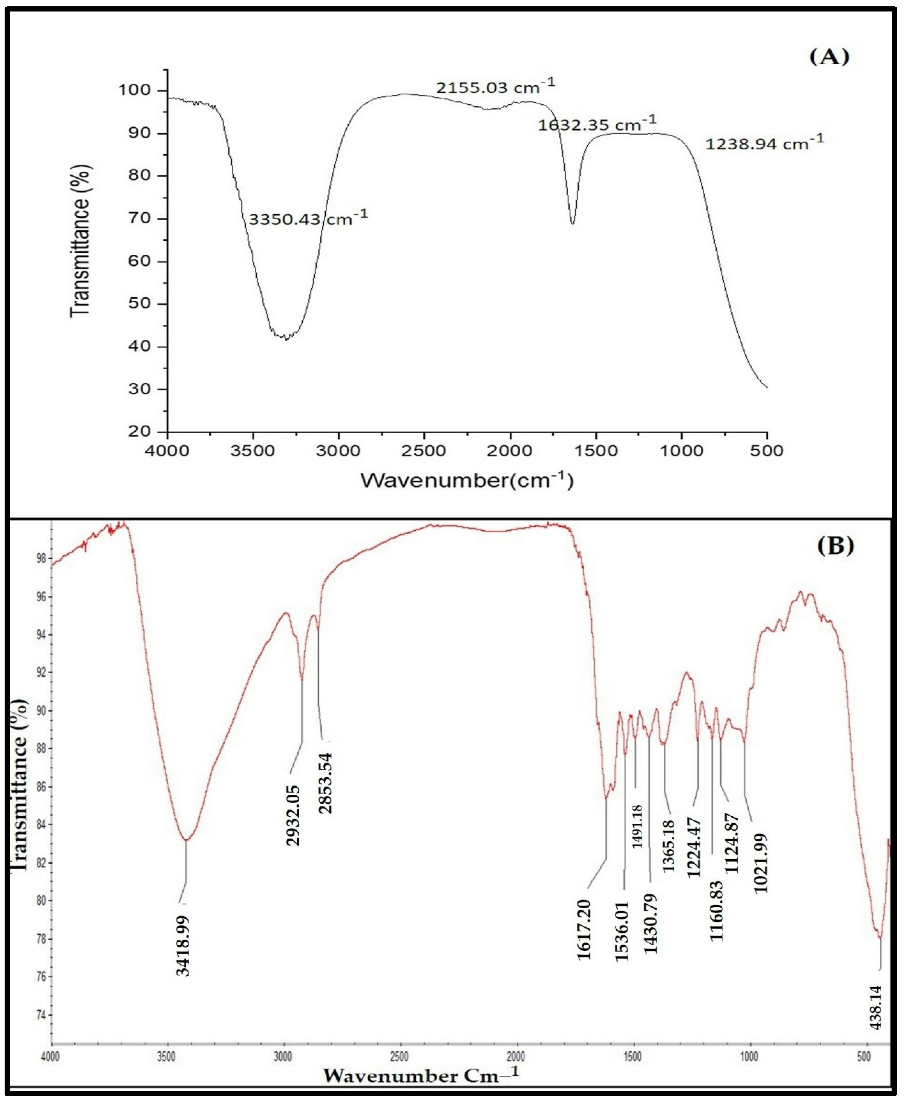
3.4. FTIR Spectral Analysis
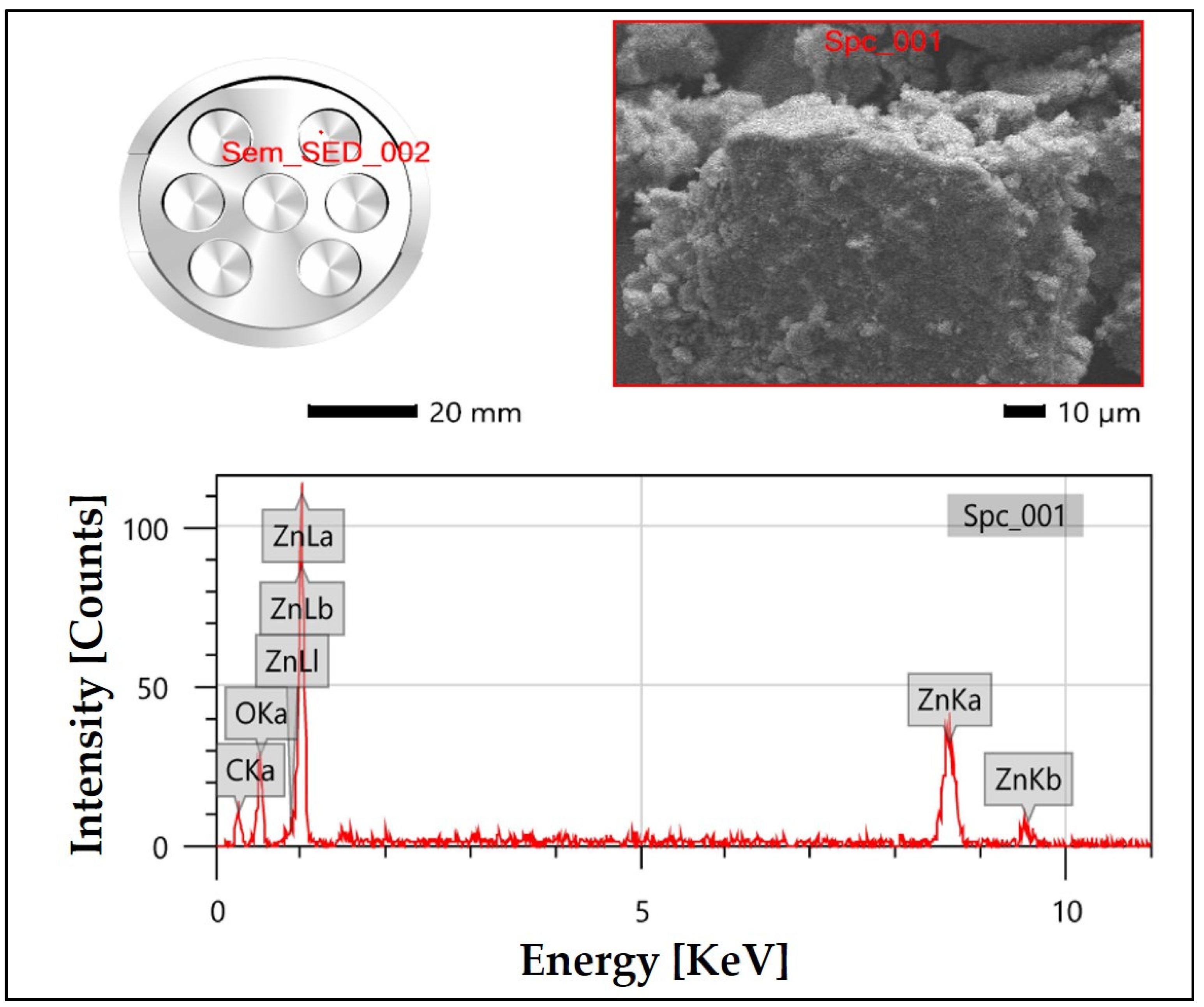
3.5. SEM Analysis
3.6. EDS Analysis
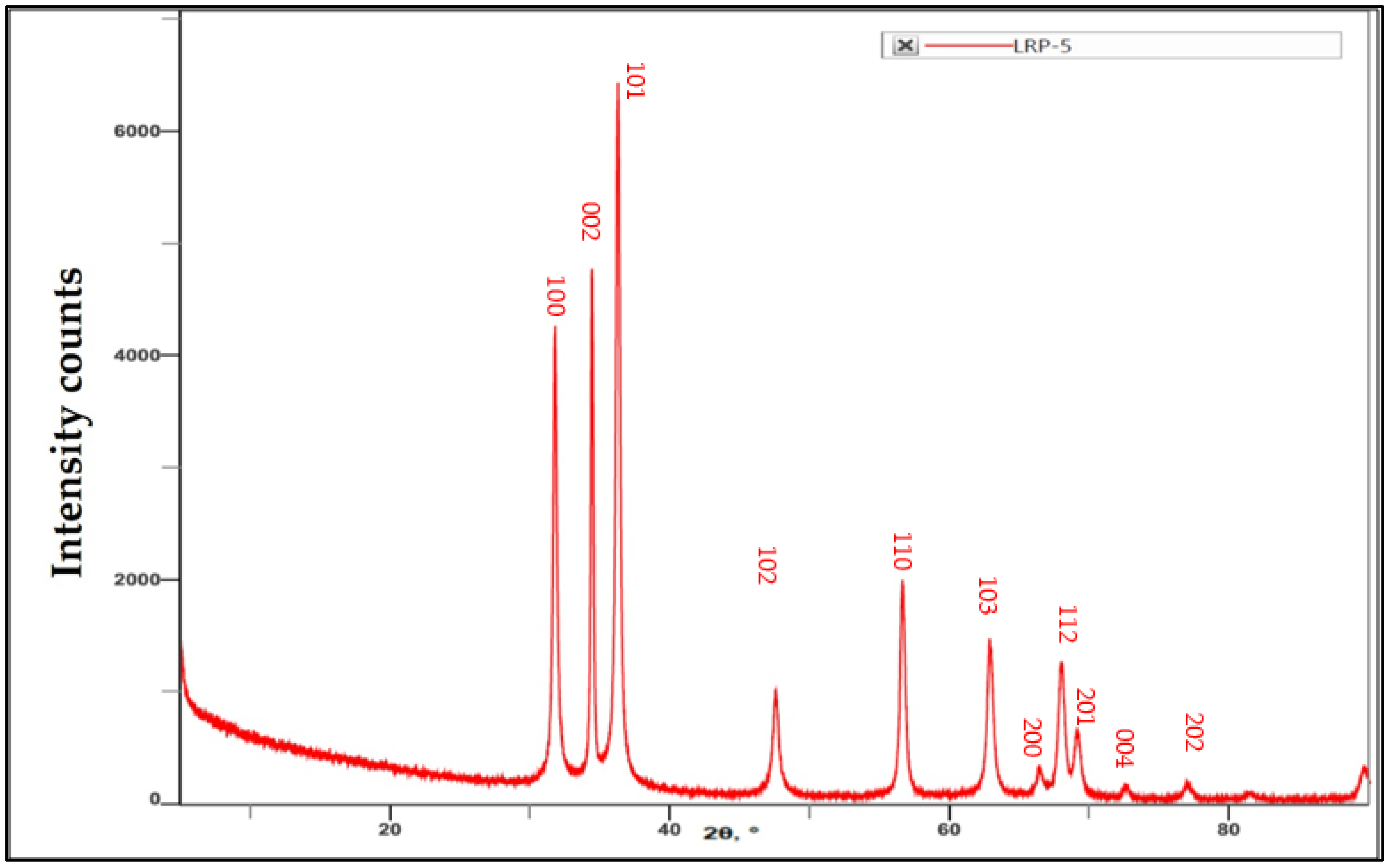
3.7. XRD Analysis
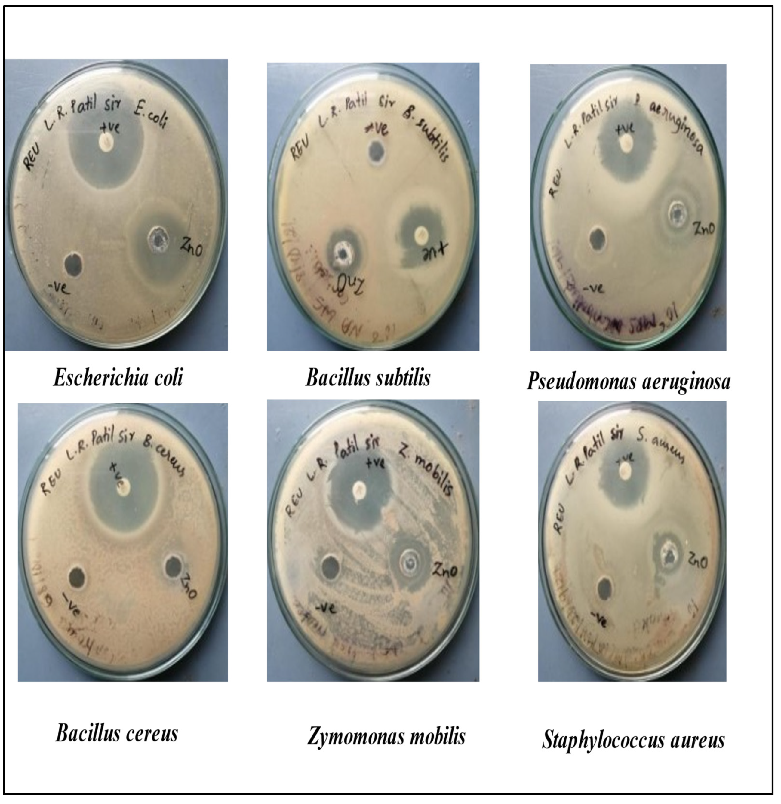
3.8. Antibacterial Activity
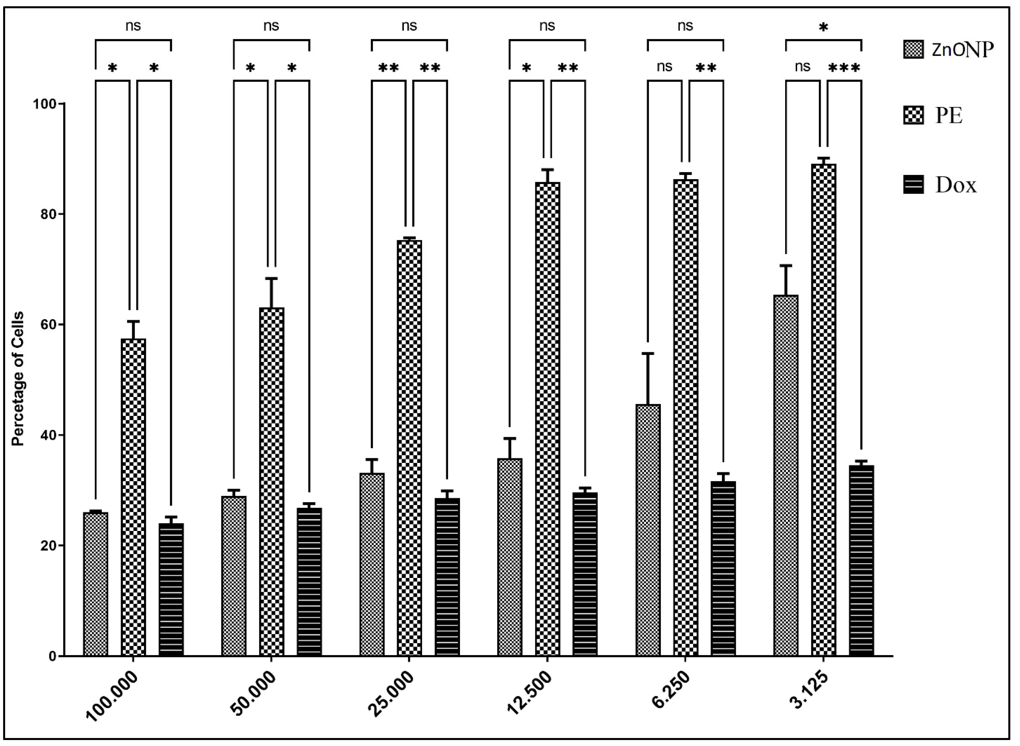
3.9. Cytotoxicity by MTT Assay
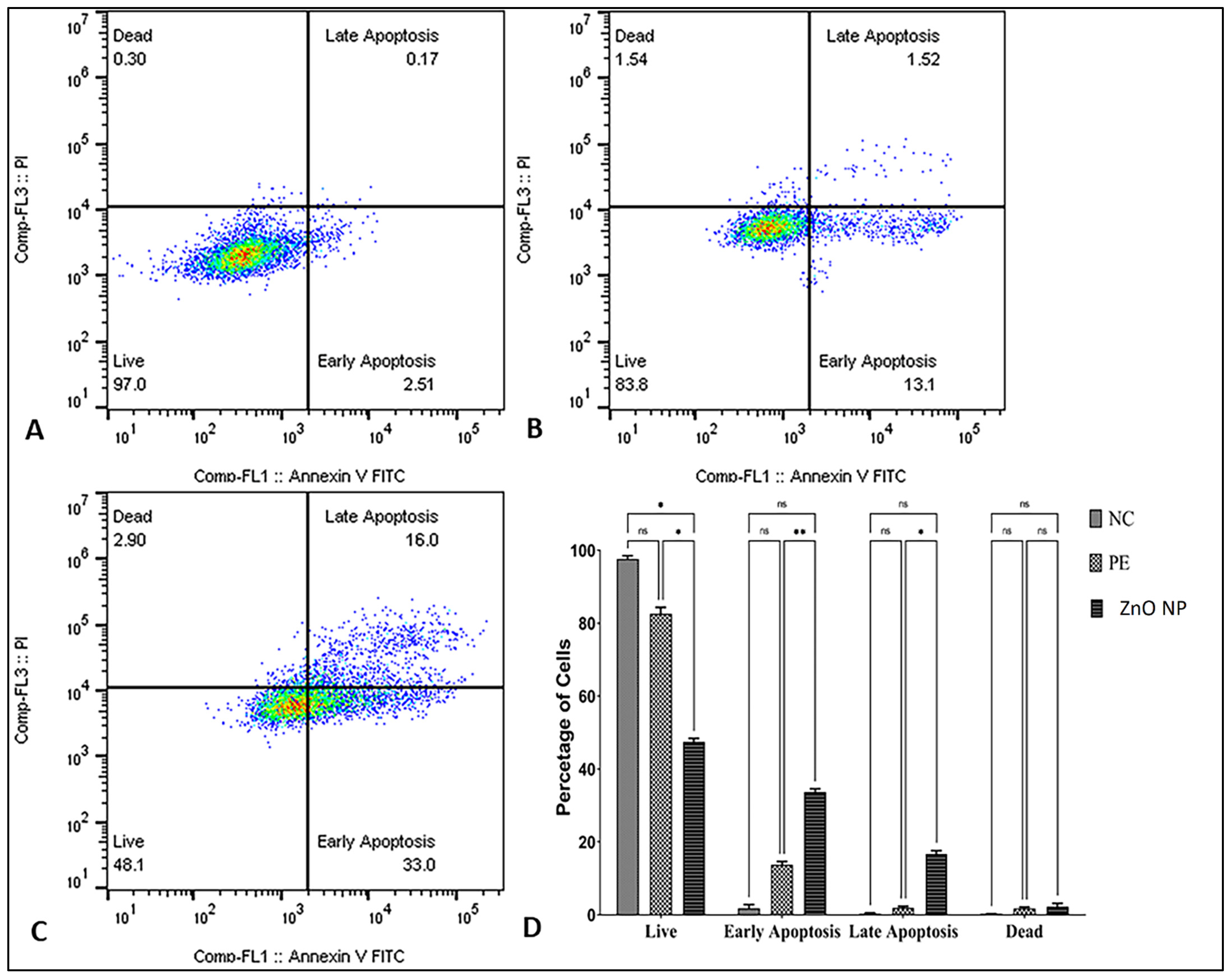
3.10. Apoptosis Assay by Flowcytometry
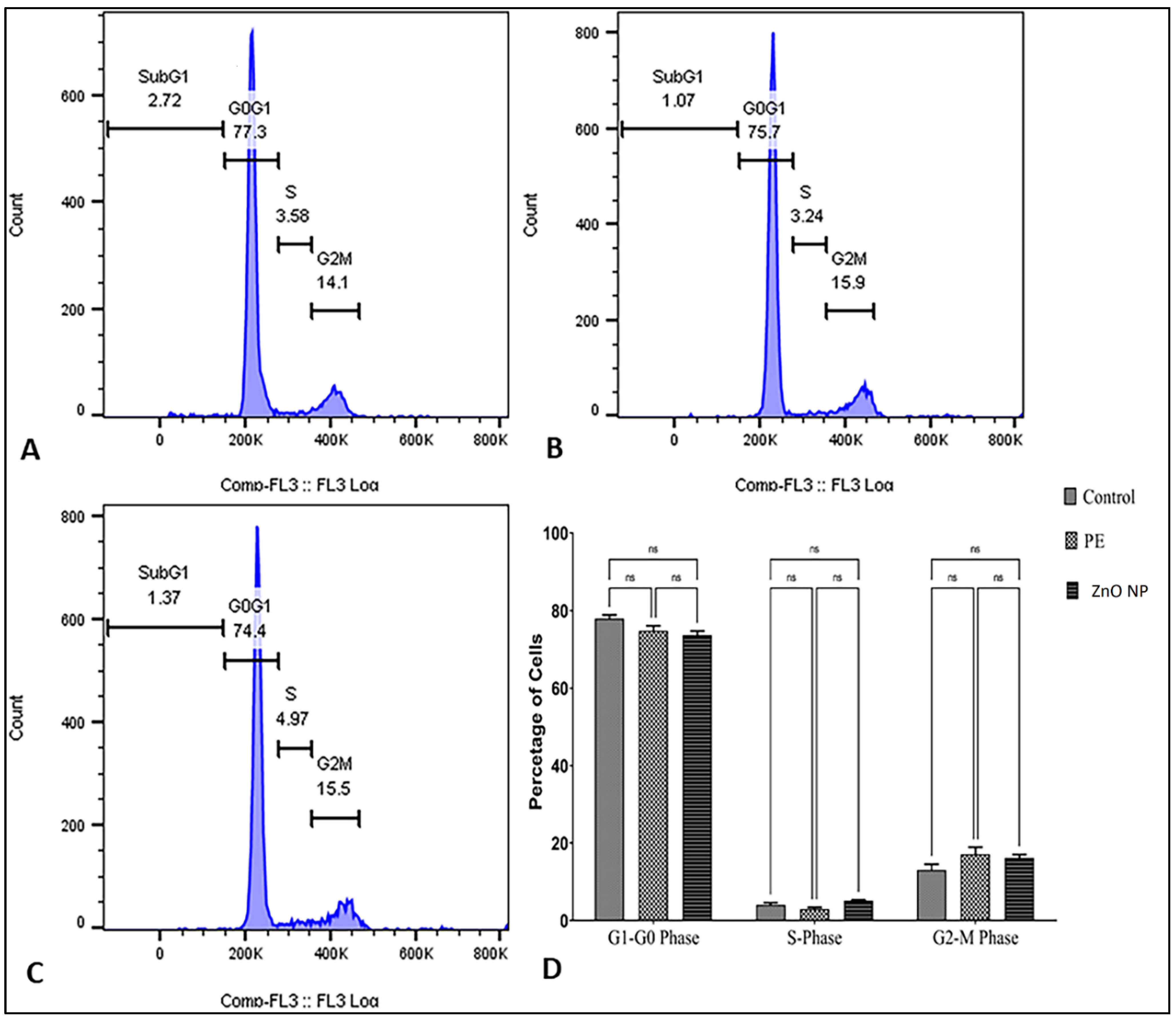
3.11. Cell Cycle Analysis by Flow Cytometer
4. Conclusions
Author Contributions
Funding
Institutional Review Board Statement
Informed Consent Statement
Data Availability Statement
Acknowledgments
Conflicts of Interest
References
- Albrecht, M.A.; Evans, C.W.; Raston, C.L. Green Chemistry and the Health Implications of Nanoparticles. Green Chem. 2006, 8, 417–432. [Google Scholar] [CrossRef]
- Singh, P.; Kim, Y.J.; Zhang, D.; Yang, D.C. Biological Synthesis of Nanoparticles from Plants and Micromicroorganisms. Trends Biotechnol. 2016, 34, 588–599. [Google Scholar] [CrossRef]
- Rouhi, J.; Mahmud, S.; Naderi, N.; Ooi, C.; Mahmood, M.R. Physical Properties of Fish Gelatin-Based Bio-nanocomposite Films Incorporated with ZnO Nanorods. Nanoscale Res. Lett. 2013, 8, 364. [Google Scholar] [CrossRef]
- Tiwari, V.; Mishra, N.; Gadani, K.; Solanki, P.S.; Shah, N.A.; Tiwari, M. Mechanism of Anti-bacterial Activity of Zinc Oxide Nanoparticle against Carbapenem-Resistant Acinetobacter baumannii. Front. Microbiol. 2018, 9, 1218. [Google Scholar] [CrossRef]
- Chaudhuri, S.K.; Malodia, L. Biosynthesis of Zinc Oxide Nanoparticles Using Leaf Extract of Calotropis gigantea: Characterization and Its Evaluation on Tree Seedling Growth in Nursery Stage. Appl. Nanosci. 2017, 7, 501–512. [Google Scholar] [CrossRef]
- Bi, C.; Li, J.; Peng, L.; Zhang, J. Biofabrication of Zinc Oxide Nanoparticles and Their In-Vitro Cytotoxicity Towards Gastric Cancer (MGC803). Cell. Biomed. Res. 2017, 28, 2065–2069. [Google Scholar]
- Zheng, Y.; Fu, L.; Han, F.; Wang, A.; Cai, W.; Yu, J.; Yang, J.; Peng, F. Green Biosynthesis and Characterization of Zinc Oxide Nanoparticles Using Corymbia Citriodora Leaf Extract and Their Photocatalytic Activity. Green Chem. Lett. Rev. 2015, 8, 59–63. [Google Scholar] [CrossRef]
- Manokari, M.; Shekhawat, M.S. Green synthesis of zinc oxide nanoparticles using whole plant extracts of Cassia tora L. and their characterization. J. Sci. Achiev. 2017, 2, 10–16. [Google Scholar]
- Khalil, A.T.; Ovais, M.; Ullah, I.; Ali, M.; Shinwari, Z.K.; Khamlich, S.; Maaza, M. Sageretiathea (Osbeck.) Mediated Synthesis of Zinc Oxide Nanoparticles and Its Biological Applications. Nanomedicine 2017, 12, 1767–1789. [Google Scholar] [CrossRef]
- Anbuvannan, M.; Ramesh, M.; Viruthagiri, G.; Shanmugam, N.; Kannadasan, N. Anisochilus carnosus Leaf Extract Mediated Synthesis of Zinc Oxide Nanoparticles for Antibacterial and Photocatalytic Activities. Mater. Sci. Semicond. Process. 2015, 39, 621–628. [Google Scholar] [CrossRef]
- Raj, L.F.A.; Jayalakshmy, E. Biosynthesis and Characterization of Zinc Oxide Nanoparticles Using Root Extract of Zingiber officinale. Orient. J. Chem. 2015, 31, 51–56. [Google Scholar] [CrossRef]
- Bhuyan, T.; Mishra, K.; Khanuja, M.; Prasad, R.; Varma, A. Biosynthesis of Zinc Oxide Nanoparticles from Azadirachta indica for Antibacterial and Photocatalytic Applications. Mater. Sci. Semicond. Process. 2015, 32, 55–61. [Google Scholar] [CrossRef]
- Bala, N.; Saha, S.; Chakraborty, M.; Maiti, M.; Das, S.; Basu, R.; Nandy, P. Green Synthesis of Zinc Oxide Nanoparticles Using Hibiscus Subdariffa Leaf Extract: Effect of Temperature on Synthesis, Anti-bacterial Activity and Anti-diabetic Activity. RSC Adv. 2015, 5, 4993–5003. [Google Scholar] [CrossRef]
- Umamaheswari, A.; Prabu, S.L.; John, S.A.; Puratchikody, A. Green Synthesis of Zinc Oxide Nanoparticles Using Leaf Extracts of Raphanus sativus var. Longipinnatus and Evaluation of Their Anticancer Property in A549 Cell Lines. Biotechnol. Rep. 2021, 29, e00595. [Google Scholar] [CrossRef]
- Francis, S.; Joseph, S.; Koshy, E.P.; Mathew, B. Microwave Assisted Green Synthesis of Silver Nanoparticles Using Leaf Extract of Elephantopus Scaber and Its Environmental and Biological Applications. Artif. Cells Nanomed. Biotechnol. 2018, 46, 795–804. [Google Scholar] [CrossRef] [PubMed]
- Shaikh, I.A.; Muddapur, U.M.; Bagewadi, Z.K.; Chiniwal, S.; Ghoneim, M.M.; Mahnashi, M.H.; Alsaikhan, F.; Yaraguppi, D.; Niyonzima, F.N.; More, S.S.; et al. Characterization of Bioactive Compounds from Acacia concinna and Citrus limon, Silver Nanoparticles’ Production by A. concinna Extract, and Their Biological Properties. Molecules 2022, 27, 2715. [Google Scholar] [CrossRef]
- Kokate, C.K.; Purohit, A.P.; Gokhale, S.B. Pharmacognosy, 47th ed.; Nirali Prakashan Publication: Pune, India, 2011. [Google Scholar]
- Gupta, M.; Tomar, R.S.; Kaushik, S.; Mishra, R.K.; Sharma, D. Effective Antimicrobial Activity of Green ZnO Nano Particles of Catharanthus Roseus. Front. Microbiol. 2018, 9, 2030. [Google Scholar] [CrossRef]
- Jeeva Lakshmi, V.; Sharath, R.; Chandraprabha, M.N.; Neelufar, E.; Hazra, A.; Patra, M. Synthesis, Characterization and Evaluation of Antimicrobial Activity of Zinc Oxide Nanoparticles. J. Biochem. Technol. 2012, 3, S151–S154. [Google Scholar]
- Weng, X.; Guo, M.; Luo, F.; Chen, Z. One-Step Green Synthesis of Bimetallic Fe/Ni Nanoparticles by Eucalyptus Leaf Extract: Biomolecules Identification, Characterization and Catalytic Activity. Chem. Eng. J. 2017, 308, 904–911. [Google Scholar] [CrossRef]
- Demissie, M.G.; Sabir, F.K.; Edossa, G.D.; Gonfa, B.A. Synthesis of Zinc Oxide Nanoparticles Using Leaf Extract of Lippia Adoensis (Koseret) and Evaluation of Its Antibacterial Activity. J. Chem. 2020, 2020, 7459042. [Google Scholar] [CrossRef]
- Zak, K.A.; Yousefi, R.; Majid, W.H.A.; Muhamad, M.R. Facile synthesis and X-ray peak broadening studies of Zn1 − xMgxO nanoparticles. Ceram. Int. 2012, 38, 2059–2064. [Google Scholar]
- Kavitha, M.K.; John, H.; Gopinath, P.; Philip, R. Synthesis of reduced graphene oxide–ZnO hybrid with enhanced optical limiting properties. J. Mater. Chem. C Mater. Opt. Electron. Devices 2013, 1, 3669–3676. [Google Scholar] [CrossRef]
- Khan, A.K.; Renouard, S.; Drouet, S.; Blondeau, J.-P.; Anjum, I.; Hano, C.; Abbasi, B.H.; Anjum, S. Effect of UV Irradiation (A and C) on Casuarina equisetifolia-Mediated Biosynthesis and Characterization of Antimicrobial and Anticancer Activity of Biocompatible Zinc Oxide Nanoparticles. Pharmaceutics 2021, 13, 1977. [Google Scholar] [CrossRef]
- Talam, S.; Karumuri, S.R.; Gunnam, N. Synthesis, characterization, and spectroscopic properties of ZnO nanoparticles. Int. Sch. Res. Not. 2012, 2012, 372505. [Google Scholar] [CrossRef]
- Vijayalakshmi, R.; Rajendran, V. Synthesis and characterization of nano-TiO2 via different methods. Arch. Appl. Sci. Res. 2012, 4, 1183–1190. [Google Scholar]
- Shah, R.; Parmar, K.; Vaghela, H. Analysing Antibacterial Efficacy of Biosynthesized Palladium Nanoparticles Using Aqueous Leaf Extract of Cocculus hirsutus As The Reducing Agent. Biosc. Biotech. Res. Commun. 2021, 14. [Google Scholar] [CrossRef]
- Slavin, Y.N.; Asnis, J.; Häfeli, U.O.; Bach, H. Metal Nanoparticles: Understanding the Mechanisms behind Antibacterial Activity. J. Nanobiotechnology 2017, 15, 65. [Google Scholar] [CrossRef]
- Yousef, J.M.; Danial, E.N. In Vitro Antibacterial Activity and Minimum Inhibitory Concentration of Zinc Oxide and Nano-Particle Zinc Oxide against Pathogenic Strains. Int. J. Health Sci. 2012, 2, 38–42. [Google Scholar] [CrossRef]
- Senthilkumar, N.; Nandhakumar, E.; Priya, P.; Soni, D.; Vimalan, M.; Vetha Potheher, I. Synthesis of ZnO nanoparticles using leaf extract of Tectona grandis (L.) and their anti-bacterial, anti-arthritic, anti-oxidant and in vitro cytotoxicity activities. New J. Chem. 2017, 41, 10347–10356. [Google Scholar] [CrossRef]
- Yedurkar, S.; Maurya, C.; Mahanwar, P. Biosynthesis of Zinc Oxide Nanoparticles Using Ixora coccinea Leaf Extract—A Green Approach. Open J. Synth. Theor. Appl. 2016, 5, 1–14. [Google Scholar] [CrossRef]
- Nour El Din, S.; El-Tayeb, T.A.; Abou-Aisha, K.; El-Azizi, M. In Vitro and in Vivo Antimicrobial Activity of Combined Therapy of Silver Nanoparticles and Visible Blue Light against Pseudomonas Aeruginosa. Int. J. Nanomed. 2016, 11, 1749–1758. [Google Scholar] [CrossRef]
- Janaki, A.C.; Sailatha, E.; Gunasekaran, S. Synthesis, Characteristics and Antimicrobial Activity of ZnO Nanoparticles. Spectrochim. Acta A Mol. Biomol. Spectrosc. 2015, 144, 17–22. [Google Scholar] [CrossRef]
- Wang, L.L.; Hu, C.; Shao, L.Q. The Antimicrobial Activity of Nanoparticles: Present Situation and Prospects for the Future. Int. J. Nanomed. 2017, 12, 1227–1249. [Google Scholar] [CrossRef]
- Ravichandran, A.; Subramanian, P.; Manoharan, V.; Muthu, T.; Periyannan, R.; Thangapandi, M.; Ponnuchamy, K.; Pandi, B.; Marimuthu, P.N. Phyto-Mediated Synthesis of Silver Nanoparticles Using Fucoidan Isolated from Spatoglossum Asperum and Assessment of Antibacterial Activities. J. Photochem. Photobiol. B 2018, 185, 117–125. [Google Scholar] [CrossRef] [PubMed]
- Mohanta, Y.K.; Panda, S.K.; Jayabalan, R.; Sharma, N.; Bastia, A.K.; Mohanta, T.K. Antimicrobial, Antioxidant and Cytotoxic Activity of Silver Nanoparticles Synthesized by Leaf Extract of Erythrina Suberosa (Roxb.). Front. Mol. Biosci. 2017, 4, 14. [Google Scholar] [CrossRef]
- Paulkumar, K.; Gnanajobitha, G.; Vanaja, M.; Rajeshkumar, S.; Malarkodi, C.; Pandian, K.; Annadurai, G. Piper Nigrum Leaf and Stem Assisted Green Synthesis of Silver Nanoparticles and Evaluation of Its Antibacterial Activity against Agricultural Plant Pathogens. Sci. World J. 2014, 2014, 829894. [Google Scholar] [CrossRef]
- Sharmila, G.; Muthukumaran, C.; Sandiya, K.; Santhiya, S.; Pradeep, R.S.; Kumar, N.M.; Suriyanarayanan, N.; Thirumarimurugan, M. Biosynthesis, Characterization, and Antibacterial Activity of Zinc Oxide Nanoparticles Derived from Bauhinia tomentosa Leaf Extract. J. Nanostruct. Chem. 2018, 8, 293–299. [Google Scholar] [CrossRef]
- Bhagwat, D.A.; Swami, P.A.; Nadaf, S.J.; Choudhari, P.B.; Kumbar, V.M.; More, H.N.; Killedar, S.G.; Kawtikwar, P.S. Capsaicin Loaded Solid SNEDDS for Enhanced Bioavailability and Anticancer Activity: In-Vitro, in-Silico, and in-Vivo Characterization. J. Pharm. Sci. 2021, 110, 280–291. [Google Scholar] [CrossRef]
- Pozarowski, P.; Grabarek, J.; Darzynkiewicz, Z. Flow Cytometry of Apoptosis. Curr. Protoc. Cell Biol. 2003, 21, 18.8.1–18.8.33. [Google Scholar] [CrossRef]
- Kumbar, V.M.; Muddapur, U.M.; Bhat, K.G.; Shwetha, H.R.; Kugaji, M.S.; Peram, M.R.; Dindawar, S. Cancer Stem Cell Traits in Tumor Spheres Derived from Primary Laryngeal Carcinoma Cell Lines. Contemp. Clin. Dent. 2021, 12, 247–254. [Google Scholar] [CrossRef] [PubMed]
- Muddapur, U.M.; Alshehri, S.; Ghoneim, M.M.; Mahnashi, M.H.; Alshahrani, M.A.; Khan, A.A.; Iqubal, S.M.S.; Bahafi, A.; More, S.S.; Shaikh, I.A.; et al. Plant-Based Synthesis of Gold Nanoparticles and Theranostic Applications: A Review. Molecules 2022, 27, 1391. [Google Scholar] [CrossRef]
- Alyamani, A.A.; Albukhaty, S.; Aloufi, S.; AlMalki, F.A.; Al-Karagoly, H.; Sulaiman, G.M. Green Fabrication of Zinc Oxide Nanoparticles Using Phlomis Leaf Extract: Characterization and In Vitro Evaluation of Cytotoxicity and Antibacterial Properties. Molecules 2021, 26, 6140. [Google Scholar] [CrossRef]
- Abdallah, Y.; Liu, M.; Ogunyemi, S.O.; Ahmed, T.; Fouad, H.; Abdelazez, A.; Yan, C.; Yang, Y.; Chen, J.; Li, B. Bioinspired Green Synthesis of Chitosan and Zinc Oxide Nanoparticles with Strong Antibacterial Activity against Rice Pathogen Xanthomonas oryzae pv. oryzae. Molecules 2020, 25, 4795. [Google Scholar] [CrossRef] [PubMed]
- Daneshvar, N.; Aber, S.; Dorraji, M.S.S.; Khataee, A.R.; Rasoulifard, M.H. Photocatalytic degradation of the insecticide diazinon in the presence of prepared nanocrystalline ZnO powders under irradiation of UV-C light. Sep. Purif. Technol. 2007, 58, 91–98. [Google Scholar] [CrossRef]
- Darvish, S.; Kahrizi, M.S.; Özbolat, G.; Khaleghi, F.; Mortezania, Z.; Sakhaei, D. Silver nanoparticles: Biosynthesis and cytotoxic performance against breast cancer MCF-7 and MDA-MB-231 cell lines. Nanomed. Res. J. 2022, 7, 83–92. [Google Scholar] [CrossRef]
- Gomathi, A.C.; Rajarathinam, S.X.; Sadiq, A.M.; Rajeshkumar, S. Anticancer activity of silver nanoparticles synthesized using aqueous fruit shell extract of Tamarindus indica on MCF-7 human breast cancer cell line. J. Drug Deliv. Sci. Technol. 2020, 55, 101376. [Google Scholar] [CrossRef]
- Gurunathan, S.; Han, J.W.; Eppakayala, V.; Jeyaraj, M.; Kim, J.H. Cytotoxicity of biologically synthesized silver nanoparticles in MDA-MB-231 human breast cancer cells. Biomed. Res. Int. 2013, 2013, 535796. [Google Scholar] [CrossRef]
- Berehu, H.M.; Anupriya, S.; Khan, M.I.; Chakraborty, R.; Lavudi, K.; Penchalaneni, J.; Mohapatra, B.; Mishra, A.; Patnaik, S. Cytotoxic Potential of Biogenic Zinc Oxide Nanoparticles Synthesized from Swertia Chirayita Leaf Extract on Colorectal Cancer Cells. Front. Bioeng. Biotechnol. 2021, 9, 788527. [Google Scholar] [CrossRef] [PubMed]
- El-Naggar, N.E.; Hussein, M.H.; El-Sawah, A.A. Bio-fabrication of silver nanoparticles by phycocyanin, characterization, in vitro anticancer activity against breast cancer cell line and in vivo cytotxicity. Sci. Rep. 2017, 7, 10844. [Google Scholar] [CrossRef] [PubMed]


| Phytochemicals | Aqueous Extract |
|---|---|
| Flavonoids | + |
| Glycosides | − |
| Alkaloids | + |
| Tannins | + |
| Phenol | + |
| Amino acid and proteins | − |
| Carbohydrates | − |
| Saponins | − |
| Temperature (°C) | Peak (nm) | Band Gap (eV) |
|---|---|---|
| 60 | 372.86 | 3.33 |
| 70 | 370.28 | 3.35 |
| 80 | 369.00 | 3.36 |
| 90 | 365.55 | 3.39 |
| Display Name | Standard Data | Quantification Method | Result Type |
|---|---|---|---|
| Spc_001 | Standardless | ZAF | Metal |
| Element | Line | Mass% | Atom% |
| C | K | 16.51 ± 0.90 | 40.76 ± 2.22 |
| O | K | 15.27 ± 0.99 | 28.30 ± 1.83 |
| Zn | K | 68.21 ± 3.20 | 30.94 ± 1.45 |
| Total | 100.00 | 100.00 | |
| Spc_001 Fitting ratio 0.3062 | |||
| Microorganism | Zone of Inhibition in mm (Mean ± SD) |
|---|---|
| Linezolid (control) | 22 ± 2 |
| Escherichia coli | 19 ± 2 |
| Pseudomonas aeruginosa | 18 ± 1 |
| Zymomonasmobilis | 17 ± 1 |
| Bacillus subtilis | 15 ± 1 |
| Bacillus cereus | 12 ± 1 |
| Staphylococcus aureus | 14 ± 1 |
Publisher’s Note: MDPI stays neutral with regard to jurisdictional claims in published maps and institutional affiliations. |
© 2022 by the authors. Licensee MDPI, Basel, Switzerland. This article is an open access article distributed under the terms and conditions of the Creative Commons Attribution (CC BY) license (https://creativecommons.org/licenses/by/4.0/).
Share and Cite
Al Awadh, A.A.; Shet, A.R.; Patil, L.R.; Shaikh, I.A.; Alshahrani, M.M.; Nadaf, R.; Mahnashi, M.H.; Desai, S.V.; Muddapur, U.M.; Achappa, S.; et al. Sustainable Synthesis and Characterization of Zinc Oxide Nanoparticles Using Raphanus sativus Extract and Its Biomedical Applications. Crystals 2022, 12, 1142. https://doi.org/10.3390/cryst12081142
Al Awadh AA, Shet AR, Patil LR, Shaikh IA, Alshahrani MM, Nadaf R, Mahnashi MH, Desai SV, Muddapur UM, Achappa S, et al. Sustainable Synthesis and Characterization of Zinc Oxide Nanoparticles Using Raphanus sativus Extract and Its Biomedical Applications. Crystals. 2022; 12(8):1142. https://doi.org/10.3390/cryst12081142
Chicago/Turabian StyleAl Awadh, Ahmed Abdullah, Anil R. Shet, Laxmikant R. Patil, Ibrahim Ahmed Shaikh, Mohammed Merae Alshahrani, Roshan Nadaf, Mater H. Mahnashi, Shivalingsarj V. Desai, Uday M. Muddapur, Sharanappa Achappa, and et al. 2022. "Sustainable Synthesis and Characterization of Zinc Oxide Nanoparticles Using Raphanus sativus Extract and Its Biomedical Applications" Crystals 12, no. 8: 1142. https://doi.org/10.3390/cryst12081142
APA StyleAl Awadh, A. A., Shet, A. R., Patil, L. R., Shaikh, I. A., Alshahrani, M. M., Nadaf, R., Mahnashi, M. H., Desai, S. V., Muddapur, U. M., Achappa, S., Hombalimath, V. S., Khan, A. A., Gouse, H. S. M., Iqubal, S. M. S., & Kumbar, V. (2022). Sustainable Synthesis and Characterization of Zinc Oxide Nanoparticles Using Raphanus sativus Extract and Its Biomedical Applications. Crystals, 12(8), 1142. https://doi.org/10.3390/cryst12081142