
Green Extraction of Cellulose Nanocrystals of Polymorph II from Cynara scolymus L.: Challenge for a “Zero Waste” Economy

 ,
,  ,
,  ,
, 
 and
and 
Abstract
:1. Introduction
2. Materials and Methods
2.1. Materials
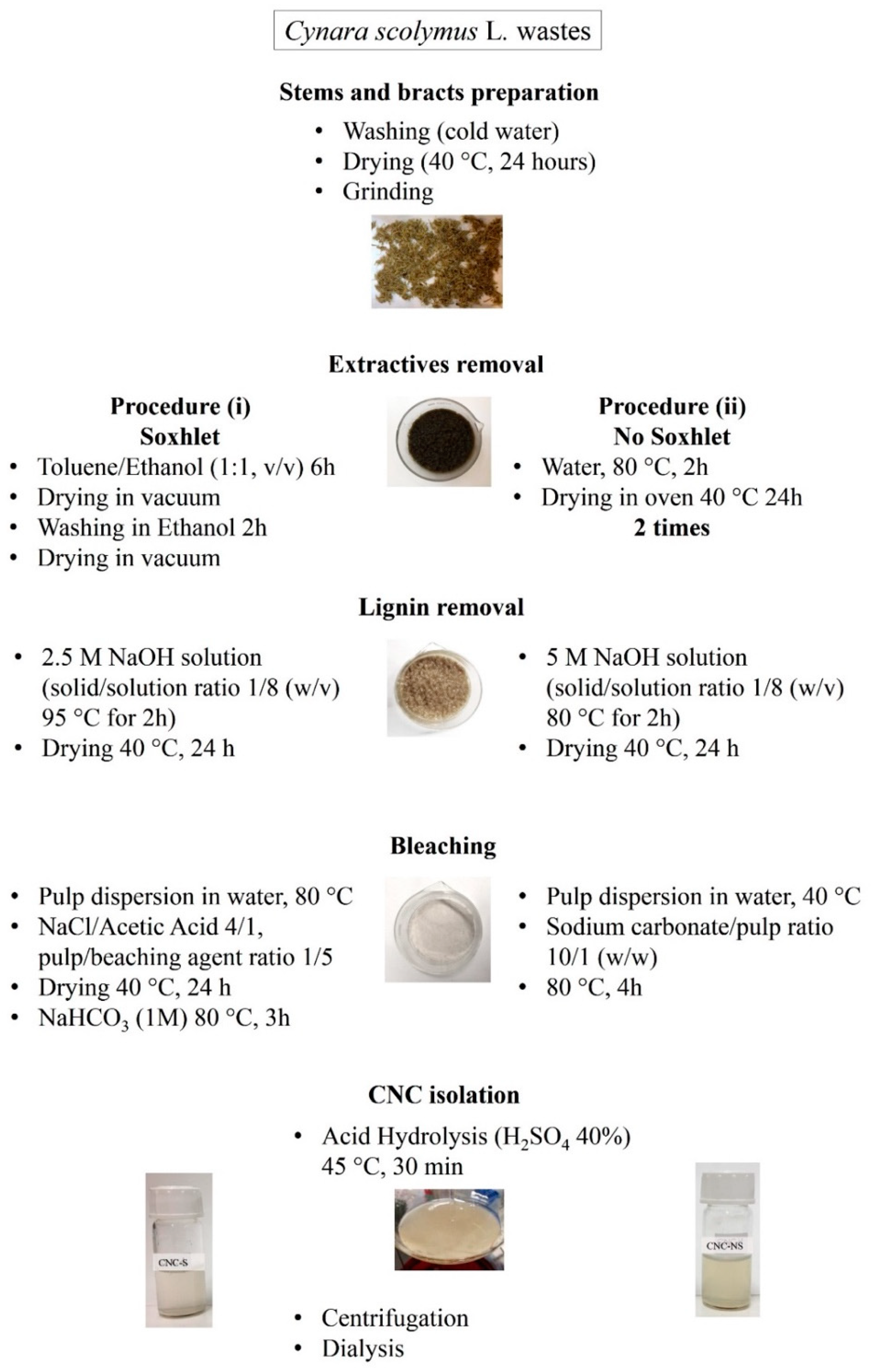
2.2. Cellulose Isolation Procedures
2.3. Cellulose Nanocrystals Preparation
2.4. Methods
2.4.1. Electrophoretic Light Scattering (ELS) Measurements
2.4.2. TEM Analysis
2.4.3. X-ray Powder Diffraction Analysis (XRPD)
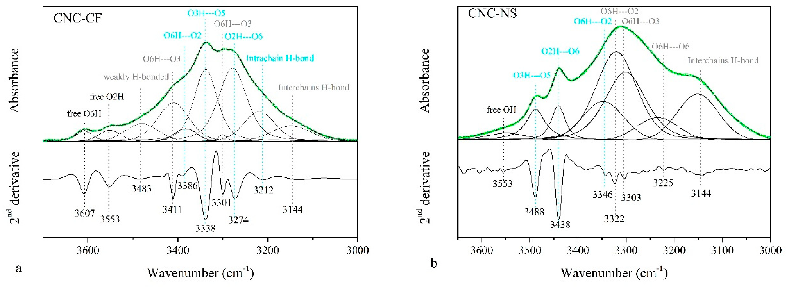
2.4.4. FT-IR Spectroscopy
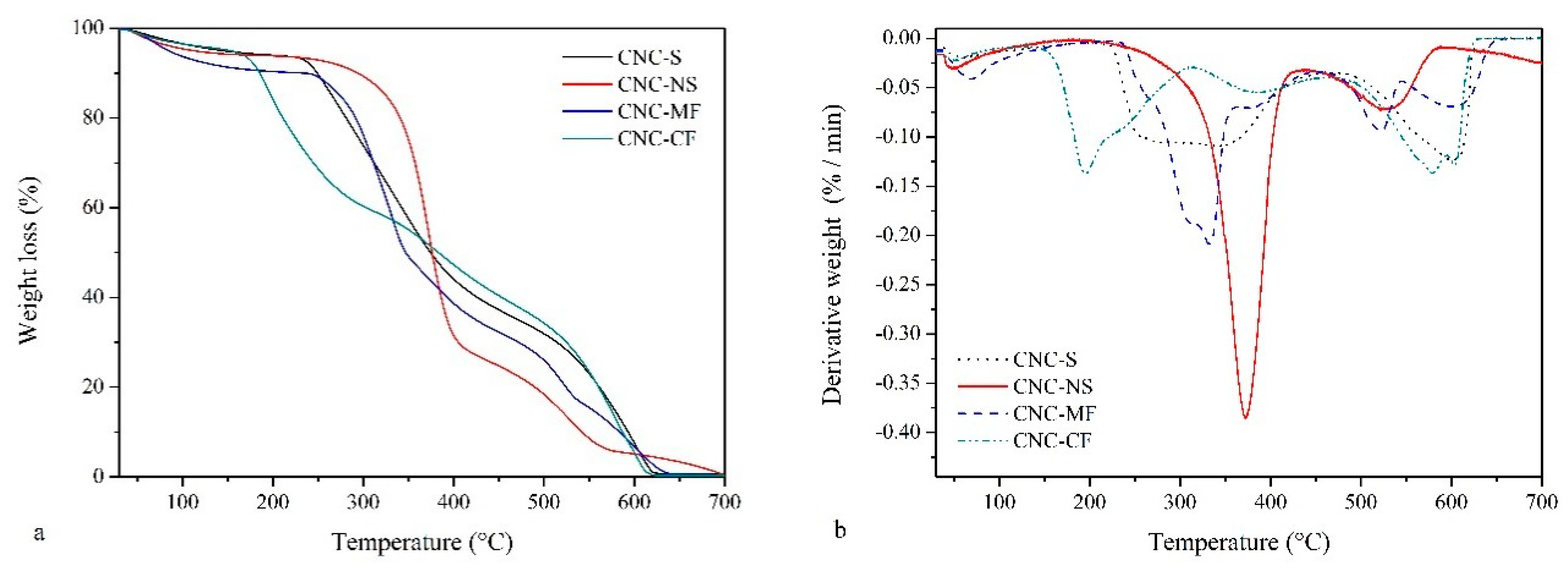
2.4.5. Thermogravimetric Analysis (TGA)
3. Results
3.1. ELS Measurements
3.2. TEM
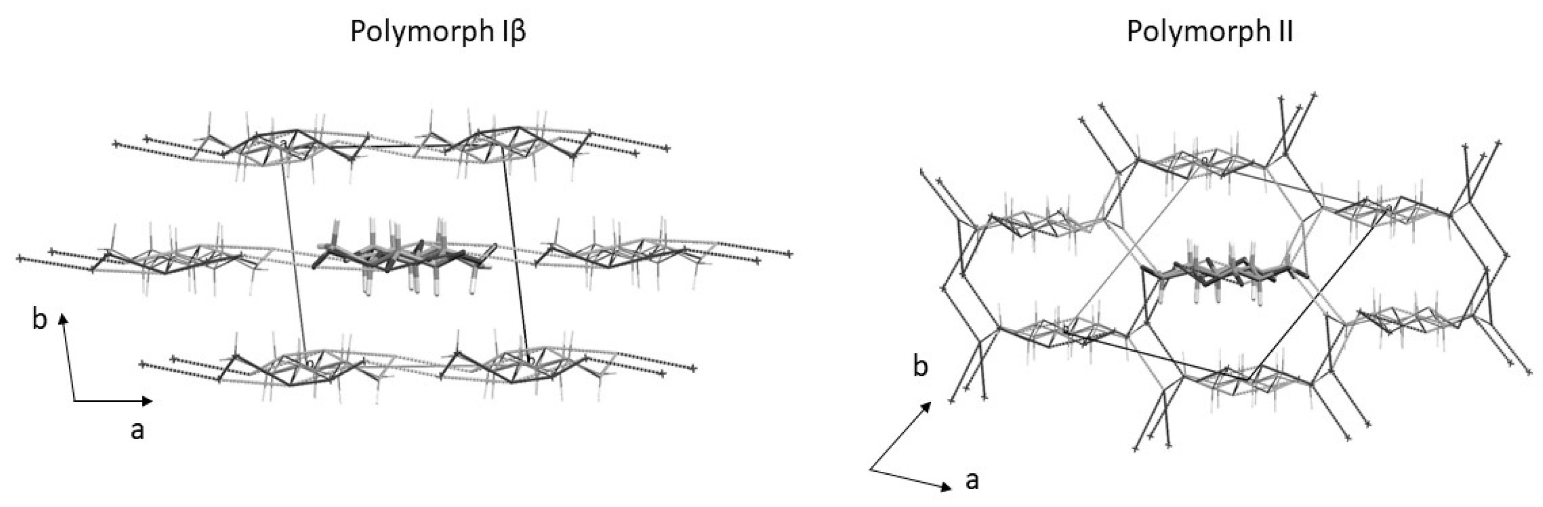
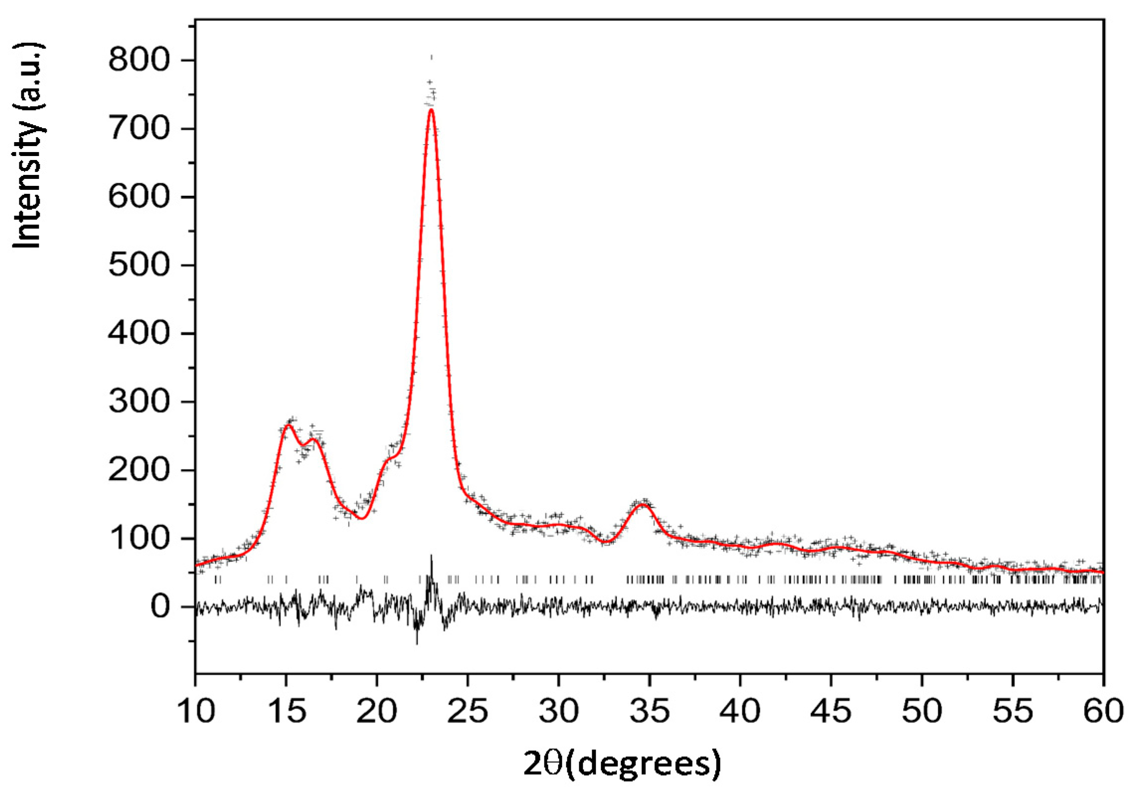
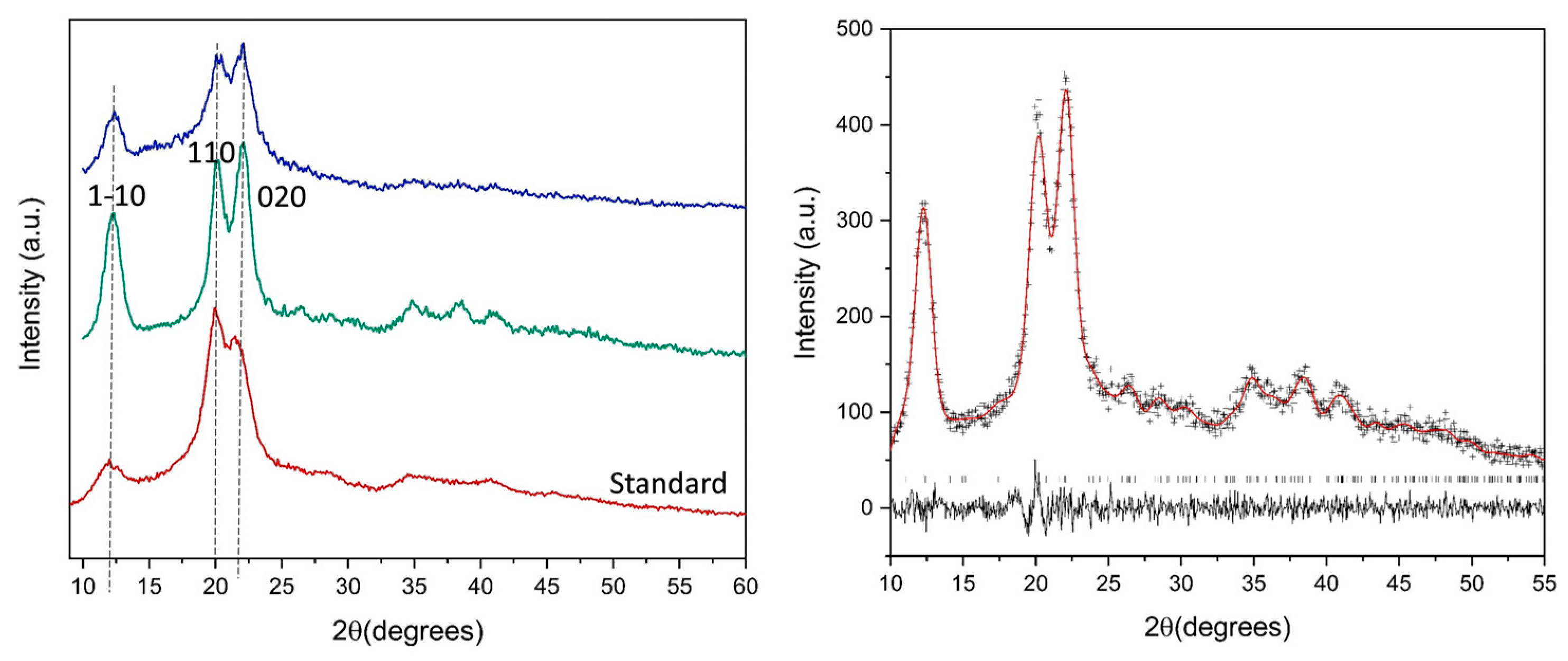
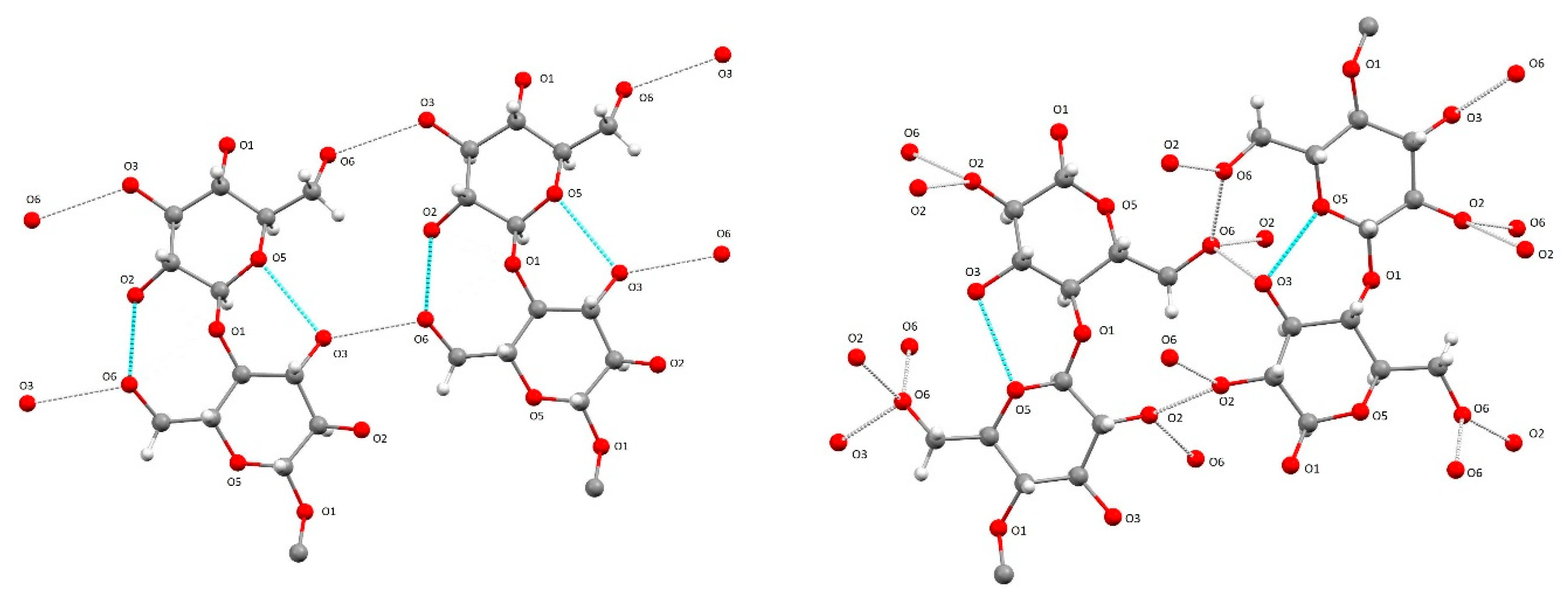
3.3. XRPD Analysis
3.4. FTIR Spectroscopy
3.5. Thermal Analysis
4. Conclusions
Author Contributions
Funding
Institutional Review Board Statement
Informed Consent Statement
Data Availability Statement
Acknowledgments
Conflicts of Interest
References
- FAO—2019: The State of Food and Agriculture. 2019. Available online: http://www.fao.org/3/ca6030en/ca6030en.pdf (accessed on 23 March 2022).
- Dhali, K.; Ghasemlou, M.; Daver, F.; Cass, P.; Adhikari, B. A review of nanocellulose as a new material towards environmental sustainability. Sci. Total Environ. 2021, 775, 145871. [Google Scholar] [CrossRef]
- Ma, T.; Hu, X.; Lu, S.; Liao, X.; Song, Y.; Hu, X. Nanocellulose: A promising green treasure from food wastes to available food materials. Crit. Rev. Food Sci. Nutr. 2022, 62, 989–1002. [Google Scholar] [CrossRef]
- Noremylia, M.B.; Hassan, M.Z.; Ismail, Z. Recent advancement in isolation, processing, characterization and applications of emerging nanocellulose: A review. Int. J. Biol. Macromol. 2022, 206, 954–976. [Google Scholar] [CrossRef]
- Lutz, M.; Henríquez, C.; Escobar, M. Chemical composition and antioxidant properties of mature and baby artichokes (Cynara scolymus L.), raw and cooked. J. Food Compos. Anal. 2011, 24, 49–54. [Google Scholar] [CrossRef]
- Meneses, M.; Martínez-Marín, A.L.; Madrid, J.; Martínez-Teruel, A.; Hernández, F.; Megías, M.D. Ensilability, in vitro and in vivo values of the agro-industrial by-products of artichoke and broccoli. Environ. Sci. Pollut. Res. 2020, 27, 2919–2925. [Google Scholar] [CrossRef]
- Zuorro, A.; Maffei, G.; Lavecchia, R. Reuse potential of artichoke (Cynara scolimus L.) waste for the recovery of phenolic compounds and bioenergy. J. Clean. Prod. 2016, 111, 279–284. [Google Scholar] [CrossRef]
- Turco, R.; Zannini, D.; Mallardo, S.; Poggetto, G.D.; Tesser, R.; Santagata, G.; Malinconico, M.; Di Serio, M. Biocomposites based on Poly(lactic acid), Cynara Cardunculus seed oil and fibrous presscake: A novel eco-friendly approach to hasten PLA biodegradation in common soil. Polym. Degrad. Stab. 2021, 188, 109576. [Google Scholar] [CrossRef]
- French, A.D. Idealized powder diffraction patterns for cellulose polymorphs. Cellulose 2014, 21, 885–896. [Google Scholar] [CrossRef]
- Nishiyama, Y. Structure and properties of the cellulose microfibril. J. Wood Sci. 2009, 55, 241–249. [Google Scholar] [CrossRef]
- Nishiyama, Y.; Sugiyama, J.; Chanzy, H.; Langan, P. Crystal Structure and Hydrogen Bonding System in Cellulose Iα from Synchrotron X-ray and Neutron Fiber Diffraction. J. Am. Chem. Soc. 2003, 125, 14300–14306. [Google Scholar] [CrossRef]
- Nishiyama, Y.; Langan, P.; Chanzy, H. Crystal Structure and Hydrogen-Bonding System in Cellulose Iβ from Synchrotron X-ray and Neutron Fiber Diffraction. J. Am. Chem. Soc. 2002, 124, 9074–9082. [Google Scholar] [CrossRef] [PubMed]
- Gupta, P.K.; Uniyal, V.; Naithani, S. Polymorphic transformation of cellulose i to cellulose II by alkali pretreatment and urea as an additive. Carbohydr. Polym. 2013, 94, 843–849. [Google Scholar] [CrossRef]
- Klemm, D.; Heublein, B.; Fink, H.-P.; Bohn, A. Cellulose: Fascinating Biopolymer and Sustainable Raw Material. Angew. Chem. Int. Ed. 2005, 44, 3358–3393. [Google Scholar] [CrossRef] [PubMed]
- Oh, S.Y.; Dong, I.Y.; Shin, Y.; Kim, H.C.; Kim, H.Y.; Chung, Y.S.; Park, W.H.; Youk, J.H. Crystalline structure analysis of cellulose treated with sodium hydroxide and carbon dioxide by means of X-ray diffraction and FTIR spectroscopy. Carbohydr. Res. 2005, 340, 2376–2391. [Google Scholar] [CrossRef] [PubMed]
- Široký, J.; Blackburn, R.S.; Bechtold, T.; Taylor, J.; White, P. Attenuated total reflectance Fourier-transform Infrared spectroscopy analysis of crystallinity changes in lyocell following continuous treatment with sodium hydroxide. Cellulose 2010, 17, 103–115. [Google Scholar] [CrossRef]
- Dinand, E.; Vignon, M.; Chanzy, H.; Heux, L. Mercerization of primary wall cellulose and its implication for the conversion of cellulose I→ cellulose II. Cellulose 2002, 9, 7–18. [Google Scholar] [CrossRef]
- Oh, S.Y.; Yoo, D.; Shin, Y.; Seo, G. FTIR analysis of cellulose treated with sodium hydroxide and carbon dioxide. Carbohydr. Res. 2005, 340, 417–428. [Google Scholar] [CrossRef]
- Tran, H.; Vakkilainnen, E.K. The Kraft Chemical Recovery Process; Tappi Kraft Pulping Short Course: St. Petersburg, FL, USA, 2008; pp. 1–8. [Google Scholar]
- Kleppe, P.J. Kraft Pulping. Tappi 1970, 53, 35–47. [Google Scholar]
- Borysiak, S. Fundamental studies on lignocellulose/polypropylene composites: Effects of wood treatment on the transcrystalline morphology and mechanical properties. J. Appl. Polym. Sci. 2013, 127, 1309–1322. [Google Scholar] [CrossRef]
- Kolpak, F.J.; Weih, M.; Blackwell, J. Mercerization of cellulose: 1. Determination of the structure of mercerized cotton. Polymer 1978, 19, 123–131. [Google Scholar] [CrossRef]
- Ferro, M.; Mannu, A.; Panzeri, W.; Theeuwen, C.H.; Mele, A. An integrated approach to optimizing cellulose mercerization. Polymers 2020, 12, 1559. [Google Scholar] [CrossRef] [PubMed]
- Heise, K.; Delepierre, G.; King, A.W.; Kostiainen, M.A.; Zoppe, J.; Weder, C.; Kontturi, E. Chemical modification of reducing end-groups in cellulose nanocrystals. Angew. Chem. Int. Ed. 2021, 60, 66–87. [Google Scholar] [CrossRef]
- Jiang, X.; Lou, C.; Hua, F.; Deng, H.; Tian, X. Cellulose nanocrystals-based flocculants for high-speed and high-efficiency decolorization of colored effluents. J. Clean. Prod. 2020, 251, 119749. [Google Scholar] [CrossRef]
- Espíndola, S.P.; Pronk, M.; Zlopasa, J.; Picken, S.J.; van Loosdrecht, M.C. Nanocellulose recovery from domestic wastewater. J. Clean. Prod. 2021, 280, 124507. [Google Scholar] [CrossRef]
- De, D.; Sai, M.S.N.; Aniya, V.; Satyavathi, B. Strategic biorefinery platform for green valorization of agro-industrial residues: A sustainable approach towards biodegradable plastics. J. Clean. Prod. 2021, 290, 125184. [Google Scholar] [CrossRef]
- De Souza, A.G.; Barbosa, R.F.S.; Rosa, D.S. Nanocellulose from Industrial and Agricultural Waste for Further Use in PLA Composites. J. Polym. Environ. 2020, 28, 1851–1868. [Google Scholar] [CrossRef]
- Adu, C.; Berglund, L.; Oksman, K.; Eichhorn, S.J.; Jolly, M.; Zhu, C. Properties of cellulose nanofibre networks prepared from never-dried and dried paper mill sludge. J. Clean. Prod. 2018, 197, 765–771. [Google Scholar] [CrossRef] [Green Version]
- Coccia, V.; Cotana, F.; Cavalaglio, G.; Gelosia, M.; Petrozzi, A. Cellulose nanocrystals obtained from Cynara cardunculus and their application in the paper industry. Sustainability 2014, 6, 5252–5264. [Google Scholar] [CrossRef] [Green Version]
- Habibi, Y.; Lucia, L.A.; Rojas, O.J. Cellulose nanocrystals: Chemistry, self-assembly, and applications. Chem. Rev. 2010, 110, 3479–3500. [Google Scholar] [CrossRef]
- Ersuo, J.; Guo, J.; Yang, F.; Zhu, Y.; Song, J.; Jin, Y.; Rojas, O.J. On the polymorphic and morphological changes of cellulose nanocrystals (CNC-I) upon mercerization and conversion CNC-II. Carbohydr. Polym. 2016, 143, 327–335. [Google Scholar] [CrossRef]
- SaifulAzry, S.O.A.; Chuah, T.G.; Paridah, M.T.; Aung, M.M.; Ridzuan, M.A.; Lee, C.H.; Juliana, A.H. Influence of cellulose II polymorph nanowhiskers on bio-based nanocomposite film from Jatropha oil polyurethane. Mater. Res. Express 2020, 8, 015003. [Google Scholar] [CrossRef]
- Grząbka-Zasadzińska, A.; Ratajczak, I.; Król, K.; Woźniak, M.; Borysiak, S. The influence of crystalline structure of cellulose in chitosan-based biocomposites on removal of Ca (II), Mg (II), Fe (III) ion in aqueous solutions. Cellulose 2021, 28, 5745–5759. [Google Scholar] [CrossRef]
- Hussin, M.H.; Pohan, N.A.; Garba, Z.N.; Kassim, M.J.; Rahim, A.A.; Brosse, N.; Haafiz, M.M. Physicochemical of microcrystalline cellulose from oil palm fronds as potential methylene blue adsorbents. Int. J. Biol. Macromol. 2016, 92, 11–19. [Google Scholar] [CrossRef]
- De Souza, A.G.; Junqueira, M.T.; De Lima, G.F.; Rangari, V.K.; Rosa, D.S. A New Proposal of Preparation of Different Polymorphs of Nanocellulose from Eucalyptus citriodora. J. Polym. Environ. 2020, 28, 1150–1159. [Google Scholar] [CrossRef]
- Bhattacharjee, S. DLS and zeta potential—What they are and what they are not? J. Control. Release 2016, 235, 337–351. [Google Scholar] [CrossRef] [PubMed]
- Zhang, J.; Elder, T.J.; Pu, Y.; Ragauskas, A.J. Facile synthesis of spherical cellulose nanoparticles. Carbohydr. Polym. 2007, 69, 607–611. [Google Scholar] [CrossRef]
- Qin, Z.; Ji, L.; Yin, X.; Zhu, L.; Lin, Q.; Qin, J. Synthesis and characterization of bacterial cellulose sulfates using a SO3/pyridine complex in DMAc/LiCl. Carbohydr. Polym. 2014, 101, 947–953. [Google Scholar] [CrossRef]
- Kuga, T.; Sunagawa, N.; Igarashi, K. Enzymatic-synthesis of cellulose in space: Gravity is a crucial factor for building cellulose II gel structure. Cellulose 2022, 29, 2999–3015. [Google Scholar] [CrossRef]
- Nagarajan, S.; Skillen, N.C.; Irvine, J.T.; Lawton, L.A.; Robertson, P.K. Cellulose II as bioethanol feedstock and its advantages over native cellulose. Renew. Sustain. Energy Rev. 2017, 77, 182–192. [Google Scholar] [CrossRef] [Green Version]
- Miao, X.; Tian, F.; Lin, J.; Li, H.; Li, X.; Bian, F.; Zhang, X. Tuning the mechanical properties of cellulose nanofibrils reinforced polyvinyl alcohol composites via altering the cellulose polymorphs. RSC Adv. 2016, 6, 83356–83365. [Google Scholar] [CrossRef]
- Borysiak, S.; Grząbka-Zasadzińska, A. Influence of the polymorphism of cellulose on the formation of nanocrystals and their application in chitosan/nanocellulose composites. J. Appl. Polym. Sci. 2016, 133, 42864. [Google Scholar] [CrossRef]
- Tang, Y.; Shen, X.; Zhang, J.; Guo, D.; Kong, F.; Zhang, N. Extraction of cellulose nano-crystals from old corrugated container fiber using phosphoric acid and enzymatic hydrolysis followed by sonication. Carbohydr. Polym. 2015, 125, 360–366. [Google Scholar] [CrossRef] [PubMed]
- Zhong, L.; Fu, S.; Peng, X.; Zhan, H.; Sun, R. Colloidal stability of negatively charged cellulose nanocrystalline in aqueous systems. Carbohydr. Polym. 2012, 90, 644–649. [Google Scholar] [CrossRef] [PubMed]
- Song, T.; Pranovich, A.; Holmbom, B. Characterization of Norway spruce hemicelluloses extracted by pressurized hot-water extraction (ASE) in the presence of sodium bicarbonate. Holtzforschung 2011, 65, 35–42. [Google Scholar] [CrossRef]
- Bergamonti, L.; Potenza, M.; Haghighi Poshtiri, A.; Lorenzi, A.; Sanangelantoni, A.M.; Lazzarini, L.; Lottici, P.P.; Graiff, C. Ag-functionalized nanocrystalline cellulose for paper preservation and strengthening. Carbohydr. Polym. 2020, 231, 115773. [Google Scholar] [CrossRef]
- Basile, R.; Bergamonti, L.; Fernandez, F.; Graiff, C.; Haghighi, A.; Isca, C.; Lottici, P.P.; Pizzo, B.; Predieri, G. Bio-inspired consolidants derived from crystalline nanocellulose for decayed wood. Carbohydr. Polym. 2018, 202, 164–171. [Google Scholar] [CrossRef]
- Bondeson, D.; Mathew, A.; Oksman, K. Optimization of the isolation of nanocrystals from microcrystalline cellulose by acid hydrolysis. Cellulose 2006, 13, 171–180. [Google Scholar] [CrossRef]
- Beltramino, F.; Roncero, M.B.; Vidal, T.; Torres, A.L.; Valls, C. Increasing yield of nanocrystalline cellulose preparation process by a cellulase pretreatment. Bioresour. Technol. 2015, 192, 574–581. [Google Scholar] [CrossRef] [Green Version]
- Segal, L.; Creely, J.J.; Martin, A.E.; Conrad, C.M. An Empirical Method for Estimating the Degree of Crystallinity of Native Cellulose Using the X-ray Diffractometer. Text. Res. J. 1959, 29, 786–794. [Google Scholar] [CrossRef]
- French, A.D.; Santiago Cintrón, M. Cellulose polymorphy, crystallite size, and the Segal Crystallinity Index. Cellulose 2013, 20, 583–588. [Google Scholar] [CrossRef]
- Popescu, C.M.; Singurel, G.; Popescu, M.C.; Vasile, C.; Argyropoulos, D.S.; Willför, S. Vibrational spectroscopy and X-ray diffraction methods to establish the differences between hardwood and softwood. Carbohydr. Polym. 2009, 77, 851–857. [Google Scholar] [CrossRef]
- Lu, P.; Hsieh, Y.L. Preparation and properties of cellulose nanocrystals: Rods, spheres, and network. Carbohydr. Polym. 2010, 82, 329–336. [Google Scholar] [CrossRef]
- Langan, P.; Nishiyama, Y.; Chanzy, H. X-ray structure of mercerized cellulose II at 1 Å resolution. Biomacromolecules 2001, 2, 410–416. [Google Scholar] [CrossRef] [PubMed]
- Bates, S.; Zografi, G.; Engers, D.; Morris, K.; Crowley, K.; Newman, A. Analysis of amorphous and nanocrystalline solids from their X-ray diffraction patterns. Pharm. Res. 2006, 23, 2333–2349. [Google Scholar] [CrossRef]
- Revol, J.F.; Dietrich, A.; Goring, D.A.I. Effect of mercerization on the crystallite size and crystallinity index in cellulose from different sources. Can. J. Chem. 1987, 65, 1724–1725. [Google Scholar] [CrossRef]
- Nam, S.; French, A.D.; Condon, B.D.; Concha, M. Segal crystallinity index revisited by the simulation of X-ray diffraction patterns of cotton cellulose Iβ and cellulose II. Carbohydr. Polym. 2016, 135, 1–9. [Google Scholar] [CrossRef]
- Bergamonti, L.; Graiff, C.; Tegoni, M.; Predieri, G.; Elviri, L.; Palanti, S.; Paris, C.; Cappelletto, E.; Di Maggio, R.; Lottici, P.P. Facile preparation of functionalized poly (amidoamine) s with biocidal activity on wood substrates. Eur. Polym. J. 2019, 116, 232–241. [Google Scholar] [CrossRef]
- Bergamonti, L.; Berzolla, A.; Chiappini, E.; Feci, E.; Maistrello, L.; Palanti, S.; Predieri, G.; Vaccari, G. Polyamidoamines (PAAs) functionalized with siloxanes as wood preservatives against fungi and insects. Holzforschung 2017, 71, 65–75. [Google Scholar] [CrossRef]
- Maréchal, Y.; Chanzy, H. The hydrogen bond network in I(β) cellulose as observed by infrared spectrometry. J. Mol. Struct. 2000, 523, 183–196. [Google Scholar] [CrossRef]
- Nada, A.M.A.; Hassan, M.L. Thermal behavior of cellulose and some cellulose derivatives. Polym. Degrad. Stab. 2000, 67, 111–115. [Google Scholar] [CrossRef]
- Goswami, P.; Blackburn, R.S.; El-Dessouky, H.M.; Taylor, J.; White, P. Effect of sodium hydroxide pre-treatment on the optical and structural properties of lyocell. Eur. Polym. J. 2009, 45, 455–465. [Google Scholar] [CrossRef]
- Di Maggio, R.; Dirè, S.; Callone, E.; Bergamonti, L.; Lottici, P.P.; Albatici, R.; Rigon, R.; Ataollahi, N. Super-adsorbent polyacrylate under swelling in water for passive solar control of building envelope. SN Appl. Sci. 2020, 2, 45. [Google Scholar] [CrossRef] [Green Version]
- Zhao, D.; Deng, Y.; Han, D.; Tan, L.; Ding, Y.; Zhou, Z.; Xu, H.; Guo, Y. Exploring structural variations of hydrogen-bonding patterns in cellulose during mechanical pulp refining of tobacco stems. Carbohydr. Polym. 2018, 204, 247–254. [Google Scholar] [CrossRef] [PubMed]
- Lu, H.; Gui, Y.; Zheng, L.; Liu, X. Morphological, crystalline, thermal and physicochemical properties of cellulose nanocrystals obtained from sweet potato residue. Food Res. Int. 2013, 50, 121–128. [Google Scholar] [CrossRef]
- Wang, N.; Ding, E.; Cheng, R. Thermal degradation behaviors of spherical cellulose nanocrystals with sulfate groups. Polymer 2007, 48, 3486–3493. [Google Scholar] [CrossRef]
- Naduparambath, S.; Jinitha, T.V.; Shaniba, V.; Sreejith, M.P.; Aparna, K.; Balan, A.K.; Purushothaman, E. Isolation and characterisation of cellulose nanocrystals from sago seed shells. Carbohydr. Polym. 2018, 180, 13–20. [Google Scholar] [CrossRef]
- Chieng, B.W.; Lee, S.H.; Ibrahim, N.A.; Then, Y.Y.; Loo, Y.Y. Isolation and Characterization of Cellulose Nanocrystals from Oil Palm Mesocarp Fiber. Polymers 2017, 9, 355. [Google Scholar] [CrossRef]
- Johar, N.; Ahmad, I.; Dufresne, A. Extraction, preparation and characterization of cellulose fibres and nanocrystals from rice husk. Ind. Crops Prod. 2012, 37, 93–99. [Google Scholar] [CrossRef]


| Source | Preparation Method | Length (nm) | CI (%) | Yield% | Reference |
|---|---|---|---|---|---|
| Buckeye cellulose | HCl/H2SO4 hydrolysis | 500 | 67 | 33 | [38] |
| Bacterial cellulose | SO3/Py | - | 69.9 | 34 | [39] |
| Cellobiose | Cellodextrin phosphorylase from the cellulolytic bacterium | 254 | - | [40] | |
| Lignicellulosic materials | NaOH Mercerization | - | 73 | 67 | [41] |
| Commercial Cellulose | NaOH Mercerization | 120 | 43 | - | [34] |
| Oil palm fronds | HCl hydrolysis | >200 | 47 | - | [35] |
| Jute fibers | TEMPO mediated oxidation and mechanical disgregation | 250 | 56 | - | [42] |
| Softwood pulp | Mercerization | 75 | 55 | - | [33] |
| Cellulose pulp | NaOH Mercerization H2SO4 hydrolysis | 140 | - | [43] |
| CNC-S | CNC-NS | CNC-CF | CNC-MF | |
|---|---|---|---|---|
| Zeta potential (mV) | −33 | −45 | −51 | −32 |
| Iβ Phase CNC-CF | II Phase CNC-MF | II Phase CNC-S | II Phase CNC-NS | |
|---|---|---|---|---|
| Symmetry | P21 | P21 | P21 | P21 |
| a (Å) | 7.826 (6) | 8.033 (4) | 8.094 (4) | 8.090 (3) |
| b (Å) | 8.316 (5) | 9.056 (5) | 9.041 (5) | 9.069 (4) |
| c (Å) | 10.349 (7) | 10.157 (6) | 10.171 (7) | 10.37 * |
| γ (°) | 95.10 (6) | 118.14 (8) | 118.14 (9) | 118.17 (2) |
| Sample | Crystallinity Index (CI%) |
|---|---|
| CNC-NS | 63 |
| CNC-MF | 67 |
| CNC-S | 75 |
| CNC-CF | 89 |
| Wavenumber (cm−1) | Δν (cm−1)/Absorbance Change | Assignment | |
|---|---|---|---|
| CNC Iβ | CNC II | ||
| 3338 | 3494 | +156/- | νO3H---O5 intramolecular H bonds |
| 3270 | 3441 | +171/- | νO2H---O6 intramolecular H bonds |
| 2900 | 2887 | −13/- | νCH |
| 2850 | νCH | ||
| 1482 | 1470 | −12/Δ | δCH2 |
| 1430 | 1419 | −10/∇ | δCH (C1) |
| 1366 | 1373 | +7/∇ | δC–H |
| 1336 | 1336 | -/∇ | δCOH in plane (C2 or C3) |
| 1236 | 1226 | −10/Δ | δCOH in plane (C6) |
| 1203 | 1195 | −8/- | δCOH in plne |
| 1160 | 1153 | −7/∇ | νasCOC (β-glycosidic bond) |
| 1030 | 1014 | −16/∇ | νC-OH of C6 |
| 984 | 994 | +10/Δ | C-O valence vibration of C6 |
| 895 | 892 | −3/Δ | COC of the ν-glycosidic bond and/or ether group |
| 660 | 665 | −5/- | δCOH out of plane |
| 550 | τOH of C6 | ||
Publisher’s Note: MDPI stays neutral with regard to jurisdictional claims in published maps and institutional affiliations. |
© 2022 by the authors. Licensee MDPI, Basel, Switzerland. This article is an open access article distributed under the terms and conditions of the Creative Commons Attribution (CC BY) license (https://creativecommons.org/licenses/by/4.0/).
Share and Cite
Potenza, M.; Bergamonti, L.; Lottici, P.P.; Righi, L.; Lazzarini, L.; Graiff, C. Green Extraction of Cellulose Nanocrystals of Polymorph II from Cynara scolymus L.: Challenge for a “Zero Waste” Economy. Crystals 2022, 12, 672. https://doi.org/10.3390/cryst12050672
Potenza M, Bergamonti L, Lottici PP, Righi L, Lazzarini L, Graiff C. Green Extraction of Cellulose Nanocrystals of Polymorph II from Cynara scolymus L.: Challenge for a “Zero Waste” Economy. Crystals. 2022; 12(5):672. https://doi.org/10.3390/cryst12050672
Chicago/Turabian StylePotenza, Marianna, Laura Bergamonti, Pier Paolo Lottici, Lara Righi, Laura Lazzarini, and Claudia Graiff. 2022. "Green Extraction of Cellulose Nanocrystals of Polymorph II from Cynara scolymus L.: Challenge for a “Zero Waste” Economy" Crystals 12, no. 5: 672. https://doi.org/10.3390/cryst12050672
APA StylePotenza, M., Bergamonti, L., Lottici, P. P., Righi, L., Lazzarini, L., & Graiff, C. (2022). Green Extraction of Cellulose Nanocrystals of Polymorph II from Cynara scolymus L.: Challenge for a “Zero Waste” Economy. Crystals, 12(5), 672. https://doi.org/10.3390/cryst12050672