Article

The High-Energy Milling Preparation and Spectroscopic Characterization of Rare-Earth Ions Doped BaY2F8 Nanoparticles

by
Aleksander V. Koshelev
1,*,
Natalia A. Arkharova
1,
Kirill V. Khaydukov
1,
Mir Saeed Seyed Dorraji
2,
Denis N. Karimov
1,* and
Vera V. Klechkovskaya
1
1
Federal Scientific Research Center «Crystallography and Photonics», Russian Academy of Sciences, Leninsky Prospekt 59, 119333 Moscow, Russia
2
Applied Chemistry Research Laboratory, Department of Chemistry, Faculty of Science, University of Zanjan, Zanjan 45371-38791, Iran
*
Authors to whom correspondence should be addressed.
Crystals 2022, 12(5), 599; https://doi.org/10.3390/cryst12050599
Submission received: 29 March 2022 / Revised: 16 April 2022 / Accepted: 22 April 2022 / Published: 24 April 2022

Abstract

:
BaY2F8 nanoparticles (NPs), doped with Yb3+ and Er3+ ions, were successfully produced by high-energy ball-milling. High-quality monoclinic single crystals (sp. gr. C2/m, a = 0.6969(3), b = 1.0502(1), c = 0.4254(1) nm, β = 99.676°) grown from the melt by the Bridgman technique were used as raw materials. The prepared nanocrystals were comprehensively studied by X-ray diffraction analysis, transmission electron microscopy, and optical spectroscopy. The fabrication possibility of single-phase irregular shaped Ba(Y0.964Yb0.030Er0.006)2F8 NPs in the size range of 20–100 nm with a milling duration of 10 h at 600 rpm is demonstrated. Ba(Y0.964Yb0.030Er0.006)2F8 NPs show intense luminescence by both up- (λ = 540 and 650 nm) and down-conversion (λ = 1540 nm) mechanisms upon IR excitation (λ = 980 nm). A qualitative comparison of the spectroscopic characteristics of the produced Ba(Y0.964Yb0.030Er0.006)2F8 NPs with the initial bulk crystal and the widely used up-conversion β-Na1.5(Y1.17Yb0.3Er0.03)F6 NPs is presented. Experimental data offer great opportunities of the Ba(Y0.964Yb0.030Er0.006)2F8 NPs applications in nanophotonics and biotechnology. High-energy ball-milling has potential as a versatile method for the scalable production of fluoride nanoparticles.

Graphical Abstract

1. Introduction

Photoluminescent (PL) nanomaterials doped with rare-earth elements (REE) have become an important basis for various photonic applications: biomedicine, photocatalysis, nanosensors, anti-counterfeiting, etc. [1,2,3,4,5,6,7]. Among the variety of the inorganic materials, the development of REE-doped fluoride NPs attracts special attention due to high chemical and thermal stability, low phonon frequencies, wide optical transparency, high isomorphic capacity, and a large selection of different dopants [7,8]. These unique features provide the ability to generate luminescence in a wide spectral range. Currently, approaches for the chemical synthesis (hydro-, solvothermal methods, coprecipitation, etc.) of fluoride NPs, based on different REE-doped crystal hosts, such as MF2 (M = Ca, Sr, Ba), LiRF4, NaRF4, BaRF5, KR3F10, and RF3, (R = Y, La-Lu) have been successfully developed [9,10,11,12,13,14,15,16], and hexagonal β-NaRF4 NPs demonstrate excellent up-conversion emission upon IR excitation [8,17]. However, the active development of new types of highly efficient nanophosphors continues at present. The preparation of new prospective fluoride hosts is limited by the technological capabilities of existing chemical techniques, which explains the active interest in the search for alternative physical “top-down” approaches, such as laser ablation [18,19] or mechanical milling (nanodispergation) of initial bulk materials [20,21].
Among fluoride PL nanocrystals, host compounds formed in the BaF2-YF3 binary system attract significant interest, and their potential for photonic and biological applications is being actively studied. There are three crystal matrices in the BaF2-YF3 system [22]: a fluorite solid solution Ba1-xYxF2+x (x < 0.36); a trigonal phase (sp. gr. R-3) with nominal composition BaxYxF17, existing in the middle concentration range [23]; and a monoclinic BaY2F8 phase (sp. gr. C2/m) [24]. Effective luminescence generation in BaYF5 and BaY2F8 bulk crystal hosts doped with various REE combinations was first demonstrated in 1969 [25]. REE-doped BaY2F8 single crystals have been studied as active laser media in a wide spectral range both up- and down-conversion excitation schemes [26,27,28,29,30]. These crystals are considered as promising scintillation materials in the vacuum UV and visible regions due to high ionizing radiation resistance [31,32,33].
The modern trend in the search for new efficient PL inorganic materials is focused on the nanoscale. Wet chemical methods for the synthesis of BaYF5 NPs with different crystal structures (cubic [14,34,35] or tetragonal [36,37,38]) were developed in detail, and the applications of these nanocrystals in bioimaging, optical thermometry, and white light emitting devices have been described. However, the stoichiometric composition of these particles needs to be specified due to the nonequilibrium state of the system during the synthesis. These BaYF5 compounds are probably the metastable fluorite-type solid solutions [39,40]. At the same time, the synthesis strategies for Ba4Y3F17 and BaY2F8 NPs are quite limited, what is associated with the significant technological complexity of preparing single-phase nanosized particles via chemical synthesis [41,42].
Monodisperse “nanobelts” BaY2F8:Yb3+/Er3+ with dimensions of 1000 × 75 nm2 were synthesized by the liquid–solid solution strategy [43]. BaY2F8:Yb3+/Ho3+ compounds were produced by solid-phase fluorination of the corresponding electrospun oxide compositions such as nanofiber [44] or submicrosized particles via co-precipitation in aqueous nitrate solutions [45]. Therefore, the development of a facile production method for BaY2F8 NPs is extremely essential.
In this regard, a promising approach for the BaY2F8 NPs fabrication is nanonization of raw single-phase bulk crystals via high-energy milling. The advantages of this method are technological simplicity, low cost, scalability, and ecological safety. However, this method is not widely used due to the possibility of pollution with milling set materials and the high polydispersity of the resulting product, which requires additional sedimentation procedures to isolate the nanofraction from larger particles. This technique was previously applied to prepare BaY2F8 NPs, doped with Pr3+ and Yb3+/Tm3+ ions [46,47], but the milling procedure is not described in detail. Utilization of such a “top-down” approach to produce new types of REE doped NPs and a comprehensive characterization of their luminescent properties is desirable to extend of photonics and biotechnology possibilities.
In this study, bulk BaY2F8 single crystals doped with Yb3+, Er3+ ions were grown from the melt by the Bridgman technique, and single-phase NPs were prepared via high-energy milling of these compounds. The size, morphology, and composition of fabricated nanocrystals were characterized by X-ray diffraction (XRD) analysis, transmission electron microscopy (TEM), and optical and fluorescent spectroscopy in a wide wavelength range. Structural and spectroscopic features of Ba(Y0.964Yb0.030Er0.006)2F8 NPs were compared with ones for bulk materials and commonly used up-conversion β-Na1.5(Y1.17Yb0.3Er0.03)F6 nanocrystals, prepared by solvothermal method.

2. Materials and Methods

2.1. Crystal Growth Process

Ba(Y0.964Yb0.030Er0.006)2F8 single crystals were successfully grown by the Bridgman technique in a double-zone resistive setup in a fluorinating atmosphere. The graphite heating unit and crucible were utilized in the growth process. Anhydrous BaF2 (99.98%, Angarsk Electrolysis Chemical Plant Ltd., Angarsk, Russia), YF3, YbF3, and ErF3 (99.99%, Lanhit Ltd., Moscow, Russia) powders were used as initial reagents. The powders were preliminarily annealed in vacuum at 500 K and melted in a fluorinating atmosphere for purification from oxygen impurities. Preliminary evacuation was carried out to the level of 5 × 10⁻³ Pa. A high-purity He + CF4 mixture was used to create a fluorinating growth atmosphere. The temperature gradient in the growth zone was 80 K/cm. W-Re thermocouples were used for continuous temperature control. The crucible pulling rate was about 3.5 mm/h. After growth, the crystals were cooled down to room temperature (RT) at a rate of 50 K/h. The losses of the substance due to evaporation during the crystallization process were less than 0.5 wt. %.

2.2. Optical Characterization

Transmission spectra of the crystals were recorded at RT using a Varian Cary 5000 spectrophotometer (Agilent Technologies, Santa Clara, CA, USA) in the spectral region λ = 190–2000 nm.

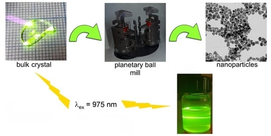
2.3. Nanodispergation of Bulk Crystals

High-energy milling of the grown crystals in the Retsch PM-200 planetary ball mill (Idar-Oberstein, Germany) was carried out for the fabrication of Ba(Y0.964Yb0.030Er0.006)2F8 NPs. Crystal pieces with volume of about 0.1 cm3 were preliminarily pestled in an agate mortar and placed in stainless steel beakers filled with 10 milling balls with a diameter of 10 mm. Trioctylphosphine oxide was used as a surface stabilizer to prevent NPs agglomeration. The crystals were milled in cycles of 5 × 5 min at 600 rpm for 10 h. Separation of the nanosized fraction was performed through centrifugation at 6000 rpm for 15 min.

2.4. Synthesis of Reference β-Na1.5(Y1.17Yb0.3Er0.03)F6 Nanoparticles

Hexagonal β-Na1.5(Y1.17Yb0.3Er0.03)F6 NPs were synthesized by the standard precursor thermolysis method in a high-boiling solvents medium [12,48]. Yttrium, ytterbium, and erbium trifluoroacetates were prepared by dissolving the corresponding rare-earth oxides in a 50 wt.% solution of trifluoroacetic acid and then used as fluoride precursors. For the synthesis of β-Na1.5(Y1.17Yb0.3Er0.03)F6 NPs, 0.78 mmol (CF3COO)3Y, 0.20 mmol (CF3COO)3Yb, 0.02 mmol (CF3COO)3Er, and 1.00 mmol CF3COONa were added to a 100 mL three-necked flask containing 15 mL of oleic acid and 15 mL of 1-octadecene. The resulting mixture was heated to 600 K with a heating rate of 20 K/min under Ar flow and kept at this temperature for 40 min. After cooling the flask to RT, NPs were collected by centrifugation of the resulting mixture at 6000 rpm for 15 min and washed three times with 2-propanol. The prepared particles were dispersed in 10 mL of hexane and used for further analysis.

2.5. XRD Analysis

XRD patterns of the analyzed samples were carried out on an X-ray powder diffractometer MiniFlex 600 (Rigaku, Tokyo, Japan) with CuKα radiation. Diffraction peaks were recorded within the angle range 2θ from 10° to 100°. The phases were identified using the ICDD PDF-2 (2014). The unit-cell parameters were calculated by the Le Bail full-profile fitting (the Jana2006 software).

2.6. Electron Microscopy Characterization

TEM, including selected area electron diffraction (SAED), high-angle annular dark-field scanning TEM (HAADF-STEM), and simultaneously acquired energy-dispersive X-ray spectroscopy (EDS) elemental mapping analysis were performed on a Tecnai Osiris (FEI, Hillsboro, OR, USA) electron microscope operated at 200 kV and equipped with a Super-X SDD high-sensitivity EDX spectrometer. TEM samples were prepared by dropping nanoparticle hexane dispersion onto the carbon-coated Cu TEM grids. The experimental images were processed using the Digital Micrograph software 2.31 (Gatan Inc., Pleasanto, CA, USA). Interpretation of the electron diffraction patterns and HRTEM diffractograms were carried out using the Java Electron Microscopy Software (JEMS) [49].

2.7. Fluorescent Spectroscopy Analysis

The photoluminescence characteristics of the produced NPs were performed on a Fluorolog-3 fluorometer (HJY, Longjumeau, France) equipped with the Hamamatsu photomultiplier R929P and H10330A-75 tubes (Hamamatsu Photonics, Shizuoka, Japan) for registration of luminescent radiation in the visible (350–850 nm) and IR (1100–1700 nm) optical ranges, respectively. A semiconductor ATC IR laser λ = 975 nm (pump power density was 25 W/cm2) was used as an excitation light source. The luminescence decay curves were recorded on a WaveRunner 9000-MS high-speed oscilloscope (Teledyne LeCroy, Chestnut Ridge, NY, USA) under excitation by a semiconductor ATC IR laser (λ = 975 nm) in a pulsed mode. The pump power density was 115 and 55 W/cm2; the pulse repetition rate was 200 and 50 Hz for recording the decay curves in the visible and IR optical ranges, respectively. The laser pulse duration was 1000 μs.

3. Results

3.1. Growth and Optical Properties of Ba(Y0.964Yb0.030Er0.006)2F8 Crystal

Ba(Y0.964Yb0.030Er0.006)2F8 crystals with a diameter of 10 mm and a length of up to 40 mm were successfully grown (Figure 1a). Crystal boules were optically transparent, no light-scattering inclusions and cracks were observed. The crystals demonstrate bright green luminescence under IR excitation (Figure 1b).
In the optical transmission spectrum (Figure 1c), the absorption bands in the UV (380 nm), visible (487, 522, 650 nm), and IR (1480–1530 nm) regions are assigned to electric dipole intra-configurational 4f-4f transitions in Er3+ ions, while the intense band in the region of 960–980 nm is attributed to the absorption of the Yb3+ ions. Weak additional bands registered at 246 and 300 nm are associated with uncontrolled contamination of a Ce3+ ions impurity [50].

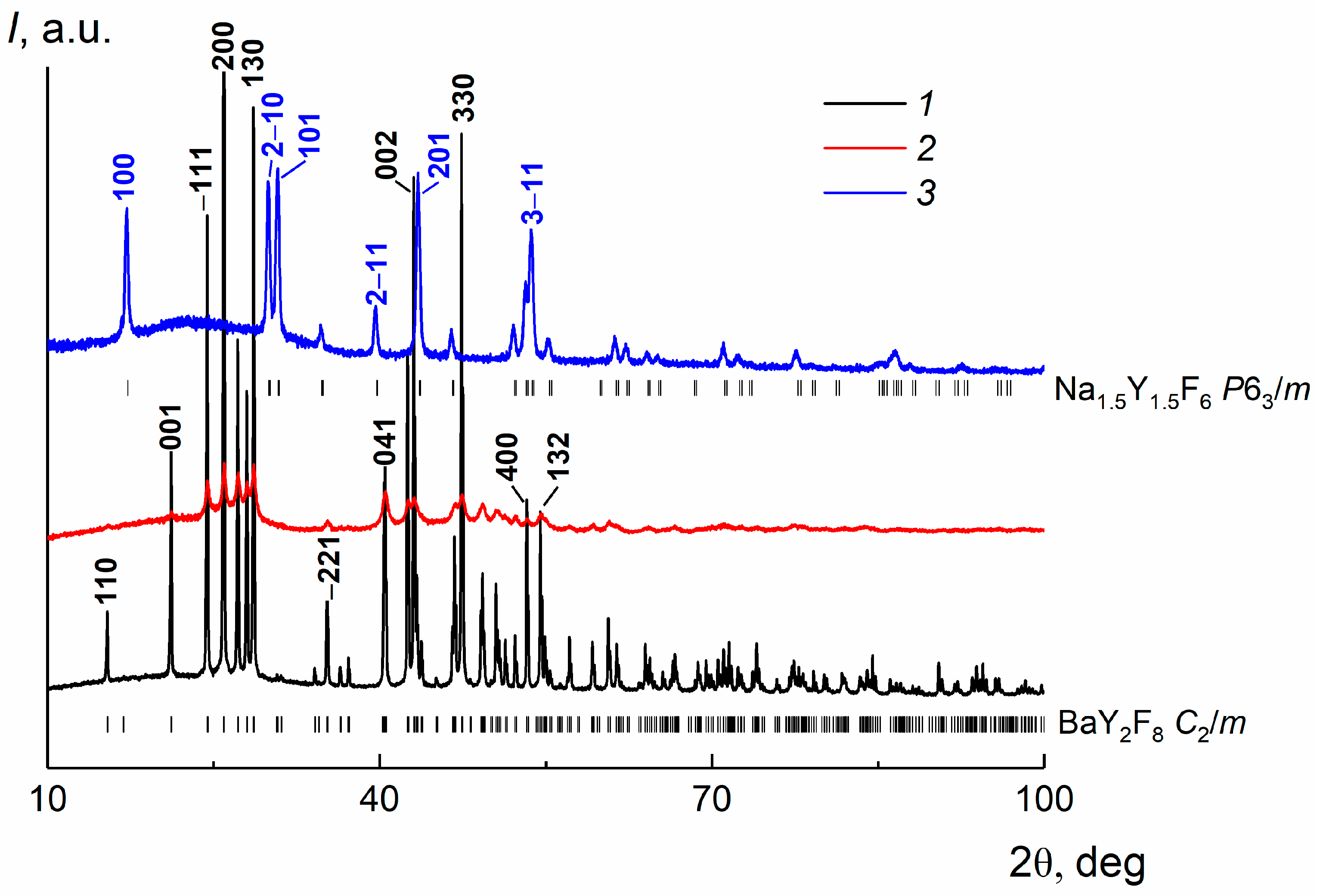
3.2. XRD Analysis of a Bulk Crystal, Ba(Y0.964Yb0.030Er0.006)2F8 and β-Na1.5(Y1.17Yb0.3Er0.03)F6 Nanoparticles

The phase purity and the assignment of grown crystals and fabricated NPs to a monoclinic (sp. gr. C2/m) BaTm2F8-type structure [51] were confirmed by XRD analysis (Figure 2). Comparison of the calculated structural characteristics of bulk and milled Ba(Y0.964Yb0.030Er0.006)2F8 samples, shown in Table 1, confirms the absence of changes in the crystal structure during the milling process. A minor reducing in the lattice parameters for the nanosized crystal sample is probably attributed to the originating mechanical compression strains during such high energy treatment. A significant broadening and a decrease in the intensity patterns for fabricated NPs indirectly indicate the crystallite sizes reducing and a transition to nanoscale. No impurity phases were observed. The reference β-Na1.5(Y1.17Yb0.3Er0.03)F6 particles synthesized by thermolysis are single-phase and crystallize in a gagarinite β-Na1.5Y1.5F6 structure type (sp. gr. P63/m) [52].

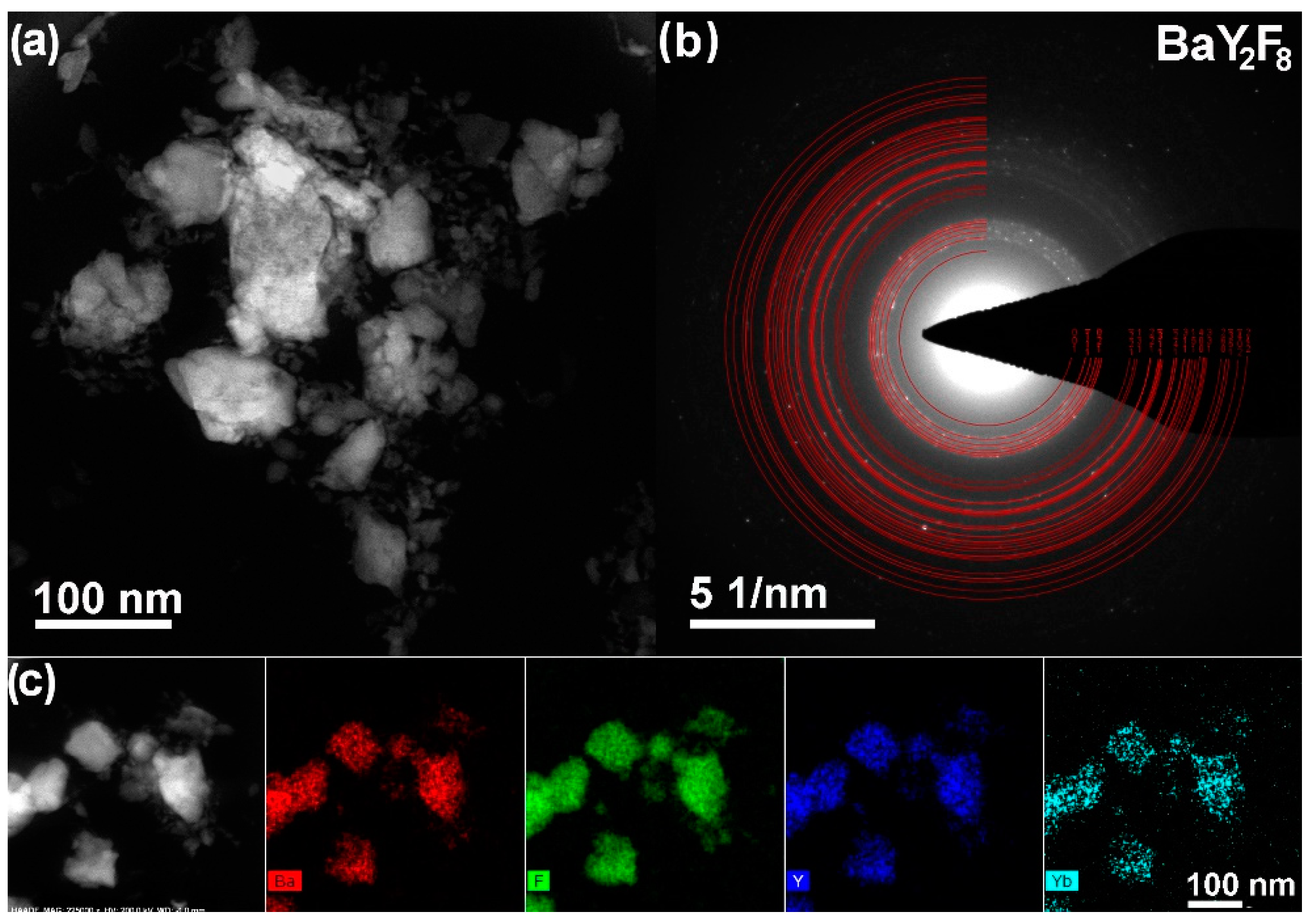
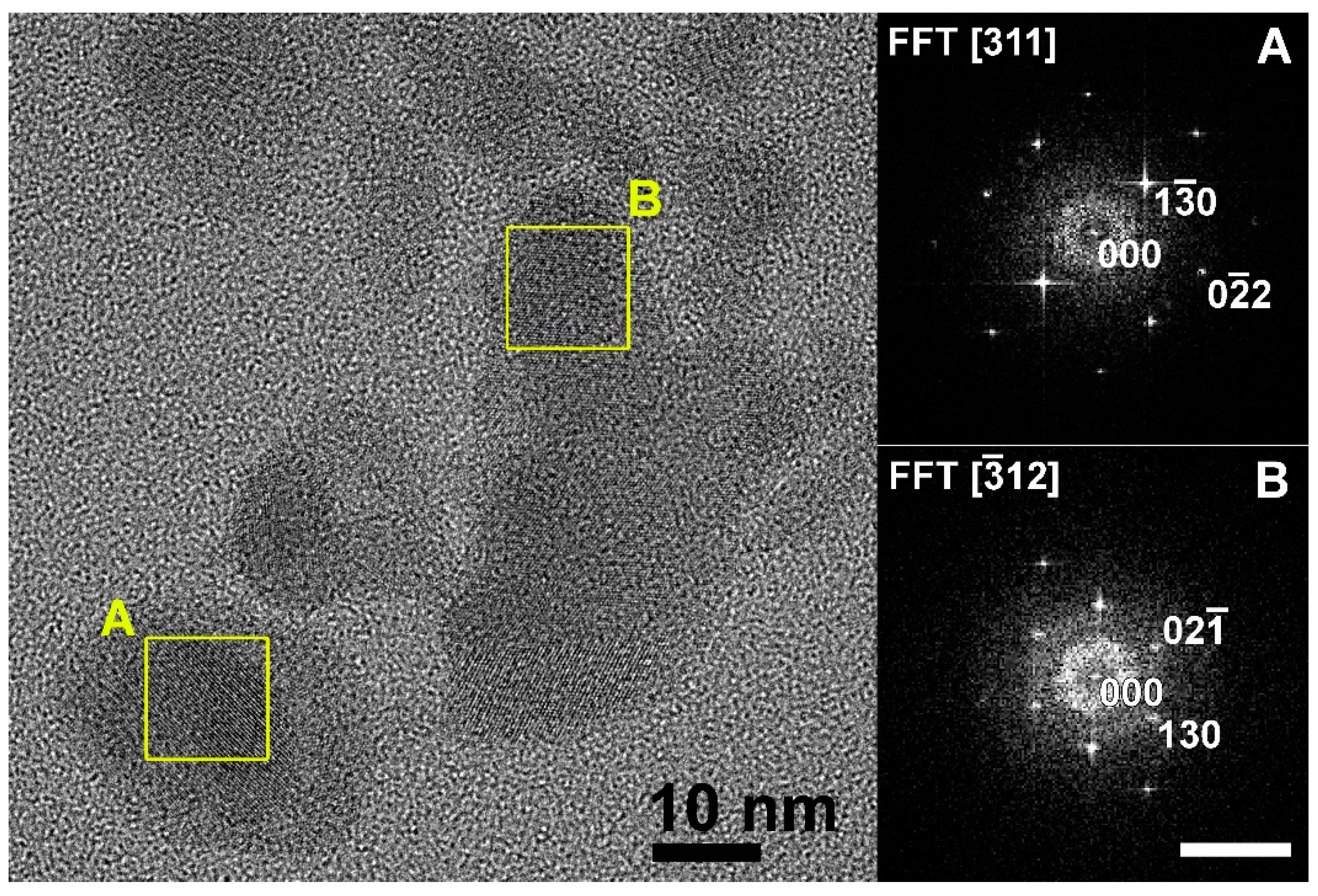
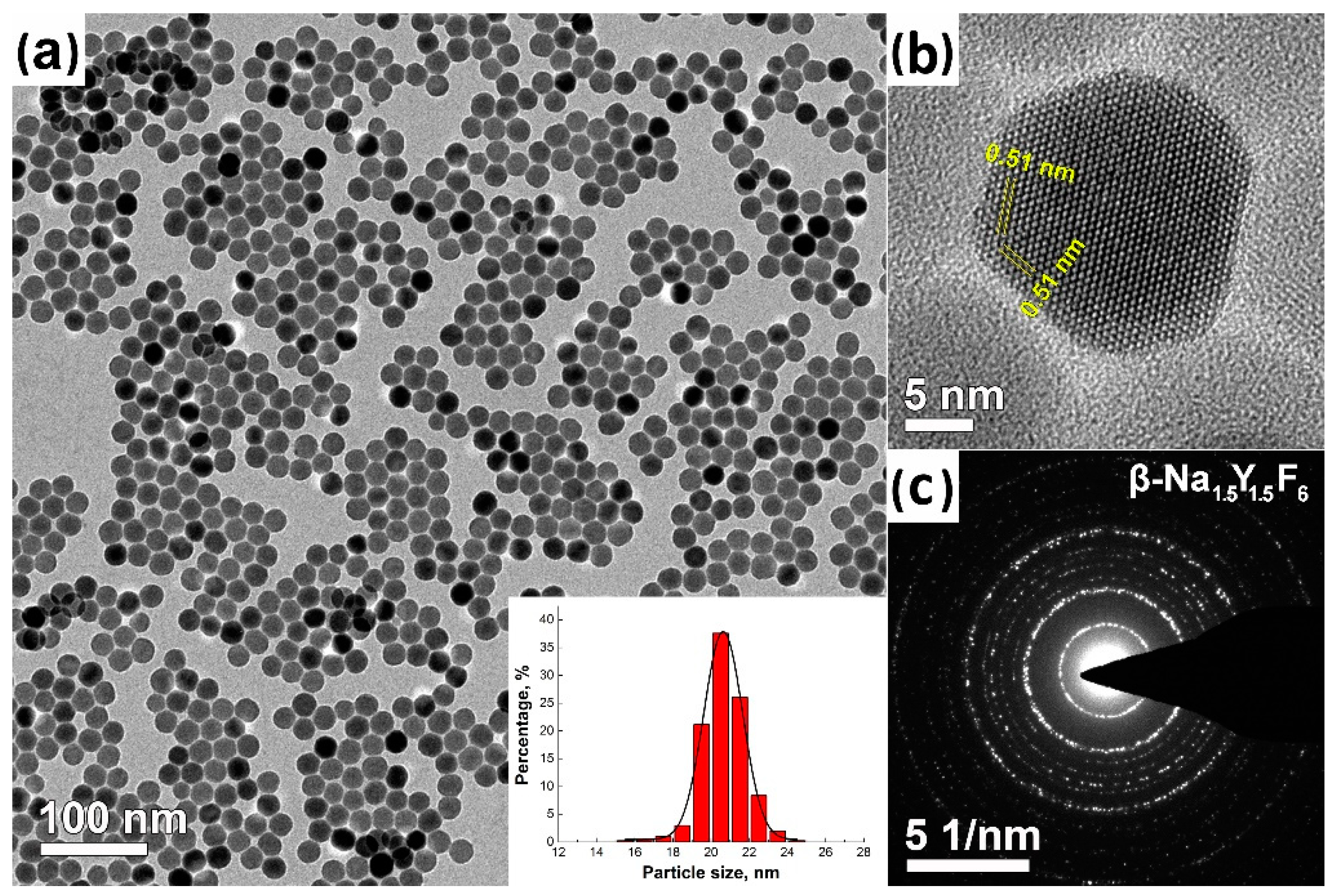
3.3. TEM Analysis of Ba(Y0.964Yb0.030Er0.006)2F8 and β-Na1.5(Y1.17Yb0.3Er0.03)F6 Nanoparticles

TEM data demonstrate that the high energy milling of the initial bulk Ba(Y0.964Yb0.030Er0.006)2F8 crystal for 10 h leads to the formation of irregular NPs. The HAADF-STEM image of the agglomerate of NPs without any prominent morphological features in a wide size range from 10 to 70 nm is presented in Figure 3a. The electron diffraction pattern (Figure 3b) taken from an agglomerate corresponds to the structure of the initial bulk monoclinic crystal and confirms the above XRD results. No other phases were found. The EDS analysis results also show no changes in the sample elemental composition during the milling process (Figure 3c).
HRTEM image of the Ba(Y0.964Yb0.030Er0.006)2F8 NPs produced by high-energy milling of a bulk crystal and the corresponding diffractograms of nanocrystals A and B are presented in Figure 4. Interpretation of FFT diffractograms using JEMS shows that A and B NPs are oriented along the [311] and [ 3 ¯ 12] directions.
TEM image of β-Na1.5(Y1.17Yb0.3Er0.03)F6 nanophosphors is shown in Figure 5. According to TEM data (Figure 5a), the thermolysis method leads to the near-monodisperse β-Na1.5(Y1.17Yb0.3Er0.03)F6 NPs in the size range of 21 ± 5 nm. SAED pattern of β-Na1.5(Y1.17Yb0.3Er0.03)F6 particles, shown in Figure 5c, corresponds to the hexagonal crystal structure of β-Na1.5Y1.5F6.

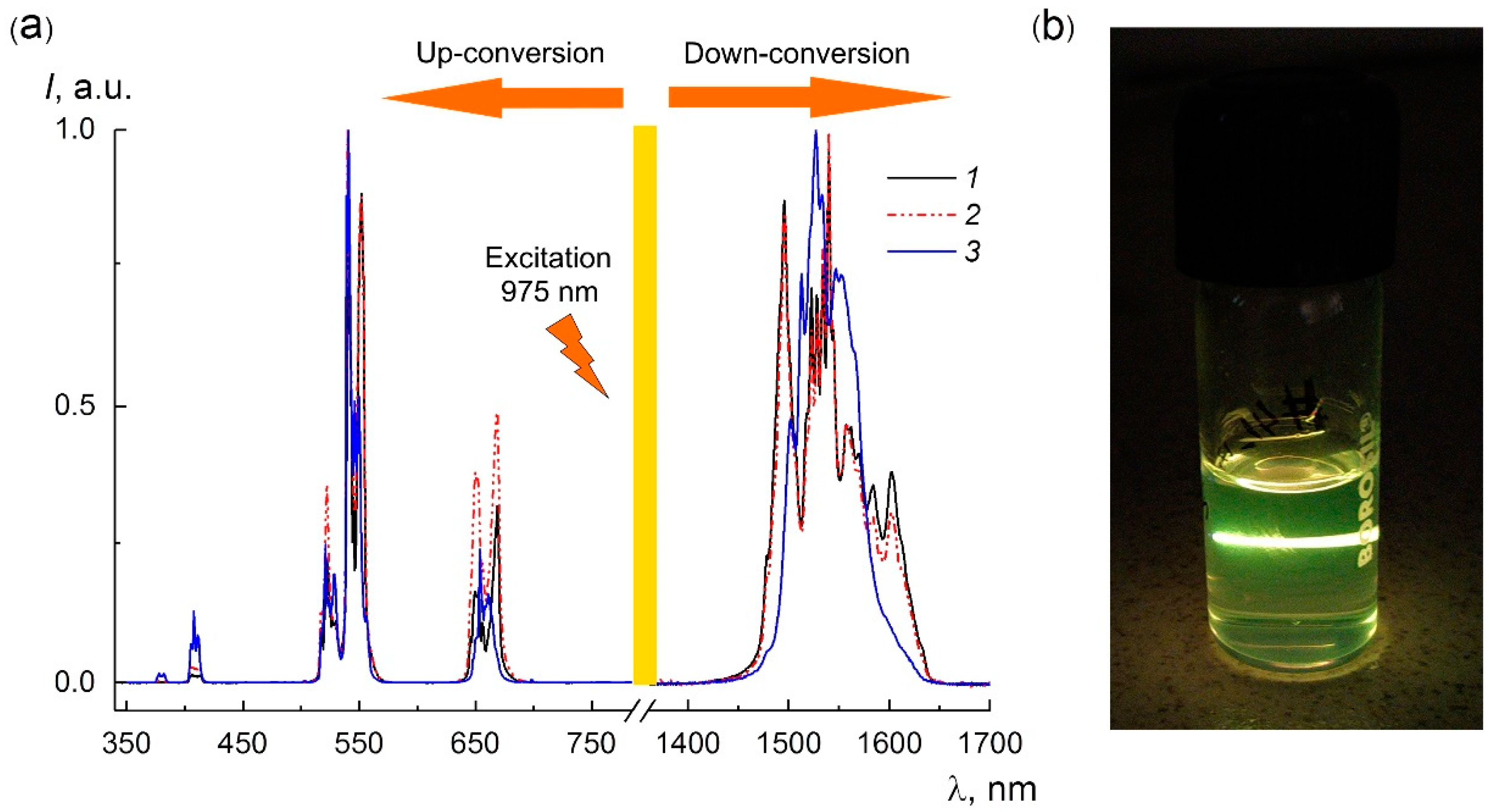
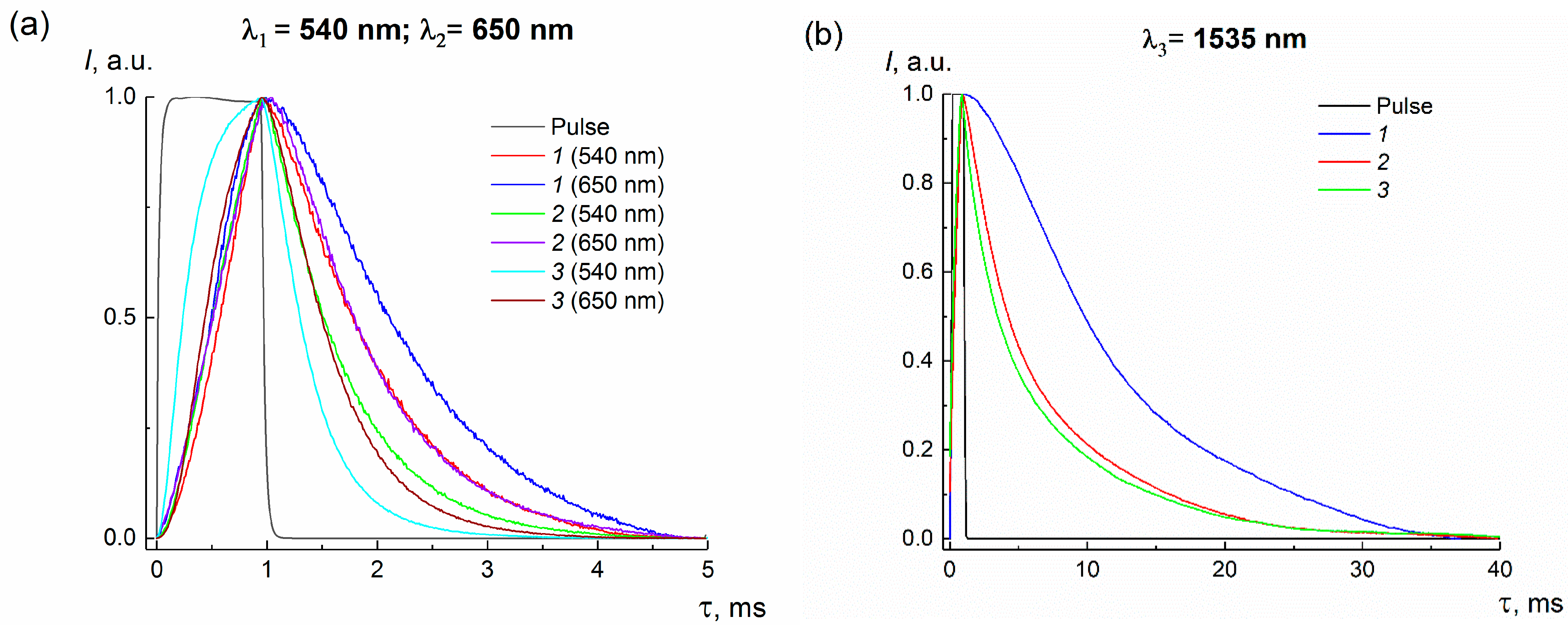
3.4. Photoluminescence Properties of Bulk and Nanocrystals

Nanosized and bulk Ba(Y0.964Yb0.030Er0.006)2F8 samples demonstrate intense visible and IR PL upon excitation by λ = 975 nm radiation at RT. Intense characteristic luminescence bands, corresponding to 4f-4f energy transitions in Er3+ ions, namely transitions from the 4F7/2 (408 nm), 4H11/2 (522 nm), 4S3/2 (540 nm), 4F9/2 (651 nm), and 4I13/2 (1535 nm) multiplet manifolds to the 4I15/2 ground state due to up- and down-conversion energy transfer processes, were observed in the PL spectra (Figure 6a). Comparison the PL spectra of nanosized and bulk Ba(Y0.964Yb0.030Er0.006)2F8 samples did not reveal any qualitative differences; no shifts or broadening of the emission bands were detected.
The energy transfer mechanism in BaY2F8 crystals co-doped with Yb3+ and Er3+ ions is shown in Figure 7. Upon absorption of 975 nm photon, Yb3+ ions are excited from the 2F7/2 ground state to 2F5/2 state, and then non-radiatively transfer energy to neighboring Er3+ ions, populating their intermediate 4I11/2 state. As a result of the successive absorption of two photons, Er3+ ions are excited to the 4F7/2 state, which can non-radiatively decay to the 2H11/2, 4S3/2, and 4F9/2 electronic states, followed by radiative green 4S3/2 → 4I15/2 (522 nm), 2H11/2 → 4I15/2 (540 nm), and red 4F9/2 → 4I15/2 (650 nm) characteristic luminescent transitions. Additionally, Er3+ ions can be excited to the 2H9/2 state by a three-photon absorption process, followed by a 2H9/2 → 4I15/2 radiative transition at a wavelength λ = 408 nm. The IR luminescence (1535 nm) results from a radiative decay of the long-lived intermediate 4I11/2 state (4H11/2 → 4I15/2).
The PL decay kinetics of the most intense spectral bands at 540, 650, and 1535 nm for Ba(Y0.964Yb0.030Er0.006)2F8 samples are shown in Figure 8. Each decay curve is well fitted in a single-exponential dependence:
I ( t ) = I 0 exp ( t τ   ) ,
where I(t) is the PL intensity at time t; I0 is the PL intensity at the end of the laser excitation pulse, τ is the average lifetime of the excited state.
The calculated PL lifetimes for the studied samples are presented in Table 2. A significant decrease in the lifetime for each PL band is observed for Ba(Y0.964Yb0.030Er0.006)2F8 NPs with respect to that for a bulk crystal. This phenomenon is associated with an increase in the surface-to-volume ratio for the nanosized particles. Milling NPs have a high concentration of surface defects generated by strains in the crystallites during the mechanical milling process. The interaction of active Er3+ centers, lying on the particle surface, with these defects leads to a significant PL quenching.
A comparative analysis of the PL spectra of Ba(Y0.964Yb0.030Er0.006)2F8 and reference β-Na1.5(Y1.17Yb0.3Er0.03)F6 NPs demonstrates the maxima position shifts and the broadening of the spectral lines for a milling sample (Figure 6a). A strong splitting of PL bands for Ba(Y0.964Yb0.030Er0.006)2F8 NPs is observed due to a local symmetry lowering of the active centers. The local symmetry of REE ions in the BaY2F8 and β-NaYF4 crystal hosts is C2 and C3h, respectively. Strong Stark splitting of Er3+ electronic levels in the more asymmetric crystal field of the monoclinic BaY2F8 matrix leads to a stronger degeneracy of existing metastable energy states, which is more clearly observed in the PL spectrum for Ba(Y0.964Yb0.030Er0.006)2F8 in the IR range. Such a degeneracy of states leads to an energy gap increasing between closely spaced 4f states of Er3+ ions and a reduction in the non-radiative relaxation processes probability and, as a consequence, to a rise in the 4S3/2, 4F9/2, and 4I13/2 states’ lifetimes. Therefore, the PL lifetimes of Er3+ excited states for the BaY2F8 matrix are higher than β-Na1.5(Y1.17Yb0.3Er0.03)F6 ones.

4. Conclusions

In this study, Ba(Y0.964Yb0.030Er0.006)2F8 NPs in the size range of 30–70 nm were successfully produced by high-energy milling of the Bridgman-grown single crystals. This scalable and versatile method provides a feasible strategy for the mass production of novel fluorescent nanomaterials in a narrow size range that are currently difficult to prepare by available chemical approaches. Ba(Y0.964Yb0.030Er0.006)2F8 NPs demonstrate a bright PL in a wide spectral range, which provides great opportunities for applications of these nanophosphors in photonics and biotechnology, for example, as visible and NIR luminescent labels. Further optimization of the milling procedure, namely the adjustment of nanodispersion technological parameters and suitable hydrophobic or hydrophilic surfactants for the nanosized fraction stabilizing, will provide the possibility to control NP sizes in a narrower range. Optimization of REE doping content and the particle surface passivation by coating a protective shell will significantly enhance the PL properties of studied NPs and are the object of future research.
We believe that BaY2F8-based NPs will occupy a rightful place in nanotechnology, the same as the commonly used β-NaREF4 nanocrystals, which are the most efficient PL nanomaterials to date.

Author Contributions

D.N.K., M.S.S.D. and V.V.K. were involved in the development of the investigation concept; D.N.K. performed crystal growth experiments; A.V.K. performed the synthesis of nanoscale samples; A.V.K. and D.N.K. performed the phase composition analysis of the prepared samples; N.A.A. performed the TEM analysis; A.V.K. and K.V.K. performed the investigation of spectroscopic properties; A.V.K. performed the original draft preparation of the manuscript; N.A.A., K.V.K. and D.N.K. reviewed and edited the draft manuscript; D.N.K. coordinated the scientific group. All authors have read and agreed to the published version of the manuscript.

Funding

This research was funded by the Russian Foundation for Basic Research (project No. 20-52-56017) and Iran National Science Foundation (project No. 99004620) in part concerning the fluoride crystal growth and nanoparticle synthesis and by the Ministry of Science and Higher Education of the Russian Federation within the State assignments of the Federal Scientific Research Centre “Crystallography and Photonics” of the Russian Academy of Sciences in part concerning crystal sample characterization using the equipment of the Shared Research Center.

Informed Consent Statement

Not applicable.

Data Availability Statement

Data sharing not applicable.

Acknowledgments

The authors are deeply grateful to I.V. Krylov and B.V. Nabatov for their help in carrying out the experiments and fruitful discussions.

Conflicts of Interest

The authors declare no conflict of interest. The funders had no role in the design of the study; in the collection, analyses or interpretation of the data; in the writing of the manuscript or in the decision to publish the results.

References

  1. Meruga, J.M.; Cross, W.M.; May, P.S.; Luu, Q.; Crawford, G.A.; Kellar, J.J. Security printing of covert quick response codes using upconverting nanoparticle inks. Nanotechnology 2012, 23, 395201. [Google Scholar] [CrossRef] [PubMed]
  2. Zhu, X.; Feng, W.; Chang, J.; Tan, Y.-W.; Li, J.; Chen, M.; Sun, Y.; Li, F. Temperature-feedback upconversion nanocomposite for accurate photothermal therapy at facile temperature. Nat. Commun. 2016, 7, 10437. [Google Scholar] [CrossRef] [PubMed]
  3. Generalova, A.; Chichkov, B.; Khaydukov, E. Multicomponent nanocrystals with anti-Stokes luminescence as contrast agents for modern imaging techniques. Adv. Colloid Interface Sci. 2017, 245, 1–19. [Google Scholar] [CrossRef] [PubMed]
  4. Liu, X.; Wang, Y.; Li, X.; Yi, Z.; Deng, R.; Liang, L.; Xie, X.; Loong, D.T.B.; Song, S.; Fan, D.; et al. Binary temporal upconversion codes of Mn2+-activated nanoparticles for multilevel anti-counterfeiting. Nat. Commun. 2017, 8, 899. [Google Scholar] [CrossRef] [PubMed]
  5. Tiwari, S.P.; Maurya, S.K.; Yadav, R.S.; Kumar, A.; Kumar, V.; Joubert, M.-F.; Swart, H.C. Future prospects of fluoride based upconversion nanoparticles for emerging applications in biomedical and energy harvesting. J. Vac. Sci. Technol. B 2018, 36, 060801. [Google Scholar] [CrossRef] [Green Version]
  6. Xu, J.; Gulzar, A.; Yang, P.; Bi, H.; Yang, D.; Gai, S.; He, F.; Lin, J.; Xing, B.; Jin, D. Recent advances in near-infrared emitting lanthanide-doped nanoconstructs: Mechanism, design and application for bioimaging. Coord. Chem. Rev. 2018, 381, 104–134. [Google Scholar] [CrossRef]
  7. Karimov, D.N.; Demina, P.A.; Koshelev, A.V.; Rocheva, V.V.; Sokovikov, A.V.; Generalova, A.N.; Zubov, V.P.; Khaydukov, E.V.; Koval’Chuk, M.V.; Panchenko, V.Y. Upconversion Nanoparticles: Synthesis, Photoluminescence Properties, and Applications. Nanotechnol. Russ. 2020, 15, 655–678. [Google Scholar] [CrossRef]
  8. Nadort, A.; Zhao, J.; Goldys, E.M. Lanthanide upconversion luminescence at the nanoscale: Fundamentals and optical properties. Nanoscale 2016, 8, 13099–13130. [Google Scholar] [CrossRef] [Green Version]
  9. Du, Y.-P.; Sun, X.; Zhang, Y.-W.; Yan, Z.-G.; Sun, L.-D.; Yan, C.-H. Uniform Alkaline Earth Fluoride Nanocrystals with Diverse Shapes Grown from Thermolysis of Metal Trifluoroacetates in Hot Surfactant Solutions. Cryst. Growth Des. 2009, 9, 2013–2019. [Google Scholar] [CrossRef]
  10. Fedorov, P.P.; Luginina, A.A.; Kuznetsov, S.; Osiko, V.V. Nanofluorides. J. Fluor. Chem. 2011, 132, 1012–1039. [Google Scholar] [CrossRef]
  11. Cheng, T.; Marin, R.; Skripka, A.; Vetrone, F. Small and Bright Lithium-Based Upconverting Nanoparticles. J. Am. Chem. Soc. 2018, 140, 12890–12899. [Google Scholar] [CrossRef] [PubMed]
  12. Mai, H.-X.; Zhang, Y.-W.; Si, R.; Yan, Z.-G.; Sun, L.-D.; You, L.-P.; Yan, C.-H. High-Quality Sodium Rare-Earth Fluoride Nanocrystals: Controlled Synthesis and Optical Properties. J. Am. Chem. Soc. 2006, 128, 6426–6436. [Google Scholar] [CrossRef] [PubMed]
  13. You, W.; Tu, D.; Zheng, W.; Shang, X.; Song, X.; Zhou, S.; Liu, Y.; Li, R.; Chen, X. Large-scale synthesis of uniform lanthanide-doped NaREF4 upconversion/downshifting nanoprobes for bioapplications. Nanoscale 2018, 10, 11477–11484. [Google Scholar] [CrossRef] [PubMed]
  14. Grzyb, T.; Balabhadra, S.; Przybylska, D.; Węcławiak, M. Upconversion luminescence in BaYF5, BaGdF5 and BaLuF5 nanocrystals doped with Yb3+/Ho3+, Yb3+/Er3+ or Yb3+/Tm3+ ions. J. Alloys Compd. 2015, 649, 606–616. [Google Scholar] [CrossRef]
  15. Li, C.; Xu, Z.; Yang, D.; Cheng, Z.; Hou, Z.; Ma, P.; Lian, H.; Lin, J. Well-dispersed KRE3F10(RE = Sm–Lu, Y) nanocrystals: Solvothermal synthesis and luminescence properties. CrystEngComm 2011, 14, 670–678. [Google Scholar] [CrossRef]
  16. Razumkova, I.A.; Denisenko, Y.G.; Boyko, A.N.; Ikonnikov, D.A.; Aleksandrovsky, A.S.; Azarapin, N.O.; Andreev, O.V. Synthesis and Upconversion Luminescence in LaF3:Yb3+, Ho3+, GdF3: Yb3+, Tm3+ and YF3:Yb3+, Er3+ obtained from Sulfide Precursors. Z. Anorg. Allg. Chem. 2019, 645, 1393–1401. [Google Scholar] [CrossRef]
  17. Boyer, J.-C.; Cuccia, A.L.A.; Capobianco, J.A. Synthesis of Colloidal Upconverting NaYF4: Er3+/Yb3+ and Tm3+/Yb3+ Monodisperse Nanocrystals. Nano Lett. 2007, 7, 847–852. [Google Scholar] [CrossRef]
  18. Gemini, L.; Schmitz, T.; Kling, R.; Barcikowski, S.; Gökce, B. Upconversion Nanoparticles Synthesized by Ultrashort Pulsed Laser Ablation in Liquid: Effect of the Stabilizing Environment. ChemPhysChem 2017, 18, 1210–1216. [Google Scholar] [CrossRef]
  19. Sajti, L.; Karimov, D.N.; Rocheva, V.V.; Arkharova, N.A.; Khaydukov, K.V.; Lebedev, O.I.; Voloshin, A.E.; Generalova, A.N.; Chichkov, B.N.; Khaydukov, E.V. Pulsed laser reshaping and fragmentation of upconversion nanoparticles—from hexagonal prisms to 1D nanorods through “Medusa”-like structures. Nano Res. 2020, 14, 1141–1148. [Google Scholar] [CrossRef]
  20. Yuan, D.; Yi, G.S.; Chow, G.M. Effects of size and surface on luminescence properties of submicron upconversion NaYF4: Yb, Er particles. J. Mater. Res. 2009, 24, 2042–2050. [Google Scholar] [CrossRef]
  21. Patel, D.N.; Sarkisov, S.S.; Darwish, A.M.; Ballato, J. Optical gain in capillary light guides filled with NaYF4: Yb3+, Er3+ nanocolloids. Opt. Express 2016, 24, 21147–21158. [Google Scholar] [CrossRef] [PubMed]
  22. Sobolev, B.; Tkachenko, N. Phase diagrams of BaF2-(Y, Ln)F3 systems. J. Less Common Met. 1982, 85, 155–170. [Google Scholar] [CrossRef]
  23. Maksimov, B.A.; Solans, K.; Dudka, A.P.; Genkina, E.A.; Font-Badria, M.; Buchinskaya, I.I.; Loshmanov, A.A.; Golubev, A.M.; Simonov, V.I.; Font-Altaba, M.; et al. The fluorite-matrix-based Ba4R3F17 (R = Y, Yb) crystal structure. Ordering of cations and specific features of the anionic motif. Crystallogr. Rep. 1996, 41, 56–64. [Google Scholar]
  24. Greis, O.; Stede, P.; Kieser, M. Darstellung und Eigenschaften der Ternären Verbindungen BaSE2F8 mit SE = Dy—Lu und Y. Z. Anorg. Allg. Chem. 1981, 477, 133–138. [Google Scholar] [CrossRef]
  25. Guggenheim, H.J.; Johnson, L.F. New fluoride compounds for efficient infrared-to-visible conversion. Appl. Phys. Lett. 1969, 15, 51–52. [Google Scholar] [CrossRef]
  26. Johnson, L.F.; Guggenheim, H.J. Infrared-Pumped Visible Laser. Appl. Phys. Lett. 1971, 19, 44–47. [Google Scholar] [CrossRef]
  27. Pollack, S.A.; Chang, D.B.; McFarlane, R.A.; Jenssen, H. Infrared (Er)BaY2F8 laser pumped through di- and tri-ionic upconversion processes. J. Appl. Phys. 1990, 67, 648–653. [Google Scholar] [CrossRef]
  28. Kaminskii, A.A.; Kochubeĭ, S.A.; Naumochkin, K.N.; Pestryakov, E.V.; Trunov, V.I.; Uvarova, T.V. Amplification of ultraviolet radiation due to the 5d–4finterconfigurational transition of the Ce3+ ion in BaY2F8. Sov. J. Quantum Electron. 1989, 19, 340–342. [Google Scholar] [CrossRef]
  29. Noginov, M.A.; Curley, M.; Venkateswarlu, P.; Williams, A.; Jenssen, H.P. Excitation scheme for the upper energy levels in a Tm:Yb:BaY2F8 laser crystal. J. Opt. Soc. Am. B 1997, 14, 2126–2136. [Google Scholar] [CrossRef]
  30. Uvarova, T.V.; Pushkar, A.A.; Uvarova, A.G. Multi-bands active media on the basis of BaY2F8 single crystals for up-conversion solid-state lasers with diode pumping. Phys. Status Solidi C 2011, 8, 2911–2914. [Google Scholar] [CrossRef]
  31. Spijker, J.V.; Dorenbos, P.; van Eijk, C.; Jacobs, J.; Hartog, H.D.; Korolev, N. Luminescence and scintillation properties of BaY2F8:Ce3+, BaLu2F8 and BaLu2F8:Ce3+. J. Lumin. 1999, 85, 11–19. [Google Scholar] [CrossRef]
  32. de Mello, A.C.; Andrade, A.B.; Nakamura, G.H.; Baldochi, S.L.; Valerio, M.E. Scintillation mechanism of Tb3+ doped BaY2F8. Opt. Mater. 2010, 32, 1337–1340. [Google Scholar] [CrossRef]
  33. Kurosawa, S.; Yanagida, T.; Pejchal, J.; Fukuda, K.; Kawaguchi, N.; Ishizu, S.; Suyama, T.; Nakagawa, M.; Yokota, Y.; Nikl, M.; et al. Evaluation of Nd:BaY2F8 for VUV scintillator. Radiat. Meas. 2013, 55, 108–111. [Google Scholar] [CrossRef]
  34. Liu, H.; Lu, W.; Wang, H.; Rao, L.; Yi, Z.; Zeng, S.; Hao, J. Simultaneous synthesis and amine-functionalization of single-phase BaYF5:Yb/Er nanoprobe for dual-modal in vivo upconversion fluorescence and long-lasting X-ray computed tomography imaging. Nanoscale 2013, 5, 6023–6029. [Google Scholar] [CrossRef] [PubMed]
  35. Nampi, P.P.; Vakurov, A.; Viswambharan, H.; Schneider, J.E.; Drummond-Brydson, R.; Millner, P.A.; Saha, S.; Jose, G. Barium yttrium fluoride based upconversion nanoparticles as dual mode image contrast agents. Mater. Sci. Eng. C 2021, 124, 111937. [Google Scholar] [CrossRef] [PubMed]
  36. Zhang, C.; Ma, P.A.; Li, C.; Li, G.; Huang, S.; Yang, D.; Shang, M.; Kang, X.; Lin, J. Controllable and white upconversion luminescence in BaYF5:Ln3+ (Ln = Yb, Er, Tm) nanocrystals. J. Mater. Chem. 2010, 21, 717–723. [Google Scholar] [CrossRef]
  37. Cao, Z.; Zhou, S.; Jiang, G.; Chen, Y.; Duan, C.; Yin, M. Temperature dependent luminescence of Dy3+ doped BaYF5 nanoparticles for optical thermometry. Curr. Appl. Phys. 2014, 14, 1067–1071. [Google Scholar] [CrossRef]
  38. Haritha, P.; Martin, I.R.; Viswanath, C.D.; Vijaya, N.; Krishnaiah, K.V.; Jayasankar, C.; Haranath, D.; Lavín, V.; Venkatramu, V. Structure, morphology and optical characterization of Dy3+-doped BaYF5 nanocrystals for warm white light emitting devices. Opt. Mater. 2017, 70, 16–24. [Google Scholar] [CrossRef]
  39. Fedorov, P.P.; Mayakova, M.N.; Kuznetsov, S.V.; Voronov, V.V.; Ermakov, R.P.; Samarina, K.S.; Popov, A.I.; Osiko, V.V. Co-precipitation of yttrium and barium fluorides from aqueous solutions. Mater. Res. Bull. 2012, 47, 1794–1799. [Google Scholar] [CrossRef]
  40. Karbowiak, M.; Cichos, J. Does BaYF5 nanocrystals exist?—The BaF2-YF3 solid solution revisited using photoluminescence spectroscopy. J. Alloys Compd. 2016, 673, 258–264. [Google Scholar] [CrossRef]
  41. Grube, J.; Krieke, G. How activator ion concentration affects spectroscopic properties on Ba4Y3F17: Er3+, Yb3+, a new perspective up-conversion material. J. Lumin. 2018, 203, 376–384. [Google Scholar] [CrossRef]
  42. Pudovkin, M.; Kuznetsov, S.; Proydakova, V.Y.; Voronov, V.; Semashko, V. Luminescent thermometry based on Ba4Y3F17:Pr3+ and Ba4Y3F17:Pr3+,Yb3+ nanoparticles. Ceram. Int. 2020, 46, 11658–11666. [Google Scholar] [CrossRef]
  43. Wang, G.; Peng, Q.; Li, Y. Synthesis and upconversion luminescence of BaY2F8:Yb3+/Er3+ nanobelts. Chem. Commun. 2010, 46, 7528–7529. [Google Scholar] [CrossRef] [PubMed]
  44. Wang, X.; Hou, Y.; Qu, J.; Ding, J.; Lin, H.; Liu, L.; Zhou, Y.; Zeng, F.; Li, C.; Su, Z. Up-conversion photoluminescence properties and energy transfer process of Ho3+,Yb3+ Co-doped BaY2F8 fine fibers. J. Lumin. 2019, 212, 154–159. [Google Scholar] [CrossRef]
  45. Nair, G.B.; Kumar, A.; Swart, H.; Dhoble, S. Facile precipitation synthesis of green-emitting BaY2F8:Yb3+, Ho3+ upconverting phosphor. Ceram. Int. 2019, 45, 14205–14213. [Google Scholar] [CrossRef]
  46. Toncelli, A.; Ahmadi, B.; Marchetti, F. Upconversion enhancement in Yb3+,Tm3+:BaY2F8 quasi-nanoparticles. J. Lumin. 2012, 132, 2268–2274. [Google Scholar] [CrossRef]
  47. Hakim, R.; Damak, K.; Gemmi, M.; Luin, S.; Maalej, R.; Toncelli, A. Pr3+:BaY2F8 Crystal Nanoparticles (24 nm) Produced by High-Energy Ball Milling: Spectroscopic Characterization and Comparison with Bulk Properties. J. Phys. Chem. C 2015, 119, 2844–2851. [Google Scholar] [CrossRef]
  48. Boyer, J.-C.; Vetrone, F.; Cuccia, L.A.; Capobianco, J.A. Synthesis of Colloidal Upconverting NaYF4 Nanocrystals Doped with Er3+, Yb3+ and Tm3+, Yb3+ via Thermal Decomposition of Lanthanide Trifluoroacetate Precursors. J. Am. Chem. Soc. 2006, 128, 7444–7445. [Google Scholar] [CrossRef]
  49. JEMS. Available online: http://www.jems-saas.ch (accessed on 15 March 2020).
  50. Sani, E.; Toncelli, A.; Tonelli, M. Effect of Cerium codoping on Er:BaY2F8 crystals. Opt. Express 2005, 13, 8980–8992. [Google Scholar] [CrossRef]
  51. Izotova, O.E.; Aleksandrov, V.B. The Crystal Structure of BaTm2F8. Sov. Phys. Dokl. 1970, 15, 525–527. [Google Scholar]
  52. Krämer, K.W.; Biner, D.; Frei, G.; Güdel, H.U.; Hehlen, M.P.; Lüthi, S.R. Hexagonal Sodium Yttrium Fluoride Based Green and Blue Emitting Upconversion Phosphors. Chem. Mater. 2004, 16, 1244–1251. [Google Scholar] [CrossRef]
Figure 1. (a) The as-grown crystal appearance, (b) intense green luminescence of the bulk crystal under excitation with λ = 975 nm radiation, and (c) the transmission spectrum of the Ba(Y0.964Yb0.030Er0.006)2F8 crystal, the sample thickness was 2 mm.
Crystals 12 00599 g001
Figure 2. XRD patterns of a bulk Ba(Y0.964Yb0.030Er0.006)2F8 crystal (1), Ba(Y0.964Yb0.030Er0.006)2F8 particles, produced by milling procedure (2), β-Na1.5(Y1.17Yb0.3Er0.03)F6 NPs, synthesized by thermolysis (3). The positions of the Bragg reflection peaks for the specified space groups are indicated.
Crystals 12 00599 g002
Figure 3. (a) HAADF-STEM image of Ba(Y0.964Yb0.030Er0.006)2F8 NPs; (b) the corresponding SAED pattern with the overlaid JEMS simulated rings of the BaY2F8 monoclinic phase (sp. gr. C2/m, a = 0.698 nm, b = 1.051 nm, c = 0.4261 nm, β = 99.676°); (c) HAADF-STEM image of Ba(Y0.964Yb0.030Er0.006)2F8 NPs and the corresponding EDX elemental mapping of Ba L-line (red), F K-line (green), Y L-line (blue), and Yb M-line (light blue).
Crystals 12 00599 g003
Figure 4. HRTEM image of Ba(Y0.964Yb0.030Er0.006)2F8 NPs (A) and (B) their corresponding diffractograms obtained along directions close to [311] and [ 3 ¯ 12]; the scale bar on the diffractograms corresponds to 5 nm−1.
Crystals 12 00599 g004
Figure 5. (a) Bright field TEM image with the particle size distribution histogram (insert); (b) HRTEM image of an individual particle close to the [0001] direction; (c) SAED pattern of β-Na1.5(Y1.17Yb0.3Er0.03)F6 NPs, synthesized by thermolysis method.
Crystals 12 00599 g005
Figure 6. (a) The RT PL spectra of a bulk Ba(Y0.964Yb0.030Er0.006)2F8 crystal (1), Ba(Y0.964Yb0.030Er0.006)2F8 particles (2), and β-Na1.5(Y1.17Yb0.3Er0.03)F6 (3) NPs upon excitation by λ = 975 nm radiation; (b) the appearance of a Ba(Y0.964Yb0.030Er0.006)2F8 colloid, performing intense green luminescence upon IR excitation (λ = 975 nm). The luminescence spectra are normalized relative to the wavelength corresponding to the maximum radiation intensity in the visible (540 nm) and IR ranges (1535 nm).
Crystals 12 00599 g006
Figure 7. Simplified energy level diagram and possible up- and down-conversion luminescence mechanisms in BaY2F8 crystals co-doped with Yb3+/Er3+ ions.
Crystals 12 00599 g007
Figure 8. Decay curves of luminescence emission bands in (a) visible and (b) IR spectral ranges of bulk Ba(Y0.964Yb0.030Er0.006)2F8 crystal (1), nanodispergated Ba(Y0.964Yb0.030Er0.006)2F8 particles (2), β-Na1.5(Y1.17Yb0.3Er0.03)F6 NPs (3) upon IR excitation (λ = 975 nm).
Crystals 12 00599 g008
Table 1. Calculated unit cell parameters for a bulk Ba(Y0.964Yb0.030Er0.006)2F8 crystal and nanodispergated particles and β-Na1.5(Y1.17Yb0.3Er0.03)F6 NPs, synthesized by thermolysis.
Space GroupUnit Cell Parameters, Å
bulk Ba(Y0.964Yb0.030Er0.006)2F8 C2/ma = 6.971(1), b = 10.506(1), c = 4.257(1), β = 99.676°
milled Ba(Y0.964Yb0.030Er0.006)2F8C2/ma = 6.969(3), b = 10.502(1), c = 4.254(1), β = 99.676°
β-Na1.5(Y1.17Yb0.3Er0.03)F6P63/ma = 5.972(1), c = 3.505(1)
Table 2. The PL lifetimes of Er3+ excited states in specified compositions.
τ, ms
λ1 = 540 nmλ2 = 650 nmλ3 = 1535 nm
bulk Ba(Y0.964Yb0.030Er0.006)2F80.8731.20011.420
milling Ba(Y0.964Yb0.030Er0.006)2F80.6790.8237.530
β-Na1.5(Y1.17Yb0.3Er0.03)F60.3800.5276.950
Publisher’s Note: MDPI stays neutral with regard to jurisdictional claims in published maps and institutional affiliations.

