Article

Fast Synthesis of Organic Copper Halide Crystals for X-ray Imaging

1
Laboratory of Optoelectronic Materials and Detection Technology, Guangxi Key Laboratory for Relativistic Astrophysics, School of Physical Science and Technology, Guangxi University, Nanning 530004, China
2
Center on Nanoenergy Research, Guangxi University, Nanning 530004, China
*
Authors to whom correspondence should be addressed.
Crystals 2022, 12(12), 1799; https://doi.org/10.3390/cryst12121799
Submission received: 11 November 2022 / Revised: 2 December 2022 / Accepted: 5 December 2022 / Published: 10 December 2022
(This article belongs to the Section Materials for Energy Applications)

Abstract

:
Copper-based metal halides are a group of potential scintillation materials with non-toxic and environmentally friendly properties. However, the slow growth rate of their crystals hinders their applications. In this paper, an organic [N(C2H5)4]2Cu2Br4 crystal was proposed for X-ray scintillation imaging. It was successfully synthesized using a fast solution-phased approach with a production rate of 100 mg/min. The photoluminescence quantum yield of the [N(C2H5)4]2Cu2Br4 crystal is 55% with good stability. More importantly, it has a bright blue emission with a large Stokes shift originating from self-trapped excitons, which contribute to the reabsorption-free characteristic. Its scintillation properties, with a light yield of 7623 photons MeV−1 and remarkable X-ray imaging performance, provide important guidance for the further study of X-ray scintillation crystals.

1. Introduction

Scintillator materials are materials that can absorb high-energy particles or rays and transform them into UV-visible light through the photoelectric effect. Although conventional scintillator materials such as CsI: Tl, Gd2O2S: Tb, Bi4Ge3O12, and PbWO4 have been successfully commercialized, they are expensive, difficult to prepare, and thus still present great challenges and limitations in their application in modern medical diagnostics or space exploration [1,2,3,4]. In recent years, metal halide materials have been considered a new class of scintillator materials with excellent properties due to their high photoluminescence quantum yields (PLQYs) and tunable optical bandgaps [5]. For example, lead chalcogenide halide materials with strong X-ray absorption and high PLQYs have been fabricated into scintillation screens for successful use in high-resolution X-ray imaging [6]. However, lead chalcogenide halide has been hampered from commercialization due to thermal stability and lead toxicity problems.
To address the problems of lead toxicity and thermal stability and to develop scintillators with non-toxic and good thermal stability, researchers have started to study lead-free halide scintillator materials [7,8]. Zhou et al. reported a solution-based treatment of lead-free chalcogenide nanocrystals (NCs) for high-resolution imaging. Lian et al. reported Cs3Cu2I5 NCs with self-captured excitons capable of excellent luminescence properties under X-ray [9]. Zhou et al. synthesized copper halide Cs3Cu2Cl5 NC films with efficient self-healing and used them for high-resolution X-ray imaging using a microwave-assisted method [10]. Unfortunately, the synthesis of these scintillation materials is relatively time-consuming. For instance, a piece of Cs3Cu2I5 single crystal that is 7 mm in diameter requires 16 h for growth using a typical Bridgman method [11], which dramatically hindered its further commercialization. It is worth noting that recent articles reported the ability to synthesize perovskite-like materials via organic–inorganic hybridization at room temperature, which was easy and fast. These organic–inorganic hybrid materials have shown high potential for scintillation applications in terms of their high crystalline qualities, even when they are obtained in the fast, large-scale solution-phased process [12,13,14].
In this study, we report the fast fabrication of TEA2Cu2Br4 (TEA stands for tetraethylammonium, [(CH3CH2)4N]+) crystalline scintillation materials by using a convenient liquid-phase cooling and crystallization technique at room temperature. This method exhibits a much higher reaction rate than that of conventional methods. The obtained TEA2Cu2Br4 crystal has broadband luminescence centered at 462 nm, and the photoluminescence quantum yield (PLQY) can reach 55%. Its long fluorescence lifetime of up to 58 μs and the broad Stokes shift between excitation/emission bands indicate the PL nature that is related to self-trapped exciton (STE) emission. Furthermore, the TEA2Cu2Br4 crystal shows strong radioluminescence (RL) under X-ray excitation with excellent linearity and good quantum yield performance. These characteristics indicate its high potential as a scintillator. Therefore, TEA2Cu2Br4 crystal powder was subsequently mixed with polymethyl methacrylate (PMMA) to form a stable and smooth scintillation screen. An X-ray imaging test based on this TEA2Cu2Br4 scintillation screen resulted in a favorable scintillation performance. The above results provide a new option for the development of low-cost, high-performance perovskite-like scintillating materials.

2. Materials and Methods

2.1. Material

Cuprous bromide (CuBr, 99%), tetraethylammonium bromide (TEAB, 98%), poly(methyl methacrylate) (PMMA, plastic injection grade), toluene, and anhydrous ethanol were purchased from Aladdin. All chemicals were used as received without any further purification.

2.2. Synthesis of TEA2Cu2Br4 Crystals

TEA2Cu2Br4 crystals were prepared with a liquid-phase cooling precipitation method. First, 2 mmol CuBr and 4 mmol TEAB were added into 10 mL of anhydrous ethanol in a vial, as shown in Figure 1. The vial was sealed in nitrogen to prevent the oxidation of Cu+. Then, the mixture was vigorously stirred and heated at 85 °C until all the CuBr and TEAB powder was dissolved so that the ions of [N(C2H5)4]+ and [Cu2Br4] formed in ethanol solvent. After that, the solution was cooled down to 25 °C and set for a few minutes. During the cooling process, the above ions became supersaturated in ethanol; thus, they began to precipitate in the form of TEA2Cu2Br4 crystal. Interestingly, the crystallization of TEA2Cu2Br4 was found to be much faster than that of other perovskite-like materials, such as CsMnBr3, which may take several days or longer to grow the same number of crystals [15]. Finally, the TEA2Cu2Br4 crystals were filtered and collected for subsequent experiments.

2.3. Preparation of the TEA2Cu2Br4 Scintillation Screen

A total of 1 g of PMMA was added into 5 mL of toluene at 60 °C, which was then stirred vigorously until the PMMA was totally dissolved. TEA2Cu2Br4 crystals were ground for five minutes to obtain powder. Then, 100 mg of TEA2Cu2Br4 powder was added to 1 mL of the PMMA toluene solution. The mixed milky suspension was poured into a glass mold and dried naturally to obtain the TEA2Cu2Br4 scintillation screen.

2.4. Characterization

The obtained structure and phase of the TEA2Cu2Br4 crystals were characterized through X-ray powder diffraction (SMARTLAB3KW, Cu Kα radiation λ = 1.5418 Å) (Rigaku SMARTLAB3KW, Tokyo, Japan). Fluorescent decay curve measurements were conducted utilizing an Edinburgh FLS1000 spectrophotometer (Edinburgh FLS1000, Livingston, UK) equipped with a Microseconds lamp. The surfaces of the TEA2Cu2Br4 crystals were characterized using a Sigma500 field emission scanning electron microscope from ZEISS, UK. The fluorescence spectra, excitation spectra, and radioluminescence spectra of TEA2Cu2Br4 crystals were measured using Zolix OmniFluo 960SP and FLS-XrayV excitation sources. The PLQY was obtained using Horiba Fluorolog®-3 systems. X-ray imaging used FLS-XrayV as the excitation source. CsI(Tl) single crystals were used as a benchmark (54,000 photons/MeV) for the radiation light yield (LY) measurement. The optical yield was calculated as LY = 54,000 × Ss/Sr, where Ss is the integrated area of the sample RL spectrum, and Sr is the integrated area of the reference CsI(Tl) RL spectrum.

3. Results and Discussion

The appearance and microstructures of the obtained TEA2Cu2Br4 crystals are shown in Figure 2. It can be observed under ambient illumination that the as-prepared sample consists of large amounts of grey fragments. With other conditions unchanged, they show a bright sky-blue luminescence under 364 nm UV illumination (right picture in Figure 2a). The detailed morphology examination using optical microscopy (Figure 1b) revealed that these fragments are particles with sizes of 50 μm~100 μm and rough surfaces. The powder XRD result (Figure 1c) confirms their highly crystalline nature. The sharp peaks located at 12.9 2θ degrees and 25.9 2θ degrees in the XRD patterns are assigned to a monoclinic system (P21/c space group) related to the TEA2Cu2Br4 crystal with a zero-dimensional (0D) electronic structure [16]. To be specific, as shown by the schematic unit cell in Figure 1d, two Cu+ ions combine with four Br ions to form a plane rhombus [Cu2Br4]2− core group. Two types of isolated [Cu2Br4]2− cores with different spatial orientations constitute the monoclinic framework, and the organic TEA+ cations fill in the space among these [Cu2Br4]2− groups to form the complete TEA2Cu2Br4 crystal. This crystalline structure has been proven to possess high stability and high defect tolerance [17,18]. The 0D electronic structure refers to the free carriers that tend to be trapped in the isolated cores and form stable localized excitons with long lifetimes.
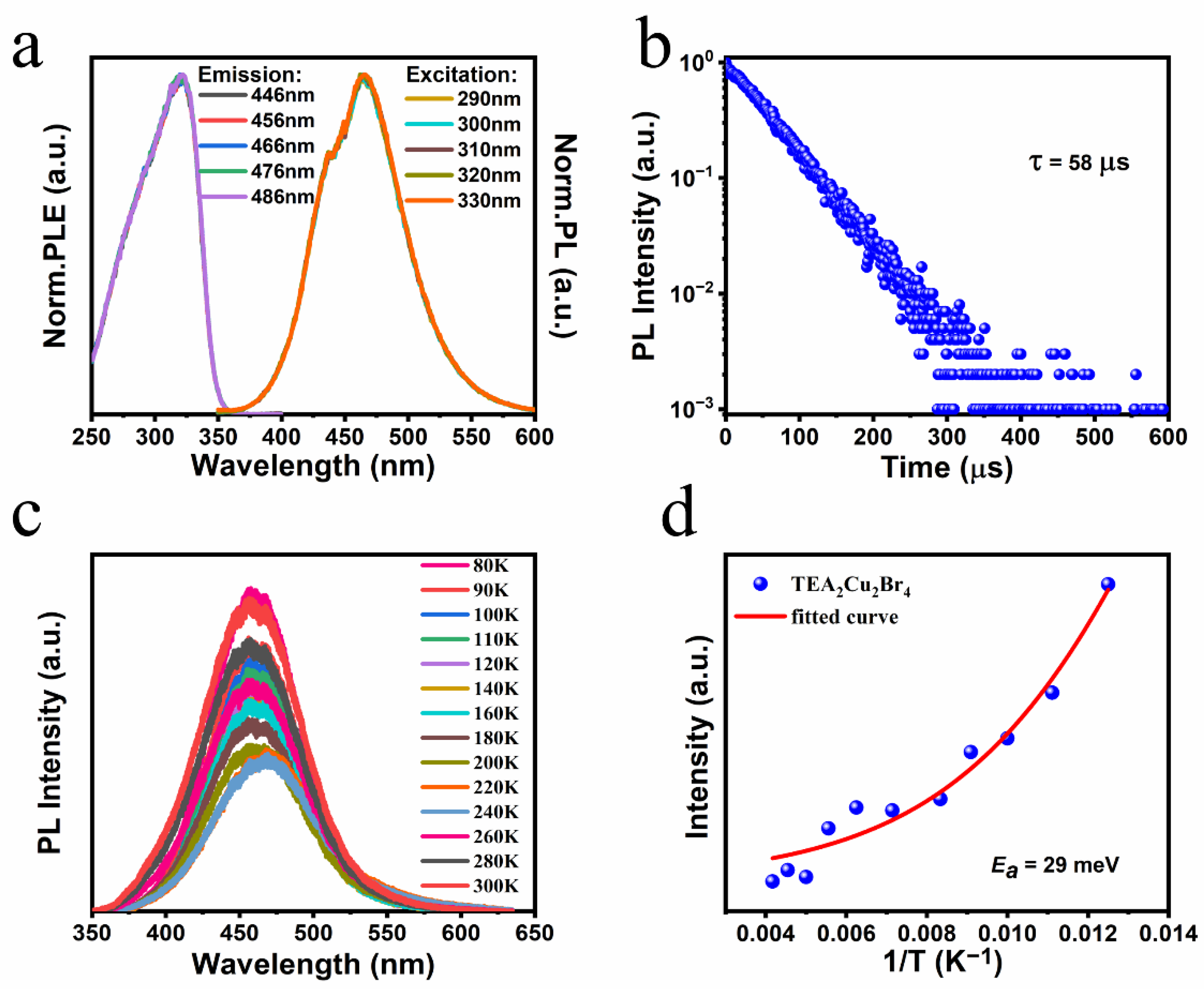
Figure 3a shows the photoluminescence excitation/emission (PLE/PL) spectra of the TEA2Cu2Br4 powder. The observed emission bands and excitation bands are at approximately 350 nm–600 nm and 250 nm–300 nm, respectively. The emission band is obviously broader than the typical perovskite emitters, such as CsPbX3 (X = Cl, Br, I), which possess emission peaks with a full width at half maximum (FWHM) no larger than 50 nm. Moreover, the positions and shapes of the emission bands are identical when the excitation wavelength was changed to a range covering the excitation bands. Correspondingly, the excitation bands, with a series of detecting wavelengths involved in the emission bands, showed the same positions and shapes. These results indicate that there was only one radiative recombination path for the excited carriers, and that all of the emission bands were generated by the same dynamic processes. Note that these emission bands have no spectra overlapping with the respective excitation bands; in other words, they have large Stokes shifts, which are quite different from those of conventional perovskite materials with band-to-band transitions. Based on the aforenoted structural characterization results, the TEA2Cu2Br4 crystal possesses a 0D structure, which facilitates the localization of free carriers. Specifically, the existing carriers easily induce Jahn–Teller distortion to the core groups. Transient defect states that are widely distributed within the forbidden bands are formed following the structural reorganization. Consequently, free carriers may be trapped by these transient defect states, thereby forming localized excitons, which are called self-trapped excitons (STEs). The recombination of STEs results in a broad emission band that has a large Strokes shift. Thus, the PL/PLE results of the TEA2Cu2Br4 crystal apparently indicate that the PL intensity arises from STE emission. Further evidence is that the measured PL decay lifetime for the 462 nm emission is as long as 58 μs (see the time-resolved PL spectrum in Figure 3b), which is also in accordance with the characteristic of STE emission. It is worth noting that the STE emission characteristic provides an advantage for the scintillator in that the reabsorption probability is low because of the large Stokes shift. Considering that the reabsorption of emitted photons by surrounding scintillation material leads to a blurred pixel, this reabsorption-free characteristic is essential for enhancing the resolution of X-ray imaging. The related PLQYs for the TEA2Cu2Br4 crystal were obtained as 55%, which is comparable to the value for those of star luminescent perovskite NCs. Figure 3c shows the variation in PL properties with the increase in the testing temperature from 80 K to 300 K. Following from the rise in temperature, the PL band shifts toward a long wavelength, and its FWHM increases slightly. This phenomenon indicates the strong electron–phonon interaction involved in the PL process [19], which is also in accordance with its STE nature. By using the Arrhenius function
I ( T ) = I 0 1 + A 1 e E a c t k T
to fit the integrated intensities of the PL bands, the activation energy E a c t was calculated as ~28 ± 1 meV, which reflected the strong binding energy of trapped excitons.
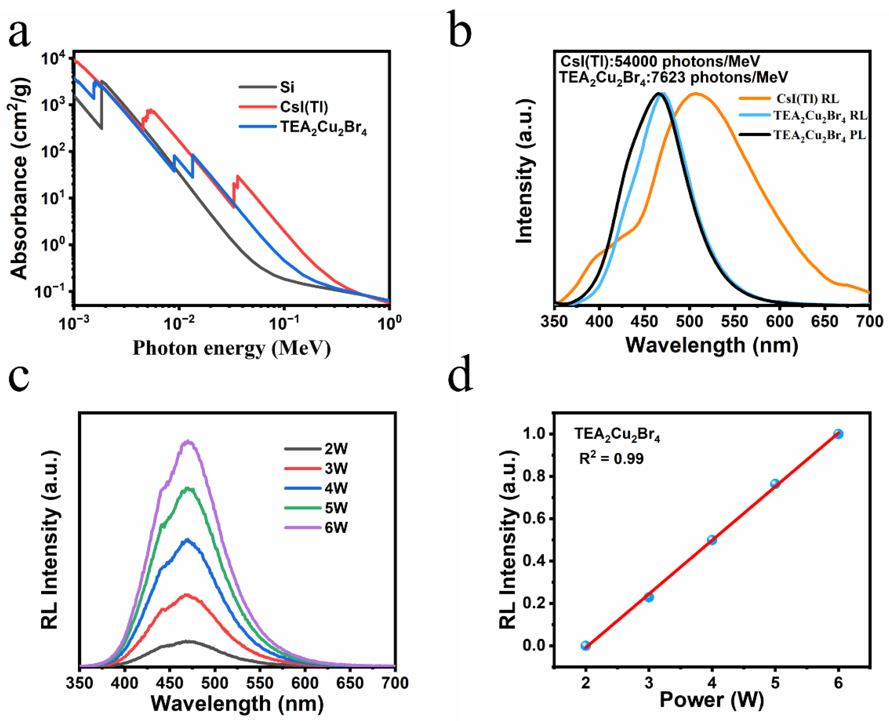
Based on the above results, the highly efficient blue STE emission with a large Stokes shift makes the obtained TEA2Cu2Br4 crystal an ideal scintillator in The X-ray detection. Therefore, their radioluminescence (RL) properties were further characterized. The X-ray absorption of TEA2Cu2Br4 crystals at different photon energies is shown in Figure 4a, which also provides a comparison with the X-ray absorption of the standard CsI(Tl) scintillator. It is worth noting that, unlike perovskite scintillators, such as CsPbBr3, the TEA2Cu2Br4 crystal delivers lower absorbance coefficients, which is due to its moderate density and lack of heavy elements such as Pb [20]. However, it is still fully sufficient for the X-ray imaging application. Figure 4b shows the normalized RL spectrum of the TEA2Cu2Br4 crystal accompanying the RL spectrum of the CsI(Tl) scintillator and the PL spectrum of itself. The broad RL band of the CsI(Tl) scintillator centers at 511 nm and covers nearly the whole visible range. In contrast, a sharper RL band of the TEA2Cu2Br4 crystal is located at 462 nm. This blue RL band is highly consistent with its PL spectrum, and no new PL band is found in the spectrum. This indicates that the RL process of the TEA2Cu2Br4 crystal shares the same recombination pathways with its PL process, which is associated with the radiative recombination of STEs. Thus, the RL process can be described as follows: Firstly, X-rays excite the electrons in the inner shells of Cu atoms to the higher energy levels of the crystal, turning them into hot electrons with very high kinetic energy. Then, these hot electrons transfer their kinetic energy through an avalanche effect to other valence band electrons, producing secondary electrons. The number of secondary electrons is proportional to the released kinetic energy. Subsequently, the generated electrons undergo relaxation and become free carriers on the bottom of the conduction band. Most importantly, unlike the ones in perovskite CsPbBr3 that recombine quickly with emitting band-edge photons, these free carriers tend to be trapped by the transient defect states that they cause by themselves from Jahn–Teller distortion, and they form STEs. Then, fast radiative recombination of STEs is suggested to take place.
LY is an important parameter in the assessment of scintillator efficiency, as it evaluates the number of generated photons absorbed per MeV of X-ray energy. Note that it is difficult to directly measure the absolute value of LY because it relates to the internal reactions of a scintillator. Thus, we used CsI(Tl) single crystal as the benchmark in LY measurements. It is already known that the LY for CsI(Tl) single crystal is 54,000 photons/MeV [21]. Firstly, the RL spectrum covering its entire emission range of CsI(Tl) was measured using our RL spectrometer. Then, a similar RL spectrum of the TEA2Cu2Br4 crystal was measured while keeping the X-ray excitation power unchanged. Based on the knowledge that the photon energy of the X-ray resource is in the range of 50 keV~70 keV, and the absorbance coefficient of both samples in this range is larger than 104 cm−1, the thicknesses of both samples are large enough to achieve complete absorption to the excitation X-ray. Thus, the relative LY of the TEA2Cu2Br4 crystal can be evaluated by comparing its integrated intensity of RL to that of CsI(Tl) single crystal. It was observed that the LY of the TEA2Cu2Br4 crystal is 7623 photons/MeV compared to CsI(Tl) (54,000 photons/MeV), which is comparable to the other perovskite-like scintillator materials, such as two-dimensional (2D) (EDBE)PbCl4 hybrid perovskite crystals (9000 photons/MeV) [22] and two-dimensional perovskite (BA)2PbBr4 microcrystals (7000 photons/MeV)) [23]. We also studied the RL spectra of the TEA2Cu2Br4 crystal excited at different X-ray powers, which are shown in Figure 4c,d. Following the increase in excitation power, the RL spectrum showed no change in shape but continuously increased in integrated intensity. Quantificationally, the integrated RL intensity showed clear linear dependence upon excitation power (X-ray dosage) without saturation, which indicates excellent scintillation performance similar to that of the commercial CsI(Tl) crystal.
The excellent STE-related PL and RL properties manifest the high potential of the TEA2Cu2Br4 crystal in radiography. Therefore, we further evaluated its X-ray imaging capability. TEA2Cu2Br4 crystalline powder was grounded and mixed with PMMA to make a scintillation screen for imaging tests [24]. The left picture in Figure 5a displays a photograph of the TEA2Cu2Br4 scintillation screen under UV illumination, which shows a uniform and bright blue florescence. The right picture in Figure 5a illustrates the same screen under exposure in X-ray, which also shows a blue RL. We set up convenient equipment for an X-ray imaging test. As shown in Figure 5b, the sample holder was straightly placed into a steel box with the TEA2Cu2Br4 scintillation screen put on the middle hole of the panel. A capsule was put right in front of the screen working as the object. The capsule was opaque, and a metal spring was placed inside the capsule, as shown in Figure 5c. Then, the capsule sample was irradiated with an X-ray resource. The X-ray beam penetrated the capsule and excited the screen, which resulted in RL on the screen. Finally, the RL photons were collected with a CCD camera through a total reflecting mirror, which formed the radiograph of the capsule sample. The X-ray imaging quality is illustrated in Figure 5d, under a minimum power of 2 W, the dark shape of the metal spring appears in the blue background. It becomes much clearer following the increase in X-ray excitation power from 2 W to 7 W. This indicates that the enhanced brightness of RL on the TEA2Cu2Br4 scintillation screen improves the resolution of the radiograph. This result confirms that the RL properties of the TEA2Cu2Br4 crystal and the uniformity of the screen are sufficient to realize the radiograph based on this prototypical TEA2Cu2Br4 scintillation screen.

4. Conclusions

In conclusion, crystalline TEA2Cu2Br4 scintillator material was synthesized using an improved liquid-phase cooling precipitation method with a fast production rate of 100 mg/minute. The product shows good optical properties at room temperature, with a high azure color under UV light, a luminescence peak at 462 nm, and a Stokes shift of 142 nm. Its PLQY reaches 55%, which is comparable to that of other perovskite-like scintillator materials. More importantly, the TEA2Cu2Br4 crystal also has good performance in X-ray scintillation, showing bright radioluminescence, good linearity, and good light yield under X-ray excitation. Better still, by using PMMA with which the scintillation screen is compounded, it also presents good X-ray imaging performance. Thus, this work provides a new pathway for the development of new non-toxic and environmentally friendly scintillator materials.

Author Contributions

Conceptualization, T.L. and X.B.; formal analysis, T.L. and X.B.; investigation, L.W., J.L. and X.B.; resources, T.L. and R.Z.; data curation, T.L., X.B. and L.W.; writing—original draft preparation, T.L., X.B. and J.L.; writing—review and editing, T.L. and X.B.; supervision, T.L. and R.Z.; project administration, T.L.; funding acquisition, T.L. All authors have read and agreed to the published version of the manuscript.

Funding

This research was funded by the National Natural Science Foundation of China (Grant Nos. 61504030, 11704081) and Guangxi Natural Science Foundation (2020GXNSFAA238043, 2020GXNSFAA297182, 2017GXNSFGA198005).

Acknowledgments

This work was supported by the National Natural Science Foundation of China.

Conflicts of Interest

On behalf of all authors, the corresponding author states that there is no conflict of interest.

References

  1. Chen, Q.; Wu, J.; Ou, X.; Huang, B.; Almutlaq, J.; Zhumekenov, A.A.; Guan, X.; Han, S.; Liang, L.; Yi, Z.J.N. All-inorganic perovskite nanocrystal scintillators. Nature 2018, 561, 88–93. [Google Scholar] [CrossRef]
  2. Dujardin, C.; Auffray, E.; Bourret-Courchesne, E.; Dorenbos, P.; Lecoq, P.; Nikl, M.; Vasil’Ev, A.; Yoshikawa, A.; Zhu, R.-Y. Needs, trends, and advances in inorganic scintillators. IEEE Trans. Nucl. Sci. 2018, 65, 1977–1997. [Google Scholar] [CrossRef] [Green Version]
  3. Zhao, J.; Zhao, L.; Deng, Y.; Xiao, X.; Ni, Z.; Xu, S.; Huang, J.J.N.P. Perovskite-filled membranes for flexible and large-area direct-conversion X-ray detector arrays. Nat. Photon. 2020, 14, 612–617. [Google Scholar] [CrossRef]
  4. Zhou, J.; An, K.; He, P.; Yang, J.; Zhou, C.; Luo, Y.; Kang, W.; Hu, W.; Feng, P.; Zhou, M.J.A.O.M. Solution-Processed Lead-Free Perovskite Nanocrystal Scintillators for High-Resolution X-Ray CT Imaging. Adv. Opt. Mater. 2021, 9, 2002144. [Google Scholar] [CrossRef]
  5. Liu, J.; Shabbir, B.; Wang, C.; Wan, T.; Ou, Q.; Yu, P.; Tadich, A.; Jiao, X.; Chu, D.; Qi, D.J.A.M. Flexible, printable soft-X-ray detectors based on all-inorganic perovskite quantum dots. Adv. Mater. 2019, 31, 1901644. [Google Scholar] [CrossRef] [PubMed]
  6. Heo, J.H.; Shin, D.H.; Park, J.K.; Kim, D.H.; Lee, S.J.; Im, S.H.J.A.M. High-performance next-generation perovskite nanocrystal scintillator for nondestructive X-ray imaging. Adv. Mater. 2018, 30, 1801743. [Google Scholar] [CrossRef]
  7. Creason, T.D.; McWhorter, T.M.; Bell, Z.; Du, M.-H.; Saparov, B. K2CuX3 (X= Cl, Br): All-inorganic lead-free blue emitters with near-unity photoluminescence quantum yield. Chem. Mater. 2020, 32, 6197–6205. [Google Scholar] [CrossRef]
  8. Yang, B.; Yin, L.; Niu, G.; Yuan, J.H.; Xue, K.H.; Tan, Z.; Miao, X.S.; Niu, M.; Du, X.; Song, H. Lead-Free Halide Rb2CuBr3 as Sensitive X-Ray Scintillator. Adv. Mater. 2019, 31, 1904711. [Google Scholar] [CrossRef]
  9. Lian, L.; Zheng, M.; Zhang, W.; Yin, L.; Du, X.; Zhang, P.; Zhang, X.; Gao, J.; Zhang, D.; Gao, L. Efficient and Reabsorption-Free Radioluminescence in Cs3Cu2I5 Nanocrystals with Self-Trapped Excitons. Adv. Sci. 2020, 7, 2000195. [Google Scholar] [CrossRef] [Green Version]
  10. Zhou, W.; Liu, S.; Chen, W.; Li, H.; Yan, S.; Yu, J.; Liu, C.; Zhao, L.; Zeng, T.; Han, T. Efficient and self-healing copper halide Cs3Cu2Cl5 film assisted by microwave treatment for high-resolution X-ray imaging. Ceram. Int. 2022, 48, 25086–25093. [Google Scholar] [CrossRef]
  11. Cheng, S.; Beitlerova, A.; Kucerkova, R.; Nikl, M.; Ren, G.; Wu, Y. Zero-Dimensional Cs3Cu2I5 Perovskite Single Crystal as Sensitive X-Ray and γ-Ray Scintillator. Phys. Status Solidi (RRL) Rapid Res. Lett. 2020, 14, 2000374. [Google Scholar] [CrossRef]
  12. Fang, S.; Li, H.; Xie, Y.; Li, H.; Wang, Y.; Shi, Y.J.S. Zero-Dimensional Organic–Inorganic Hybrid Copper-Based Halides with Highly Efficient Orange–Red Emission. Small 2021, 17, 2103831. [Google Scholar] [CrossRef] [PubMed]
  13. Xu, L.-J.; Lin, X.; He, Q.; Worku, M.; Ma, B. Highly efficient eco-friendly X-ray scintillators based on an organic manganese halide. Nat. Commun. 2020, 11, 1–7. [Google Scholar] [CrossRef]
  14. Zhou, Y.; Wang, X.; He, T.; Yang, H.; Yang, C.; Shao, B.; Gutiérrez-Arzaluz, L.; Bakr, O.M.; Zhang, Y.; Mohammed, O. Large-Area Perovskite-Related Copper Halide Film for High-Resolution Flexible X-ray Imaging Scintillation Screens. ACS Energy Lett. 2022, 7, 844–846. [Google Scholar] [CrossRef]
  15. Xu, Y.; Li, J.; Zhao, F.; Gao, Y.; Chen, R.; He, T. Optical Properties of a CsMnBr3 Single Crystal. ACS omega 2022, 7, 29415–29419. [Google Scholar] [CrossRef]
  16. Liu, X.; Yuan, F.; Zhu, C.; Li, J.; Lv, X.; Xing, G.; Wei, Q.; Wang, G.; Dai, J.; Dong, H. Near-unity blue luminance from lead-free copper halides for light-emitting diodes. Nano Energy 2022, 91, 106664. [Google Scholar] [CrossRef]
  17. Asplund, M.; Jagner, S.; Hamalainen, R.; Kohl, F.; Seip, R. Crystal Structure of bis-(tetraethylammonium) di-mu-bromo-dibromodicuprate (I),[N-(C2H5) 4] 2 [Cu2Br4]. Acta Chem. Scand. A 1984, 38, 135–139. [Google Scholar] [CrossRef]
  18. Lian, L.; Wang, X.; Zhang, P.; Zhu, J.; Zhang, X.; Gao, J.; Wang, S.; Liang, G.; Zhang, D.; Gao, L. Highly luminescent zero-dimensional organic copper halides for X-ray scintillation. J. Phys. Chem. Lett. 2021, 12, 6919–6926. [Google Scholar] [CrossRef]
  19. Han, L.; Zhang, H.; Ning, Y.; Chen, H.; Guo, C.; Cui, J.; Peng, G.; Ci, Z.; Jin, Z. Environmentally stable one-dimensional copper halide based ultra-flexible composite film for low-cost X-ray imaging screens. Chem. Eng. J. 2022, 430, 132826. [Google Scholar] [CrossRef]
  20. Wang, B.; Peng, J.; Yang, X.; Cai, W.; Xiao, H.; Zhao, S.; Lin, Q.; Zang, Z.J.L.; Reviews, P. Template Assembled Large-Size CsPbBr3 Nanocomposite Films toward Flexible, Stable, and High-Performance X-Ray Scintillators. Laser Photonics Rev. 2022, 16, 2100736. [Google Scholar] [CrossRef]
  21. Li, N.; Xu, Z.; Xiao, Y.; Liu, Y.; Yang, Z.; Liu, S. Flexible, High Scintillation Yield Cu3Cu2I5 Film Made of Ball-Milled Powder for High Spatial Resolution X-Ray Imaging. Adv. Opt. Mater. 2022, 10, 2102232. [Google Scholar] [CrossRef]
  22. Birowosuto, M.; Cortecchia, D.; Drozdowski, W.; Brylew, K.; Lachmanski, W.; Bruno, A.; Soci, C. X-ray scintillation in lead halide perovskite crystals. Sci. Rep. 2016, 6, 1–10. [Google Scholar] [CrossRef] [PubMed] [Green Version]
  23. Li, Y.; Chen, L.; Liu, B.; Jin, P.; Gao, R.; Zhou, L.; Wan, P.; Xu, Q.; Ouyang, X. Scintillation performance of two-dimensional perovskite (BA) 2 PbBr 4 microcrystals. J. Mater. Chem. C 2021, 9, 17124–17128. [Google Scholar] [CrossRef]
  24. Hu, X.; Yan, P.; Ran, P.; Lu, L.; Leng, J.; Yang, Y.M.; Li, X. In Situ Fabrication of Cs3Cu2I5: Tl Nanocrystal Films for High-Resolution and Ultrastable X-ray Imaging. J. Phys. Chem. Lett. 2022, 13, 2862–2870. [Google Scholar] [CrossRef]
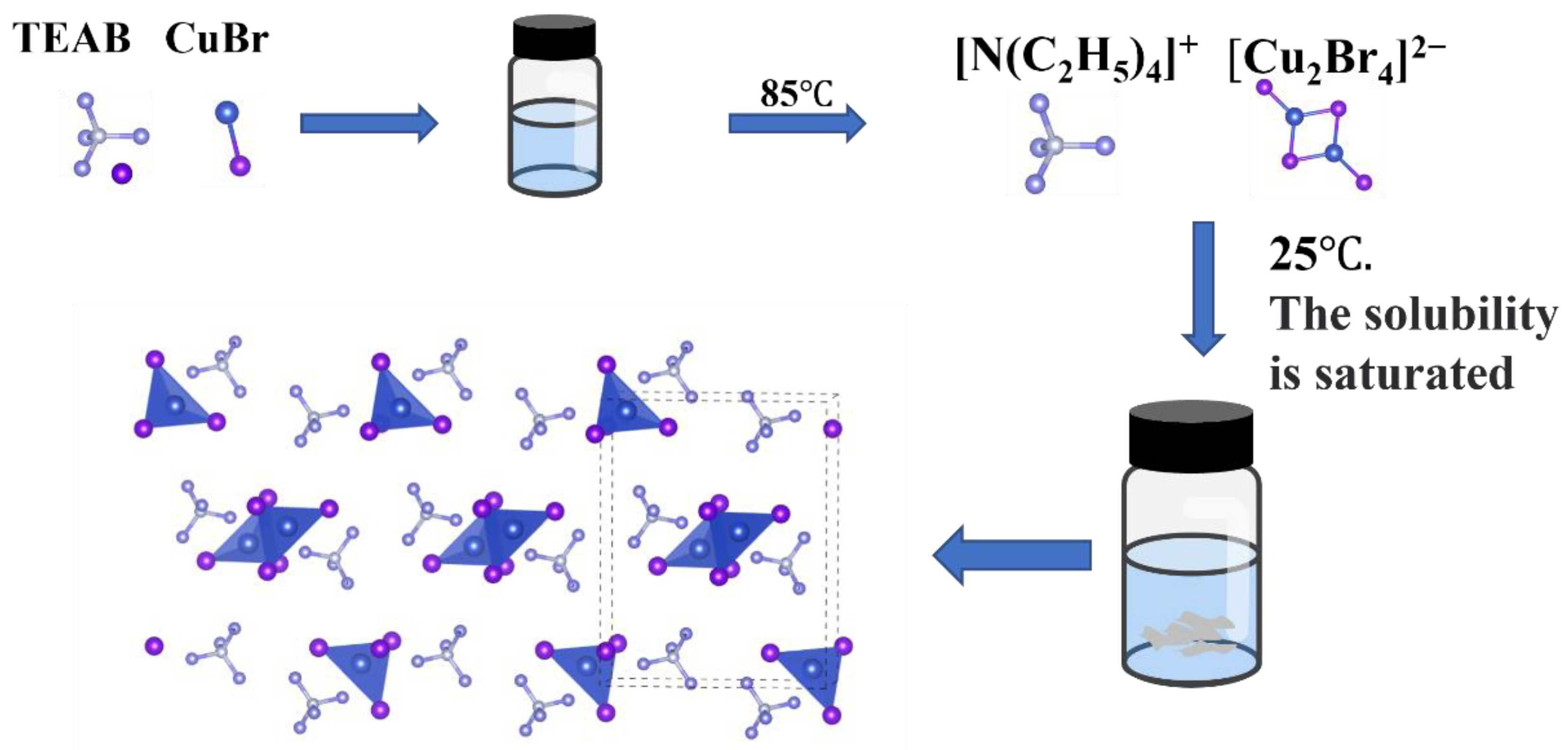
Figure 1. Schematics of the preparation of the TEA2Cu2Br4 crystals with liquid-phase cooling precipitation.
Crystals 12 01799 g001
Figure 2. Morphology and crystalline structure of TEA2Cu2Br4 crystals. (a) Pictures of TEA2Cu2Br4 crystals in ambient light (left) and under UV illumination (right). (b) Optical microscopy images of TEA2Cu2Br4 powder. (c) XRD pattern of TEA2Cu2Br4 crystals. (d) Schematics of crystal structure of TEA2Cu2Br4.
Crystals 12 01799 g002
Figure 3. PL performance of TEA2Cu2Br4 crystal. (a) Normalized PL and PLE spectra of TEA2Cu2Br4. (b) Time-resolved PL decay curve of TEA2Cu2Br4 at room temperature. (c) Temperature-dependent PL spectra of TEA2Cu2Br4. (d) Integrated PL intensities vs. temperature plots of TEA2Cu2Br4 crystal. The plots were fitted with Arrhenius function, and the obtained activation energy is ~28 ± 1 meV.
Crystals 12 01799 g003
Figure 4. X-ray absorption and fluorescence spectra of TEA2Cu2Br4 crystal. (a) Absorbance coefficients of Si, CsI(Tl), and TEA2Cu2Br4. (b) TEA2Cu2Br4 PL spectra, RL spectra, and CsI RL spectra. (c) TEA2Cu2Br4 RL spectra with different X-ray excitation power. (d) The excitation power dependence of the RL intensity.
Crystals 12 01799 g004
Figure 5. X-ray fluorescence imaging properties of the sample. (a) Photomicrographs of TEA2Cu2Br4 scintillation screen under UV and X-ray illumination. (b)Test specimen capsules and springs. (c) X-ray imaging of the internal spring of the capsule at different powers. (d) X-ray tube imaging pictures at different powers.
Crystals 12 01799 g005
Publisher’s Note: MDPI stays neutral with regard to jurisdictional claims in published maps and institutional affiliations.