Article

Maleic Anhydride-β-Cyclodextrin Functionalized Magnetic Nanoparticles for the Removal of Uranium (VI) from Wastewater

1
State Key Laboratory of Nuclear Resources and Environment, East China University of Technology, Nanchang 330013, China
2
School of Chemical Engineering and Technology, Tianjin University, Tianjin 300072, China
*
Authors to whom correspondence should be addressed.
Crystals 2022, 12(12), 1731; https://doi.org/10.3390/cryst12121731
Submission received: 12 November 2022 / Revised: 25 November 2022 / Accepted: 26 November 2022 / Published: 30 November 2022

Abstract

:
New maleic anhydride-β-cyclodextrin functionalized magnetic iron oxide nanoparticles (IONPs@MAH-β-CD) were prepared through the simple chemical coprecipitation method. Macroscopic batch experiments were carried out to study its absorption behavior for uranium (VI) under different environmental conditions. Using the magnetic separation technique, it was possible to easily separate IONPs@MAH-β-CD from uranium-containing wastewater. The equilibrium for uranium (VI) adsorption was reached for 180 min. The results showed that the kinetics and isotherm of the adsorption process were consistent with the pseudo-second-order kinetic model and Langmuir model, respectively, indicating that it was a chemisorption process. Compared with pure IONPs, the maximum adsorption quantity of maleic anhydride-β-cyclodextrin-coated iron oxide nanoparticles for uranium (VI) was significantly improved, which could reach 163.93 mg g−1. The recyclability of the IONPs@MAH-β-CD was investigated, and FTIR and XPS characterization were used to explore the possible mechanism of U(VI) adsorption.

1. Introduction

Due to its unsustainability and the emission of greenhouse gases, traditional energy has not met the requirements of an ecological civilization. As a kind of clean and efficient energy, the research and development of nuclear energy has been gradually paid more and more attention [1,2,3]. However, uranium mining and tailing, nuclear fuel processing and manufacturing, and fuel reprocessing produce a large amount of uranium (U(VI))-containing wastewater, causing serious pollution of the environment [4]. How to effectively treat U(VI)-containing wastewater has become an important problem in the sustainable development of nuclear energy and environmental safety, which is also a research focus in the field of environmental chemistry [5,6].
Up to now, the effective treatments for uranium-containing wastewater are chemical precipitation [7], membrane separation [8], ion exchange [9], photocatalysis [10], adsorption [11], and so on. Adsorption, which is characterized by high adsorption efficiency, excellent selectivity, simple operation, and low cost, is a research hotspot in the field of nuclear research. Adsorbents such as clay minerals, graphene oxide materials, metal–organic frameworks, and so on, are the most important component in adsorption [12,13]. Nevertheless, the existing adsorbent materials are difficult to use in industrial applications due to their complicated synthesis process and high separation difficulty. As a new kind of adsorption material, magnetic material is low-cost, easily prepared, and has a high removal efficiency. Moreover, magnetic materials can quickly complete solid–liquid separation in the presence of an external magnetic field, and these advantages have attracted more and more attention to magnetic materials [14,15]. Among the various magnetic materials, iron oxide nanoparticles (IONPs), which include maghemite (γ-Fe2O3) and magnetite (Fe3O4), are usually used for adsorption [16]. However, pure IONPs have large surface energy and strong magnetic properties, which easily produce agglomeration. As a result, the specific surface area decreases, the distribution is uneven in water, and the adsorption efficiency decreases [17]. Therefore, modifying IONPs to construct a uniform distribution structure is an effective way to improve their adsorption performance.
Cyclodextrin, a green, environmental, non-toxic oligosaccharide formed by enzyme action on long amylose, has a hydrophobic inner cavity and hydrophilic outer cavity and has attracted a great amount of attention in the field of environmental remediation. Badruddoza et al. investigated a magnetic material prepared using immobilized carboxymethyl-β-cyclodextrin on Fe3O4 with cyanamide as the linker group, which was applied to the adsorption of methyl green and Cu2+. The equilibrium adsorption capacities were 140.8 mg g−1 and 47.2 mg g−1, respectively [18,19]. Yang et al. developed β-cyclodextrin (β-CD) grafted halloysite nanotubes/iron oxide to adsorb U(VI) in aqueous solution. The optimal pH value was 7.0, and it adsorbed 92% of U(VI) in aqueous solution [20]. Helal et al. grafted succinyl-β-cyclodextrin onto magnetic hematite nanoparticles to obtain SβCD-APTES@Fe2O3, which had remarkable potential for the recovery of uranium from aqueous solution. At pH 6, the maximum adsorption capacity of uranyl ions reached 286 mg g−1 [21]. Due to host–guest supramolecular interaction and synergistic effect [22], β-CD has great potential in U(VI) adsorption [21]. β-CD and IONP components play the roles of inclusion complexation cooperator and magnetic carrier, respectively, which not only ensures the dispersion of materials but also has the advantage of ease of recycling,
Maleic anhydride-β-cyclodextrin (MAH-β-CD) has rich carboxyl and hydroxyl functional groups that can provide a large number of binding sites. However, until now MAH-β-CD-linked magnetic materials have not been studied for radionuclides. Hence, in this study, MAH-β-CD was selected to combine with IONPs and obtain an enhanced magnetic adsorption material for U(VI). The aims of this investigation were to develop a new magnetic nanoparticle, maleic anhydride-β-cyclodextrin functionalized magnetic nanoparticle (IONPs@MAH-β-CD), which was prepared using MAH-β-CD and IONPs through a simple chemical coprecipitation method, in order to remove U(VI) from wastewater. The morphology, physicochemical properties, and removal mechanism of U(VI) were studied, combining microscopic characterization and macroscopic testing. Furthermore, the adsorbent, which is appropriate for easy separation using an external magnetic field, is expected to have good potential due to its easy separation; it is of great significance to the development of nuclear wastewater purification.

2. Materials and Methods

2.1. Materials

β-cyclodextrin (β-CD, 99%), maleic anhydride (MAH, 99%), N,N-dimethylformamide (DMF, 99.8%, extra dry over molecular sieve), ethyl acetate (99.5%), iron(III) chloride hexahydrate (96%, FeCl3·6H2O), iron(II) chloride tetrahydrate (98%, FeCl2·4H2O), ammonium hydroxide (NH4OH, 25% in water), and arsenazo III (AR) were purchased from Energy Chemical. Uranyl nitrate hexahydrate (UO2(NO3)2·6H2O, 99%) was purchased from Macklin Reagent. Ultrapure water (18.25 MΩ cm) was used to prepare all aqueous solutions.

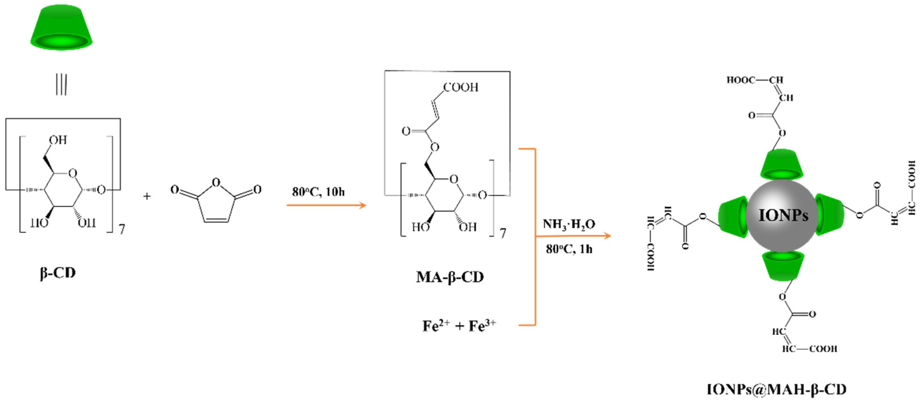
2.2. Synthesis of MAH-β-CD

MAH-β-CD was synthesized by improving the synthesis methods reported in [23,24]. Specifically, 5.68 g dried β-CD (0.005 mol) was dissolved in anhydrous DMF and 4.90 g maleic anhydride (0.05 mol, 10eq) was added and dissolved under the protection of Ar atmosphere. The mixture was stirred in an oil bath at 80 °C for 10 h and then cooled to room temperature. Then, 300 mL ethyl acetate was poured in with a magnetic stirrer and a white precipitate appeared. The precipitate was collected by filtration and washed with acetone. Finally, MAH-β-CD was obtained by drying in a vacuum oven.

2.3. Synthesis of IONPs@MAH-β-CD

IONPs@MAH-β-CD was prepared by one-step chemical coprecipitation. Briefly, 1.5 g MAH-β-CD was dispersed under an Ar atmosphere in deoxygenated ultrapure water containing 2.36 g FeCl3·6H2O and 0.86 g FeCl2·4H2O. Then, 25 mL ammonia water (25%) was quickly added through a syringe at 80 °C while blending. The reaction was continued for 1 h under constant stirring in an Ar gas environment at 80 °C, and then the black precipitated product was collected. The product was washed three times with ultrapure water and absolute ethanol in turn, and finally dried under vacuum at 70 °C to obtain IONPs@MAH-β-CD, as shown in Scheme 1.

2.4. Characterization

Scanning electron microscopy (SEM, Hitachi Regulus 8100, Tokyo, Japan) was used to observe the microstructure of IONPs@MAH-β-CD. Energy-dispersive X-ray spectrometry (EDS, Hitachi Regulus 8100) was carried out to study the elemental content and composition of the material. A UV spectrophotometer (UV-VIS, Shanghai UV-5800, Shanghai, China) was employed to determine the U(VI) concentration at a wavelength of 650 nm. A Fourier transform infrared spectrometer (FTIR, NICOLET 5700, Waltham, MA, USA) was used to measure the functional groups of materials, using the KBr pellet technique in a region of 4000–400 cm−1. Furthermore, the specific surface area and pore size distribution of IONPs@MAH-β-CD were measured using N2 adsorption and desorption experiments at 77 K. The standard Brunauer–Emmett–Teller (BET) equation was employed to calculate the specific surface area. The pore size distribution was obtained from the desorption branch of the isotherms via the Barrett–Joyner–Halenda (BJH) method using Quantachrome Autosorb software (Quantachrome Instruments, Boynton Beach, FL, USA) [25,26].
For the zeta potential measurement, the IONPs@MAH-β-CD samples were diluted with distilled water to 0.02 wt% concentration before measurement to prevent multiple scattering effects. pH values were adjusted using HCl or NaOH aqueous solution. After ultrasonic treatment for 10 min, zeta potential was measured. Three parallel measurements were made at each pH, and the results were averaged.
The structural properties were characterized by X-ray diffractogram (XRD, Bruker D8 Advance) analysis with the use of Cu Kα radiation. The samples were scanned from 20 to 80° with a scanning speed of 2°·min−1. X-ray photoelectron spectroscopy (XPS, Thermo ESCALAB 250XI) was used to analyze the types and chemical states of elements before and after adsorption by the adsorbent, and the scanning range was changed from 0 to 700 eV.

2.5. Adsorption Experiment

The batch adsorption experiment was carried out in batches and the influence of different factors on the adsorption of U(VI) by IONPs@MAH-β-CD, such as pH values, adsorbent dosage, contact time, temperature, U(VI) initial concentration, and so on were explored successively. A certain amount of adsorbent was put in to a 125 mL polyethylene bottle, and then 25 mL U(VI) solution in a certain concentration was added. After adjusting the pH values by 0.1 mol L−1 HCl or 0.1 mol L−1 NaOH, the mixture was placed in the constant-temperature oscillator, working at a speed of 200 r min−1 for a certain time. Finally, the residual concentration of U(VI) was detected by a UV spectrophotometer using arsenazo III as the chromogenic agent after being separated by 0.22 μm filter membrane.

3. Results and Discussion

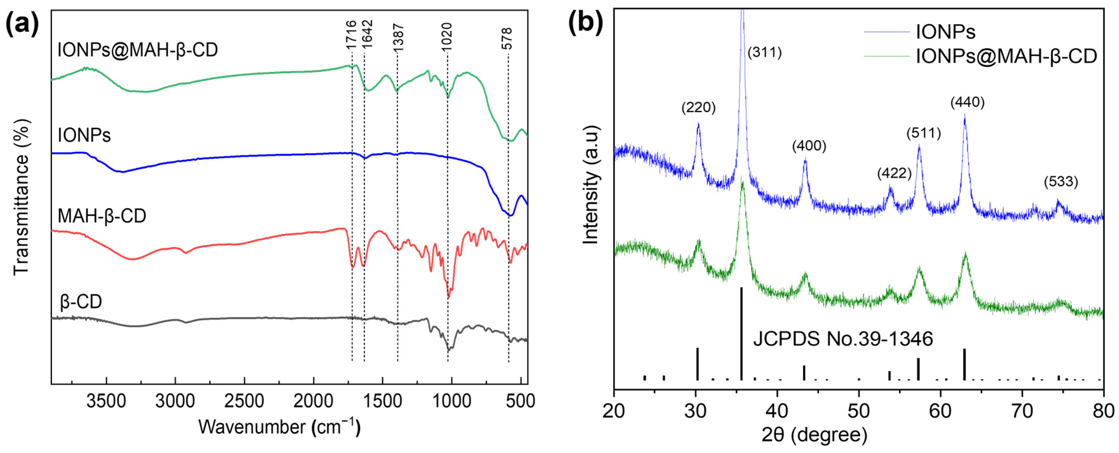
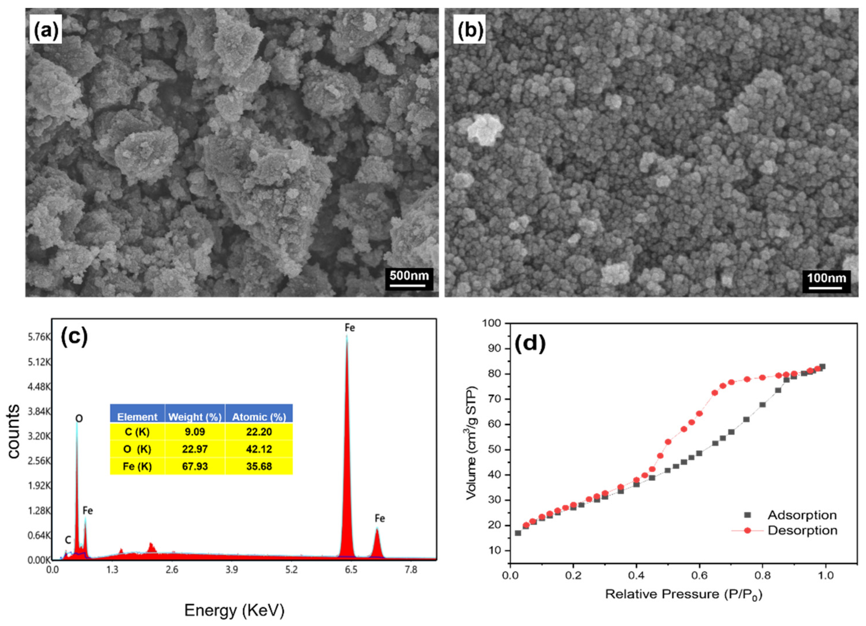
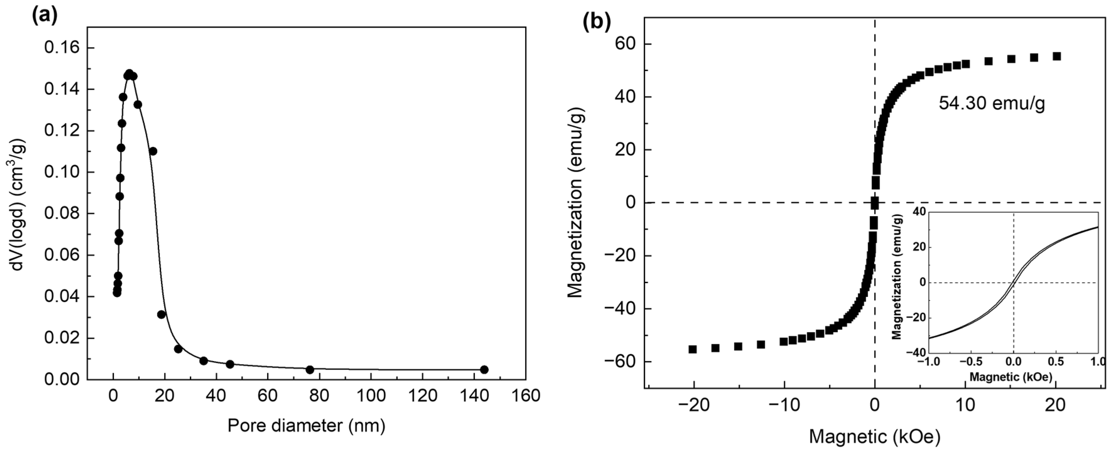
3.1. Characterization Results

FTIR was used to analyze the functional groups of β-CD, MAH-β-CD, IONPs and IONPs@MAH-β-CD, and results are shown in Figure 1a. The wide and strong peak of β-CD at 3293 cm−1 was assigned to the stretching vibration of -OH, and the strong absorption peak at 1020 cm−1 was assigned to the stretching vibration of C-O [27,28]. The characteristic peaks of β-CD were also observed in the spectrum of MAH-β-CD, the wide and strong -OH stretching vibration peak at 3309 cm−1, and the stretching vibration peak of C-O at 1021 cm−1. In addition, three absorption peaks were found at 1716, 1642, and 1387 cm−1, corresponding to the C=O stretching vibration peak, C=C stretching vibration peak, and carboxylic acid group symmetric stretching vibration on maleic anhydride, respectively [29,30]; that indicates the formation of MAH-β-CD. Compared with MAH-β-CD, a typical Fe-O stretching vibration peak was found in the infrared spectrum of magnetically functionalized IONPs@MAH-β-CD, meaning that IONPs@MAH-β-CD was successfully synthesized.
XRD testing was used to analyze pure IONPs and IONPs@MAH-β-CD, and the results are exhibited in Figure 1b. The pure IONPs had seven characteristic diffraction peaks at 2θ of 30.34°, 35.74°, 43.48°, 53.92°, 57.36°, 63.01°, and 74.44°, which correspond to the (220), (311), (400), (422), (511), (440), and (533) planes of cubic γ-Fe2O3 ( JCPDS:No.39-1346) [31]. In addition, the IONPs@MAH-β-CD sample also showed significant γ-Fe2O3 diffraction peaks, which indicates that iron oxide nanoparticles successfully modified the surface of the composite. Since Mössbauer spectroscopy was not carried out on the samples, it could not be determined whether the IONPs and IONPs@MAH-β-CD contained only γ-Fe2O3 particles [32]. In addition, the peak intensity of the IONPs@MAH-β-CD hybrid particles was lower than that of pure IONPs, probably because the IONPs were embedded inside the MAH-β-CD’s amorphous organic shell [33].
SEM was employed to study the surface morphology and microstructure of IONPs@MAH-β-CD, and the results are shown in Figure 2a. From the SEM patterns, it was observed that IONPs@MAH-β-CD had an abundant pore structure, and the surface was fluffy and rough on the micron scale, showing an irregular porous structure. In addition, it was found that IONPs@MAH-β-CD had a regular spherical shape at the nanoscale and the average diameter was about 10–20 nm. This was due to the van der Waal force between particles and the magnetization caused by the magnetic field separation, which could encourage the particles to attract each other and flock together. From the EDS results shown in Figure 2c, the spectra showed the presence of a 0.27 keV(Kα) carbon peak and a 0.52 keV(Kα) oxygen peak, and 0.71 keV(Lα), 6.40 keV(Kα), and 7.04 keV(Kβ) iron peaks [34].The weight percentages of C, O, and Fe were 9.09%, 22.97%, and 67.93%, respectively. These data also supported the successful synthesis of IONPs@MAH-β-CD.
The N2 adsorption/desorption isotherms of IONPs@MAH-β-CD are shown in Figure 2d. It can be seen that there is an obvious H1 hysteresis loop, and it exhibits a class IV adsorption isotherm, indicating that the adsorbent was a mesoporous material [35]. The surface area of IONPs@MAH-β-CD is 126.62 m2 g−1, as calculated by the BET equation. The corresponding pore size distribution curve of IONPs@MAH-β-CD is shown in Figure 3a. It can also be seen that the pore size distribution of IONPs@MAH-β-CD was concentrated at 10 nm, which was basically consistent with the SEM results. Thus, the porous structure and high specific surface area of IONPs@MAH-β-CD provided sufficient binding sites for U(VI). Furthermore, the hysteresis loop of IONPs@MAH-β-CD was acquired using a vibrating sample magnetometer, and its saturation magnetization value was 54.30 emu g−1, which was slightly lower than that of pure IONPs alone, due to the introduction of maleic anhydride-β-cyclodextrin [18].The saturation magnetization value was lower than the reported value of IONPs, which was not only due to the presence of MAH-β-CD but also possibly due to the maghemite phase [36]; It can be seen in the inner illustration at the lower right corner of the hysteresis loop. The remanent magnetization (Br) of IONPs@MAH-β-CD was only 0.76 emu g−1, and the coercivity (Hc) was only 0.015 kOe, indicating that there was no obvious Br or Hc, which indicated that IONPs@MAH-β-CD has superparamagnetism [37]. Essentially, IONPs@MAH-β-CD has good magnetic responsiveness, and it can be easily separated from the solution by an applied magnetic field in one minute.

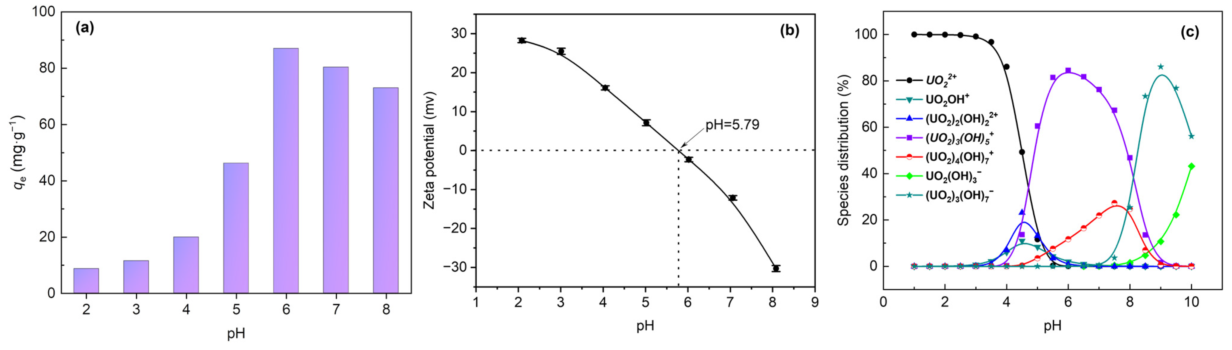
3.2. Effect of Initial pH

The U(VI) removal capacity of the adsorbent was characterized by the removal rate (%) and the adsorption quantity qe (mg g−1) at equilibrium, which were calculated by equations (1) and (2) respectively [38]:
  Removal   rate   % = C 0 C e C 0 × 100
q e = C 0 C e × V m
Here, the removal rate and qe were determined by the initial U(VI) concentration C0 (mg L−1) and the equilibrium U(VI) concentration Ce (mg L−1), where m (mg) and V (mL) were the amount of adsorbent and solution volume, respectively.
The adsorption quantity of IONPs@MAH-β-CD for U(VI) was examined at different pH levels, and the results are displayed in Figure 4a. It was observed that the adsorption quantity for U(VI) was greatly affected by pH. Specifically, the adsorption quantity of IONPs@MAH-β-CD for U(VI) increased slowly with the increase of pH in the range of 2.0~4.0 and increased rapidly in the range of 4.0~6.0. The maximum adsorption quantity can reach 87.02 mg g−1 at pH = 6.0, and the adsorption quantity decreased with the increase in pH.
The influence of pH on the zeta potential of the IONPs@MAH-β-CD solution is shown in Figure 4b. It can be seen that IONPs@MAH-β-CD was electrically neutral at pH = 5.79, and the surface of the adsorbent material was positively charged at pH < 5.79 and negatively charged at pH > 5.79. Figure 4b combined with the species distribution map of aqueous solution of U(VI) at different pH (Figure 4c) could be used to explain why the adsorption quantity changed with pH. At pH < 5.79, U(VI) ions mainly existed in the form of UO22+, UO2OH+, (UO2)3(OH)5+, and (UO2)2(OH)22+ cations. Due to the positive charge on the surface of the adsorbent, electrostatic repulsion led to poor adsorption of U(VI). In addition, at low pH values H+ and H3O+ in the solution would compete with UO22+ to be adsorbed on the active sites of IONPs@MAH-β-CD, and H+ would also protonate IONPs@MAH-β-CD to reduce the adsorption capacity. With the increase in pH, the protonated carboxyl and hydroxyl groups are gradually deprotonated, increasing the uranium adsorption capacity [39]. U(VI) ions are still mainly in the form of cations at pH = 6.0, while the adsorbent surface is negatively charged, and the electrostatic attraction effect led to a significant increase in adsorption level. Furthermore, when pH = 6, the adsorbent began to be carboxylated, leading to the formation of negative charges on the surface. The carboxylate groups of IONPs@MAH-β-CD could combine with positively charged uranium ions, forming relatively stable outer surface complexes and leading to a high adsorption capacity [40]. Subsequently, with the increase in pH value, the negative charge on the surface of IONPs@MAH-β-CD increased, and the morphology of U(VI) gradually transferred to negatively charged UO2(OH)3 and (UO2)3(OH)7, and the electrostatic repulsion between the adsorbent and negatively charged U(VI) increased, resulting in the decrease in adsorption quantity [41].

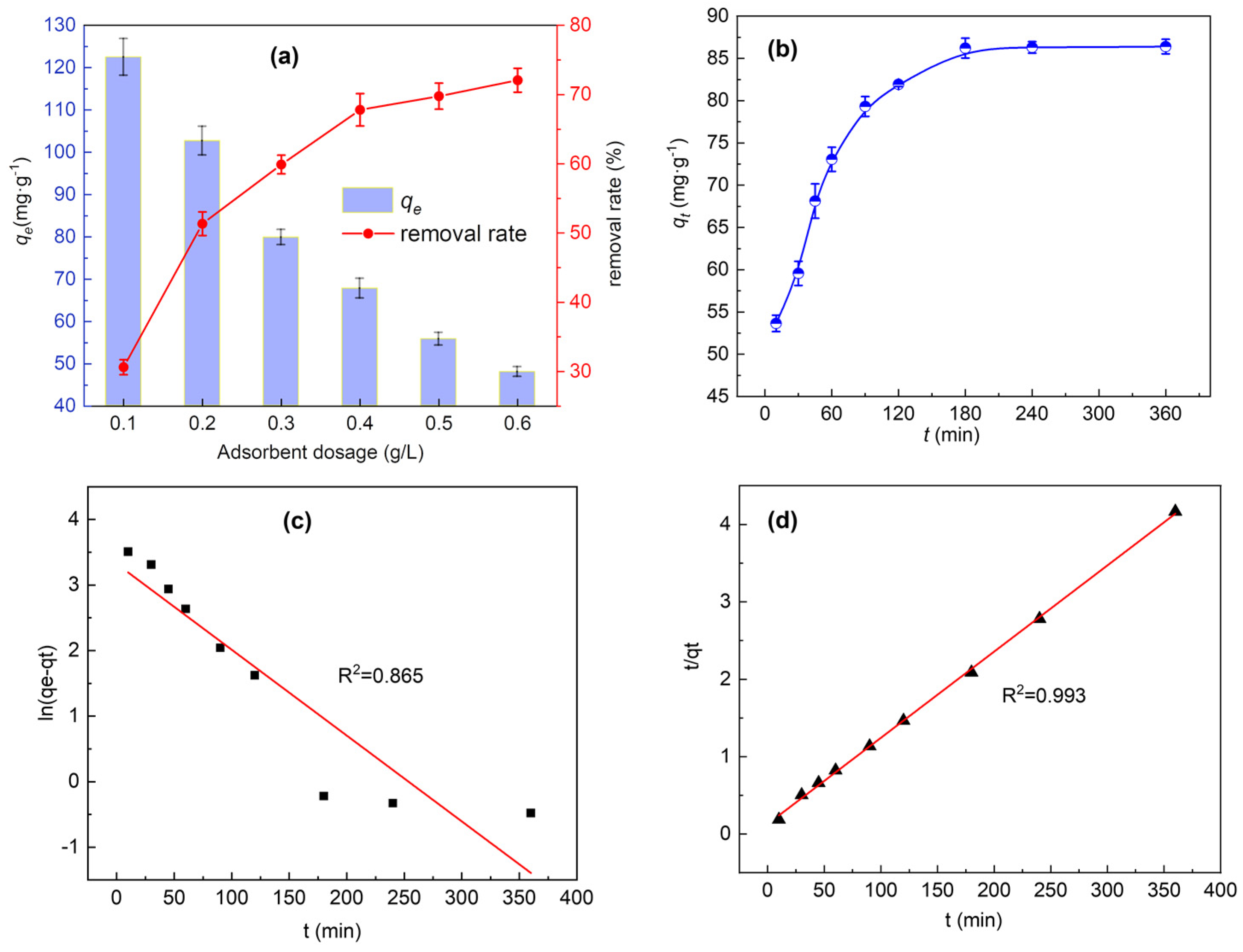
3.3. Effect of Adsorbent Dosage

Effect of adsorbent dosage on the adsorption performance of U(VI) is shown in Figure 5a, and it can be clearly observed that the removal rate of U(VI) increased sharply from 30 % to 70 % with the solid–liquid ratio increasing from 0.1 g L−1 to 0.4 g L−1. It came down to the surface area and adsorption sites of the adsorbent added as the adsorbent dosage increased, which made IONPs@MAH-β-CD adsorb more uranyl ions and the removal rate of U(VI) was significantly increased [42]. When the solid–liquid ratio was between 0.4~0.6 g L−1, the removal rate did not increase significantly, and that might be because the adsorption sites of IONPs@MAH-β-CD were reduced, which made it more difficult for U(VI) to contact with the adsorbent. Therefore, considering the characteristics of the removal rate varied according to adsorbent dosage and the practicability of the adsorbent, 0.4 g L−1 was selected as the optimal adsorbent dosage to further study the influence of subsequent factors on adsorption.

3.4. Effect of Contact Time and Adsorption Kinetics

The removal rate is also an important parameter to measure adsorbent efficiency. In order to evaluate the adsorption performance of IONPs@MAH-β-CD on U(VI), the effect of contact time on the adsorption kinetic behavior was studied. Figure 5b exhibited the adsorption kinetic of IONPs@MAH-β-CD as a function of contact time. It was observed that the adsorption quantity of the adsorbent rose sharply in the first 10 min and it was a rapid binding process between U(VI) and active sites on the adsorbent surface. After that, the adsorption process gradually stabilized and reached equilibrium at 180 min, when the adsorbent was nearly saturated.
Two typical kinetic models (the pseudo-first-order and pseudo-second-order kinetic models) were used to simulate the adsorption process, which are represented in Equations (3) and (4). qt (mg g−1) is the adsorption quantity at contact time t (min), and k1 (min−1) and k2 (g·mg−1 min−1) are the pseudo-first-order and pseudo-second-order kinetic rate constants, respectively [40].
l n ( q e q t ) = l n q e k 1 t
t q t = 1 k 2 q e 2 + t q e
The fitting results of the pseudo-first-order and pseudo-second-order kinetic models for adsorption of U(VI) are displayed in Figure 5c,d. The results indicated that the pseudo-first-order and pseudo-second-order kinetic rate constants were 0.0131 min−1 (k1) and 0.00095 g mg−1 min−1 (k2), respectively, and the correlation coefficient of the pseudo-second-order kinetic model (R2 = 0.999) is higher than that of the pseudo-first-order kinetic model (R2 = 0.865). Moreover, the theoretical value of adsorption quantity qe,cal obtained from the pseudo-second-order kinetic model was 90.09 mg g−1, which was close to the experimental adsorption quantity (87.02 mg g−1), indicating that the adsorption process between IONPs@MAH-β-CD and U(VI) was in accordance with the pseudo-second-order kinetic model [35]. It was proven that the adsorption process of IONPs@MAH-β-CD for U(VI) was carried out on the surface of the material, which was controlled by the active sites. The active sites bound to the pollutant U(VI) rapidly, which was mainly a chemical adsorption process [43]. This might be due to the interaction between the carboxyl groups of IONPs@MAH-β-CD and U(VI) [44].

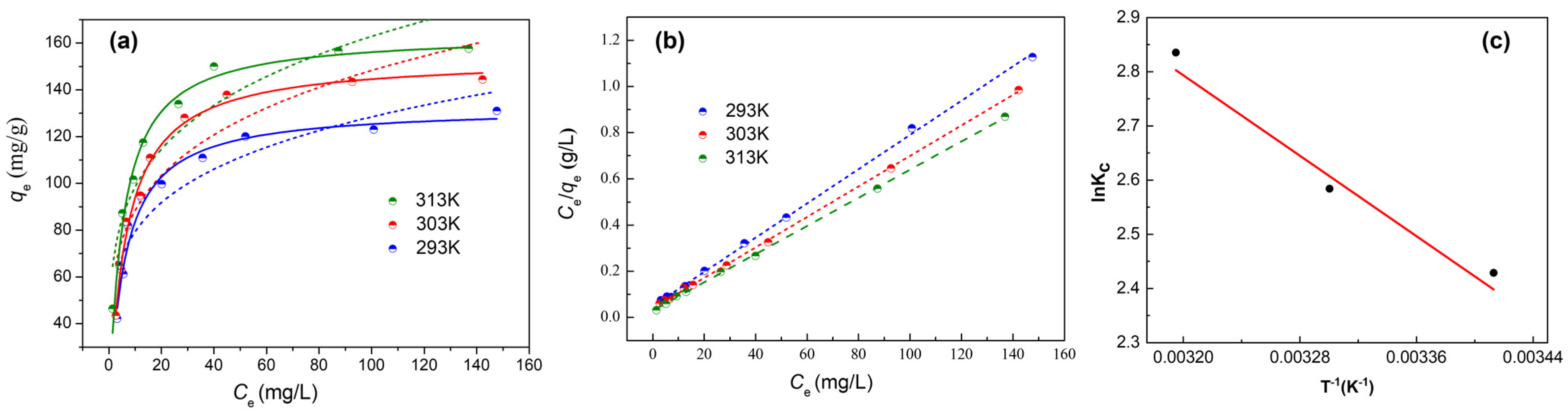
3.5. Adsorption Isotherms and Thermodynamics

The adsorption performance of IONPs@MAH-β-CD for U(VI) was carried out at 293, 303, and 313 K to explore the trends with variation in temperature and study the adsorption models. As shown in Figure 6a–c, the Langmuir and Freundlich models were used to fit the isotherm data and study the adsorption mechanism for U(VI). The Langmuir model is based on monolayer adsorption theory, which could well describe the adsorption process of monolayer absorbents with uniform surface [28] and is represented in Equation (5) [29]. The Freundlich model is suitable for a multilayer adsorption process or adsorption system with uneven distribution of adsorbent and is represented in Equation (6) [30]:
q e = q m K L C e 1 + K L C e
q e = K F C e 1 n
where qm (mg g−1) is the adsorption quantity at the maximum theoretically calculated adsorption and Ce (mg L−1) is the concentration of residual U(VI) in solution at reaction equilibrium. KL and KF are constants calculated by fitting the Langmuir model and the Freundlich model, respectively, and n is a dimensionless constant in the Freundlich model.
The isotherm fitting results are shown in Figure 6a,b. The solid lines are fitting curves of the Langmuir model and the dashed lines are the fitting curves of the Freundlich model, based on experimental data. The fitting parameters of IONPs@MAH-β-CD with the Langmuir and Freundlich models are shown in Table 1. It can be seen that the fitting coefficient (R2) of the Langmuir model was higher than that of the Freundlich model, indicating that the adsorption behavior of IONPs@MAH-β-CD for U(VI) was more consistent with the Langmuir model and that it was a surface uniform monolayer adsorption process. The maximum adsorption quantity of IONPs@MAH-β-CD was 134.77, 151.52, and 163.93 mg g−1 at 293, 303, and 313 K, respectively, while the maximum adsorption quantity of pure IONPs was 65.3 mg g−1at 313 K [45]. The maximum adsorption quantity was nearly three times that of IONPs, which fully reflected that the introduction of MAH-β-CD significantly improved the adsorption quantity and increased the contact opportunity between the active sites of the adsorbent and U(VI).
In addition, from Figure 6a it can be seen that the adsorption quantity for U(VI) increased with the temperature increment. This was because the higher temperature enhanced the molecular thermal motion of the adsorbent and U(VI) [46]. Therefore, the contact probability increased and the adsorption reaction was more likely to occur. In order to explain this phenomenon more accurately, Equations (7) and (8) were employed to determine the standard Gibbs free energy (ΔG0, kJ mol−1), the standard entropy change (ΔS0, J mol−1·K−1), and the standard enthalpy change (ΔH0, kJ mol−1) [47]:
l n K c = Δ S 0 R Δ H 0 R T
Δ G 0 = Δ H 0 T Δ S 0
where R (8.314 J·mol−1·K−1) and Kc are the molar gas constant and distribution constant, respectively, and Kc is obtained by K c = q e / c e . Figure 6c is the linear relation fitting of lnKc to 1/T, and the thermodynamic parameters obtained by the fitting equation are listed in Table 2.
The positive value of ΔH0 indicated that it was an endothermic process. This was because U(VI) in aqueous solution was usually wrapped by the hydration layer, and it needed to absorb external energy to break off the hydration layer when the adsorption occurred [48]. The positive value of ΔS0 demonstrated that the adsorption system tends to be disordered. The negative value of ΔG0 illustrated that the adsorption reaction can proceed spontaneously, and the decrease of ΔG0 with the increase of temperature indicated that the higher temperature could accelerate to break off the hydration layer and make adsorption more likely to occur.

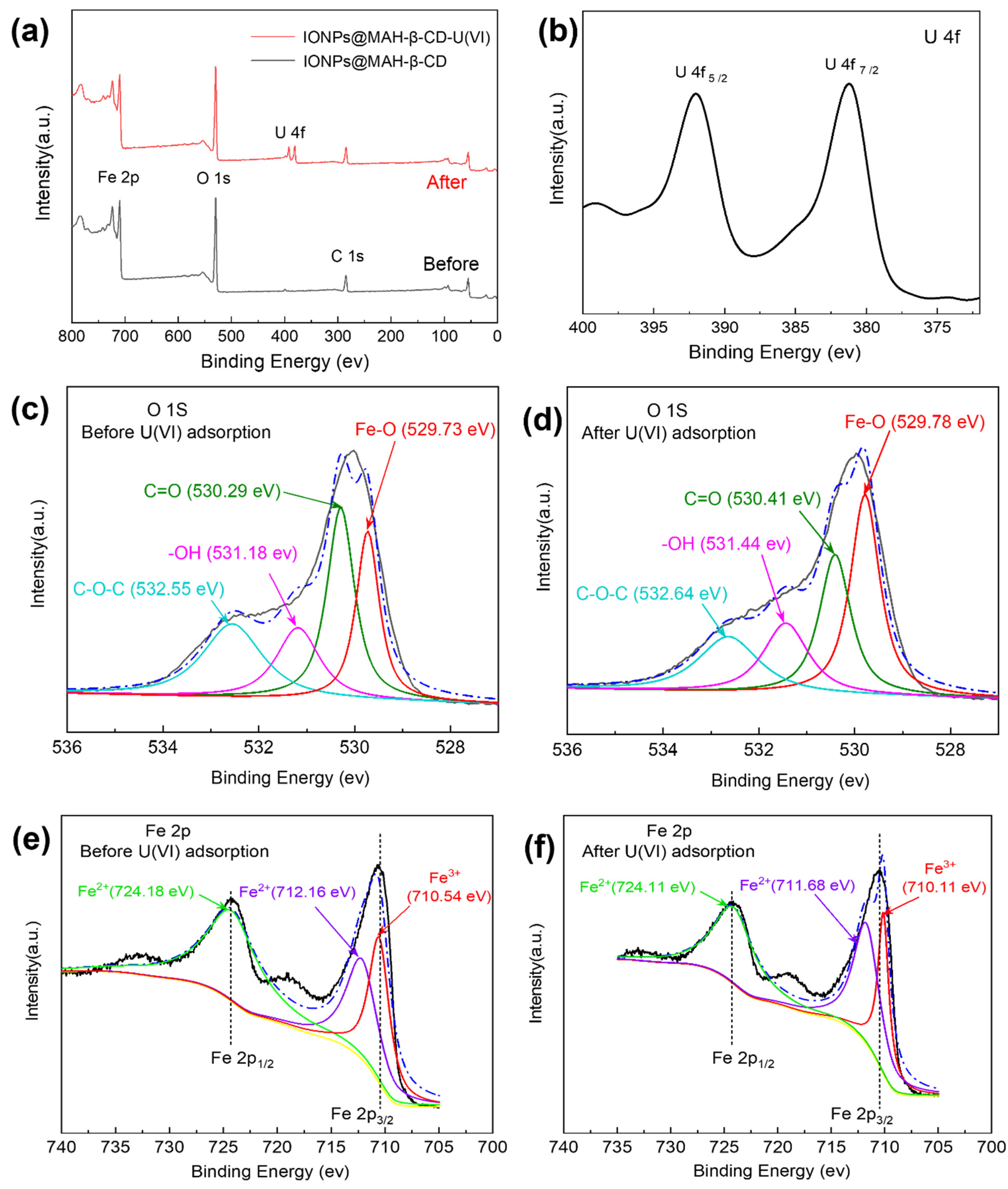
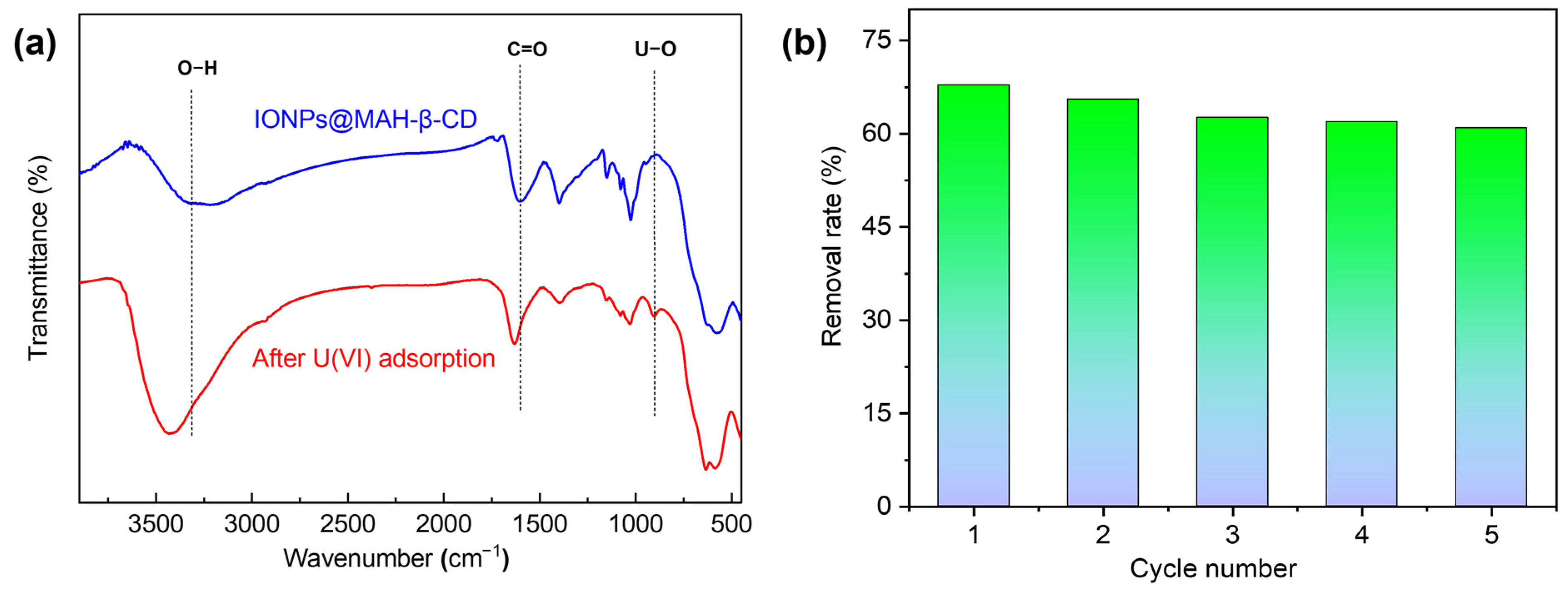
3.6. Mechanism of Adsorption

In order to determine the adsorption mechanism of IONPs@MAH-β-CD for U(VI), changes in C, O, and Fe elements before and after adsorption were investigated by measuring the XPS spectrum. As seen in Figure 7a, the strong peaks of C1s, O1s, and Fe 2p are observed, showing that C, O, and Fe are the main elements in IONPs@MAH-β-CD; it is further proven that the material is successfully prepared. It also can be seen that U4f existed in the XPS map after adsorption by comparing the full-scan spectra before and after adsorption, indicating that U(VI) was successfully adsorbed by IONPs@MAH-β-CD [49]. In addition, two new strong peaks could be observed in Figure 7b at 380.97 and 391.97 eV, which stand for U4f 7/2 and Uf 5/2 components, respectively; it is suggested that U(VI) was successfully adsorbed by IONPs@MAH-β-CD [15,50].
As shown in Figure 7c, the high resolution O1s of the prepared IONPs@MAH-β-CD could be deconvolved into four peaks at 529.73, 530.29, 531.18, and 532.55 eV, which were attributed to Fe-O, C=O (suggesting carboxylate groups), OH, and C-O-C, respectively. The four peaks of the O1s spectrum after adsorption had position shifts of +0.05, +0.12, +0.26, and +0.09 eV, respectively, implying that oxygen-containing functional groups played an important role in the trapping of U(VI) during the adsorption process of IONPs@MAH-β-CD [51]. In other words, the introduction of maleic anhydride containing carboxyl groups and cyclodextrin containing rich amounts of -OH and C-O-C enhanced the adsorption performance of IONPs. As displayed in Figure 7e, the binding energies Fe 2p 1/2 and Fe 2p 3/2 were located at 724.18 eV and 710.52 eV, respectively. In addition, the high-resolution Fe 2p 3/2 spectra of IONPs@MAH-β-CD could be deconvoluted into two peaks at 712.16 eV and 710.54 eV, respectively, corresponding to the binding energies of Fe (II) and Fe (III) bonds [40,52,53,54]. The peak positions of Fe (II) and Fe (III) changed after adsorption (Figure 7(f)), suggesting that IONPs were involved in the coordination with uranium during the adsorption process. Moreover, as shown in FTIR in Figure 8, the new absorption peak at 904 cm−1 could be attributed to the asymmetric vibration peak of the U-O group, which confirmed the adsorption of uranyl ions on the sample. FTIR analysis also showed that after the adsorption of U(VI) by IONPs@MAH-β-CD, the C=O (suggesting carboxylate groups) and OH peaks red-shifted from 1650 and 3217 cm−1to 1633 and 3434 cm−1, respectively, and the intensity changed significantly, indicating that the carboxyl group and cyclodextrin were the main adsorption sites [55]. Based on the above analysis, it was found that the surface of IONPs@MAH-β-CD contained abundant functional groups and active sites, which were conductive to the binding of U(VI) [56].

3.7. Recyclability of IONPs@MAH-β-CD

The recyclability of the magnetic IONPs@MAH-β-CD nanoparticles was studied. First, 0.01mol/L HCl were used as eluent to desorb the adsorbed IONPs@MAH-β-CD, and then the IONPs@MAH-β-CD were washed with ultrapure water. As seen in Figure 8b, the experiment found that after five repeated adsorption/desorption cycles, the removal rate of U(VI) decreased slightly and the removal rate of adsorbent uranium remained above 60.9%, indicating that IONPs@MAH-β-CD had relatively good recoverability.

4. Conclusions

The IONPs@MAH-β-CD magnetic material was prepared by a simple chemical co-precipitation method and was analyzed using SEM, BET, VSM, and EDS. The results showed that IONPs@MAH-β-CD contained 10–20 nm nano-spherical particles with good magnetic properties and abundant pore structure. Kinetic experiments demonstrated that the adsorption could reach equilibrium within 180 min and thermodynamic analysis showed that it was a spontaneous endothermic and entropy-increasing process. Moreover, the adsorption isotherms illustrated that the adsorption capacity of IONPs for U(VI) could be improved by the introduction of MAH-β-CD, and the maximum adsorption quantity could reach 163.93 mg g−1. In addition, research on the adsorption mechanism indicated that there were a large number of functional groups and active sites on the surface of IONPs@MAH-β-CD, which were conductive to the binding of U(VI). Because IONPs@MAH-β-CD is an easily obtained material, with simple operation, and is easy to separate, and because the removal rate of adsorbent uranium remained above 60.9% after five cycles of regeneration, it will have great potential in nuclear wastewater treatment and other practical applications.

Author Contributions

Conceptualization, methodology, investigation, and writing—original draft preparation, X.Z.; investigation, Q.Y.; validation, Q.W.; writing—review and editing: C.H. and K.G.; supervision, J.O. and M.C. All authors have read and agreed to the published version of the manuscript.

Funding

This research was supported by the National Natural Science Foundation of China (No. 52102214), and the Jiangxi Provincial Natural Science Foundation (20202BABL203026, 20202BAB213008).

Data Availability Statement

Not Applicable.

Conflicts of Interest

The authors declare no conflict of interest.

References

  1. Tsouris, C. Uranium extraction: Fuel from seawater. Nat. Energy 2017, 2, 17022. [Google Scholar] [CrossRef]
  2. Degueldre, C. Uranium as a renewable for nuclear energy. Prog. Nucl. Energ. 2017, 94, 174–186. [Google Scholar] [CrossRef]
  3. Cui, W.R.; Zhang, C.R.; Jiang, W.; Li, F.F.; Liang, R.P.; Liu, J.; Qiu, J.D. Regenerable and stable sp2 carbon-conjugated covalent organic frameworks for selective detection and extraction of uranium. Nat. Commun. 2020, 11, 436. [Google Scholar] [CrossRef] [PubMed] [Green Version]
  4. Kong, L.; Ruan, Y.; Zheng, Q.; Su, M.; Diao, Z.; Chen, D.; Hou, L.a.; Chang, X.; Shih, K. Uranium extraction using hydroxyapatite recovered from phosphorus containing wastewater. J. Hazard. Mater. 2020, 382, 120784. [Google Scholar] [CrossRef] [PubMed]
  5. Zhou, J.; Zhang, X.; Zhang, Y.; Wang, D.; Zhou, H.; Li, J. Effective inspissation of uranium (VI) from radioactive wastewater using flow electrode capacitive deionization. Sep. Purif. Technol. 2022, 283, 120172. [Google Scholar] [CrossRef]
  6. Tang, N.; Liang, J.; Niu, C.; Wang, H.; Luo, Y.; Xing, W.; Ye, S.; Liang, C.; Guo, H.; Guo, J.; et al. Amidoxime-based materials for uranium recovery and removal. J. Mater. Chem. A 2020, 8, 7588–7625. [Google Scholar]
  7. Yuan, Y.; Yu, Q.; Yang, S.; Wen, J.; Guo, Z.; Wang, X.; Wang, N. Ultrafast recovery of uranium from seawater by bacillus velezensis strain UUS-1 with innate anti-biofouling activity. Adv. Sci. 2019, 6, 1900961. [Google Scholar] [CrossRef] [Green Version]
  8. Wang, Y.; Zhang, Y.; Li, Q.; Li, Y.; Cao, L.; Li, W. Amidoximated cellulose fiber membrane for uranium extraction from simulated seawater. Carbohydr. Polym. 2020, 245, 116627. [Google Scholar] [CrossRef]
  9. Chen, Z.; Liang, Y.; Jia, D.; Chen, W.; Cui, Z.; Wang, X. Layered silicate RUB-15 for efficient removal of UO2 2+ and heavy metal ions by ion-exchange. Environ. Sci. Nano. 2017, 4, 1851–1858. [Google Scholar] [CrossRef]
  10. Cui, W.R.; Li, F.F.; Xu, R.H.; Zhang, C.R.; Chen, X.R.; Yan, R.H.; Liang, R.P.; Qiu, J.D. Regenerable covalent organic frameworks for photo-enhanced uranium adsorption from seawater. Angew. Chem. Int. Ed. Engl. 2020, 59, 17684–17690. [Google Scholar] [CrossRef]
  11. Zhong, L.; He, F.; Liu, Z.; Dong, B.; Ding, J. Adsorption of uranium (VI) ions from aqueous solution by acrylic and diaminomaleonitrile modified cellulose. Colloids Surf. A 2022, 641, 128565. [Google Scholar] [CrossRef]
  12. Zhao, L.; Wang, S.; Zhuang, H.; Lu, B.; Sun, L.; Wang, G.; Qiu, J. Facile synthesis of low-cost MnPO4 with hollow grape-like clusters for rapid removal uranium from wastewater. J. Hazard. Mater. 2022, 434, 128894. [Google Scholar] [CrossRef] [PubMed]
  13. Jun, B.M.; Lee, H.K.; Park, S.; Kim, T.J. Purification of uranium-contaminated radioactive water by adsorption: A review on adsorbent materials. Sep. Purif. Technol. 2021, 278, 119675. [Google Scholar] [CrossRef]
  14. Zhao, M.; Cui, Z.; Pan, D.; Fan, F.; Tang, J.; Hu, Y.; Xu, Y.; Zhang, P.; Li, P.; Kong, X.Y.; et al. An efficient uranium adsorption magnetic platform based on amidoxime-functionalized flower-like Fe3O4@TiO2 core-shell microspheres. ACS Appl. Mater. Interfaces 2021, 13, 17931–17939. [Google Scholar] [CrossRef]
  15. Duan, S.; Xu, X.; Liu, X.; Wang, Y.; Hayat, T.; Alsaedi, A.; Meng, Y.; Li, J. Highly enhanced adsorption performance of U(VI) by non-thermal plasma modified magnetic Fe3O4 nanoparticles. J. Colloid. Interf. Sci. 2018, 513, 92–103. [Google Scholar] [CrossRef]
  16. Ji, W.C.; Hu, P.; Wang, X.Y.; Saji, S.E.; Chang, T.; Zhu, X.Y.; Yang, F.F.; Cao, Q.G.; Dang, R.; Wang, K.S. One-step carbothermal synthesis of super nanoadsorbents for rapid and recyclable wastewater treatment. Crystals 2021, 11, 75. [Google Scholar] [CrossRef]
  17. Liu, S.; Yu, B.; Wang, S.; Shen, Y.; Cong, H. Preparation, surface functionalization and application of Fe3O4 magnetic nanoparticles. Adv. Colloid. Interfac. 2020, 281, 102165. [Google Scholar] [CrossRef]
  18. Badruddoza, A.Z.M.; Hazel, G.S.S.; Hidajat, K.; Uddin, M.S. Synthesis of carboxymethyl-β-cyclodextrin conjugated magnetic nano-adsorbent for removal of methylene blue. Colloids Surf. A 2010, 367, 85–95. [Google Scholar] [CrossRef]
  19. Badruddoza, A.Z.; Tay, A.S.; Tan, P.Y.; Hidajat, K.; Uddin, M.S. Carboxymethyl-beta-cyclodextrin conjugated magnetic nanoparticles as nano-adsorbents for removal of copper ions: Synthesis and adsorption studies. J. Hazard. Mater. 2011, 185, 1177–1186. [Google Scholar] [CrossRef]
  20. Yang, S.; Zong, P.; Hu, J.; Sheng, G.; Wang, Q.; Wang, X. Fabrication of β-cyclodextrin conjugated magnetic HNT/iron oxide composite for high-efficient decontamination of U(VI). Chem. Eng. J. 2013, 214, 376–385. [Google Scholar] [CrossRef]
  21. Helal, A.S.; Mazario, E.; Mayoral, A.; Decorse, P.; Losno, R.; Lion, C.; Ammar, S.; Hémadi, M. Highly efficient and selective extraction of uranium from aqueous solution using a magnetic device: Succinyl-β-cyclodextrin-APTES@maghemite nanoparticles. Environ. Sci. Nano. 2018, 5, 158–168. [Google Scholar] [CrossRef]
  22. Li, N.; Yang, L.; Wang, D.; Tang, C.; Deng, W.; Wang, Z. High-capacity amidoxime-functionalized beta-cyclodextrin/graphene aerogel for selective uranium capture. Environ. Sci. Technol. 2021, 55, 9181–9188. [Google Scholar] [CrossRef] [PubMed]
  23. Liu, F.; Zhang, S.; Wang, G.; Zhao, J.; Guo, Z.J.R.A. A novel bifunctional molecularly imprinted polymer for determination of Congo red in food. RSC Adv. 2015, 5, 22811–22817. [Google Scholar] [CrossRef]
  24. Binello, A.; Cravotto, G.; Nano, G.M.; Spagliardi, P. Synthesis of chitosan–cyclodextrin adducts and evaluation of their bitter-masking properties. Flavour Frag. J. 2004, 19, 394–400. [Google Scholar] [CrossRef]
  25. Zhang, M.; Li, A.; Zhou, Q.; Shuang, C.; Zhou, W.; Wang, M. Effect of pore size distribution on tetracycline adsorption using magnetic hypercrosslinked resins. Micropor. Mesopor. Mat. 2014, 184, 105–111. [Google Scholar] [CrossRef]
  26. Zhang, J.; Li, X.; Rosenholm, J.M.; Gu, H.C. Synthesis and characterization of pore size-tunable magnetic mesoporous silica nanoparticles. J. Colloid Interf. Sci. 2011, 361, 16–24. [Google Scholar] [CrossRef]
  27. Golovnev, N.N.; Molokeev, M.S.; Samoilo, A.S.; Atuchin, V.V. Influence of alkyl substituents in 1,3-diethyl-2-thiobarbituric acid on the coordination environment in M(H2O)2 (1,3-diethyl-2-thiobarbiturate)2 M = Ca2+, Sr2+. J. Coord. Chem. 2016, 69, 957–965. [Google Scholar] [CrossRef]
  28. Denisenko, Y.G.; Molokeev, M.S.; Oreshonkov, A.S.; Krylov, A.S.; Aleksandrovsky, A.S.; Azarapin, N.O.; Andreev, O.V.; Razumkova, I.A.; Atuchin, V.V. Crystal structure, vibrational, spectroscopic and thermochemical properties of double sulfate crystalline hydrate [CsEu(H2O)3(SO4)2]·H2O and its thermal dehydration product CsEu(SO4)2. Crystals 2021, 11, 1027. [Google Scholar] [CrossRef]
  29. Luo, C.; Shea, J.J.; Huang, J. A carboxylate group-based organic anode for sustainable and stable sodium ion batteries. J. Power Sources 2020, 453, 227904. [Google Scholar] [CrossRef]
  30. Yang, Y.; Liu, Y.; Chen, S.; Cheong, K.L.; Teng, B. Carboxymethyl β-cyclodextrin grafted carboxymethyl chitosan hydrogel-based microparticles for oral insulin delivery. Carbohyd. Polym. 2020, 246, 116617. [Google Scholar] [CrossRef]
  31. Chen, B.; Sun, J.; Fan, F.; Zhang, X.; Qin, Z.; Wang, P.; Li, Y.; Zhang, X.; Liu, F.; Liu, Y. Ferumoxytol of ultrahigh magnetization produced by hydrocooling and magnetically internal heating co-precipitation. Nanoscale 2018, 10, 7369–7376. [Google Scholar] [CrossRef] [PubMed]
  32. Ramos-Guivar, J.A.; Flores-Cano, D.A.; Caetano Passamani, E. Differentiating nanomaghemite and nanomagnetite and discussing their importance in arsenic and lead removal from contaminated effluents: A critical review. Nanomaterials 2021, 11, 2310. [Google Scholar] [CrossRef] [PubMed]
  33. Kim, M.W.; Bae, D.H.; Kwon, S.H.; Choi, H.J. Magnetite embedded mini-emulsion polymerized polystyrene particles and their magnetorheology. Macromol. Res. 2018, 26, 353–358. [Google Scholar] [CrossRef]
  34. Davar, F.; Majedi, A.; Abbasi, A. Synthesis of Fe3O4@ZrO2 core–shell nanoparticles through new approach and its solar light photocatalyst application. J. Mater. Sci-Mater. El. 2017, 28, 4871–4878. [Google Scholar] [CrossRef]
  35. Li, N.; Gao, P.; Chen, H.; Li, F.; Wang, Z. Amidoxime modified Fe3O4@TiO2 particles for antibacterial and efficient uranium extraction from seawater. Chemosphere 2021, 287, 132137. [Google Scholar] [CrossRef] [PubMed]
  36. Ramos-Guivar, J.A.; Krohling, A.; López, E.O.; Litterst, F.J.; Passamani, E. Superspinglass behavior of maghemite nanoparticles dispersed in mesoporous silica. J. Magn. Magn. Mater. 2019, 485, 142–150. [Google Scholar] [CrossRef]
  37. Lajevardi, A.; Sadr, M.H.; Yaraki, M.T.; Badiei, A.; Armaghan, M. A pH-responsive and magnetic Fe3O4@silica@MIL-100 (Fe)/β-CD nanocomposite as a drug nanocarrier: Loading and release study of cephalexin. New J. Chem. 2018, 42, 9690–9701. [Google Scholar] [CrossRef]
  38. Dai, Y.; Jin, J.; Zhou, L.; Li, T.; Li, Z.; Liu, Z.; Huang, G.; Adesina, A.A. Preparation of hollow SiO2 microspheres functionalized with amidoxime groups for highly efficient adsorption of U(VI) from aqueous solution. J. Radioanal. Nucl. Chem. 2016, 311, 2029–2037. [Google Scholar] [CrossRef]
  39. Li, W.; Han, X.; Wang, X.; Wang, Y.; Wang, W.; Xu, H.; Tan, T.; Wu, W.; Zhang, H. Recovery of uranyl from aqueous solutions using amidoximated polyacrylonitrile/exfoliated Na-montmorillonite composite. Chem. Eng. J. 2015, 279, 735–746. [Google Scholar] [CrossRef]
  40. Zong, P.; Xu, X.; Shao, M.; Cao, D.; Wang, S.; Zhang, H. Efficient elimination of uranium by carboxymethyl-β-cyclodextrin nanoparticles decorated iron oxides: Water chemistry influences and mechanism researches. Int. J. Hydrogen Energ. 2021, 46, 1370–1384. [Google Scholar] [CrossRef]
  41. Liu, X.; Sun, J.; Xu, X.; Alsaedi, A.; Hayat, T.; Li, J. Adsorption and desorption of U(VI) on different-size graphene oxide. Chem. Eng. J. 2019, 360, 941–950. [Google Scholar] [CrossRef]
  42. Ma, F.; Nian, J.; Bi, C.; Yang, M.; Zhang, C.; Liu, L.; Dong, H.; Zhu, M.; Dong, B. Preparation of carboxylated graphene oxide for enhanced adsorption of U(VI). J. Solid State Chem. 2019, 277, 9–16. [Google Scholar] [CrossRef]
  43. Wang, J.; Chen, Z.; Shao, D.; Li, Y.; Xu, Z.; Cheng, C.; Asiri, A.M.; Marwani, H.M.; Hu, S. Adsorption of U(VI) on bentonite in simulation environmental conditions. J. Mol. Liq. 2017, 242, 678–684. [Google Scholar] [CrossRef]
  44. Li, P.; Wang, Y.; Wang, J.; Dong, L.; Zhang, W.; Lu, Z.; Liang, J.; Pan, D.; Fan, Q. Carboxyl groups on g-C3N4 for boosting the photocatalytic U(VI) reduction in the presence of carbonates. Chem. Eng. J. 2021, 414, 128810. [Google Scholar] [CrossRef]
  45. Diao, Z.; Pang, H.; Tang, H.; Wang, X.; Yu, S.; Song, G. Magnetic-dictyophora indusiata derived biochar composite for efficient removal of U(VI) and mechanism investigation. Chinese Sci. Bull. 2020, 65, 3954–3964. [Google Scholar] [CrossRef]
  46. Xu, Y.; Yu, J.; Zhu, J.; Liu, Q.; Zhang, H.; Liu, J.; Chen, R.; Li, Y.; Wang, J. Cyclized polyacrylonitrile amidoxime with local conjugate domain for high-efficiency extraction of uranium from seawater. Appl. Catal. B-Environ 2022, 316, 121677. [Google Scholar] [CrossRef]
  47. Kavci, E.; Erkmen, J.; Bingöl, M.S. Removal of methylene blue dye from aqueous solution using citric acid modified apricot stone. Chem. Eng. Commun. 2021, 210, 1–16. [Google Scholar] [CrossRef]
  48. Han, H.; Cheng, C.; Hu, S.; Li, X.; Wang, W.; Xiao, C.; Xu, Z.; Shao, D. Facile synthesis of gelatin modified attapulgite for the uptake of uranium from aqueous solution. J. Mol. Liq. 2017, 234, 172–178. [Google Scholar] [CrossRef]
  49. Song, W.; Wang, X.; Wang, Q.; Shao, D.; Wang, X. Plasma-induced grafting of polyacrylamide on graphene oxide nanosheets for simultaneous removal of radionuclides. Phys. Chem. Chem. Phys. 2015, 17, 398–406. [Google Scholar] [CrossRef]
  50. Atuchin, V.V.; Zhang, Z. Chemical bonding between uranium and oxygen in U6+-containing compounds. J. Nucl. Mater. 2012, 420, 222–225. [Google Scholar] [CrossRef]
  51. Zhong, X.; Wang, R.; Xu, G.; Guo, K.; Hu, C. Adsorptive removal of uranium (VI) from wastewater using a crosslinked amidoxime-functionalized β-cyclodextrin polymer. New J. Chem. 2022, 46, 20592–20601. [Google Scholar] [CrossRef]
  52. Meng, Y.; Wang, Y.; Liu, L.; Fang, Y.; Ma, F.; Zhang, C.; Dong, H. Efficient and magnetically recoverable U(VI) adsorbent: Fe3O4 loaded hypercrosslink copoly (styrene/maleic anhydride). Colloids Surf. A 2022, 632, 127644. [Google Scholar] [CrossRef]
  53. Atuchin, V.V.; Vinnik, D.; Gavrilova, T.; Gudkova, S.; Isaenko, L.; Jiang, X.; Pokrovsky, L.; Prosvirin, I.; Mashkovtseva, L.; Lin, Z. Flux crystal growth and the electronic structure of BaFe12O19 hexaferrite. J. Phys. Chem. C 2016, 120, 5114–5123. [Google Scholar] [CrossRef]
  54. Pinaeva, L.; Prosvirin, I.; Chesalov, Y.; Atuchin, V. High-temperature abatement of N2O over FeOx/CeO2-Al2O3 catalysts: The effects of oxygen mobility. Catalysts 2022, 12, 938. [Google Scholar] [CrossRef]
  55. Li, B.; Ma, L.; Tian, Y.; Yang, X.; Li, J.; Bai, C.; Yang, X.; Zhang, S.; Li, S.; Jin, Y. A catechol-like phenolic ligand-functionalized hydrothermal carbon: One-pot synthesis, characterization and sorption behavior toward uranium. J. Hazard. Mater. 2014, 271, 41–49. [Google Scholar] [CrossRef]
  56. Radu, T.; Iacovita, C.; Benea, D.; Turcu, R. X-ray photoelectron spectroscopic characterization of iron oxide nanoparticles. Appl. Surf. Sci. 2017, 405, 337–343. [Google Scholar] [CrossRef]
Scheme 1. Synthesis of IONPs@MAH-β-CD.
Crystals 12 01731 sch001
Figure 1. FTIR spectra (a) and XRD patterns (b).
Crystals 12 01731 g001
Figure 2. SEM images (a,b), EDS spectra (c), and N2 adsorption–desorption isotherm (d) of IONPs@MAH-β-CD.
Crystals 12 01731 g002
Figure 3. Pore-size distributions (a) and magnetization curves (b) of IONPs@MAH-β-CD.
Crystals 12 01731 g003
Figure 4. (a) The effect of initial pH on the capacity to adsorb U(VI). (b) The zeta potential of IONPs@MAH-β-CD versus pH. (c) The morphology distribution of U(VI) at different pH.
Crystals 12 01731 g004
Figure 5. Effect of adsorbent dosage (a) and contact times (b) for IONPs@MAH-β-CD at pH = 6.0, C0 = 40 mg L−1, 303 K. Pseudo-first-order (c) and pseudo-second-order (d) kinetic models of U(VI) adsorption.
Crystals 12 01731 g005
Figure 6. Adsorption isotherms of IONPs@MAH-β-CD at pH = 6.0 and 293, 303, and 313 K using the Langmuir and Freundlich model fittings (a), linear plot of Ce/qe versus Ce (b), and linear plot of lnK0 versus 1/T (c) on IONPs@MAH-β-CD.
Crystals 12 01731 g006
Figure 7. XPS spectra of IONPs@MAH-β-CD before and after U(VI) adsorption, full spectrum (a), U 4f (b), deconvolution of O1s before (c) and after adsorption (d), and deconvolution of Fe 2p before (e) and after adsorption (f).
Crystals 12 01731 g007
Figure 8. (a) The FTIR spectra of IONPs@MAH-β-CD before and after U(VI) adsorption; (b) recyclability of IONPs@MAH-β-CD.
Crystals 12 01731 g008
Table 1. The U(VI) adsorption isotherm parameters of IONPs@MAH-β-CD.
AdsorbentT(K)Langmuir ModelFreundlich Model
qm (mg g−1)KL (L mg−1)R2nKFR2
IONPs@MAH-β-CD293134.770.1530.9763.9041.5520.814
303151.520.1690.9893.5243.0440.839
313163.930.2020.9783.7050.2530.881
Table 2. Thermodynamic parameters of IONPs@MAH-β-CD.
AdsorbentΔG0 (KJ mol−1)ΔH0 (KJ mol−1)ΔS0 (J mol−1 K−1)
293 K303 K313 K
IONPs@MAH-β-CD−5.84−6.57−7.2915.4372.60
Publisher’s Note: MDPI stays neutral with regard to jurisdictional claims in published maps and institutional affiliations.

