The scanning of a certain surface area in STM to record an image is often performed using the tunneling current feedback mode. Usually, to obtain a single image, it is necessary to set specific scanning parameters: scanning area, as well as such important parameters as operating voltage—Usc (voltage across the tunnel gap) and operating tunnel current Isc. The joint choice of these two parameters can be called the choice of “working or operating point” for the scanning. The value of the tunnel gap between the probe and sample is not measured in many cases and it is not known exactly, or otherwise, an independent measurement of this parameter is additionally required. During a single scan, the voltage is usually kept unchanged, and the tunneling current is supported at the constant value by means of feedback which properly regulates the probe spacing, with respect to the sample surface. During the lateral movement of the probe from point to point along the surface, the tunneling current may change due to the surface morphology (i.e., a change in the tunnel gap) or a change in the density of electronic states involved — due to the tunneling, because of, say, a composition variation at different points on the surface. The feedback returns the current to the set value due to the compensating vertical probe displacement, with respect to the surface: if the current drops, then the probe tip approaches the surface, if the current increases, then the probe moves away from the surface. Both parameters current and voltage, or in other words, this one or that choice of the operating point, affect the image contrast but in different ways. Take a closer look at the examples of how these parameters can affect the contrast observed in the STM image.
3.1. Testing the Influence of the Scanning Parameters on the STM Image Contrast for the Clean Silicon Surface with the (7 × 7) Superstructure
In the scanning of a clean silicon surface with a well-known (7 × 7) superstructure, it is assumed that only a slight change in the density of states is possible when the probe is moved laterally from point to point (or from a small area to the next similar area) along the surface since this is a pure single-component material with the same reconstruction on the terraces. Examples of spectra at different points of the (7 × 7) structure can be found in [
41]. In this case, obviously, one should expect the formation of an image contrast that adequately reflects the surface morphology. The contrast on the Si(111) bilayer step was measured previously [
40]. However, even in this seemingly simple case, there are certain features.
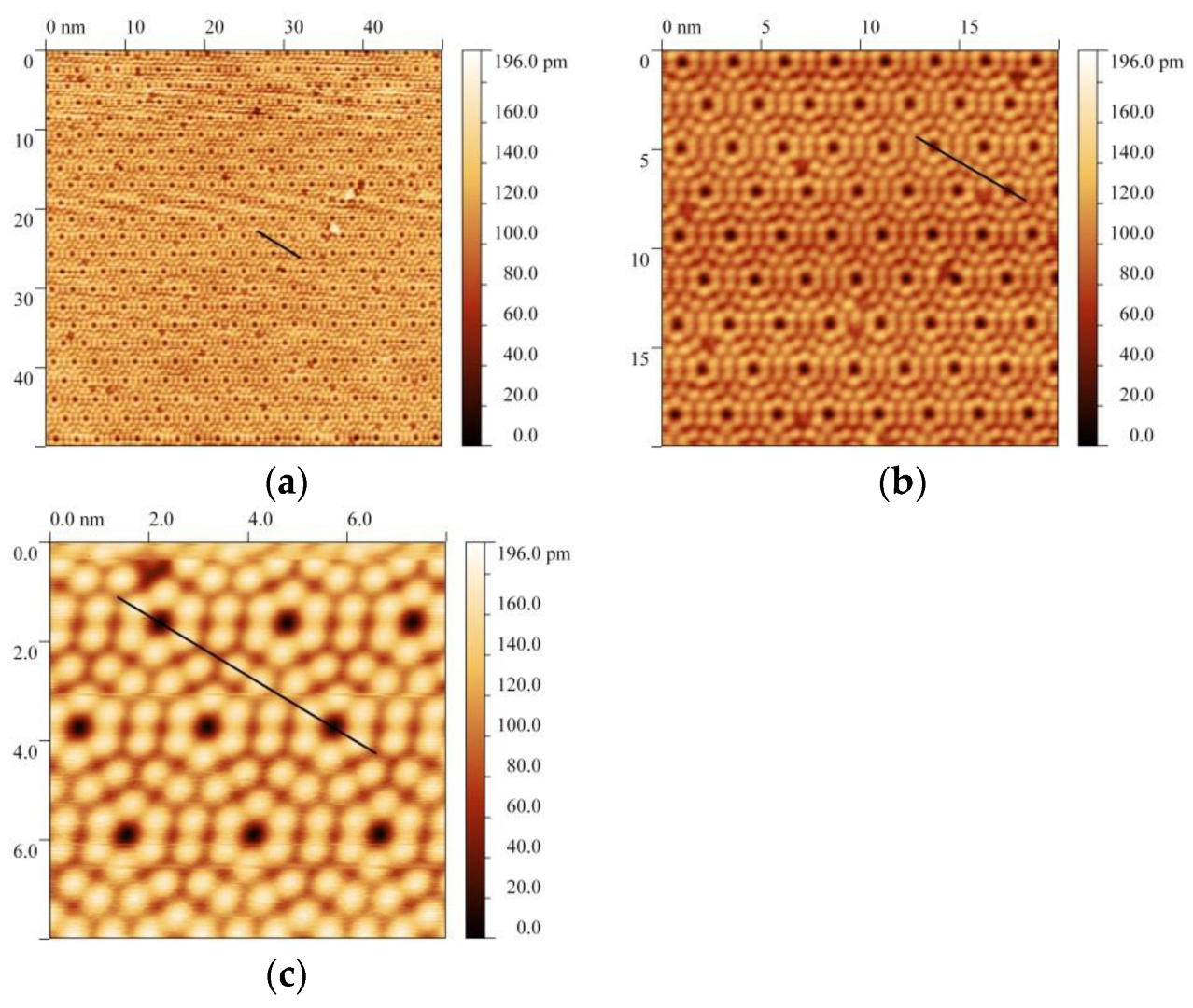
Figure 1 presents the STM images of several successive scans of the same Si(111)-(7 × 7) surface, obtained by varying only one parameter, namely the scan area, which is accompanied by a corresponding improvement in the lateral resolution with a decrease in the scan area, since the number of recorded points of 400 × 400 was not changed.
In this case, the same operating point was used to obtain all the presented images—the tunnel current was 0.025 nA and the voltage was +1.5 V. In general, it is obvious that the main visible features are light adatoms and dark corner holes, and the images look almost the same. Note that the pseudocolor scale was deliberately chosen to be the same for a more adequate comparison of the images. However, the differences in the contrast of light adatoms and dark corner holes in the presented images with different scan area sizes cannot be visually determined, despite the fact that adatoms and corner holes are clearly visible and differ sharply in color. If we compare the respective height profiles, the difference becomes apparent. The measured height profiles along the lines indicated in
Figure 1 are shown in
Figure 2. The profile registration line crosses two corner holes along the long diagonal of the elementary diamond cell of the (7 × 7) structure.
It is clearly shown in
Figure 2 that, as the scan area decreases, the height difference between the adatom and the corner hole increases, and it can be measured quantitatively. The blue profile corresponding to
Figure 1a has a height difference of 0.053 nm; the red profile from
Figure 1b has a height difference of 0.08 nm and the black profile from
Figure 1c has a height difference of 0.13 nm. This means that the contrast of the images increases accordingly. Note that this effect cannot be explained by the changes occurring on the surface or by a variation in the probe dimensions (for example, due to its degradation), since the contrast and height profiles are reproduced when a certain area is rescanned after a scanning area was changed in any way.
It should be noticed that it is very easy to bring all the images in
Figure 1 to the same visible contrast or rescale the intensity difference from maximum to minimum and, hence, to distort the real change of the contrast. Moreover, many pieces of the STM/AFM measuring software perform this correction automatically. In this case, the contrast change in the image may not be recognized at all if the intensity scale color is not presented. This simplest notice is given here not only to emphasize the importance of accompanying any STM image with a scale that relates the pseudocolor (or gray scale) to the z-coordinate normal to the surface, i.e., to the height–depth coordinate. This would help to avoid possible ambiguities in the perception and interpretation of the pseudocolor. What is more important is that it is for pointing out one simple but not so obvious feature. Images with a noticeably higher lateral resolution of the (7 × 7) structure than that shown here in
Figure 1 and
Figure 2 can be found in the literature [
44]. Indeed, high-resolution images show that, in the vertical direction, the difference in the heights of the adatoms and the corner hole in the experimental images and the corresponding contrast reaches 0.23 nm (see
Figure 3c) in the referenced paper [
44]. However, it is necessary to pay attention to the fact that this value is almost 2 times less than the vertical geometric distance from adatoms to the bottom in the center of the corner hole, which should be about 0.4 nm (i.e., 4 Å) according to the generally accepted dimer–adatom–stacking fault (DAS) model [
45]. A detailed diagram of the DAS model indicating the vertical distances between different atoms is given, for example, in the recent work by Demuth [
46]. In this case, we are interested in the vertical distance from the adatoms to the corner hole bottom. The authors of [
44] explained the difference between the experimental profile and the DAS model by the effect of the probe size, which seems to be the most obvious explanation. However, using ab initio calculations of STM images even for an ideal probe with a zero tip radius, the authors obtained a height difference of about 0.35 nm or 3.5 Å, i.e., still less than the expected 3.98 Å for the DAS model. This fact indicates the existence of additional reasons for a discrepancy, such as the influence of the spatial tunneling current distribution, besides the sensitivity of the lateral and vertical resolution to the probe tip dimensions.
The observed effect of the contrast dependence on the scanning area could be explained, for example, by a simple dependence of the vertical resolution on the lateral one, that is, by a decrease in the number of measured points per scanning length unit. It is also likely that there are certain spatial tunneling current integration effects here due to the finite area involved in the tunneling. An additional uncontrolled contribution, probably not too much, may originate from the probe displacement relative to the surface (such as random deviation and/or drift) during the measurement process at each surface point with an increase in lateral displacements during the scanning of increased area.
The images obtained by varying the working tunnel current when two other parameters, namely the scanning area and the operating voltage, were fixed are shown in
Figure 3. The image obtained at the current of 0.035 nA (
Figure 3a) with the scanning area of 20 × 20 nm
2 should be compared with a similar image with a tunnel current of 0.025 nA, which is shown in
Figure 1b above, and a comparison of the height profiles at these currents is shown in
Figure 3b.
It is shown in
Figure 3b that as the operating current increases, the profile depth increases. Obviously, the morphological features of the surface have not been changed, since the contrast is reproduced when the working tunneling current is reproduced. However, it should still be noted that, sometimes, changes in the probe state (because of possible events of some atoms jumping from the surface to the STM probe or vice versa, or due to the probe tip shape changing, caused by a strong electrical field applied between the probe and the surface as well as the high tunneling current density during the scanning along the surface) are possible, which can lead to an uncontrolled change in the contrast. In the absence of these random processes, the observed current variation effect is that the average tip-to-sample distance decreases as the operating current increases, and an increased feedback response is required to compensate for the increased current changes during the surface scanning. Again, it should be noted that the contrast here is not determined unambiguously by the surface morphology itself, but is associated with the features of the tunnel current formation and measurement and the feedback operation.
In the high-resolution images, the change in contrast while the tunneling current is varied appears to be much more pronounced (see
Figure 3c,d). In
Figure 3d, the profiles for the STM images obtained at 0.035 nA and 0.025 nA shown in
Figure 1c and
Figure 3c, respectively, are compared. One can see that the height difference between the adatoms and corner holes reaches the value of 2.8 Å. It should be noted that the range of the pseudocolor scale in
Figure 3c is deliberately increased, from 0 to 340 pm, in comparison with other images in order to reflect the increased contrast adequately.
The variation of another operating parameter, the voltage, from +1.5 V to +2.0 V with a fixed scanning area, in this case 50 × 50 nm
2, and fixed operating current 0.025 nA does not lead to noticeable contrast changes. Here, the images in
Figure 1a and
Figure 4a are compared, along with the corresponding profiles shown in
Figure 4b.
It is shown in
Figure 4b that the profiles along the cell diagonal almost coincide; only the elementary cell orientation changed (it is rotated by 180 degrees). The difference between the halves of the elementary cell (faulted and unfaulted halves) of the (7 × 7) structure turns out to be noticeable, which is a well-known fact following from the DAS model of (7 × 7). An increase in the voltage at a fixed operating current captures a wider range of the spectrum of electronic states involved in the tunneling current formation (which is discussed below in detail). In this case, the voltage variation did not affect the image contrast (depth profile), since the spectra at different points on the surface of one material change synchronously.
Thus, even for the case of a clean surface of a single-component material, in our case, silicon, the contrast dependence on the scanning parameters can show a more complex nature of the tunneling current formation than just an unambiguous presentation of the surface morphology.
3.2. The Contrast Dependence on the Voltage on the Nitridated Silicon Surface. Simultaneous Observation of the Contrast Behavior on the Morphological Step and on the (7 × 7)N/(8 × 8) Boundary as a Function of Tunnel Bias
A more interesting behavior of the contrast appears when the voltage is varied on scanning the nitridated silicon surface Si(111), in contrast to a clean surface. In this work, the contrast dependence on the operating voltage applied to the tunnel gap on the samples treated with a relatively small dose of ammonia was experimentally observed. To prepare the samples, a clean silicon surface was treated under ammonia flux
p = 3 × 10
−7 Torr at
T = 1000 °C for several tens of seconds (10–30 s), and the corresponding nitridation dose was 3–10 Langmuir (L) (1 L corresponds to an exposition of 1 s at pressure 1 × 10
−6 Torr). As a result of such treatment, the main part of the surface is covered by the (7 × 7)
N structure; along with this, islands of the (8 × 8) structure are also formed on the surface. We attributed the appearance of such an (8 × 8) structure to the graphene-like layer formation of silicon nitride g-Si
3N
3 [
40,
41].
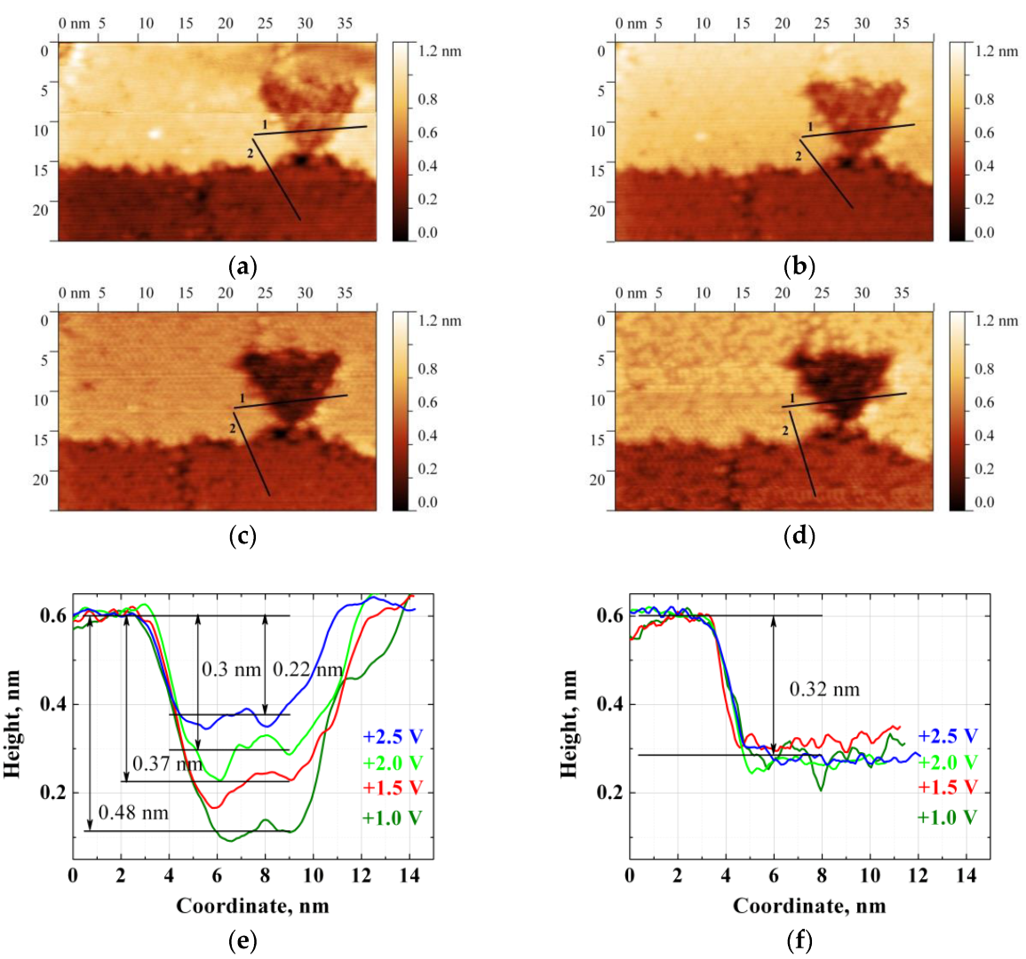
The images of the same nitridated sample area recorded sequentially with a decrease in the operating voltage from +2.5 V to +1.0 V are shown in
Figure 5a–d.
On the STM images of
Figure 5a–d, an extended one-dimensional (1D) morphological feature, i.e., a step, as well as a triangular island associated with the formation of the (8 × 8) structure, is observed. Somewhat lighter and darker backgrounds on the terraces on both step sides are observed, and the terraces are covered by the (7 × 7)
N structure. This fact was established in the STM images with a higher resolution. Characteristic images of the (7 × 7)
N structure with a high resolution were presented earlier [
40], and this structure also becomes noticeable, for example, in the image of
Figure 5d, where the characteristic dark trenches appear. This image was taken at the scan voltage of +1 V. It was shown that, at a low voltage, the characteristic contrast of the central and corner adatoms of the (7 × 7)
N images is more pronounced. On the other hand, for the triangular island in this image, as well as for other similar triangular islands observed on this surface, higher resolution images show the (8 × 8) structure. High-resolution images of the (8 × 8) structure were also presented earlier in [
40,
41]. In the images in
Figure 5a–d, it is possible to observe simultaneously the contrast behavior at the morphological step and at the (7 × 7)
N/(8 × 8) interface with a varying voltage. It is shown in
Figure 5 that the contrast does not depend on the voltage at the morphological step and that at the boundaries of these two structures ((7 × 7)
N and (8 × 8)), the contrast is decreased with the increase in operating voltage.
Figure 5e,f present the height profiles along the lines indicated in images a–d, where image e presents a profile along line #1 crossing the island of the (8 × 8) structure, and f presents a profile along line #2 crossing the morphological step. The profiles more clearly show the different behavior of the contrast or apparent “depth” at the morphological step and at the boundaries of the structures as a function of bias. The color contrast depth and the corresponding height of the morphological step do not change, despite the presence of random noise and a certain “shift” of the color tone of both terraces being observed. The “shift” is associated with a change in the contrast on the (7 × 7)
N structure itself [
40]. The morphological step height was determined as ≈0.32 nm (≈3.2 Å) after the profile normalization, as described in [
40], which is quite close to the normal well-known distance between the atomic bilayers of 3.14 Å for bulk silicon in the [111] direction. In turn, one can clearly see the apparent decrease in the depth of the “etch pit” (8 × 8) with the increasing voltage. In a previous work, we related this effect to the difference in the density of states for these two structures and gave a qualitative explanation of how such a difference in the DOS can manifest itself in the image contrast. Below, we will take a closer look and quantify the changes in the contrast within a simple empirical model based on the 1D WKB approximation.
The experimentally observed results do not agree with the expected behavior based on the generally accepted (8 × 8) structure model. In ref. [
28] and other articles devoted to the island formation of the (8 × 8) structure, two main points were assumed. First, the (8 × 8) structure is formed in arising etch pits because the ammonia or another nitrogen-containing active component is supposed to chemically etch the silicon surface. The etched pits are the reason for the darker contrast of the islands in the STM images. Second, the (8 × 8) structure is connected with the crystalline phase formation of β-Si
3N
4 silicon nitride. It is known that this crystalline modification of silicon nitride is a good insulator with a bandgap of about 5.3 eV [
27,
47]. Then, one could expect a sharp variation in the contrast of the images or even the appearance of “new objects” associated with the β-Si
3N
4 dielectric islands when the voltage on the tunnel gap reaches and crosses the edge of the conduction band of the β-Si
3N
4 dielectric, i.e., at a voltage of about +2.5 eV. However, in our case, as can be seen in
Figure 5, there is a smooth change in the image contrast: the contrast of (7 × 7)
N/(8 × 8) images systematically changes with voltages in the energy range below +2.5 V, which indicates the presence of a nonzero electron DOS in the voltage range that would be within the β-Si
3N
4 bandgap. Consequently, the (8 × 8) structure bandgap is smaller than that of the crystalline phase of silicon nitride β-Si
3N
4, as was already demonstrated earlier in [
40]. This behavior of the contrast in STM images is in qualitative agreement with the experimental data of direct measurements of the STS spectra of the DOS (8 × 8) structure [
41].
In order to understand in more detail the nature of this contrast and the observed voltage dependence, we will further consider and compare the STS data for areas with (7 × 7)N and (8 × 8) structures.
3.3. Comparison of the Current–Voltage Characteristics of (7 × 7)N and (8 × 8) Structures
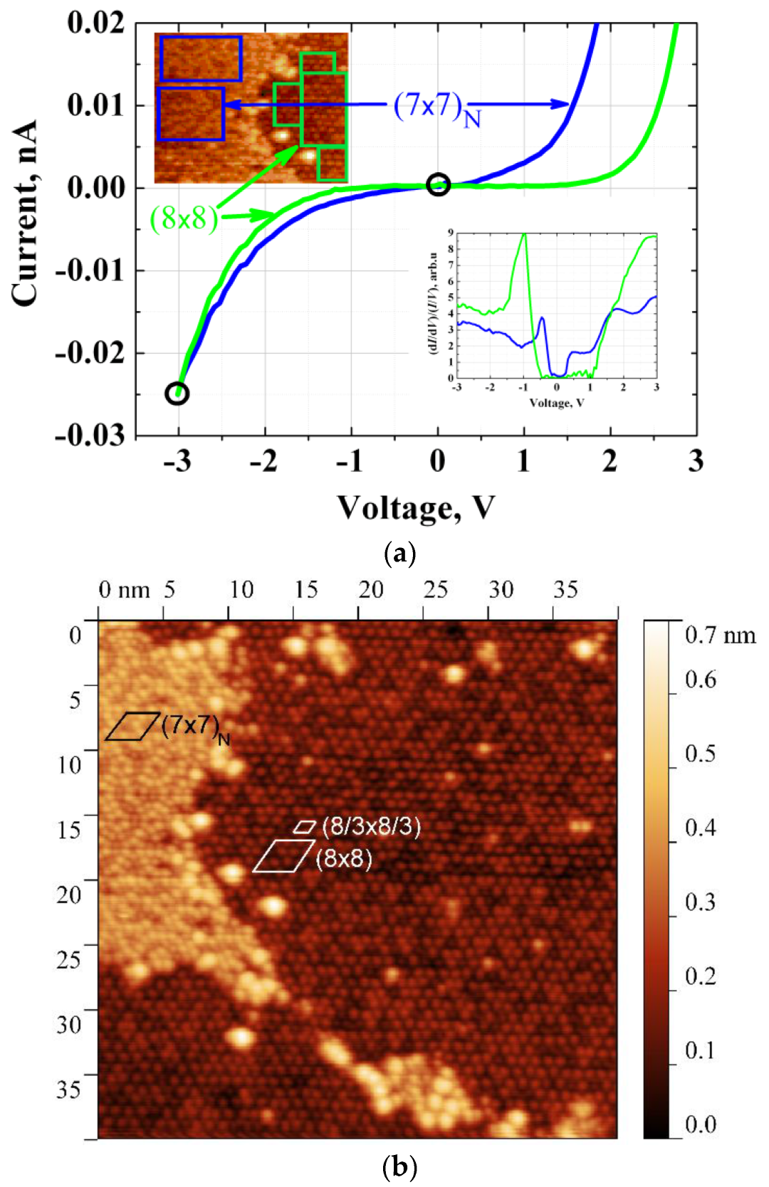
Figure 6a presents the current–voltage (
I–V) characteristics measured in the areas covered by the (7 × 7)
N and (8 × 8) structures. The scanning image was obtained in the current-imaging tunneling spectroscopy (CITS) mode, and a fragment of the image is shown in the inset in
Figure 6a. The operating current at the scan voltage of −3 V was −0.025 nA, the image size was 20 × 20 nm
2 and the number of scan points was 400 × 400.
I–V characteristics were measured at the nodes of the 80 × 80 grid points when the scanning was stopped at each of these points, the probe–sample spacing was fixed and the feedback was turned off. The current was measured while the voltage was varied in the range from −3.0 V to +3.0 V. After that, the operating voltage and current were returned, the feedback was turned on and obtaining the scanning image was continued.
To measure the
I–V characteristics, the typical regions of both structures were selected, as shown in the upper inset of
Figure 6a; as an example, an image with the coexistence of (7 × 7)
N and (8 × 8) structures in higher resolution (scanned at tunneling current 0.025 nA and bias −3 V) with marked (7 × 7)
N, (8 × 8) and (8/3 × 8/3) unit cells is shown in
Figure 6b. The tunnel current was averaged over the region area corresponding to the sum of the selected rectangles on each of the structures. This helps to reduce the noise of the experimental
I–V curves. The “locality degree” of the measurements, in this case, is set by an operator selecting the sites for the analysis. It can be seen in
Figure 6a that the
I–V characteristics measured on the (7 × 7)
N and (8 × 8) structures differ markedly. For the purposes of this work, it is important to know the DOS for these structures. As mentioned above, the DOS is related to the value d
σn = (d
I/d
V)/(
I/
V) measured in the experiment.
It is clear that, for these experimental current–voltage characteristics, the curves necessarily coincide, at least, at two voltage points, regardless of how the local surface composition (and, accordingly, the local density of states) or surface morphology are varied. Namely, there is a match at
V = 0, where the tunneling current is zero (this is the equilibrium point of the probe and the sample), and also at
Vsc—the operating scanning voltage, where the operating tunnel current always coincides owing to the feedback. In this case, the operating point
Vsc can and should be used to normalize the spectra d
σn(
V) in order to ensure a proper determination of the empirical densities of states. The details of the proposed procedure are described below. Meantime, let us note that the attempt to estimate the tunneling current directly by numerical integration in the frames of 1D WKB approximation using the obtained d
σn(
V) curves “as is”, which are shown in the bottom inset of
Figure 6a, results in a failure. The curves shown in the inset were obtained by the numerical differentiation using the free software WSxM program for the STM image analysis [
48]. In fact, these dependences do not even allow one to reproduce the coincidence of currents at the operating point
Vsc for the compared structures.
3.4. Tunnel Current Model
The probe and the sample, as well as the emerging potential barrier in the tunnel gap between it at two voltage values +1.0 V and +2.5 V, are schematically shown in
Figure 7a,b, respectively. Shown are a tungsten probe (left), the sample surface (right) and a tunnel gap between them. The diagram also shows the Fermi level positions of the probe and sample, the potential barrier in the tunnel gap, and the spectra of the density of states for the surface areas with the (7 × 7)
N and (8 × 8) structures. The green and blue colors indicate the energy range in which the tunneling occurs. Here the work functions are denoted as WF
(7×7)N/(8×8) for the sample and WF
W for the tungsten probe (WF—work function).
We describe the tunneling current behavior quantitatively in the framework of the classical model based on the one-dimensional WKB approximation [
17]:
where
A is the normalizing factor,
φ is the average tunnel barrier height at zero voltage and
α = 8m/h
2,
z is the distance from the probe to the surface, or the tunnel gap, and
E is the integration variable that covers the energy range corresponding to the difference between the Fermi levels of the probe and the sample. The function
T(
E,V,z) describes the transmission coefficient of the tunneling barrier, and it was assumed that the barrier has the shape of a trapezoid;
ρs(
E) is the sample density of states. The tungsten probe tip density of states
ρt(
E–V) in some works, for example [
49], is taken into account, but it is usually considered as a smooth slowly changing function that can be replaced by a constant [
17,
50]. In ref. [
50], the density of states is given for a tungsten surface near the Fermi level (Figure 3b in [
50]), which demonstrates the acceptability of the substitution by a constant for the voltage range from −3 V to +3 V. Thus, in the present paper, the probe density of states is assumed to be a constant.
Using Equations (1) and (2), one can compare the calculated contrast with the experiment in two stages: first, an estimate of the expected tunneling currents at a given operating voltage for both structures (7 × 7)N and (8 × 8) coexisting on the surface; then, an evaluation of the contrast change with a varying voltage. First, assume that the structures (7 × 7)N and (8 × 8) are geometrically located on the surface at the same level, i.e., the tunnel gap (z0) remains unchanged during the scanning. Then, at a given voltage value Vsc, the function describing the transmission coefficient becomes a function of the only variable E; that is, T(E,eVsc,z0) = f(E). Changes in the tunneling current in the selected surface areas are determined by the integral (over the E variable) of the product of the transmission function and the local DOS T(E) × ρs(E) within the integration range from 0 to the operating voltage V.
The density of states in the system under study is proposed to be determined based on the experimental dependence of the differential conductivity dσn = (dI/dV)/(I/V) on voltage. This allows determining the density of states originating from the entire system participating in the tunneling, i.e., from all layers arising during the nitridation process on the conducting Si(111) substrate surface.
For the one-dimensional WKB approximation, the formula describing the relation between the density of states and the logarithmic derivative of conductivity, which is presented in the work of Feenstra et al. [
17] (see Equation (3)), can be rewritten as follows:
where
and
It is fair to say that the relation between
ρs(
E) and d
σn(
E) becomes a simple linear function if we assume that the parameters
a and
b are the numerical coefficients independent of voltage
V. An even simpler relation, namely the proportionality of
ρs(
E)~d
σn(
E) is usually assumed, but in the general case, as can be seen from the above equations, this is not necessarily true. The constants
a and
b can be chosen in such a way that, after the numerical integration of the product
T(
E) ×
ρs(
E) to calculate the tunnel current, two conditions are satisfied. The first condition is to obtain the same tunnel current for both structures (7 × 7)
N and (8 × 8) at the operating point at −3 V, as shown in
Figure 6a. The second condition is to reach the contrast values or the tunnel gap (Δ
z) variation for the (7 × 7)
N and (8 × 8) structures at different scanning voltages to be close to the experiment. In this case, the spectrum d
σn(
E) for the (8 × 8) structure will be corrected using a linear transformation, and the spectrum of the (7 × 7)
N structure will be assumed to be the reference one. Then both spectra are normalized at the operating point to match the experimental operating current.
3.5. Correction Results of the Differential Conductivity Spectrum and Tunneling Current Estimation
The densities of states and the integrand of Equation (1), that is, of the product
T(
E) ×
ρs(
E), for (7 × 7)
N and (8 × 8) structures as a function of energy are shown in
Figure 8a. The curves in
Figure 8b were calculated with the following parameters for the transmission coefficient
T(
E):
z0 = 15 Å,
φ = 4.5 eV and
V = −3 V. In this work, to correct the (8 × 8) structure spectrum, the coefficients
a = 0.565 and
b = 0.4 were found; as is shown further, these coefficients satisfy the two formulated criteria.
A comparison of the density of states for the (8 × 8) structure, obtained empirically in this work, and the DOS calculated in [
42] is shown in
Figure 8c. In both cases, it is assumed that the atomic structure of this array is based on a two-dimensional graphene-like g-Si
3N
3 layer. To compare the total DOS, we summarized the partial DOS given in [
42].
It is clear in
Figure 8c that the calculated and empirical DOS values are not identical, although there is a certain similarity in the spectra. The difference manifested itself despite the assumption that we are dealing with the same basis for the atomic structures of (8 × 8) and graphene-like g-Si
3N
3. Anyway, based on the presented graphs, it can be argued that the experimental and calculated bandgaps agree quite well, taking a value of about 2.3 eV. According to this fact and strong discrepancy with the expected β-Si
3N
4 bandgap of 5.3 eV, the graphene-like nature of the (8 × 8) structure became clearer, as follows from the better agreement between the experimental spectrum and the calculation of Guo at al. [
42]. In contrast, if one sticks to the generally accepted model of the (8 × 8) structure as a thin β-Si
3N
4 layer, then one would expect an increase in the bandgap, in comparison with the bulk value of silicon nitride (>5.3 eV), due to the quantum confinement effect, as was shown, for example, in [
51,
52,
53]. In turn, there may be several reasons for the detected discrepancies between the ab initio calculation of g-Si
3N
3 and the experimental spectra in
Figure 8c. One of the main reasons, in our opinion, is as follows: the authors [
42] considered the perfect isolated (not interacting with anything) two-dimensional layer when calculating from the first principles, and in our case, a similar layer (but, of course, nonperfect) is located on the silicon surface; moreover, the g-Si
3N
3 graphene-like layer is covered by an adsorption silicon layer [
41]. In other words, in the STM experiment, a more complex nonperfect structure is studied, and there is the graphene-like g-Si
3N
3 layer interaction with the silicon substrate and the adsorption layer. Basically, between the layers, there should be van der Waals interactions (if some kinds of defects are neglected), although they are supposed to be weak, distorting the spectrum of an isolated g-Si
3N
3 layer. It is also possible that there is a certain “mixing” of the states of all these layers as was mentioned in the model considered in [
33]. In addition, the states of the silicon substrate and the adsorption layer, immediately, in accordance with its geometric arrangement relative to the probe, are also more or less involved in the tunneling current formation. Another significant reason for the discrepancy may be related to the finite energy and spatial resolution of the scanning microscope, which usually reduces the amplitude and broadens the initially narrow peaks in the perfect spectra. It should be also noted that, in ab initio calculations, in one or another approximation and according to the chosen basis, a certain underestimation of the density of states in the region of unoccupied electronic states, that is, for the states lying above the Fermi level, may appear.
The changes in the expected tunneling current for the (7 × 7)
N and (8 × 8) structures obtained by the integration of
T(
E) ×
ρs(
E), based on the corrected DOS of the (8 × 8) structure, as well as keeping the function
T(
E,eV,z) without variations, are shown in
Figure 9.
To avoid possible ambiguities, it is necessary to make the following clarification: The calculated current values shown in
Figure 9 do not correspond to the attempt at an accurate reproduction of the experimental
I–V characteristics (how it might seem from the comparison with
Figure 6a), even despite the coincidence of currents for both structures at the operating point of −3 V, for which the correction of the differential conductivity spectra was actually carried out. The meaning of the points presented in
Figure 9 is estimating the hypothetically possible change in current Δ
I, while the probe is laterally displaced from the structure (7 × 7)
N to (8 × 8) (or in the opposite direction) during the scanning at the fixed tunnel gap for the selected operating voltage. In this case, the current can be changed only due to the difference in the density of states of these structures. The empirical
I–V characteristics of the structures shown in
Figure 6a were actually measured at different tunnel gaps. Let us recall one more time that a change in the tunnel current usually does not occur during the real scanning of the surface at selected operating conditions (i.e., at selected voltage and current values), since the scanning is carried out with the feedback on the tunnel current.
3.6. Estimation of the Vertical Probe Offset Compensating the ΔI Value. Current Feedback Simulation
The necessary vertical displacement of the probe (offset) to compensate for the expected current changes during the lateral displacement from the surface region with the (7 × 7)
N structure to the region with the (8 × 8) structure is estimated: that is, this time, we are going to vary the only value of tunnel gap
z which will change only the function
T(
E,
eV,
z), keeping the corrected DOS value without any variations, to reach the equalization of the tunnel currents in the areas for both structures. To be more precise, we determine the required vertical probe offset on the (8 × 8) structure to return to the same current that was calculated on the (7 × 7)
N structure at the given bias. In other words, one needs to eliminate the Δ
I value calculated above by means of variation of
T(
E,eV,z), i.e.,
z0 is replaced by
z0 + Δ
z when calculating this function, and it is necessary to determine a suitable Δ
z. All other parameters (except for variable Δ
z) used to calculate the function
T(
E,
eV,z) were presented above and were kept constant, and suitable normalization parameters for DOS were found (as presented above). A comparison of the experimental data and calculation results for Δ
z at various biases is shown in
Figure 10.
The black solid dots and the dotted line show the experimental contrast versus voltage (see
Figure 5e), and the red blank dots present the estimation of the necessary probe offset Δ
z to compensate for the expected change in the current. It can be seen that as the voltage across the tunnel gap decreases, the required vertical probe offset increases, which corresponds qualitatively and quantitatively to the empirical behavior of the contrast versus voltage.
Thus, it is proposed to use the procedure for correcting the STS spectra of differential conductivity to match the behavior of the STM image contrast, as a function of bias, which helps to solve the problem of quantitative determination of DOS by the STM/STS technique. In this work, the (7 × 7)
N structure state spectrum was assumed to be known, and the correction of the (8 × 8) structure spectrum, according to the contrast behavior with the bias variation, turned out to be quite effective, since it made it possible to reproduce the empirical behavior of the contrast at the boundary between these two structures in the calculations. Then, the result shown in
Figure 10 allows us to put forward a more general speculation. The proposed procedure for correcting the STS spectra d
σn(
V) = (d
I/d
V)/(
I/
V) can be actually useful for the STM studies of systems comprising surface boundaries between regions with a well-known DOS value and regions covered by poorly known structures or materials, the DOS values of which should be determined. The empirical determination of the differential conductance spectrum of this material from the STS measurements and the simple linear adjustment of this spectrum makes it possible to quantify the DOS. This approach is applicable to the study of a wide range of materials, both conductive and dielectric ones. In addition, this approach allows distinguishing the contrast associated with the density of states from the topographical contrast in STM images and a more adequate determination of the height of topographic surface features.


