Wet Chemical Synthesis and Characterization of Au Coatings on Meso- and Macroporous Si for Molecular Analysis by SERS Spectroscopy
Abstract
1. Introduction
2. Materials and Methods
2.1. PS Substrate Fabrication
2.2. Gold Deposition and Samples’ Characterization
3. Results and Discussion
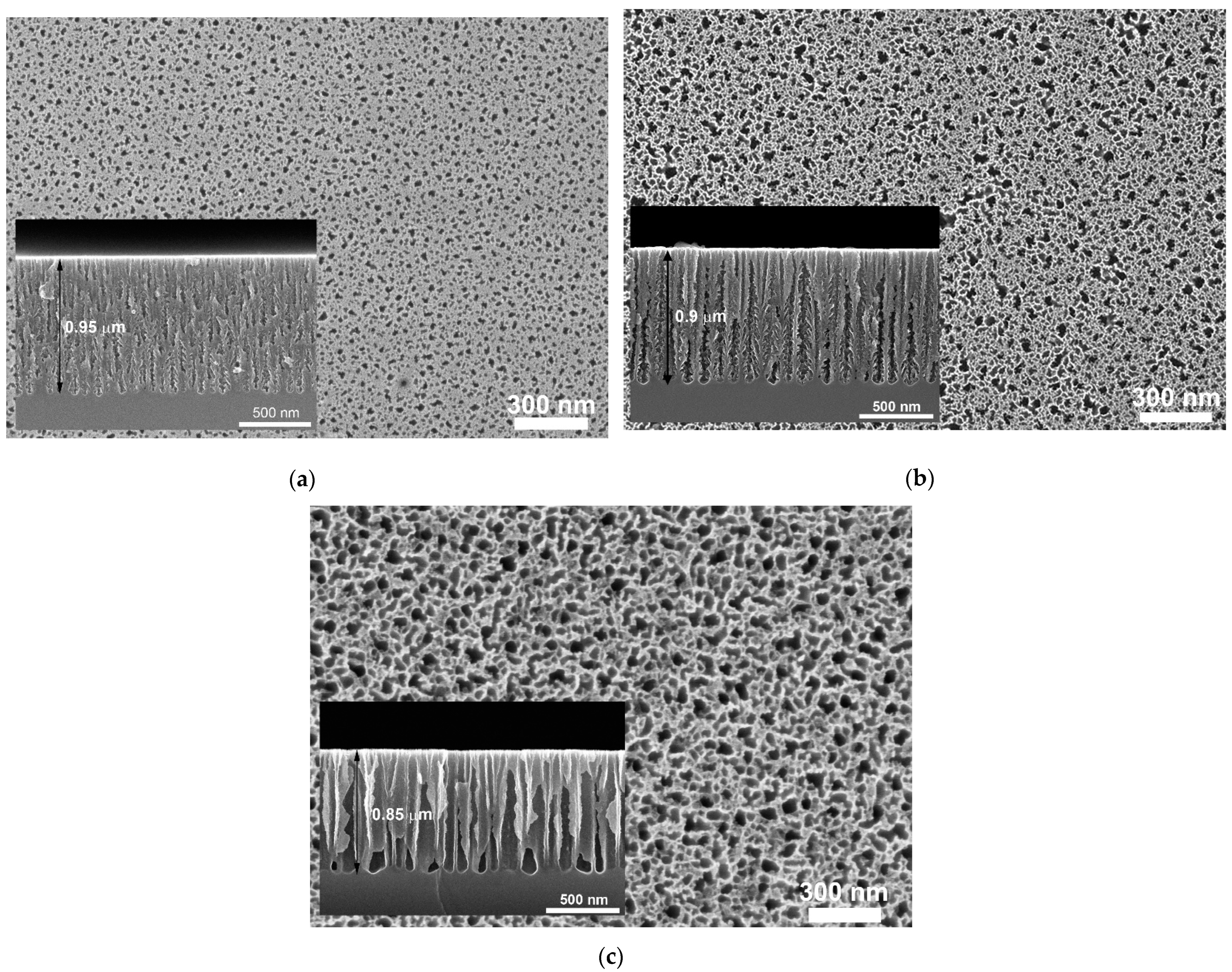
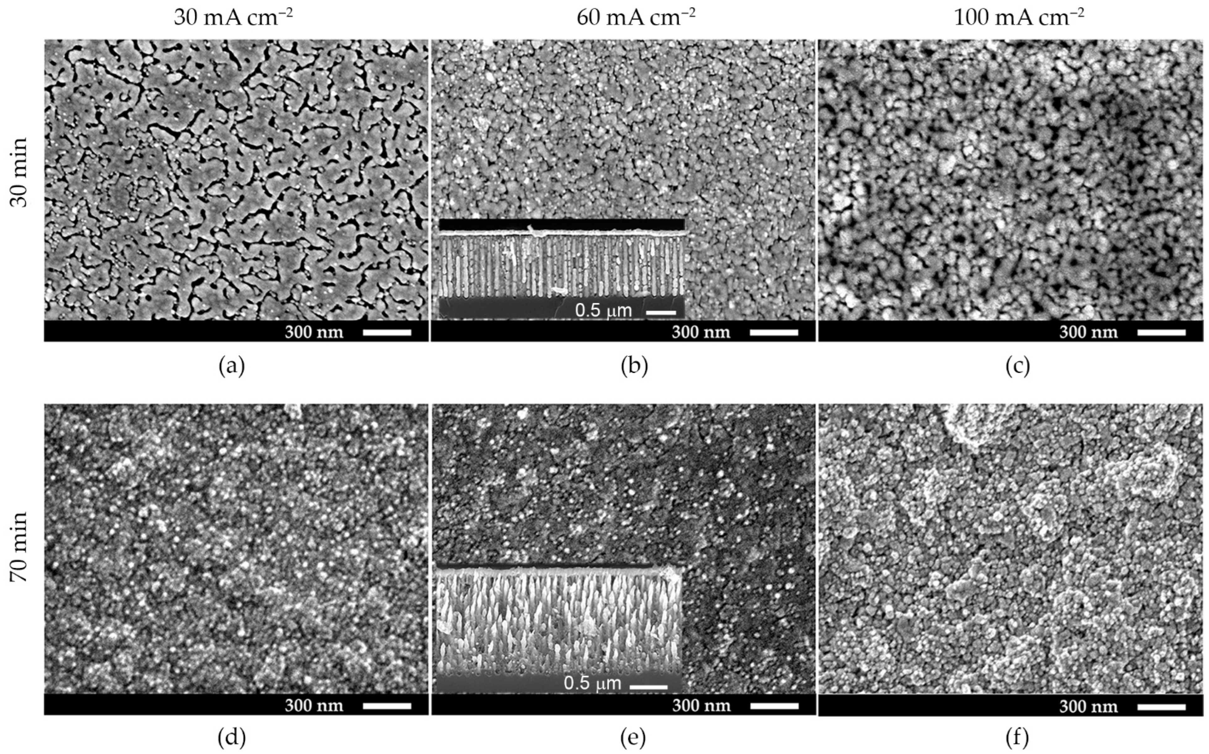
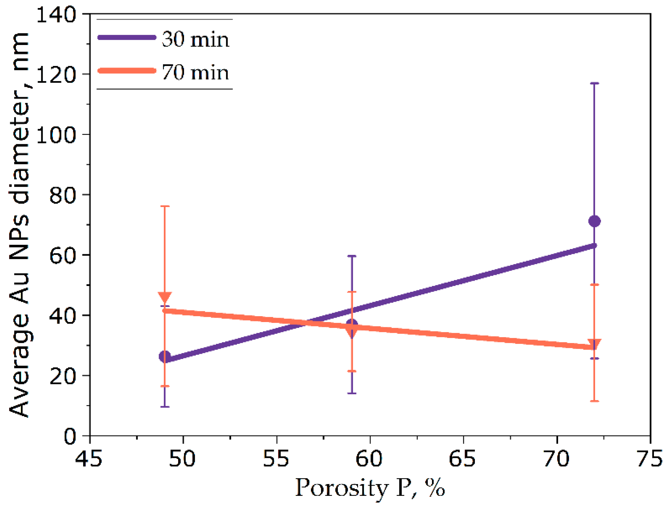
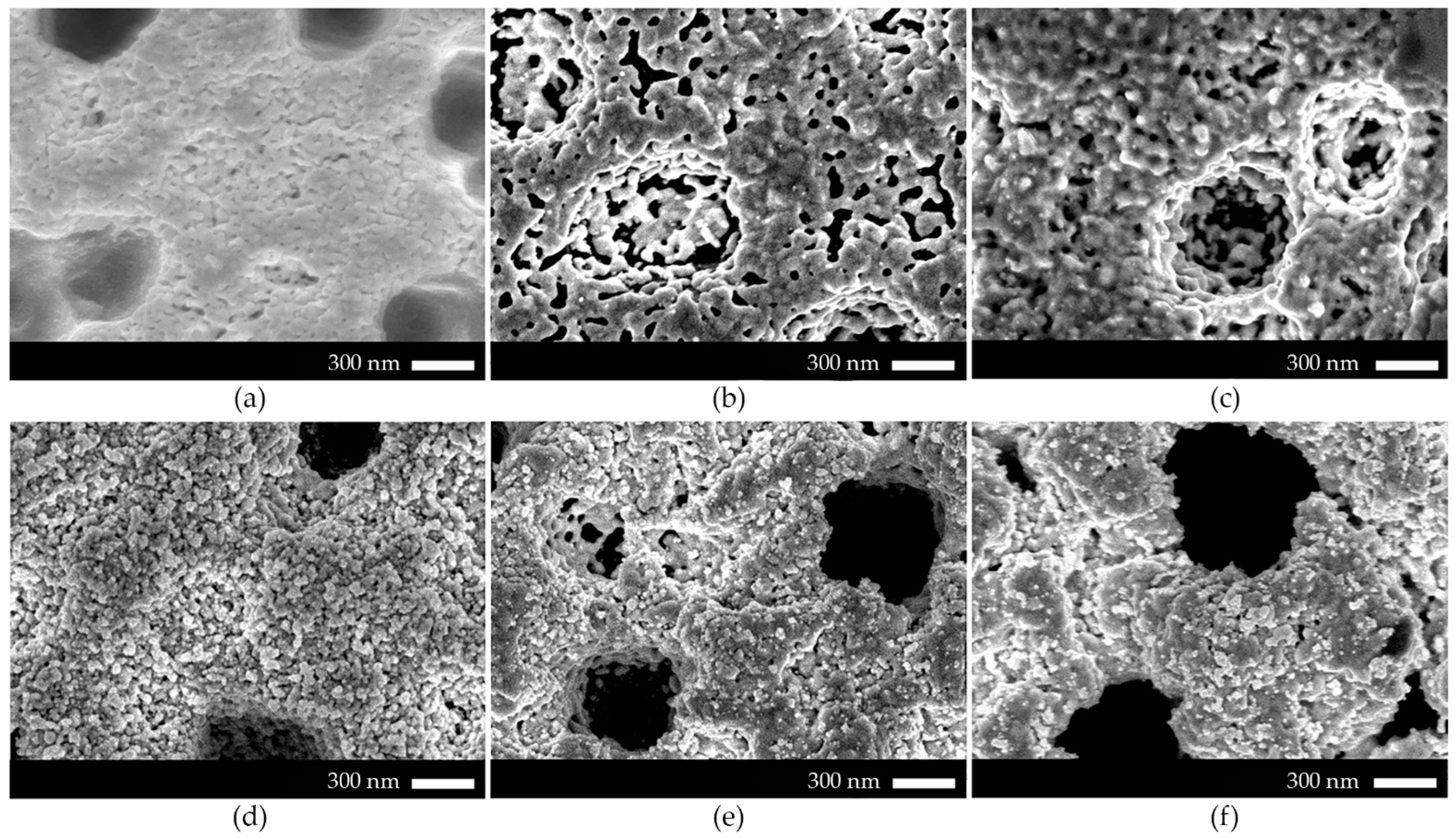
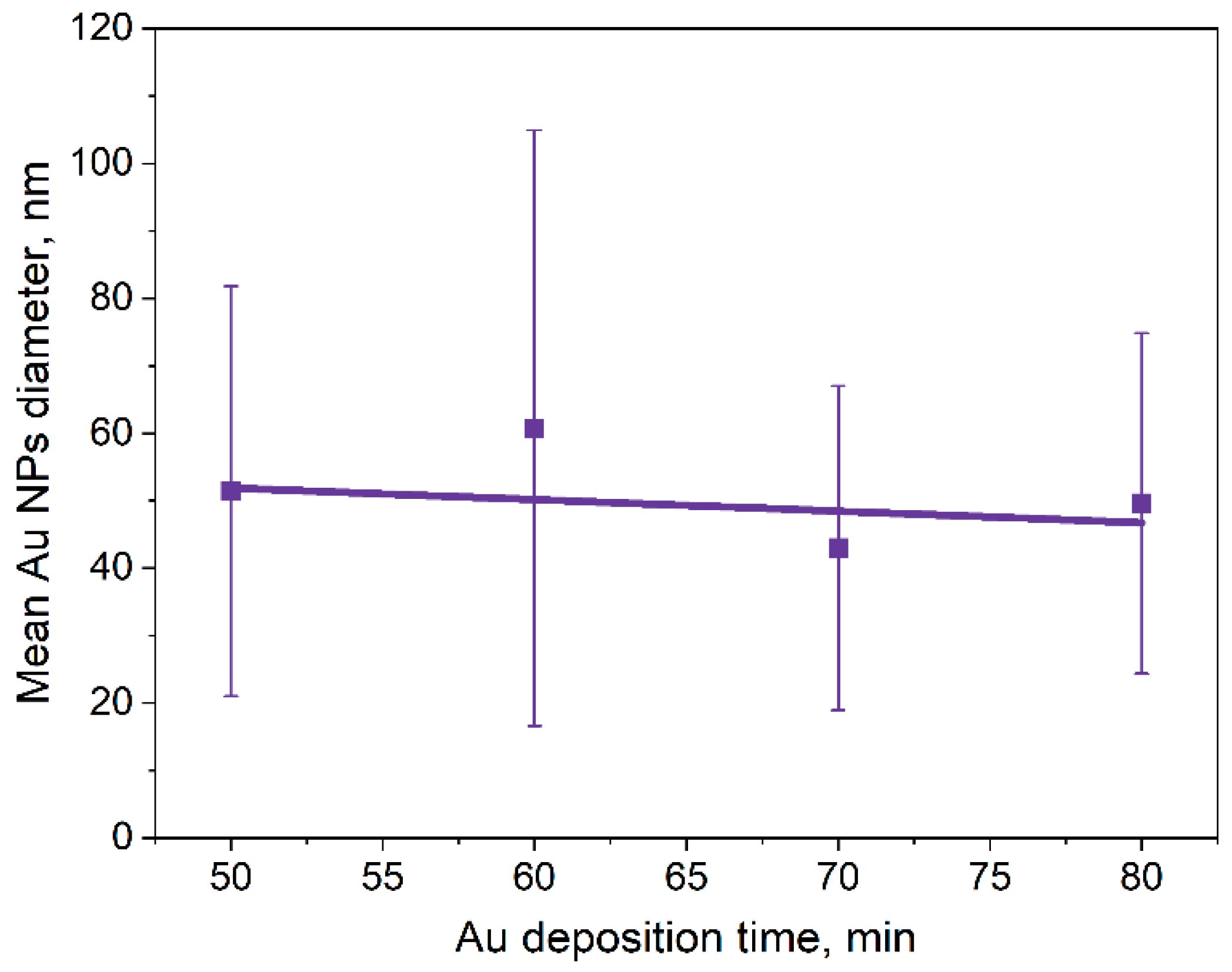
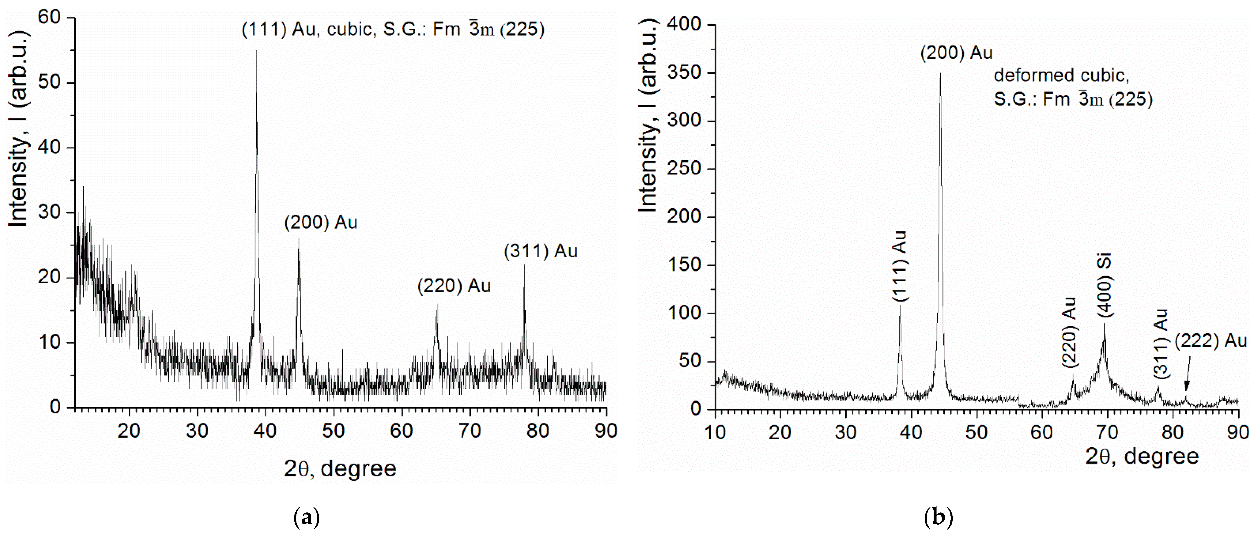
3.1. Morphology of the Au-Coated Meso-PS Substrates
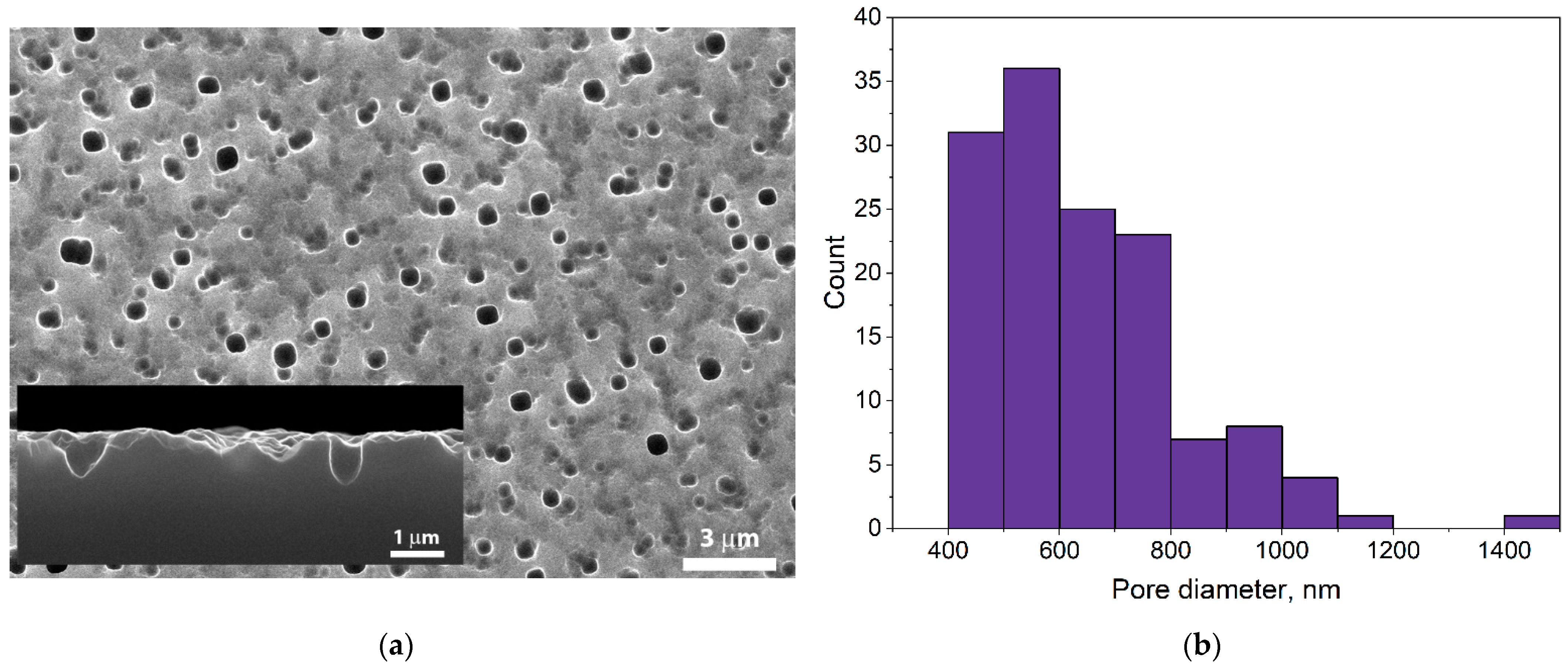
3.2. Morphology of the Au-Coated Macro-PS Substrates
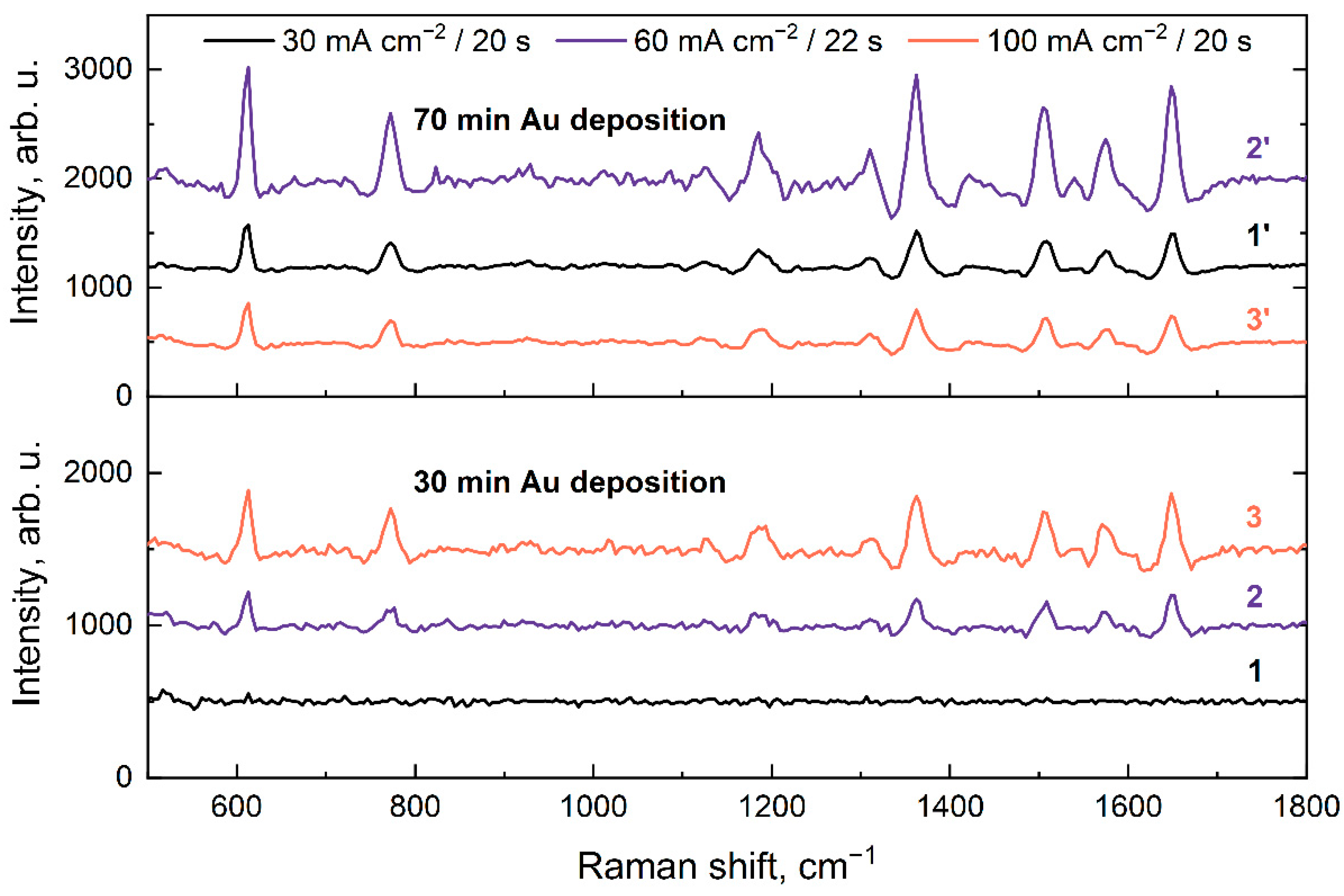
3.3. UV-Vis and SERS Measurements
4. Conclusions
Author Contributions
Funding
Data Availability Statement
Acknowledgments
Conflicts of Interest
References
- Robertus, J. Principles of Protein X-Ray Crystallography, 3rd Edition By Jan Drenth (University of Groningen, The Netherlands). With a Major Contribution from Jeroen Mester (University of Lübeck, Germany). Springer Science + Business Media LLC: New York. 2007. Xiv + 3. J. Am. Chem. Soc. 2007, 129, 5782–5783. [Google Scholar] [CrossRef]
- Bax, A.D.; Grzesiek, S. Methodological Advances in Protein NMR. Acc. of Chem. Res. 1993, 26, 131–138. [Google Scholar] [CrossRef]
- Cavanagh, J.W.; Fairbrother, A.; Palmer, N., III; Skelton, M.R. Protein NMR Spectroscopy: Principles and Practice, 2nd ed.; Elsevier: Amsterdam, The Netherlands, 2007. [Google Scholar]
- Kelly, S.M.; Jess, T.J.; Price, N.C. How to Study Proteins by Circular Dichroism. Biochim. Biophys. Acta (BBA)-Proteins Proteom. 2005, 1751, 119–139. [Google Scholar] [CrossRef]
- Whitmore, L.; Miles, A.J.; Mavridis, L.; Janes, R.W.; Wallace, B.A. PCDDB: New Developments at the Protein Circular Dichroism Data Bank. Nucleic Acids Res. 2017, 45, D303–D307. [Google Scholar] [CrossRef] [PubMed]
- Amendola, V.; Pilot, R.; Frasconi, M.; Maragò, O.M.; Iatì, M.A. Surface Plasmon Resonance in Gold Nanoparticles: A Review. J. Phys. Condens. Matter 2017, 29, 203002. [Google Scholar] [CrossRef]
- Lane, L.A.; Qian, X.; Nie, S. SERS Nanoparticles in Medicine: From Label-Free Detection to Spectroscopic Tagging. Chem. Rev. 2015, 115, 10489–10529. [Google Scholar] [CrossRef]
- Kneipp, J.; Kneipp, H.; Kneipp, K. SERS—A Single-Molecule and Nanoscale Tool for Bioanalytics. Chem. Soc. Rev. 2008, 37, 1052–1060. [Google Scholar] [CrossRef]
- Zavatski, S.; Khinevich, N.; Girel, K.; Redko, S.; Kovalchuk, N.; Komissarov, I.; Lukashevich, V.; Semak, I.; Mamatkulov, K.; Vorobyeva, M.; et al. Surface Enhanced Raman Spectroscopy of Lactoferrin Adsorbed on Silvered Porous Silicon Covered with Graphene. Biosensors 2019, 9, 34. [Google Scholar] [CrossRef]
- Xie, X.; Pu, H.; Sun, D.W. Recent Advances in Nanofabrication Techniques for SERS Substrates and Their Applications in Food Safety Analysis. Crit. Rev. Food Sci. Nutr. 2018, 58, 2800–2813. [Google Scholar] [CrossRef]
- Burtsev, V.; Erzina, M.; Guselnikova, O.; Miliutina, E.; Kalachyova, Y.; Svorcik, V.; Lyutakov, O. Detection of Trace Amounts of Insoluble Pharmaceuticals in Water by Extraction and SERS Measurements in a Microfluidic Flow Regime. Analyst 2021, 146, 3686–3696. [Google Scholar] [CrossRef]
- Cailletaud, J.; De Bleye, C.; Dumont, E.; Sacré, P.Y.; Netchacovitch, L.; Gut, Y.; Boiret, M.; Ginot, Y.M.; Hubert, P.; Ziemons, E. Critical Review of Surface-Enhanced Raman Spectroscopy Applications in the Pharmaceutical Field. J. Pharm. Biomed. Anal. 2018, 147, 458–472. [Google Scholar] [CrossRef] [PubMed]
- Khinevich, N.; Zavatski, S.; Bandarenka, H.; Belyatsky, V.; Galyuk, E.; Ryneiskaya, O. Study of Diluted Meldonium Solutions by Surface Enhanced Raman Scattering Spectroscopy. Int. J. Nanosci. 2019, 18, 1940054. [Google Scholar] [CrossRef]
- Khrustalev, V.V.; Khrustaleva, T.A.; Kahanouskaya, E.Y.; Rudnichenko, Y.A.; Bandarenka, H.V.; Arutyunyan, A.M.; Girel, K.V.; Khinevich, N.V.; Ksenofontov, A.L.; Kordyukova, L.V. The Alpha Helix 1 from the First Conserved Region of HIV1 Gp120 Is Reconstructed in the Short NQ21 Peptide. Arch. Biochem. Biophys. 2018, 638, 66–75. [Google Scholar] [CrossRef]
- Kneipp, K.; Kneipp, H.; Bohr, H.G. Single-Molecule SERS Spectroscopy. In Surface-Enhanced Raman Scatt; Springer: Berlin/Heidelberg, Germany, 2006; pp. 261–277. [Google Scholar] [CrossRef]
- Nie, S.; Emory, S.R. Probing Single Molecules and Single Nanoparticles by Surface-Enhanced Raman Scattering. Science 1997, 275, 1102–1106. [Google Scholar] [CrossRef] [PubMed]
- Stiles, P.L.; Dieringer, J.A.; Shah, N.C.; Van Duyne, R.P. Surface-Enhanced Raman Spectroscopy. Annu. Rev. Anal. Chem. 2008, 1, 601–626. [Google Scholar] [CrossRef] [PubMed]
- Langer, J.; Jimenez de Aberasturi, D.; Aizpurua, J.; Alvarez-Puebla, R.A.; Auguié, B.; Baumberg, J.J.; Bazan, G.C.; Bell, S.E.J.; Boisen, A.; Brolo, A.G. Present and Future of Surface-Enhanced Raman Scattering. ACS Nano 2019, 14, 28–117. [Google Scholar] [CrossRef] [PubMed]
- Ding, S.-Y.; Yi, J.; Li, J.-F.; Ren, B.; Wu, D.-Y.; Panneerselvam, R.; Tian, Z.-Q. Nanostructure-Based Plasmon-Enhanced Raman Spectroscopy for Surface Analysis of Materials. Nat. Rev. Mater. 2016, 1, 16021. [Google Scholar] [CrossRef]
- Natan, M.J. Concluding Remarks Surface Enhanced Raman Scattering. Faraday Discuss. 2006, 132, 321–328. [Google Scholar] [CrossRef]
- Corni, S.; Tomasi, J. Surface Enhanced Raman Scattering from a Single Molecule Adsorbed on a Metal Particle Aggregate: A Theoretical Study. J. Chem. Phys. 2002, 116, 1156–1164. [Google Scholar] [CrossRef]
- Mosier-Boss, P.A. Review of SERS Substrates for Chemical Sensing. Nanomaterials 2017, 7, 142. [Google Scholar] [CrossRef]
- Poston, P.E.; Harris, J.M. Stable, Dispersible Surface-Enhanced Raman Scattering Substrate Capable of Detecting Molecules Bound to Silica-Immobilized Ligands. Appl. Spectrosc. 2010, 64, 1238–1243. [Google Scholar] [CrossRef] [PubMed]
- Feng, L.; Xu, Y.-L.; Fegadolli, W.S.; Lu, M.-H.; Oliveira, J.E.B.; Almeida, V.R.; Chen, Y.-F.; Scherer, A. Experimental Demonstration of a Unidirectional Reflectionless Parity-Time Metamaterial at Optical Frequencies. Nat. Mater. 2013, 12, 108–113. [Google Scholar] [CrossRef]
- Dubkov, S.V.; Savitskiy, A.I.; Trifonov, A.Y.; Yeritsyan, G.S.; Shaman, Y.P.; Kitsyuk, E.P.; Tarasov, A.; Shtyka, O.; Ciesielski, R.; Gromov, D.G. SERS in Red Spectrum Region through Array of Ag–Cu Composite Nanoparticles Formed by Vacuum-Thermal Evaporation. Opt. Mater. X 2020, 7, 100055. [Google Scholar] [CrossRef]
- Kneipp, K.; Kneipp, H.; Manoharan, R.; Hanlon, E.B.; Itzkan, I.; Dasari, R.R.; Feld, M.S. Extremely Large Enhancement Factors in Surface-Enhanced Raman Scattering for Molecules on Colloidal Gold Clusters. Appl. Spectrosc. 1998, 52, 1493–1497. [Google Scholar] [CrossRef]
- Jeon, T.Y.; Jeon, H.C.; Yang, S.-M.; Kim, S.-H. Hierarchical Nanostructures Created by Interference of High-Order Diffraction Beams. J. Mater. Chem. C 2016, 4, 1088–1095. [Google Scholar] [CrossRef]
- Radu, A.I.; Ryabchykov, O.; Bocklitz, T.W.; Huebner, U.; Weber, K.; Cialla-May, D.; Popp, J. Toward Food Analytics: Fast Estimation of Lycopene and β-Carotene Content in Tomatoes Based on Surface Enhanced Raman Spectroscopy (SERS). Analyst 2016, 141, 4447–4455. [Google Scholar] [CrossRef]
- Kim, S.; Zhang, W.; Cunningham, B.T. Coupling Discrete Metal Nanoparticles to Photonic Crystal Surface Resonant Modes and Application to Raman Spectroscopy. Opt. Express 2010, 18, 4300–4309. [Google Scholar] [CrossRef]
- Gopalakrishnan, A.; Chirumamilla, M.; De Angelis, F.; Toma, A.; Zaccaria, R.P.; Krahne, R. Bimetallic 3D Nanostar Dimers in Ring Cavities: Recyclable and Robust Surface-Enhanced Raman Scattering Substrates for Signal Detection from Few Molecules. ACS Nano 2014, 8, 7986–7994. [Google Scholar] [CrossRef]
- Yao, X.; Jiang, S.; Luo, S.; Liu, B.-W.; Huang, T.-X.; Hu, S.; Zhu, J.; Wang, X.; Ren, B. Uniform Periodic Bowtie SERS Substrate with Narrow Nanogaps Obtained by Monitored Pulsed Electrodeposition. ACS Appl. Mater. Interfaces 2020, 12, 36505–36512. [Google Scholar] [CrossRef]
- Lazarouk, S.; Bondarenko, V.; Pershukevich, P.; La Monica, S.; Maiello, G.; Ferrari, A. Visible Electroluminescence from Al-Porous Silicon Reverse Bias Diodes Formed on the Base of Degenerate N-Type Silicon. MRS Online Proc. Libr. 1994, 358, 659. [Google Scholar] [CrossRef]
- Bandarenka, H.; Redko, S.; Smirnov, A.; Panarin, A.; Terekhov, S.; Nenzi, P.; Balucani, M.; Bondarenko, V. Nanostructures Formed by Displacement of Porous Silicon with Copper: From Nanoparticles to Porous Membranes. Nanoscale Res. Lett. 2012, 7, 477. [Google Scholar] [CrossRef] [PubMed]
- Bandarenka, H.V.; Khinevich, N.V.; Burko, A.A.; Redko, S.V.; Zavatski, S.A.; Shapel, U.A.; Mamatkulov, K.Z.; Vorobyeva, M.Y.; Arzumanyan, G.M. 3D Silver Dendrites for Single-Molecule Imaging by Surface-Enhanced Raman Spectroscopy. ChemNanoMat 2021, 7, 141–149. [Google Scholar] [CrossRef]
- Korotcenkov, G. Porous Silicon: From Formation to Application: Biomedical and Sensor Applications; CRC Press: Boca Raton, FL, USA, 2016; Volume 2, ISBN 1482264579. [Google Scholar]
- Bandarenka, H.V.; Girel, K.V.; Bondarenko, V.P.; Khodasevich, I.A.; Panarin, A.Y.; Terekhov, S.N. Formation Regularities of Plasmonic Silver Nanostructures on Porous Silicon for Effective Surface-Enhanced Raman Scattering. Nanoscale Res. Lett. 2016, 11, 262. [Google Scholar] [CrossRef] [PubMed]
- Khajehpour, K.J.; Williams, T.; Bourgeois, L.; Adeloju, S. Gold Nanothorns–Macroporous Silicon Hybrid Structure: A Simple and Ultrasensitive Platform for SERS. Chem. Commun. 2012, 48, 5349–5351. [Google Scholar] [CrossRef] [PubMed]
- Bandarenka, H.; Dolgiy, A.; Chubenko, E.; Redko, S.; Girel, K.; Prischepa, S.L.; Panarin, A.; Terekhov, S.; Pilipenko, V.; Bondarenko, V. Nanostructured Metal Films Formed onto Porous Silicon Template. J. Nano Res. Trans. Tech. Publ. 2016, 39, 235–255. [Google Scholar] [CrossRef]
- Khinevich, N.; Bandarenka, H.; Zavatski, S.; Girel, K.; Tamulevičienė, A.; Tamulevičius, T.; Tamulevičius, S. Porous Silicon—A Versatile Platform for Mass-Production of Ultrasensitive SERS-Active Substrates. Microporous Mesoporous Mater. 2021, 323, 111204. [Google Scholar] [CrossRef]
- Bandarenka, H.V.; Girel, K.V.; Zavatski, S.A.; Panarin, A.; Terekhov, S.N. Progress in the Development of SERS-Active Substrates Based on Metal-Coated Porous Silicon. Materials. 2018, 11, 852. [Google Scholar] [CrossRef]
- Chernousova, S.; Epple, M. Silver as Antibacterial Agent: Ion, Nanoparticle, and Metal. Angew. Chem.-Int. Ed. 2013, 52, 1636–1653. [Google Scholar] [CrossRef]
- Franci, G.; Falanga, A.; Galdiero, S.; Palomba, L.; Rai, M.; Morelli, G.; Galdiero, M. Silver Nanoparticles as Potential Antibacterial Agents. Molecules 2015, 20, 8856–8874. [Google Scholar] [CrossRef]
- Dolgiy, A.; Redko, S.V.; Bandarenka, H.; Prischepa, S.L.; Yanushkevich, K.; Nenzi, P.; Balucani, M.; Bondarenko, V. Electrochemical Deposition and Characterization of Ni in Mesoporous Silicon. J. Electrochem. Soc. 2012, 159, D623. [Google Scholar] [CrossRef]
- Magagnin, L.; Maboudian, R.; Carraro, C. Gold Deposition by Galvanic Displacement on Semiconductor Surfaces: Effect of Substrate on Adhesion. J. Phys. Chem. B 2002, 106, 401–407. [Google Scholar] [CrossRef]
- Kong, L.; Dasgupta, B.; Ren, Y.; Mohseni, P.K.; Hong, M.; Li, X.; Chim, W.K.; Chiam, S.Y. Evidences for Redox Reaction Driven Charge Transfer and Mass Transport in Metal-Assisted Chemical Etching of Silicon. Sci. Rep. 2016, 6, 36582. [Google Scholar] [CrossRef]
- Wang, C.H.; Sun, D.C.; Xia, X.H. One-Step Formation of Nanostructured Gold Layers via a Galvanic Exchange Reaction for Surface Enhancement Raman Scattering. Nanotechnology 2006, 17, 651–657. [Google Scholar] [CrossRef]
- Grevtsov, N.; Burko, A.; Redko, S.; Khinevich, N.; Zavatski, S.; Niauzorau, S.; Bandarenka, H. Silicon Nanowire Arrays Coated with Ag and Au Dendrites for Surface-Enhanced Raman Scattering. MRS Adv. 2020, 5, 2023–2032. [Google Scholar] [CrossRef]
- Merkus, H.G. Particle Size Measurements, 1st ed.; Springer: Dordrecht, The Netherlands, 2009; ISBN 1402090161. [Google Scholar]
- Herino, R.; Bomchil, G.; Barla, K.; Bertrand, C.; Ginoux, J.L. Porosity and Pore Size Distributions of Porous Silicon Layers. J. Electrochem. Soc. 1987, 134, 1994. [Google Scholar] [CrossRef]
- Smith, R.L.; Collins, S.D. Porous Silicon Formation Mechanisms. J. Appl. Phys. 1992, 71, R1–R22. [Google Scholar] [CrossRef]
- Korotcenkov, G. Porous Silicon: From Formation to Application: Formation and Properties; CRC Press: Boca Raton, FL, USA, 2016; Volume 1, ISBN 1482264552. [Google Scholar]
- Nativ-Roth, E.; Rechav, K.; Porat, Z. Deposition of Gold and Silver on Porous Silicon and inside the Pores. Thin Solid Films 2016, 603, 88–96. [Google Scholar] [CrossRef]
- Panarin, A.Y.; Terekhov, S.N.; Kholostov, K.I.; Bondarenko, V.P. SERS-Active Substrates Based on n-Type Porous Silicon. Appl. Surf. Sci. 2010, 256, 6969–6976. [Google Scholar] [CrossRef]
- Sing, K.S.W.; Everett, D.H.; Haul, R.A.W.; Moscou, L.; Pierotti, R.A.; Rouquerol, J.; Siemieniewska, T. Reporting Physisorption Data for Gas/Solid Systems with Special Reference to the Determination of Surface Area and Porosity (Recommendations 1984). Pure Appl. Chem. 1985, 57, 603–619. [Google Scholar] [CrossRef]
- Venables, J. Introduction to Surface and Thin Film Processes; Cambridge University Press: Cambridge, UK, 2000; ISBN 0521785006. [Google Scholar]
- Pimpinelli, A.; Villain, J. Physics of Crystal Growth; Cambridge University Press: Cambridge, UK, 1998; ISBN 0521551986. [Google Scholar]
- Oura, K.; Lifshits, V.G.; Saranin, A.A.; Zotov, A.V.; Katayama, M. Surface Science: An Introduction; Springer: Berlin/Heidelberg, Germany, 2013; ISBN 3662051796. [Google Scholar]
- Ponomarev, E.A.; Lévy-Clément, C. Macropore Formation on P-Type Si in Fluoride Containing Organic Electrolytes. Electrochem. Solid State Lett. 1998, 1, 42. [Google Scholar] [CrossRef]
- Wehrspohn, R.B.; Chazalviel, J.; Ozanam, F. Macropore Formation in Highly Resistive P-Type Crystalline Silicon. J. Electrochem. Soc. 1998, 145, 2958. [Google Scholar] [CrossRef]
- Cattarin, S.; Pantano, E.; Decker, F. Investigation by Electrochemical and Deflectometric Techniques of Silicon Dissolution and Passivation in Alkali. Electrochem. Commun. 1999, 1, 483–487. [Google Scholar] [CrossRef]
- Cole, R.M.; Mahajan, S.; Bartlett, P.N.; Baumberg, J.J. Engineering SERS via Absorption Control in Novel Hybrid Ni/Au Nanovoids. Opt. Express 2009, 17, 13298–13308. [Google Scholar] [CrossRef] [PubMed]
- Kelf, T.A.; Sugawara, Y.; Cole, R.M.; Baumberg, J.J.; Abdelsalam, M.E.; Cintra, S.; Mahajan, S.; Russell, A.E.; Bartlett, P.N. Localized and Delocalized Plasmons in Metallic Nanovoids. Phys. Rev. B 2006, 74, 245415. [Google Scholar] [CrossRef]
- Haiss, W.; Thanh, N.T.K.; Aveyard, J.; Fernig, D.G. Determination of Size and Concentration of Gold Nanoparticles from UV-Vis Spectra. Anal. Chem. 2007, 79, 4215–4221. [Google Scholar] [CrossRef]
- Han, X.; Liu, Y.; Yin, Y. Colorimetric Stress Memory Sensor Based on Disassembly of Gold Nanoparticle Chains. Nano Lett. 2014, 14, 2466–2470. [Google Scholar] [CrossRef]
- Dieringer, J.A.; Wustholz, K.L.; Masiello, D.J.; Camden, J.P.; Kleinman, S.L.; Schatz, G.C.; Van Duyne, R.P. Surface-Enhanced Raman Excitation Spectroscopy of a Single Rhodamine 6G Molecule. J. Am. Chem. Soc. 2009, 131, 849–854. [Google Scholar] [CrossRef]
- Zeman, E.J.; Schatz, G.C. An Accurate Electromagnetic Theory Study of Surface Enhancement Factors for Silver, Gold, Copper, Lithium, Sodium, Aluminum, Gallium, Indium, Zinc, and Cadmium. J. Phys. Chem. 1987, 91, 634–643. [Google Scholar] [CrossRef]
- Krug, J.T.; Wang, G.D.; Emory, S.R.; Nie, S. Efficient Raman Enhancement and Intermittent Light Emission Observed in Single Gold Nanocrystals. J. Am. Chem. Soc. 1999, 121, 9208–9214. [Google Scholar] [CrossRef]


| Sample | Etching Current Density for PS, mA cm−2 | Etching Time for PS, s | Au Deposition Time, min |
|---|---|---|---|
| 1/1′ | 30 | 20 | 30/70 |
| 2/2′ | 60 | 22 | 30/70 |
| 3/3′ | 100 | 20 | 30/70 |
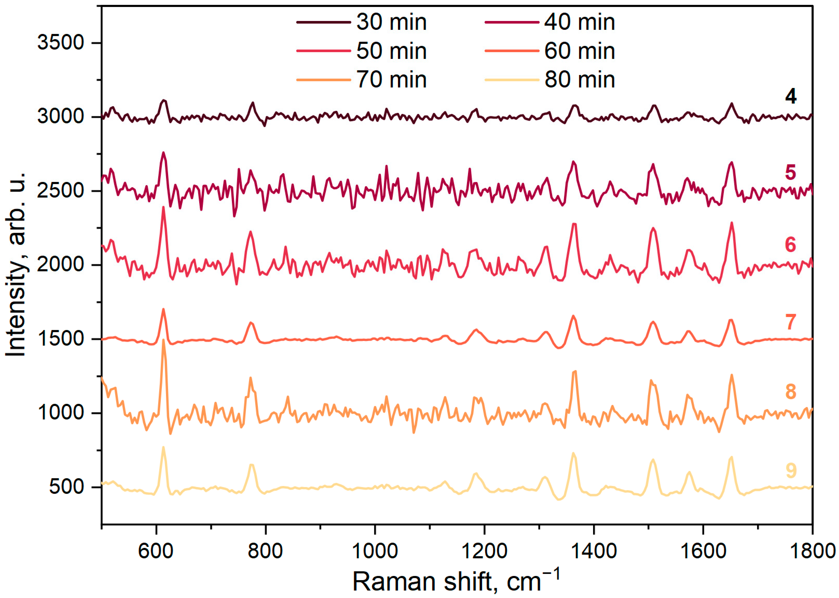
| 4 | 8 | 240 | 30 |
| 5 | 8 | 240 | 40 |
| 6 | 8 | 240 | 50 |
| 7 | 8 | 240 | 60 |
| 8 | 8 | 240 | 70 |
| 9 | 8 | 240 | 80 |
Publisher’s Note: MDPI stays neutral with regard to jurisdictional claims in published maps and institutional affiliations. |
© 2022 by the authors. Licensee MDPI, Basel, Switzerland. This article is an open access article distributed under the terms and conditions of the Creative Commons Attribution (CC BY) license (https://creativecommons.org/licenses/by/4.0/).
Share and Cite
Zavatski, S.; Popov, A.I.; Chemenev, A.; Dauletbekova, A.; Bandarenka, H. Wet Chemical Synthesis and Characterization of Au Coatings on Meso- and Macroporous Si for Molecular Analysis by SERS Spectroscopy. Crystals 2022, 12, 1656. https://doi.org/10.3390/cryst12111656
Zavatski S, Popov AI, Chemenev A, Dauletbekova A, Bandarenka H. Wet Chemical Synthesis and Characterization of Au Coatings on Meso- and Macroporous Si for Molecular Analysis by SERS Spectroscopy. Crystals. 2022; 12(11):1656. https://doi.org/10.3390/cryst12111656
Chicago/Turabian StyleZavatski, Siarhei, Anatoli I. Popov, Andrey Chemenev, Alma Dauletbekova, and Hanna Bandarenka. 2022. "Wet Chemical Synthesis and Characterization of Au Coatings on Meso- and Macroporous Si for Molecular Analysis by SERS Spectroscopy" Crystals 12, no. 11: 1656. https://doi.org/10.3390/cryst12111656
APA StyleZavatski, S., Popov, A. I., Chemenev, A., Dauletbekova, A., & Bandarenka, H. (2022). Wet Chemical Synthesis and Characterization of Au Coatings on Meso- and Macroporous Si for Molecular Analysis by SERS Spectroscopy. Crystals, 12(11), 1656. https://doi.org/10.3390/cryst12111656


