Abstract
The early stages of nanocrystallization in amorphous Fe73.8Si13B9.1Cu1Nb3.1 ribbons and microwires were compared in terms of their internal stress effects. The microstructure was investigated by the X-ray diffraction method. Classical expressions of crystal nucleation and growth were modified for microwires while accounting for the internal stress distribution, in order to justify the XRD data. It was assumed that, due to the strong compressive stresses on the surface part and tensile stresses on the central part, crystallization on the surface part of the microwire proceeded faster than in the central part. The results revealed more rapid nanocrystallization in microwires compared to that in ribbons. During the initial period of annealing, the compressive surface stress of a microwire caused the formation of a predominantly crystallized surface layer. The results obtained open up new possibilities for varying the high-frequency properties of microwires and their application in modern sensorics.
1. Introduction
An amorphous-nanocrystalline Fe73.5Si13.5B9Cu1Nb3 alloy was first mentioned in [1]. It was established as a soft magnetic material that demonstrated high values of magnetic permeability and saturation magnetization, combined with low magnetostriction and low magnetization losses [2,3]. An amorphous alloy of this composition can be produced in the form of a ribbon by the melt spinning method, and in the form of a microwire by the Ulitovsky-Taylor method. An amorphous-nanocrystalline state is reached as a result of heat treatment. Due to the peculiarities of the production processes, microwires and ribbons have different stress states, although their chemical compositions can be identical [4,5]. The value of quenching stresses in a ribbon is in the tens of MPa. The stress level in microwires is 1–2 orders of magnitude higher than that in ribbons. In microwires, stresses are distributed in homogeneously over the radius. Strong compressive (in units of GPa) stresses prevail in the near-surface region of a microwire, while tensile stresses (in hundreds of MPa) prevail in the central part [5,6]. When the shell is removed, the total level of stresses decreases by several hundreds of MPa [6]. Despite shell removing, the character of the total stress distribution does not change (strong compressive stress on the surface part and weak tensile stress in the central part). This difference in the stress state not only affects the magnetization reversal process and the shape of a hysteresis loop [7,8] but it should also affect the processes of crystal formation at the initial stages. Previously, the effect of mechanical stresses on the crystallization and magnetic properties of Co-rich microwires was observed in [9,10,11]. It was found that isothermal annealing led to the formation of crystallites with an elongated shape, and there existed a preferential orientation along the microwire axis [12]. This tendency was stronger in microwires that had undergone a directional crystallization in the presence of a magnetic field [13]. It was shown that the growth of crystals oriented along the microwire axis led to a giant increase in the coercivity (by an order of magnitude or more). Moreover, the cumulative effects of stresses on the crystallization and high-frequency properties of microwires was demonstrated in [11].
The crystallization of the Fe73.5Si13.5B9Cu1Nb3 alloy considered in this work led to the formation of nanocrystals with the density being higher than that of the amorphous matrix. Thus, the density of the amorphous Fe73.5Si13.5B9Cu1Nb3 alloy was 7.14·103 kg/m3 [14], the density of the Fe3Si nanocrystals was 7.39·103 kg/m3 [15]. Therefore, under crystallization, a negative bulk effect would be the reason for the facilitated formation of nanocrystals. Then, the combination of existing internal stresses and additional stresses arising due to the compensation of a bulk crystallization effect would affect the kinetics of crystal formation under the crystallization of amorphous alloys.
It is well-known that mechanical stresses arising under rolling [16], bending [17], milling [18], plastic deformation [19,20], and static load [21,22] affect crystal formation.
Upon undergoing a negative bulk effect, the Gibbs energy of formation of a critical nucleus of Fe(Si) nanocrystals decreases with an increase in the compressive stresses and, on the contrary, increases under tension. Surely, this affects nucleation frequency and, hence, the size of nanocrystals and their volume fraction in the material. Strong compressive stresses prevail in the near-surface region of a microwire. At that region, as mentioned above, a negative bulk effect was observed under the crystallization of the Fe73.8Si13B9.1Cu1Nb3.1 alloy. Then, nanocrystal formation should have been facilitated in the near-surface region of a microwire. At the same time, there were no regions with compressive stresses in the amorphous ribbons [4]. Therefore, when comparing the initial crystallization stages of the amorphous ribbons and microwires, the effect of compressive stresses on them could be estimated. Thus, the present work aimed to study the effect of a high level of mechanical stresses on nanocrystal formation in the amorphous Fe73.8Si13B9.1Cu1Nb3.1 alloy, and compared initial nanocrystallization stages in amorphous microwires and ribbons, which had the same composition but significantly different levels of internal stresses.
2. Materials and Methods
Amorphous Fe73.8Si13B9.1Cu1Nb3.1 alloys were produced in the form of a ribbon by rapid melt quenching; their thickness was 16–18 µm, and the width was 9 mm. Amorphous alloys of the same composition were produced in the form of a glass-coated microwire by the Ulitovsky-Taylor method; the average diameter of the metallic part was 16.5 µm, and the thickness of the shell was 3.5 µm. Initial samples were produced using high-purity (99.9%) components. As shown in [3], the best values of coercivity and magnetic permeability are achieved by heat treatment of the Fe73.5Si13.5B9Cu1Nb3 alloy in the temperature range of 773–833 K. DTA analysis performed in [23] revealed that crystallization began at 803 K. In contrast, results from paper [24] indicated that isothermal annealing for 1 h at 723–823 K led to Fe(Si) nanocrystal precipitation. Accordingly, the ribbons and uncoated microwires were annealed in a vacuum at temperatures in the range of 753–823 K for 1 h. The most illustrative results were obtained after annealing at 753 K. Before annealing, the glass shell was removed by chemical etching in hydrofluoric acid.
Since crystals are formed at an enhanced temperature, the value of the stresses corresponding to these conditions needed to be estimated. To study the effect of temperature on the level of mechanical stresses, the degree of bending stress relaxation in the ribbons was estimated. For this to be done, samples of the ribbons were placed into a quartz ampoule along its inner diameter. The length of the samples coincided with the circumference of the ampoule. After that, heat treatment was performed at 753 K. Then, the samples were pulled out of the ampoule, and the curvature radii of the annealed samples were measured. To estimate the degree of stress relaxation, the parameter γ = 1 − R0/R was used [25], where R0 is the radius of a quartz ampoule and R is the curvature radius of a ribbon after heat treatment.
The structure and phase compositions of the samples before and after annealing were investigated with a SIEMENS D-500 (Manufacturer: Siemens AG. Location: Östliche Rheinbrückenstraße 50, 76187 Karlsruhe) diffractometer using Co Kα-radiation. Since the samples contained the amorphous and crystalline phases, the experimental curves were decomposed into a diffuse component caused by scattering from the amorphous phase, and a diffraction component caused by the presence of crystals. When decomposing, the characteristics of the curve of scattering by the initial amorphous phase (the half-width and the position of the diffuse maximum) were taken into account. The size of the nanocrystals was calculated according to the half-width of a diffraction spectrum component using the Scherrer equation [26,27].
The fraction of the amorphous and crystalline phases was estimated by the ratio of the integral intensities of the diffraction and diffuse components of the X-ray diffraction patterns, according to [28].
3. Results
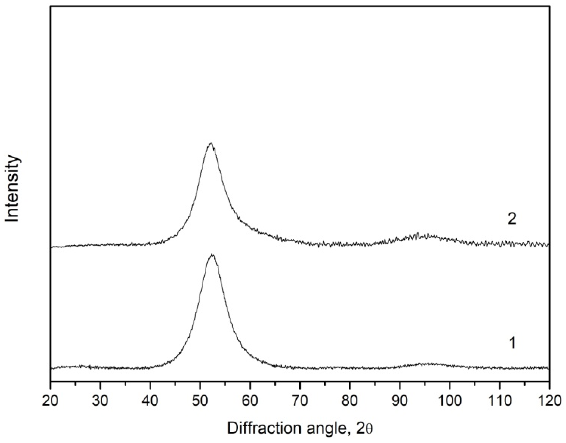
After the production, the samples were amorphous. The X-ray diffraction patterns of initial samples contained only broad diffuse maxima; no diffraction reflections from the crystalline phases were observed (Figure 1).
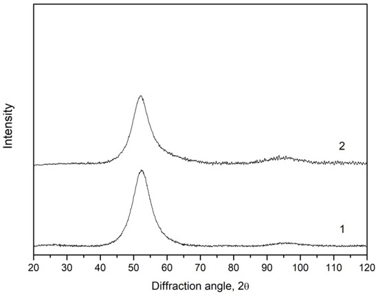
Figure 1.
X-ray diffraction patterns of the initial amorphous Fe73.8Si13B9.1Cu1Nb3.1 ribbons (1) and microwires (2).
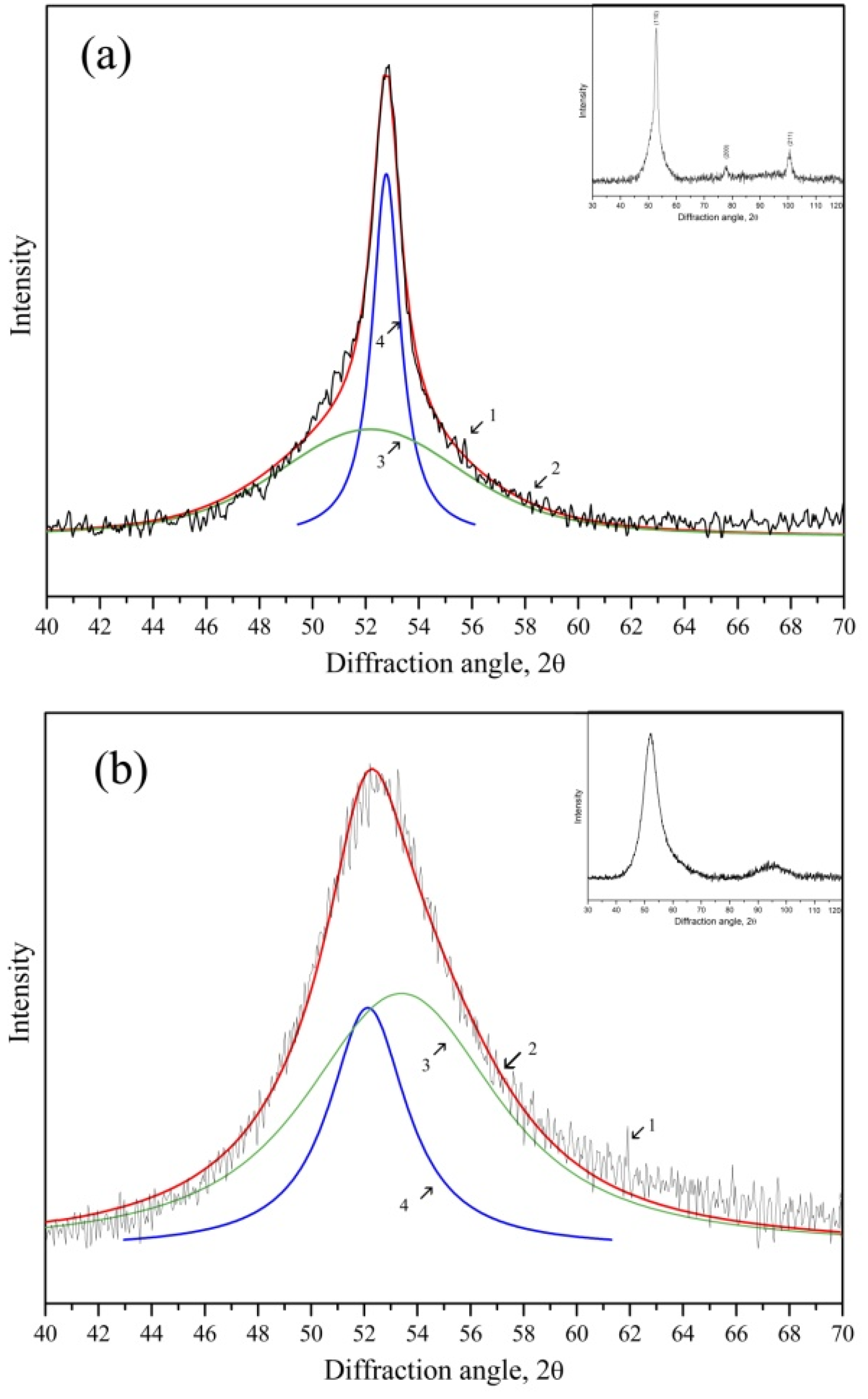
After the annealing, the structure of both samples changed markedly. Figure 2 shows the X-ray diffraction patterns of the microwires (Figure 2a) and ribbons (Figure 2b) annealed at 753 K for 1 h.
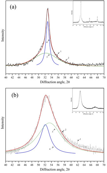
Figure 2.
X-ray diffraction patterns of the microwires (a) and ribbons (b) of Fe73.8Si13B9.1Cu1Nb3.1 composition annealed at 753 K for 1 h (1—experimental curve, 3—scattering from the amorphous phase, 4—diffraction reflections from nanocrystals, 2—summary of curves 3–4).
Figure 2 illustrates regions of the main diffuse maximum in the X-ray diffraction patterns; the insets show complete curves. In the figure, curve 1 is an experimental X-ray diffraction pattern, curve 3 corresponds to scattering by the amorphous phase, curve 4 describes diffraction reflections from nanocrystals, and curve 2 is the sum of curves 3 and 4. In the complete X-ray diffraction pattern of the microwires (Figure 2a), diffraction reflections were indexed. They corresponded to the bcc phase with the parameter a = 2.845 Å and a Fe(Si) solid solution. One can see that the intensity of the diffraction reflections (curve 4) in Figure 2a was significantly higher than that of the diffraction reflections in Figure 2b. The analysis of the reflection intensities showed that the fraction of the precipitated nanocrystalline phase in the microwires was 20% higher than that in the ribbons. The half-widths of the diffraction reflections in Figure 2a,b were also different, which was the evidence of different sizes of the nanocrystals. The size of the nanocrystals in the microwires was determined using the Scherrer equation as 16 nm, and that in the ribbons was 6 nm.
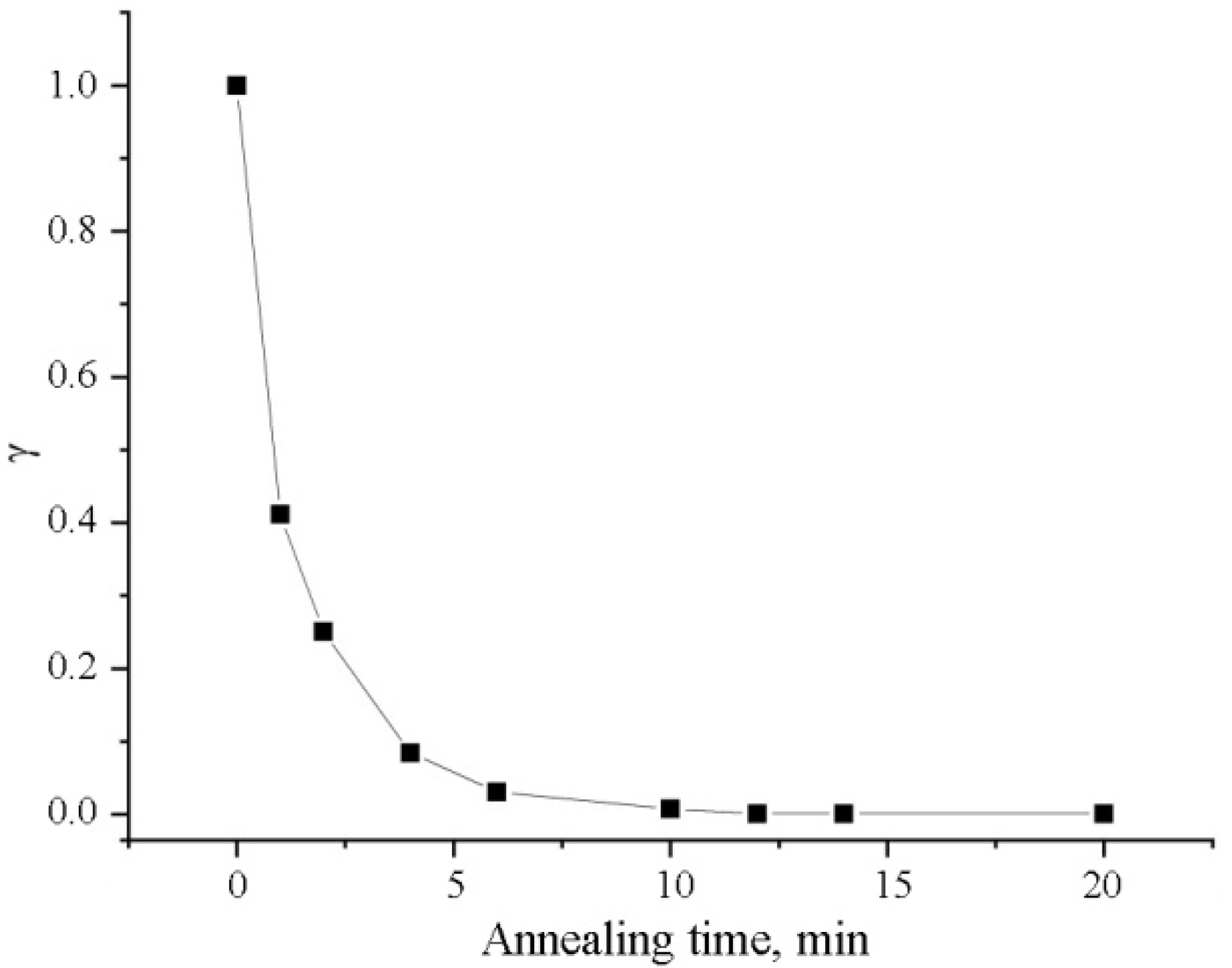
To estimate the degree of mechanical stress relaxation under annealing, in the assumption that the initial internal stresses in a microwire decrease with the temperature at the same rate as ribbons induced under bending do, an experiment on the estimation of the degree of bending stress relaxation was performed.
Figure 3 depicts the change in the parameter γ with time. One can see that about 20 min later, bending stresses were almost completely relaxed. To facilitate further calculations, γ = 0.1 was selected as the “effective” value.
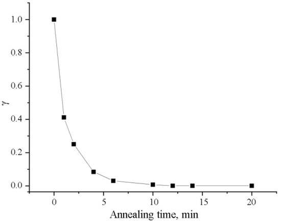
Figure 3.
Relaxation curve of bending stresses for initial samples of the ribbon under annealing at 753 K.
4. Discussion
We shall assume that nanocrystals nucleate by a homogeneous mechanism. As is well-known, Cu clusters are formed in this alloy under heating, which can act as heterogeneous nucleation centers [29]. However, under consideration of the first approximation, we will not take into account their effect on the kinetics of the Fe(Si) nanocrystal nucleation.
To consider the effect of the internal stresses of the microwire on nucleation frequency, one should first modify the expression for the Gibbs energy ΔG of a spherical nucleus with the radius r, proposed in [16,17,18,19,20,21,22]:
where ΔGch is the driving force of crystallization from an amorphous state, Eε is the elastic energy that includes deformation contributions from internal stresses and the emergence of a nanocrystal different from the amorphous density matrix, and σ is the surface energy.
ΔG = − 4/3πr3(ΔGch + Eε) + 4πr2σ
We shall write the elastic energy in more detail [30], assuming that the shear component of deformation γ is absent:
where G = E/(2(1 + ν)) is the shear modulus, and E = 139 GPa is Young’s modulus at 753 K. Under normal conditions, the value of Young’s modulus is 154 GPa [31]; however, considering the temperature effect, there is a decrease by about 10% [32], ν is the Poisson’s ratio that is taken to be 0.3 [33,34], x, is the radial from the microwire axis, and ε(x) is the normal component of deformation. It is presented in the form:
where σ(x) = σrr + σθθ + σzz is the sum of diagonal components of a stress tensor of the metallic core of a microwire with respect to the sign, calculated according to [6,8], γ is the effective value of the stress relaxation parameter, obtained from an experiment with the ribbons. ΔV/V is the value of a bulk effect calculated from the difference between densities of amorphous material and formed nanocrystals:
where ρam,cr are the densities of an amorphous alloy and nanocrystals, respectively. According to [14], the density of the amorphous Fe73.5Si13.5B9Cu1Nb3 alloy was 7.14·103 kg/m3, the density of nanocrystals of a solid solution of 25% Si in Fe, Fe3Si, was 7.39·103 kg/m3 [15], and the density of Fe was 7.87·103 kg/m3. Considering that density changes linearly with a change in Si concentration, the density of nanocrystals was taken to be 7.55·103 kg/m3 at Si content of 16.5% [35].
Eε = πGε(x)2/4
ε(x) = ΔV/3V + γσ(x)/E
ΔV/V = (ρam − ρcr)/ρam
It is assumed in Equation (3) that compressive stresses are negative, and tension stresses are positive. From (1), we obtain the Gibbs energy of a critical nucleus:
ΔG* = 16πσ3/(3(ΔGch + Eε)2)
The next assumption is that the value of internal stresses in a ribbon is significantly lower than that in a microwire [4]. Therefore, the second term in (3) could be neglected for calculations in a ribbon. The difference between free energies of the alloy in amorphous and crystalline states was calculated according to [36]:
where ΔHcr = 58 J/g is the crystallization enthalpy [37], Tm is the melting temperature taken to be 1450 K [38], T is the temperature of heat treatment at 753 K, and ρam is the density of the amorphous matrix at 7.14·103 kg/m3. Here, we assumed that the crystallization enthalpy for ribbons and microwires was the same and did not depend on internal stresses. Then, ΔGch is 0.14 GJ/m3.
ΔGch = 2TΔHcrΔTρam/(Tm(Tm + T))
The growth rate was calculated by the formula [39,40]:
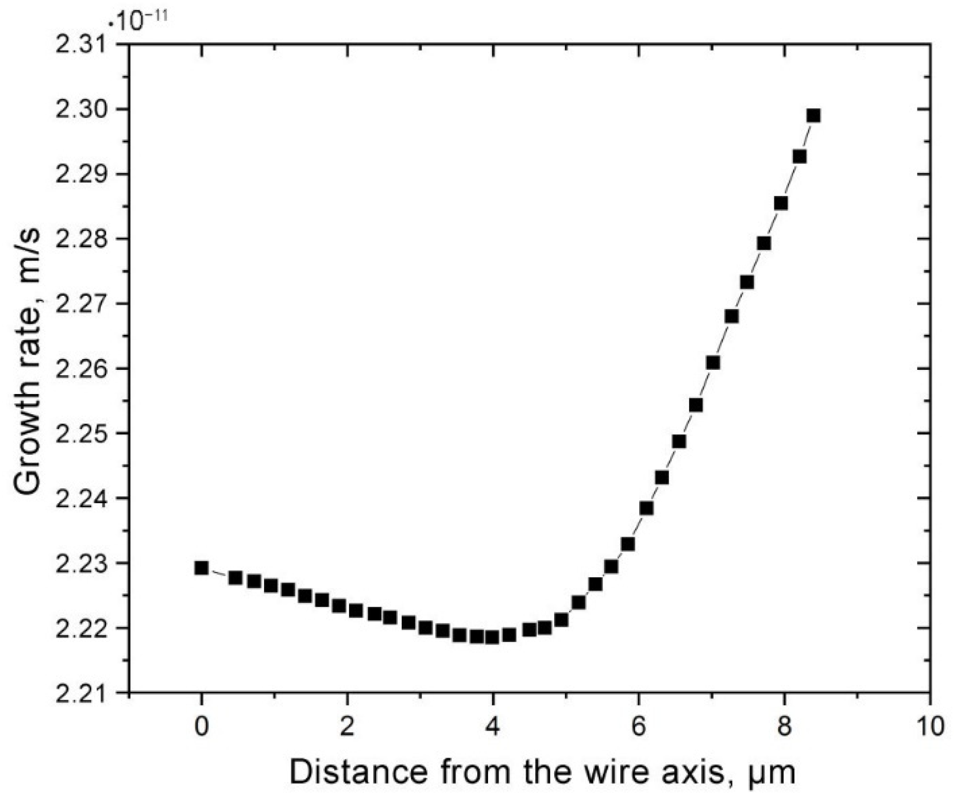
where f ≈ 1 and D is the diffusion coefficient. The growth rate for the ribbon was 2.23·10−11 m/s, assuming that only the bulk effect was presented and σ(x) = 0 in (3). By using the stress distribution σ(x) as a function of the distance to the microwire axis calculated according to [6,8], as well as Equation (2), the distribution uc(x) was calculated (Figure 4). The obtained values of nanocrystal growth rate were within the range of 2.22·10−11–2.30·10−11 m/s.
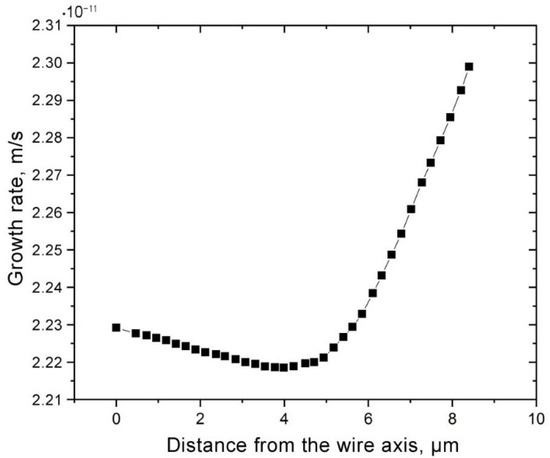
Figure 4.
Distribution of nanocrystal growth rate over the microwire radius calculated by the Formula (7).
It follows from the data of the X-ray diffraction studies that the content of nanocrystals in the microwire was 20% higher than that in the ribbon. Assuming that, at the initial crystallization stages, the growth rate and nucleation frequency of the nanocrystals did not change with time, we used the well-known expression for the crystallized volume fraction [41], and wrote a ratio for the crystallized fractions in a microwire and ribbon:
The left side of Equation (8) is the relative fraction of nanocrystals determined experimentally, with the average over the volume fraction resulting in the microwire. On the right side, there are unknown values of nanocrystal nucleation frequencies, which, in turn, contain an unknown value of surface energy. The value of σ was determined from the condition that the value on the left side of (8) determined experimentally, which corresponded to the value of the final expression on the right side of (8) with a precision of approximately 1%.
The nucleation frequency was determined according to [17]:
where D is the diffusion coefficient, is the average atomic concentration, is the average molar mass, and a0 is the average interatomic spacing. The diffusion coefficient was estimated using the technique from [42]; it turned out to be 3.6·10−20 m2/s. The average atomic concentration NV was 8.66·1028 m−3. The average interatomic spacing was estimated from the Ehrenfest equation for a radius of the first coordination sphere, a0 = 2.5 Å. The values necessary for further calculations are summarized in Table 1.

Table 1.
Data used for the calculations.
Taking into account Equations (5) and (9) can be rewritten as:
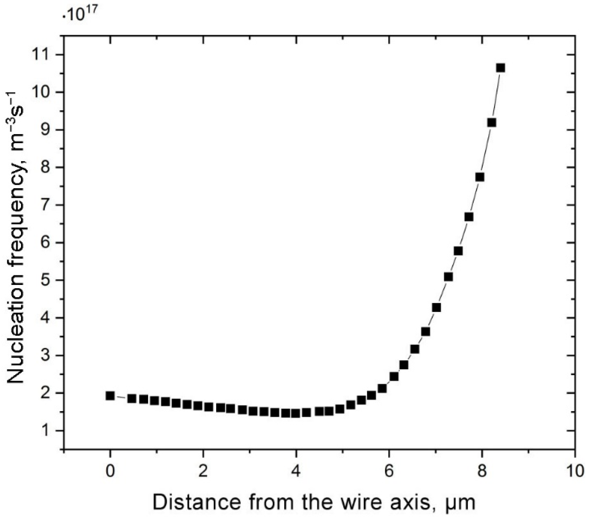
Calculations using (10) showed that the nucleation frequency of the ribbon was 1.9·1017 m−3s−1, with a surface energy of 0.072 J/m2. The value of surface energy estimated in [18] for a Fe crystal in the amorphous environment at room temperature was 0.13 J/m2. This difference may be related to the purely homogeneous mechanism of crystal nucleation assumed in this work. However, nanocrystals are formed in this alloy not only by a homogeneous mechanism, but also by nucleation on Cu clusters. The estimated values of nucleation frequencies were quite close in the order of magnitude to (10 ± 5)·1019 m−3s−1 obtained under heat treatment at 763 K [24]. The distribution of nucleation frequencies over the microwire radius calculated from (12) is demonstrated in Figure 5.
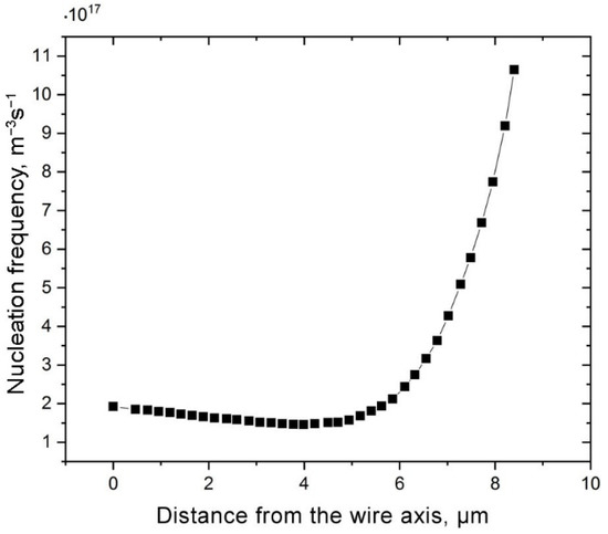
Figure 5.
Distribution of nucleation frequency over the microwire radius.
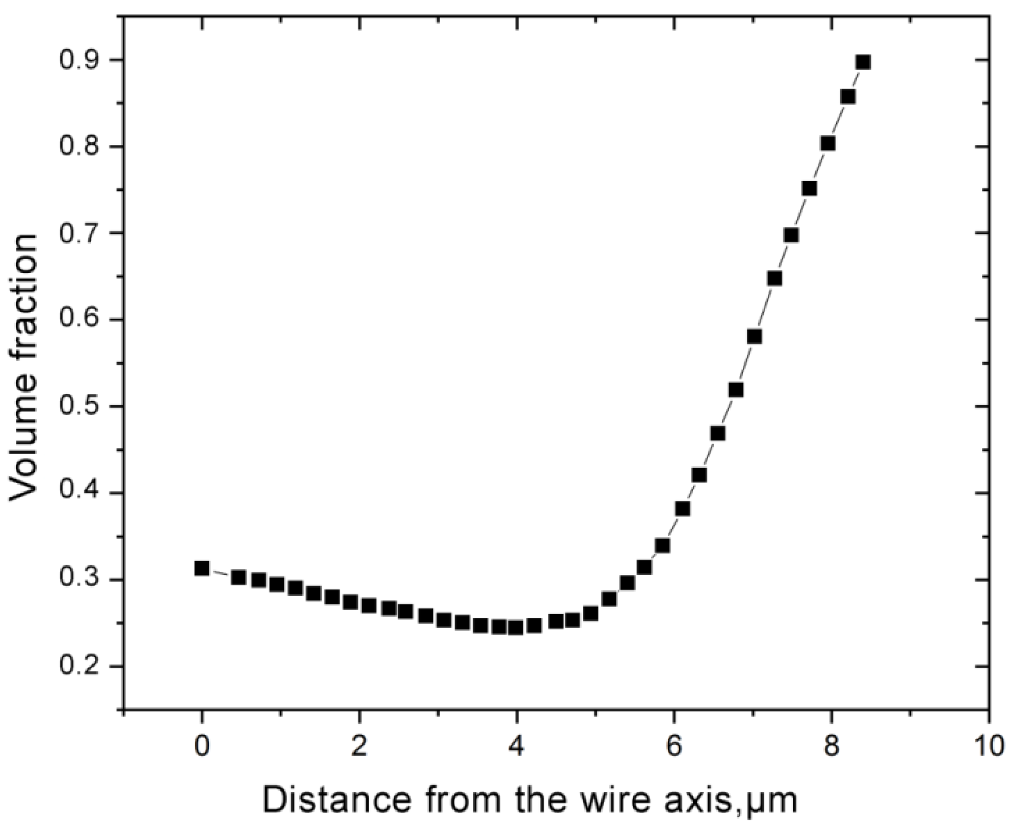
The obtained distributions of the nucleation frequency and growth rate of the nanocrystals allowed for calculating the distribution of the crystallized volume fraction X over the microwire radius (Figure 6). One can see from Figure 6 that the volume fraction of the formed nanocrystals significantly increased in the near-surface region, where the value of compressive stresses can reach 2 GPa [6,8]. At that region, the average volume fraction of nanocrystals in the microwire, calculated from the distribution in Figure 6, was about 29%. For the ribbon, the content of nanocrystals, calculated using the expression for the crystallized volume fraction X, was 24%, which was about 20% lower and corresponded to the experimentally revealed differences.
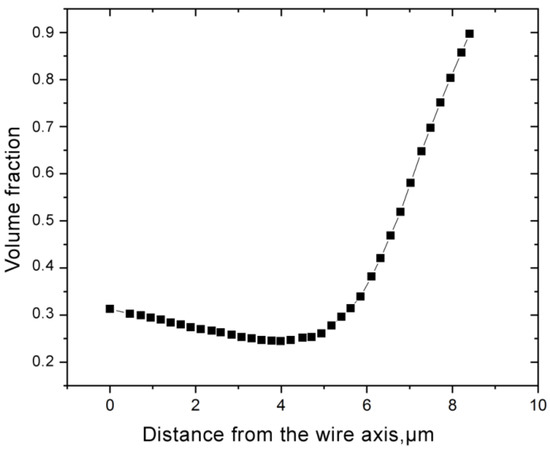
Figure 6.
Volume fraction of the nanocrystalline phase depending on the microwire axis.
As one can see from Figure 2, at the initial crystallization stages, microwires crystallized more rapidly than ribbons. The obtained estimates suggested that, at early stages, nanocrystallization in microwires occurred predominantly in the microwire part where compressive inner stresses prevailed, i.e., in a small, near-surface region, which was about 2–2.5 µm for the microwire with a diameter of 16.8 µm. In addition, one of the reasons for the predominant crystallization of the surface layer could be not only compressive stresses, but also defects on the microwire surface.
In some amorphous alloys, compressive stresses can lead to different effects [43,44,45,46]. Due to compressive stresses, the slowdown of the crystallization kinetics led to an increase in the crystallization temperature in Pd-, La-, and Pr-based amorphous alloys. In such alloys, the glass transition temperature is below the crystallization temperature, Tg < Tx. This means that the crystallization of such alloys occurs from the supercooled liquid state, not from the amorphous state. During the nucleation and growth of crystals from the supercooled liquid state, the bulk effect may not affect the overall crystallization kinetics, due to the low viscosity of the alloy and rapid relaxation. In this case, a decrease in the diffusion coefficient (atomic mobility) may have played a decisive role. We assumed that the bulk effect played a crucial role in our case, since in the Fe73.8Si13B9.1Cu1Nb3.1 alloy Tg > Tx and crystallization occurs from the amorphous state, not from the supercooled liquid state. In this case, the viscosity of the alloy remains high and the stresses do not have time to relax. In addition, it should be noted that the effect of pressure on the crystallization of alloys with Tg < Tx is not unambiguous. Thus, it was demonstrated in [17] that the application of bending stresses of more than 1.5 GPa to a Pd40Cu30Ni10P20 ribbon led to the fact that the tension side (above the neutral axis) of the glassy Pd40Cu30Ni10P20 ribbon showed no crystallinity. At the same time, the compressed part of the ribbon thickness showed the presence of nanocrystals. The bulk effect in Pd-based alloys is about 3 times lower than that in the Fe73.8Si13B9.1Cu1Nb3.1 alloy. Therefore, we supposed that, in our case, the influence of the bulk effect on the crystallization was stronger.
The crystallization of the Fe73.5Si13.5B9Cu1Nb3 alloy led to a simultaneous increase in the magnetic permeability and a decrease in the saturation magnetostriction [2,3]. Since the surface part of the microwire crystallized faster than its central part, the high-frequency properties of the microwire were improved in the surface part. For instance, there is the GMI effect, which is essentially an increase in the impedance Z of a soft magnetic conductor placed in a static magnetic field. The GMI ratio ΔZ/Z of annealed amorphous-nanocrystalline Fe73.5Si13.5B9Cu1Nb3 [47] and Fe73.8Si13B9.1Cu1Nb3.1 [48] microwires increased significantly (up to 200%) in a wide range of frequencies, in comparison with as-prepared samples. It is noteworthy that the ΔZ/Z value depended on the depth of skin layer; the alternating current flows effectively in the near-surface region. The selection of heat treatment conditions could avoid the crystallization of the central part of the microwire during the crystallization of its surface part. In this case, the microwire would be a semblance of layered composite, with enhanced soft magnetic properties (like magnetic permeability) near the surface This opens up new prospects for varying the high-frequency properties of Fe73.5Si13.5B9Cu1Nb3 microwires and their application in modern sensorics.
5. Conclusions
It has been determined experimentally that the sizes and volume fraction of nanocrystals formed as a result of heat treatment at 753 K in the Fe73.8Si13B9.1Cu1Nb3.1 microwires were greater than those in a ribbon. It was found that crystallization proceeded more intensively in the near-surface region of the microwire. These data are the evidence of different rates of crystallization processes in a microwire and a ribbon, which were caused by the presence of a high level of internal stresses in microwires.
The estimated calculations of the dependence of the nucleation frequency of Fe(Si) nanocrystals on mechanical stresses were carried out. Based on the performed calculations and the experimental data, it was found that the nucleation frequency of the nanocrystals in the near-surface region of a microwire increases significantly and reaches a maximum on the surface. The average value of nucleation frequency in an amorphous microwire was 1.5 times larger than that in a ribbon and was 2.9·1017 m−3s−1.
The possibility of the formation of microwires during heat treatment with a predominant nucleation of nanocrystals in the near-surface region opens up broad prospects for controlling the high-frequency properties of microwires, and for using them as sensitive elements of strain and magnetic field sensors.
Author Contributions
Investigation, A.F. and O.A.; Supervision, A.A., G.A. and M.C.; Writing—original draft preparation, A.F.; Writing—review and editing, A.F., O.A., G.A. and A.A. All authors have read and agreed to the published version of the manuscript.
Funding
This work was supported by the Russian Science Foundation (grant no. 22-72-00067).
Informed Consent Statement
Not applicable.
Data Availability Statement
Not applicable.
Conflicts of Interest
The authors declare no conflict of interest.
References
- Yoshizawa, Y.; Oguma, S.; Yamauchi, K. New Fe-based soft magnetic alloys composed of ultrafine grain structure. J. Appl. Phys. 1988, 64, 6044–6066. [Google Scholar] [CrossRef]
- Chiriac, H.; Lupu, N.; Stoian, G.; Ababei, G.; Corodeanu, S.; Óvári, T.-A. Ultrathin Nanocrystalline Magnetic Wires. Crystals 2017, 7, 48. [Google Scholar] [CrossRef]
- Herzer, G. Nanocrystalline soft magnetic materials. Phys. Scr. 1993, 1993, T49A. [Google Scholar] [CrossRef]
- Carara, M.; Baibich, M.N.; Sommer, R.L. Stress level in Finemet materials studied by impedanciometry. J. Appl. Phys. 2002, 91, 8441–8443. [Google Scholar] [CrossRef]
- Zhukov, A.; Ipatov, M.; Talaat, A.; Blanco, J.M.; Hernando, B.; Gonzalez-Legarreta, L.; Suñol, J.J.; Zhukova, V. Correlation of Crystalline Structure with Magnetic and Transport Properties of Glass-Coated Microwires. Crystals 2017, 7, 41. [Google Scholar] [CrossRef]
- Chiriac, H.; Ovari, T.A.; Pop, G. Internal stress distribution in glass-covered amorphous magnetic wires. Phys. Rev. B 1995, 52, 10104–10113. [Google Scholar] [CrossRef]
- Baranov, S.A.; Larin, V.S.; Torcunov, A.V. Technology, Preparation and Properties of the Cast Glass-Coated Magnetic Microwires. Crystals 2017, 7, 136. [Google Scholar] [CrossRef]
- Aksenov, O.I.; Fuks, A.A.; Aronin, A.S. The effect of stress distribution in the bulk of a microwire on the magnetization processes. J. Alloys Compd. 2020, 836, 155472. [Google Scholar] [CrossRef]
- Morchenko, A.T.; Panina, L.V.; Larin, V.S.; Churyukanova, M.N.; Salem, M.M.; Hashim, H.; Trukhanov, A.V.; Korovushkin, V.V.; Kostishyn, V.G. Structural and magnetic transformations in amorphous ferromagnetic microwires during thermomagnetic treatment under conditions of directional crystallization. J. Alloys Compd. 2017, 698, 685–691. [Google Scholar] [CrossRef]
- Evstigneeva, S.; Morchenko, A.; Trukhanov, A.; Panina, L.; Larin, V.; Volodina, N.; Yudanov, N.; Nematov, M.; Hashim, H.; Ahmad, H. Structural and magnetic anisotropy of directionally-crystallized ferromagnetic microwires. EPJ Web Conf. 2018, 185, 04022. [Google Scholar] [CrossRef]
- Panina, L.; Dzhumazoda, A.; Nematov, M.; Alam, J.; Trukhanov, A.; Yudanov, N.; Morchenko, A.; Rodionova, V.; Zhukov, A. Soft Magnetic Amorphous Microwires for Stress and Temperature Sensory Applications. Sensors 2019, 19, 5089. [Google Scholar] [CrossRef] [PubMed]
- Li, Z.-D.; Zhang, W.-W.; Li, G.-T.; Li, S.-S.; Ding, H.-S.; Zhang, T.; Song, Y.-J. Magnetic field annealing of FeCo-based amorphous alloys to enhance thermal stability and Curie temperature. Rare Met. 2018, 1–7. [Google Scholar] [CrossRef]
- Serrano, I.G.; Hernando, A.; Marín, P. Low temperature magnetic behavior of glass-covered magnetic microwires with gradient nanocrystalline microstructure. J. Appl. Phys. 2014, 115, 033903. [Google Scholar] [CrossRef]
- Parsons, R.; Ono, K.; Li, Z.; Kishimoto, H.; Shoji, T.; Kato, A.; Hill, M.R.; Suzuki, K. Prediction of density in amorphous and nanocrystalline soft magnetic alloys: A data mining approach. J. Alloys Compd. 2021, 859, 157845. [Google Scholar] [CrossRef]
- The Materials Project. Available online: https://materialsproject.org/materials/mp-2199/ (accessed on 19 September 2022).
- Yan, Z.; Song, K.; Hu, Y.; Dai, F.; Chu, Z.; Eckert, J. Localized crystallization in shear bands of a metallic glass. Sci. Rep. 2016, 6, 19358. [Google Scholar] [CrossRef] [PubMed]
- Yavari, A.R.; Georgarakis, K.; Antonowicz, J.; Stoica, M.; Nishiyama, N.; Vaughan, G.; Chen, M.; Pons, M. Crystallization during bending of a Pd-based metallic glass detected by X-ray microscopy. Phys. Rev. Lett. 2012, 109, 085501. [Google Scholar] [CrossRef] [PubMed]
- Gheiratmand, T.; Hosseini, H.R.M.; Davami, P.; Ababei, G.; Song, M. Mechanism of mechanically induced nanocrystallization of amorphous FINEMET ribbons during milling. Met. Mater. Trans. A 2015, 46, 2718–2725. [Google Scholar] [CrossRef]
- Aronin, A.S.; Abrosimova, G.E. Reverse martensite transformation in iron nanocrystals under severe plastic deformation. Mater. Lett. 2012, 83, 183–185. [Google Scholar] [CrossRef]
- Vasil’ev, L.S.; Lomaev, I.L. On possible mechanisms of nanostructure evolution upon severe plastic deformation of metals and alloys. Phys. Met. Metallogr. 2006, 101, 386–392. [Google Scholar] [CrossRef]
- Ye, F.; Lu, K. Pressure effect on crystallization kinetics of an Al–La–Ni amorphous alloy. Acta Mater. 1999, 47, 2449–2454. [Google Scholar] [CrossRef]
- Lee, S.-W.; Huh, M.-Y.; Fleury, E.; Lee, J.-C. Crystallization-induced plasticity of Cu–Zr containing bulk amorphous alloys. Acta Mater. 2006, 54, 349–355. [Google Scholar] [CrossRef]
- Noh, T.H.; Lee, M.B.; Kim, H.J.; Kang, I.K. Relationship between crystallization process and magnetic properties of Fe-(Cu-Nb)-Si-B amorphous alloys. J. Appl. Phys. 1990, 67, 5568–5570. [Google Scholar] [CrossRef]
- Clavaguera, N.; Pradell, T.; Jie, Z.; Clavaguera-Mora, M.T. Thermodynamic and kinetic factors controlling the formation of nanocrystalline FeCuNbSiB materials. Nanostruct. Mater. 1995, 6, 453–456. [Google Scholar] [CrossRef]
- Luborsky, F.; Walter, J. Stress relaxation in amorphous alloys. Mater. Sci. Eng. 1978, 35, 255–261. [Google Scholar] [CrossRef]
- Guinier, A. Тheоrie et Technique de la Radiocristallographie; Dumond: Paris, France, 1956. [Google Scholar]
- Cruz, M.E.; Li, J.; Gorni, G.; Durán, A.; Mather, G.C.; Balda, R.; Fernández, J.; Castro, Y. Crystallization Process and Site-Selective Excitation of Nd3+ in LaF3/NaLaF4 Sol–Gel-Synthesized Transparent Glass-Ceramics. Crystals 2021, 11, 464. [Google Scholar] [CrossRef]
- Abrosimova, G.E.; Aronin, A.S.; Kholstinina, N.N. On the determination of volume fraction of the crystalline phase in amorphous-crystalline alloys. Phys. Solid State 2010, 52, 445–451. [Google Scholar] [CrossRef]
- Vásquez, M.; Marín, P.; Davies, H.A.; Olofinjana, A. Magnetic hardening of FeSiBCuNb ribbons and wires during the first stage of crystallization to a nanophase structure. Appl. Phys. Lett. 1994, 64, 3184–3186. [Google Scholar] [CrossRef]
- Cahn, R.W.; Haasen, P. Physical Metallurgy, 3rd ed.; North-Holland Physics Publishing: Amsterdam, The Netherlands, 1983. [Google Scholar]
- Duong, A.H.; Malkinski, L.; Grossinger, R. Magnetomechanical properties in FINEMET type alloy. In Proceedings of the VACETS Technical International Conference, San Jose, CA, USA, 17–19 July 1997; p. 40. [Google Scholar]
- Kikuchi, M.; Fukamichi, K.; Masumoto, T. Young’s modulus and delay time characteristics of ferromagnetic Fe-Si-B amorphous alloys. Sci. Rep. Res. Inst. Tohoku Univ. 1976, 26, 232–239. [Google Scholar]
- Yang, G.N.; Sun, B.A.; Chen, S.Q.; Gu, J.L.; Shao, Y.; Wang, H.; Yao, K.F. Understanding the effects of Poisson’s ratio on the shear band behavior and plasticity of metallic glasses. J. Mater. Sci. 2017, 52, 6789–6799. [Google Scholar] [CrossRef]
- Bansal, N.P.; Doremus, R.H. Handbook of Glass Properties; Academic Press: Cambridge, MA, USA, 1986. [Google Scholar]
- Gheiratmand, T.; Hosseini, H.R.M. Finemet nanocrystalline soft magnetic alloy: Investigation of glass forming ability, crystallization mechanism, production techniques, magnetic softness and the effect of replacing the main constituents by other elements. J. Magn. Magn. Mater. 2016, 408, 177–192. [Google Scholar] [CrossRef]
- Thompson, C.; Spaepen, F. On the approximation of the free energy change on crystallization. Acta Metall. 1979, 27, 1855–1859. [Google Scholar] [CrossRef]
- Antoszewska, M.; Wasiak, M.; Gwizdałła, T.; Sovak, P.; Moneta, M. Thermal induced structural and magnetic transformations in Fe73.5−xCex=0,3,5,7Si13.5B9Nb3Cu1 amorphous alloy. J. Therm. Anal. Calorim. 2014, 115, 1381–1386. [Google Scholar] [CrossRef][Green Version]
- Clavaguera-Mora, M.T.; Clavaguera, N.; Crespo, D.; Pradell, T. Crystallisation kinetics and microstructure development in metallic systems. Prog. Mater. Sci. 2002, 47, 559–619. [Google Scholar] [CrossRef]
- Nishiyama, N.; Inoue, A. Supercooling investigation and critical cooling rate for glass formation in Pd–Cu–Ni–P alloy. Acta Mater. 1999, 47, 1487–1495. [Google Scholar] [CrossRef]
- Uhlmann, D.R. A kinetic treatment of glass formation. J. Non Cryst. Solids 1972, 7, 337–348. [Google Scholar] [CrossRef]
- Christian, J.W. The Theory of Phase Transformations in Metals and Alloys; Pergamon Press: Oxford, UK, 1965. [Google Scholar]
- Pershina, E.; Matveev, D.; Abrosimova, G.; Aronin, A. Formation of nanocrystals in an amorphous Al90Y10 alloy. Mat. Char. 2017, 133, 87–93. [Google Scholar] [CrossRef]
- Jiang, J.Z.; Zhuang, Y.X.; Rasmussen, H.; Nishiyama, N.; Inoue, A.; Lathe, C. Crystallization of Pd40Cu30Ni10P20 bulk glass under pressure. Eur. Lett. 2001, 54, 182. [Google Scholar] [CrossRef]
- Jiang, J.Z.; Saksl, K.; Nishiyama, N.; Inoue, A. Crystallization in Pd40Ni40P20 glass. J. Appl. Phys. 2002, 92, 3651–3656. [Google Scholar] [CrossRef]
- Wang, Y.; Zhao, W.; Li, G.; Li, Y.; Liu, R. Structural evolution of Lanthanide-based metallic glasses under high-pressure annealing. J. Alloys Compd. 2013, 551, 185–188. [Google Scholar] [CrossRef]
- Falqui, A.; Loche, D.; Casu, A. In Situ TEM Crystallization of Amorphous Iron Particles. Crystals 2020, 10, 41. [Google Scholar] [CrossRef]
- Knobel, M.; Sánchez, M.L.; Gómez-Polo, C.; Marin, P.; Vazquez, M.; Hernando, A. Giant magneto-impedance effect in nanostructured magnetic wires. J. Appl. Phys. 1996, 79, 1646–1654. [Google Scholar] [CrossRef]
- Talaat, A.; Zhukova, V.; Ipatov, M.; Blanco, J.M.; Gonzalez-Legarreta, L.; Hernando, B.; del Val, J.J.; González, J.; Zhukov, A. Optimization of the giant magnetoimpedance effect of Finemet-type microwires through the nanocrystallization. J. Appl. Phys. 2014, 115, 17A313. [Google Scholar] [CrossRef]
Publisher’s Note: MDPI stays neutral with regard to jurisdictional claims in published maps and institutional affiliations. |
© 2022 by the authors. Licensee MDPI, Basel, Switzerland. This article is an open access article distributed under the terms and conditions of the Creative Commons Attribution (CC BY) license (https://creativecommons.org/licenses/by/4.0/).

