Article

Synthesis of Zirconia Micro-Nanoflakes with Highly Exposed (001) Facets and Their Crystal Growth

1
State Key Laboratory of Isotope Geochemistry, Guangzhou Institute of Geochemistry, Guangzhou 510640, China
2
CAS Center for Excellence in Deep Earth Science, Guangzhou 510640, China
3
College of Earth and Planetary Sciences, University of Chinese Academy of Sciences, Beijing 100049, China
4
School of Earth Science and Resources, Chang’an University, Xi’an 710054, China
*
Author to whom correspondence should be addressed.
Crystals 2021, 11(8), 871; https://doi.org/10.3390/cryst11080871
Submission received: 9 June 2021 / Revised: 21 July 2021 / Accepted: 23 July 2021 / Published: 27 July 2021
(This article belongs to the Special Issue Advanced Functional Oxide Ceramics)

Abstract

:
This study reports a novel preparation method of zirconia micro-nanoflakes with high (001) facets that is generated through a hydrolysis reaction of the fluozirconic acid (H2ZrF6). Zirconia micro-nanoflakes synthesized at varied conditions were analyzed by the SEM, EDS, μ-XRD, and Raman spectroscopy to characterize the morphology and probe into the crystal growth mechanism. The synthesized zirconia crystals in the form of elliptical micro-nanoflakes or irregular nanoflakes generally display the highly exposed (001) facets with a thickness of 1–100 nm and a length of 0.1–2.0 μm. As the temperature and initial solution concentration increased, the particle sizes of the synthesized zirconia micro-nanoflakes became more uniform and the thicknesses of the (001) facets became larger, suggesting that the synthesized zirconia crystals grow along the (001) facets and mostly along the c-axis direction. This is confirmed by the data from the μ-XRD patterns. The results also demonstrate that an oriented attachment-based growth occurring in a fluorine-rich solution environment was involved in the aggregation and coarsening of zirconia micro-nanoflakes. Meanwhile, synthesized zirconia micro-nanoflakes also evolved from a mixture of monoclinic and tetragonal systems to a pure monoclinic system (i.e., baddeleyite) with the temperature increasing, suggesting a key role of temperature regarding zirconia’s growth.

1. Introduction

Zirconia (ZrO2), as a kind of significant structural and functional material, has excellent physical and chemical properties including a high melting point (as high as 2700 °C), high boiling point, high strength, low thermal conductivity, a large thermal expansion coefficient, and good ionic conductivity, and is wear-resistant, high-temperature-resistant, corrosion-resistant, and oxidation-resistant [1,2]. As of 1980s, apart from its use as a traditional refractory material and ceramic glaze, zirconia has received increasingly more attention for application in the fields of energy, environment, and material [1,3,4,5,6]. The performances of micro-nano zirconia in the aspects of optics, mechanics, thermodynamics, and electromagnetism are different from those of conventional materials due to its tiny size. As a result, micro-nano zirconia displays extensive application uses in the industrial synthesis, catalysts and catalyst carriers, unique ceramics, thermal barrier coatings, refractory materials, cutting tools, sensors, solid batteries, decoration, adsorption, and reinforcing and toughening fields [5,7,8,9,10,11,12,13,14]. Therefore, micro-nano zirconia has been considered one of the most valuable functional materials in the 21st century.
Preparation of micro-nano zirconia is generally divided into physical and chemical approaches. The physical approach includes a reactive plasma processing method, freeze-drying method, explosive compaction-coating method, and spray pyrolysis method, among others [13,15,16]. In contrast, the chemical approach mainly involves an emulsion method, direct precipitation method, lamellar liquid crystal template method, sol-gel method, and hydrothermal method [4,14,17,18,19]. Multiple reagents such as raw materials, precipitators, stabilizing agents, and solvents are adopted in the foregoing synthesis processes. Currently, the most widely adopted synthetic raw materials mainly include zirconium oxychloride octahydrate (ZrOCl2·8H2O), aqueous zirconium nitrate (ZrO(NO3)2·xH2O), zirconium nitrate (Zr(NO3)4·5H2O), and zirconium (IV) chloride (ZrCl4), as well as inorganic or organic reagents as the precipitators (such as sodium hydroxide, ammonia water, docusate sodium, and urea), inorganic reagents as the stabilizing agents (such as silver nitrate, calcium chloride, yttrium chloride, and yttrium oxide), and organic reagents as the solvents or polymerization inhibitors (such as polyethylene glycol, absolute ethyl alcohol, sodium lauryl sulfate, n-butanol, cyclohexane, isopropanol, n-ethanol, and Span-80) [10,20,21,22,23].
Previous studies demonstrated that the physicochemical properties of zirconia not only depend on the chemical components and phase structure of zirconia but also on the morphology, size, and surface area of zirconia [24]. Therefore, in recent years, many methods have been used to prepare zirconia materials with different morphologies and sizes so as to promote the performances or exploit new properties of zirconia. Currently, zirconia crystals with various morphologies or structures have already been successfully synthesized by using different synthetic raw materials and preparation methods such as nanopowder, nanorod, nanofiber, micro-nanofiber, micro-nano whisker, nanotube, microsphere, films, and composite materials, among other [3,4,5,6,7,8,9,11,14,16,17,19,25,26,27,28,29,30,31].
In this paper, we first adopted a pyrohydrolysis method to synthesize a kind of micro-nanoflaky zirconia with high (001) facets. Throughout the whole process, only two reagents (fluozirconic acid and ethanol) were used as the starting materials. The zirconia micro-nanoflakes are uniform and have high exposed (001) facets, and could have great application prospects in the fields of adsorption, catalysis, sensors, and ceramic materials.

2. Materials and Methods

2.1. Reagents and Instruments

The starting reagent, fluozirconic acid (H2ZrF6, 45 wt.% in H2O, Macklin, Shanghai, China) with a density of 1.512 g/cm3, is one of the most stable chemicals among Zr-F complexes at room temperature and ambient pressure, and gradually hydrolyzes at high temperatures and high pressures. The starting reagent was diluted with deionized water to produce the initial solutions with concentrations of 0.01 and 0.02 mol·L−1. Note that the fluozirconic acid is a highly corrosive and toxic chemical, and all operations related to fluozirconic acid should be conducted in a fume hood and operators must protect themselves by wearing protective masks, clothing, and gloves when working with it.
All synthesis experiments were performed in the cold-seal pressure vessels with a 27 mm outer diameter, 6 mm inner diameter, and 250 mm length using water as a pressure medium. It is connected with a high-precision high-pressure gauge (±5 MPa) measured with a NiCr-Ni thermocouple (±5 °C) [32,33,34]. A constant temperature drying oven (±1 °C, DGG-9070B, Jiangdong) was used for the post-processing of synthetic products [35,36]. All synthesis processes were conducted at the hydrothermal laboratory of the Guangzhou Institute of Geochemistry, China.
A SU8010 field-emission scanning electron microscope (SEM) (HITACHI, Tokyo, Japan) equipped with an EDAX Apollo x-SDD energy dispersive spectrometer (Core Lab, Tulsa, OK, USA) with a working voltage of 15 kV and magnification of 600–80,000 was used for the morphological analysis of the synthesized crystals at the State Key Laboratory of Organic Geochemistry of the Guangzhou Institute of Geochemistry, China. A Dmax RAPID V micro-area X-ray Diffraction (μ-XRD) (Rigaku, Tokyo, Japan) with a working voltage of 40 kV, working current of 30 mA, exposure time of 100 s, and collimator of 0.1 mm was used for the structure analysis of the synthesized crystals at the CAS Key Laboratory of Mineralogy and Metallogeny of the Guangzhou Institute of Geochemistry, China. The qualitative analysis of the synthesized crystals was conducted by a WITec alpha 300R high-resolution confocal Raman spectrometer (Germany) that was equipped with three lasers (488, 532, and 633 nm), three gratings (300, 600, and 1800 grooves/mm), and four Zeiss objectives (5×, 20×, 50×, and 100×) at the hydrothermal laboratory of Guangzhou Institute of Geochemistry, China [37].

2.2. Synthesis Procedure

First, well-prepared diluted initial solutions of H2ZrF6 with concentrations of 0.01 and 0.02 mol·L−1 were injected into a gold capsule with a diameter of 4 mm and length of 30 mm to account for 30–80% of the volume of the capsule while the capsule was sealed at both ends with a tungsten insert gas welding system (PUK U3, Germany). Before and after all synthesis experiments, the capsule was checked for leaks by placing it into the drying oven at 110 °C for at least 2 h. Only those capsules with negligible mass differences were used in the synthesis experiments. The gold capsule was then placed into the end of the pressure vessel and pressurized and heated to the designed pressure and temperature. After the run was completed, the vessel was quickly quenched in an ice-water mixture. Finally, the gold capsule was opened and washed 2–3 times with deionized water and ethanol and dried in a constant temperature drying oven. A zirconia layer of micro-nanoflakes with high (001) facets that was formed on the inner surface of the gold was also subsequently analyzed.

3. Results and Discussion

During the synthesis experiment, the dissolved H2ZrF6 could become unstable with increasing temperature and pressure, and could gradually hydrolyze due to the hydrolysis of Zr (IV) cation [38,39]. The cumulative hydrolysis reaction and formation of zirconia precipitates can be described as:
H2ZrF6 + 2H2O = ZrO2↓ + 6HF
The above reaction generally reached an equilibrium of hydrolysis within a few hours at 200 °C and 100 MPa, producing zirconia crystals and hydrogen fluoride, and demonstrated a strong dependence on temperature and initial solution concentration [33,36]. To avoid the effect of runtime for synthesis products, all synthesis experiments were conducted with a longer duration (12–14 h). The synthesized products are identified as zirconia crystals by the EDS (Figure 1), Raman spectrum (Figure 2), and μ-XRD (Figure 3). The morphology analysis results from the SEM images demonstrated that synthesized zirconia crystals were in the form of elliptical micro-nanoflakes or irregular micro-nanoflakes, 1–100 nm thick and 0.1–2 μm long, with highly exposed (001) facets (Figure 4 and Figure 5). The histogram of the long-axis sizes for these synthesized zirconia crystals almost displayed a normal distribution (Figure 6). The long-axis sizes of the synthesized zirconia at 200 °C, 250 °C, 300 °C varied between 0.1–2.0 μm, 0.1–1.0 μm, and 0.1–2.0 μm, and had peaks at 0.8–1.0 μm, 0.5–0.8 μm, and 0.6–1.0 μm, respectively. By contrast, the sizes of zirconia crystals synthesized at 400 °C and 500 °C were more uniform, ranging between 0.4–1.0 μm and 0.4–2.0 μm, and higher peaks ranged between 0.7–0.9 μm and 1.2–1.5 μm than those of 200–300 °C (Figure 5). Other synthesis procedure and results are shown in Table 1.

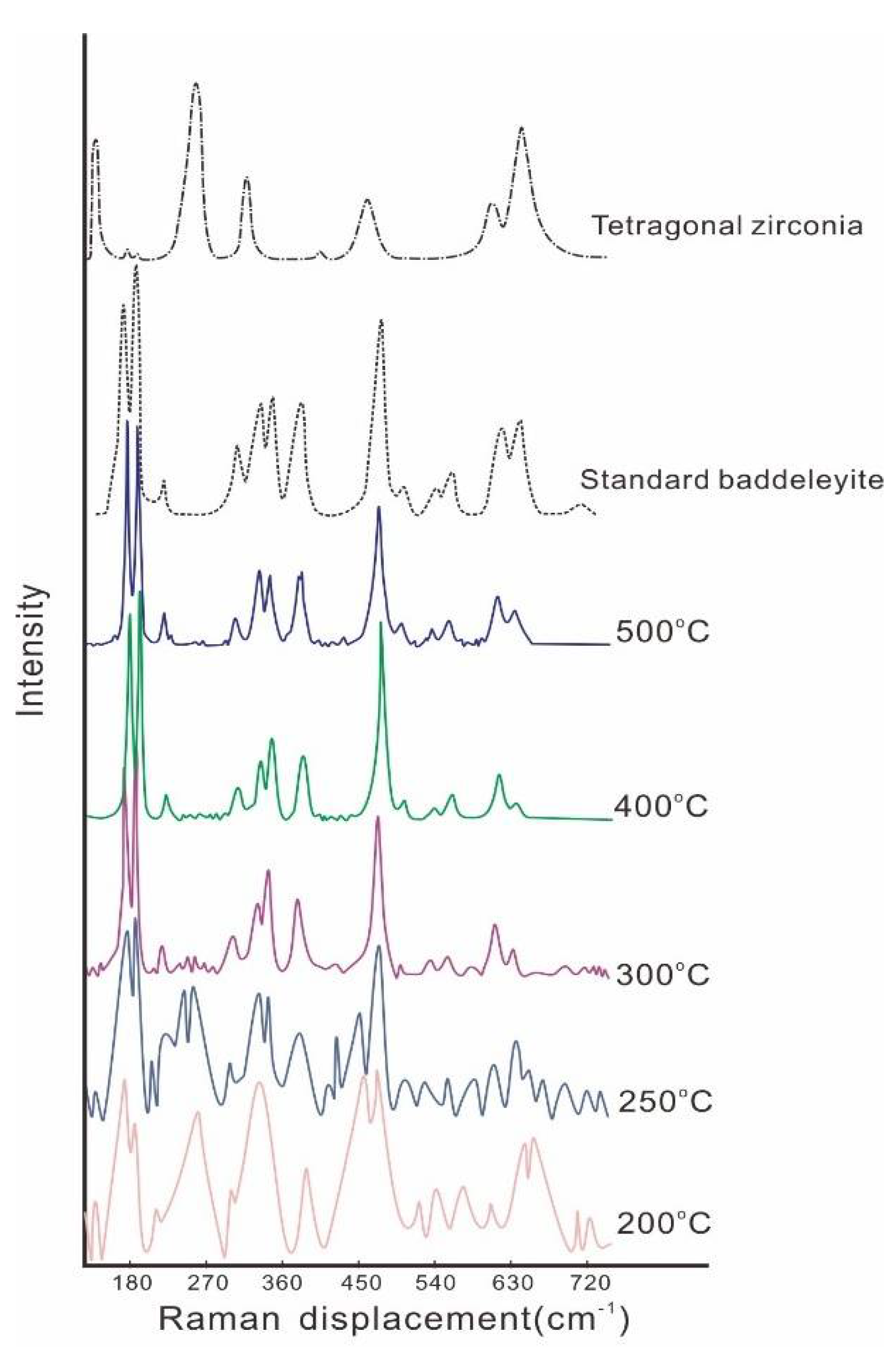
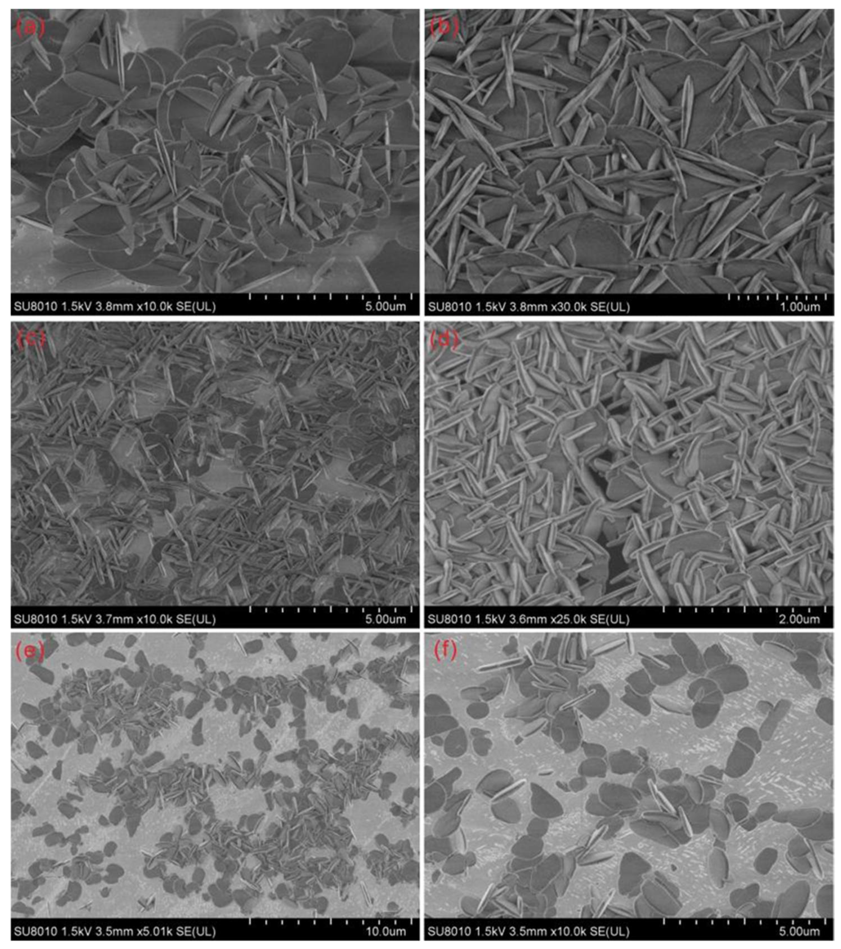
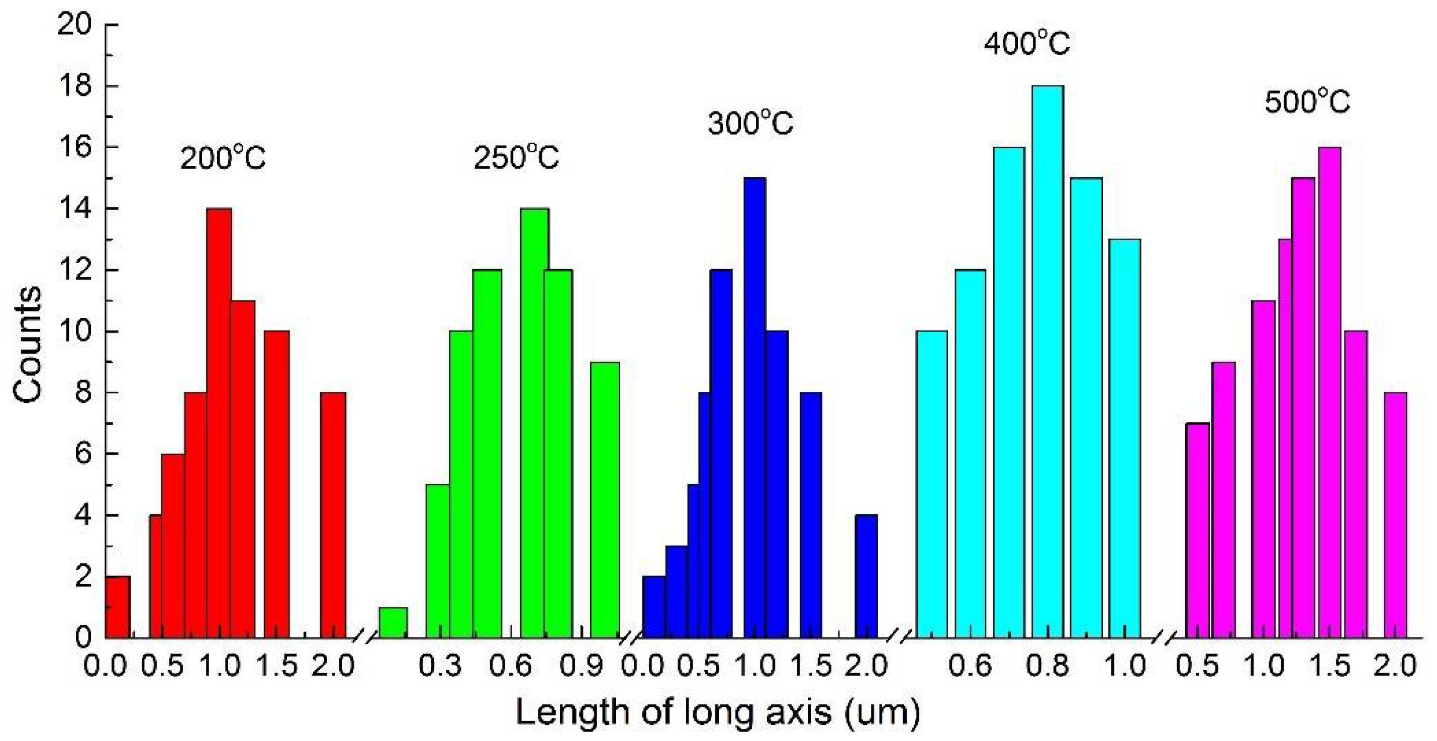
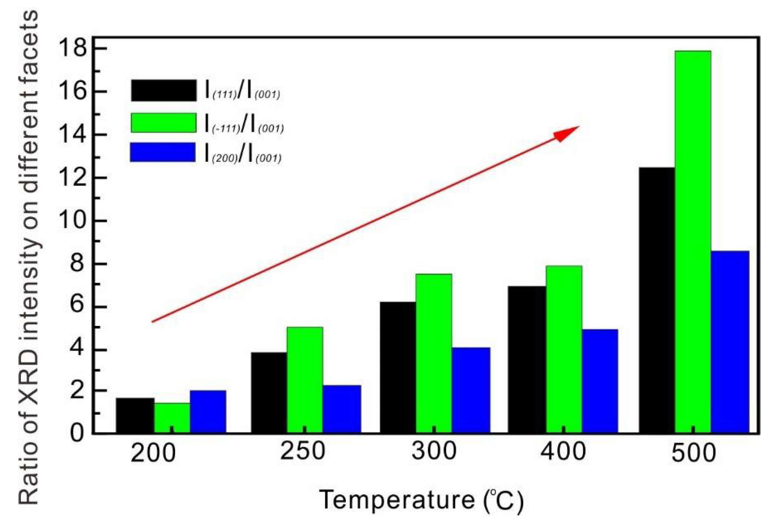
3.1. Temperature Dependence on the Crystal Form and the Morphology of the Synthesized Zirconia

The synthesis temperature has an important influence on the crystal form of synthesized zirconia based on the result of the Raman analysis. As shown in Figure 2, the results of the Raman spectrum show that 14 Raman characteristic peaks of synthesized zirconia at 200 °C and 250 °C are similar to that of the standard baddeleyite (monoclinic system). In addition, 6 Raman characteristic peaks of synthesized zirconia appear at 144 cm−1, 258 cm−1, 318 cm−1, 462 cm−1, 612 cm−1, and 640 cm−1, which is consistent to that of the Raman spectrum for the tetragonal system, indicating a mixture of monoclinic and tetragonal systems at 200 °C and 250 °C. The Raman spectrum of synthesized zirconia demonstrated that it is a monoclinic system mixed with a few amounts of the tetragonal system at 300 °C but is a pure monoclinic system at 400 °C and 500 °C (Figure 2). The consistent results were also demonstrated by the μ-XRD spectrum (Figure 3). Therefore, the synthesized zirconia is a pure monoclinic system at elevated temperatures (≥400 °C) but is a mixture of monoclinic and tetragonal systems at a relatively lower temperature (≤300 °C). This suggests that elevated temperatures can promote the growth of a monoclinic system and reduce the formation of a tetragonal system in the experimental temperature range.
In addition to the crystal form of synthesized products, temperature also has an important effect on the hydrolytic reaction’s degree and morphology of hydrolytic products [32,34,35,36,40]. In this study, as the temperature increases, the morphology of synthesized zirconia also varied. For example, at similar synthesis conditions such as an initial solution concentration of 0.02 mol·L−1, pressure of 100 MPa, and runtime of 12 h, the synthesized zirconia is in the form of an elliptical micro-nanoflake with a thickness of 1–50 nm and length of 0.1–2.0 μm at 200 °C (Figure 4a), and in contrast, at 250 °C, it is in the form of an elliptical micro-nanoflake or irregular micro-nanoflake with a thickness of 10–50 nm and length of 0.1–1.0 μm (Figure 4b); additionally, it changes to a form with a thickness of 50–100 nm and length of 0.5–1.0 μm at 400 °C (Figure 4d). The results demonstrate that the synthesized zirconia becomes more uniform in particle size and thicker in the (001) facets from 10–50 nm to 50–100 nm and longer in the diameter from 0.1–1.0 μm to 0.5–1.0 μm as the temperature increases from 200 °C to 400 °C (Figure 6). Compared to the morphology of zirconia at other synthesis conditions, zirconia synthesized at 500 °C has a larger crystal size (i.e., 50–100 nm thick and 0.5–2.0 μm long) than those at 200–400 °C (Figure 4e,f and Figure 5). This also suggests that elevated temperatures can promote the growth of zirconia by increasing its size and the thickness of the (001) facet considering the possible effect of pressure. Similarly, compared to zirconia’s morphology synthesized at 250–500 °C and a lower initial solution concentration of 0.01 mol·L−1 (Figure 5), the effect of temperature for the growth of zirconia with the (001) facets can be observed even in the case of inconsistent pressure and time. The influence of temperature on the morphology of synthesized zirconia could be explained by the process of hydrolytic reaction. As the temperature increases, the degree of hydrolytic reaction will increase, resulting in Equation (1) moving to the right and forming more precipitates and HF [36]. Hence, a large amount of ZrO2 precipitated from the solution can facilitate zirconia’s growth, resulting in an increase of zirconia in size.

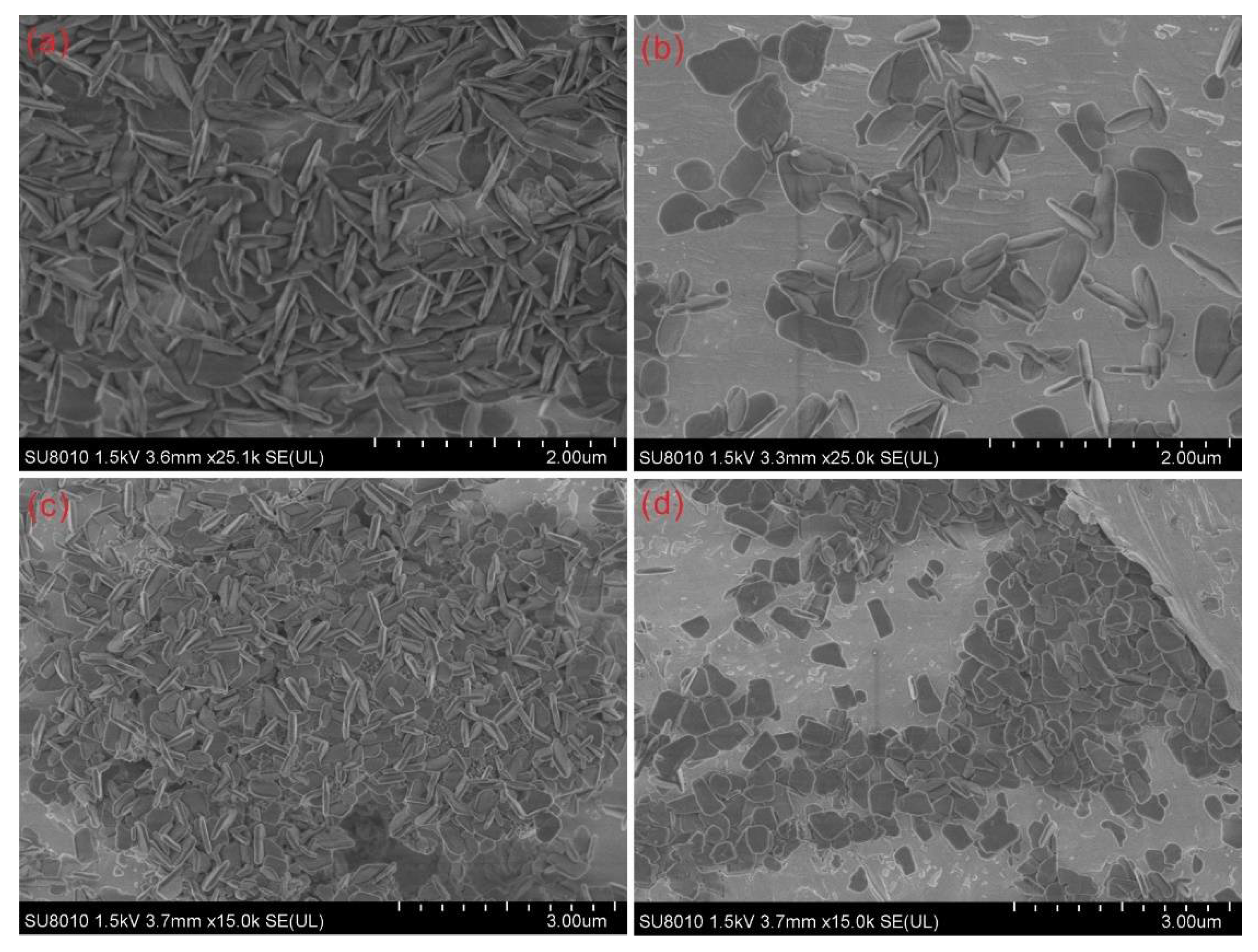
3.2. Effect of Initial Solutions’ Concentration on the Morphology of the Synthesized Zirconia

Apart from the temperature, the initial solution concentration of H2ZrF6 also has a positive effect on the morphology of the synthesized zirconia. As shown in Figure 4e,f and Figure 5c,d, at the same synthesis conditions including the temperature of 500 °C, pressure of 80 MPa, and runtime of 12 h, the synthesized zirconia crystals grow from small irregular micro-nanoflakes (50–80 nm thick and 0.5–1.0 μm long) at 0.01 mol·L−1 of the initial solution to larger elliptical micro-nanoflakes or irregular micro-nanoflakes (50–100 nm thick and 0.5–2.0 μm long) at 0.02 mol·L−1 of the initial solution. The synthesized zirconia at a high initial solution concentration is also larger in size than that of a low initial solution concentration (Figure 4b,d and Figure 5a,b). These results suggest a promotive effect of high initial solution concentration on the growth of the synthesized zirconia. This could also be explained by the degree of the hydrolytic reaction of H2ZrF6. With the concentration of the initial H2ZrF6 solution increasing, Equation (1) will move to the right and form a large amount of precipitates and HF, resulting in the growth and aggregation of the synthesized zirconia micro-nanoflake with high (001) facets [34,36].

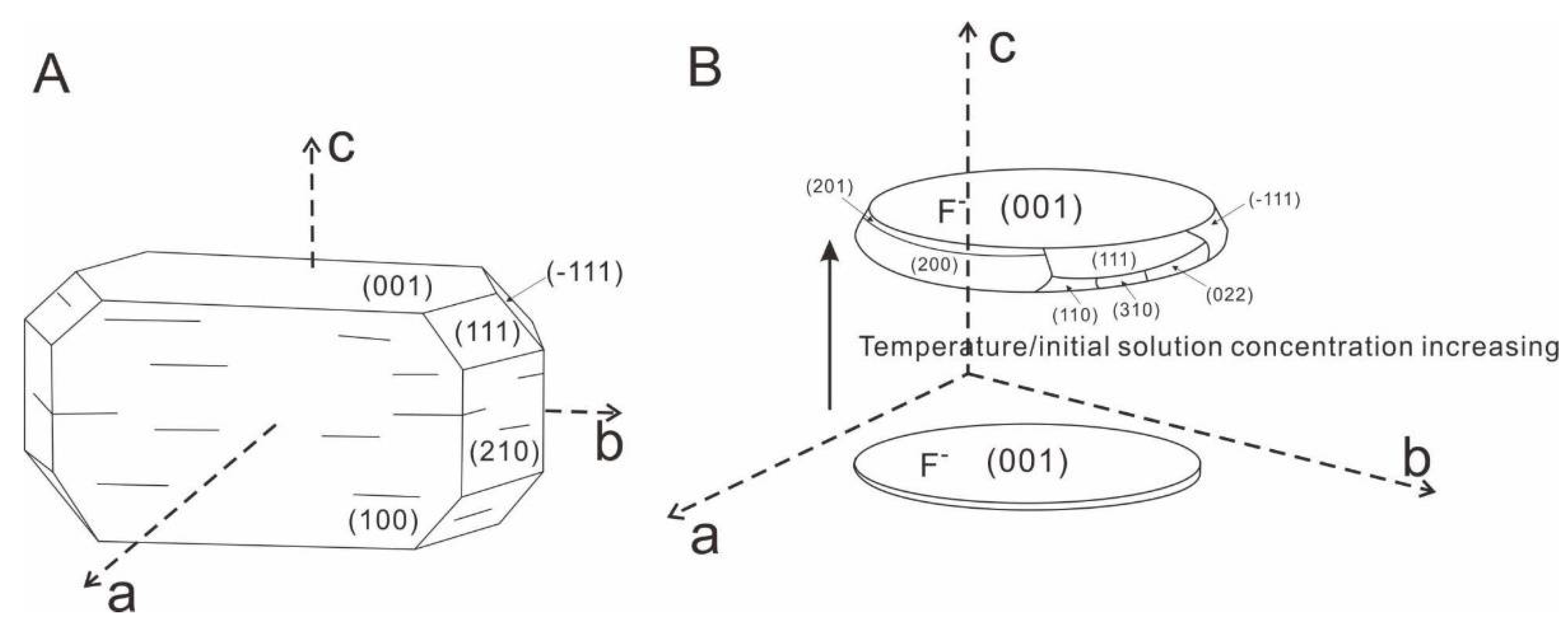
3.3. Growth Mechanism of the Synthesized Zirconia

Crystal nucleation and growth is usually involved in the conventional atom-by-atom growth model, non-classical particle aggregation, and amorphous transformation [41,42]. In the present study, as the temperature increased, H2ZrF6 in the solutions hydrolyzed to produce zirconia precipitates that subsequently experienced nucleation, aggregation, and coarsening to generate zirconia micro-nanoflakes in addition to the phase transformation. A basic observation would state that as the temperature and initial solution concentration increases, the particle size of the synthesized zirconia micro-nanoflakes becomes more uniform and the thicknesses of the (001) facets becomes larger, suggesting that the synthesized zirconia crystals grow mostly along the (001) facets and c-axis. This is also confirmed by the data derived from the μ-XRD patterns. As shown in Figure 3, the peak intensity of (001), (111), (−111), and (200) facets increases as the temperature increase from 200 °C to 500 °C. Given that the (111), (−111), (022), and (200) facets are perpendicular or nearly perpendicular to the (001) facets, they grow mainly along the c-axis and can induce the thickening of the (001) facets significantly. Remarkably, the ratios of I(111)/I(001), I(−111)/I(001), I(200)/I(001) all rise from 200 °C to 500 °C (Figure 7) which could indicate a fast growth rate of the flank facets along the c-axis direction at high temperatures, resulting in the faster growth of those facets over the (001) facets.
Note that the synthesized zirconia crystals could grow the (001) facets mostly prior to the hydrolysis equilibrium of H2ZrF6. As the hydrolysis of H2ZrF6 can produce HF, surface fluorine of the zirconia micro-nanoflake could play a key role in stabilizing the (001) facets considering fluorine’s role in the growth of the anatase (001) facets [43]. During the growth of zirconia, fluorine anions generated by ionization of HF from the hydrolytic reaction adhere to the (001) facets of zirconia to reduce the surface energy and promote the growth of the (001) facets. Meanwhile, HF or H+ in the solutions could inhibit the growth of other facets of zirconia micro-nanoflakes compared to the ideal monoclinic zirconia crystal (Figure 8A). During the hydrolysis, the amount of precipitation driven by the hydrolysis of H2ZrF6 could be maximized which induces the (001) facets to grow at a very high rate; however, when the hydrolysis reaches equilibrium, the amount of precipitation would be reduced and causes a slower growth rate [44]. In the present study, all zirconia micro-nanoflakes were synthesized within a long duration that is sufficient for the H2ZrF6 to reach equilibrium, finally causing a major growth along the c-axis direction by the oriented attachment mechanism (Figure 8B). The oriented attachment-based growth results in the changes of zirconia micro-nanoflakes in terms of size, thickness, and superficial area to ensure that the (001) facets are well developed as other crystal facets gradually develop (Figure 8B). The highly exposed (001) facets for the new zirconia micro-nanoflakes likely have great application potentials on catalysis, adsorption, sensors, and ceramic materials [5,26].
Figure 2. Raman spectra of zirconia micro-nanoflakes synthesized in the present study and standard monoclinic (e.g., baddeleyite) and tetragonal zirconia (ZrO2) crystals. The standard Raman spectrum of baddeleyite is derived from an online Raman database (https://rruff.info/baddeleyite/display=default/R060016 (accessed on 8 May 2021), while the Raman spectrum of tetragonal zirconia (ZrO2) is from Reference [45].
Crystals 11 00871 g002
Figure 3. Representative XRD analysis of zirconia micro-nanoflakes synthesized at 200–500 °C. The XRD spectra of Au, monoclinic zirconia (ZrO2), and tetragonal zirconia (ZrO2) are from the database (No.00-004-0784, 00-037-1484, and 00-002-0733).
Crystals 11 00871 g003
Figure 4. Representative SEM micrographs of zirconia micro-nanoflakes synthesized by 0.02 mol/L of the initial solution concentration: (a) No. 1 at 200 °C, 100 MPa, and 12 h; (b) No. 3 at 250 °C, 100 MPa, and 12 h; (c) No. 4 at 300 °C, 120 MPa, and 12 h; (d) No. 5 at 400 °C, 100 MPa, and 12 h; and (e,f) No. 8 at 500 °C, 80 MPa, and 12 h.
Crystals 11 00871 g004
Figure 5. Representative SEM micrographs of zirconia micro-nanoflakes synthesized by 0.01 mol/L of the initial solution concentration: (a) No. 2 at 250 °C, 80 MPa, and 14 h; (b) No. 6 at 400 °C, 80 MPa, and 12 h; and (c,d) No. 7 at 500 °C, 80 MPa, and 12 h.
Crystals 11 00871 g005
Figure 6. Statistical histograms of long-axis sizes for zirconia micro-nanoflakes (ZrO2) synthesized at 250–500 °C.
Crystals 11 00871 g006
Figure 7. A histogram illustrating the ratios on the μ-XRD intensity of the (111), (−111), and (200) facets compared to the (001) facets. As the temperature increases, all the ratios rise, suggesting that the growth of flank facets causes the thickening of zirconia micro-nanoflakes.
Crystals 11 00871 g007
Figure 8. A schematic diagram of zirconia crystals with high (001) facets, demonstrating (A) the crystallographic structure of an ideal monoclinic zirconia (e.g., baddeleyite) and (B) a growth model for zirconia micro-nanoflakes.
Crystals 11 00871 g008

4. Conclusions

In this paper, we synthesized zirconia micro-nanoflakes with high (001) facets at 200–500 °C and 80–120 MPa by a pyrohydrolysis procedure of H2ZrF6. Zirconia crystals synthesized are 1–100 nm thick and 0.1–2.0 μm long elliptical or irregular micro-nanoflakes that are highly exposed in the (001) facets. The zirconia micro-nanoflakes belong to a mixture of monoclinic and tetragonal systems at the temperatures of ≤300 °C but belong to a pure monoclinic system at the temperatures of ≥400 °C. As the temperature and initial H2ZrF6 concentration increase, the synthesized zirconia micro-nanoflakes become more uniform, mostly derived from the growth along the (001) facets. The increasing μ-XRD intensity of the (111), (−111), and (200) facets and the increasing ratios of I(111)/I(001), I(−111)/I(001), I(200)/I(001) from 200 °C to 500 °C indicate an oriented attachment-based major growth along the c-axis that results in the thickening of the (001) facets with increasing temperatures. This suggests that the oriented attachment-based growth occurring in the fluorine-rich solution environment was involved in the aggregation and coarsening of zirconia micro-nanoflakes.

Author Contributions

Conceptualization, X.D.; methodology, X.D., Z.L. and J.L. (Junfeng Liu); formal analysis, J.D., H.Y. and J.L. (Jiahao Li); investigation, H.Y., J.D., Z.L. and J.L. (Jiahao Li); writing—original draft preparation, X.D. and H.Y.; writing—review and editing, J.L. (Junfeng Liu); visualization, X.D. and H.Y.; project administration, X.D. and J.L. (Junfeng Liu); funding acquisition, X.D. All authors have read and agreed to the published version of the manuscript.

Funding

This study was funded by the National Natural Science Foundation of China, grant number (41773054).

Institutional Review Board Statement

Not applicable.

Informed Consent Statement

Not applicable.

Data Availability Statement

Data sharing is not applicable to this article.

Acknowledgments

We thank the two anonymous reviewers and also Xavier Zang, Ulla Simon, and Maged F. Bekheet for their constructive comments and suggestions. This is also a contribution from GIGCAS, number IS-3053.

Conflicts of Interest

The authors declare no conflict of interest.

References

  1. Zhou, P. Preparation and Characterization of Zirconia Ceramic Hollow Shells; Harbin Institute of Technology: Harbin, China, 2009. [Google Scholar]
  2. Chang, J.P.; Lin, Y.S.; Chu, K. Rapid thermal chemical vapor deposition of zirconium oxide for metal-oxide-semiconductor field effect transistor application. J. Vac. Sci. Technol. B Microelectron. Nanometer Struct. Proc. Meas. Phenom. 2001, 19, 1782–1787. [Google Scholar] [CrossRef]
  3. Yu, H.; Wang, Z.Y.; Zhu, G.D.; Dai, Y.; Wang, Y.B.; Liu, J.Y. Electrospun Zirconia Nanofibre-modified Glassy Carbon Electrode for Determination of Methyl Parathion. Chin. J. Anal. Chem. 2018, 46, 1982–1989, (In Chinese with English). [Google Scholar]
  4. He, W.Y.; Liu, J.R.; Cao, Z.Z.; Li, C.H.; Gao, Y.F. Preparation and characterization of monodisperse zirconia spherical nanometer powder via lamellar liquid crystal template method. Chin. J. Chem. Eng. 2015, 23, 1721–1727. [Google Scholar] [CrossRef]
  5. Wijayanti, R.B.; Rosmayanti, I.; Wahyudi, K.; Maryani, E.; Hernawan, H.; Septawendar, R. Preparation of magnesia partially stabilized zirconia nanomaterials from zirconium hydroxide and magnesium carbonate precursors using PEG as a template. Crystals 2021, 11, 635. [Google Scholar] [CrossRef]
  6. Gao, Q.W.; Jiang, W.H.; Liu, J.M.; Feng, G.; Chen, T.; Miao, L.F. Preparation of Zirconia Nanorods by Molten Salt Method. Chin. J. Inorg. Chem. 2017, 33, 1555–1560, (In Chinese with English Abstract). [Google Scholar]
  7. Kang, J.; Ji, W.L.; Bu, J.L.; Chen, Y.; Wei, H.Y.; Lv, D.F.; Cui, Y.; Chen, Y.J.; Wei, Y.N.; Gong, L.P.; et al. Study on process of electrospinning preparation of zirconia hollow microspheres. Refract. Mater. 2018, 53, 170–175, (In Chinese with English Abstract). [Google Scholar]
  8. Gao, Q.W.; Liu, J.M.; Jiang, W.H.; Feng, G.; Chen, T.; Miao, L.F. Effect of Solvothermal Temperature on the Preparation of Zirconia Whiskers. J. Ceram. 2018, 39, 154–158, (In Chinese with English Abstract). [Google Scholar]
  9. Kwon, J.H.; Choi, J.H.; Bae, J.H.; Park, J. Hysteresis reduction for organic thin film transistors with multiple stacked functional zirconia polymeric films. Crystals 2019, 9, 634. [Google Scholar] [CrossRef]
  10. Zhu, J.F.; Xiao, D.; Li, C.Y.; Huang, J.F.; Yong, X.; Cao, L.Y.; Fei, J. Microwave hydrothermal synthesis and crystallization mechanism of tetragonal phase zirconia nanocrystalline. J. Shaanxi Univ. Sci. Technol. (Nat. Sci. Ed.) 2015, 33, 60–68. [Google Scholar]
  11. Wu, J.; Lu, Y.F.; Liu, Y.; Yu, Z.Y.; Xin, B.F. Fabrication and Characterization of ZrO2 Nanofibers with Controllable Crystalline Phase by a Halid-free Approach. Chem. J. Chin. Univ. Chin. Ed. 2015, 36, 1403–1408, (In Chinese with English Abstract). [Google Scholar]
  12. Pan, L.; Xu, H.B.; Sun, Y.Y.; Zhao, J.P.; Li, Y. Preparation of three-dimensional photonic crystals of zirconia by electrodeposition in a colloidal crystals template. Crystals 2016, 6, 76. [Google Scholar] [CrossRef]
  13. Jayakumar, S.; Ananthapadmanabhan, P.V.; Perumal, K.; Thiyagarajan, T.K.; Mishra, S.C.; Su, L.T.; Tok, A.; Guo, J. Characterization of nano-crystalline ZrO2 synthesized via reactive plasma processing. Mater. Sci. Eng. B 2011, 176, 894–899. [Google Scholar] [CrossRef]
  14. Noh, H.J.; Seo, D.S.; Kim, H.; Lee, J.K. Synthesis and crystallization of anisotropic shaped ZrO2 nanocrystalline powders by hydrothermal process. Mater. Lett. 2003, 57, 2425–2431. [Google Scholar] [CrossRef]
  15. Binner, J.; Vaidhyanathan, B. Processing of bulk nanostructured ceramics. J. Eur. Ceram. Soc. 2008, 28, 1329–1339. [Google Scholar] [CrossRef] [Green Version]
  16. Gaudon, M.; Djurado, E.; Menzler, N.H. Morphology and sintering behaviour of yttria stabilised zirconium (8-YSZ) powders synthesised by spray pyrolysis. Ceram. Int. 2004, 30, 2295–2303. [Google Scholar] [CrossRef]
  17. Soo, M.T.; Prastqmo, N.; Matsuda, A.; Kawamura, G.; Muto, H.; Noor, A.; Lockman, Z.; Cheong, K.Y. Elaboration and characterization of sol–gel derived ZrO2 thin films treated with hot water. Appl. Surf. Sci. 2012, 258, 5250–5258. [Google Scholar] [CrossRef]
  18. Garg, N.; Mittal, V.K.; Bera, S.; Dasgupta, A.; Sankaralingam, V. Preparation and characterization of tetragonal dominant nanocrystalline ZrO2 obtained via direct precipitation. Ceram. Int. 2012, 38, 2507–2512. [Google Scholar] [CrossRef]
  19. Chang, Y.; Li, X.B. Preparation of ZrO2 spherical nanometer powders. J. Cent. South. Univ. (Sci. Technol.) 2007, 38, 46–50, (In Chinese with English Abstract). [Google Scholar]
  20. Silva Junior, E.; Antonio, S.G.; Longo, E. Synthesis and structural evolution of partially and fully stabilized ZrO2 from a versatile method aided by microwave power. Ceram. Int. 2018, 44, 3517–3522. [Google Scholar] [CrossRef] [Green Version]
  21. Wang, X.X.; Lin, J.W.; Zhan, Y.H.; Zhang, Z.B.; Chu, M. Adsorption of Phosphate from Aqueous Solution on Hydrous Zirconium Oxides Precipitated at Different pH Values. Enviorn. Sci. 2017, 38, 1936–1946. [Google Scholar] [CrossRef]
  22. Ji, Y.; Han, L.Z.; Zheng, W.Q.; Wang, Q.; Yang, H.Z. Influence of pH values on different zirconia systems. Chin. J. Tissue Eng. Res. 2017, 21, 4131–4136. [Google Scholar]
  23. Wang, H.Y.; Guo, Z.S.; Xing, G.E.; Song, X.Q. Study on Preparing Nano-sized ZrO2 by Different Techniques. J. Synth. Cryst. 2006, 35, 753–756. [Google Scholar]
  24. Wang, J.J. Research progress of photocatalytic performance of nanometer zirconia. Light Ind. Stand. Qual. 2017, 2, 68–69. (In Chinese) [Google Scholar]
  25. Li, W.; Liu, F.H.; Wu, D.W. Preparation of Flexible Yttria-stabilized Zirconia Nanofibers. Guangzhou Chem. Ind. 2018, 46, 45–51, (In Chinese with English Abstract). [Google Scholar]
  26. Xu, G.F.; Song, H.Z.; Yang, D.L. The Hdrothermal Preparation of Stable Zirconia Nano Powder. Surf. Technol. 2017, 46, 95–100. [Google Scholar] [CrossRef]
  27. Du, P.; Zhang, J.F.; Zhao, J.L.; Wang, X.X.; Zhang, L.B. Study on decolourization and degradation of methyl rrange by stir-ultrasonic ZrO2 nanotubes. J. Synth. Cryst. 2015, 44, 2518–2523, (In Chinese with English Abstract). [Google Scholar]
  28. Wang, X.X.; Zhao, J.L.; Guo, L.M.; Yu, X.; Tang, C.C. Fabrication of Zirconia Nanotube Arrays on Curved Surface of Zirconium Wire. Rare Met. Mat. Eng. 2012, 41, 35–37, (In Chinese with English Abstract). [Google Scholar]
  29. Li, J. Study on performance of nanometer zirconia-cerium oxide composites. Sci. Technol. Inf. 2014, 3, 51–79, (In Chinese with English Abstract). [Google Scholar]
  30. Guo, J.H.; Li, X.H.; Li, Z.W.; Du, Z.L.; Zhang, Z.J. Synthesis of sheet-like silica/zirconia nanocomposites by sol-gel process. Chem. Res. 2011, 22, 49–54. [Google Scholar]
  31. Yang, S.F. Study on liquid phase reaction self-assembly preparation and performance of ZrO2 films. Tianjin Chem. Ind. 2015, 29, 31–33. [Google Scholar]
  32. Yan, H.B.; Sun, W.D.; Liu, J.F.; Tu, X.L.; Ding, X. Thermodynamic properties of ruthenium (IV) chloride complex and the transport of ruthenium in magmatic-hydrothermal fluids. Ore Geol. Rev. 2021, 131, 104043. [Google Scholar] [CrossRef]
  33. He, J.J.; Ding, X.; Wang, Y.R.; Sun, W.D. The effects of precipitation-aging-re-dissolution and pressure on hydrolysis of fluorine-rich titanium complexes in hydrothermal fluids and its geological implications. Acta Pet. Sin. 2015, 31, 1870–1878, (In Chinese with English Abstract). [Google Scholar]
  34. He, J.J.; Ding, X.; Wang, Y.R.; Sun, W.D. The effect of temperature and concentration on hydrolysis of fluorine-rich titanium complexes in hydrothermal fluids: Constraints on titanium mobility in deep geological processes. Acta Pet. Sin. 2015, 31, 802–810, (In Chinese with English Abstract). [Google Scholar]
  35. Di, J.; Yan, H.B.; Liu, Z.Y.; Ding, X. Synthesis and Characterization of Anatase TiO2 Microspheres Self-Assembled by Ultrathin Nanosheets. Materials 2021, 14, 2870. [Google Scholar] [CrossRef]
  36. Yan, H.B.; He, J.J.; Liu, X.W.; Wang, H.B.; Liu, J.F.; Ding, X. Thermodynamic Investigation of the Hydrolysis Behavior of Fluorozirconate Complexes at 423.15–773.15 K and 100 MPa. J. Solut. Chem. 2020, 49, 836–848. [Google Scholar] [CrossRef]
  37. Zhang, L.; Wang, Q.; Ding, X.; Li, W.C. Diverse serpentinization and associated abiotic methanogenesis within multiple types of olivine-hosted fluid inclusions in orogenic peridotite from northern Tibet-Science Direct. Geochim. Cosmochim. Acta 2021, 296, 1–17. [Google Scholar] [CrossRef]
  38. Baes, J.C.F.; Mesmer, R.E. The thermodynamics of cation hydrolysis. Am. J. Sci. 1980, 281, 935–962. [Google Scholar] [CrossRef]
  39. Ding, X.; Harlov, D.E.; Chen, B.; Sun, W.D. Fluids, metals, and mineral/ore deposits. Geofluids 2018, 1452409. [Google Scholar] [CrossRef] [Green Version]
  40. Wang, Y.R.; Chou, I.M. Characteristics of hydrolysis of the complex Na2SnF6 in hydrothermal solutions—An experimental study. Chin. J. Geochem. 1987, 6, 372–382. [Google Scholar] [CrossRef]
  41. Lee, G.S.; Lee, Y.J.; Yoon, K.B. Layer-by layer assembly of zeolite crystals on glass with polyelectrolytes as ionic linkers. J. Am. Chem. Soc. 2001, 123, 9769–9779. [Google Scholar] [CrossRef]
  42. De Yoreo, J.J.; Gilbert, P.U.P.A.; Sommerdijk, N.A.J.M.; Penn, R.L.; Whitelam, S.; Joester, D.; Zhang, H.Z.; Rimer, J.D.; Navrotsky, A.; Banfield, J.F.; et al. Crystallization by particle attachment in synthetic, biogenic, and geologic environments. Science 2015, 349, aaa6760. [Google Scholar] [CrossRef]
  43. Yang, H.G.; Sun, C.H.; Qiao, S.Z.; Zou, J.; Liu, G.; Smith, S.C.; Cheng, H.M.; Lu, G.Q. Anatase TiO2 single crystal with a large percentage of reactive facets. Nature 2008, 453, 638–642. [Google Scholar] [CrossRef] [PubMed] [Green Version]
  44. Ding, X.; He, J.J.; Liu, Z.Y. Experimental studies on crystal growth of anatase under hydrothermal conditions. Earth Sci. 2018, 43, 1763–1772. [Google Scholar]
  45. Bouvier, P.; Lucazeau, G. Raman spectra and vibrational analysis of nanometric tetragonal zirconia under high pressure. J. Phys. Chem. Solids 2000, 61, 569–578. [Google Scholar] [CrossRef]
Figure 1. Representative EDS analysis of zirconia micro-nanoflakes adhered to the inner surface of gold capsule. Note that N could derive from the starting initial solutions and Au is derived from the gold capsule.
Crystals 11 00871 g001
Table 1. Preparation on zirconia micro-nanoflakes with high (001) facets under varied conditions.
No.Initial Concentration (mol/L)Temperature (°C)Pressure (MPa)Time (h)Thickness (nm)Length (μm)
10.02200100121–500.1–2.0
20.01250801410–500.1–1.0
30.022501001210–500.1–1.0
40.023001201220–800.1–2.0
50.024001001250–1000.5–1.0
60.01400801220–800.5–1.0
70.01500801250–800.5–1.0
80.02500801250–1000.5–2.0
Publisher’s Note: MDPI stays neutral with regard to jurisdictional claims in published maps and institutional affiliations.

