Article

Synthesis and Optical Characterizations of Yb3+: CaxSr1−xF2 Transparent Ceramics

State Key Laboratory of Advanced Technology for Materials Synthesis and Processing, Wuhan University of Technology, Wuhan 430070, China
*
Author to whom correspondence should be addressed.
Crystals 2021, 11(6), 652; https://doi.org/10.3390/cryst11060652
Submission received: 11 May 2021 / Revised: 3 June 2021 / Accepted: 5 June 2021 / Published: 8 June 2021

Abstract

:
In this study, 3 at.% Yb3+: CaxSr1−xF2 nanopowders were synthesized via the chemical co-precipitation method. Highly transparent 3 at.% Yb3+: CaxSr1−xF2 ceramics with various CaF2 concentrations were fabricated by hot-pressed sintering. The 3 at.% Yb3+: CaxSr1−xF2 nanopowders exhibited a spherical shape with slight agglomeration, and their particle size ranged from 26 nm to 36 nm. With an increase of the CaF2 concentration, the peak shape changed significantly and the width of the emission band increased inhomogeneously. The minimal fluorescence lifetime at the wavelength of 1011 nm of 3 at.% Yb3+: CaxSr1−xF2 transparent ceramics with various CaF2 concentrations was higher than 3.25 ms, which was longer than that of the 3 at.% Yb3+: CaF2 (2.6 ms) and the 3 at.% Yb3+: SrF2 (3.22 ms) reported in previous literature. The results indicate that incorporating Ca2+ ions into the SrF2 is an effective method to modulate the optical properties of transparent ceramics.

1. Introduction

Since the first Dy: CaF2 transparent ceramics were prepared by Hatch et al. in 1964 [1], alkaline-earth fluorides (AeF2, Ae = Ca, Sr, Ba) have attracted much attention in the field of solid-state lasers because of their low phonon energy, high transmittance, and wide range of light transmission [2,3,4]. For instance, Su et al. prepared Tm3+: CaF2 single crystals and their slope efficiency reached up to 64.4%, which was higher than Tm3+: YAG single crystals with 31.8% [5,6]. Basiev, T. T. et al. fabricated Nd3+: SrF2 laser ceramics with a laser slope efficiency of 19% [7]. On the other hand, AeF2 possessed the cubic structure ( F 3 m m ) with fluoride ions occupying the centers of the octants and Ae2+ ions occupying the nodes in a face-centered lattice, which made it possible for the Re3+ (rare earth) ions to incorporate in the AeF2 crystal structure and achieve high dopant concentrations. Moreover, various Re3+ ions have been successfully doped into alkaline earth fluorides, such as Nd3+: CaF2 crystals [8], Eu3+: CaF2 transparent ceramics [9], Er3+: CaF2 glass-ceramics [10], and Nd3+: SrF2 nanoparticles [11].
Among the Re3+ ions, Yb is one of the most promising elements for solid-state laser materials like high power or ultrafast lasers. Yb3+ ions possess a simple electronic-level structure with only two multiples 2F7/2 and 2F5/2, enabling efficient diode-pump laser systems [12,13,14]. A. Lucca et al. prepared the first diode-pumped laser based on the Yb3+: CaF2 single crystal and achieved a laser output of 5.8 W at 1053 nm [15]. Subsequently, a Yb3+: CaF2 single crystal achieved a 150 femtosecond, 880 mW laser output at the central wavelength of 1043 nm [16]. However, the local environment of Yb3+ ions affects its optical properties [17].
One of the effective methods to alter the local environment of Yb3+ ions is introducing non-active ions, like Ga3+ ions. Y.J. Wu and his co-workers modified their lattice structure by co-doping Ga3+ ions into Yb3+: SrF2 to form a disordered lattice site, broadening the absorption and emission cross-sections, and explored the potential of the Yb3+, Ga3+: SrF2 crystal in tunable and passively mode-locked lasers [18]. Further, due to the different radii of Ca2+ ions and Sr2+ ions, the local environment of rare earth ions in CaxSr1−xF2 ceramics varies with the concentration of CaF2. Recently, a few studies have researched the CaF2-SrF2 mixed matrix because incorporating the Ca2+ ions into the SrF2 destroys the symmetry of the crystal structure, thereby affecting its luminescence performance [19,20,21]. Compared with the Yb3+: SrF2 single crystal, the slope efficiency of CaF2-SrF2-YbF3 nanoceramics is 1.7 times higher than the former [22]. Therefore, it is meaningful to investigate the influence of CaF2 concentration on the optical properties of CaxSr1−xF2 transparent ceramics. However, according to the previous literature on CaxSr1−xF2 transparent ceramics, the ratio of SrF2 to CaF2 is relatively small [23]. When the proportion of SrF2 is higher than 25 at.%, the Sr2+ cannot incorporate into the CaF2 structure, and two single phases (CaF2 and SrF2) exist in the products [24]. In this paper, high purity of 3 at.% Yb3+: CaxSr1−xF2 (x = 0.1, 0.3, 0.5, 0.7 and 0.9) nanopowders were synthesized via the coprecipitation method and the corresponding transparent ceramics were prepared by hot-pressed sintering. The morphology of 3 at.% Yb3+: CaxSr1−xF2 nanopowders was observed by a scanning electron microscope (SEM). The optical quality, microstructure, and photoluminescence properties of 3 at.% Yb3+: CaxSr1−xF2 transparent ceramics were also characterized and discussed.

2. Experimental Procedure

The 3 at.% Yb3+: CaxSr1−xF2 (x = 0.1, 0.3, 0.5, 0.7 and 0.9) nanopowders were synthesized with the coprecipitation method by dropping the KF·2H2O aqueous solution into the cationic aqueous solution (Sr(NO3)2, Ca(NO3)2·4H2O and Yb(NO3)3·5H2O) under stirring. The 3 at.% Yb3+: CaxSr1−xF2 nanopowders were prepared following this formulation:
0.03Yb(NO3)3 + xCa(NO3)2 + (1 − x)Sr(NO3)2 + 2.09KF → Yb0.03CaxSr1−xF2.09↓ + 2.09KNO3
The obtained precipitates were aged for 3 h, and finally, the precipitates were washed with deionized water, filtered, and freeze-dried under vacuum for 8 h. The fully dried powder was ground in a mortar to remove large aggregates.
The 3 at.% Yb3+: CaxSr1−xF2 transparent ceramics were fabricated by hot-pressing the as-synthesized nanopowders in vacuum. The nanopowders were calcined at 400 °C for 2 h to remove residual water and nitrates from the powder, then sintered at 800 °C under an axial pressure of 40 MPa for 2 h followed by cooling down to room temperature. Then, the 3 at.% Yb3+: CaxSr1−xF2 transparent ceramics were both-side polished to 2 mm for further measurements.
The phase compositions of 3 at.% Yb: CaxSr1−xF2 ceramics were identified by X-ray diffraction (XRD, D8 Advance, Bruker, Karlsruhe, Germany). The morphology of nanopowders and the fracture microstructure of transparent ceramics of Yb3+: CaxSr1−xF2 were observed by a scanning electron microscope (SEM, S4800, Hitachi, Tokyo, Japan). The average particle size of the Yb3+: CaxSr1−xF2 transparent ceramic was calculated by scanning software. The in-line transmittance and absorption spectra of the Yb3+: CaxSr1−xF2 transparent ceramics were measured by a spectrophotometer (U-3500, Hitachi, Tokyo, Japan). The photoluminescence (PL) spectra and fluorescence lifetimes were recorded by a spectrofluorometer (FLS980, Edinburgh, Livingston, UK) at room temperature.

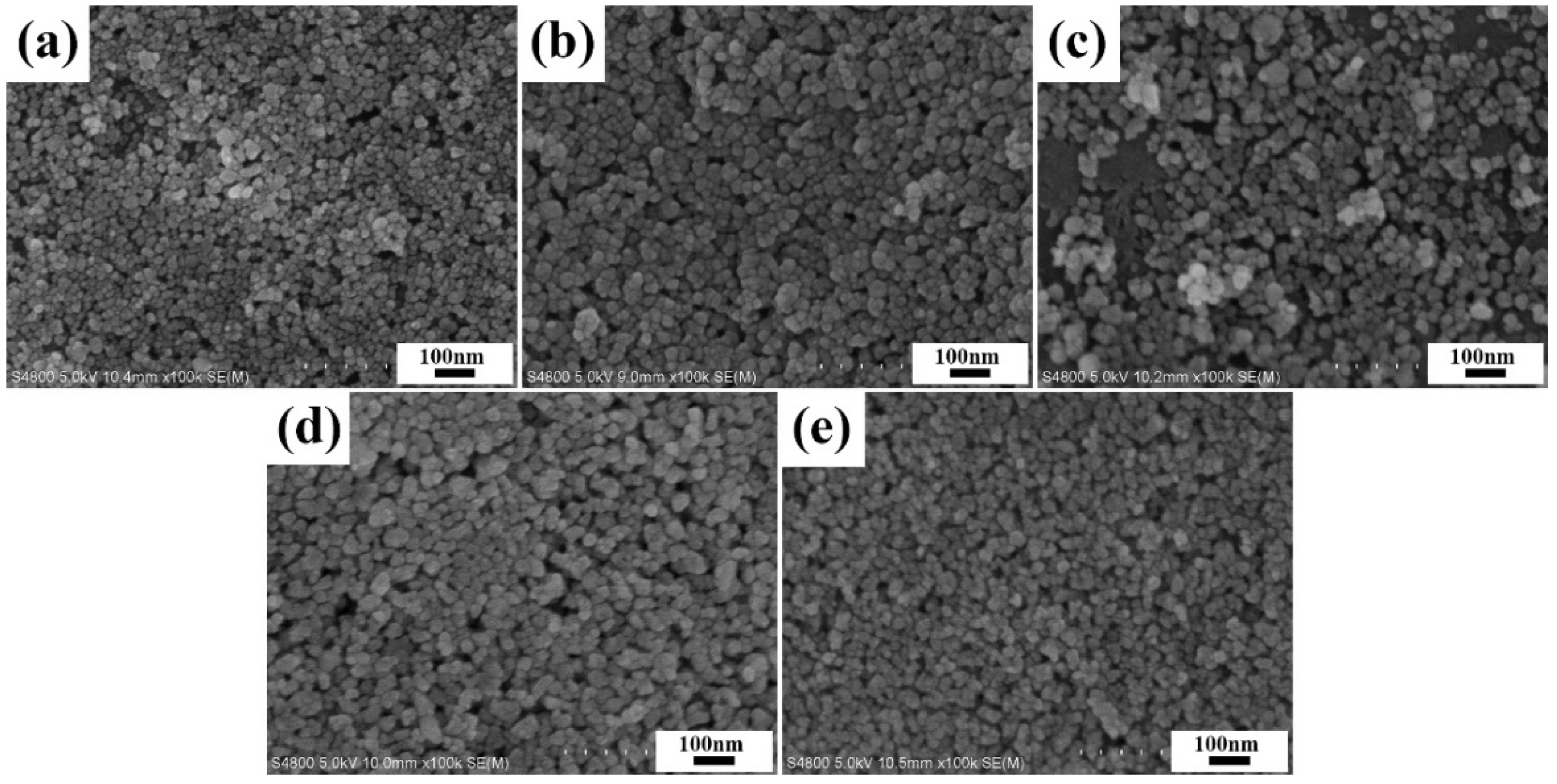
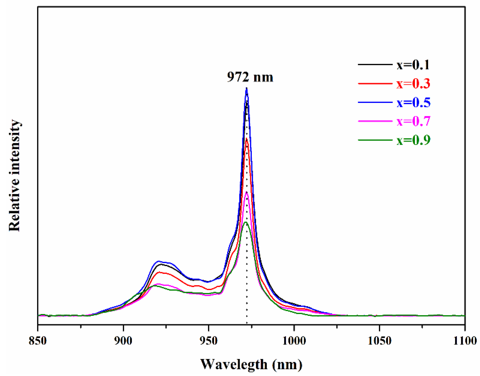
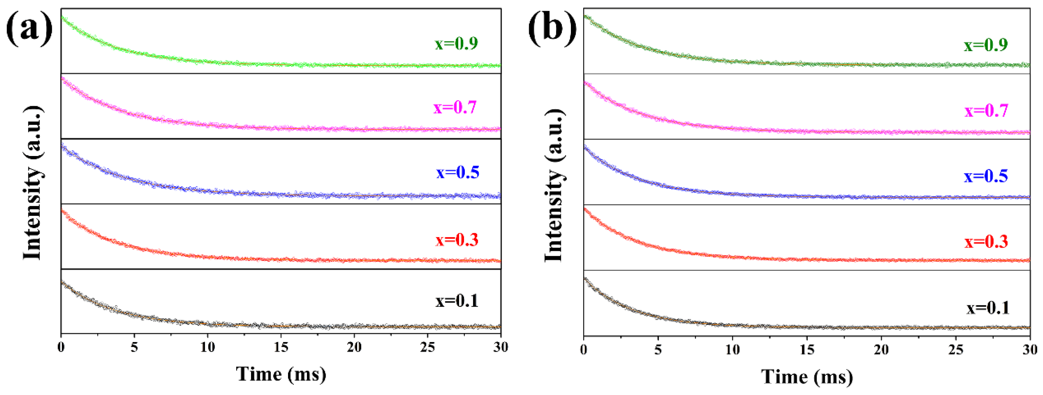
3. Results and Discussion

The XRD patterns of 3 at.% Yb3+: CaxSr1−xF2 transparent ceramics with various CaF2 concentrations and standard XRD patterns of CaF2 (JCPDS file number 65-0535) and SrF2 (JCPDS file number 06-0262) are shown in Figure 1a. All the diffraction peaks of 3 at.% Yb: CaxSr1−xF2 transparent ceramics corresponded well to the cubic CaF2 and/or SrF2 phase without redundant peaks. With the increase of CaF2 concentration, the position of all diffraction peaks shifted to a high angle, and was closer to the peak of pure CaF2, indicating that the position of Sr2+ ions was replaced by the Ca2+ ions in the SrF2 unit cell and formed a solid solution structure instead of a two-phase composite structure of CaF2 and SrF2. According to the Hume-Rothery solid solution theory, taking r1 and r2 to represent the ionic radius of different ions, there is the empirical formula [25]
Δ r = | r 1 r 2 r 1 |
According to the formula, the obtained value of Δ r was 10%. Further, the two ions had the same valence state and CaF2 and SrF2 possess the same fluorite structure. Therefore, Ca2+ ions can be incorporated into the SrF2 unit cell at any ratio to form a continuous solid solution instead of destroying the unit cell structure. In addition, the lattice constant of Yb3+: CaxSr1−xF2 was calculated by the Barrage equation and is presented in Figure 1b. At lower CaF2 concentrations, the lattice constant was close to that of SrF2, while at higher CaF2 concentrations, the lattice constant was close to that of CaF2. The linear relationship between the lattice constant and CaF2 concentration was corresponded well to the 2θ shift trend in Figure 1b. In the condition of the expansion of the lattice constant when larger Sr2+ ions were substituted, the stability of the unit cell structure was still maintained.
The SEM images of 3 at.% Yb3+: CaxSr1−xF2 (x = 0.1, 0.3, 0.5, 0.7 and 0.9) nanopowders are shown in Figure 2. All 3 at.% Yb3+: CaxSr1−xF2 nanopowders exhibited a spherical shape and uniform distribution in the field of vision with slight agglomeration. With the increase of the CaF2 concentration, there was nearly no significant change in the shape and particle size of the powder. The average particle size of the powders ranged from 26 nm to 34 nm.
The in-line transmittance spectra and photographs of as-fabricated 3 at.% Yb3+: CaxSr1−xF2 (x = 0.1, 0.3, 0.5, 0.7 and 0.9) transparent ceramics are presented in Figure 3. The 3 at.% Yb3+: Ca0.5Sr0.5F2 transparent ceramics had the highest optical transmittance among the five samples. The text on the paper can be clearly identified through the ceramics. The transmittance of all ceramics was higher than 80% over the wavelength of 1130 nm and reached 90% around 2000 nm, which might be attributed to the well-dispersed nanopowders. The small and spherical-shaped nanopowders had relatively large surface energy and high sintering activity, which was favorable for the sintering process. In addition, well dispersion of the nanopowders was conducive to the discharge of pores. However, the incorporation of CaF2 caused the lattice distortion of the matrix material, which affected the sintering activity of the synthesized nanopowders. Then, the diffusion rate of the powder was affected by the nanopowders which obsess different sintering activity, resulting in different pores in the sample. At ultraviolet wavelength range, the 3 at.% Yb3+: Ca0.5Sr0.5F2 transparent ceramic possessed the highest in-line transmittance, which might have been caused by the absence of impurities and fewer micropores in the ceramic. However, a significant transmittance loss was observed at the ultraviolet wavelength range similar to the CaF2 and SrF2 transparent ceramics [26,27], which might have been caused by fewer micro-obturator pores existing in the sample. When the size of the scattering source was much smaller than the wavelength of the incident light, the scattering intensity and the incident light wavelength conformed to Rayleigh’s law of scattering [28]:
S = ( 128 π 5 d 6 / 3 λ 4 ) { [ ( n 2 n 1 ) 2 1 ] / [ ( n 2 n 1 ) 2 2 ] } 2
where   S ,   d , λ , and n 2 and n 1 are the cross section for the scattering of a particle, the radius of the scattering body, the measuring wavelength, and the refractive indexes for the host materials and scattering body, respectively. According to Rayleigh’s law of scattering, scattering intensity is inversely proportional to the fourth power of the wavelength of the incident light. Therefore, the micropores inside the ceramic strongly led to the scatter of short-wavelength light such as ultraviolet light, which affected the transmittance of transparent ceramics in the ultraviolet wavelength range.
As can be seen in SEM micrographs of the fracture surface of 3 at.% Yb3+: CaxSr1−xF2 transparent ceramics (Figure 4), at a low CaF2 concentration (x = 0.1, 0.3), transgranular fractures accounted for a large proportion. With an increase of the CaF2 concentration, the transgranular fractures gradually decreased and tended to disappear, and intergranular fractures were the main fracture mode. Intracrystalline pores can affect the fracture mode of ceramic samples. It can be clearly seen that there were some intracrystalline pores inside the ceramics when the CaF2 concentration was low. The intracrystalline pores were the starting point of the crack propagation during the fracture process, which extended to the grain boundary to form transgranular fractures. When the intracrystalline pores gradually decreased, the proportion of transgranular fractures also decreased.
Figure 5 shows the relative absorption (divide intensity by Yb concentration) spectra of 3 at.% Yb3+: CaxSr1−xF2 (x = 0.1, 0.3, 0.5, 0.7 and 0.9) transparent ceramics. The absorption spectra of all samples possessed a similar shape with two absorption peaks, located at 922 nm and 972 nm, respectively. This was caused by the stark split of ground state 2F7/2 energy levels and the excited state 2F5/2 energy levels. In general, the absorption intensity increased with an increase of the Yb3+ concentration. Considering the influence of the Yb3+ concentration on the absorption intensity, we conducted an ICP test (within ±0.01 margin of error) to determine the practical doping concentration of Yb3+, and have attached the test result in Table 1. The test result was consistent with the result of the absorption spectrum. Thus, we attributed the change in absorption intensity to the change in the practical doping concentration of Yb3+.
The emission spectra of the 3 at.% Yb3+: CaxSr1−xF2 transparent ceramics with different concentrations of CaF2 (x = 0.1, 0.3, 0.5, 0.7 and 0.9) are shown in Figure 6. With the increase of CaF2 concentrations, the peak shape changed significantly and the width of the emission band increased inhomogeneously. It can be seen in Figure 6 that there were three different emission peaks located at 976 nm, 1011 nm and 1027 nm, respectively. The intensity of the emission peaks at 976 nm and 1011 nm gradually increased with the increase of the CaF2 concentration. The emission peak at 1027 became much clearer when the CaF2 concentration was higher than 0.7, which might be attributed to the tendency of Yb3+ ions in the AeF2 matrix to form different centers of symmetry. With the increase of the cation radius (Ca Sr Ba), the symmetric system gradually changed from C4v to C3v. The emission peak of the tetragonal symmetry center of Yb3+ in AF2 (A = Ca, Sr) was located at 1025 nm [29]. Therefore, when the concentration of CaF2 increased, the proportion of the symmetry center of C4v gradually increased, and the emission peak near 1027 became much clearer. Further, Youngman et al. have proved that the substitution of Ca 2+ ions for Sr2+ ions is random in Ca1−xSrxF2 single crystals [30]. Similarly, the linear relationship in Figure 1b proved that it also conformed to Vegard’s law in CaxSr1−xF2 transparent ceramics. The substitution of Ca2+ ions in CaxSr1−xF2 transparent ceramics is random, too [31]. Therefore, on the one hand, when Ca2+ ions replaced Sr2+ ions, the distance between Yb3+ decreased, leading to an increase in the degree of clustering of Yb3+. A wealth of lattice sites formed, combining the Yb3+ cluster with different symmetry, resulting in the broadening of the spectrum. On the other hand, the substitution process caused lattice distortion and affected the Yb3+ crystal field environment. The combined effect of the two aspects caused the inhomogeneous broadening of the emission spectrum [24].
The room-temperature luminescence decay curves of 3% at. Yb3+: CaxSr1−xF2, with different concentrations of CaF2 (x = 0.1, 0.3, 0.5, 0.7 and 0.9) at 976 nm and 1011 nm pumped by the xenon lamp of 896 nm, are shown in Figure 7a,b, respectively. To visualize the change of lifetime with the concentration of CaF2, the change curve of the fluorescence lifetime at 976 nm and 1011 nm as a function of the CaF2 concentration is presented in Figure 8. A single exponential function was used to fit the decay curve of all ceramics:
I(t) = A1exp(−t/τ1) + I0
where I(t) and I0 represent the luminescence intensity at time t and t0, respectively; t is time; A1 is a constant; τ1 is the decay time.
The fluorescence lifetime at 976 nm gradually increased from 3.69 ms to 4.34 ms and then decreased to 3.89 ms with the increase of the CaF2 concentration. Similarly, the fluorescence lifetime at 1011 nm increased from 3.25 ms to 3.84 ms and then decreased to 3.82 ms (the fluorescence lifetime value is the average value of the four test results). The fluorescence lifetime at 976 nm was longer than that at 1011 nm, which may have been caused by the re-absorption at 976 nm. This is because there was an overlap area between the absorption peak at 972 nm and the emission peak at 976 nm. Therefore, when Yb3+: CaxSr1−xF2 transparent ceramics were excited by the 896 nm xenon lamp to emit 976 nm light, this wavelength was in the absorption band, so it was reabsorbed. Similarly, there was no reabsorption at 1011 nm because Yb3+: CaxSr1−xF2 transparent ceramics have extremely weak absorption at 1011 nm. Moreover, compared with Yb3+: CaF2 [32] (about 2.6 ms) and Yb3+: SrF2 [33] (about 3.22 ms), the fluorescence lifetime of Yb3+: CaxSr1−xF2 with the same rare earth doping concentration was longer than both of them. This can be attributed to the incorporation of CaF2. On the one hand, the incorporation of Ca2+ broke the clusters of Yb3+ ions, thereby increasing the fluorescence lifetime. On the other hand, when the doping ratio was higher, the crystal lattice tended to be CaF2, and the fluorescence lifetime of Yb3+: CaF2 transparent ceramics were lower than that of Yb3+: SrF2 transparent ceramics, so the fluorescence lifetime was reduced. Under the combined effect of two factors, the fluorescence lifetime of Yb3+: CaxSr1−xF2 transparent ceramics presented a curve that first increased and then decreased.

4. Conclusions

Highly pure 3 at.% Yb3+: CaxSr1−xF2 (x = 0.1, 0.3, 0.5, 0.7 and 0.9) nanopowders with various CaF2 concentrations were successfully synthesized by the chemical co-precipitation method. The phase analysis showed that CaF2 can be incorporated into the crystal lattice of SrF2 at any ratio. With an increase of the CaF2 concentration, the lattice constant of 3 at.% Yb3+: CaxSr1−xF2 decreased. All the as-fabricated 3 at.% Yb3+: CaxSr1−xF2 transparent ceramics possessed high optical quality, with their in-line transmittance higher than 80% at the wavelength of 1130 nm. The absorption spectra of all 3 at.% Yb3+: CaxSr1−xF2 transparent ceramics samples had similar spectra shapes. With the increase of CaF2 concentrations, the peak shape changed significantly and the width of the emission bands gradually increased. The fluorescence lifetime of all 3 at.% Yb3+: CaxSr1−xF2 transparent ceramics at 1011 nm exceeded 3.25 ms, which is suitable for high-power laser output. Above all, incorporating Ca2+ ions into the SrF2 is an effective method to modulate the optical properties of transparent ceramics. The present results can stimulate further studies on the modulation of optical spectra and the performance of solid-state laser materials.

Author Contributions

H.L. and B.M. conceived this research and experimental plan. H.L. conducted experiments and wrote the first draft of the paper. W.L., Y.Y., Y.Z., X.L. reviewed and edited the manuscript. All authors have read and agreed to the published version of the manuscript.

Funding

This research was funded by National Natural Science Foundation of China, grant number No: 51972245.

Conflicts of Interest

The authors declare that they have no conflict of interest to this work. We declare that we do not have any commercial or associative interest that represents a conflict of interest in connection with the work submitted.

References

  1. Hatch, S.E.; Parsons, W.F.; Weagley, R.J. Hot pressed polycrystalline CaF2: Dy ceramic lasers. Appl. Phys. Lett. 1964, 5, 153–154. [Google Scholar] [CrossRef]
  2. Srivastava, R.; Lauer, H.; Chase, L.; Bron, W. Raman frequencies of fluorite crystals. Phys. Lett. 1971, 36A, 333–334. [Google Scholar] [CrossRef]
  3. Rubloff, G.W. Far-ultraviolet reflectance spectra and the electronic structure of ionic crystals. Phys. Rev. 1972, B5, 662–684. [Google Scholar] [CrossRef]
  4. Lyberis, A.; Patriarche, G.; Gredin, P.; Vivien, D.; Mortier, M. Origin of light scattering in ytterbium doped calcium fluoride transparent ceramic for high power lasers. J. Eur. Ceram. Soc. 2011, 31, 1619–1630. [Google Scholar] [CrossRef]
  5. Wang, Y.; Wang, S.; Wang, J.; Zhang, Z.; Zhang, Z.; Liu, R.; Zu, Y.; Liu, J.; Su, L. High-efficiency ∼2 µm CW laser operation of LD-pumped Tm3+: CaF2 single-crystal fibers. Opt. Expreess 2020, 28, 6684–6695. [Google Scholar] [CrossRef]
  6. Jinge, L.; Ya, W.; Shuo, L.; Liangyu, G.; Chunting, W.; Yongji, Y.; Chao, W.; Guangyong, J. Study on LD end-pumped multi-segment bonded Tm: YAG solid-state laser. Opt. Commun. 2021, 480, 126452. [Google Scholar] [CrossRef]
  7. Basiev, T.T.; Doroshenko, M.E.; Konyushkin, V.A.; Osiko, V.V. SrF2: Nd3+ laser fluoride ceramics. Opt Lett. 2010, 35, 4009–4011. [Google Scholar] [CrossRef] [PubMed]
  8. Alimov, O.K.; Basiev, T.T.; Doroshenko, M.E.; Fedorow, P.P.; Konyushkin, V.A.; Nakladov, A.N.; Osiko, V.V. Investigation of Nd3+ ions spectroscopic and laser properties in SrF2 fluoride single crystal. Opt Mater. 2012, 34, 799–802. [Google Scholar] [CrossRef]
  9. Samuel, P.; Ishizawa, H.; Ezura, Y.; Ueda, K.I.; Babu, S.M. Spectroscopic analysis of Eu doped transparent CaF2 ceramics at different concentration. Opt. Mater. 2011, 33, 735–737. [Google Scholar] [CrossRef]
  10. Fan, J.; Chen, S.; Yuan, X.; Jiang, Y.; Pan, L.; Jiang, B.; Mao, X.; Li, R.; Jiang, X.; Zhang, L. Recrystallization of Er3+: CaF2 in Transparent Fluorophosphate Glass-Ceramics with the Co-Firing Method. J. Am. Ceram. Soc. 2016, 99, 2971–2976. [Google Scholar] [CrossRef]
  11. Zhou, Z.W.; Li, W.W.; Song, J.H.; Yi, G.; Mei, B.; Su, L. Synthesis and characterization of Nd3+ doped SrF2 nanoparticles prepared by precipitation method. Ceram. Int. 2018, 44, 4344–4350. [Google Scholar] [CrossRef]
  12. Lu, X.; Jiang, B.; Li, J.; Liu, W.; Wang, L.; Ba, X.; Hu, C.; Liu, B.; Pan, Y. Synthesis of highly sinterable Yb: Sc2O3 nanopowders for transparent ceramic. Ceram. Int. 2013, 39, 4695–4700. [Google Scholar] [CrossRef]
  13. Zhang, L.; Pan, W.; Feng, J. Dependence of spectroscopic and thermal properties on concentration and temperature for Yb: Y2O3 transparent ceramics. J. Eur. Ceram. Soc. 2015, 35, 2547–2554. [Google Scholar] [CrossRef]
  14. Takaichi, K.; Yagi, H.; Shirakawa, A.; Ueda, K.; Hosokawa, S.; Yanagitani, T.; Kaminskii, A.A. Lu2O3:Yb3+ ceramics-a novel gain material for high-power solid-state lasers. Phys. Status. Solidi A 2005, 202, 1–3. [Google Scholar] [CrossRef]
  15. Lucca, A.; Jacquemet, M.; Druon, F.; Balembois, F.; Georges, P.; Camy, P.; Doualan, J.L.; Moncorgé, R. High-power tunable diode-pumped Yb3+: CaF2 laser. Opt. Lett. 2004, 29, 1879–1881. [Google Scholar] [CrossRef] [Green Version]
  16. Lucca, A.; Debourg, G.; Jacquemet, M.; Druon, F.; Balembois, F.; Georges, P.; Camy, P.; Doualan, J.L.; Moncorgé, R. High-power diode-pumped Yb3+: CaF2 femtosecond laser. Opt. Lett. 2004, 29, 2767–2769. [Google Scholar] [CrossRef] [Green Version]
  17. Petit, V.; Camy, P.; Doualan, J.-L. Spectroscopy of Yb3+: CaF2: From isolated centers to clusters. Phys. Rev. B. 2008, 78, 085131. [Google Scholar] [CrossRef]
  18. Wu, Y.; Zou, Z.; Wang, C.; Liu, J.; Zheng, L.; Su, L. Broadly tunable and passively mode-locked operations of Yb3+, Gd3+: SrF2 Laser. IEEE J. Sel. Top. Quant. 2019, 25, 1100405. [Google Scholar] [CrossRef]
  19. Jiang, Y.G.; Wang, Z.Y.; Zhang, L.F.; Yuan, C.; Zhang, L. Transparent Nd, Y- Codoped Ca1−xSrxF2 glass-ceramic with large emission bandwidth tailored by a controllable spontaneous precipitation under supersaturated state. Ceram. Int. 2019, 45, 24651–24655. [Google Scholar] [CrossRef]
  20. Veselský, K.; Šulc, J.; Jelínková, H.; E Doroshenko, M.; A Konyushkin, V.; Nakladov, A.N. Spectroscopic and laser properties of a broadly tunable diode-pumped Tm3+: CaF2-SrF2 laser. Laser Phys. Lett. 2020, 17, 025802. [Google Scholar] [CrossRef]
  21. Tian, X.Q.; Ma, F.K.; Zhang, Z. Linear correlation of crystal structure and spectral properties of Nd3+ in Ca1−xSrxF2 mixed crystals. J. Am. Ceram. Soc. 2020, 103, 3650–3656. [Google Scholar] [CrossRef]
  22. Basiev, T.T.; Doroshenko, M.E.; Fedorov, P.P.; Konyushkin, V.A.; Kuznetsov, S.; Osiko, V.V.; Akchurin, M.S. Efficient laser based on CaF2-SrF2-YbF3 nanoceramics. Opt. Lett. 2008, 33, 521–523. [Google Scholar] [CrossRef] [PubMed]
  23. Zhu, C.; Song, J.; Mei, B.; Li, W.; Liu, Z. Fabrication and optical characterizations of CaF2-SrF2-NdF3 transparent ceramic. Mater. Lett. 2016, 167, 115–117. [Google Scholar] [CrossRef]
  24. Zhou, Z.; Mei, B.; Song, J.; Li, W.; Yang, Y.; Yi, G. Effects of Sr2+ content on microstructure and spectroscopic properties of Nd3+ doped Ca1−xSrxF2 transparent ceramics. J. Alloy Compd. 2019, 811, 152046. [Google Scholar] [CrossRef]
  25. Hume-Rothery, W. Comments on papers resulting from Hume-Rothery´s Note-1965. Acta Metall. 1967, 15, 567–569. [Google Scholar] [CrossRef]
  26. Wan, Z.; Li, W.; Mei, B.; Liu, Z.; Yang, Y. Fabrication and spectral properties of Ho-doped calcium fluoride transparent ceramics. J. Lumin. 2020, 223, 117188. [Google Scholar] [CrossRef]
  27. Xiong, F.; Song, J.; Li, W.; Mei, B.; Su, L. Influence of sintering conditions on the microstructure and optical properties of Eu: CaF2 transparent ceramic. Mater. Res. Bull. 2017, 95, 138–145. [Google Scholar] [CrossRef]
  28. Ikesue, A.; Yoshida, K.; Yamamoto, T.; Yamaga, I. Optical scattering centers in polycrystalline Nd: YAG laser. J. Am. Ceram. Soc. 1997, 80, 1517–1522. [Google Scholar] [CrossRef]
  29. Aidilibike, T.; Guo, J.; Li, Y.; Liu, X.; Qin, W. Triplet cooperative luminescence of Yb3+-doped AF2 (A=Ca, Sr) crystals. J. Lumin. 2017, 188, 107–111. [Google Scholar] [CrossRef]
  30. Youngman, R.E.; Smith, C.M. Multinuclear NMR studies of mixed Ca1−xSrxF2 crystals. Phys. Rev. B 2008, 78, 014112. [Google Scholar] [CrossRef]
  31. Vegard, L. Die konstitution der mischkristalle und die raumfellung der atome. Z. Phys. 1921, 5, 17–26. [Google Scholar] [CrossRef]
  32. Lyberis, A.; Stevenson, A.J.; Suganuma, A.; Ricaud, S.; Druon, F.; Herbst, F.; Vivien, D.; Gredin, P.; Mortier, M. Effect of Yb3+ concentration on optical properties of Yb: CaF2 transparent ceramics. Opt. Mater. 2012, 34, 965–968. [Google Scholar] [CrossRef]
  33. Gao, Y.W.; Mei, B.C.; Li, W.W.; Zhou, Z.; Liu, Z. Effect of Yb3+ concentration on microstructure and optical properties of Yb: SrF2 transparent ceramics. Opt. Mater. 2020, 105, 109869. [Google Scholar] [CrossRef]
Figure 1. (a) XRD patterns of 3 at.% Yb3+: CaxSr1−xF2 (x = 0.1, 0.3, 0.5, 0.7 and 0.9) transparent ceramics and (b) lattice constant as a function of CaF2 concentration.
Crystals 11 00652 g001
Figure 2. SEM images of 3 at.% Yb3+: CaxSr1−xF2 nanopowders with various CaF2 concentrations: (a) x = 0.1, (b) x = 0.3, (c) x = 0.5, (d) x = 0.7, and (e) x = 0.9.
Crystals 11 00652 g002
Figure 3. In-line transmittance spectra of as-fabricated 3 at.% Yb3+: CaxSr1−xF2 (x = 0.1, 0.3, 0.5, 0.7 and 0.9) transparent ceramics, and insert shows the appearance of ceramics with the thickness of 2 mm.
Crystals 11 00652 g003
Figure 4. SEM micrographs of the fracture surface of 3 at.% Yb3+: CaxSr1−xF2 transparent ceramics: (a) x = 0.1, (b) x = 0.3, (c) x = 0.5, (d) x = 0.7 and (e) x = 0.9.
Crystals 11 00652 g004
Figure 5. Absorption spectra of 3 at.% Yb3+: CaxSr1−xF2 (x = 0.1, 0.3, 0.5, 0.7 and 0.9) transparent ceramics.
Crystals 11 00652 g005
Figure 6. Emission spectra of the 3 at.% Yb3+: CaxSr1−xF2 (x = 0.1, 0.3, 0.5, 0.7 and 0.9) transparent ceramics.
Crystals 11 00652 g006
Figure 7. Luminescence decay curves at (a) 976 nm and (b) at 1011 nm of 3 at.% Yb3+: CaxSr1−xF2 (x = 0.1, 0.3, 0.5, 0.7 and 0.9) transparent ceramics with various CaF2 concentrations.
Crystals 11 00652 g007
Figure 8. Luminescence lifetime curves of 3 at.% Yb3+: CaxSr1−xF2 (x = 0.1, 0.3, 0.5, 0.7 and 0.9) transparent ceramics with various CaF2 concentrations.
Crystals 11 00652 g008
Table 1. Designed concentration and practical concentration of Yb3+ in 3 at.% Yb3+: CaxSr1−xF2 (x = 0.1, 0.3, 0.5, 0.7 and 0.9) transparent ceramics.
SampleYb3+ (mol%)
Designed ValueICP Result (±0.01)
3 at.% Yb: Ca0.1Sr0.9F232.82
3 at.% Yb: Ca0.3Sr0.7F232.67
3 at.% Yb: Ca0.5Sr0.5F232.84
3 at.% Yb: Ca0.7Sr0.3F232.63
3 at.% Yb: Ca0.9Sr0.1F232.60
Publisher’s Note: MDPI stays neutral with regard to jurisdictional claims in published maps and institutional affiliations.

