Article

Spectroscopic Investigation of Wall Paintings in the Alhambra Monumental Ensemble: Decorations with Red Bricks

by
Paz Arjonilla
1,
María José Ayora-Cañada
1,
María José de la Torre-López
2,
Elena Correa Gómez
3,
Ramón Rubio Domene
3 and
Ana Domínguez-Vidal
1,*
1
Department of Physical and Analytical Chemistry, Faculty of Experimental Sciences, Campus Las Lagunillas, Universidad de Jaén, E-23071 Jaén, Spain
2
Department of Geology, EPSL, Campus Científico Tecnológico de Linares, Universidad de Jaén, Cinturón Sur s/n, Linares, E-23700 Jaén, Spain
3
Conservation Department, Council of The Alhambra and Generalife, E-18009 Granada, Spain
*
Author to whom correspondence should be addressed.
Crystals 2021, 11(4), 423; https://doi.org/10.3390/cryst11040423
Submission received: 12 March 2021 / Revised: 26 March 2021 / Accepted: 11 April 2021 / Published: 14 April 2021
(This article belongs to the Section Inorganic Crystalline Materials)

Abstract

:
The Alhambra Monumental Ensemble (Granada, Spain) is a unique well-preserved palatine city from the medieval Islamic period, and it constitutes the best example of Nasrid architecture. In this work, we focus on the study of one of its most unknown decorations: Wall paintings with the appearance of red bricks. These faux-brick decorations are found in many different locations within the Alhambra complex, including both exterior and interior walls, arches and vaults. We have considered locations from different Nasrid reigns to gain information about their characteristics in terms of materials, execution techniques and conservation state. They have been studied combining a non-invasive methodology using portable equipment (X-ray fluorescence (XRF) and Raman spectroscopy) with complementary studies on selected samples (Scanning Electron Microscopy-Energy Dispersive X-ray Spectroscopy (SEM-EDS) and Raman microimaging). In general, those located in the exterior are poorly preserved, in contrast with the good conservation state of the interior motifs. The red rectangles of these decorations were painted over a white finishing layer, which acted also as the edge lines between these false bricks. The red colour was always due to hematite (α-Fe2O3), as revealed by its characteristic Raman bands. The use of a natural red ochre pigment (very abundant in the region) could be hypothesised considering XRF and SEM-EDS results. In general, the white layer was made of lime mortar and the presence of CaCO3 in the painting layers suggests the use of lime-based techniques (either fresco or mezzo fresco). Only in one of the indoor locations, a different execution technique, based on gypsum (CaSO4·2H2O) plaster, was used. The identification of calcium oxalate in this location, in the form of weddellite (CaC2O4·2H2O), can be interpreted as the result of organic binder degradation. Furthermore, superficial contamination with gypsum was always detected in outdoor locations.

1. Introduction

The Alhambra complex is the best conserved medieval Islamic civil construction. It started to be built in 1238 with the Nasrid dynasty during the Hispano-Muslim period in the Iberian Peninsula [1]. Some additional spaces were added to the complex after the Christian conquest in 1492. This monumental site was a fortress and a residence area with palaces and gardens for the Royal Family and separate military and citadel zone. It belongs to the World Heritage UNESCO (The United Nations Educational, Scientific and Cultural Organization) List since 1984.
Thousands of tourists visit the Alhambra every year due to its special beauty and become impressed by the decorative revetments. The most popular decorations of the Alhambra are plasterworks, both in mocarabes vaults and in the panels that cover most of the walls. However, in this paper, we consider a less awesome decorative element that can be found in many vaults and walls in the Monumental Complex. They can be described as “faux red brick” mural paintings.
In most locations, the supporting materials of these mural paintings were brick walls covered with a mixture of sand and lime plaster [2] to unify and smooth the surface before applying the painting that, curiously, seems to imitate red bricks (see Figure 1).
Despite being a very common feature in the Nasrid architecture, there are no previous scientific investigations published about this kind of decorative works. Probably, the reason is their stylistic simplicity in contrast with the richness of other decorative revetments like polychrome plasterworks in the form of mocarabes vaults, carved marble motifs or wooden ceilings, which have received more attention [3,4,5,6,7,8,9,10,11]. It is difficult to interpret if the objective of these paintings was to give the impression of a more robust building or if maybe it was just an aesthetical style of the moment. Here, we consider several of these faux-brick decorations belonging to different locations and periods in order to gain information about their characteristics in terms of materials, execution techniques and conservation state. This will provide insight on the historical significance of such decorations in the Alhambra monument and it will also be useful to carry out adequate posterior restoration or conservation works [12,13]. In analytical studies involving Cultural Heritage items the minimal intervention is an important premise and the availability of portable equipment [14,15] has allowed in situ investigations in a complete non-invasive way in the last years [16,17,18,19], including studies of wall paintings [20,21]. Thus, in our study we have exploited the potential of Raman and X-ray fluorescence portable instrumentation for in situ analysis that have been complemented with further investigation of samples from selected locations.

2. Materials and Methods

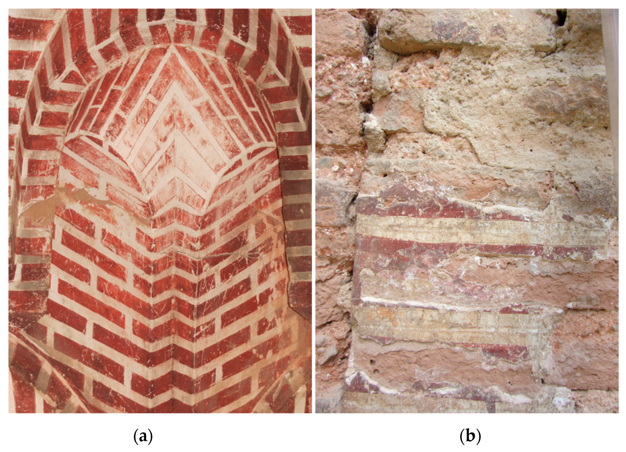
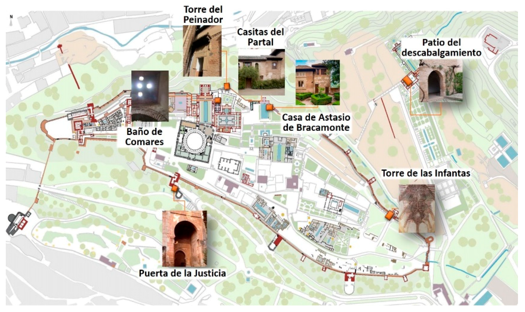
2.1. Description of the Sites of Study

The wall decorations imitating red-bricks can be found in several locations, including interior vaults, arches and walls, spread throughout the entire Monumental Ensemble. The locations studied are summarized in Figure 2 and described below.

2.1.1. Indoor Locations

  • Casitas del Partal (Partal Dwellings)–Torre de las Damas (Tower of the Ladies, TL)
These are a group of Nasrid two-storey houses built in the fourteenth century. In one of them, the so-called House of the Paintings, wall paintings were discovered in 1907 hidden behind layers of plaster. Among different motifs of courtly life and hunting scenes, which are not the object of this study, mural paintings imitating red bricks can be found in both ground and first floor. This wall of the house belonged originally to the façade of the TL of the Partal Palace, the oldest palace in the Alhambra complex, built during Muhammad III reign (1302–1309).
  • Baño del Palacio de Comares (The Bath of Comares Palace, BC)
The faux-brick mural painting is decorating the vault of the corridor that connects the Hall of the Beds of the Royal Bath of Comares palace with the Cool Hall (Ismail I (1314–1325)). The decoration has a good conservation aspect due to the restoration works carried out by the conservation department of the Alhambra Council in 2015–2017.
  • Torre de las Infantas (Infant’s Tower, IT)
It is a small tower-palace located in the main wall of the Alhambra in the way from the Partal Gardens to the Generalife. Two areas were studied in this location, a painted mocarabes vault in the entrance passageway and another one located on the first floor. According to different authors [22], the decoration of this tower is representative of a period of decadence in the late fourteenth century and early fifteenth century, coinciding with the reign of sultan Muhammad VII (1392–1408).

2.1.2. Outdoor Locations

  • Patio del descabalgamiento (Court of the Dismount, CD)
This court is located in the entrance to the Generalife Palace. It is dated from the first period of the Nasrid dynasty during the reign of Muhammad II (1273–1302) or Muhammad III (1302–1309). The mural paintings are located in the interior part of the entrance arch. The remains of mural painting in this area are severely deteriorated.
  • Casa de Astasio de Bracamonte (Astasio de Bracamonte´s House, AH)
The scarce remnants of mural painting studied are located in the facade of this Nasrid house dated to Muhammad III (1302–1309) period.
  • Puerta de la Justicia (The Gate of Justice, GC)
This is the largest and most impressive of the four gates in the Alhambra walls and it belongs to the Yusuf I period (1333–1354). The vaults of the interior of the gate conserve areas of red bricks-like decoration.
  • Peinador de la Reina (Queen’s Robing Room, QR)
The faux-brick paintings are located in the facade of the entrance to this tower. The mural painting remains are quite small areas distributed along the facade. They dated to the period of Yusuf I (1333–1354)–Muhammad V (1354–1359).

2.2. Instrumentation and Measurements

2.2.1. Non-Invasive In Situ Study

A handheld ED-XRF analyser Niton XL3t GOLDD+ (Thermo Fisher Scientific Inc., Waltham, MA, USA) was employed for identification of chemical elements. The X-ray radiation was emitted by an Ag tube anode of 45 kV. It uses a geometrically optimised large drift detector (GOLDD) and the radiation can reach a penetration of 40 µm maximum. It is provided with an integrated CCD camera, which allowed for visualizing the measurement area. The diameter of the measurement spot could be reduced from the standard 8 to 3 mm thanks to a collimator. Four filters were available, namely main (40 kV), high (30 kV), low (20 kV) and light (10 kV). Each of them could be selected specifically for the optimum excitation of a certain range of elements. The measurement method employed was “Mining and soils”, which was optimised to detect elements from Mg to U at levels higher than 1%. The four filters with a measurement time of 20 s each were used.
A portable Raman spectrometer innoRam (B&W TEK Inc., Newark, NJ, USA) was used. This spectrometer is equipped with a 785 nm laser for excitation and a CCD detector thermoelectrically cooled to −20 °C. The Raman shift covered was from 61 to 3018 cm−1 and the spectral resolution was about 2 cm−1. In order to adapt it to the in situ studies, the fibre optic probe was 5 m long and it was coupled to a video-microscope with a long focal distance objective (20×), giving an 85 µm sampling spot. This facilitated focusing in the area required and the possibility of saving the sampling area image. The video microscope with the attached Raman probe was mounted on a tripod with an extensive bar that could reach a height up to 3 m. An accessory motorised in the X–Y–Z axes with remote control allowed the movement of the probe and the focusing of the laser beam on the surface of the artwork. The analysis conditions were optimized in each case to achieve a good signal-to-noise ratio always using reduced laser power to avoid any damage. Typical conditions were below 30 mW of nominal laser power, 10 s of exposition time and 5 accumulations.

2.2.2. Laboratory Studies on Selected Samples

Selected samples of wall paintings were taken from different locations for laboratory analysis. The morphology of the samples was first studied using a Leica M205C stereomicroscope equipped with a Leica DFC450C digital camera (Wetzlar, Germany). The samples were hardened by embedding them in a polyester resin. Then they were cut with a diamond saw blade to obtain cross-sections in order to study the stratigraphy.
A FEI Quanta 400f field-emission, environmental scanning electron microscope (ESEM) equipped with EBSD, BSE and SE detectors was used to obtain both secondary electron (SE) and retro-dispersive electrons (BSEs) images. It could also perform elemental microanalysis for elements with Z > 5, with an energy dispersive X-ray spectroscopy detector (EDS) XFlash 6/30 (Bruker, Billerica, Massachusetts, US). The samples were studied without metallisation.
Raman spectra and images were registered using a Renishaw inVia Qontor Raman microspectrometer. This instrument is provided with three excitation lasers, green (532 nm), red (633 nm) and one near-infrared (785 nm). Four objectives (5×, 20×, 50× or 100×) are also available. Acquisition conditions such as exposition time and number of accumulations were adapted to the characteristics of each sample. The laser power was always less than 10 mW in order to prevent sample damage.

3. Results and Discussion

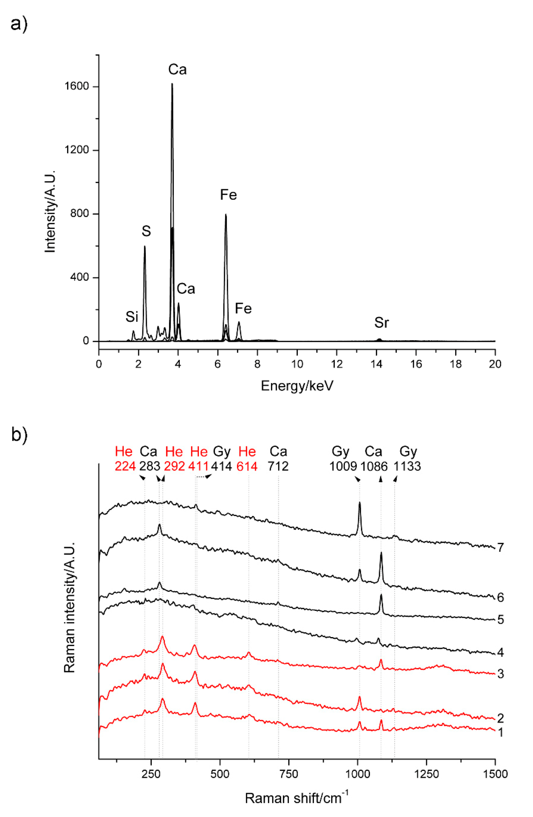
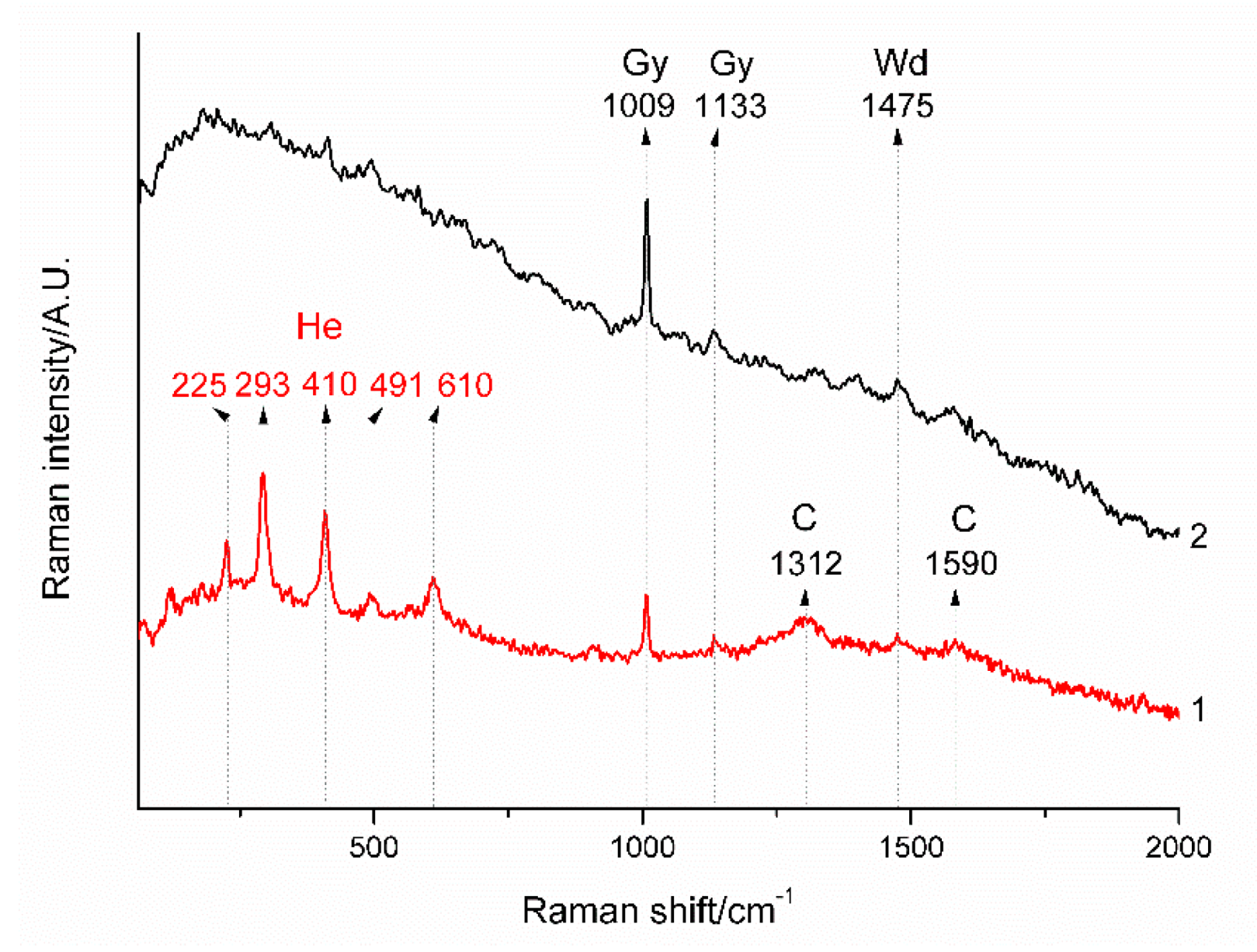
3.1. Non-Invasive In Situ Study

The visual inspection of the different motifs revealed the application of a continuous white base of plaster, above which the red rectangles simulating bricks were painted. Iron, calcium, sulphur and silicon were identified in the red faux-bricks using X-ray fluorescence in all the locations analysed (Figure 3a). These results suggest the use of natural iron oxide pigments (red ochre) to achieve the red tonalities.
The spectra obtained in situ with the portable Raman spectrometer in red areas showed clear characteristic bands at 224, 292, 411, 614 cm−1 (Figure 3b, 1–3) and; therefore, confirmed the presence of hematite (α-Fe2O3) [23]. Red ochre pigments are obtained from clays which have a high amount of iron in the form of hematite, being the mineral responsible for the red colour [24]. This type of pigment has been employed widely in history since prehistoric times [25,26]. During the Nasrid period, they were named as almagre. The origin of the name came from the word almáḡra belonging to the hispano-arab and it is modified from the classic arab maḡ[a]rah, which means red land. The term almagre is usually employed to define the naturally occurring red pigment. However, these pigments can also be obtained artificially by calcination of yellow ochres thanks to the transformation of goethite (α–FeOOH) into hematite [24]. Previous studies have demonstrated that although Raman spectroscopy can be useful in the distinction between well-ordered hematite from a more disordered one, it cannot be used to differentiate heated goethite from natural hematite [27]. Nevertheless, considering the abundance of red ochre in the region, the use of a natural pigment could be considered. Apart from the Raman features characteristic of hematite, other phases were identified. The Raman spectra of the red areas usually showed additional bands at 1009 and 1133 cm−1 (see Figure 3b, 1–2). These bands are typical of gypsum (CaSO4·2H2O) [28]. The presence of gypsum together with hematite in most red areas analysed can have different interpretations. It could rarely be a natural impurity of the red ochre, although it has been reported for Italian ochre pigments [29,30], or it could have been intentionally used as an extender material mixed with the pigment to modify the tonality. However, the more plausible explanation is that gypsum forms because of the reaction between CaCO3 present in the painting layers with sulphur containing air pollutants [31,32], particularly in exterior locations, which were much more altered. In fact, in most locations another additional band at 1086 cm−1, typical of calcite (Figure 3b, 1,3), also appears in the red areas. This band could be attributed to calcite impurities present in red ochre, but it could also be due to a lime-based pictorial technique. The contribution of the underlying white base must also be considered since calcite features were more clearly detected in areas with a certain loose of the red pigment. Further investigations on sample stratigraphy will be provided afterwards.
The analysis of the white areas between the red bricks using X-ray fluorescence revealed calcium as the main element in all of the locations. They also showed the presence of sulphur, but the relative amounts were very different depending on the locations. Raman spectroscopic studies confirmed these differences. In most cases, the white background layer was made of lime mortar showing the characteristic Raman bands of calcite at 1086 (s), 712 (w), 283 (m) cm−1 (see Figure 3b, 4–6). This is, for example, the case of the House of the Paintings of the Partal Dwellings. This is an interesting location because although now the wall paintings are located inside of the house, this wall was, in fact, the external wall originally belonging to the Tower of the Ladies of the Partal Palace to which the house was adjoined. When later the houses were built, this wall was kept as the interior wall of the house and the paintings were covered with gypsum plaster (Figure 3b, 7). Once this covering was removed, we can find these decorations rather well preserved.
In other locations, additional gypsum bands were observed in the white areas (Figure 3b, 4). The origin of gypsum in such cases is, probably, superficial contamination but further investigation of stratigraphy is necessary to confirm this hypothesis, as it will be discussed afterwards.
Another interesting location is the IT. We studied the wall paintings of the vault of the entrance, as well as the walls and the vaults of the first floor, with very different results. Only gypsum and no calcite was found in the white areas of the vault of the entrance and the wall of the first floor, which suggests a different execution technique, over gypsum plaster (see Figure 4). In this indoor decoration, Raman spectra also showed an additional band at 1475 cm−1 (and another weak at 910 cm−1, more difficult to see) associated to calcium oxalates in the form associated to calcium oxalates in the form of weddellite CaC2O4·2H2O [33]. Calcium oxalates are degradation products typically found in many artworks. They could be generated due to the microbial degradation of the organic material employed as binder in a calcium-rich ambient proportioned by the porous gypsum base [7,34].
Furthermore, in some cases, broad Raman bands of carbon located at 1312 and 1590 cm−1 were observed (Figure 4) [35]. This could be interpreted as an intentional addition of black carbon to provide a darker tonality, due to environmental pollution or contamination from fire soot.
In the case of the vaults of the first floor, the visual inspection revealed grey areas at the surface of the paintings. They were particularly evident in the white lines between the bricks. The results of the in situ Raman study in this location were not satisfactory. Spectra recorded in these areas showed only an intense fluorescence background with a small band at 1086 cm−1 attributed to calcite, independently of being registered on white or red colours. These decorations seem clearly different from the entrance, but further studies over samples are essential in this case.

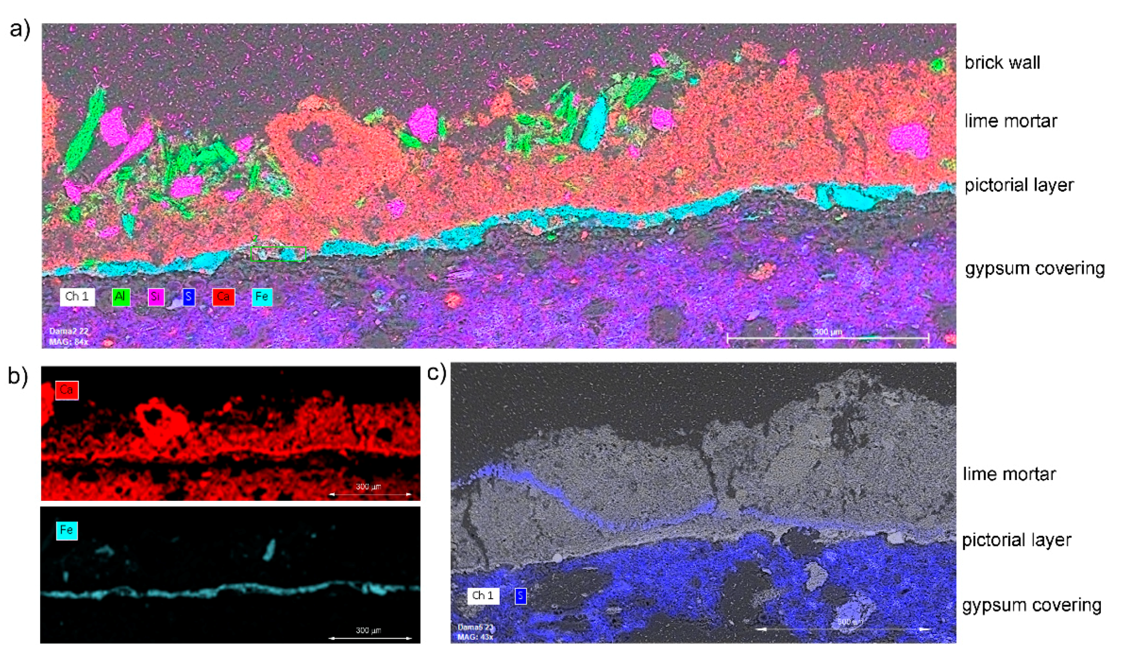
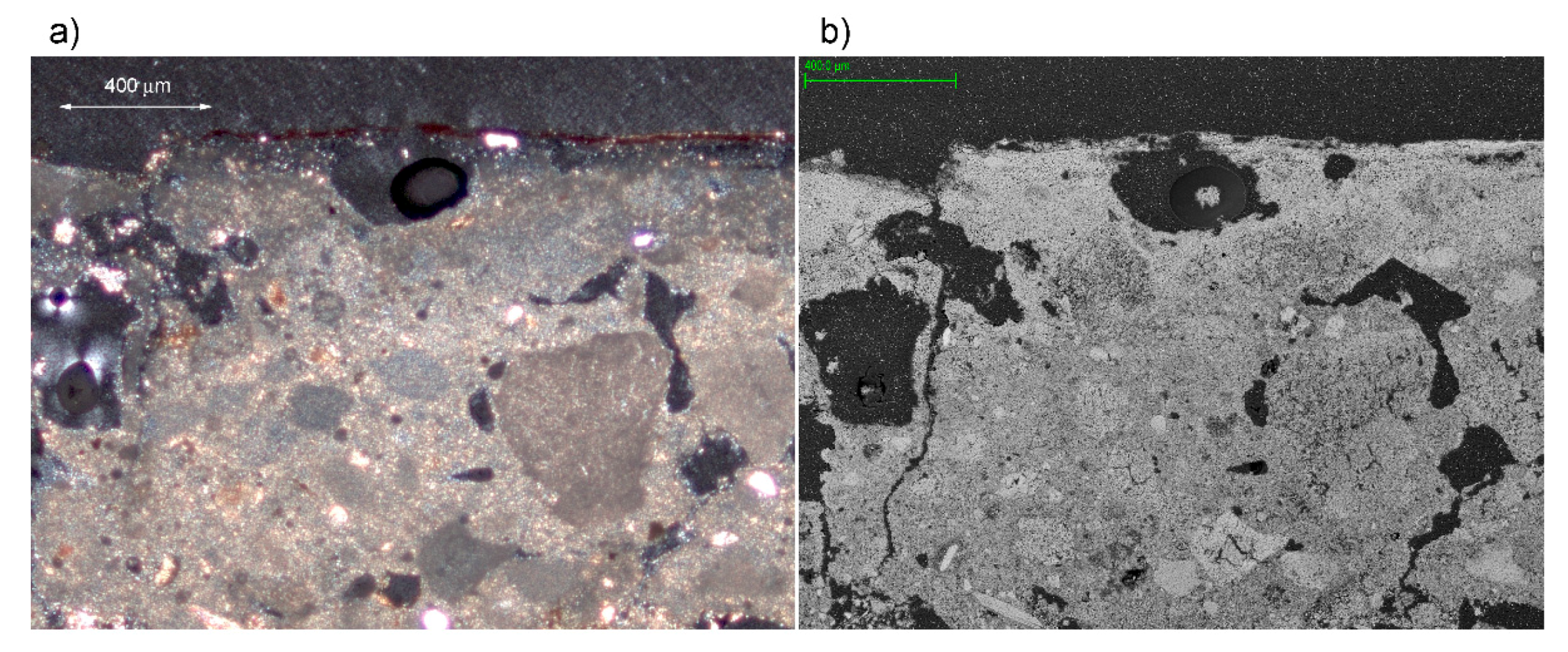
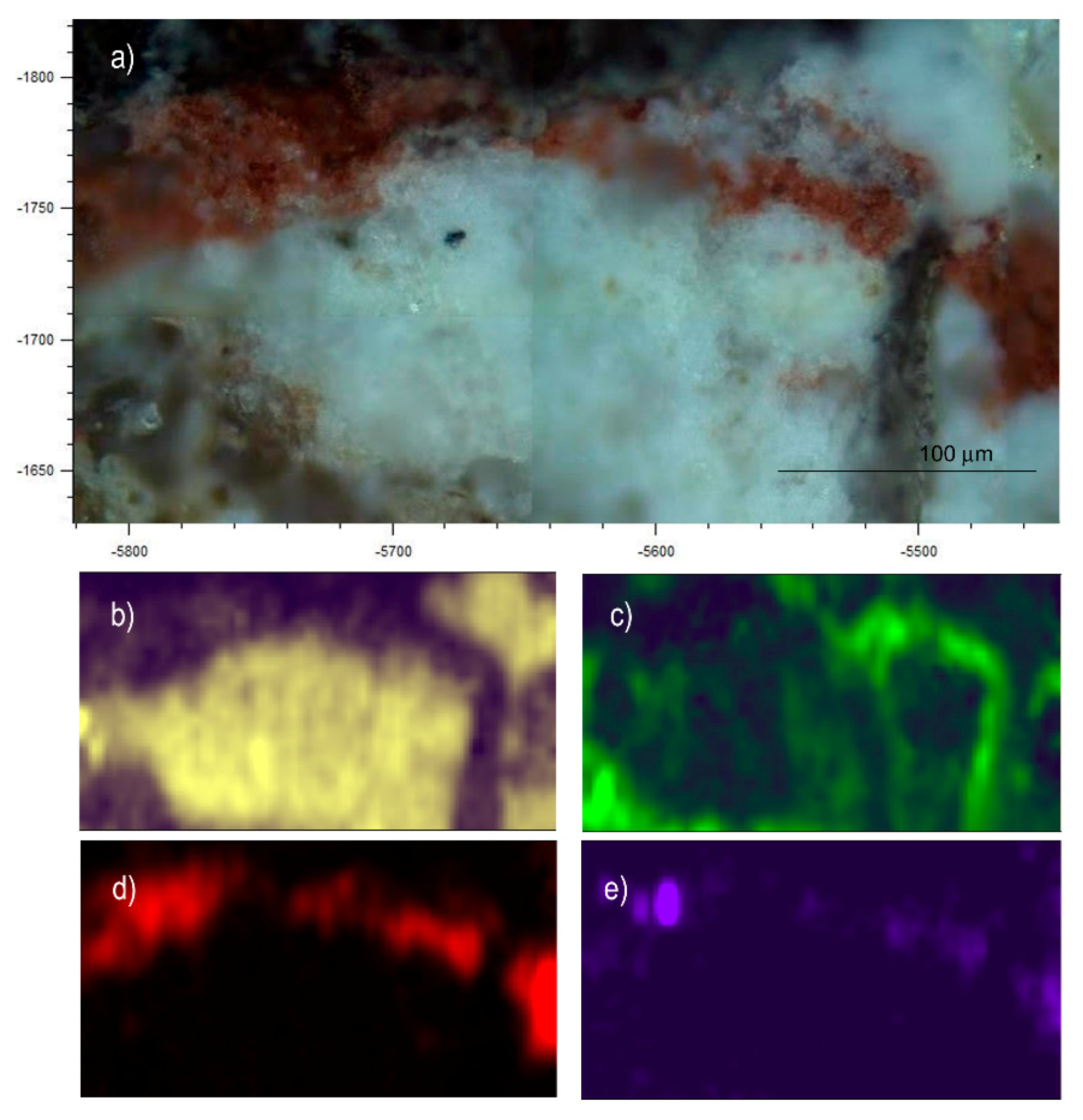
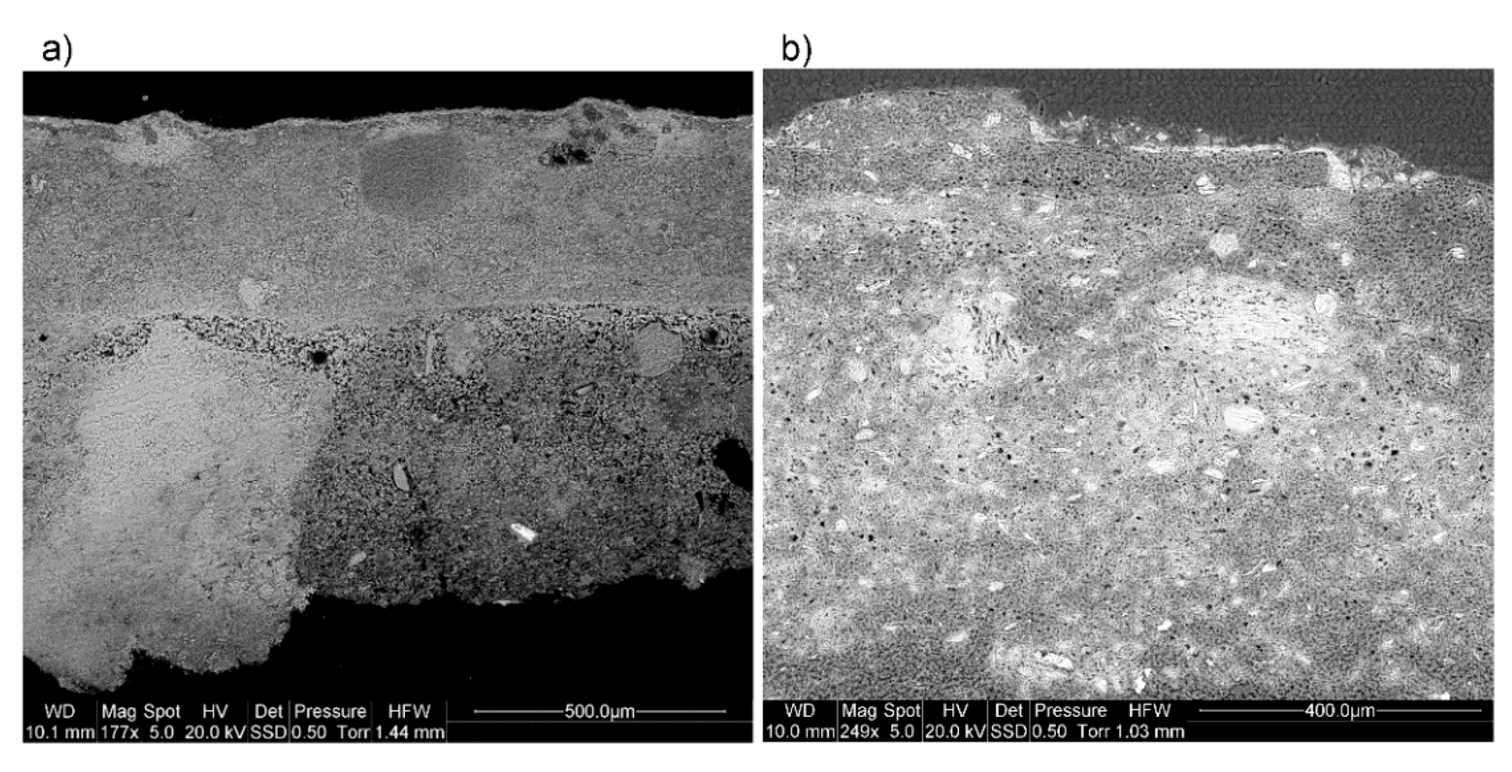
3.2. Laboratory Analyses

The sampling was performed taking into account the information obtained from in situ analysis and restricted to three selected locations. Thus samples from outdoor (QR), indoor (IT) and false interior (TL) locations were considered. Samples cross sections were investigated by Scanning Electron Microscopy-Energy Dispersive X-ray Spectroscopy (SEM-EDS) and micro-Raman spectroscopy. In this way, we can access the stratigraphy of the painting layers to gain more information about the execution techniques and the degradation processes.
Figure 5 shows the typical structure of the samples from TL, which illustrates the most common execution technique found. From top to bottom in the figure it can be seen: A preparation layer of lime mortar with few aggregates, applied over the real brick wall, a thin pictorial layer of hematite and a thick layer of gypsum plaster over the pictorial layer. The pigment was very pure in this location, not showing the presence of phyllosilicates. Regarding the execution technique, the results obtained suggest the use of lime-based painting techniques, either fresco or lime-paint. In the fresco technique, a very fine-grained pigment dispersed in water was laid on the surface of a fresh fine-grained lime mortar while in the case of lime-paint (or mezzo fresco) pigments were mixed with slaked lime or limewater and spread on dry mortar. The distinction between them is not straightforward, although the thin Ca-rich layer observed at the top of the preparation layer immediately below the pigment (see Figure 6b) suggests the use of lime-paint according to the criteria proposed by Piovesan et al. [36]. This layer is due to superficial carbonation already occurring before pigment application, but it was not always clearly observed and; therefore, we cannot definitely prove the use of this execution technique, although well separated layers of pigment and lime mortar also point in this direction.
It is interesting to mention that, although the thick gypsum covering played an important role in the preservation of these decorations along the centuries, as commented before, these results show that it also had a certain negative impact on the conservation state of the pictorial layers. As can be seen in Figure 5b, gypsum has mobilized and re-crystallized in a crack within the lime mortar.
Despite the incipient conservation problems, these decorations are very well preserved in contrast with those located outdoor. As an example, we show here the results from the samples taken from the scarce remains of decoration of the QR tower facade. The execution technique over lime mortar is very similar to that of TL, but in this case, no clear signs of the Ca-rich layer indicative of mezzo fresco are observed, maybe due to the poor conservation condition. According to EDS results, the hematite pigment employed in this location is also quite pure. However, as expected, the state of conservation is not good, and the mortar show holes due to dissolution and cracks (see Figure 6).
According to EDS results, the lime mortar, without aggregates, contains mainly Mg as an impurity in lime, whereas the presence of S and Cl should be considered as ions from soluble salts, related to deterioration. Furthermore, the deposition of gypsum due to the interaction between the lime mortar and atmospheric pollutants, hypothesized during the in-situ analysis, is confirmed by studying these samples. As can be seen in the distribution map of the different mineral phases detected by Raman microimaging, gypsum crystallized in the cracks of the mortar (see Figure 7). Additionally, TiO2 in the form of anatase was detected with a characteristic strong band at 143 cm−1 [37]. According to the distribution map, this mineral is present as an impurity in the red ochre pigment [30,38]. Anatase is a common impurity in ochre pigments, and it can be easily detected when using Raman spectroscopy. The clear identification of this mineral, even when present in low amounts, is due to its exceptionally strong Raman scattering, in contrast to the weak signals produced by other phases present in this type of pigments, like clays [39].
Finally, we considered the samples from the first floor of IT, where the in-situ studies did not provide much information due to the intense fluorescence background. We took samples from the vault and the wall and the results showed two different techniques of execution. In the vault, the stratigraphy (Figure 8) consists of a thin pictorial layer (<10 µm) of hematite with appreciable amounts of Si, Al and K, compatible with phyllosilicates, as well as Ca and Mg. Calcite and, in minor proportions, gypsum were also identified in this layer by Raman microspectroscopy. The abundance of phyllosilicates is probably the reason for the strong fluorescence background observed during the in situ Raman measurements. According to SEM observations hematite grains were very small (1–2 µm). The pigment was applied over a preparation layer of fine lime plaster, uniform, and without sand or other aggregates. Few impurities can be found in this area, being Cl the most remarkable. Under this preparation layer, the most interior lime mortar contained appreciable amounts of aggregates (siliceous fragments, gypsum crystals and ilmenite).
Curiously, in the sample taken from the wall of the same room, the base substrate was gypsum plaster, as confirmed by Raman microspectroscopy. It shows few impurities, low porosity and seems very well-preserved, without signs of dissolution (see Figure 8b). The preparation layer located just below the pigment was also made of gypsum plaster but according to EDS results it contained appreciable amounts of Mg, Cl and Na, whose presence is more difficult to explain. Their presence could be, in principle, attributed to environmental pollution due to marine aerosol contributions [40], but chlorine here is more abundant than in other locations. This can point to an intentional addition related to the secco technique with an organic binder. This execution technique over gypsum instead of lime mortar is similar to that found at the entrance of this tower during the in situ study, where no calcite was detected.

4. Conclusions

The complete study carried out, combining in situ analysis with portable equipment and laboratory measurements, on samples from selected locations allowed for the characterization of Nasrid mural paintings that imitates red bricks walls. The decorations were executed by painting the red bricks with red ochre over a white background of lime mortar, which also acted as the white edges between the decorative bricks. The ubiquitous presence of calcite and the absence of organic binders in the pictorial layer support the hypothesis of the use of lime-based techniques, probably mezzo fresco. Only in the entrance vault and in the wall of the Infant´s Tower a different execution technique was revealed. Here, the decoration was performed over gypsum plaster and evidence of the use of an organic binder is provided by the identification of calcium oxalate, as a degradation product. Hence, it is not clear if the motifs in this particular location are the original Nasrid decorations or can be the result of posterior interventions, since their execution does not coincide with the rest of the decorations studied.
Regarding the conservation condition, the decorations in interior locations are rather well preserved. In the case of the TL, the complete removal of the gypsum plaster covering will prevent further deterioration due to the penetration of gypsum in the pictorial layer. In contrast, exterior decorations are in very poor condition and only remnants of the original paintings are preserved. However, the study and the conservation of these remains is important, since it keeps testimony of the original aspect of many buildings of the Alhambra, with most of their exterior walls painted in bright red colour imitating bricks.

Author Contributions

Conceptualization: M.J.A.-C., M.J.d.l.T.-L., E.C.G., R.R.D. and A.D.-V.; Experimental and methodology: P.A., M.J.A.-C., M.J.d.l.T.-L., E.C.G., R.R.D. and A.D.-V.; writing—original draft preparation: P.A., M.J.A.-C. and A.D.-V.; writing—review and editing: M.J.A.-C., M.J.d.l.T.-L., E.C.G., R.R.D. and A.D.-V.; supervision: M.J.A.-C. and A.D.-V.; funding acquisition: M.J.d.l.T.-L. and A.D.-V. All authors have read and agreed to the published version of the manuscript.

Funding

This work has been financed by the research project BIA2017-87131-R (Spanish Ministry of Economy and Competitiveness and FEDER funds). P. Arjonilla is also grateful for financial support from Spanish Ministry of Economy and Competitiveness through BES-2014-068793 predoctoral contract.

Acknowledgments

This work was carried out in collaboration with Alhambra and Generalife Council. Technical and human support from the CICT-Universidad de Jaén is acknowledged (UJA, MINECO, Junta de Andalucía, FEDER).

Conflicts of Interest

The authors declare no conflict of interest.

References

  1. Fernández Puertas, A.; Jones, O. The Alhambra I: From the Ninth Century to Yūsuf I (1354); Saqi Books: London, UK, 1997; ISBN 0863564666. [Google Scholar]
  2. De La Torre López, M.J.; Sebastián, P.E.; Rodríguez, G.J. A study of the wall material in the Alhambra (Granada, Spain). Cem. Concr. Res. 1996, 26, 825–839. [Google Scholar] [CrossRef]
  3. Dominguez-Vidal, A.; de la Torre-Lopez, M.J.; Rubio-Domene, R.; Ayora-Cañada, M.J. In situ noninvasive Raman microspectroscopic investigation of polychrome plasterworks in the Alhambra. Analyst 2012, 137, 5763–5769. [Google Scholar] [CrossRef]
  4. Gómez-Morón, M.A.; Ortiz, P.; Martín-Ramírez, J.M.; Ortiz, R.; Castaing, J. A new insight into the vaults of the kings in the Alhambra (Granada, Spain) by combination of portable XRD and XRF. Microchem. J. 2016, 125, 260–265. [Google Scholar] [CrossRef]
  5. Arjonilla, P.; Domínguez-Vidal, A.; de la Torre López, M.J.; Rubio-Domene, R.; Ayora-Cañada, M.J. In situ Raman spectroscopic study of marble capitals in the Alhambra monumental ensemble. Appl. Phys. A Mater. Sci. Process. 2016, 122, 1014/1–1014/8. [Google Scholar] [CrossRef]
  6. Cardell-Fernández, C.; Navarrete-Aguilera, C. Pigment and plasterwork analyses of Nasrid polychromed Lacework stucco in the Alhambra (Granada, Spain). Stud. Conserv. 2006, 51, 161–176. [Google Scholar] [CrossRef]
  7. De la Torre-López, M.J.; Dominguez-Vidal, A.; Campos-Suñol, M.J.; Rubio-Domene, R.; Schade, U.; Ayora-Cañada, M.J. Gold in the Alhambra: Study of materials, technologies, and decay processes on decorative gilded plasterwork. J. Raman Spectrosc. 2014, 45, 1052–1058. [Google Scholar] [CrossRef]
  8. Llorent-Martínez, E.J.; Domínguez-Vidal, A.; Rubio-Domene, R.; Pascual-Reguera, M.I.; Ruiz-Medina, A.; Ayora-Cañada, M.J. Identification of lipidic binding media in plasterwork decorations from the Alhambra using GC-MS and chemometrics: Influence of pigments and aging. Microchem. J. 2014, 115, 11–18. [Google Scholar] [CrossRef]
  9. Cardell, C.; Rodriguez-Simon, L.; Guerra, I.; Sanchez-Navas, A. Analysis of Nasrid polychrome carpentry at the Hall of the Mexuar Palace, Alhambra complex (Granada, Spain), combining microscopic, chromatographic and spectroscopic methods. Archaeometry 2009, 51, 637–657. [Google Scholar] [CrossRef]
  10. Bueno, A.G.; Medina Flórez, V.J. The Nasrid plasterwork at “qubba Dar al-Manjara l-kubra” in Granada: Characterisation of materials and techniques. J. Cult. Herit. 2004, 5, 75–89. [Google Scholar] [CrossRef]
  11. Burgio, L. Dating Alhambra stuccoes. V&A Conserv. J. 2005, 49, 2–3. [Google Scholar]
  12. Pollard, A.M. Analytical Chemistry in Archaeology; Cambridge University Press: Cambridge, UK, 2007; ISBN 0521655722. [Google Scholar]
  13. Madariaga, J.M. Analytical chemistry in the field of cultural heritage. Anal. Methods 2015, 7, 4848–4876. [Google Scholar] [CrossRef]
  14. Cuñat, J.; Palanco, S.; Carrasco, F.; Simón, M.D.; Laserna, J.J. Portable instrument and analytical method using laser-induced breakdown spectrometry for in situ characterization of speleothems in karstic caves. J. Anal. At. Spectrom. 2005, 20, 295–300. [Google Scholar] [CrossRef]
  15. Vandenabeele, P.; Weis, T.L.; Grant, E.R.; Moens, L.J. A new instrument adapted to in situ Raman analysis of objects of art. Anal. Bioanal. Chem. 2004, 379, 137–142. [Google Scholar] [CrossRef]
  16. Lauwers, D.; Hutado, A.G.; Tanevska, V.; Moens, L.; Bersani, D.; Vandenabeele, P. Characterisation of a portable Raman spectrometer for in situ analysis of art objects. Spectrochim. Acta A Mol. Biomol. Spectrosc. 2014, 118, 294–301. [Google Scholar] [CrossRef] [PubMed]
  17. Marcaida, I.; Maguregui, M.; Morillas, H.; Prieto-Taboada, N.; de Vallejuelo, S.F.-O.; Veneranda, M.; Madariaga, J.M.; Martellone, A.; De Nigris, B.; Osanna, M. In situ non-invasive characterization of the composition of Pompeian pigments preserved in their original bowls. Microchem. J. 2018, 139, 458–466. [Google Scholar] [CrossRef]
  18. Olivares, M.; Castro, K.; Corchón, M.S.; Gárate, D.; Murelaga, X.; Sarmiento, A.; Etxebarria, N. Non-invasive portable instrumentation to study Palaeolithic rock paintings: The case of La Peña Cave in San Roman de Candamo (Asturias, Spain). J. Archaeol. Sci. 2013, 40, 1354–1360. [Google Scholar] [CrossRef]
  19. Brunetti, B.G.; Matteini, M.; Miliani, C.; Pezzati, L.; Pinna, D. MOLAB, a Mobile Laboratory for In Situ Non-Invasive Studies in Arts and Archaeology. In Lasers in the Conservation of Artworks; Springer: Berlin/Heidelberg, Germany, 2007; pp. 453–460. [Google Scholar]
  20. Mollica Nardo, V.; Renda, V.; Bonanno, S.; Parrotta, F.; Anastasio, G.; Caponetti, E.; Saladino, M.L.; Vasi, C.S.; Ponterio, R.C. Non-Invasive Investigation of Pigments of Wall Painting in S. Maria Delle Palate di Tusa (Messina, Italy). Heritage 2019, 2, 147. [Google Scholar] [CrossRef] [Green Version]
  21. Alberghina, M.F.; Germinario, C.; Bartolozzi, G.; Bracci, S.; Grifa, C.; Izzo, F.; La Russa, M.F.; Magrini, D.; Massa, E.; Mercurio, M.; et al. The Tomb of the Diver and the frescoed tombs in Paestum (southern Italy): New insights from a comparative archaeometric study. PLoS ONE 2020, 15, e0232375/1-26. [Google Scholar] [CrossRef] [Green Version]
  22. Torres Balbás, L. Cronología de las construcciones de la Casa Real de La Alhambra. Al-Andalus 1959, XXIV, 52–62. [Google Scholar]
  23. Zoppi, A.; Lofrumento, C.; Castellucci, E.M.; Migliorini, M.G. The Raman spectrum of hematite: Possible indicator for a compositional or firing distinction among Terra Sigillata wares. Ann. Chim. 2005, 95, 239–246. [Google Scholar] [CrossRef] [PubMed]
  24. Eastaugh, N.; Walsh, V.; Chaplin, T.; Siddall, R. Pigment Compendium: A Dictionary of Historical Pigments; Elsevier: Oxford, UK, 2007; ISBN 0750657499. [Google Scholar]
  25. Iriarte, M.; Hernanz, A.; Gavira-Vallejo, J.M.; Alcolea-González, J.; de Balbín-Behrmann, R. μ-Raman spectroscopy of prehistoric paintings from the El Reno cave (Valdesotos, Guadalajara, Spain). J. Archaeol. Sci. Rep. 2017, 14, 454–460. [Google Scholar] [CrossRef]
  26. Siddall, R. Mineral Pigments in Archaeology: Their Analysis and the Range of Available Materials. Minerals 2018, 8, 201. [Google Scholar] [CrossRef] [Green Version]
  27. Gialanella, S.; Girardi, F.; Ischia, G.; Lonardelli, I.; Mattarelli, M.; Montagna, M. On the goethite to hematite phase transformation. J. Therm. Anal. Calorim. 2010, 102, 867–873. [Google Scholar] [CrossRef]
  28. Prieto-Taboada, N.; Gómez-Laserna, O.; Martínez-Arkarazo, I.; Olazabal, M.Á.; Madariaga, J.M. Raman spectra of the different phases in the CaSO4-H2O system. Anal. Chem. 2014, 86, 10131–10137. [Google Scholar] [CrossRef]
  29. Elias, M.; Chartier, C.; Prévot, G.; Garay, H.; Vignaud, C. The colour of ochres explained by their composition. Mater. Sci. Eng. B Solid-State Mater. Adv. Technol. 2006, 127, 70–80. [Google Scholar] [CrossRef]
  30. Froment, F.; Tournié, A.; Colomban, P. Raman identification of natural red to yellow pigments: Ochre and iron-containing ores. J. Raman Spectrosc. 2008, 39, 560–568. [Google Scholar] [CrossRef]
  31. Cultrone, G.; Arizzi, A.; Sebastián, E.; Rodriguez-Navarro, C. Sulfation of calcitic and dolomitic lime mortars in the presence of diesel particulate matter. Environ. Geol. 2008, 56, 741–752. [Google Scholar] [CrossRef]
  32. Charola, A.E.; Pühringer, J.; Steiger, M. Gypsum: A review of its role in the deterioration of building materials. Environ. Geol. 2007, 52, 207–220. [Google Scholar] [CrossRef]
  33. Edwards, H.G.M.; Seaward, M.R.D.; Attwood, S.J.; Little, S.J.; De Oliveira, L.F.C.; Tretiach, M. FT-Raman spectroscopy of lichens on dolomitic rocks: An assessment of metal oxalate formation. Analyst 2003, 128, 1218–1221. [Google Scholar] [CrossRef] [PubMed]
  34. Rampazzi, L. Calcium oxalate films on works of art: A review. J. Cult. Herit. 2019, 40, 195–214. [Google Scholar] [CrossRef]
  35. Tomasini, E.P.; Halac, E.B.; Reinoso, M.; Di Liscia, E.J.; Maier, M.S. Micro-Raman spectroscopy of carbon-based black pigments. J. Raman Spectrosc. 2012, 43, 1671–1675. [Google Scholar] [CrossRef]
  36. Piovesan, R.; Mazzoli, C.; Maritan, L.; Cornale, P. Fresco and lime-paint: An experimental study and objective criteria for distinguishing between these painting techniques. Archaeometry 2012, 54, 723–736. [Google Scholar] [CrossRef]
  37. Frank, O.; Zukalova, M.; Laskova, B.; Kürti, J.; Koltai, J.; Kavan, L. Raman spectra of titanium dioxide (anatase, rutile) with identified oxygen isotopes (16, 17, 18). Phys. Chem. Chem. Phys. 2012, 14, 14567–14572. [Google Scholar] [CrossRef] [PubMed]
  38. Marcaida, I.; Maguregui, M.; Morillas, H.; Veneranda, M.; Prieto-Taboada, N.; Fdez-Ortiz de Vallejuelo, S.; Madariaga, J.M. Raman microscopy as a tool to discriminate mineral phases of volcanic origin and contaminations on red and yellow ochre raw pigments from Pompeii. J. Raman Spectrosc. 2019, 50, 143–149. [Google Scholar] [CrossRef]
  39. Košařová, V.; Hradil, D.; Němec, I.; Bezdička, P.; Kanický, V. Microanalysis of clay-based pigments in painted artworks by the means of Raman spectroscopy. J. Raman Spectrosc. 2013, 44, 1570–1577. [Google Scholar] [CrossRef]
  40. Horemans, B.; Cardell, C.; Bencs, L.; Kontozova-Deutsch, V.; De Wael, K.; Van Grieken, R. Evaluation of airborne particles at the Alhambra monument in Granada, Spain. Microchem. J. 2011, 99, 429–438. [Google Scholar] [CrossRef]
Figure 1. Examples of faux-brick paintings on (a) indoor vault in Infant’s Tower and (b) outdoor wall in Astasio de Bracamonte’s house.
Crystals 11 00423 g001
Figure 2. Map of the Alhambra Monumental Ensemble showing the location of the study sites.
Crystals 11 00423 g002
Figure 3. Non-invasive analyses results. (a) Typical XRF spectra on red areas of the faux-brick mural paintings and (b) typical Raman spectra of (1–3) red and (4–7) white areas. He: Hematite, Ca: Calcite, Gy: Gypsum. Raman spectra were stacked for clarity.
Crystals 11 00423 g003
Figure 4. In situ Raman spectra registered on the red (1) and white (2) motifs in the vault of the entrance of the Infant´s Tower (IT). He: Hematite, Ca: Calcite, Gy: Gypsum, C: Carbon, Wd: Weddellite. Spectra were stacked for clarity.
Crystals 11 00423 g004
Figure 5. SEM–EDS results for two cross-sections samples of the Tower of the Ladies (TL). (a) SEM image with overlapping combined elementals maps of aluminium (green); silicon (pink); sulphur (dark blue); calcium (red); iron (light blue); (b) individual elemental maps of calcium and iron of the sample shown in (a). (c) SEM image and sulphur (dark blue) map in a sample where conservation problems can be seen.
Crystals 11 00423 g005
Figure 6. Petrographic (a) and SEM (b) images of the cross-section from Queen’s Robing Room (QR).
Crystals 11 00423 g006
Figure 7. (a) Visible microscopic image of a sample cross-section from Queen’s Robing Room (QR). Distribution maps obtained by micro-Raman spectroscopy (using 785 nm as excitation wavelength): (b) Calcite (1086 cm−1), (c) gypsum (1009 cm−1), (d) hematite (292 cm−1) and (e) anatase (143 cm−1).
Crystals 11 00423 g007
Figure 8. SEM images of cross-sections from the first floor of Infant´s Tower (IT). Samples taken from red decorations of: (a) The vault and (b) the wall.
Crystals 11 00423 g008
Publisher’s Note: MDPI stays neutral with regard to jurisdictional claims in published maps and institutional affiliations.

