Article

Low-Temperature Co-Precipitation Synthesis of HoFeO3 Nanoparticles

1
Faculty of Pedagogy in Natural Sciences, Sai Gon University, Ho Chi Minh City 700000, Vietnam
2
Informetrics Research Group, Ton Duc Thang University, Ho Chi Minh City 700000, Vietnam
3
Faculty of Applied Sciences, Ton Duc Thang University, Ho Chi Minh City 700000, Vietnam
*
Author to whom correspondence should be addressed.
Crystals 2021, 11(3), 238; https://doi.org/10.3390/cryst11030238
Submission received: 16 February 2021 / Revised: 24 February 2021 / Accepted: 24 February 2021 / Published: 26 February 2021
(This article belongs to the Section Inorganic Crystalline Materials)

Abstract

:
In this research, we investigate and discuss the characteristics of HoFeO3 nanoparticles synthesized by the co-precipitation method at low temperature (t° ≤ 4 °C). The single-phase HoFeO3 samples with the orthorhombic structure formed after annealing of the precipitates at different temperatures up to 950 °C. The annealed HoFeO3 nanoparticles have an average crystal size of 10–20 nm (SEM, TEM). UV-Vis spectrum of HoFeO3 sample annealed at 750 °C showed strong UV and Vis absorption with small optical energy gap (Eg = 1.56 eV). In the range temperature of 100–300 K, the HoFeO3 samples showed superparamagnetic behaviour at 5 kOe with high magnetization (Ms = 1.3–2.4 emu/g) and very low susceptibility (χ << 1).

Graphical Abstract

1. Introduction

The features of rare-earth orthoferrites RFeO3 (R = rare-earth elements) such as thermal, electrical, magnetic and optical properties depend on chemical component, particle size and synthetic methods [1,2,3,4,5,6]. Based on specific use purposes, there are different requirements about performance of orthoferrites. This can be carried out by adjusting morphology and particle size via preparation method or doping other elements in the RFeO3 [7,8,9,10]. Noteworthy, one of the rare-earth orthoferrites receives much attention as HoFeO3, which is synthesized by different methods such as solid-state reaction technique [11], hydrothermal [12] and sol-gel technique [13,14,15,16]. Especially, HoFeO3 particles with a micrometer size synthesized by solid-state reaction technique possess an optical energy gap (Eg) of 3.39 eV; saturation magnetization (Ms) of 25.5 emu/g, coercivity (Hc) of 2659 Oe and remanent magnetization (Mr) of 4.08 emu/g at 10 K [11]. Meanwhile, with a size of 10–20 µm, HoFeO3 particles prepared by hydrothermal method acquire a Ms < 0.1 emu/g at 300 K [12].
In the previous reports [13,14], HoFeO3 nanoparticles (40–100 nm) with Eg value of 2.12–2.14 eV have been employed as photo-catalysts in the visible light. The Ms value of HoFeO3 nanoparticles (100–300 nm) gradually decrease with increase of temperature [15], specifically, which reach the amount of 5.14, 0.6 and 0.2, respectively, at 50, 100 and 300 K. HoFeO3 particles in size 25–30 nm was also prepared by co-precipitation, using ethanol solvent [17]. However, ethanol is volatile, pollutant, and flammable solvent compared to water. Furthermore, the water solvent is cheaper. The orthoferrite HoFeO3 nanoparticles with a size of 10–20 nm and without conglomeration, which are prepared by simple co-precipitation method at low temperature (t° ≤ 4 °C), have not been reported, so far.
In this work, HoFeO3 nanoparticles (with a size of 10–20 nm and without conglomeration) were successfully synthesized and studied on structure, thermal, optical and magnetic properties.

2. Experimental

A mixture containing 25 mL of Fe(NO3)3 solution (0.2 M) and 25 mL of Ho(NO3)3 solution (0.2 M) was mixed at room temperature (t° ~ 300 K) and then gradually added to 400 mL of cool water (t° ≤ 4 °C). This strategy is to stabilize the obtained precipitate; consequently, it results in the controllable growth of crystals better than the co-precipitation at room temperature [4,10]. Subsequently, a 5% NH3 solution was slowly introduced until pH = 9.2–9.4 to entirely precipitate the cations of Fe (III) and Ho (III) [18,19,20]. The precipitate was filtered, washed by deionized water until pH ~ 7 and dried in air. Finally, the products were then grinded and carried out thermogravimetric analysis to determine appropriate temperature for obtaining the single-phase of HoFeO3 orthoferrite.
The crystal structure of the products has been determined via X-ray diffraction (XRD) on a D8-ADVANCE (Brucker, Bremen, Germany) with Cu Kα radiation (λ = 1.540 Å) using a step size of 0.019° in the range of 10–80°. The materials’ element component and surface morphology have been investigated via energy dispersive analysis of X-ray (EDAX) and scanning electron microscopy (SEM) on S-4800 FE-SEM (Hitachi, Tokyo, Japan), Raman spectrometer on Horiba XploRa ONE (American), and transmission electron microscopy (TEM) on JEM-1400 instrument (Jeol Ltd., Tokyo, Japan). The thermal property has been determined via thermal gravimetric analysis and differential scanning calorimetry (TGA-DSC) on a LABSYS Evo 1600° (SETERAM Instrumentation, Caluire, France). While the optical and magnetic properties have been studied via UV-Visible spectrophotometer (UV-Vis) on UV-2600 (Shimadzu, Tokyo, Japan) and vibrating sample magnetometer (VSM) on a EV11 (MicroSense, Tokyo, Japan) with a maximum applied field of 16,000 Oe.

3. Results and Discussion

In order to determine the optimal temperature range for the structural investigation, thermal gravimetric analysis (TGA) was performed with the results revealed in Figure 1. As a consequence, the initial weight loss (~35%) starts from 50 to 650 °C, meanwhile, which has not considerably changed at t° > 650 °C (<1%).
The endothermic and exothermic peaks specifically appear on the DSC curve at 125.49 and 749.37 °C, respectively. Herein, the weight loss after annealing is attributed to the water removal from the surface, resulting from the crystallization and dehydration of Fe2O3.xH2O (x = 1–5) [21] and HoO(OH).yH2O [22]. Hence, the temperatures of 650, 750, 850 and 950 °C, were chosen for investigating structure and morphology of HoFeO3 nanoparticles.
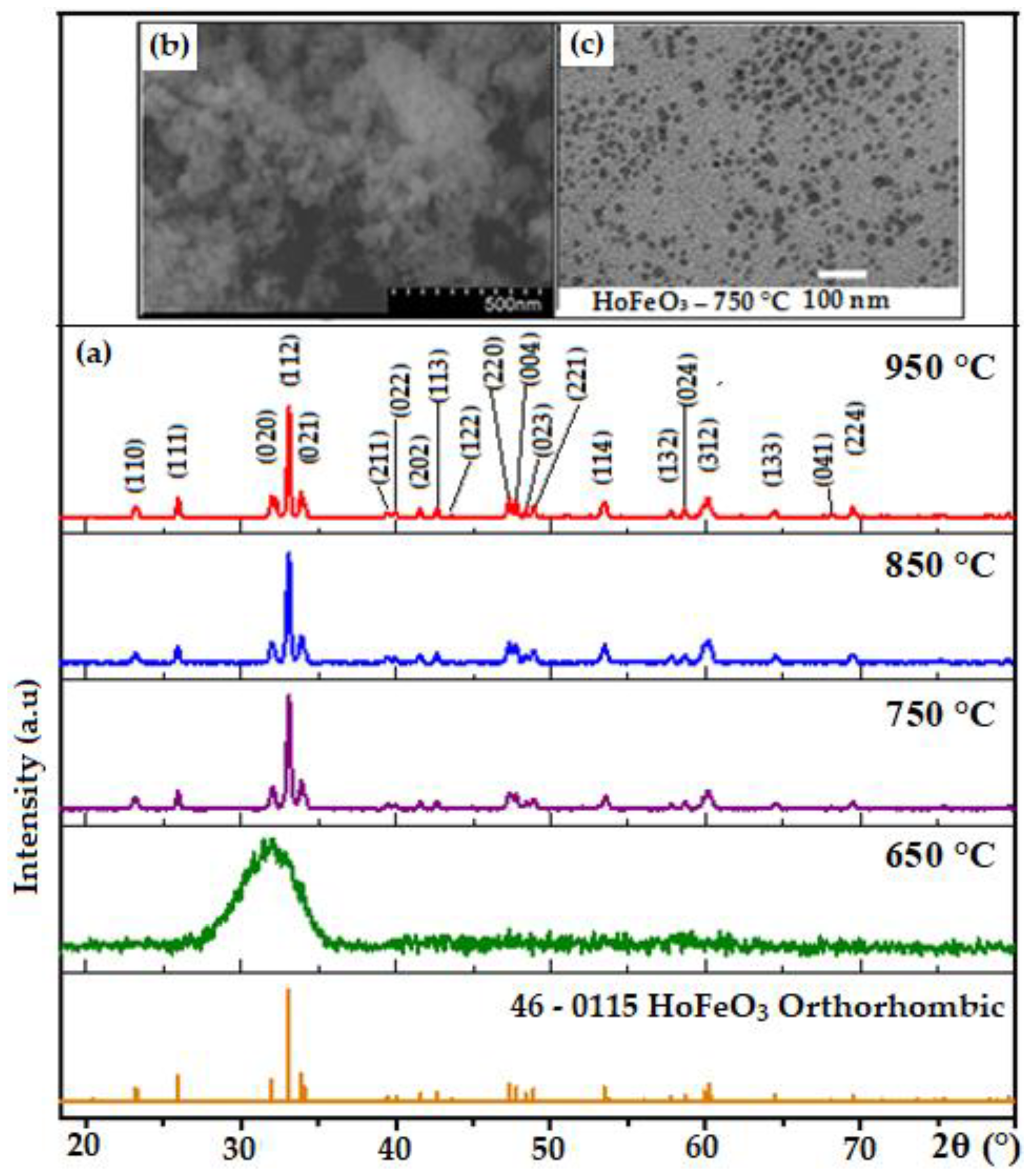
Figure 2a shows the XRD pattern of HoFeO3 after annealing at 650, 750, 850 và 950 °C for 1 h. As a result, HoFeO3 reveals an amorphous state at 650 °C. Whereas, at the different annealing temperatures, the samples are in good accordance with the standard JCPDS: 46-0115 of the orthorhombic HoFeO3 single-phase, which are correspondent to the exothermic peak on the DSC curve (749.37 °C).
Furthermore, the SEM and TEM images of HoFeO3 nanoparticles annealed at 750 °C are shown in Figure 2b,c. Herein, the orthoferrite HoFeO3 particles are discrete with an average size of 10–20 nm, which are smaller and without conglomeration of particles comparing to HoFeO3 nanoparticles prepared by hydrothermal [12], sol-gel [13,14,15], and co-precipitation using ethanol [17]. Figure 3a shows the EDAX spectra of HoFeO3, in which presence of Ho and Fe ions are clearly seen. An analysis of the weight and atomic percent of HoFeO3 nanoparticles is in agreement with the theoretical value calculated from the chemical formula.
In the Raman spectrum of the HoFeO3 sample at room temperature, three modes are observed at 129.1, 514.1 and 594.2 cm−1, which possess high peak intensities (Figure 3b). The Raman active modes of the HoFeO3 were assigned based on the method recently proposed by Gupta et al. [23] for RFeO3 (R = Tb, Dy, Ho, Er, Tm) compounds. Noteworthy, the wavenumber, appearing at 129.2 cm−1, was attributed Ho-O vibration modes. The Raman bands above 200 cm−1 correspond to oxygen ions. The high frequency mode in the RFeO3 crystal may be assigned to the internal vibration related to the mutual Fe-O motion within the oxygen octahedron [24], in this work, which presents at wavenumbers of 514.1 and 594.2 cm−1. The UV-Vis absorption spectra of the HoFeO3 nanoparticles revealed a strong absorption in the visible light region (~400–600 nm) (Figure 3c). This is interesting because HoFeO3 could be developed as a new visible light photocatalyst. The direct band gap energy (Eg, eV) was determined similarly to the work [11] and is shown in Figure 3d. As a consequence, the band gap value of HoFeO3 nanoparticles is ~1.56 eV which is lower than in the previous works (Table 1).
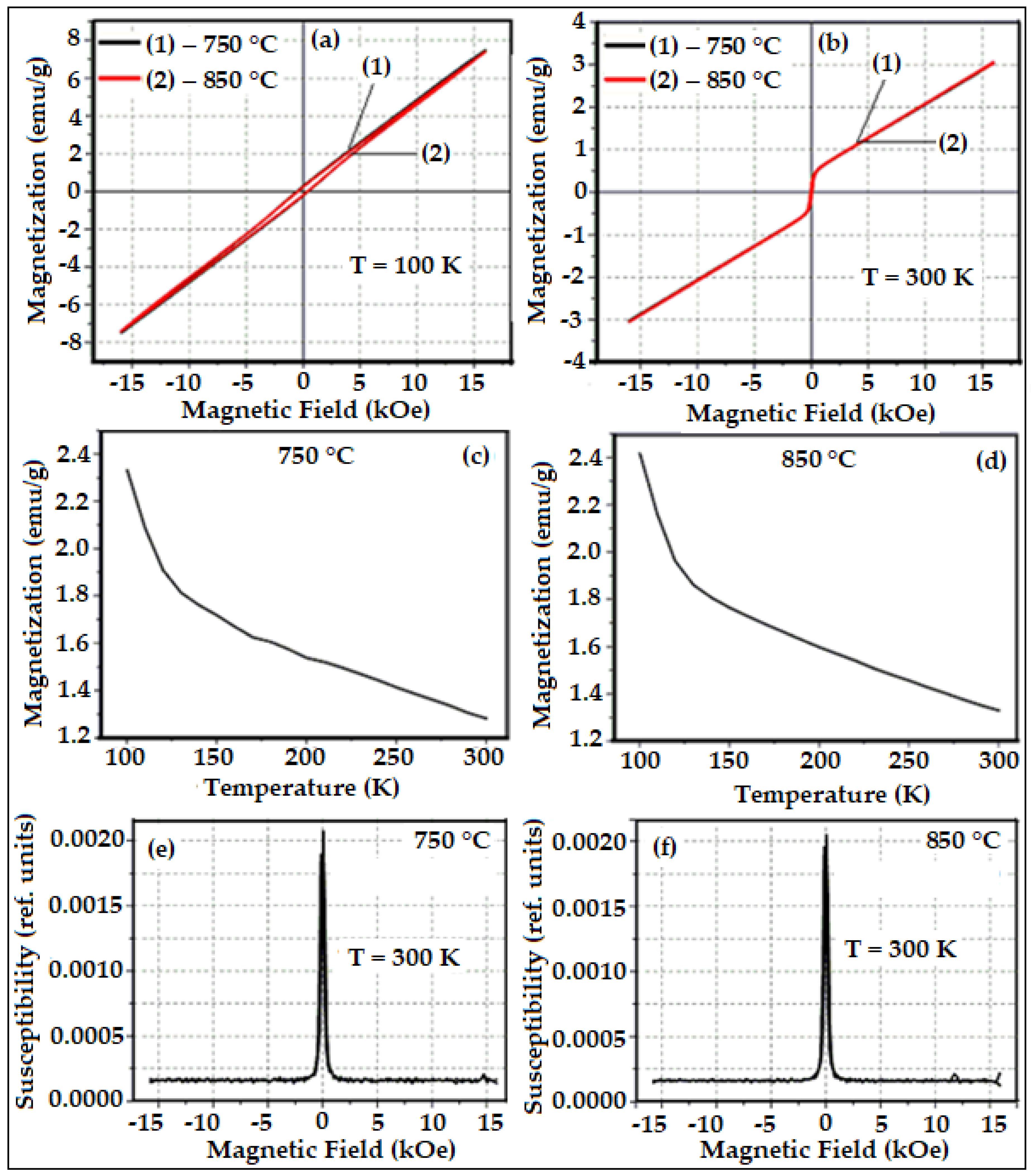
The obtained results on magnetic properties of nanoparticles HoFeO3 indicated that the magnetization increases when the temperature decreases (Figure 4c,d). Additionally, the magnetization is much greater than reported papers (Table 1). Interestingly, the magnetization curve continuously goes up when the external magnetic field increases (Figure 4a,b) and magnetic susceptibility is very low (χ << 1) (Figure 4e,f). HoFeO3 nanoparticles are super-paramagnetic material at temperatures greater than 100 K [25]. When the particles do not aggregate and the particle size is very small, the thermal energy is more dominant than directional energy, resulting in the superparamagnetic property of nano HoFeO3 (See Figure 2b,c).

4. Conclusions

In conclusion, syntheses and full characterizations of HoFeO3 nanoparticles are performed. With property analysis, HoFeO3 revealed a single-phase state after annealing at 750, 850 and 950 °C for 1h adopted the grain sizes in the range of 10–20 nm. HoFeO3 nanopowders have low band gap energy (Eg = 1.56 eV), which is beneficial for application in optical catalysis. The HoFeO3 nanoparticles are antiferromagnetic with a superparamagnetic behaviour (low susceptibility χ << 1 and high magnetization Ms = 3.2–7.5 emu/g), which makes them the potential candidates for making the devices operating at a high magnetic field.

Author Contributions

Conceptualization, methodology, and formal analysis, N.A.T. and V.Q.M.; validation, N.A.T.; writing—original draft preparation, V.Q.M.; writing—review and editing, N.A.T. All authors have read and agreed to the published version of the manuscript.

Funding

This research was funded by Sai Gon University, grant number TĐ 2020-1.

Institutional Review Board Statement

Not applicable.

Informed Consent Statement

Not applicable.

Data Availability Statement

Not applicable.

Acknowledgments

The authors would like to thank the Sai Gon University for financial support of this research.

Conflicts of Interest

The authors declare no conflict of interest.

References

  1. Rempel, A.A. Nanotechnologies, Properties and applications of nanostructured materials. Russ. Chem. Rev. 2007, 76, 435–461. [Google Scholar] [CrossRef]
  2. Atta, N.F.; Galal, A.; El-Ads, E.H. Perovskite nanomaterials—Synthesis, characterization, and applications. In Perovskite Materials—Synthesis, Characterisation, Properties, and Applications; Pan, L., Zhu, G., Eds.; IntechOpen: London, UK, 2016. [Google Scholar]
  3. Mounkachi, O.; Masrour, R.; Hamedoun, M.; Hlil, E.K.; Benyoussef, A. Synthesis and super-paramagnetic properties of neodymium ferrites nanorods. J. Alloys Compd. 2013, 581, 776–781. [Google Scholar]
  4. Tien, N.A.; Almjasheva, O.V.; Mittova, I.Y.; Stognei, O.V.; Soldatenko, S.A. Synthesis and magnetic properties of YFeO3 nanocrystals. Inorg. Mater. 2009, 45, 1304–1308. [Google Scholar] [CrossRef]
  5. Haron, W.; Thaweechai, T.; Wattanathana, W.; Laobuthee, A.; Manaspiya, H.; Veranitisagul, C.; Koonsaeng, N. Structural characteristics and dielectric properties of La1-xCoxFeO3 and LaFe1-xCoxO3 synthesized via metal organic complexes. Energy Procedia 2013, 34, 791–800. [Google Scholar] [CrossRef] [Green Version]
  6. Bhat, M.; Kaur, B.; Kumar, R.; Joy, P.A.; Kulkarni, S.D.; Bamzai, K.K.; Kotru, P.N.; Wanklyn, B.M. Swift heavy ion irradiation effect on structural and magnetic characteristics of RFeO3 (R = Er, Ho and Y) crystals. Nucl. Instrum. Methods Phys. Res., Sect. B 2006, 243, 134–142. [Google Scholar] [CrossRef]
  7. Sasikala, C.; Durairaj, N.; Baskaran, I.; Sathyaseelan, B.; Henini, M.; Manikandan, E. Transition metal titanium (Ti) doped LaFeO3 nanoparticles for enhanced optical structural and magnetic properties. J. Alloys Compd. 2017, 712, 870–877. [Google Scholar] [CrossRef] [Green Version]
  8. Nguyen, A.T.; Chau, H.D.; Nguyen, T.T.L.; Mittova, V.O.; Do, T.H.; Mittova, I.Y. Structural and magnetic properties of YFe1-xCoxO3 (0.1 ≤ x ≤ 0.5) perovskite nanomaterials synthesized by co-precipitation method. Nanosyst. Phys. Chem. Math. 2018, 9, 424–429. [Google Scholar] [CrossRef]
  9. Cheon, C.I.; Kim, J.S.; Jang, P.W. Ferroelectric and magnetic properties of PrFeO3-PbTiO3 and PrFeO3-BiFeO3-PbTiO3 thin films. Jpn. J. Appl. Phys. 2002, 41, 67–77. [Google Scholar] [CrossRef]
  10. Tien, N.A.; Mittova, I.Y.; Al’myasheva, O.V. Influence of the synthesis conditions on the particle size and morphology of yttrium orthoferrite obtained from aqueous solutions. Russ. J. Appl. Chem. 2009, 82, 1915–1918. [Google Scholar] [CrossRef]
  11. Habib, Z.; Majid, K.; Ikram, M.; Sultan, K.; Mir, S.A.; Asokan, K. Influence of Ni substitution at B-site for Fe3+ ions on morphological, optical and magnetic properties of HoFeO3 ceramics. Appl. Phys. A 2016, 122, 550. [Google Scholar] [CrossRef]
  12. Zhou, Z.; Guo, L.; Yang, H.; Liu, Q.; Ye, F. Hydrothermal synthesis and magnetic properties of multiferroic rare-earth orthoferrites. J. Alloys Compd. 2014, 583, 21–31. [Google Scholar] [CrossRef]
  13. Martinson, K.D.; Kondrashkova, I.S.; Omarov, S.O.; Sladkovskiy, D.A.; Kiselev, A.S.; Kiseleva, T.Y.; Popkov, V.I. Magnetically recoverable catalyst based on porous nanocrystalline HoFeO3 for processes of n-hexane conversion. Adv. Powder Technol. 2019, 31, 402–408. [Google Scholar] [CrossRef]
  14. Kondrashkova, I.S.; Martinson, K.D.; Zakharova, N.V.; Popkov, V.I. Synthesis of nanocrystalline HoFeO3 photocatalyst via heat treatment of products of glycine-nitrate combustion. Russ. J. Gen. Chem. 2018, 88, 2465–2471. [Google Scholar] [CrossRef]
  15. Jiang, L.; Liu, W.; Wu, A.; Xu, J.; Liu, Q.; Qian, G.; Zhang, H. Low-temperature combustion synthesis of nanocrystalline HoFeO3 powders via a sol-gel method using glycine. Ceram. Int. 2012, 38, 3667–3672. [Google Scholar] [CrossRef]
  16. Popkov, V.I.; Almjasheva, O.V.; Semenova, A.S.; Kellerman, D.G.; Nevedomskiy, V.N.; Gusarov, V.V. Magnetic properties of YFeO3 nanocrystals obtained by different soft-chemical methods. J. Mater. Sci. Mater. Electron. 2017, 28, 7163–7170. [Google Scholar] [CrossRef]
  17. Nguyen, T.A.; Nguyen, L.T.T.; Bui, V.X.; Nguyen, D.H.; Lieu, H.D.; Le, L.M.; Pham, V. Optical and magnetic properties of HoFeO3 nanocrystals prepared by a simple co-precipitation method using ethanol. J. Alloys Compd. 2020, 834, 155098. [Google Scholar] [CrossRef]
  18. Nguyen, T.A.; Pham, V.N.; Le, H.T.; Chau, D.H.; Mittova, V.O.; Nguyen, L.T.T.; Dinh, D.A.; Hao, T.N.; Mittova, I.Y. Crystal structure and magnetic properties of LaFe1-xNixO3 nanomaterials prepared via a simple co-precipitation method. Ceram. Int. 2019, 45, 21768–21772. [Google Scholar] [CrossRef]
  19. Nguyen, A.T.; Pham, V.N.; Nguyen, T.T.L.; Mittova, V.O.; Vo, Q.M.; Berezhnaya, M.V.; Mittova, I.Y.; Do, T.H.; Chau, H.D. Crystal structure and magnetic properties of perovskite YFe1-xMnxO3 nanopowders synthesized by co-precipitation method. Solid State Sci. 2019, 96, 105922. [Google Scholar] [CrossRef]
  20. Nguyen, T.A.; Pham, V.; Chau, D.H.; Mittova, V.O.; Mittova, I.Y.; Kopeychenko, E.I.; Nguyen, L.T.T.; Bui, V.X.; Nguyen, A.T. Effect of Ni substitution on phase transition, crystal structure and magnetic properties of nanostructured YFeO3 perovskite. J. Mol. Struct. 2020, 1215, 128293. [Google Scholar] [CrossRef]
  21. Belous, A.G.; Pashkova, E.V.; Elshanskii, V.A.; Ivanitskii, V.P. Effect of precipitation conditions on the phase composition, particle morphology and properties of iron (III, II) hydroxide precipitates. Inorg. Mater. 2000, 36, 343–351. [Google Scholar] [CrossRef]
  22. Imanaka, N. Physical and Chemical Properties of Rare Earth Oxides. In Binary Rare Earth Oxides; Adachi, G., Imanaka, N., Kang, Z.C., Eds.; Springer: Dordrecht, The Netherland; Berlin, Germany, 2004; pp. 111–113. [Google Scholar]
  23. Gupta, H.C.; Kumar Singh, M.; Tiwari, L.M. Lattice dynamic investigation of Raman and infrared wavenumbers at the zone center of orthorhombic RFeO3 (R = Tb, Dy, Ho, Tm) perovskite. J. Raman Spectrosc. 2002, 33, 67–70. [Google Scholar] [CrossRef]
  24. Habib, Z.; Ikram, M.; Majid, K.; Asokan, K. Structural, dielectric and ac conductivity properties of Ni-doped HoFeO3 before and after gamma irradiation. Appl. Phys. A 2014, 116, 1327–1335. [Google Scholar] [CrossRef]
  25. Cullity, B.D.; Graham, C.D. Introduction to Magnetic Materials, 2nd ed.; Wiley-IEEE Press: Hoboken, NJ, USA, 2009; ISBN 978-0-471-47741-9. [Google Scholar]
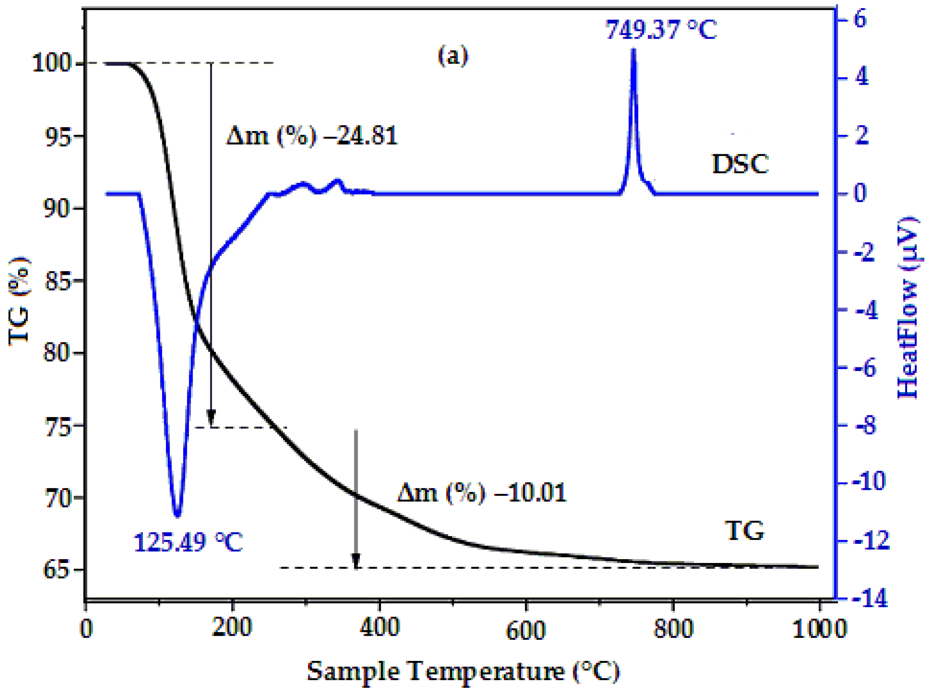
Figure 1. Combined TGA-DSC plots of HoFeO3 precipitate sample.
Crystals 11 00238 g001
Figure 2. XRD patterns of the nano-sized HoFeO3 particles annealed at 650, 750, 850 and 950 °C for 1 h (a) and SEM (b), TEM (c) of the HoFeO3 annealed at 750 °C for 1 h.
Crystals 11 00238 g002
Figure 3. EDXA (a), Raman (b), UV-Vis (c) spectra, and Plots of (Ahv)2 as a function of photon energy (d) of HoFeO3 nanoparticles annealed at 750 °C for 1 h.
Crystals 11 00238 g003
Figure 4. Magnetization versus applied field plots of HoFeO3 nanoparticles at 100 K (a) and 300 K (b); Temperature dependence of zero-field-cold (ZFC) magnetization (c,d), and (e,f) susceptibility plots of HoFeO3 nanoparticles at 300 K.
Crystals 11 00238 g004
Table 1. Characteristics of HoFeO3 nanoparticles in this work and that from the published literatures as a comparison.
HoFeO3Particle Size, nmEg, eVMs, emu/gχ
100 K300 K100 K300 K
750 °C10–201.56~7.5 ~3.2~0.53× 10⁻³~2.1× 10⁻³
850 °C~0.51× 10⁻³
[9]149.33.392.55
[10]10–20 μm<0.1
[11,12]40–1002.12–2.14
[13]100–3000.60.2
Publisher’s Note: MDPI stays neutral with regard to jurisdictional claims in published maps and institutional affiliations.