Communication

Synthesis and Characterization of New Guanine Complexes of Pt(IV) and Pd(II) by X-ray Diffraction and Hirshfeld Surface Analysis

by
Anton Petrovich Novikov
,
Mikhail Alexandrovich Volkov
,
Alexey Vladimirovich Safonov
,
Mikhail Semenovich Grigoriev
and
Evgeny Vladilenovich Abkhalimov
*
Frumkin Institute of Physical Chemistry and Electrochemistry, Russian Academy of Sciences, Leninskii Prospect 31-4, 119071 Moscow, Russia
*
Author to whom correspondence should be addressed.
Crystals 2021, 11(11), 1417; https://doi.org/10.3390/cryst11111417
Submission received: 20 October 2021 / Revised: 14 November 2021 / Accepted: 15 November 2021 / Published: 19 November 2021

Abstract

:
The aim of the work was to synthesize new perspective compounds of palladium and platinum with nitrogenous bases (guanine), promising for use in biomedicine and catalysis. The article describes the synthesis of new [PdCl2(HGua)2]Cl2·H2O and [PtCl5(HGua)]·2H2O compounds using wet chemistry methods. The structure of the obtained single crystals was established by the method of single crystal X-ray diffraction. The complexes have an M-N bond, and the organic ligand is included in the first coordination sphere. The analysis of Hirshfeld surfaces for the obtained complexes and their analogues for the analysis of intermolecular interactions was carried out. In the palladium complex we obtained, π-halogen and π-stacking interactions were found; in analogues, such interactions were not found. π-halogen and halogen interactions were found in structure of platinum complex and its analogues.

Graphical Abstract

1. Introduction

The study of Pt and Pd complexes with various organic compounds is of great fundamental and practical interest. One of the areas of research is focused on the synthesis of nanoscale metal clusters [1] and nanoparticles with DNA using Pd and Pt complexes [2,3,4]. Such works have a wide range of applications, including biosensors, DNA visualization [5], and catalysis [2,6].
First of all, coordination compounds of platinum group metals occupy an important place in anticancer therapy [7]. Cisplatin is most successfully used, although serious side-effects (toxic effect, nonspecificity, development of resistance during treatment) stimulate the search for new coordination compounds of metals (palladium, ruthenium, etc.) [8]. The basis of the operation of cisplatin (cis-diamminedichloroplatinum (II)) lies in the alkylating effect of DNA due to the rapid replacement of chlorine in an aqueous environment. In coincident complexes of cisplatin, when replacing platinum with palladium, the lability of chlorine significantly decreases and for this reason palladium analogues to cisplatin are unusable in anti-cancer therapy. However, there is evidence of the successful use of complexes of paladium II). For example, Pd(II) complexes of dithiocarbamate [9] cis-Diamminedichloropalladium are promising in anticancer therapy, which are characterized by the much lower toxicity in comparison with platinum complexes for tissues [10]. In the work [11], the antitumoral activity of some palladium complexes of compounds against Sarcoma is described. As in the case of platinum, the interaction of palladium with DNA leads to a cytotoxic effect during the formation of inter and intrastrand cross-links due to complexation with purine and pyrimidine bases [12]. Nitrogen bases of DNA and other similar nucleobases are ambidentate (or ambivalent) ligands since they have two or more donor atoms capable of binding metal ions; therefore, Pd(II) complexes with various heterocycles (xanthine, theophylline, theobromine, 3,8-dimethylxanthine, caffeine and 1,3,8-trimethylxanthine), promising for use in medicine were synthetized and described earlier [13].
It is well known that the interaction of platinum and palladium with DNA occurs mainly on guanine-rich DNA fragments [14,15,16,17,18]. In this sense, guanine N(7) is the most commonly accepted atom for primary attachment of platinum compounds to DNA [19,20,21] because its N(9) position in guanine is unsubstituted. Some studies have demonstrated that N(9) of guanine is also the main site of binding interaction with metals [22]. Guanine is one of the major purine nucleobases present in DNA and RNA. RNA has considerably higher content of guanine. In general, purines are basic components of nucleotides in cell proliferation, because they provide the necessary energy and cofactors to promote cell survival and proliferation, thus purine metabolism is associated with the progression of cancer. High concentrations of purine metabolites have been indicated in tumor cells [23].
Metal complexes of unsubstituted guanine have been investigated by various groups [24,25,26]. Both purine and pyrimidine DNA derivatives belong to antimetabolites of nucleic metabolism, and their application as active carriers of alkylating agents is based on the biochemical data relating to the accumulation of purines and pyrimidines in a tumour cell. It is for this reason that platinum complexes with purine and pyrimidine bases and their derivatives are considered as potential antitumor agents [27]. In addition to medicine, it was proposed to use guanine palladium complexes in organic catalysis, including in the form of graphene deposited on nanoparticles [28,29].
Some previous works on the synthesis of palladium derivatives of guanine proposed to use guanine derivatives with ethylenediamine and diethylenetriamine complexes of palladium(II) [30]. In their research works, they obtained complexes of palladium and platinum with nucleoside fragments [31,32], but the complexes described in these works have different geometry [33] or X-ray data were not obtained or the structures were not described [34]. Thus, the aim of our work was to obtain Pd(II) and Pt(IV) compounds with unsubstituted guanine as a ligand and describe the crystal structure and intermolecular interactions by the method of Hirshfeld surfaces.

2. Materials and Methods

2.1. Materials

Complexes 1 and 2 were synthesized using hexachloroplatinic (IV) acid hexahydrate (H2PtCl6·6H2O, ~40% Pt), palladium (II) chloride (PdCl2, ≥99%), guanine (C5H5N5O, ≥98%) and hydrochloric acid (HCl, 36.5%), all being ACS Reagent Grade chemicals purchased from Sigma-Aldrich (Darmstadt, Germany) and used without additional purification. All solutions were prepared using distilled water additionally deionized with the Arium setup (Sartorius AG, Goettingen, Germany).

2.2. Synthesis Procedure of [PdCl2HGua2]Cl2·H2O (1)

To begin with, we dissolved 15 mg (0.1 mmol) of guanine in 15 mL of 1 mol L−1 hydrochloric acid aqueous solution in 25 mL two-necked flask equipped with a thermometer. To prepare H2PdCl4 acid, we dissolved 9 mg (0.05 mmol) of PdCl2 in 1 mol L−1 hydrochloric acid aqueous solution. Then, both guanine and tetrachloropalladic (II) acid solutions were mixed at room temperature under stirring for 5 min at 23 °C. After that, we raised the temperature to 50 °C and continued stirring for 60 min. After turning off the stirring, the solution was transferred to a vacuum desiccator and left for 24 h over anhydrous CaCl2. To the resulting brown-red crystals, 500 μL of anhydrous methanol were added. The resulting suspension was heated to 40 °C and cooled in air to room temperature; the resulting red crystals were analyzed by XRD.

2.3. Synthesis Procedure of PtCl5HGua·2H2O (2)

We dissolved ~10 mg of guanine in 5 mL of distilled water at room temperature in a 10 mL two-necked flask equipped with a thermometer. To this suspension we added 100 μL of 0.7 mol L−1 hexachloroplatinic acid aqueous solution. The mixture was stirred for 5 min at 23 °C, then the temperature was raised to 50 °C and held for 60 min. After turning off the stirring, the flask with the solution was transferred to a vacuum desiccator and left for 24 h over anhydrous CaCl2. To the resulting yellow-green powder was added 500 μL of anhydrous methanol. The resulting suspension was heated to 40 °C and cooled in air to room temperature; the formed thin needles of light green color were analyzed by XRD.

2.4. Single-Crystal XRD Analysis

The crystal structure of all synthesized substances was determined by X-ray structural analysis using an automatic four-circle area-detector diffractometer Bruker KAPPA APEX II with MoKα radiation at a temperature of 100 K (for 1) and 296 K (for 2). The cell parameters were refined over the entire data set by using SAINT-Plus software [35]. Absorption corrections were introduced using the SADABS program [36]. The structures were solved using the SHELXT-2018/2 program [37] and refined by full-matrix least squares on F2 in the anisotropic approximation for all non-hydrogen atoms (SHELXL-2018 [38]). The H atoms were objectively located from the difference Fourier synthesis and refined in an idealized geometry with isotropic temperature factors equal to 1.2Ueq (N, C) for NH- and CH-groups, and 1.5Ueq (O) for water molecules. Tables and pictures for structures were generated using Olex2 [39].
Crystal data, data collection, and structure refinement details are summarized in Table 1. All other crystallographic parameters of structures 1 and 2 are indicated in Tables S1−S8. The atomic coordinates were deposited at the Cambridge Crystallographic Data Centre [40], CCDC № 2116634 and 2116635 for 1 and 2, respectively. The Supplementary crystallographic data can be obtained free of charge from the Cambridge Crystallographic Data Centre via www.ccdc.cam.ac.uk/data_request/cif (accessed on 14 November 2021).

3. Results and Discussions

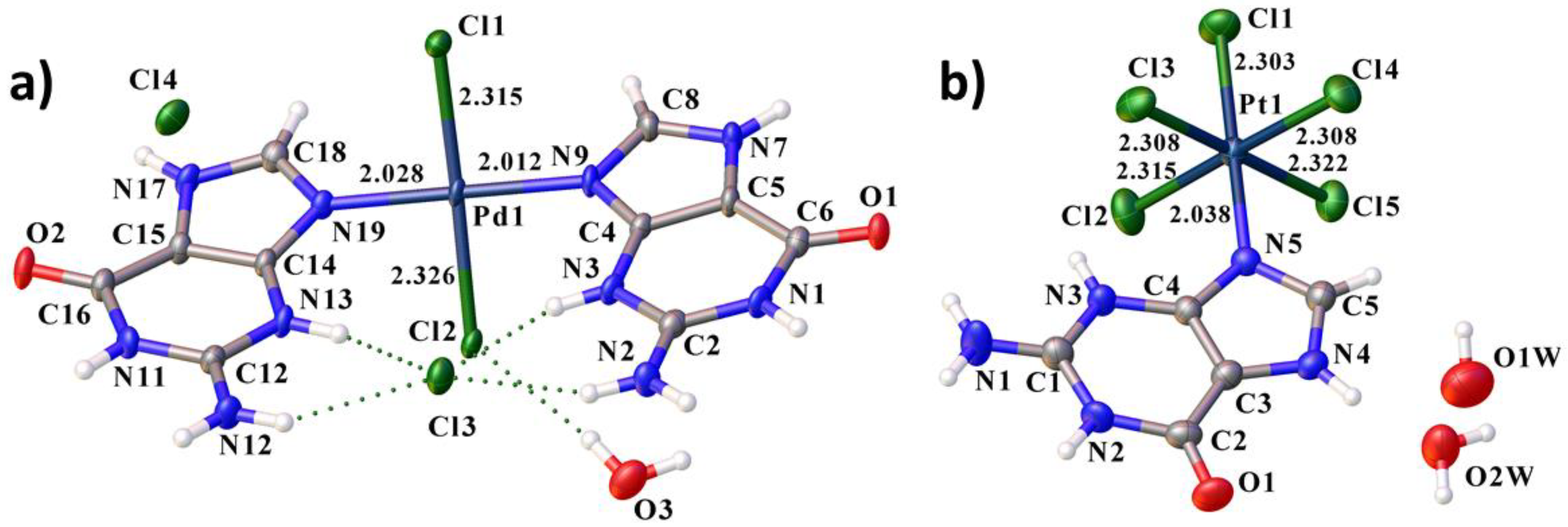
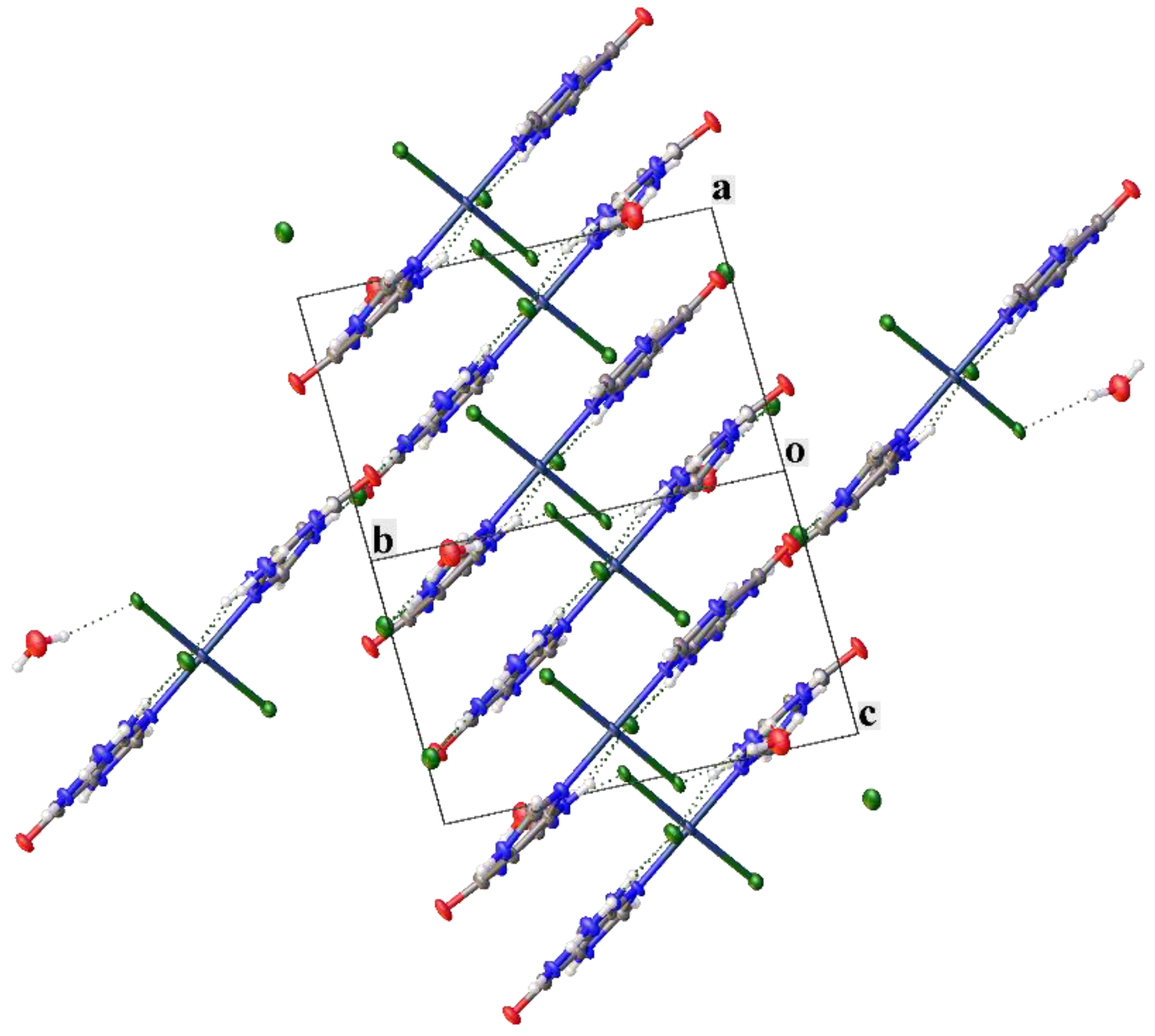
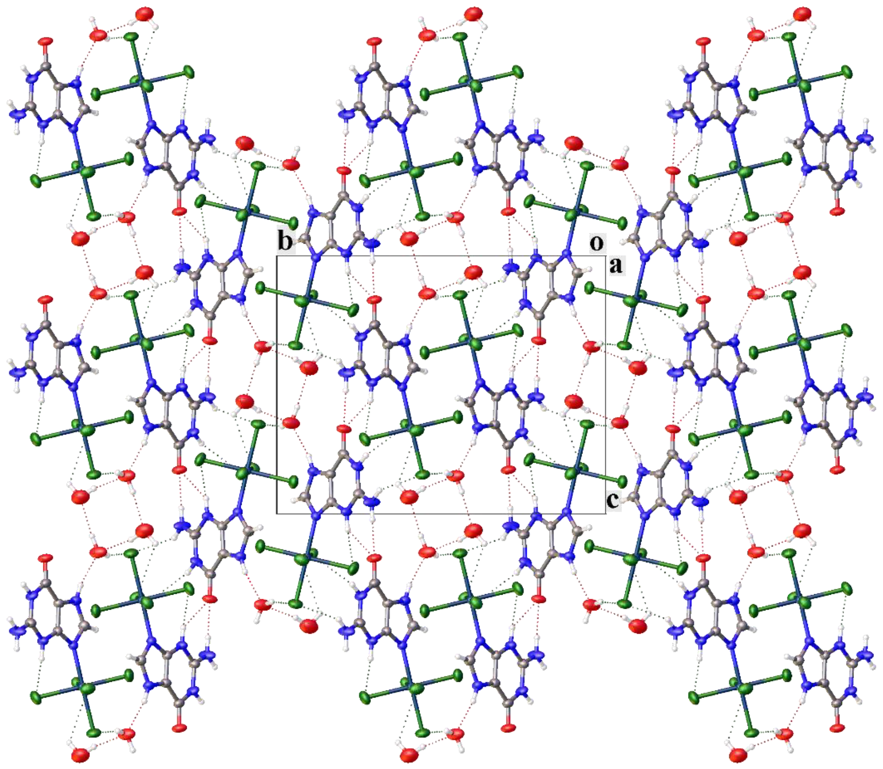
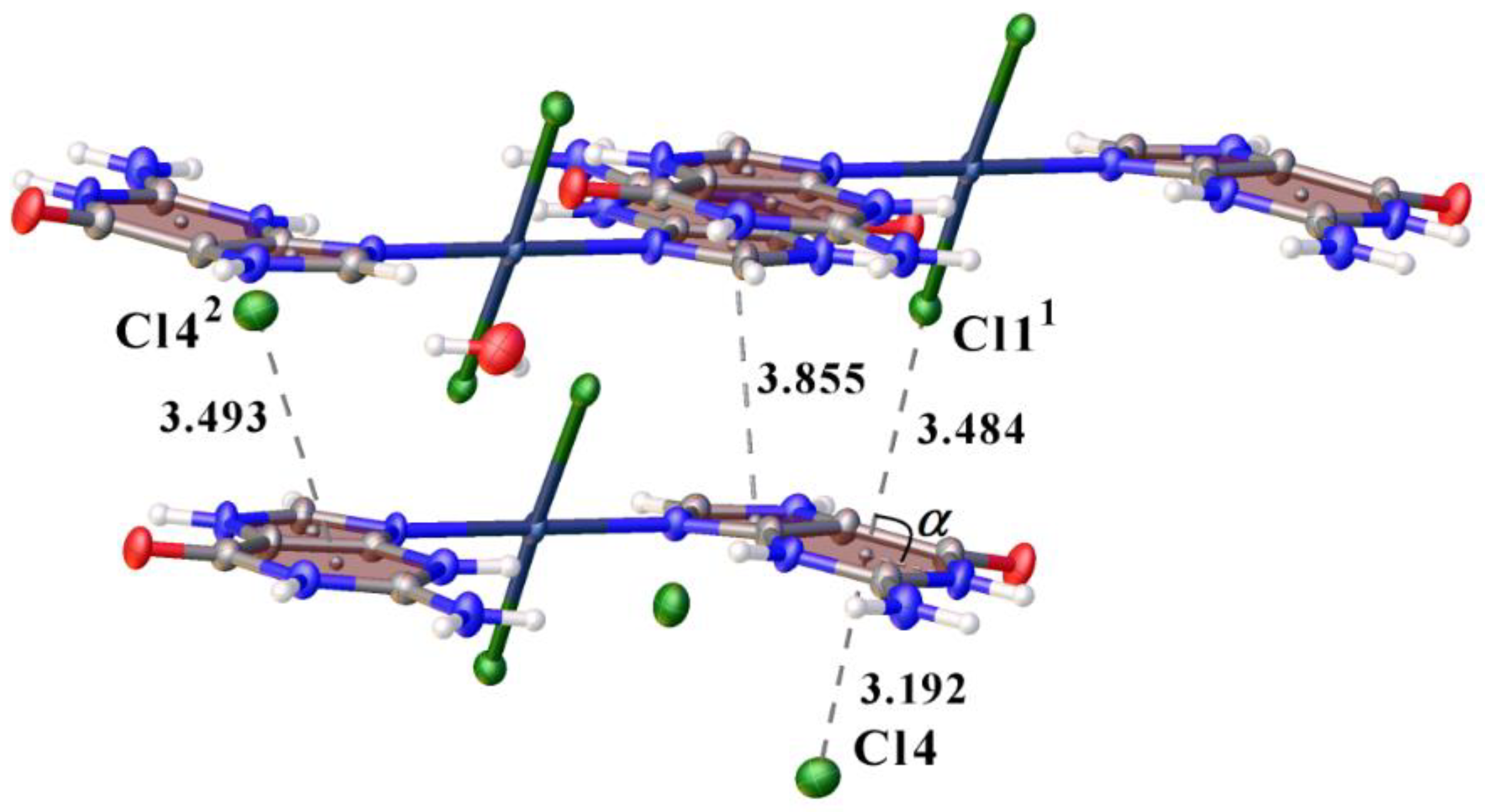
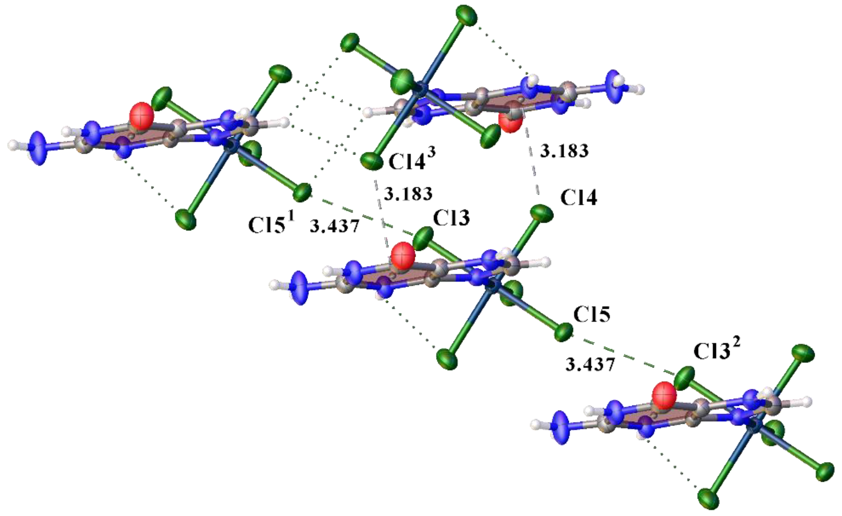
3.1. Structural Description

Compound 1 crystallizes in the triclinic space group P-1, 2 in the monoclinic space group P21/n. The asymmetric fragments of 1 and 2 are shown in Figure 1. In both structures, the metal atom forms a M-N bond with the nitrogen atom of the five-membered ring of the guanine fragment. Also, in both structures, the nitrogen atom of the six-membered ring, which is in the para-position to the oxygen atom of the guanine molecule, is protonated. However, the molecule of complex 1 has a charge of +2, and the molecule of complex 2 is electroneutral. This can be used for selective delivery of the complex in organisms. As counter-ions in structure 1, there are two chloride anions. In both structures, in addition to the molecules of the complex, there are molecules of crystallization water. There are one and two molecules of water in 1 and 2, respectively.
The guanine fragments in structures 1 and 2 are plane. However, in structure 1 in the second ring, the atoms deviate from the plane more strongly than for the first ring in 2 and for the ring in 1 (maximum displacement of 0.128, 0.047 and 0.003 Å for C15, C2 in 1 and C3 in 2 respectively).
The planes of guanine fragments are significantly rotated relative to the plane perpendicular to the direction determined by the practically linear grouping Cl1-Pd1-Cl2. The torsion angles Cl2-Pd1-N19-C14 of 57.05° and Cl2-Pd1-N9-C4 of −60.32° differ greatly from 90° as in similar compounds [41,42,43]. However, in this case, the planes of both ligands almost coincide (angle (plane normal-plane normal): 3.000°), which is obviously due to H-bonding with the outer-sphere chloride ion.
The Pd-N distance varies from 2.012 to 2.028 Å. The average distance found with the data from the Cambridge Structural Database (CSD Version 5.42, update of September 2021) is 2.018 Å [40]. The Pt-N distance is slightly longer than Pd-N and is 2.038 Å, which is slightly shorter than the average distance found in the CSD, which is 2.046 Å. The Pd–Cl distances vary from 2.315 to 2.326 Å, which is longer than the average distance found in CSD for the structures with analogous coordination, which is 2.296 Å. The Pt–Cl distances change from 2.303 to 2.322 Å, which is close to 2.310 Å (average distance from CSD). The slight lengthening of bonds may be due to the different participation of chlorine atoms in hydrogen bonds.
In structures 1 and 2, a complex system of hydrogen bonds is formed. The hydrogen bond parameters are listed in Tables S3 and S7. Crystal water plays an important role in the formation of a system of hydrogen bonds in both structures. In crystal 1, bifurcate H-bonds are absent, but the chlorine atom Cl3 is linked by four hydrogen bonds to the amino groups of six-membered rings (Figure 2a). In 2, there is a trifurcate hydrogen bond N3–H3A⋯Cl2, N3–H3A⋯Cl3, N3–H3A⋯O15 with bond lengths 3.377 (2), 3.159 (3), 2.942 (3) Å, respectively. The strongest hydrogen bonds N11–H11⋯O11 and N7–H7⋯O22 in 1 are formed between nitrogen atoms of five- and six-membered rings and oxygen atoms of guanine fragments. In 2, the strongest H-bonds are formed between water molecules (O2W⋯O1W3 distance is 2.795 (4) Å), nitrogen molecule N4 and water molecule O2W (N4⋯O2W distance is 2.746 (3)Å) and amino group and oxygen atom guanine fragment (N4⋯O2W distance is 2.734 (4) Å).
Crystal packing in 1 can be represented as layered (Figure 3). In 2, the crystal packing can be represented by columns (Figure 4). In both structures, hydrogen bonds form a three-dimensional framework and connect the layers and columns to each other. The layers are additionally connected by π-halogen interactions in 1. The complex molecules in 2 in the columns are connected by halogen bonds (XB). In structure 2, four water molecules form a cluster linked by H-bonds. There is π-staking interaction in 1 of six-membered ring (C14 N19 C18 N17 C15) with five-membered ring (C4 N9 C8 N7 C5) with angle: 3.543°, centroid-centroid distance: 3.855 Å and shift distance 1.851 Å, which additionally stabilizes the structure.
Previously, only a few compounds similar to ours were described. Complexes are described in the literature in which the Pd atom is bound by a Pd-N bond with an organic ligand [41,42,43,44]. But in almost all works, electrically neutral complexes are obtained. In our work, the complex has a charge of +2. Besides, the palladium atom binds to two guanine molecules. This fact presupposes the use of our complex as a selective delivery of palladium to various organs and tissues. We have found only one work with structure of the platinum atoms with molecules of guanine or 7-methylguanine [45]. However, in the work of Gupta et al., Pt binds not only to the guanine fragment, but also to the nitrogen atoms of the diethyleneamine fragments. Also, the guanine fragment in compound 2 is not protonated.

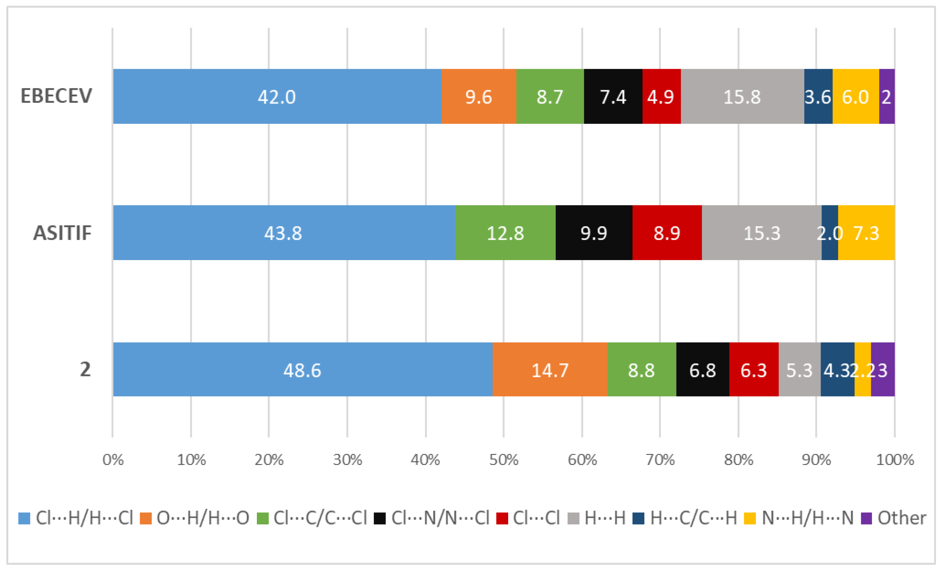
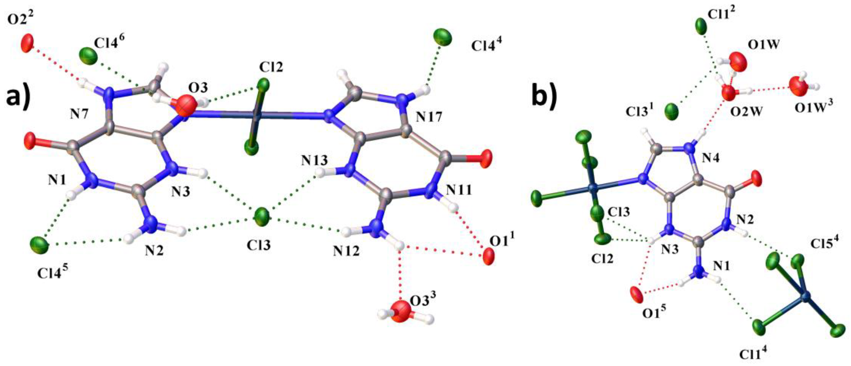
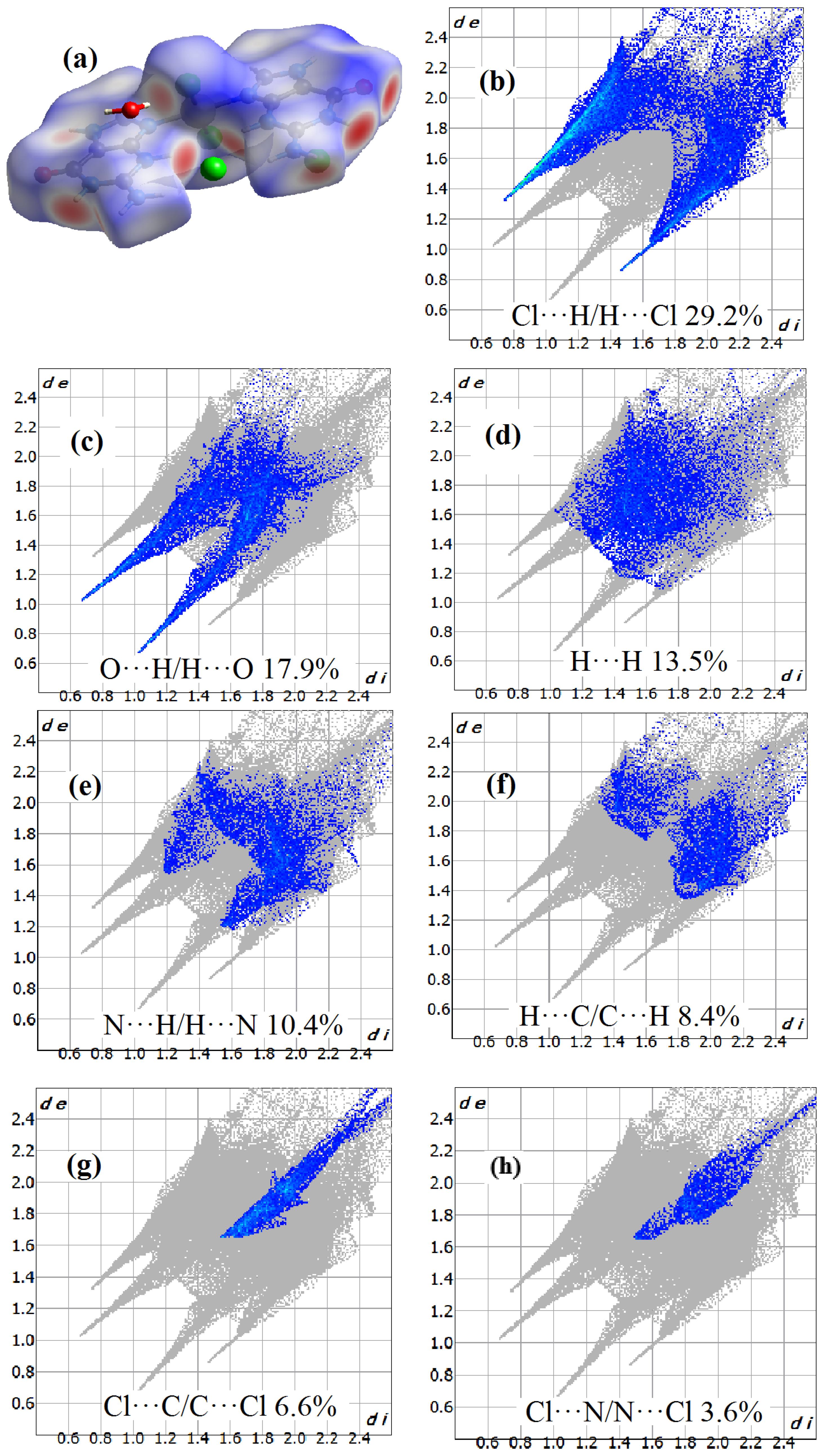
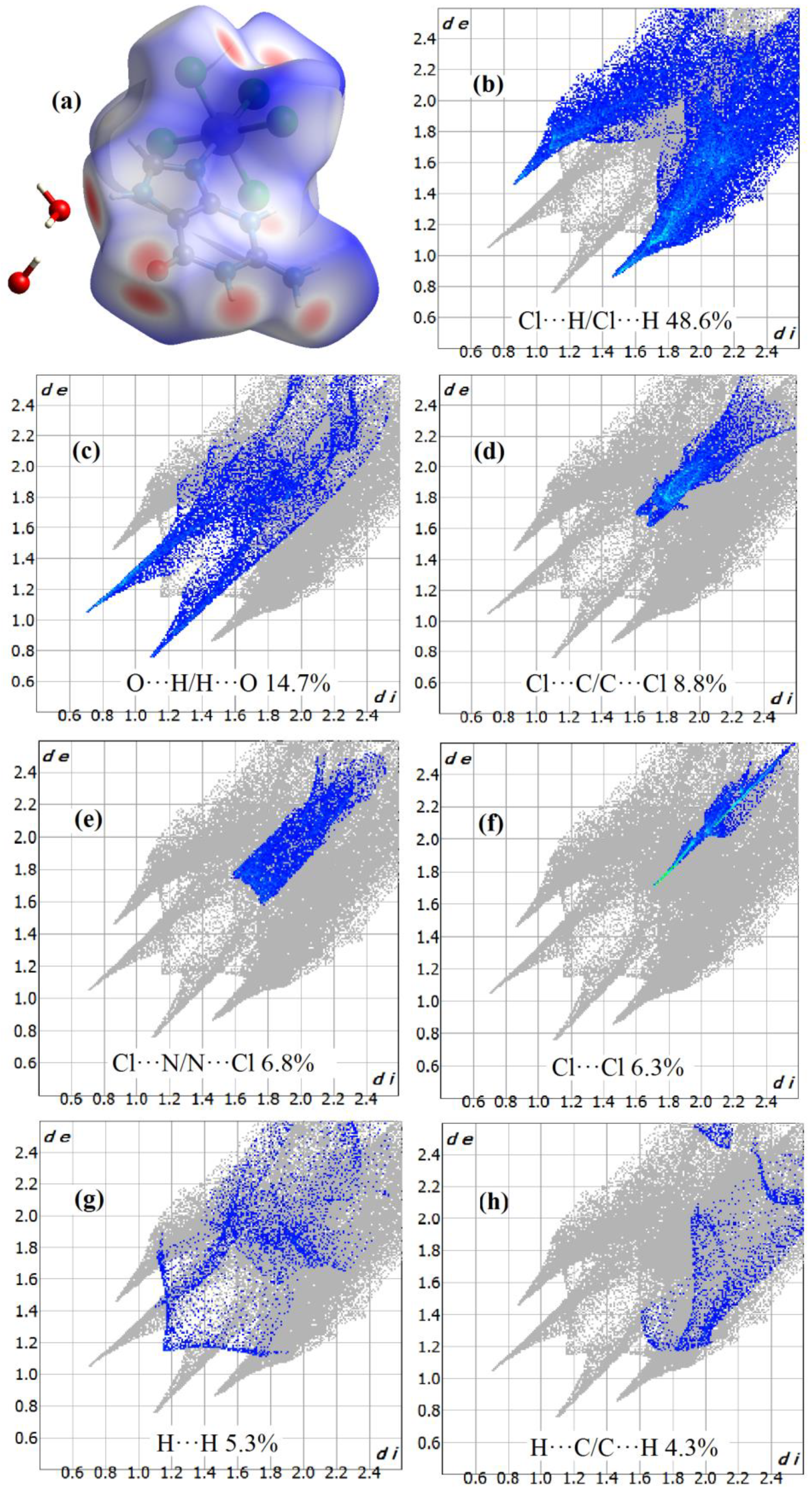
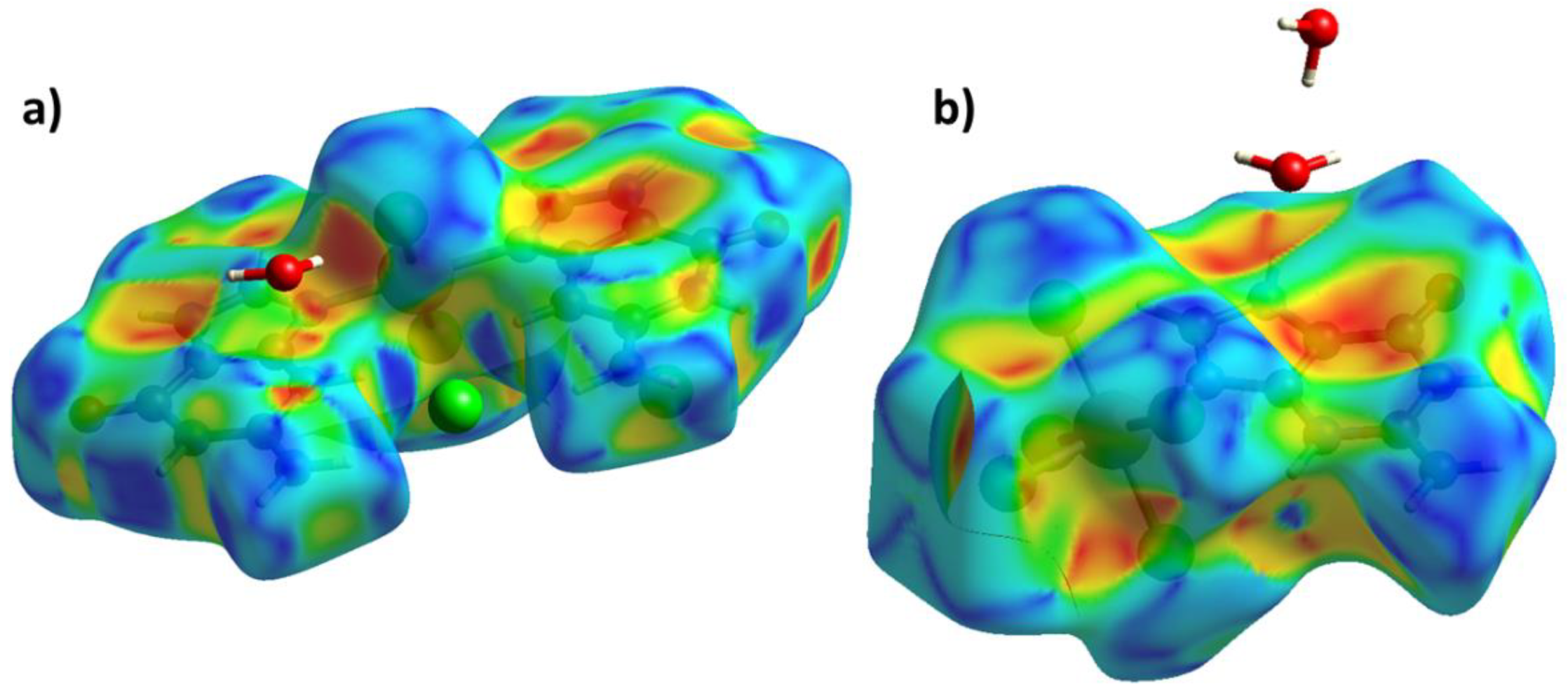
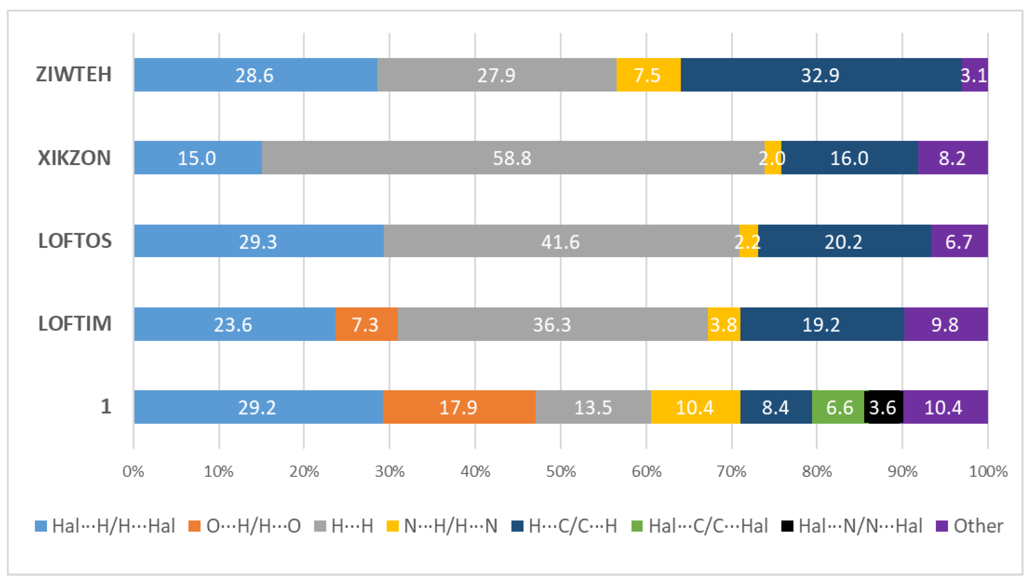
3.2. Hirshfeld Surface Analysis

Crystal Explorer 21 was used to calculate the Hirshfeld surfaces (HS) and two-dimensional fingerprint plots [46]. The donor-acceptor groups are visualized using a standard (high) surface resolution and dnorm surfaces mapped over a fixed color scale of −0.705 (red) to 1.266 (blue) for 1 and −0.665 to 1.650 a.u. for 2, as illustrated in Figure 5a and Figure 6a, respectively. Additionally, the surface of shape-index was constructed, with bright spots on which short interactions are also visible (Figure 7).
Red spots on the dnorm surface for both compounds correspond to hydrogen bonds. In compounds 1 and 2, the main contribution to intermolecular interactions is made by hydrogen bonds of the Cl⋯H/H⋯Cl type (29.2% and 48.6%, respectively) and O⋯H/H⋯O type (17.9% and 14.7%, respectively). Such a difference in the contribution of contacts may be associated with a large percentage of chlorine atoms in complex 2 and with a different composition of the complexes. Van der Waals interactions H⋯H (13.6%), N⋯H/H⋯N (10.4%), and H⋯C/C⋯H (8.4%) play important role in crystal packing. Cl⋯C/C⋯Cl contacts in complexes 1 and 2 (8.8% and 6.6%, respectively) indicate the presence of π-halogen interaction. In the structure with a palladium atom, there are the π -anion interactions between the six-member ring and the chloride anions and chlorine atom of other complex, as described in [47,48]. This is additionally confirmed by short distances (plane N1C2N3C4C5C6 centroid-atom Cl42 (symmetry code: x, 1 + y, z): 3.493 Å, plane N11C12N13C14C15C16 centroid-atom Cl4: 3.192 Å and plane N11C12N13C14C15C16 centroid-atom Cl1 (symmetry code: 1 + x, y, z): 3.484) (Figure 8). The angles α confirm this fact (67.07, 80.0, and 82.82°, respectively). In structure 2, π-halogen interaction also occurs, but this time the ring of the guanine fragment with the chlorine atom Cl43 (plane C1N2C2C3C4N3 centroid-atom Cl43 (symmetry code: 1−x, 1−y, 1−z) is 3.183 Å and α is 83.97°) [47,49,50,51] (Figure 9). The short contact Cl⋯Cl (6.3%) in structure 2 indicates the presence of a halogen bond. The short distance Cl3···Cl5 and the angle Pd-Cl···Cl equal to 163° confirms this fact [52]. Significant contribution to intermolecular interactions in 2 is made by contacts of the type Cl⋯N/N⋯Cl, H⋯H, H⋯C/C⋯H.
We have analyzed the HS for analogues of the title compounds. As an analogue for compound 1, we found only a few compounds in which there is a Pd-N bond and in which the ligand is represented by five- and six-member rings. We have taken compounds with flat substituents. Moreover, all the analogues we found are electrically neutral. The analysis performed for analogues of the complex 1 showed that in neutral complexes the main contribution to intermolecular interactions is made by van der Waals interactions (Figure 10). Hydrogen bonds contribute less to intermolecular interactions than in 1. In these compounds, there is no π-halogen interaction, as in our compound. This may be since in 1 the π-halogen interaction occurs predominantly between the chloride anion and the rings of the guanine fragments, while the analogues we have found are electrically neutral. It can be noted that with lengthening of the substituent, the proportion of H⋯H interactions increases.
As an analogue for the HS analysis, we found only two structures with similar geometry (Pt-N bond with the benzoimidazole ligand, Pt(IV) bonded to five halogen atoms) for 2. Moreover, the CSD contains data only on pentachloroplatinates structures; for other halogens, such structures are absent. The complex in ASITIF is electrically neutral, and in EBECEV it is negatively charged, but in these complexes the charge does not affect strongly the HS. A similar type of packing is formed in all structures, and hydrogen bonds play an important role in the formation of crystal packing (Figure 11). All compounds also contain Cl⋯C/C⋯Cl and Cl⋯N/N⋯Cl contacts. This shows that, as in our complex, in similar structures, the molecules of the complexes are additionally bound by the π-halogen interaction. Short Cl⋯Cl contacts show that in similar structures, a halogen bonding is present. It should also be noted that the fraction of van der Waals interactions in similar structures is higher, which may be due to the absence of crystallization water molecules in the structure.
In this work, complexes of palladium and platinum with guanine were obtained for the first time. When analyzing the literature, we found only a few similar Pt or Pd compounds with M-N bonds with imidazole rings or molecules of nitrogenous bases. In both complexes, a system of hydrogen bonds is formed, which additionally stabilizes the structures in the crystals. The analysis of non-bonded interactions carried out in this work showed their different nature in two complexes under consideration. In structure 1, the molecules of the complex are linked by both π-stacking interaction and a π-halogen bonding. In 2, the molecules of the complex are additionally linked to HB by a π-halogen and halogen bonding.
The detailed analysis of non-bonded interactions carried out in this work may be of interest in the context of self-assembly [57]. In the future, when replacing, for example, chlorine atoms with bromine atoms, we can assume that there will be an increase in non-bonded interactions and a better association of complexes in the crystal and in solution. And replacing chlorine with fluorine, on the contrary, can lead to a different type of packing. The analysis of the influence of halogen atoms on the character of interactions was carried out in [58]. It has been shown that the strength of the XB donor increases in the following order as the XB donor capacity increases: F < Cl <Br < I.
Analysis of non-bonded interactions and H-bond systems can also predict the behavior of complexes in solutions. Such an analysis may be of interest from the point of view of predicting the formation of solvates or micelles in the process of creating targeted dosage forms, as well as in modeling receptor activity in silico. At a certain concentration of the solution, these complexes will most likely form aggregates due to non-bonded interactions, but in this case, they will interact with solvent molecules through a system of H-bonds. Platinum and palladium do not participate in intermolecular interactions and are completely screened by their coordination spheres, which suggests that compounds 1 and 2 are resistant to hydrolytic processes.

4. Conclusions

In this work, we obtained the compounds of four-coordinated palladium(II) and six-coordinated platinum(IV) with guanine entering the coordination sphere through the M-N bond for the first time. The compounds were synthesized from acidic solutions, which resulted in additional protonation of the formed complexes at the nitrogen atoms N3. The resulting palladium complex is a doubly charged cation, and the platinum compound is an electrically neutral molecule. The noble metals complexes usage may be interesting for medical purposes. The main intermolecular interactions are shown by the Hirshfeld surface method, and the significant contribution of hydrogen bonds to the crystal packing is proved. π-halogen, π-stacking, and halogen interactions were found in the obtained compounds and compared with their analogues.

Supplementary Materials

The following are available online at https://www.mdpi.com/article/10.3390/cryst11111417/s1, Table S1: Bond Lengths for 1, Table S2: Bond Angles for 1, Table S3: Hydrogen Bonds for 1, Table S4: Torsion Angles for 1, Table S5: Bond Lengths for 2, Table S6: Bond Angles for 2, Table S7: Hydrogen Bonds for 2, Table S8: Torsion Angles for 2.

Author Contributions

Conceptualization, E.V.A. and A.V.S.; methodology, M.A.V. and E.V.A.; validation, M.S.G.; investigation, E.V.A., A.P.N., M.S.G. and M.A.V.; writing—original draft preparation, A.P.N., A.V.S. and M.S.G.; writing—review and editing, E.V.A.; visualization, A.P.N.; supervision, M.S.G.; project administration, E.V.A. All authors have read and agreed to the published version of the manuscript.

Funding

The study was supported by the Ministry of Science and Higher Education of the Russian Federation (program no. AAAA-A18-118040590105-4).

Institutional Review Board Statement

Not applicable.

Informed Consent Statement

Not applicable.

Acknowledgments

X-ray diffraction experiments were performed at the Center for Shared Use of Physical Methods of Investigation at the Frumkin Institute of Physical Chemistry and Electrochemistry, RAS.

Conflicts of Interest

The authors declare no conflict of interest.

References

  1. Richter, J.; Seidel, R.; Kirsch, R.; Mertig, M.; Pompe, W.; Plaschke, J.; Schackert, H.K. Nanoscale Palladium Metallization of DNA. Adv. Mater. 2000, 12, 507–510. [Google Scholar] [CrossRef]
  2. Van Nguyen, K.; Minteer, S.D. DNA-functionalized Pt nanoparticles as catalysts for chemically powered micromotors: Toward signal-on motion-based DNA biosensor. Chem. Commun. 2015, 51, 4782–4784. [Google Scholar] [CrossRef]
  3. Nguyen, K.; Monteverde, M.; Filoramo, A.; Goux-Capes, L.; Lyonnais, S.; Jegou, P.; Viel, P.; Goffman, M.; Bourgoin, J.-P. Synthesis of Thin and Highly Conductive DNA-Based Palladium Nanowires. Adv. Mater. 2008, 20, 1099–1104. [Google Scholar] [CrossRef]
  4. Ford, W.E.; Harnack, O.; Yasuda, A.; Wessels, J.M. Platinated DNA as Precursors to Templated Chains of Metal Nanoparticles. Adv. Mater. 2001, 13, 1793–1797. [Google Scholar] [CrossRef]
  5. Pekarik, V.; Peskova, M.; Duben, J.; Remes, M.; Heger, Z. Direct fluorogenic detection of palladium and platinum organometallic complexes with proteins and nucleic acids in polyacrylamide gels. Sci. Rep. 2020, 10, 12344. [Google Scholar] [CrossRef]
  6. Miller, M.A.; Askevold, B.; Mikula, H.; Kohler, R.H.; Pirovich, D.; Weissleder, R. Nano-palladium is a cellular catalyst for in vivo chemistry. Nat. Commun. 2017, 8, 15906. [Google Scholar] [CrossRef] [Green Version]
  7. Fong, C.W. Platinum anti-cancer drugs: Free radical mechanism of Pt-DNA adduct formation and anti-neoplastic effect. Free Radic. Biol. Med. 2016, 95, 216–229. [Google Scholar] [CrossRef]
  8. Clarke, M.J.; Zhu, F.; Frasca, D.R. Non-Platinum Chemotherapeutic Metallopharmaceuticals. Chem. Rev. 1999, 99, 2511–2533. [Google Scholar] [CrossRef]
  9. Hadizadeh, S.; Najafzadeh, N.; Mazani, M.; Amani, M.; Mansouri-Torshizi, H.; Niapour, A. Cytotoxic Effects of Newly Synthesized Palladium(II) Complexes of Diethyldithiocarbamate on Gastrointestinal Cancer Cell Lines. Biochem. Res. Int. 2014, 2014, 813457. [Google Scholar] [CrossRef]
  10. Hill, G.A.; Forde, G.; Gorb, L.; Leszczynski, J. cis-Diamminedichloropalladium and its interaction with guanine and guanine-cytosine base pair. Int. J. Quantum Chem. 2002, 90, 1121–1128. [Google Scholar] [CrossRef]
  11. Butour, J.L.; Wimmer, S.; Wimmer, F.; Castan, P. Palladium(II) compounds with potential antitumour properties and their platinum analogues: A comparative study of the reaction of some orotic acid derivatives with DNA in vitro. Chem. Biol. Interact. 1997, 104, 165–178. [Google Scholar] [CrossRef]
  12. Tikhomirov, A.G.; Ivanova, N.A.; Erofeeva, O.S.; Gorbacheva, L.B.; Efimenko, I.A. Interaction of Palladium(II) Acido Complexes with DNA. Russ. J. Coord. Chem. 2003 297 2003, 29, 489–493. [Google Scholar] [CrossRef]
  13. Tewari, B.B. Critical Reviews: Spectroscopic Studies On Palladium (Ii)-Complexes With Xanthine And Its Derivatives At Normal And High External Pressure Part-I: Spectroscopic Studies On Palladium (Ii)-Complexes With Xanthine And Its Derivatives At Normal Pressure. Boliv. J. Chem. 2013, 30, 122–130. [Google Scholar]
  14. Sigel, H. (Ed.) Metal Ions in Biological Systems Volume 42: Metal Complexes in Tumor Diagnosis and as Anticancer Agents, 1st ed.; CRC Press: Boca Raton, FL, USA, 2004; ISBN 9780824754945. [Google Scholar]
  15. Stone, P.J.; Kelman, A.D.; Sinex, F.M. Specific binding of antitumour drug cis-Pt(NH3)2Cl2 to DNA rich in guanine and cytosine. Nature 1974, 251, 736–737. [Google Scholar] [CrossRef]
  16. Cohen, G.L.; Ledner, J.A.; Bauer, W.R.; Ushay, H.M.; Caravana, C.; Lippard, S.J. Sequence dependent binding of cis-dichlorodiammineplatinum(II) to DNA. J. Am. Chem. Soc. 1980, 102, 2487–2488. [Google Scholar] [CrossRef]
  17. Tullius, T.D.; Lippard, S.J. cis-Diamminedichloroplatinum(II) binds in a unique manner to oligo(dG).oligo(dC) sequences in DNA—a new assay using exonuclease III. J. Am. Chem. Soc. 1981, 103, 4620–4622. [Google Scholar] [CrossRef]
  18. Galea, A.M.; Murray, V. The Anti-tumour Agent, Cisplatin, and its Clinically Ineffective Isomer, Transplatin, Produce Unique Gene Expression Profiles in Human Cells. Cancer Inform. 2008, 6, CIN.S802. [Google Scholar] [CrossRef]
  19. Kistenmacher, T.J.; Orbell, J.D.; Marzilli, L.G. Conformational Properties of Purine and Pyrimidine Complexes of cis-Platinum. In Platinum, Gold, and Other Metal Chemotherapeutic Agents; ACS Symposium Series; American Chemical Society: Washington, DC, USA, 1983; Volume 209, pp. 191–207. ISBN 9780841207585. [Google Scholar]
  20. Zeng, W.; Zhang, Y.; Zheng, W.; Luo, Q.; Han, J.; Liu, J.; Zhao, Y.; Jia, F.; Wu, K.; Wang, F. Discovery of Cisplatin Binding to Thymine and Cytosine on a Single-Stranded Oligodeoxynucleotide by High Resolution FT-ICR Mass Spectrometry. Molecules 2019, 24, 1852. [Google Scholar] [CrossRef] [Green Version]
  21. Baik, M.H.; Friesner, R.A.; Lippard, S.J. Theoretical Study of Cisplatin Binding to Purine Bases: Why Does Cisplatin Prefer Guanine over Adenine? J. Am. Chem. Soc. 2003, 125, 14082–14092. [Google Scholar] [CrossRef]
  22. Pezzano, H.; Podo, F. Structure of binary complexes of mono- and polynucleotides with metal ions of the first transition group. Chem. Rev. 1980, 80, 365–401. [Google Scholar] [CrossRef]
  23. Siddiqui, A.; Ceppi, P. A non-proliferative role of pyrimidine metabolism in cancer. Mol. Metab. 2020, 35. [Google Scholar] [CrossRef]
  24. Cochran, K.; Forde, G.; Hill, G.A.; Gorb, L.; Leszczynski, J. cis-Diamminodichloronickel and Its Interaction with Guanine and Guanine–Cytosine Base Pair. Struct. Chem. 2002, 13, 133–140. [Google Scholar] [CrossRef]
  25. Mikulski, C.M.; Mattucci, L.; Smith, Y.; Tran, T.B.; Karayannis, N.M. Guanine complexes with first row transition metal perchlorates. Inorg. Chim. Acta 1983, 80, 127–133. [Google Scholar] [CrossRef]
  26. Savchenkov, A.; Demina, L.; Safonov, A.; Grigoriev, M.; Solovov, R.; Abkhalimov, E. Syntheses and crystal structures of new aurate salts of adenine or guanine nucleobases. Acta Crystallogr. Sect. C Struct. Chem. 2020, 76, 139–147. [Google Scholar] [CrossRef] [Green Version]
  27. Efimenko, I.A.; Kurbakova, A.P.; Matović, Z.D.; Ponticelli, G. Synthesis and structure of palladium(II) mixed complexes with DNA purine or pyrimidine bases and imidazole derivatives. Part I. Transit. Met. Chem. 1994, 19, 539–541. [Google Scholar] [CrossRef]
  28. Trost, B.M.; Madsen, R.; Guile, S.D.; Brown, B. Palladium-Catalyzed Enantioselective Synthesis of Carbanucleosides. J. Am. Chem. Soc. 2000, 122, 5947–5956. [Google Scholar] [CrossRef]
  29. Ng, J.C.; Tan, C.Y.; Ong, B.H.; Matsuda, A.; Basirun, W.J.; Tan, W.K.; Singh, R.; Yap, B.K. Novel palladium-guanine-reduced graphene oxide nanocomposite as efficient electrocatalyst for methanol oxidation reaction. Mater. Res. Bull. 2019, 112, 213–220. [Google Scholar] [CrossRef]
  30. Uchida, K.; Toyama, A.; Tamura, Y.; Sugimura, M.; Mitsumori, F.; Furukawa, Y.; Takeuchi, H.; Harada, I. Interactions of guanine derivatives with ethylenediamine and diethylenetriamine complexes of palladium(II) in solution: Pd binding sites of the guanine ring and formation of a cyclic adduct, [{Pd(en)(guanine ring)}4]. Inorg. Chem. 1989, 28, 2067–2073. [Google Scholar] [CrossRef]
  31. Hadjiliadis, N. Pt(II) and Pd(II) interactions with nucleosides-binding sites-new compounds. Inorg. Chim. Acta 2016, 452, 279–284. [Google Scholar] [CrossRef]
  32. Gaballa, A.; Schmidt, H.; Hempel, G.; Reichert, D.; Wagner, C.; Rusanov, E.; Steinborn, D. Protonated nucleobase ligands: Synthesis, structure and characterization of 9-methyladeninium hexachloroplatinate and pentachloro(9-methyladeninium)platinum(IV). J. Inorg. Biochem. 2004, 98, 439–446. [Google Scholar] [CrossRef]
  33. Terzis, A.; Mentzafos, D. Trichloro(9-methylguaninium)platinum(II) hydrate and 9-methylguaninium hexachloroplatinate(IV) dihydrate: Synthesis and structure. Inorg. Chem. 1983, 22, 1140–1143. [Google Scholar] [CrossRef]
  34. Basallote, M.G.; Vilaplana, R. Palladium and platinum guanine complexes. Transit. Met. Chem. 1986, 11, 232–235. [Google Scholar] [CrossRef]
  35. Bruker AXS Inc. SAINT-Plus, Version 7.68; Bruker AXS Inc.: Madison, WI, USA, 2012. [Google Scholar]
  36. Sheldrick, G.M. SADABS; Bruker AXS: Madison, WI, USA, 2008. [Google Scholar]
  37. Sheldrick, G.M. SHELXT—Integrated space-group and crystal-structure determination. Acta Crystallogr. Sect. A Found. Adv. 2015, 71, 3–8. [Google Scholar] [CrossRef] [Green Version]
  38. Sheldrick, G.M. Crystal structure refinement with SHELXL. Acta Crystallogr. Sect. C Struct. Chem. 2015, 71, 3–8. [Google Scholar] [CrossRef]
  39. Dolomanov, O.V.; Bourhis, L.J.; Gildea, R.J.; Howard, J.A.K.; Puschmann, H. OLEX2: A complete structure solution, refinement and analysis program. J. Appl. Crystallogr. 2009, 42, 339–341. [Google Scholar] [CrossRef]
  40. Groom, C.R.; Bruno, I.J.; Lightfoot, M.P.; Ward, S.C. The Cambridge Structural Database. Acta Crystallogr. Sect. B Struct. Sci. Cryst. Eng. Mater. 2016, 72, 171–179. [Google Scholar] [CrossRef]
  41. De la Cruz, R.; Espinet, P.; Gallego, A.M.; Martín-Alvarez, J.M.; Martínez-Ilarduya, J.M. Structural and dynamic studies in solution of anionic dinuclear azolato-bridged palladium(II) complexes. J. Organomet. Chem. 2002, 663, 108–117. [Google Scholar] [CrossRef]
  42. Navarro-Ranninger, M.C.; Martínez-Carrera, S.; García-Blanco, S. Structure of trans-dichlorobis(1-methylimidazole)palladium(II), [Pd(C4H6N2)2Cl2]. Acta Crystallogr. Sect. C Cryst. Struct. Commun. 1983, 39, 186–188. [Google Scholar] [CrossRef]
  43. Liebing, P.; Edelmann, F.T. Trifluoromethylated 3-(Pyrazol-1-yl)propanamide (PPA) Ligands. Helv. Chim. Acta 2020, 103, e2000148. [Google Scholar] [CrossRef]
  44. Qin, Z.; Jennings, M.C.; Puddephatt, R.J. Self-Assembly of Polymer and Sheet Structures from Palladium(II) Complexes by Hydrogen Bonding between Carboxamide Substituents. Inorg. Chem. 2001, 40, 6220–6228. [Google Scholar] [CrossRef]
  45. Gupta, D.; Nowak, R.; Lippert, B. Pt(ii) complexes of unsubstituted guanine and 7-methylguanine. Dalt. Trans. 2010, 39, 73–84. [Google Scholar] [CrossRef]
  46. Spackman, P.R.; Turner, M.J.; McKinnon, J.J.; Wolff, S.K.; Grimwood, D.J.; Jayatilaka, D.; Spackman, M.A. CrystalExplorer: A program for Hirshfeld surface analysis, visualization and quantitative analysis of molecular crystals. J. Appl. Crystallogr. 2021, 54, 1006–1011. [Google Scholar] [CrossRef]
  47. Escobar, C.A.; Artigas, V.; Bacho, M.; Trujillo, A. π-halogen interaction on the crystalline packing of 1,3,5-tris(4-bromophenyl)-1,3,5-triazine-2,4,6-trione·[solvate]. J. Mol. Struct. 2022, 1247, 131307. [Google Scholar] [CrossRef]
  48. Lucas, X.; Bauzá, A.; Frontera, A.; Quiñonero, D. A thorough anion–π interaction study in biomolecules: On the importance of cooperativity effects. Chem. Sci. 2016, 7, 1038–1050. [Google Scholar] [CrossRef] [PubMed] [Green Version]
  49. Youn, I.S.; Kim, D.Y.; Cho, W.J.; Madridejos, J.M.L.; Lee, H.M.; Kołaski, M.; Lee, J.; Baig, C.; Shin, S.K.; Filatov, M.; et al. Halogen−π Interactions between Benzene and X 2 /CX 4 (X = Cl, Br): Assessment of Various Density Functionals with Respect to CCSD(T). J. Phys. Chem. A 2016, 120, 9305–9314. [Google Scholar] [CrossRef]
  50. Zhuo, H.; Li, Q.; Li, W.; Cheng, J. Is π halogen bonding or lone pair⋯π interaction formed between borazine and some halogenated compounds? Phys. Chem. Chem. Phys. 2014, 16, 159–165. [Google Scholar] [CrossRef] [PubMed]
  51. Kellett, C.W.; Kennepohl, P.; Berlinguette, C.P. π covalency in the halogen bond. Nat. Commun. 2020, 11, 3310. [Google Scholar] [CrossRef] [PubMed]
  52. Albright, E.; Cann, J.; Decken, A.; Eisler, S. Halogen⋯halogen interactions in diiodo-xylenes. CrystEngComm 2017, 19, 1024–1027. [Google Scholar] [CrossRef]
  53. Sadaf, H.; Imtiaz-ud-Din; Fettouhi, M.; Fazal, A.; Ahmad, S.; Farooqi, B.A.; Nadeem, S.; Ihsan-ul-Haq; Ahmad, W. Synthesis, crystal structures and biological activities of palladium(II) complexes of benzimidazole and 2-methylbenzimidazole. Polyhedron 2019, 170, 537–543. [Google Scholar] [CrossRef]
  54. Huang, Z.-J.; Du, L.; Xie, M.-J.; Chen, J. Dichlorido[bis(2-hexylsulfanyl)-1 H -benzimidazole-κ N 3 ]palladium(II). Acta Crystallogr. Sect. E Struct. Reports Online 2007, 63, m2474. [Google Scholar] [CrossRef]
  55. Sadaf, H.; Imtiaz-ud-Din; Zahra, S.S.; Ihsan-ul-Haq; Nadeem, S.; Tahir, M.N.; Ahmad, S.; Andleeb, S. Synthesis, X-ray structures and biological properties of palladium(II) complexes of 1,2-dimethylimidazole and benzimidazole. Polyhedron 2019, 160, 101–107. [Google Scholar] [CrossRef]
  56. Łakomska, I.; Fandzloch, M.; Wojtczak, A.; Szłyk, E. Platinum(IV) coordination compounds containing 5-methyl-1,2,4-triazolo[1,5-a]pyrimidin-7(4H)-one as nonleaving ligand. Molecular and cytotoxicity in vitro characterization. Spectrochim. Acta Part A Mol. Biomol. Spectrosc. 2011, 79, 497–501. [Google Scholar] [CrossRef] [PubMed]
  57. Chongboriboon, N.; Samakun, K.; Dungkaew, W.; Kielar, F.; Sukwattanasinitt, M.; Chainok, K. Halogen-Bonding-Driven Self-Assembly of Solvates of Tetrabromoterephthalic Acid. Crystals 2021, 11, 198. [Google Scholar] [CrossRef]
  58. Politzer, P.; Murray, J.S.; Clark, T. Halogen bonding: An electrostatically-driven highly directional noncovalent interaction. Phys. Chem. Chem. Phys. 2010, 12, 7748. [Google Scholar] [CrossRef]
Figure 1. Asymmetric fragments of 1 (a) and 2 (b) with labeling of atoms and some bond lengths. Displacement ellipsoids are drawn at the 50% probability level.
Crystals 11 01417 g001
Figure 2. View showing hydrogen bonds in 1 (a) and in 2 (b). Symmetrical molecules of the complex are not shown in full for clarity.
Crystals 11 01417 g002
Figure 3. Packing diagram of 1, view along the [101] direction. 0 indicates the origin of the unit cell and a, b, c—lattice translations.
Crystals 11 01417 g003
Figure 4. Packing diagram of 2, view along the [100] direction. 0 indicates the origin of the unit cell and a, b, c—lattice translations.
Crystals 11 01417 g004
Figure 5. (a) Hirshfeld surface dnorm for complex 1 and (b) two-dimensional fingerprint plot for Cl⋯H/H⋯Cl, (c) O⋯H/H⋯O, (d) H⋯H, (e) N⋯H/H⋯N, (f) H⋯C/C⋯H, (g) Cl⋯C/C⋯Cl, (h) Cl⋯N/N⋯Cl contacts. Surface color scale: red (distances shorter than sum of vdW radii), through white to blue (distances longer than sum of vdW radii).
Crystals 11 01417 g005
Figure 6. (a) Hirshfeld surface dnorm for complex 2 and (b) two-dimensional fingerprint plot for Cl⋯H/H⋯Cl, (c) O⋯H/H⋯O, (d) Cl⋯C/C⋯Cl, (e) Cl⋯N/N⋯Cl, (f) Cl⋯Cl, (g) H⋯H, (h) H⋯C/C⋯H contacts. Surface color scale: red (distances shorter than sum of vdW radii), through white to blue (distances longer than sum of vdW radii).
Crystals 11 01417 g006
Figure 7. Shape-index of 1 (a) and 2 (b).
Crystals 11 01417 g007
Figure 8. View showing π-stacking and π-halogen interactions with bond lengths between layers in the structure 1.
Crystals 11 01417 g008
Figure 9. View showing π-halogen interactions and halogen bonds with bond lengths within and between columns respectively in 2.
Crystals 11 01417 g009
Figure 10. Percentage contributions of contacts to the Hirschfeld surface in structures 1 and taken from CSD. LOFTIM: bis(1H-benzimidazole)-(dibromo)-palladium(II) dimethyl sulfoxide solvate [53]; LOFTOS: bis(iodo)-bis(2-methyl-1H-benzimidazole)-palladium(II) [53]; XIKZON: Dichlorido[bis(2-hexylsulfanyl)-1H-benzimidazole-κN3]palladium(II) [54]; ZIWTEH: trans-Dichlorido-bis(1H-benzimidazole)-palladium(II) [55].
Crystals 11 01417 g010
Figure 11. Percentage contributions of contacts to the Hirschfeld surface in structures 2 and taken from CSD. ASITIF: Pentachloro-(9-methyladeninio-N)-platinum(IV) [32]; N-Methylmethanaminium pentachl ro-(5-methyl[1,2,4]triazolo[1,5-a]pyrimidin-7(4H)-one)-platinum(IV) [56].
Crystals 11 01417 g011
Table 1. Crystal data and structure refinement for structure 1 and 2.
Identification Code12
Empirical formulaC10H14Cl4N10O3PdC5H10Cl5N5O3Pt
Formula weight570.51560.52
Temperature/K100(2)296(2)
Crystal systemTriclinicMonoclinic
Space groupP-1P21/n
a/Å7.7566(4)7.9869(2)
b/Å10.7651(5)15.8000(5)
c/Å11.4193(5)13.0000(3)
α/°97.127(3)90
β/°99.974(3)107.173(1)
γ/°90.128(3)90
Volume/Å3931.58(8)1567.37(7)
Z24
ρcalcg/cm32.0342.375
μ/mm−11.6089.812
F(000)564.01048.0
Crystal size/mm30.9 × 0.6 × 0.110.4 × 0.1 × 0.08
RadiationMoKα (λ = 0.71073)MoKα (λ = 0.71073)
2 Θ range for data collection/8.23 to 608.348 to 60
Index ranges−10 ≤ h ≤ 10, −15 ≤ k ≤ 15, −15 ≤ l ≤ 16−10 ≤ h ≤ 11, −21 ≤ k ≤ 22, −17 ≤ l ≤ 18
Reflections collected18,83114,606
Independent reflections5402 [Rint = 0.0711, Rsigma = 0.0736]4556 [Rint = 0.0263, Rsigma = 0.0286]
Data/restraints/parameters5402/0/2574556/3/178
Goodness-of-fit on F21.0271.039
Final R indexes [I ≥ 2σ (I)]R1 = 0.0477, wR2 = 0.1168R1 = 0.0206, wR2 = 0.0408
Final R indexes [all data]R1 = 0.0778, wR2 = 0.1318R1 = 0.0293, wR2 = 0.0432
Largest diff. peak/hole/e Å−31.30/−1.120.86/−0.97
Publisher’s Note: MDPI stays neutral with regard to jurisdictional claims in published maps and institutional affiliations.

