Article

Backbone Conformation Shifts in X-ray Structures of Human Acetylcholinesterase upon Covalent Organophosphate Inhibition

Skaggs School of Pharmacy and Pharmaceutical Sciences, University of California San Diego, La Jolla, CA 92093, USA
*
Author to whom correspondence should be addressed.
Contributed equally.
Crystals 2021, 11(11), 1270; https://doi.org/10.3390/cryst11111270
Submission received: 29 September 2021 / Revised: 15 October 2021 / Accepted: 17 October 2021 / Published: 20 October 2021

Abstract

:
Conformations of Cα backbones in X-ray structures of most organophosphate (OP)-inhibited human acetylcholinesterases (hAChEs) have been previously shown to be similar to that of the native hAChE. One of the exceptions is the structure of the diethylphosphoryl-hAChE conjugate, where stabilization of a large ethoxy group into the acyl pocket (AP) of hAChE-triggered notable loop distortions and consequential dissociation of the hAChE homodimer. Recently, six X-ray structures of hAChE conjugated with large OP nerve agents of the A-type, Novichoks, have been deposited to PDB. In this study we analyzed backbone conformation shifts in those structures, as well as in OP-hAChE conjugates formed by Paraoxon, Soman, Tabun, and VX. A Java-based pairwise alpha carbon comparison tool (PACCT 3) was used for analysis. Surprisingly, despite the snug fit of large substituents on phosphorus, inside Novichok-conjugated hAChEs only minor conformational changes were detected in their backbones. Small magnitudes of observed changes were due to a 1.2–2.4 Å shift of the entire conjugated OP away from the AP. It thus appears that the small AP of AChEs can accommodate, without distortion, substituents of the size of ethoxy or butyryl groups, provided that conjugated OP is “pulled” away from the AP. This observation has practical consequences in the structure-based design of nucleophilic reactivation antidotes as well as in the definition of the AChE specificity that relies on the size of its AP.

1. Introduction

Triesters of phosphoric acid, diesters of phosphonic acid as well as phosphoramidic acid esters are commonly known as organophosphate (OP) inhibitors of acetylcholinesterase (AChE; EC 3.1.1.7) an essential hydrolytic enzyme in cholinergic neurotransmission of vertebrates [1]. OPs form a stable covalent bond with nucleophilic active serine of the AChE catalytic triad leading to their hydrolysis. OPs are, therefore, very poor substrates of AChE [2]. Extremely slow rates of de-phosphylation in this reaction are measured in time-intervals of hours and days, rendering OPs inhibitory and AChE unavailable for its physiological function, which is the catalysis of acetylcholine (ACh) hydrolysis that has to occur within a microsecond time frame [3].
Although some of the most toxic substances made by man are OPs, such as nerve agent poisons VX and Novichok OPs, compounds from this class are frequently used as pesticides. General susceptibility to hydrolytic degradation allows non-volatile OP pesticides to convert to non-toxic products within days of their application in the field. Nerve agent warfare OPs, on the other hand, are both more stable (VX, Tabun, Novichoks) [4] and much more volatile [1] (sarin, soman) rendering them extremely hazardous. Furthermore, some of the OP pesticides such as parathion, chlorpyrifos, or malathion are applied in their low toxicity phosphorothioate (P=S), forms that oxidative enzymes in insects convert to much more toxic OP “oxons” (P=O). Both pesticide and nerve agent OPs owe their exquisite toxicity additionally to their uncharged chemical nature, which allows them to quickly traverse biological membranes (such as skin or alveoli), enter blood and penetrate both peripheral and central nervous systems, reaching their target AChE [1].
Inadequate or malicious handling of otherwise safe OP pesticides may result in exposure to still reactive, toxic compounds. OP intoxication is a global issue, and the OPCW (Organization for the Prohibition of Chemical Weapons) has recently warned about the potential for further escalation of this threat and the pending need for effective antidotes [5,6].
Intoxication by OPs is the cause of more than 200,000 deaths per year worldwide. Most fatalities are from human exposure to OP pesticides [7], and more recently also from exposure to nerve agent OPs. Sarin fatally wounded hundreds of innocent civilians in the 2017 Syrian conflict [8]. VX was used in the 2017 terrorist assassination of dissident Kim Jong Nam at the Kuala Lumpur airport, and Sergei and Julia Skripal were insidiously poisoned in 2018 in the UK [9], as was Alexey Navalny in 2020 in Russia [10], with new A-type nerve agents, Novichoks [11].
Current antidotes to OP poisoning are nucleophilic oxime reactivators of covalently inhibited AChE. All regulatory-approved antidotes, such as pralidoxime, asoxime (HI6), and obidoxime are cationic pyridinium derivatives that can access OP inhibited AChE in skeletal muscles and the peripheral system. However, they cannot cross the Blood Brain Barrier and rescue AChE activity inhibited in the CNS, resulting in their limited efficacy [12].
The search for novel and enhanced oxime antidotes relies on X-ray structures of native and OP-conjugated AChE, in particular those of human AChE (hAChE) that are used as valuable drug discovery templates.
In this computational study, we analyzed in a pairwise fashion the X-ray structures of native hAChE, OP-hAChE conjugates formed by Paraoxon (POX), VX, Tabun, or Soman inhibition and structures of those OP-AChEs in complex with antidote HI6 (Figure 1).
Structures of most OP-hAChE conjugates have been previously shown to be similar to that of native hAChE. Thus, we aimed to search for small but systematic deviations. The exception is the structure of the POX-hAChE conjugate where OP binding triggered notable loop distortions [13] and consequential dissociation of the hAChE homodimer [14].
In addition, we were interested in the properties of OP-hAChE conjugates formed by new A-type nerve agents, Novichoks. Those nerve agent OPs were added to the OPCW “Schedule 1A” in 2020. A set of six X-ray structures of A230, A232 and A234 conjugated hAChE in the absence and presence of HI6 were deposited to PDB in 2020 and they served as a primary structural resource for our analysis [15]. Our goal was to find whether any shifts of Cα backbones could be detected in structures of Novichok-conjugated hAChE. The Novichok OPs are exquisitely toxic [11] and their large size fills the active center gorge of hAChE more tightly than any other covalent or reversible ligand (Figure 2).

2. Materials and Methods

The source of all X-ray structural data used in our analyses was the RCSB PDB database [16]. The frame of reference in our all pairwise structural comparisons was the structure of unliganded Apo-hAChE PDB ID 4EY4 [17]. We have used X-ray structures of following OP-hAChE conjugates: POX-hAChE (PDB ID 5HF5), VX-hAChE (PDB ID 6CQZ), soman-hAChE (PDB ID 6WVC), tabun-hAChE (PDB ID 6WUV), A230-hAChE (PDB ID 6NTO), A232-hAChE (PDB ID 6NTK) and A234-hAChE (PDB ID 6NTL). Structures of following complexes of pyridinium oxime antidote HI6 with OP-hAChE conjugates were used: POX-hAChE*HI6 (PDB ID 5HF9), VX-hAChE*HI6 (PDB ID 6CQW), soman-hAChE*HI6 (PDB ID 6WVO), tabun-hAChE*HI6 (PDB ID 6WUY), A230-hAChE*HI6 (PDB ID 6NTN), A232-hAChE*HI6 (PDB ID 6NTM) and A234-hAChE*HI6 (PDB ID 6NTG). Each of the above-listed structures contains nearly identical, yet structurally slightly distinct, chains A and B of the hAChE homodimer solved in the P 31 2 1 space group at resolutions between 2.05 Å and 2.70 Å.
Pairwise Alpha Carbon Comparison Tool (PACCT 3) was used for pairwise comparisons of α-carbon positions/shifts in structural backbones. This Java-based tool, available for download from Zenodo (www.ZENODO.org, DOI 10.5281/zenodo.3992329. Last accessed on 20 September 2021), was used as shown earlier [3,14]. Among several PACCT 3 outputs, a bar chart of shifts of backbone Cα positions (given in Å) was used for comparison as a function of amino acid residue number. For each of OP-hAChE structures and HI6 complexes, chain A of each structural homodimer was compared to the respective chain B first, and resulting Cα shifts were used as a baseline. Next, the chain A of the OP-hAChE structure was compared to the chain A of the Apo-hAChE (PDB ID 4EY4). The resulting bar chart was overlaid with baseline bar charts of the structure in question (chain A vs. chain B comparison) and with baseline bar chart of Apo-hAChE (chain A vs. chain B of the PDB ID 4EY4 entry). Average Cα shift values were indicated in bar charts for each of the baseline comparisons and for inter-structural comparisons as horizontal dashed lines. Peaks of Cα shifts extending above the inter-structural average were taken as significant. Excepted were peaks observed at the C- and N-termini and around breaks (missing parts of compared structures). Also excepted were peaks coinciding with peaks observed in baselines.
For visual analysis of structures in comparison, PACCT 3-generated overlays were used and visualized in the VR-based macromolecule visualization suite Nanome (Nanome Inc., San Diego, CA, USA). Those overlays were based on the superposition of twenty Cα atom coordinates with the smallest observed values of Cα shifts in the respective comparison.

3. Results

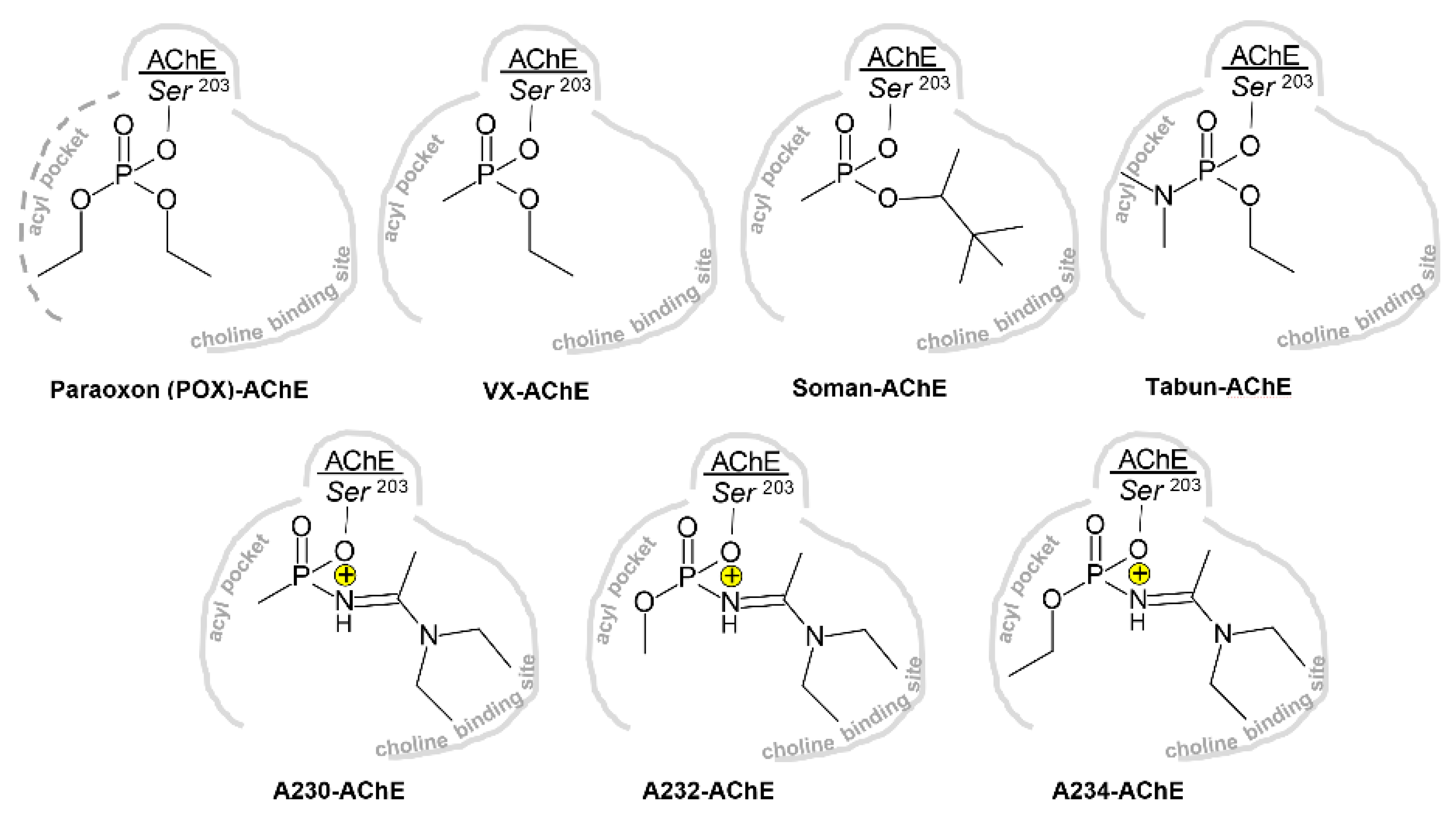
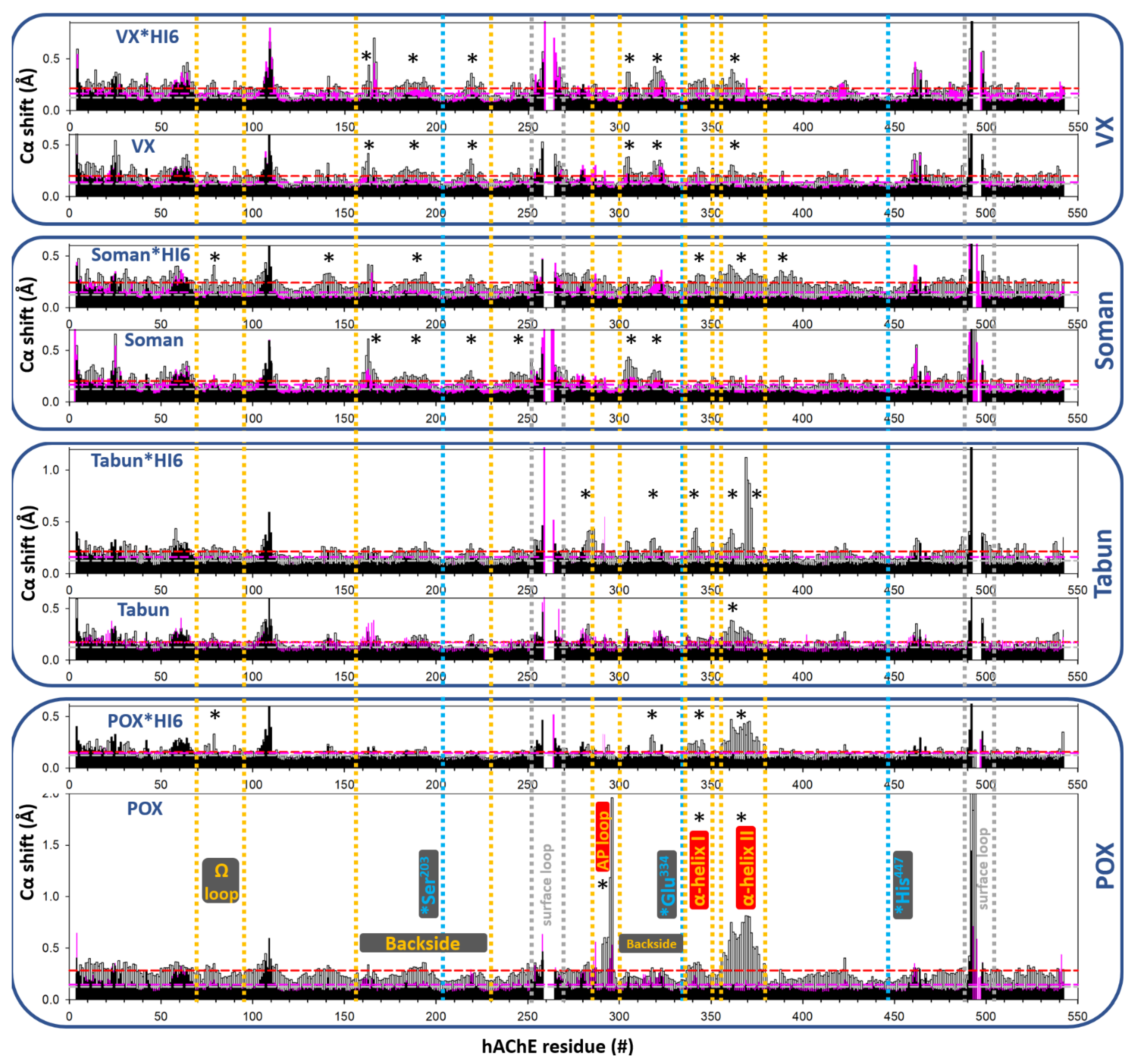
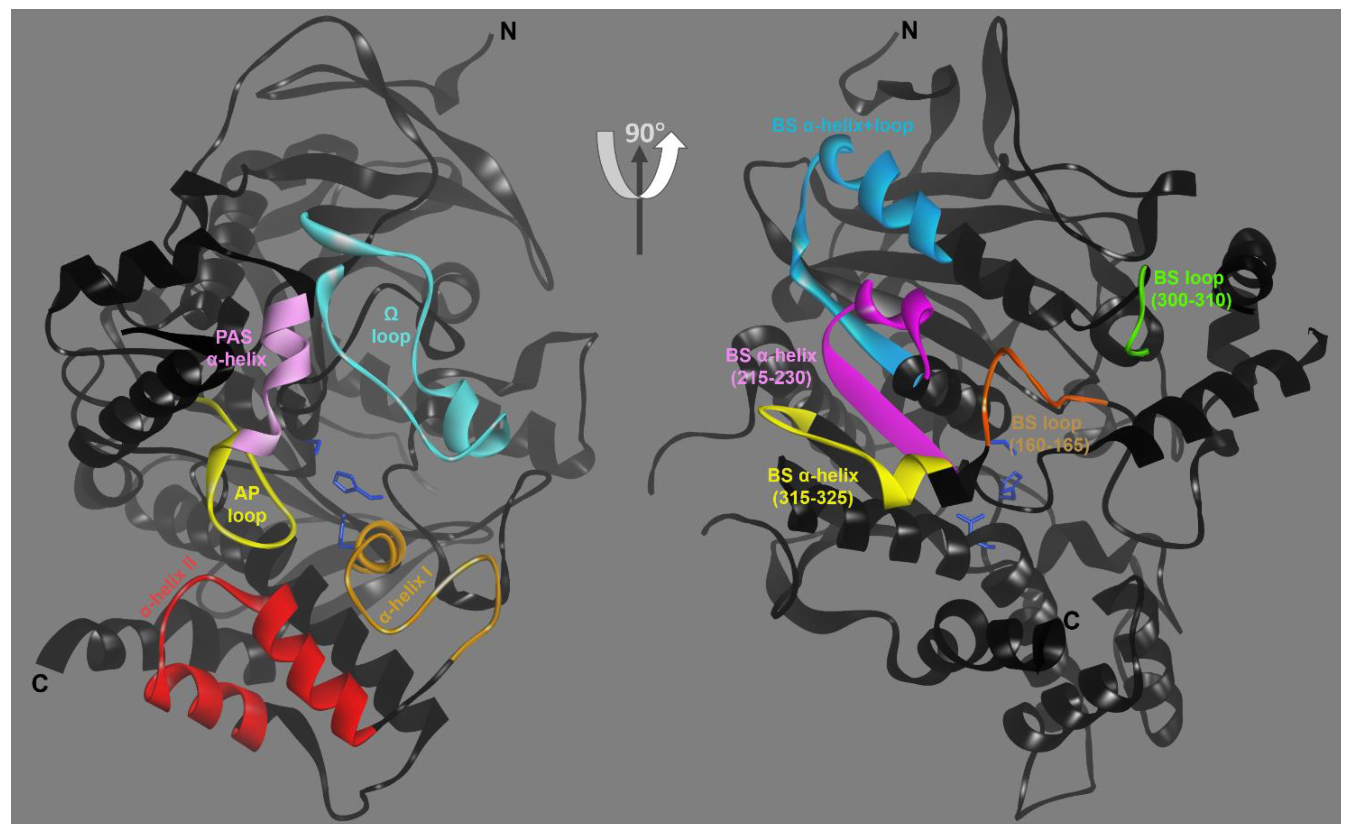
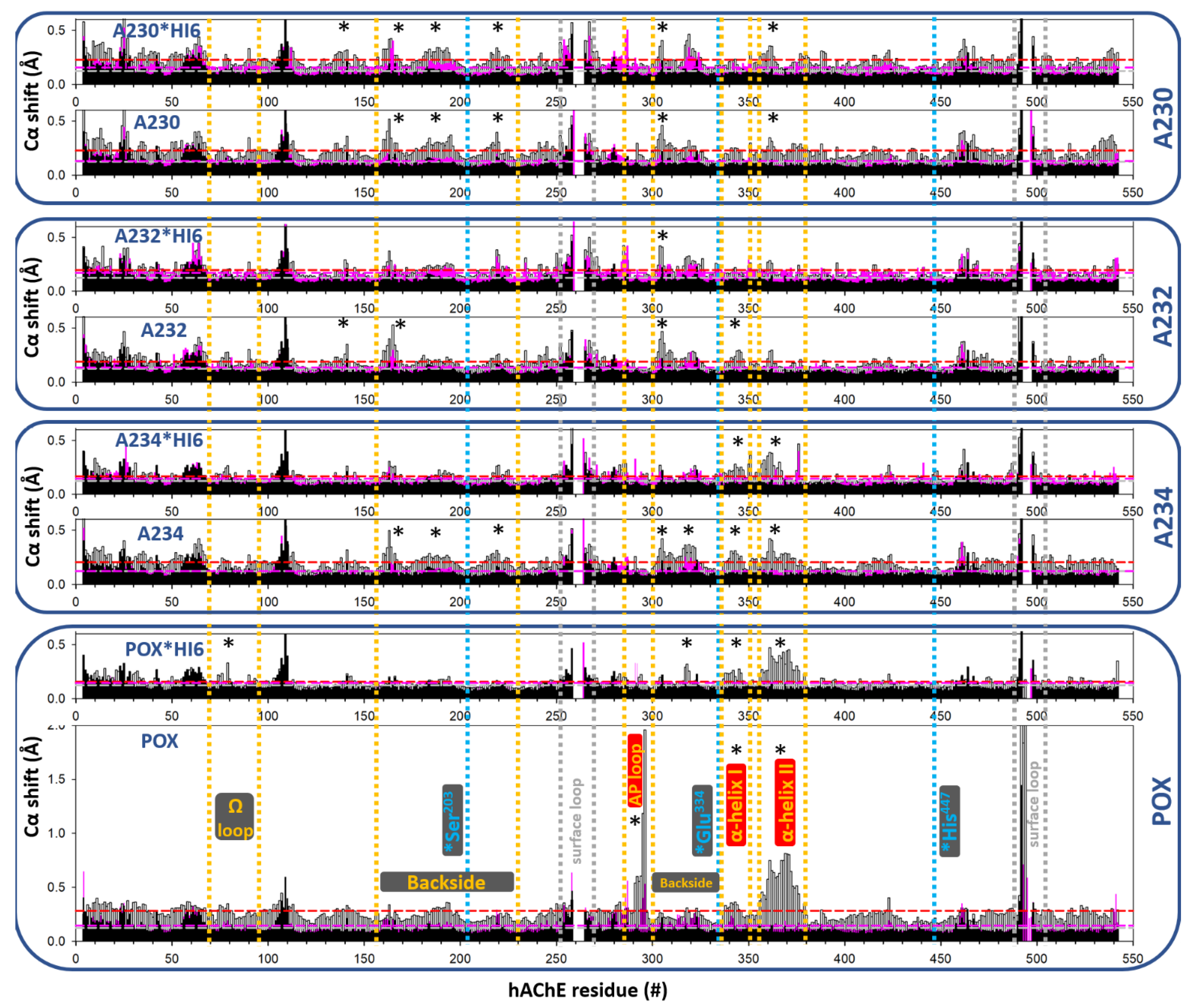
3.1. Effects of Conjugation of an OP Pesticide and “Classical” Nerve Agent OPs to hAChE

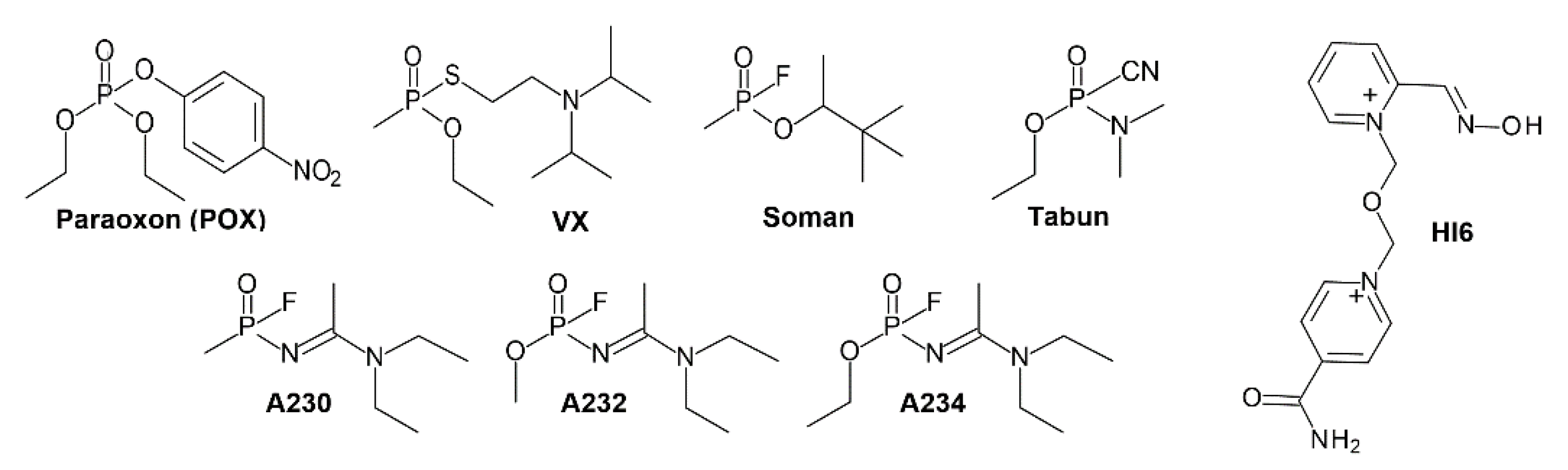
3.1.1. Conjugation of Paraoxon (POX)

Distortion of the hAChE backbone upon conjugation of diethylphosphate of POX, an active inhibitory form of the pesticide parathion, to the Ser203 has been well documented earlier based on RMSD overlay and Rapido-server analysis [13]. Our PACCT 3 comparison confirmed the acyl pocket loop (AP loop) distortion, as well as distortions of C-terminal α-helices (Figure 3 and Figure 4 and Table 1). As a consequence, the size of the active center gorge opening in the POX-hAChE conjugate is reduced, and a positively charged electrostatic barrier is introduced at the gorge opening (Figure 3B,C). Both effects compromise access of cationic pyridinium-based antidotes, such as HI6, towards conjugated phosphorus and its nucleophilic displacement that could reactivate inhibited hAChE. Subtle shifts in C- terminal α-helices were previously reported to be involved in the allosteric promotion of hAChE homodimer dissociation [14]. Interestingly, the conformation of the Ω-loop that serves as a “lid” of the active center gorge and also regulates the size of the active-center gorge opening was not affected in the POX-hAChE structure. The flexibility of this loop was previously suspected of playing a major role in controlling the volume and access to the hAChE active center similar to the one documented for the Ω-loop of Candida rugosa lipase [18].
Reversible binding of HI6 to POX-hAChE reverts the AP loop conformation to the one found in Apo-hAChE (Figure 4 and Table 1), allowing the slender HI6 molecule to insert itself into the active center gorge opening, which otherwise would not be possible. The reversal attests to the flexibility of the AP loop even in the flash-frozen crystal, where the short (10–30 min) soak of HI6 was sufficient to revert loop conformation into the native one [13], that appears to be better stabilized by the bound HI6. C-terminal α-helices, however, are located slightly farther and remain in their equally distorted conformation seen in the POX-hAChE conjugate, even in the presence of bound HI6.

3.1.2. Conjugation of “Classical” Nerve Agent OPs

In all of the “classical” nerve agent OP conjugates that we analyzed (VX, Soman and Tabun) the smallest of substituents on phosphorus bound to Ser203 were smaller than the ethoxy group of POX, and were stabilized in the acyl pocket without any distortion of the associated loop (Figure 4 and Table 1). This fact is consistent with more than an order of magnitude faster inhibition rates of hAChE by those OPs compared to POX. It is also consistent with substrate specificity of AChEs [19], where up to two carbon atoms bound to the carbonyl group of a substrate (acetyl- and propionyl-) fit the AP well and are hydrolyzed by AChEs equally efficiently. The two orders of magnitude reduction in turnover observed, for butyryl-choline [19] (a substrate where three linear carbon atoms need to be stabilized in the AP of AChE during fast catalytic turnover) could coincide with a need for distortion of the AP loop, similar to the one observed in X-ray structure of the POX-hAChE conjugate (with “three-atom” ethoxy group of POX in the AP). The latter structure could be captured primarily due to a much slower de-acylation step, where de-phosphorylation was much slower than the de-butyrylation of AChE [13].
Otherwise, conformations of C-terminal α-helices were consistently affected upon conjugation by those OPs, although to a smaller magnitude than the one observed in POX-hAChE. In particular, shifts were observed in the α-helix II (356–380). The same helix was affected in complexes with HI6 as well (Figure 4 and Table 1). The HI6 effect was largest for Tabun and Soman, the most voluminous of those OPs. The α-helix I (335–352) distortion was smaller in magnitude and observed primarily in complexes with HI6, indicating larger sensitivity of that helix to the presence of the active center gorge channel-filling ligands (HI6) when the choline-binding site is occupied with another voluminous ligand. In this case, that ligand was substituent on the phosphorus covalently attached to the Ser203.
Shifts in surface loops and helices located in the “backside” of the hAChE structure, opposite from the active center gorge opening, were smaller and of unclear significance for hAChE catalysis. They may primarily act as connectors of helices that protrude into the interior of the enzyme. Those internal helices were not identified to shift significantly in our comparisons, probably due to their internal intramolecular stabilization. However, their smaller shifts might have been amplified at connecting elements on the surface as overall structures adjusted to OP conjugation.
Overall, conjugation of hAChE with VX, Soman, and Tabun did not result in structural alteration of its backbone comparable to the one observed in POX-hAChE, except for some C-terminal α-helical shifts.

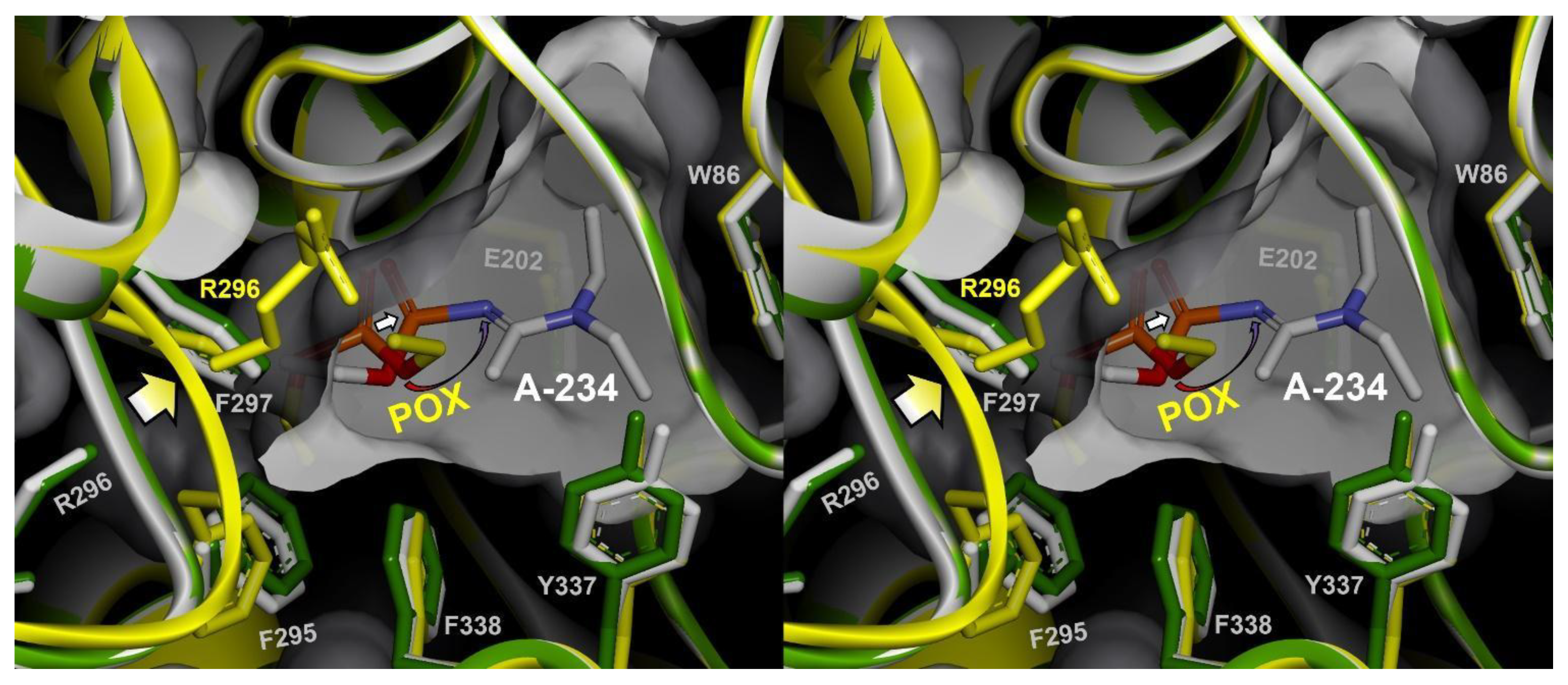
3.2. Effects of Conjugation of the Novel, A-Type Nerve Agent OPs to hAChE

Considering their fairly large size (Figure 1 and Figure 2) it is surprising that the covalent binding of A-type nerve agents, or Novichoks, to hAChE did not result in more extensive changes in its backbone conformation (Figure 6 and Table 1).
A quick look at Table 1 data reveals their similar influences on C-terminus and backside surface of hAChE to those observed for “classical” nerve agent OPs, but no effects on the active center Ω-loop, AP-loop, or α-helix of the peripheral anionic site (PAS). In particular, the largest of the three Novichoks, A234, in spite of inserting its large ethoxy substituent into the AP and simultaneously filling the choline-binding site with large phosphoramide substituent, did not promote any distortions in the AP loop or elsewhere in hAChE. Its effects on C-terminal α-helices and backside surface elements were larger than for the other two Novichoks, yet modest in magnitude.
Why did the diethyl-phosphoryl conjugate formed by POX force the AP loop to distort when the much larger A234-hAChE conjugate (Figure 2) did not? The answer could be in the ~1.2–2.4 Å shift of the entire conjugated phosphoramidate towards the choline-binding site with the potential of relieving steric pressure on the confines of the AP (Figure 7, VR supplement S1, Video supplement S1).
Very similar shifts are seen in X-ray structures of all three Novichocks and in their complexes with HI6. Since much smaller AP groups are found in A232-hAChE and A230-hAChE conjugates (Figure 2 and Table 1) observed shifts are likely not a consequence of a steric “push-out” from the AP towards the choline-binding site. It is more likely that large and hydrophobic phosphoramide substituents of Novichoks find attractive stabilization in the choline-binding site where their size fits well. This stabilization then pulls the whole substituted phosphorus towards W86. A similar shift is observed in the structure of the just-slightly-smaller Soman-hAChE conjugate [20].
An additional component of the driving force for the shift is in possible hydrogen bonding/Coulombic attraction between possibly protonated imido nitrogen of Novichoks bound to P atom and anionic Glu202 located within 3–4 Å distance. That attraction could directly pull the conjugate towards the choline-binding site. An example for such an electronically-driven shift can be found in the X-ray structure of Fenamiphos-conjugated mouse AChE (PDB ID 2JGF). The phosphorus-bound N atom there has hydrogen and small isopropyl group as substituents, while the AP-stabilized phosphorus-bound ethoxy group does not distort the AP loop.

4. Discussion

The small volume of the active center gorge of AChEs and the narrow and tortuous path that connects it to the enzyme’s surface have promoted the development of hypotheses on the need for conformational flexibility of its backbone and side-chains ever since the time the first X-ray structure was solved [21]. The flexibility was needed to explain how thousands of narrowly fitting substrate molecules can reach the site of catalytic hydrolysis at the base of the gorge, and the resulting products be expelled in a single second. Computational molecular dynamics simulations predicted rapid, small-magnitude breathing motions, including those of Ω-loop and AP loop [22], to result in the concerted gorge opening. A popular analogy was that Ω-loop in AChEs could open and close similar to distinct Ω-loop conformations observed in separate X-ray structures of structurally similar Candida rugosa lipase [18]. None of the PDB-deposited X-ray structures, however, captured hAChE in a distinctly different (open) conformation. The largest differences were observed for the covalent conjugates, POX-hAChE [13] (Figure 7) and dealkylated (aged) DFP-AChEs [23,24] where the AP loop was considerably distorted by the protrusion of ethoxy- or isopropoxy- substituents on phosphorus conjugated to the Ser203. No significant deviations have been observed in the choline-binding site of AChE, formed in part by the Ω-loop, even in complexes of large reversible ligands that occupy primarily that site, such as huperzine A [17,25] or galanthamine [17].
The recent availability of X-ray structures of hAChE conjugated with A-type nerve agents (Novichoks) prompted us to investigate them with respect to eventual distortions of their active center gorge backbone elements, the AP loop, the choline-binding site, and Ω-loop. This is because these large OPs form uniquely tight conjugates where both AP and choline-binding sites of the active center are occupied with large substituents, nearly too large to fit the available volumes. We have not, however, observed distortions even for the conjugate with the largest of the three Novichoks, A234, that projects large ethoxy- substituent into the AP and large phosphoramido substituent into the choline-binding site.
Thus, the choline-binding site of hAChE formed by internal α-helices and covered by the Ω-loop, has the capacity to attract and stabilize large OP conjugates, such as Soman and Novichok OPs, in its native geometry—without any need for enlargement-associated distortion. Smaller adjustments of the gorge volume seem to be done via C-terminal α-helical shifts that contribute to formation of the lower part of the gorge wall.
The AP-loop has to adjust its conformation and distort in order to accommodate binding only for large AP-directed substituents on conjugated phosphorus, such as ethoxy or isopropoxy groups. This, however, happens only when those conjugates are not exposed to additional “pulling interactions” that could shift them towards choline-binding sites. In addition to the POX-hAChE structure, good examples for this kind of effect are structures of aged DFP-AChE conjugates obtained with both Torpedo californica AChE [23] and with mouse AChE [24], where large distortions of the AP loop were observed. Similar AP loop distortions were reported in the N-ethyl-N-methyl- carbamylated Torpedo californica AChE [26]. The suggested “pulling interactions” could be of hydrophobic nature, (observed for pinacolyl group of unaged soman-hAChE), electrostatic nature (observed for Fenamiphos-mAChE) or a combination of both (observed in Novichok A-type conjugates).
One practical consequence of “pulling shift” interactions is that they additionally stabilize OP-hAChE conjugates and render them more resistant towards nucleophilic reactivation with oxime antidotes. It seems that, because of those additional conjugate stabilizations from both AP and choline-binding sites, reactivating nucleophiles possess a better chance of accessing the conjugated phosphorus when they approach from the direction of the PAS and align with the axis of the active center gorge channel with their extended nucleophilic aldoxime. In case when antidotes rely on stabilizations (or flexibility) in either AP or the choline-binding site, their substituted pyridinium rings appear misaligned for nucleophilic attack. This was observed for 2PAM and HI6 in the respective X-ray structures (PDB IDs 6NTN, 6NTM, 6NTG, 6CQW, 6WVO, 6WUY, 5HF9). Reactive nucleophilic aldoxime groups attached to their pyridinium rings are found 9–12 Å far from the conjugated P atom, in orientations non-productive for reactivation. The more recent and promising generation of zwitterionic monoximes (RS194) or zwitterionic bis-oximes (LG-bisoximes) [27] approach conjugated phosphorus immediately from the direction of the PAS and seem to comply with this structure-based requirement better.
An additional consequence of “pulling shift” interactions is for the molecular basis of substrate/inhibitor specificity of AChEs. It appears that, contrary to a widespread assumption, a small acyl pocket of AChEs can in fact accommodate, without distortion, substituents of the size of a linear three-carbon chain, such as ethoxy or butyryl groups. This phenomenon is demonstrated in the structures of A234 and Fenamiphos conjugates. It can be shown that stabilization of the choline group of ACh or BCh in the choline-binding site pulls their carbonyl carbon by ~0.5 Å out from the AP (as inferred from comparison of reported structures [28,29]). Although smaller in magnitude than observed for a choline-binding site substituent of Novichok A234, this effect could reduce the AP loop distortion in the acylation step of BCh hydrolysis. This cannot happen in the deacylation step. The structure-based reduction in the rate of BCh deacylation might thus dominate as a substrate specificity-defining step responsible for the larger part of the two orders of magnitude slower turnover of BCh compared to ACh (19).

Supplementary Materials

The following are available online at https://www.mdpi.com/article/10.3390/cryst11111270/s1, VR supplement S1: Immersive VR Nanome session recording file: “Ca_shifts_1.22.nanosr”, compatible with Oculus Rift, Quest and HTC Vive—based VR visualization software by Nanome Inc. (www.nanome.ai, accessed on 20 September 2021). Video supplement S1: https://youtu.be/CDZilvzTf6o, accessed on 20 September 2021.

Author Contributions

Conceptualization, Z.R.; methodology, Z.R.; software, Z.R.; validation, S.L., C.B., Y.L., E.L., B.P. and Z.R.; formal analysis, S.L., C.B., Y.L., E.L., B.P. and Z.R.; investigation, S.L., C.B., Y.L., E.L., B.P. and Z.R.; resources, Z.R.; data curation, S.L., C.B., Y.L., E.L., B.P. and Z.R.; writing—original draft preparation, Z.R.; writing—review and editing, S.L., C.B., Y.L., E.L., B.P. and Z.R.; visualization, S.L., C.B., Y.L., E.L., B.P. and Z.R.; supervision, Z.R.; project administration, Z.R.; funding acquisition, Z.R. All authors have read and agreed to the published version of the manuscript.

Funding

This research was supported by the CounterACT Program, National Institutes of Health Office of the Director (NIH OD), and the National Institute of Neurological Disorders and Stroke (NINDS), [Grant Numbers U01 NS083451 and R21 NS098998] and by the UCSD Academic Senate grant BG084144.

Data Availability Statement

All data is contained within the manuscript or available to be shared upon request (please contact corresponding author).

Conflicts of Interest

The authors declare no conflict of interest.

References

  1. Taylor, P. Anticholinesterase Agents. In Goodman & Gilman’s The Pharmacological Basis of Therapeutics, 13th ed.; Brunton, L.L., Hilal-Dandan, R., Knollman, B.C., Eds.; McGraw-Hill: New York, NY, USA, 2018; pp. 163–177. [Google Scholar]
  2. Aldridge, W.N.; Reiner, E. Enzyme Inhibitors as Substrates: Interactions of Esterases with Esters of Organophosphorus and Carbamic Acids, 1st ed.; North-Holland Pub. Co.: Amsterdam, Holland, 1972; 328p. [Google Scholar]
  3. Gerlits, O.; Blakeley, M.P.; Keen, D.A.; Radić, Z.; Kovalevsky, A. Room temperature crystallography of human acetylcholinesterase bound to a substrate analogue 4K-TMA: Towards a neutron structure. Curr. Res. Struct. Biol. 2021, 3, 206–215. [Google Scholar] [CrossRef]
  4. Harvey, S.P.; McMahon, L.R.; Berg, F.J. Hydrolysis and enzymatic degradation of Novichok nerve agents. Heliyon 2020, 6, 1–4, e03153. [Google Scholar] [CrossRef]
  5. OPCW. Decision: Recommendation for a Change to Schedule 1 of the Annex on Chemicals to the Chemical Weapons Convention. In Proceedings of the Sixty-Second Meeting of the Executive Council, The Hague, The Netherlands, 14 January 2019. EC-M-62/DEC.1*. [Google Scholar]
  6. OPCW. Opening Statement by the Director-General to the Executive Council at its Ninetieth Session. In Proceedings of the Ninetieth Meeting of the Executive Council, The Hague, The Netherlands, 12 March 2019. Notes by Director-General; EC-90/DG.16*. [Google Scholar]
  7. Dawson, A.H.; Eddleston, M.; Senarathna, L.; Mohamed, F.; Gawarammana, I.; Bowe, S.J.; Manuweera, G.; Buckley, N.A. Acute human lethal toxicity of agricultural pesticides: A prospective cohort study. PLoS Med. 2010, 7, e1000357. [Google Scholar] [CrossRef] [PubMed] [Green Version]
  8. Dolgin, E. Syrian gas attack reinforces need for better anti-sarin drugs. Nat. Med. 2013, 19, 1194−1195. [Google Scholar] [CrossRef]
  9. Stone, R.U.K. attack shines spotlight on deadly nerve agent developed by Soviet scientists. Science 2018, 359, 1314–1315. [Google Scholar] [CrossRef]
  10. Stone, R. How German military scientists likely identified the nerve agent used to attack Alexei Navalny. Science 2020. [Google Scholar] [CrossRef]
  11. Mirzayanov, V.S. State Secrets: An Insider’s Chronicle of the Russian Chemical Weapons Program; Outskirts Press: Denver, Colorado, 2009; ISBN 978-1-4327-2566-2. [Google Scholar]
  12. Sit, R.K.; Kovarik, Z.; Maček Hrvat, N.; Žunec, S.; Green, C.; Fokin, V.V.; Sharpless, K.B.; Radić, Z.; Taylor, P. Pharmacology, Pharmacokinetics, and Tissue Disposition of Zwitterionic Hydroxyiminoacetamido Alkylamines as Reactivating Antidotes for Organophosphate Exposure. J. Pharmacol. Exp. Ther. 2018, 367, 363–372. [Google Scholar] [CrossRef]
  13. Franklin, M.C.; Rudolph, M.J.; Ginter, C.; Cassidy, M.S.; Cheung, J. Structures of paraoxon-inhibited human acetylcholinesterase reveal perturbations of the acyl loop and the dimer interface. Proteins 2016, 84, 1246–1256. [Google Scholar] [CrossRef]
  14. Blumenthal, D.K.; Cheng, X.; Fajer, M.; Ho, K.Y.; Rohrer, J.; Gerlits, O.; Taylor, P.; Juneja, P.; Kovalevsky, A.; Radić, Z. Covalent inhibition of hAChE by organophosphates causes homodimer dissociation through long-range allosteric effects. J. Biol. Chem. 2021, 297, 1–14. [Google Scholar] [CrossRef]
  15. Bester, S.M.; Guelta, M.A.; Height, J.J.; Pegan, S.D. Crystal Structure of Recombinant Human Acetylcholinesterase Inhibited by A-234. 2020. Available online: https://www.rcsb.org/structure/6NTL (accessed on 20 September 2021).
  16. wwPDB Consortium. Protein Data Bank: The single global archive for 3D macromolecular structure data. Nucleic Acids Res. 2019, 47, D520–D528. [Google Scholar]
  17. Cheung, J.; Rudolph, M.J.; Burshteyn, F.; Cassidy, M.S.; Gary, E.N.; Love, J.; Franklin, M.C.; Height, J.J. Structures of human acetylcholinesterase in complex with pharmacologically important ligands. J. Med. Chem. 2012, 55, 10282–10286. [Google Scholar] [CrossRef] [PubMed]
  18. Grochulski, P.; Li, Y.; Schrag, J.D.; Bouthillier, F.; Smith, P.; Harrison, D.; Rubin, B.; Cygler, M. Insights into interfacial activation from an open structure of Candida rugosa lipase. J. Biol. Chem. 1993, 268, 12843–12847. [Google Scholar] [CrossRef]
  19. Radić, Z.; Pickering, N.A.; Vellom, D.C.; Camp, S.; Taylor, P. Three distinct domains in the cholinesterase molecule confer selectivity for acetyl- and butyrylcholinesterase inhibitors. Biochemistry 1993, 32, 12074–12084. [Google Scholar] [CrossRef]
  20. McGuire, J.R.; Bester, S.M.; Guelta, M.A.; Cheung, J.; Langley, C.; Winemiller, M.D.; Bae, S.Y.; Funk, V.; Myslinski, J.M.; Pegan, S.D.; et al. Structural and Biochemical Insights into the Inhibition of Human Acetylcholinesterase by G-Series Nerve Agents and Subsequent Reactivation by HI-6. Chem. Res. Toxicol. 2021, 34, 804–816. [Google Scholar] [CrossRef] [PubMed]
  21. Sussman, J.L.; Harel, M.; Frolow, F.; Oefner, C.; Goldman, A.; Toker, L.; Silman, I. Atomic structure of acetylcholinesterase from Torpedo californica: A prototypic acetylcholine-binding protein. Science 1991, 253, 872–879. [Google Scholar] [CrossRef]
  22. Cheng, Y.; Suen, J.K.; Radić, Z.; Bond, S.D.; Holst, M.J.; McCammon, J.A. Continuum simulations of acetylcholine diffusion with reaction-determined boundaries in neuromuscular junction models. Biophys. Chem. 2007, 127, 129–139. [Google Scholar] [CrossRef] [PubMed] [Green Version]
  23. Millard, C.B.; Kryger, G.; Ordentlich, A.; Greenblatt, H.M.; Harel, M.; Raves, M.L.; Segall, Y.; Barak, D.; Shafferman, A.; Silman, I.; et al. Crystal structures of aged phosphonylated acetylcholinesterase: Nerve agent reaction products at the atomic level. Biochemistry 1999, 38, 7032–7039. [Google Scholar] [CrossRef]
  24. Hörnberg, A.; Tunemalm, A.K.; Ekström, F. Crystal structures of acetylcholinesterase in complex with organophosphorus compounds suggest that the acyl pocket modulates the aging reaction by precluding the formation of the trigonal bipyramidal transition state. Biochemistry 2007, 46, 4815–4825. [Google Scholar] [CrossRef]
  25. Raves, M.L.; Harel, M.; Pang, Y.P.; Silman, I.; Kozikowski, A.P.; Sussman, J.L. Structure of acetylcholinesterase complexed with the nootropic alkaloid, (-)-huperzine A. Nat. Struct. Biol. 1997, 4, 57–63. [Google Scholar] [CrossRef]
  26. Santoni, G.; de Sousa, J.; de la Mora, E.; Dias, J.; Jean, L.; Sussman, J.L.; Silman, I.; Renard, P.Y.; Brown, R.; Weik, M.; et al. Structure-Based Optimization of Nonquaternary Reactivators of Acetylcholinesterase Inhibited by Organophosphorus Nerve Agents. J. Med. Chem. 2018, 61, 7630–7639. [Google Scholar] [CrossRef] [Green Version]
  27. Gorecki, L.; Gerlits, O.; Kong, X.; Cheng, X.; Blumenthal, D.K.; Taylor, P.; Ballatore, C.; Kovalevsky, A.; Radić, Z. Rational design, synthesis, and evaluation of uncharged, “smart” bis-oxime antidotes of organophosphate-inhibited human acetylcholinesterase. J. Biol. Chem. 2020, 295, 4079–4092. [Google Scholar] [CrossRef] [PubMed] [Green Version]
  28. Colletier, J.P.; Fournier, D.; Greenblatt, H.M.; Stojan, J.; Sussman, J.L.; Zaccai, G.; Silman, I.; Weik, M. Structural insights into substrate traffic and inhibition in acetylcholinesterase. EMBO J. 2006, 25, 2746–2756. [Google Scholar] [CrossRef] [PubMed]
  29. Bourne, Y.; Radić, Z.; Sulzenbacher, G.; Kim, E.; Taylor, P.; Marchot, P. Substrate and product trafficking through the active center gorge of acetylcholinesterase analyzed by crystallography and equilibrium binding. J. Biol. Chem. 2006, 281, 29256–29267. [Google Scholar] [CrossRef] [PubMed] [Green Version]
Figure 1. Structures of OPs and an oxime antidote HI6 used in this study. Paraoxon is an active form of the OP pesticide parathion. VX, Soman and Tabun are “classical” nerve agent OPs and A230, A232 and A234 are Novichok nerve agent OPs.
Crystals 11 01270 g001
Figure 2. Schematic representation of OP-hAChE conjugates formed by OPs from Figure 1. Orientations of substituents on phosphorus atoms with respect to “acyl pocket”, active Ser203 and “choline-binding site” of hAChE were inferred from respective X-ray structures. Protonation of the imido nitrogen in A-type nerve agent inhibited conjugates is suggested based on semi-empirical calculations.
Crystals 11 01270 g002
Figure 3. PACCT 3 based overlay of POX-hAChE (Chain A of 5HF5) on Apo-hAChE (Chain A of 4EY4) in stereo. (A) Ribbon representation of Cα backbones for POX-hAChE (yellow) and Apo-hAChE (black) with N- and C- terminus labels. Diethylphosphate of POX covalently attached at Ser203 is represented by sticks surrounded with a semi-transparent surface. Asterisks indicate fragments of significant backbone deviation: the acyl pocket loop (orange), and α-helices I (red) and II (blue) in the C- terminal region, as indicated in Figure 4 and summarized in Table 1. (B) The influence of the POX-hAChE backbone distortion on the size of the active center gorge opening. Solvent-accessible Connolly surface is colored by atom charge (red-negative, blue-positive). Arrows indicate points of constriction and blue electrostatic barrier imposed by the flipped Arg296 in the AP loop. (C) Size of the gorge opening and charge distribution in the Apo-hAChE (4EY4).
Crystals 11 01270 g003
Figure 4. Alpha carbon shifts in hAChE backbone upon covalent conjugation by OPs as indicated in each panel and further reversible complex formation with HI6. Each X-ray structure is compared to Apo hAChE structure (PDB ID 4EY4) in a pairwise fashion using PACCT 3 tool. Black and magenta bars represent baseline obtained by pairwise comparison of chains A and B of the same structure (black for 4EY4 and magenta for each of analyzed structures as labelled above). White bars indicate difference between chains A of each indicated structure and Apo hAChE (4EY4). Average values of Cα shifts for baselines are indicated by horizontal dashed lines: black for Apo hAChE and magenta for the structure compared. Red, dashed horizontal line indicates average shift obtained from white bars. Location of different structural elements in the linear hAChE amino acid sequence, as well as position of catalytic triad residues is indicated with vertical dashed lines. Those elements are mapped onto 3D structural backbone of hAChE in Figure 5. Asterisks indicate significant shifts in Cα.
Crystals 11 01270 g004
Figure 5. Location of structural elements of hAChE backbone affected by OP conjugation, as detected by PACCT 3 analysis (Figure 4 and Figure 6 and Table 1), shown in the ribbon representation of the X-ray structure of Apo-hAChE (PDB ID 4EY4). Two different sets of elements are shown. (Left) those surrounding the opening of the active center gorge (AP loop, PAS α-helix, Ω-loop, and α-helices I and II). (Right) Backside Surface (BS) elements, BS loops (160–165 and 300–310), BS α-helices (215–230 and 315–325) and BS α-helix+loop. Catalytic triad Ser203, His447 and Glu334 are rendered as blue sticks.
Crystals 11 01270 g005
Figure 6. Alpha carbon shifts in the hAChE backbone upon covalent OP conjugation by A-type nerve agents A230, A232 and A234 and further reversible complex formation with HI6. The POX panel from Figure 4 is added for comparison. Graph description is the same as for Figure 4.
Crystals 11 01270 g006
Figure 7. Stereo representation of PACCT 3 overlays of POX-hAChE (yellow ribbon and sticks) and A234-hAChE (grey ribbon and sticks) onto Apo-hAChE (green ribbon and sticks). Cut-off of the grey solvent accessible Connolly surface belongs to Apo-hAChE and illustrates internal volume of the hAChE active center gorge. Opening of the gorge is perpendicular to the plane of the paper. Arrows indicate 4.9 Å shift of the AP loop (yellow arrow), 1.2 Å shift of conjugated P atom (white arrow) and 2.4 Å shift of O to N atoms directly bound to P (red-blue curved arrow).
Crystals 11 01270 g007
Table 1. Summary of effects of OP conjugation of hAChE and of consecutive, reversible HI6 binding, as evaluated by PACCT 3 from pairwise comparisons of listed X-ray structures with Apo-hAChE (4EY4). Identities of the acyl pocket (AP) stabilized substituents (AP group) on phosphorus atoms for each OP conjugate are given as observed from corresponding X-ray structures. Listed in the table are magnitudes of Cα shifts identified in Figure 4 and Figure 6 as significant, from weak (+) to very significant (++++). Locations of ten identified elements in the hAChE Cα backbone are shown in Figure 5. All analyzed structures were of the same space group, P 31 2 1.
StructurePDB IDResolution
2)
AP GroupAffected Structural Element in hAChE
Active CenterPASC-TerminusBackside Surface
Ω Loop
(69–96)
AP Loop
(285–300)
α-Helix
(280–288)
α-Helix I
(335–352)
α-Helix II
(356–380)
Loop
(160–165)
α-Helix + Loop
(180–200)
α-Helix
(215–230)
Loop
(300–310)
α-Helix
(315–325)
Apo hAChE4EY42.16-----------
POX-hAChE5HF52.15CH3CH2-O--++++-+++++-----
POX-hAChE *HI65HF92.20CH3CH2-O-+--++++----+
VX-hAChE6CQZ2.22CH3 -----++++++
VX-hAChE*HI66CQW2.28CH3 -----++++++++
soman-hAChE6WVC2.60CH3 ------++++++
soman-hAChE*HI66WVO2.19CH3 -+--+++-++--
tabun-hAChE6WUV2.63NH2 -----++-----
tabun-hAChE*HI66WUY2.46NH2 ---++++++++---+
A230-hAChE6NTO2.05CH3 -----+-+++-
A230-hAChE*HI66NTN2.70CH3 -----+-+++-
A232-hAChE6NTK2.41CH3-O----+-+--+-
A232-hAChE*HI66NTM2.55CH3-O------+--+-
A234-hAChE6NTL2.25CH3CH2-O----+++++++++
A234-hAChE*HI66NTG2.65CH3CH2-O----++++-----
Publisher’s Note: MDPI stays neutral with regard to jurisdictional claims in published maps and institutional affiliations.

