Synthesis, X-ray Structure, Hirshfeld Analysis of Biologically Active Mn(II) Pincer Complexes Based on s-Triazine Ligands


1
Department of Chemistry, Faculty of Science, Alexandria University, P.O. Box 426, Ibrahimia, Alexandria 21321, Egypt
2
Department of Chemistry, College of Science, King Saud University, P.O. Box 2455, Riyadh 11451, Saudi Arabia
*
Authors to whom correspondence should be addressed.
Crystals 2020, 10(10), 931; https://doi.org/10.3390/cryst10100931
Submission received: 20 September 2020
/
Revised: 7 October 2020
/
Accepted: 12 October 2020
/
Published: 13 October 2020
Abstract
:Herein, the synthesis and antimicrobial activities of [Mn(MorphBPT)(H2O)2NO3]NO3; (1) and [Mn(PipBPT)(H2O)2NO3]NO3; (2) complexes of the pincer-type tridentate ligands MorphBPT; 4-(4,6-di(1H-pyrazol-1-yl)-1,3,5-triazin-2-yl)morpholine and PipBPT; 2-(piperidin-1-yl)-4,6-di(1H-pyrazol-1-yl)-1,3,5-triazine are presented. Both complexes have slightly distorted octahedral coordination geometry. Their molecular packing depends on O–H···O, C–H···O hydrogen bonds and anion–π stacking contacts. Hirshfeld analysis was used to quantify the different contacts. Both complexes exhibited better anti-fungal activity than the standard Fluconazole and comparable antibacterial activity to Gentamycin against Staphylococcus aureus and Escherichia coli microbes. Moreover, complexes 1 and 2 are biologically more active than the free ligands against these microbes.

1. Introduction
The use of transition metal complexes as antimicrobial agents, and for their potential applications as inorganic pharmaceuticals and in medicine for diagnostics, has gained great interest from researchers [1,2,3,4]. In this regard, large and rising numbers of mono- and polynuclear complexes of different metals, and with different classes of chelating ligands involving Schiff bases, bipyridine, and phenanthroline with different anions, were presented in literature [5,6,7,8,9,10,11,12,13,14].
Of transition metals, manganese is considered an essential micronutrient in living organisms and has an important role in a broad range of enzyme-catalyzed reactions. There is no doubt about the potential use of Mn(II) complexes as catalytic scavengers for H2O2 against oxidative stress [7,8,9,10,11,12,13,14,15]. From this point of view, an increasing interest with the synthesis of more Mn(II) complexes in order to investigate their catalytic and antimicrobial activities. Mn(II) complexes of bipyridine and phenanthroline ligands were found to have promising antifungal activity comparable to the antifungal drug ketoconazole [15,16]. Mn(II) plays a key role in biology as required enzyme activator, which is responsible for metabolism and apoptosis [17,18]. In addition, a Mn(II) complex of the Schiff base ligand derived from 1,4-diaminobutane and pyridoxal hydrochloride showed a great anticancer activity against breast cancer [19]. More recently, a Mn(II) complex of a Schiff base derived from vitamin B6 was found as an apoptosis inducer in human MCF7 and HepG2 cancer cells [20].
In continuation to our interest with the s-triazine pincer complexes [21,22,23,24], and in light of the interesting recently reported data in literature [25,26,27,28,29,30,31], we are presenting here the synthesis of two new Mn(II) complexes with the s-triazine based NNN-pincer ligands shown in Figure 1. The structural features of both complexes are elucidated. In addition, their antimicrobial activities as antibacterial and antifungal agents are presented.
2. Materials and Methods
Chemicals, reagents, and solvents used in this work were purchased from their commercial suppliers. The CHN analyses were determined using Perkin-Elmer 2400 instrument (PerkinElmer, Inc. 940 Winter Street, Waltham, MA, USA).
2.1. Preparation of the Organic Ligands
The organic ligands were prepared using the method reported in literature [32] (Supplementary data, Method S1, Figures S1 and S2).
2.2. Syntheses of [Mn(MorphBPT)(H2O)2NO3]NO3; (1) and [Mn(PipBPT)(H2O)2NO3]NO3; (2)
In a 50 mL conical flask, the ligand solution (0.05 mmol) in 10 mL methanol was added to an aqueous solution of the Mn(NO3)2·4H2O (~0.126 g, 0.05 mmol) in 10 mL water. The resulting clear solution was kept at room temperature for slow evaporation. Colorless crystals of the titled complexes were obtained after three days and were collected by filtration.
Yield:C17H26MnN10O9 (1) 80% with respect to the ligand. Anal. Calc. C, 35.86; H, 4.60; N, 24.60%. Found: C, 35.65; H, 4.52; N, 24.48%.
Yield; C18H28MnN10O8 (2) 77% with respect to the ligand. Anal. Calc. C, 38.10; H, 4.97; N, 24.69%. Found: C, 37.91; H, 4.91; N, 24.46%.
2.3. Crystal Structure Determination
The crystal structures of complexes 1 and 2 were determined by using a Bruker D8 Quest (Bruker Corporation, Massachusetts, MA, USA) diffractometer employing SHELXTL and SADABS programs [33,34,35]. Table 1 illustrated the refinement and crystal details. Hirshfeld calculations were performed using the default parameters of the Crystal Explorer 17.5 program [36,37,38,39,40].
2.4. Antimicrobial Studies
The bio-activities of the free MorphBPT and PipBPT ligands, as well as the corresponding Mn(II) complexes against different microbes, were determined [32]. More details regarding the bio-experiments are found in Supplementary data.
2.5. Density Functional Theory (DFT) Calculations
3. Results and Discussion
3.1. X-ray Crystal Structure Description
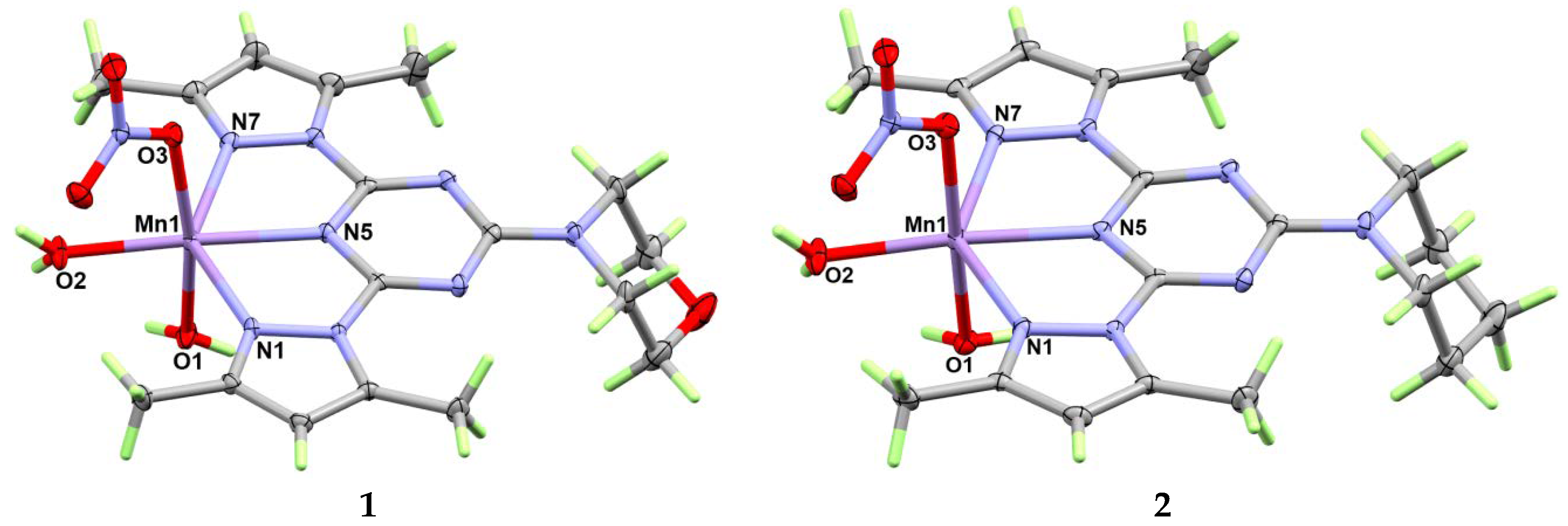
The structure with atomic numbering of [Mn(MorphBPT)(H2O)2NO3]NO3 complex (1) are shown in Figure 2 and list of the most important geometric parameters are given in Table 2. It crystallized in the triclinic crystal system and P-1 space group, and Z = 2. This cationic complex has a hexa-coordinated Mn(II) with one tridentate pincer ligand, one monodentate nitrate ion, and two water molecules in its inner sphere while the outer sphere comprised two halves of nitrate ions. The manganese to nitrogen distance is significantly shorter with s-triazine (2.2170(14) Å) than the corresponding Mn–N(pyrazole) bonds (2.3187(15)–2.3269(14) Å). Moreover, the equatorial Mn–O bond is the shortest (2.1419(13) Å) where the length of the manganese to oxygen distances is in the order of Mn–O(equatorial water) < Mn–O(nitrate) < Mn–O(axial water). The two bite angles of the tridentate chelate are 69.49(5) and 69.09(5)° for N5–Mn1–N1 and N5–Mn1–N7, respectively while the angle between the two trans Mn–N(pyrazole) bonds is 138.55(5)° for N1–Mn1–N7. The O2–Mn1–O3 and O2–Mn1–O1 bond angles of these cis bonds are 90.33(5) and 80.66(6)°, respectively while the O3–Mn1–O1 trans bond angle is 167.72(5)°. The hexa-coordinated Mn(II) has a distorted octahedral configuration with a distorted square comprised the N1N5N7O2 atoms while the O1 and O3 atoms are located at the apexes. Using Shape 2.1 software (Barcelona, Spain), the continuous shape measure (CShM) values of 4.1 and 11.8 relative to perfect octahedron and trigonal prism, respectively were computed. The CShM values revealed slightly distorted octahedral coordination geometry.
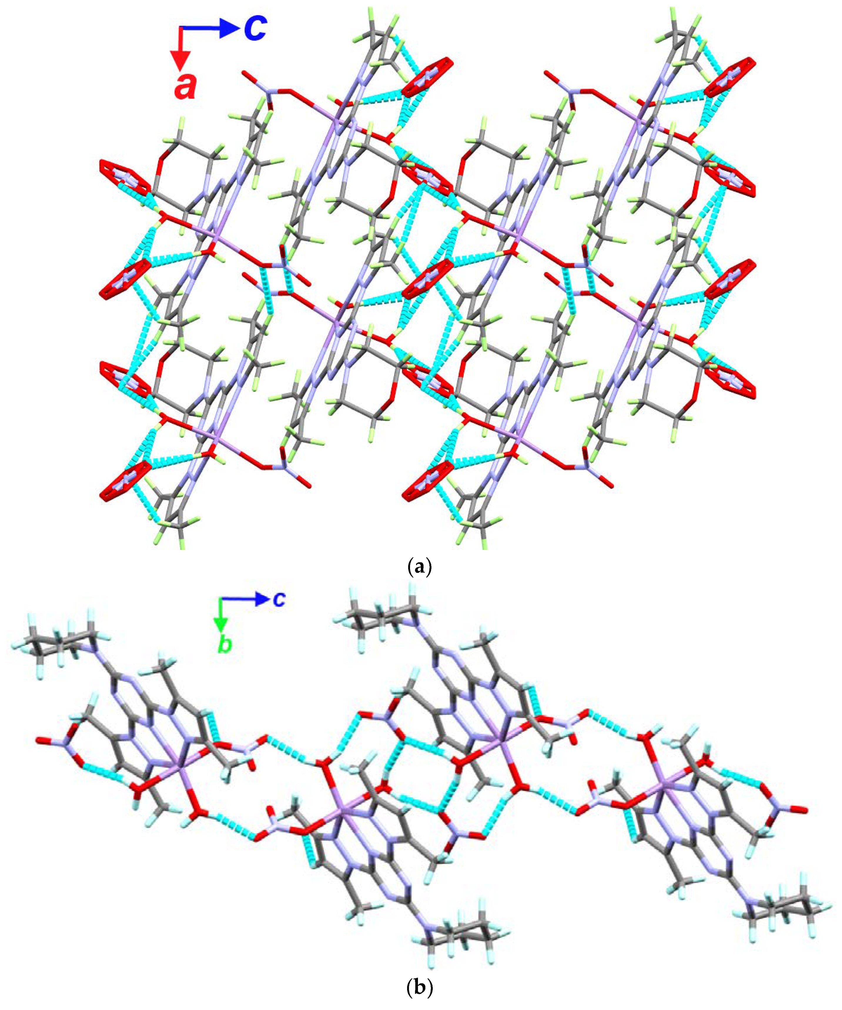
The three dimensional structure of 1 is built by O–H···O hydrogen bonds and C–H···O interactions as shown in the left part of Figure 3. The donor-acceptor distances are generally shorter (2.662(3)–2.924(2) Å) in the former than in the latter (3.271(3)–3.455(3) Å) (Table 3). The packing of 1 is shown in the upper part of Figure 4. In addition, anion–π contacts play an important role in the packing of 1 (Figure S3 (Supplementary data) and Table 4).
The structure of [Mn(PipBPT)(H2O)2NO3]NO3 complex 2 is very similar to 1. It also crystallized in the primitive triclinic unit cell with P-1 space group and two of the molecular formula per unit cell. In this case, the asymmetric unit comprised one cationic [Mn(PipBPT)(H2O)2NO3] and one NO3¯ counter anion. In general, the Mn–N and Mn–O bonds are slightly shorter in this complex than those in 1 except one of the Mn–N(pyrazole) bonds as well as one Mn–O(water) bond, which is trans to the Mn–O(nitrate). The hexa-coordinated Mn(II) coordination configuration is slightly less distorted than that in 1 where the continuous shape measure values for 2 were computed to be 3.3 and 11.9 with respect to the perfect octahedron and trigonal prism, respectively. The most important O–H···O and C–H···O hydrogen bond contacts as well as the anion–π stacking contacts in 2 are shown in the right part of Figure 3 and Figure S3, respectively. The packing of 2 could be considered as 1D hydrogen bond polymer (Figure 4) while list of the hydrogen bonds is given in Table 3.
A slight structural difference between complexes 1 and 2 is the deviation of the Mn and coordinated equatorial oxygen atoms from the s-triazine plane. The plane passing through the perfectly planar aromatic s-triazine moiety is nearly passing through the center of Mn atom in complex 2 with only 0.040(2) Å deviation from this mean plane. The corresponding distances in complex 1 is 0.142(2) Å. The equatorial oxygen atom is deviated from this plane by a distance of 0.603(3) and 0.515(3) Å in complexes 1 and 2, respectively. The reason could be simply attributed to the involvement of the s-triazine in larger number of anion–π stacking contacts in 1 compared to 2.
There is another structural difference between complexes 1 and 2. It is the orientation of the nitrate counter anion, with respect to the s-triazine moiety. In complex 1, the nitrate anion is nearly perpendicular to the s-triazine mean plane where the angle between the two planes is 86.67(3)°. It seems that such situation allowed further anion–π stacking interactions in 1 compared to 2. The corresponding angle between the two mean planes in complex 2 is only 26.64(3)°.
3.2. Analysis of Molecular Packing
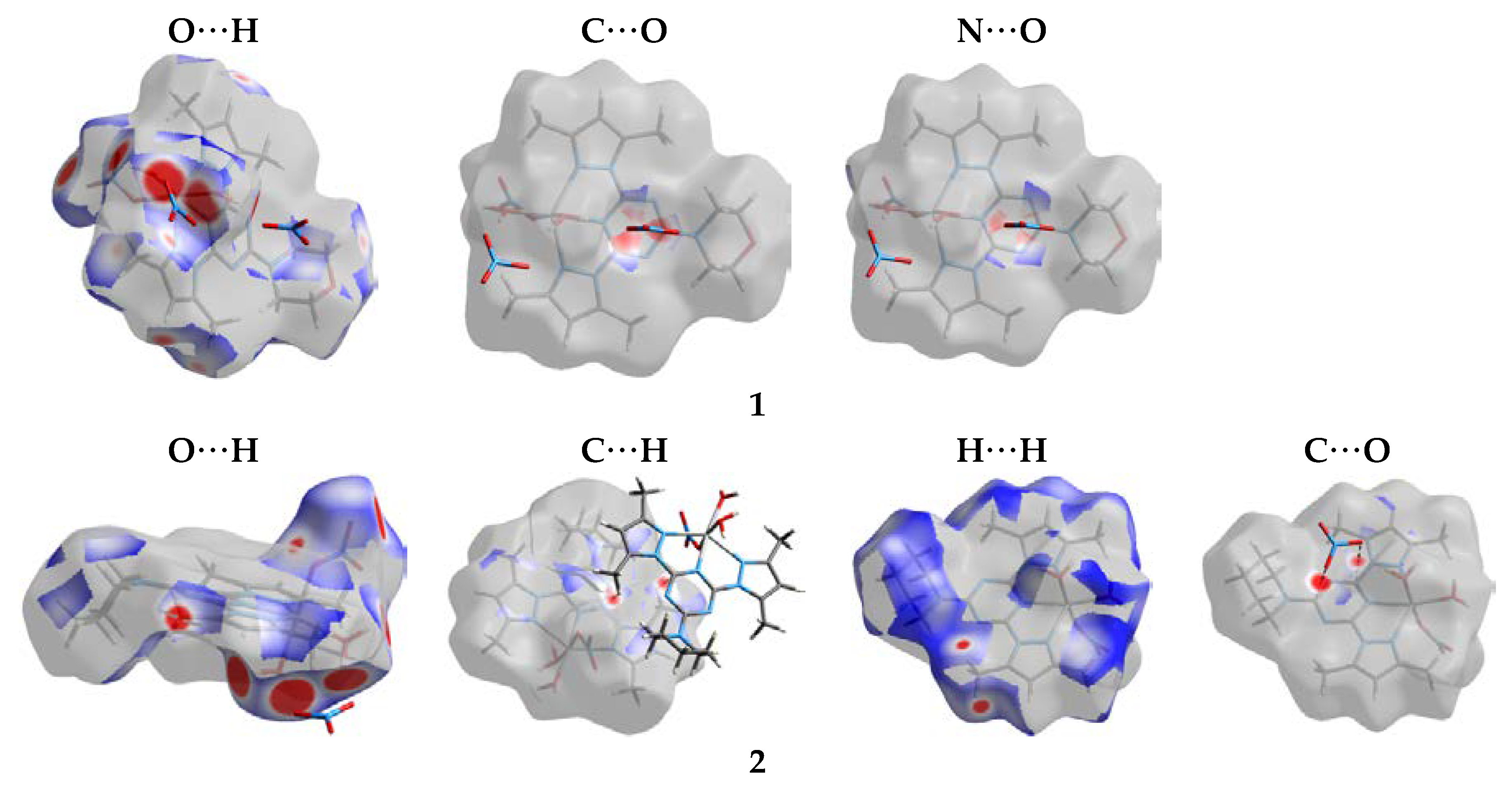
Hirshfeld surfaces for 1 and 2 are given in Figure S4 (Supplementary data) while all possible contacts are shown in Figure 5. The decomposed dnorm maps of the short and most significant contacts are collected in Figure 6. The H···H, O···H, N···H and C···H interactions are the most frequent in both complexes. In complex 1, these contacts contributed by 38.4, 37.5, 9.9 and 6.1%, respectively from the whole fingerprint area while the corresponding values in complex 2 are 45.2, 32.8, 8.8 and 5.2%, respectively. In addition, both complexes showed comparable amounts of anion–π stacking interactions with net C(s-triazine)···O(nitrate) and N(s-triazine)···O(nitrate) contacts of 2.8 and 2.5% for complexes 1 and 2, respectively. The latter is weaker in complex 2 and not showed the characteristics of short contacts. The shortest C···O contact is C7···O8 (2.982(2) Å) in 2 while in 1 the shortest contact is C8···O9 (2.889(3) Å). Regarding the N···O contacts in complexes 1 and 2, the shortest contact distances are N5···O9 (2.831(3) Å) and N6···O7 (3.077(2) Å), respectively. The N···O interaction in 2 is slightly longer than the vdW radii sum of nitrogen and oxygen indicating weaker interaction than 1. The O···H contacts appeared strong in both complexes where the O11···H1 (1.742 Å) and O7···H1B (1.770 Å) are the shortest in complexes 1 and 2, respectively. The values are different from those obtained from the CIF data given in Table 3 because in the Hirshfeld analysis the X–H (X = C, O) distances are normalized using the default criteria of Crystal Explorer 17.5 program. Many of the O···H contacts appeared as red regions in the dnorm map indicated shorter distance than the vdW radii sum of hydrogen and oxygen. In addition, complex 2 showed H···H and C–H···π interactions as red regions in the dnorm map with remarkable short distances of 1.959 Å (H5B···H5B) and 2.646 Å (C6···H5C). The latter occurred between the C–H of one methyl group from a complex unit with the s-triazine π-system from another complex unit. There are no observable π–π stacking interactions from the shape index and curvedness surfaces in both complexes.
3.3. AIM Topology Analysis
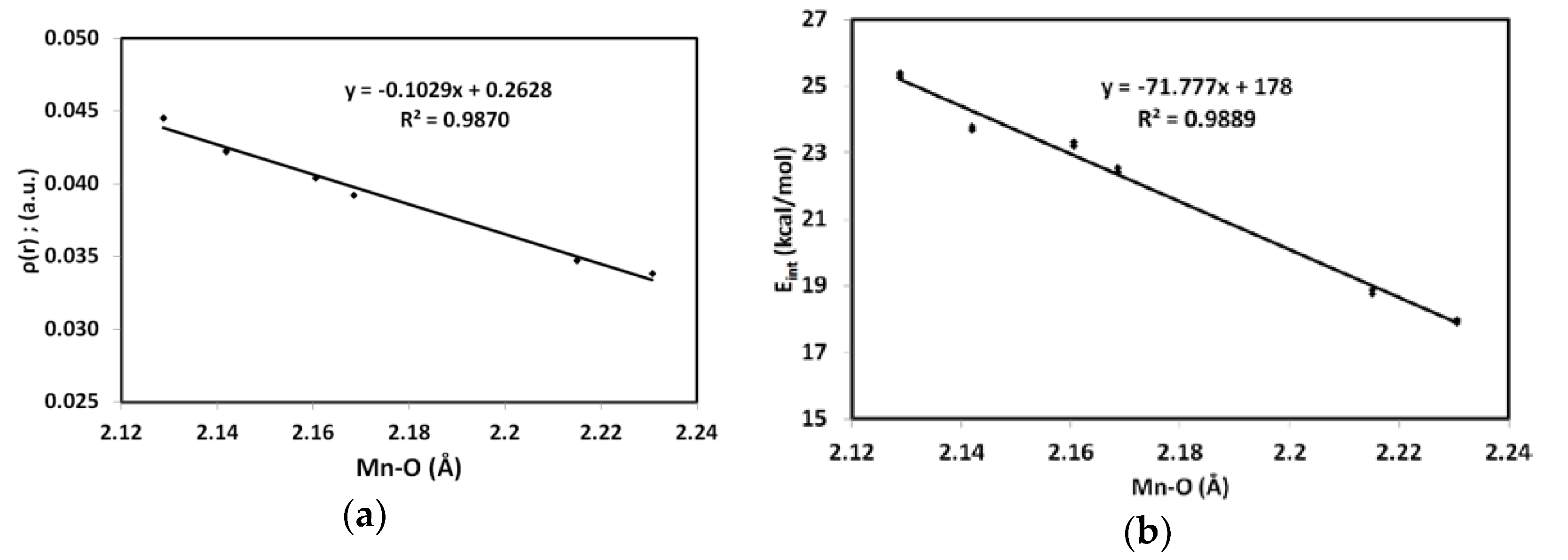
The nature and strength of Mn–N and Mn–O interactions in the studied complexes were analyzed using atoms in molecules (AIM) calculations [44,45,46,47,48,49,50,51,52,53,54]. The electron density (ρ(r)) of the Mn–O and Mn–N bondings are in the range of 0.028–0.046 and 0.035–0.045 a.u, respectively which are generally lower than 0.1 a.u indicating weak interactions with closed shell characters (Table 5). With the exception of the Mn–N(s-triazine), the positive H(r) and V(r)/G(r) < 1 for the rest of Mn–N and Mn–O interactions are the typical characteristics of the closed shell interactions. The Mn–N(s-triazine) bonds have very slightly small negative H(r) values and V(r)/G(r) very slightly higher than one indicating that the Mn–N(s-triazine) bonds have higher covalent characters than the Mn–N(pyrazole). Among the Mn–O bonds, the equatorial bond which is located trans to the Mn–N(s-triazine) has the highest ρ(r) value and the highest interaction energy. As clearly seen in Figure 7, the Mn–O distances correlate well with the ρ(r) values as well as interaction energies (Eint). Similar observation could be noted for the Mn–N distances where the correlation coefficients (R2) are found to be 0.992 and 0.993, respectively.
Bond orbital analysis (Table 6) of the Mn–N and Mn–O coordination interactions agreed very well with the experimental bond distances observed experimentally. It is clear that the bond order is the highest for Mn–N(s-triazine) compared to the Mn–N(pyrazole). Similarly, the equatorial Mn–O bond has the highest bond order compared to the rest of Mn–O bonds where the order of the Mn–O bond length is Mn–O(equatorial water) < Mn–O(nitrate) < Mn–O(axial water). The correlation coefficients of the Mn–N and Mn–O distances with the calculated bond order values are 0.996 and 0.976, respectively.
Charge calculations of the free ligands allowed us to investigate the charge variations at the coordinating sites due to the chelation with the Mn(II) cation. It is obvious from the natural charges listed in Table 7 that all the coordinated donor atoms have more negative charge than those in the free ligand. The natural charge variation is higher (0.12–0.13 e) for the s-triazine N-site than the pyrazole (0.08–0.09 e) nitrogen atoms. As a conclusion, the coordination of the pincer ligand with the positively charged Mn(II) ion produced further polarization in the electron density towards the donor atom.
3.4. Antimicrobial Activity
In the current study, the bio-activity of MorphBPT and PipBPT as well as their Mn(II) complexes were tested as antibacterial and antifungal agents (Supplementary data, Method S2) [32,55,56,57,58]. The Mn(II) complexes showed good bio-activities against the target pathogenic microbes more than original ligand as illustrated from the inhibition zones (mm), which were measured as indicator for bioactivity of the tested compounds (Table 8) at concentration 200 µg/mL. MorphBPT is completely inactive against all tested microbes while PipBPT showed good activity against Staphylococcus aureus and Candida albicans, while completely inactive against Escherichia coli (Table 8). Both Mn(II) complexes have better antibacterial and antifungal activity than the free ligands against S. aureus, E. coli, and C. albicans. Moreover, complexes 1 and 2 have better antifungal activity than the standard Fluconazole. In addition, the studied complexes have comparable antibacterial activity to Gentamycin against S. aureus and E. coli.
The minimum inhibitory concentration (MIC) and minimum bactericidal concentration (MBC) values of all the tested complexes against S. aureus, E. coli, and C. albicans are reported (Table 9). It is clear that the studied Mn(II) complexes showed good bio-activity against S. aureus and E. coli as well as the fungus C. albicans. The MIC values are less for [Mn(PipBPT)(H2O)2NO3]NO3 than [Mn(MorphBPT)(H2O)2NO3]NO3 against S. aureus, while both compounds showed similar antifungal actions against C. albicans and almost similar bio-activity against E. coli. Similarly, the MBC revealed these results very well.
4. Conclusions
[Mn(MorphBPT)(H2O)2NO3]NO3; (1) and [Mn(PipBPT)(H2O)2NO3]NO3; (2) were synthesized using self-assembly of the pincer MorphBPT and PipBPT ligands with Mn(NO3)2·4H2O in water-alcohol mixture. The molecular and supramolecular structures of complexes 1 and 2 were investigated using X-ray single crystal diffraction combined with Hirshfeld calculations. Their anti-microbial activities were compared with the free ligands and with Fluconazole and Gentamycin as standard agent. Both complexes showed better anti-fungal activity than the standard Fluconazole. Complexes 1 and 2 are biologically more active than the free ligands against S. aureus, E. coli, and C. albicans microbes.
Supplementary Materials
The following are available online at https://www.mdpi.com/2073-4352/10/10/931/s1. Method S1. General method for preparation of ligands. Figure S1. 1H NMR and 13C NMR of compound ligand MorphBPT. Figure S2. 1H NMR and 13C NMR of compound ligand PipBPT. Method S2. Antimicrobial studies. Figure S3. Anion–π interactions in 1 and 2. Figure S4. Hirshfeld surfaces mapped over dnorm, shape index and curvedness.
Author Contributions
Conceptualization, S.M.S.; formal analysis, S.M.S., H.H.A.-R. and A.E.-F.; funding acquisition, A.E.-F.; investigation, S.M.S. and H.H.A.-R.; resources, S.M.S. and A.E.-F.; software, S.M.S.; supervision, S.M.S.; writing—original draft, S.M.S., H.H.A.-R. and A.E.-F.; writing—review editing, S.M.S. All authors contributed to the first draft and the final version. All authors have read and agreed to the published version of the manuscript.
Funding
Deanship of Scientific Research, King Saud University, Saudi Arabia, grant number: RGP-1441-234.
Acknowledgments
The authors extend their thanks to the Deanship of Scientific Research at King Saud University for funding this work through research group no. (RGP-1441-234, Saudi Arabia).
Conflicts of Interest
The authors declare no conflict of interest.
References
- Ronconi, L.; Sadler, P.J. Using coordination chemistry to design new medicines. Coord. Chem. Rev. 2007, 251, 1633–1648. [Google Scholar] [CrossRef]
- Storr, T.; Thompson, K.H.; Orvi, C. Design of targeting ligands in medicinal inorganic chemistry. Chem. Soc. Rev. 2006, 35, 534–544. [Google Scholar] [CrossRef]
- Biswas, B.; Kole, N.; Patra, M.; Shampa Dotta, S.; Ganguly, M. Synthesis, structural characterization and biological activity of a trinuclear zinc(II) complex: DNA interaction study and antimicrobial activity. J. Chem. Sci. 2013, 125, 1445–1453. [Google Scholar] [CrossRef] [Green Version]
- Patel, M.N.; Joshi, H.N.; Patel, C.R. Cytotoxic, DNA binding, DNA cleavage and antibacterial studies of ruthenium–fluoroquinolone complexes. J. Chem. Sci. 2014, 126, 739–749. [Google Scholar] [CrossRef] [Green Version]
- Madalan, A.M.; Kravtsov, V.C.; Pajic, D.; Zadro, K.; Simonov, Y.A.; Stanica, N.; Ouahab, L.; Lipkowski, J.; Andruh, M. Chemistry at the apical position of square-pyramidal copper(II) complexes: Synthesis, crystal structures, and magnetic properties of mononuclear Cu(II), and heteronuclear Cu(II)–Hg(II) and Cu(II)–Co(II) complexes containing [Cu(AA)(BB)]+ moieties (AA = acetylacetonate, salicylaldehydate; BB = 1,10-phenanthroline, Me2bipy = 4,4′-dimethyl-2,2′-bipyridine). Inorg. Chim. Acta 2004, 357, 4151–4164. [Google Scholar]
- Paulovicova, A.; El-Ayaan, U.; Fukuda, Y. Synthesis, characterization and X-ray crystal structures of two five-coordinate ternary copper(II) complexes containing acetylacetonate with 1,10-phenanthroline and 2,9-dimethyl phenanthroline. Inorg. Chim. Acta 2001, 321, 56–62. [Google Scholar] [CrossRef]
- Triller, M.U.; Hsieh, W.Y.; Pecoraro, V.L.; Rompel, A.; Krebs, B. Preparation of Highly Efficient Manganese Catalase Mimics‖. Inorg. Chem. 2002, 4, 5544–5554. [Google Scholar] [CrossRef]
- Wu, A.J.; Penner-Hahn, J.E.; Pecoraro, V.L. Structural, Spectroscopic, and Reactivity Models for the Manganese Catalases. Chem. Rev. 2004, 104, 903–938. [Google Scholar] [CrossRef]
- Reddig, N.; Pursche, D.; Kloskowski, M.; Slinn, C.; Baldeau, S.M.; Rompel, A. Tuning the Catalase Activity of Dinuclear Manganese Complexes by Utilizing Different Substituted Tripodal Ligands. Eur. J. Inorg. Chem. 2004, 2004, 879–887. [Google Scholar] [CrossRef]
- Signorella, S.; Rompel, A.; Buldt-Karentzopoulos, K.; Krebs, B.; Pecoraro, V.L.; Tuchagues, J.P. Reevaluation of the Kinetics of Polynuclear Mimics for Manganese Catalases. Inorg. Chem. 2007, 46, 10864–10868. [Google Scholar] [CrossRef]
- Signorella, S.; Hureau, C. Bioinspired functional mimics of the manganese catalases. Coord. Chem. Rev. 2012, 256, 1229–1245. [Google Scholar] [CrossRef]
- Biava, H.; Palopoli, C.; Duhayon, C.; Tuchagues, J.P.; Signorella, S. Synthesis, Structure, and Catalase-Like Activity of Dimanganese(III) Complexes of 1,5-Bis[(2-hydroxy-5-X-benzyl)(2-pyridylmethyl)amino]pentan-3-ol (X = H, Br, OCH3). Inorg. Chem. 2009, 48, 3205–3214. [Google Scholar] [CrossRef] [PubMed]
- Biju, A.R.; Rajasekharan, M.V. Structure, magnetic properties, catalase activity and DFT studies of [Mn2(μ-RCOO)2(μ-OR)2]2+ type dinuclear manganese(III,III) complexes. Inorg. Chim. Acta 2011, 372, 275–280. [Google Scholar] [CrossRef]
- Vazquez-Fernandez, M.A.; Bermejo, M.R.; Fernandez-Garcia, M.I.; Gonzalez-Riopedre, G.; Rodriguez-Doutun, M.J.; Maneiro, M.J. Influence of the geometry around the manganese ion on the peroxidase and catalase activities of Mn(III)–Schiff base complexes. Inorg. Biochem. 2011, 105, 1538–1547. [Google Scholar] [CrossRef] [PubMed]
- Kani, I.; Atlier, Ö.; Güven, K. Mn(II) complexes with bipyridine, phenanthroline and benzoic acid: Biological and catalase-like activity. J. Chem. Sci. 2016, 128, 523–536. [Google Scholar] [CrossRef] [Green Version]
- Devereux, M.; McCann, M.; Leonl, V.; Geraghty, M.; McKee, V.; Wikaira, J. Synthesis and Biological Activity of Manganese (II) Complexes of Phthalic and Isophthalic Acid: X-Ray Crystal Structures of [Mn(ph)(Phen)2(H2O)] 4H2O, [Mn(Phen)2(H2O)2]2(Isoph)2(Phen) 12H2O and {[Mn(Isoph)(bipy)]4·2.75biby}n(phH2 = Phthalic Acid; isoph = Isophthalic Acid; phen = 1,10-Phenanthroline; bipy = 2,2-Bipyridine). Metal Based Drugs 2000, 7, 275–288. [Google Scholar]
- Avila, D.S.; Puntel, R.L.; Aschner, M. Interrelations between Essential Metal Ions and Human Diseases; Springer: Berlin, Germany, 2013; pp. 199–227. [Google Scholar]
- Yoon, H.; Kim, D.S.; Lee, G.H.; Kim, K.W.; Kim, H.R.; Chae, H.J. Apoptosis Induced by Manganese on Neuronal SK-N-MC Cell Line: Endoplasmic Reticulum (ER) Stress and Mitochondria Dysfunction. Environ. Health Toxicol. 2011, 26, e2011017. [Google Scholar] [CrossRef]
- Yadamani, S.; Neamati, A.; Homayouni-Tabrizi, M.; Beyramabadi, S.A.; Yadamani, S.; Gharib, A.; Morsali, A.; Khashi, M. Treatment of the breast cancer by using low frequency electromagnetic fields and Mn(II) complex of a Schiff base derived from the pyridoxal. Breast 2018, 41, 107–112. [Google Scholar] [CrossRef]
- Tabatabayi, Z.S.; Tabrizi, M.H.; Neamati, A.; Beyramabadi, S.A. Mn(II) complex of a vitamin B6 Schiff base as an exclusive apoptosis inducer in human MCF7 and HepG2 cancer cells: Synthesis, characterization, and biological studies. J. Cell. Biochem. 2020, 121, 2677–2689. [Google Scholar] [CrossRef]
- Soliman, S.M.; Almarhoon, Z.; El-Faham, A. Synthesis, Molecular and Supramolecular Structures of New Cd(II) Pincer-Type Complexes with s-Triazine Core Ligand. Crystals 2019, 9, 226. [Google Scholar] [CrossRef] [Green Version]
- Soliman, S.M.; Elsilk, S.E.; El-Faham, A. Synthesis, structure and biological activity of zinc(II) pincer complexes with 2,4-bis(3,5-dimethyl-1H-pyrazol-1-yl)-6-methoxy-1,3,5-triazine. Inorg. Chim. Acta 2020, 508, 119627. [Google Scholar] [CrossRef]
- Soliman, S.M.; Elsilk, S.E.; El-Faham, A. Syntheses, structure, Hirshfeld analysis and antimicrobial activity of four new Co(II) complexes with s-triazine-based pincer ligand. Inorg. Chim. Acta 2020, 510, 119753. [Google Scholar] [CrossRef]
- Soliman, S.M.; Elsilk, S.E.; El-Faham, A. Novel one-dimensional polymeric Cu(II) complexes via Cu(II)-assisted hydrolysis of the 2,4-bis(3,5-dimethyl-1H-pyrazol-1-yl)-6-methoxy-1,3,5-triazine pincer ligand: Synthesis, structure, and antimicrobial activities. Appl. Organometall. Chem. 2020, 510, 119753. [Google Scholar]
- Chen, W.; Chu, J.F.; Wang, Y.Q. Synthesis, characterization and preliminary reactivity behaviors with transitional metals of a new polydentate N-donor ligand. J. Mol. Struct. 2014, 1068, 237–244. [Google Scholar] [CrossRef]
- Chu, J.F.; Zhang, M.Y.; Wang, Y.Q. Syntheses, Crystal Structures, and Properties of Three Copper(II) Complexes Constructed by 4-(4,6-Bis(1H-pyrazol-1-yl)-1,3,5-triazin-2-yl)morpholine. Z. Anorg. Allg. Chem. 2017, 643, 1101–1106. [Google Scholar] [CrossRef]
- Aleksanyan, D.V.; Churusova, S.G.; Brunova, V.V.; Rybalkina, E.; Susova, O.; Peregudov, A.S.; Klemenkova, Z.S.; Denisov, G.L.; Kozlov, V.A. Synthesis, characterization, and cytotoxic activity of N -metallated rhenium(I) pincer complexes with (thio)phosphoryl pendant arms. J. Organometall. Chem. 2020, 926, 121498. [Google Scholar] [CrossRef]
- Shakirova, J.R.; Hendi, Z.; Zhukovsky, D.D.; Sokolov, V.V.; Jamali, S.; Pavlovskiy, V.V.; Porsev, V.V.; Evarestov, R.A.; Tunik, S.P. NIR emitting platinum pincer complexes based on the N^N^C ligand containing {benz[4,5]imidazo[1,2-a]pyrazin} aromatic system; synthesis, characterization and photophysical study. Inorg. Chim. Acta 2020, 511, 119776. [Google Scholar] [CrossRef]
- Fayoumi, A.; Lyubov, D.M.; Tolpygin, A.O.; Shavyrin, A.S.; Cherkasov, A.V.; Ob’edkov, A.M.; Trifonov, A.A. Sc and Y Hetero-alkyl Complexes with NCsp3N Pincer Type Diphenylmethanido Ligand: Synthesis, Structure and Reactivity. Eur. J. Inorg. Chem. 2020, 2020, 3259–3267. [Google Scholar] [CrossRef]
- Gafurov, Z.N.; Bekmukhamedov, G.E.; Kagilev, A.A.; Kantyukov, A.O.; Sakhapov, I.F.; Mikhailov, I.K.; Khayarov, K.R.; Zaripov, R.B.; Islamov, D.R.; Usachev, K.S.; et al. Unsymmetrical pyrazole-based PCN pincer Ni(II) halides: Reactivity and catalytic activity in ethylene oligomerization. J. Organometall. Chem. 2020, 912, 121163. [Google Scholar] [CrossRef]
- Safronov, S.V.; Gutsul, E.I.; Golub, I.E.; Dolgushin, F.M.; Nelubina, Y.V.; Filippov, O.A.; Epstein, L.M.; Peregudov, A.S.; Belkova, N.V.; Shubin, S.E. Synthesis, Structural Properties and Reactivity of Ruthenocene-based Pincer Pd(II) Tetrahydroborate. Dalton Trans. 2019, 48, 12720–12729. [Google Scholar] [CrossRef]
- Sharma, A.; Ghabbour, H.; Khan, S.T.; de la Torre, B.G.; Albericio, F.; El-Faham, A. Novel pyrazolyl-s-triazine derivatives, molecular structure and antimicrobial activity. J. Mol. Struct. 2017, 1145, 244–253. [Google Scholar] [CrossRef]
- Sheldrick, G.M. Program for Empirical Absorption Correction of Area Detector Data; University of Göttingen: Göttingen, Germany, 1996. [Google Scholar]
- Sheldrick, G.M. SHELXT-Integrated space-group and crystal-structure determination. Acta Cryst. A 2015, 71, 3–8. [Google Scholar] [CrossRef] [PubMed] [Green Version]
- Spek, A.L. Structure validation in chemical crystallography. Acta Cryst. D 2009, 65, 148–155. [Google Scholar] [CrossRef] [PubMed]
- Turner, M.J.; McKinnon, J.J.; Wolff, S.K.; Grimwood, D.J.; Spackman, P.R.; Jayatilaka, D.; Spackman, M.A. Crystal Explorer 17; University of Western Australia: Perth, Australia, 2017. Available online: http://hirshfeldsurface.net (accessed on 12 June 2017).
- Spackman, M.A.; Jayatilaka, D. Hirshfeld surface analysis. Cryst. Eng. Comm. 2009, 11, 19–32. [Google Scholar] [CrossRef]
- Spackman, M.A.; McKinnon, J.J. Fingerprinting intermolecular interactions in molecular crystals. Cryst. Eng. Commun. 2002, 4, 378–392. [Google Scholar] [CrossRef]
- Bernstein, J.; Davis, R.E.; Shimoni, L.; Chang, N.-L. Patterns in hydrogen bonding: Functionality and graph set analysis in crystals. Angew. Chem. Int. Ed. Engl. 1995, 34, 1555–1573. [Google Scholar] [CrossRef]
- McKinnon, J.J.; Jayatilaka, D.; Spackman, M.A. Towards quantitative analysis of intermolecular interactions with Hirshfeld surfaces. Chem. Commun. 2007, 37, 3814–3816. [Google Scholar] [CrossRef]
- Frisch, M.J.; Trucks, G.W.; Schlegel, H.B.; Scuseria, G.E.; Robb, M.A.; Cheeseman, J.R.; Scalmani, G.; Barone, V.; Mennucci, B.; Petersson, G.A.; et al. GAUSSIAN 09.Revision A02; Gaussian Inc.: Wallingford, CT, USA, 2009. [Google Scholar]
- Adamo, C.; Barone, V. Exchange functionals with improved long-range behavior and adiabatic connection methods without adjustable parameters: The mPWmPW and mPW1PWmPW1PW models. J. Chem. Phys. 1998, 108, 664–675. [Google Scholar] [CrossRef]
- Glendening, E.D.; Reed, A.E.; Carpenter, J.E.; Weinhold, F. NBO Version 3.1, CI; University of Wisconsin: Madison, WI, USA, 1998. [Google Scholar]
- Lu, T.; Chen, F. Multiwfn: A multifunctional wavefunction analyzer. J. Comput. Chem. 2012, 33, 580–592. [Google Scholar] [CrossRef]
- Bader, R.F.W. Atoms in Molecules: A Quantum Theory; Oxford University Press: Oxford, UK, 1990. [Google Scholar]
- Matta, C.F.; Hernandez-Trujillo, J.; Tang, T.-H.; Bader, R.F.W. Hydrogen-hydrogen bonding: A stabilizing interaction in molecules and crystals. Chem. Eur. J. 2003, 9, 1940–1951. [Google Scholar] [CrossRef]
- Grabowski, S.J.; Pfitzner, A.; Zabel, M.; Dubis, A.T.; Palusiak, M. Intramolecular H–H interactions for the Crystal Structures of [4-((E)-But-1-enyl)-2,6-dimethoxyphenyl]pyridine-3-carboxylate and [4-((E)-Pent-1-enyl)-2,6-dimethoxyphenyl]pyridine-3-carboxylate; DFT calculations on modeled styrene derivatives. J. Phys. Chem. B 2004, 108, 1831–1837. [Google Scholar] [CrossRef]
- Matta, C.F.; Castillo, N.; Boyd, R.J. Characterization of a closed-shell fluorine-fluorine bonding interaction in aromatic compounds on the basis of the electron density. J. Phys. Chem. A 2005, 109, 3669–3681. [Google Scholar] [CrossRef] [PubMed]
- Pendás, A.M.; Francisco, E.; Blanco, M.A.; Gatti, C. Bond paths as privileged exchange channels. Chem. Eur. J. 2007, 13, 9362–9371. [Google Scholar] [CrossRef] [PubMed]
- Bobrov, M.F.; Popova, G.V.; Tsirelson, V.G. A topological analysis of electron density and chemical bonding in cyclophosphazenes PnNnX2n (X = H, F, Cl; n = 2, 3, 4). Russ. J. Phys. Chem. 2006, 80, 584–590. [Google Scholar] [CrossRef]
- Gatti, C. Chemical bonding in crystals: New directions. Zeitschrift für Kristallographie 2005, 220, 399–457. [Google Scholar] [CrossRef]
- Gibbs, G.V.; Downs, R.T.; Cox, D.F.; Ross, N.L.; Boisen, M.B., Jr.; Rosso, K.M. Shared and closed-shell O-O interactions in silicates. J. Phys. Chem. A 2008, 112, 3693–3699. [Google Scholar] [CrossRef] [PubMed] [Green Version]
- Espinosa, E.; Molins, E.; Lecomte, C. Hydrogen bond strengths revealed by topological analyses of experimentally observed electron densities. Chem. Phys. Lett. 1998, 285, 170–173. [Google Scholar] [CrossRef]
- Cremer, D.; Kraka, E. Chemical Bonds without Bonding Electron Density—Does the Difference Electron-Density Analysis Suffice for a Description of the Chemical Bond? Angew. Chem. Int. Ed. Engl. 1984, 23, 627–628. [Google Scholar] [CrossRef]
- De Souza, S.M.; Delle Monache, F.; Smânia, A. Antibacterial activity of coumarins. Zeitschrift fuer Naturforschung C 2005, 60, 693–700. [Google Scholar] [CrossRef]
- Arshad, M.; Bhat, A.R.; Hoi, K.K.; Choi, I.; Athar, F. Synthesis, characterization and antibacterial screening of some novel 1,2,4-triazine derivatives. Chin. Chem. Lett. 2017, 28, 1559–1565. [Google Scholar] [CrossRef]
- Torshizi, H.M.; Jahromi, S.Z. Synthesis, Characterization and in Vitro Antimicrobial Screening of the Xanthate Derivatives and their Iron(II) Complexes. Iran. J. Chem. Chem. Eng. 2017, 36, 43–54. [Google Scholar]
- Dhokale, N.T.; Karale, B.K.; Nagawadw, A.V. Synthesis, Characterization and Antibacterial Studies on Mn(II) and Fe(II) Complexes of N, O Donor Salicyloyl Pyrazole Oxime Schiff Bases. Orient. J. Chem. 2017, 33, 165–172. [Google Scholar] [CrossRef] [Green Version]
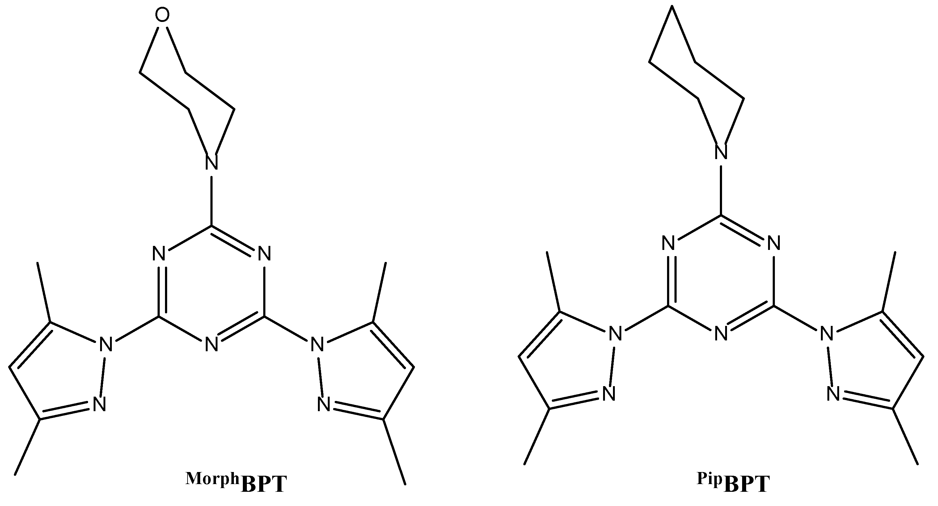
Figure 1.
Structure of the pincer ligands [32].
Figure 1.
Structure of the pincer ligands [32].

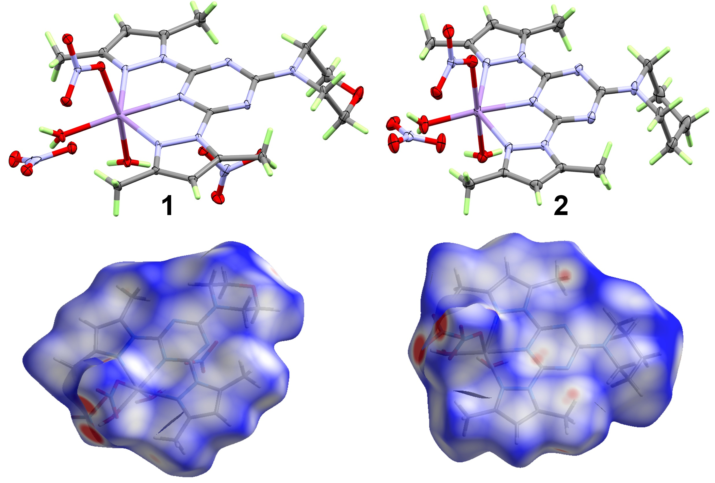
Figure 2.
Structure and atomic numbering of the symmetric unit of 1 and 2. Thermal ellipsoids were drawn at 50% probability level. Moreover, the nitrate counter ions were omitted for better clarity.
Figure 2.
Structure and atomic numbering of the symmetric unit of 1 and 2. Thermal ellipsoids were drawn at 50% probability level. Moreover, the nitrate counter ions were omitted for better clarity.

Figure 3.
Important H–bond contacts in 1 and 2. The green and blue colored contacts refer to the C–H···O and O–H···O interactions, respectively.
Figure 3.
Important H–bond contacts in 1 and 2. The green and blue colored contacts refer to the C–H···O and O–H···O interactions, respectively.

Figure 4.
H–bond networks in 1 (a) and 2 (b).

Figure 5.
Summary of the intermolecular interactions in 1 and 2.
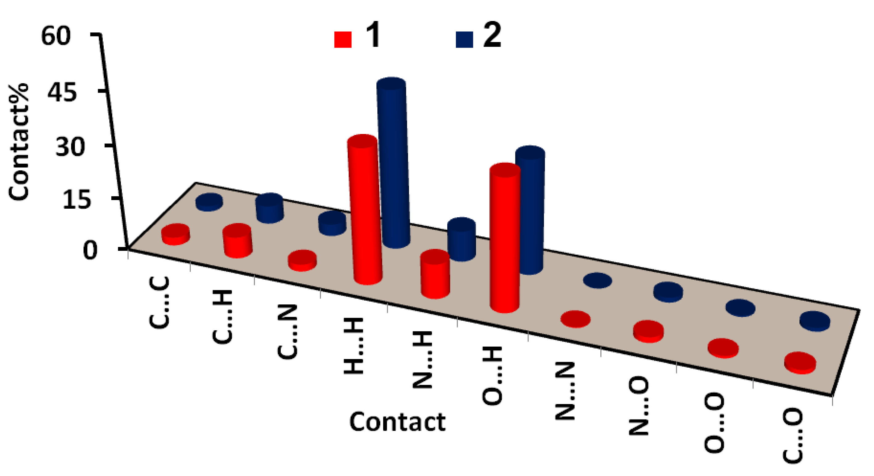
Figure 6.
The dnorm maps of the most important contacts in 1 and 2.

Figure 7.
Correlation between ρ(r); (a) and interaction energies (Eint); (b) with Mn–O distances.


Table 1.
Crystal and refinement data of 1 and 2.
| Compound | 1 | 2 |
|---|---|---|
| Empirical formula | C17H26MnN10O9 | C18H28MnN10O8 |
| Formula weight (g/mol) | 569.42 | 567.44 |
| Temperature (K) | 124(2) | 117(2) |
| λ (Mo-Kα, Å) | 0.71073 | 0.71073 |
| Crystal system | Triclinic | Triclinic |
| Space group | P-1 | P-1 |
| Unit cell dimensions | a = 8.2794(5) Å | a = 7.830(5) Å |
| b = 12.1167(7) Å | b = 12.951(7) Å | |
| c = 12.1738(7) Å | c = 13.990(7) Å | |
| α = 88.3660(19)° | α = 116.533(10)° | |
| β = 89.605(2)° | β = 94.832(14)° | |
| γ = 79.184(2)° | γ = 101.694(16)° | |
| Volume (Å3) | 1199.08(12) | 1217.9(12) |
| Z | 2 | 2 |
| Density (calc. g/cm3) | 1.577 | 1.547 |
| Absorption coefficient (mm−1) | 0.621 | 0.608 |
| F(000) | 590 | 590 |
| Crystal size (mm3) | 0.29 × 0.21 × 0.11 | 0.31 × 0.23 × 0.19 |
| θ range for data collection | 2.36 to 26.34° | 2.71 to 25.31° |
| Index ranges | −10 ≤ h ≤ 10, −15 ≤ k ≤ 15, −15 ≤ l ≤ 15 | −9 ≤ h ≤ 9, −15 ≤ k ≤ 15, −16 ≤ l ≤ 16 |
| Reflections collected | 21,451 | 19,570 |
| Independent reflections | 4863 [R(int) = 0.0315] | 4437 [R(int) = 0.0554] |
| Completeness to θ | 99.30% | 99.80% |
| Refinement method | Full-matrix least-squares on F2 | |
| Data/restraints/parameters | 4863/0/385 | 4437/0/351 |
| Goodness-of-fit on F2 | 0.894 | 1.06 |
| Final R indices [I>2sigma(I)] | R1 = 0.0275, wR2 = 0.0671 | R1 = 0.0298, wR2 = 0.0732 |
| R indices (all data) | R1 = 0.0341, wR2 = 0.0721 | R1 = 0.0371, wR2 = 0.0775 |
| Extinction coefficient | 0.0100(8) | 0.0157(17) |
| Largest diff. peak and hole | 0.371 and −0.318 | 0.409 and −0.308 |
| CCDC | 2025609 | 2025610 |
Table 2.
The most important bond distances and angles of 1 and 2.
| Atoms | 1 | 2 |
|---|---|---|
| Mn1–O2 | 2.1416(12) | 2.1288(19) |
| Mn1–O3 | 2.1683(11) | 2.1604(16) |
| Mn1–O1 | 2.2159(12) | 2.2306(17) |
| Mn1–N5 | 2.2172(12) | 2.1973(18) |
| Mn1–N1 | 2.3188(13) | 2.3097(19) |
| Mn1–N7 | 2.3267(13) | 2.3580(18) |
| O2–Mn1–O3 | 90.35(5) | 96.78(6) |
| O2–Mn1–O1 | 80.70(5) | 83.33(6) |
| O3–Mn1–O1 | 167.76(5) | 172.64(5) |
| O2–Mn1–N5 | 170.23(5) | 166.35(6) |
| O3–Mn1–N5 | 97.55(4) | 94.69(6) |
| O1–Mn1–N5 | 90.62(5) | 84.34(6) |
| O2–Mn1–N1 | 115.16(5) | 116.12(6) |
| O3–Mn1–N1 | 98.37(5) | 94.40(6) |
| O1–Mn1–N1 | 93.02(5) | 92.13(6) |
| N5–Mn1–N1 | 69.49(4) | 70.18(5) |
| O2–Mn1-N7 | 106.03(5) | 103.93(6) |
| O3–Mn1–N7 | 85.47(4) | 86.40(6) |
| O1–Mn1–N7 | 88.98(5) | 86.44(6) |
| N5–Mn1–N7 | 69.09(4) | 69.39(5) |
| N1–Mn1–N7 | 138.55(4) | 139.49(6) |
Table 3.
The Geometric parameters of the H–bonds in 1 and 2.
| Atoms | D–H (Å) | H···A (Å) | D···A (Å) | D–H···A (°) |
|---|---|---|---|---|
| 1 | ||||
| O2–H1···O11 | 0.82(2) | 1.89(2) | 2.662(3) | 157(2) |
| O2–H1···O12 | 0.82(2) | 1.95(2) | 2.766(3) | 175(2) |
| O1–H14···O9 | 0.83(2) | 1.97(2) | 2.728(3) | 153(2) |
| O1–H14···O7i | 0.83(2) | 1.96(2) | 2.758(3) | 161(2) |
| O1–H16···O10 | 0.85(2) | 2.07(2) | 2.884(3) | 161(2) |
| O1–H16···O11 | 0.85(2) | 2.07(2) | 2.831(4) | 150(2) |
| O2–H21···O5 | 0.85(2) | 2.54(2) | 2.924(2) | 109(2) |
| O2–H21···O4ii | 0.85(2) | 2.01(2) | 2.816(2) | 159(2) |
| C3–H3···O3iii | 0.95 | 2.52 | 3.319(2) | 142 |
| C9–H9A···O7iv | 0.98 | 2.49 | 3.455(3) | 170 |
| C11–H11···O7v | 0.95 | 2.39 | 3.328(3) | 171 |
| C13–H13C···O10 | 0.98 | 2.49 | 3.271(3) | 136 |
| Symmetry Code: (i) 1-x,1-y,2-z; (ii) -x,2-y,1-z; (iii) 1+x,y,z; (iv) -1+x,y,z; (v) -x,1-y,2-z | ||||
| 2 | ||||
| O1–H1A···O7i | 0.83(3) | 2.04(3) | 2.839(3) | 162(3) |
| O1–H1B···O7 | 0.86(2) | 1.89(2) | 2.723(3) | 163(2) |
| O2–H2A···O6 | 0.87(2) | 1.92(2) | 2.758(3) | 161(2) |
| O2–H2B···O4ii | 0.83(4) | 1.99(4) | 2.795(3) | 162(4) |
| C3–H3···O3iii | 0.95 | 2.53 | 3.240(3) | 132 |
| C9–H9C···O8iv | 0.98 | 2.42 | 3.329(3) | 154 |
| Symmetry Code: (i) 1-x,1-y,-z; (ii) 1-x,1-y,1-z; (iii) 1+x,y,z; (iv) -x,1-y,-z | ||||
Table 4.
Anion–π contacts in complexes 1 and 2.
| 1 | 2 | ||
|---|---|---|---|
| C7···O8i | 2.940(3) | C8···O7ii | 3.090(2) |
| N3···O8i | 3.077(3) | C7···O8ii | 2.982(2) |
| N4···O8i | 2.995(3) | ||
| C8···O8i | 3.044(3) | ||
| C6···O8i | 3.141(3) | ||
| C6···O9i | 3.052(3) | ||
| N5···O9i | 2.831(3) | ||
| C8···O9i | 2.889(3) | ||
| Symmetry Code: (i) x,y,z in 1 (ii) 1-x,1-y,-z in 2 | |||
Table 5.
Atoms in molecules (AIM) indices (a.u.) for the Mn–O and Mn–N bonds.
| Bond | Ρ(r) | G(r) | V(r) | Eint a | H(r) b | V(r)/G(r) c |
|---|---|---|---|---|---|---|
| 1 (MPW1PW91) | ||||||
| Mn1–N1 | 0.0315 | 0.0526 | −0.0511 | 16.0302 | 0.0015 | 0.9712 |
| Mn1–N7 | 0.0310 | 0.0508 | −0.0495 | 15.5195 | 0.0013 | 0.9737 |
| Mn1–N5 | 0.0446 | 0.0730 | −0.0749 | 23.5085 | −0.0019 | 1.0257 |
| Mn1–O1 | 0.0348 | 0.0617 | −0.0602 | 18.8813 | 0.0015 | 0.9754 |
| Mn1–O2 | 0.0423 | 0.0764 | −0.0759 | 23.8078 | 0.0006 | 0.9927 |
| Mn1–O3 | 0.0392 | 0.0731 | −0.0719 | 22.5571 | 0.0012 | 0.9835 |
| 1 (WB97XD) | ||||||
| Mn1–N1 | 0.0315 | 0.0524 | −0.0507 | 15.9153 | 0.0016 | 0.9689 |
| Mn1–N7 | 0.0309 | 0.0506 | −0.0491 | 15.4165 | 0.0014 | 0.9714 |
| Mn1–N5 | 0.0445 | 0.0729 | −0.0746 | 23.3928 | −0.0017 | 1.0234 |
| Mn1–O1 | 0.0347 | 0.0615 | −0.0599 | 18.7798 | 0.0016 | 0.9734 |
| Mn1–O2 | 0.0422 | 0.0763 | −0.0755 | 23.6955 | 0.0007 | 0.9904 |
| Mn1–O3 | 0.0392 | 0.0728 | −0.0715 | 22.4445 | 0.0013 | 0.9820 |
| 2 (MPW1PW91) | ||||||
| Mn1–N1 | 0.0323 | 0.0536 | −0.0522 | 16.3859 | 0.0014 | 0.9746 |
| Mn1–N7 | 0.0285 | 0.0464 | −0.0449 | 14.0784 | 0.0016 | 0.9662 |
| Mn1–N5 | 0.0465 | 0.0774 | −0.0796 | 24.9777 | −0.0022 | 1.0283 |
| Mn1–O1 | 0.0339 | 0.0592 | −0.0574 | 17.9982 | 0.0018 | 0.9698 |
| Mn1–O2 | 0.0446 | 0.0815 | −0.0810 | 25.4111 | 0.0005 | 0.9940 |
| Mn1–O3 | 0.0404 | 0.0754 | −0.0743 | 23.3263 | 0.0010 | 0.9862 |
| 2 (WB97XD) | ||||||
| Mn1–N1 | 0.0323 | 0.0533 | −0.0518 | 16.2604 | 0.0015 | 0.9724 |
| Mn1–N7 | 0.0284 | 0.0462 | −0.0445 | 13.9667 | 0.0017 | 0.9638 |
| Mn1–N5 | 0.0464 | 0.0772 | −0.0792 | 24.8540 | −0.0020 | 1.0260 |
| Mn1–O1 | 0.0338 | 0.0590 | −0.0570 | 17.8993 | 0.0019 | 0.9676 |
| Mn1–O2 | 0.0445 | 0.0812 | −0.0806 | 25.2873 | 0.0006 | 0.9921 |
| Mn1–O3 | 0.0404 | 0.0751 | −0.0740 | 23.2066 | 0.0012 | 0.9846 |
a kcal/mol; b total energy density; c potential to kinetic energy density.
Table 6.
Bond order analysis of the Mn–N and Mn–O coordination interactions.
| Bond | MPW1PW91 | WB97XD | MPW1PW91 | WB97XD |
|---|---|---|---|---|
| 1 | 2 | |||
| Mn1–N1 | 0.143 | 0.144 | 0.142 | 0.143 |
| Mn1–N7 | 0.136 | 0.137 | 0.122 | 0.123 |
| Mn1–N5 | 0.198 | 0.199 | 0.206 | 0.207 |
| Mn1–O1 | 0.129 | 0.129 | 0.119 | 0.119 |
| Mn1–O2 | 0.163 | 0.163 | 0.170 | 0.171 |
| Mn1–O3 | 0.157 | 0.158 | 0.159 | 0.161 |
Table 7.
The calculated natural charge at the N-sites of the free and coordinated pincer ligands using MPW1PW91(WB97XD) methods.
Table 7.
The calculated natural charge at the N-sites of the free and coordinated pincer ligands using MPW1PW91(WB97XD) methods.
| Atom | MorphBPT | 1 | PipBPT | 2 |
|---|---|---|---|---|
| N5 | −0.4618(−0.4740) | −0.5921(−0.6022) | −0.4671(−0.4794) | −0.5892(−0.5987) |
| N1 | −0.2237(−0.2271) | −0.3163(−0.3191) | −0.2243(−0.2275) | −0.3042(−0.3069) |
| N7 | −0.2015(−0.2275) | −0.3084(−0.3106) | −0.2224(−0.2256) | −0.3085(−0.3114) |
Table 8.
Inhibition zones at 200 µg of the tested compounds by agar well diffusion method.
| Compound | S. aureus | E. coli | C. albicans |
|---|---|---|---|
| MorphBPT | - | - | - |
| PipBPT | 11 | - | 12 |
| 1 | 21 | 17 | 15 |
| 2 | 27 | 20 | 17 |
| Fluconazole | - | - | 14 |
| Gentamycin | 28 | 21 | - |
Table 9.
The minimum inhibitory concentration (MIC) and minimum bactericidal concentration (MBC) of complexes 1 and 2.
Table 9.
The minimum inhibitory concentration (MIC) and minimum bactericidal concentration (MBC) of complexes 1 and 2.
| Microbes | 1 | 2 | ||
|---|---|---|---|---|
| MIC (µg/mL) | MBC (µg/mL) | MIC (µg/mL) | MBC (µg/mL) | |
| S. aureus | 8.3 | 16.5 | 6.5 | 13.5 |
| E. coli | 8.3 | 16.5 | 8.3 | 16.8 |
| C. albicans | 18.5 | 100 | 18.5 | 100 |
© 2020 by the authors. Licensee MDPI, Basel, Switzerland. This article is an open access article distributed under the terms and conditions of the Creative Commons Attribution (CC BY) license (http://creativecommons.org/licenses/by/4.0/).
Share and Cite
MDPI and ACS Style
Soliman, S.M.; Al-Rasheed, H.H.; El-Faham, A. Synthesis, X-ray Structure, Hirshfeld Analysis of Biologically Active Mn(II) Pincer Complexes Based on s-Triazine Ligands. Crystals 2020, 10, 931. https://doi.org/10.3390/cryst10100931
AMA Style
Soliman SM, Al-Rasheed HH, El-Faham A. Synthesis, X-ray Structure, Hirshfeld Analysis of Biologically Active Mn(II) Pincer Complexes Based on s-Triazine Ligands. Crystals. 2020; 10(10):931. https://doi.org/10.3390/cryst10100931
Chicago/Turabian StyleSoliman, Saied M., Hessa H. Al-Rasheed, and Ayman El-Faham. 2020. "Synthesis, X-ray Structure, Hirshfeld Analysis of Biologically Active Mn(II) Pincer Complexes Based on s-Triazine Ligands" Crystals 10, no. 10: 931. https://doi.org/10.3390/cryst10100931
Note that from the first issue of 2016, this journal uses article numbers instead of page numbers. See further details here.