Article

Growth of Nacre Biocrystals by Self-Assembly of Aragonite Nanoparticles with Novel Subhedral Morphology

School of Resources, Environment and Materials, Guangxi University, 100 Daxue Road, Nanning 530004, Guangxi, China
*
Author to whom correspondence should be addressed.
Crystals 2020, 10(1), 3; https://doi.org/10.3390/cryst10010003
Submission received: 21 October 2019 / Revised: 13 December 2019 / Accepted: 14 December 2019 / Published: 18 December 2019
(This article belongs to the Section Crystalline Materials)

Abstract

:
Nacre has long served as a research model in the field of biomineralization and biomimetic materials. It is widely accepted that its basic components, aragonite biocrystals, namely, tablets, are formed by the nanoparticle-attachment pathway. However, the details of the nanoparticle morphology and arrangement in the tablets are still a matter of debate. Here, using field emission scanning electron microscopy (FESEM) and transmission electron microscopy (TEM), we observed the nanostructure of the growing tablets at different growth stages and found that: (1) the first detectable tablet looked like a rod; (2) tablets consisted of subhedral nanoparticles (i.e., partly bounded by crystal facets and partly by irregular non-crystal facets) that were made of aragonite single crystals with a width of 160–180 nm; and (3) these nanoparticles were ordered in orientation but disordered in position, resulting in unique subhedral and jigsaw-like patterns from the top and side views, respectively. In short, we directly observed the growth of nacre biocrystals by the self-assembly of aragonite nanoparticles with a novel subhedral morphology.

1. Introduction

Nacre is an iridescent material of biogenic crystals formed by mollusks for mechanical protection. It is synthesized at ambient conditions and from readily-available materials, namely, >95wt% CaCO3 mineral and minor biopolymers [1]. However, it exhibits a striking combination of high strength and toughness that are still difficult to achieve synthetically using current technologies [2,3]. Therefore, nacre has long been an important research subject in a variety of fields such as biology, material science, chemistry, and bioinspired material synthesis [4,5,6,7].
Generally, the basic structural units of nacre are aragonite biocrystals with a tablet-like shape that, when isolated, look like short pseudo-hexagonal prisms bounded by flat surfaces [8,9,10], which can be indexed as {010}, {110}, and {001} of aragonite, respectively (Figure 1a) [1,8,9,11,12,13,14]. For this reason, such tablets are often called “flat tablets” in the literature [15,16,17]. Interestingly, flat tablets are similar in shape to the equilibrium habit of aragonite (Figure 1b) [18,19], except that they have large flat basal surfaces. Thus, their projected shapes on the ab plane (i.e., horizontal plane) are nearly identical (Figure 1c). Sometimes they also appear in the form of triple twinning (Figure 1d).
Flat tablets have been widely studied in order to understand their formation mechanism. It is generally accepted that they form via a nonclassical, nanoparticle-attachment pathway [21]. However, the existing literature (Table S1) shows a variety of data about the shape and size of the nanoparticles. Regarding their shape, there are two conflicting views. The first proposes that the nanoparticles are anhedral (i.e., not bounded by crystal faces) such as cobble-like, globular, and nanosheet-like, with a size of 5–50nm. The second proposes that they are euhedral (i.e., pseudo-hexagonal) with a width of 20–180nm and unknown thickness [22]. However, there are few reports on the self-assembly process of nanoparticles. (See Supplementary Materials).
With a few exceptions [23], most investigators have implicitly or explicitly focused on the nanostructure of the mature tablet. Therefore, the original morphology and arrangement of nanoparticles have either been neglected or deduced on the basis of observing the mature tablets. Here, we focus on the nanostructure of the growing tablets in different growth stages aiming at discovering the features of nanoparticles that may disappear in mature tablets. We found that the tablets consist of subhedral aragonite nanoparticles (SANs) with granular and straight facets. These nanoparticles have never been reported. This finding may provide new insights into the nacre formation mechanism.

2. Materials and Methods

The bivalve Perna viridis is a common mussel which is widely distributed in the Indo-Pacific region of Asia with rapid growth rates of between 6 and 10 mm/month [24]. Live samples with a shell length of 2–3 cm (about 3–5 months old) were collected on the coast of Beihai in Guangxi and Zhanjiang in Guangdong, southern China. They were first processed as follows: (1) soft tissue was removed from the shells with a scalpel; (2) the shells were cleaned with tap water followed by distilled water and air-dried at room temperature; (3) shell fragments, about 5 × 5 mm in size, were mechanically broken away from the shell margin.
For X-ray diffraction (XRD) analysis, the shell fragments were scratched along their inner surface with a scalpel to obtain powder samples. Then, the diffraction pattern was recorded with a diffractometer (Rigaku D/max-2500, Nanning, Guangxi, China) in a θ–2θ configuration with Cu Kα radiation (λ = 0.15406 nm). The operating voltage and current were 40 kV and 200 mA, respectively.
For Raman spectra analysis, the prepared shell fragments were fixed to the slide. Raman spectra of the nacre were obtained on a Raman spectrometer (Renishaw Invia, Nanning, Guangxi, China), using a 785 nm laser with an exposure time of 10 s.
For field emission scanning electron microscopy (FESEM) analysis, the formerly prepared shell fragments were mounted onto the sample holder with the nacre surface either parallel or normal to the holder plane. Then, samples were sputtered with gold and observed with FESEM (Hitachi, SU8020, Nanning, Guangxi, China) operated at 8–10 kV.
For field emission transmission electron microscopy (FETEM) analysis, samples were prepared by the focused ion beam (FIB) method. A vertical cross-section slice was prepared from the nacreous inner surface, using a dual-beam FIB system (FEI Helios NanoLab 600i, Guiyang, Guizhou, China). After that, TEM images and selected area electron diffraction (SAED) patterns were obtained with FETEMs (JEOL JEM-2010, Jingdezhen, Jiangxi, China and FEI G20, Changsha, Hunan, China) operated at 200 kV. TEM images were analyzed with the software Digital Micrograph software (Gatan, 3.11.2, Nanning, China).

3. Results

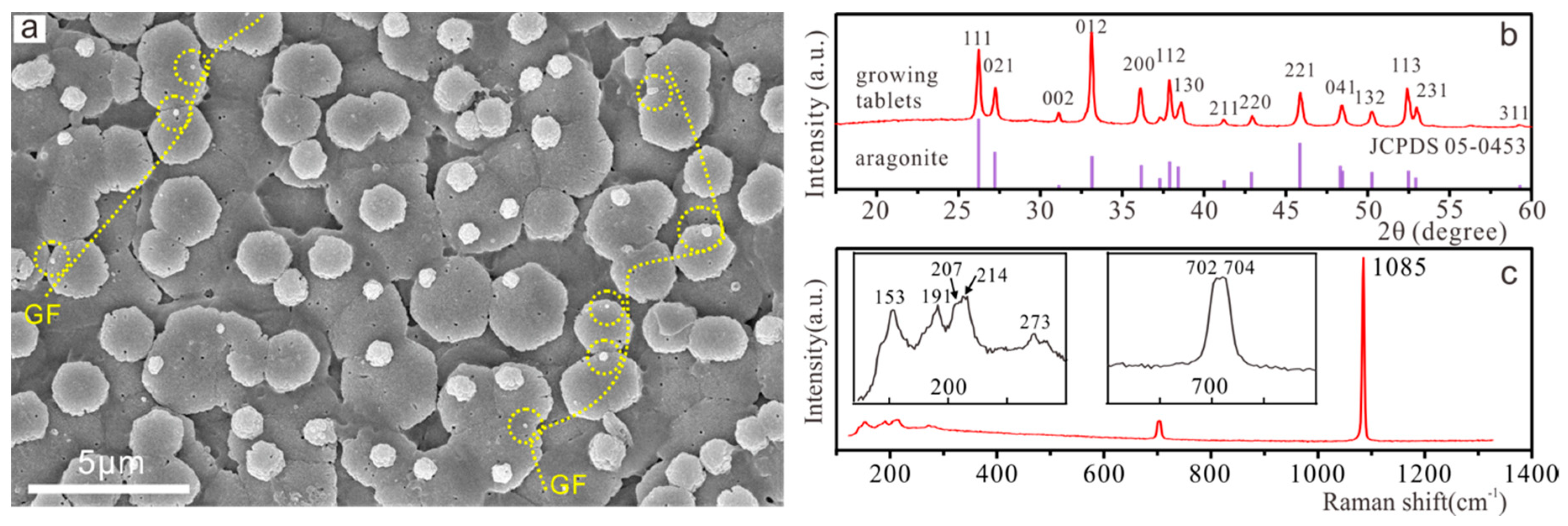
3.1. Nacre Growth Pattern and Composition

The growth pattern of nacre in Perna vridis is shown in Figure 2a, which is similar to that of a classical sheet nacre in bivalves [25]. That is, at the growth front (GF) of each tablet layer, new tablets (Figure 2a and Figure S1a, circles) nucleate on the top peripheries of the underlying tablets and gradually increase in size until merging with the already-formed tablet layer. The growth front is a line connecting the smallest growing tablets, which is perpendicular to the horizontal growth direction of the nacre, suggesting the position where the new layer of nacre begins to grow.
Figure 2b shows powder XRD patterns of the growing tablets that match with the JCPDS data for aragonite (with lattice parameters a = 4.9633Å, b = 7.9703Å, c = 5.7441Å, file No. 05-0453), indicating that the growing tablets contain only one crystalline phase, aragonite. In addition, Figure 2c shows the Raman shift signals obtained from the growing tablets on the transitional area, which can identify the tablets as aragonite by lattice modes at 153 cm−1 (B1g), 191 cm−1 (B2g), 207 cm−1 (B2g), 214 cm−1 (Ag), and 273 cm−1 (B3g), and internal modes at 702 cm−1 (B3g), 704 cm−1 (Ag), and 1085 cm−1 (Ag), with the results of XRD analysis [26].

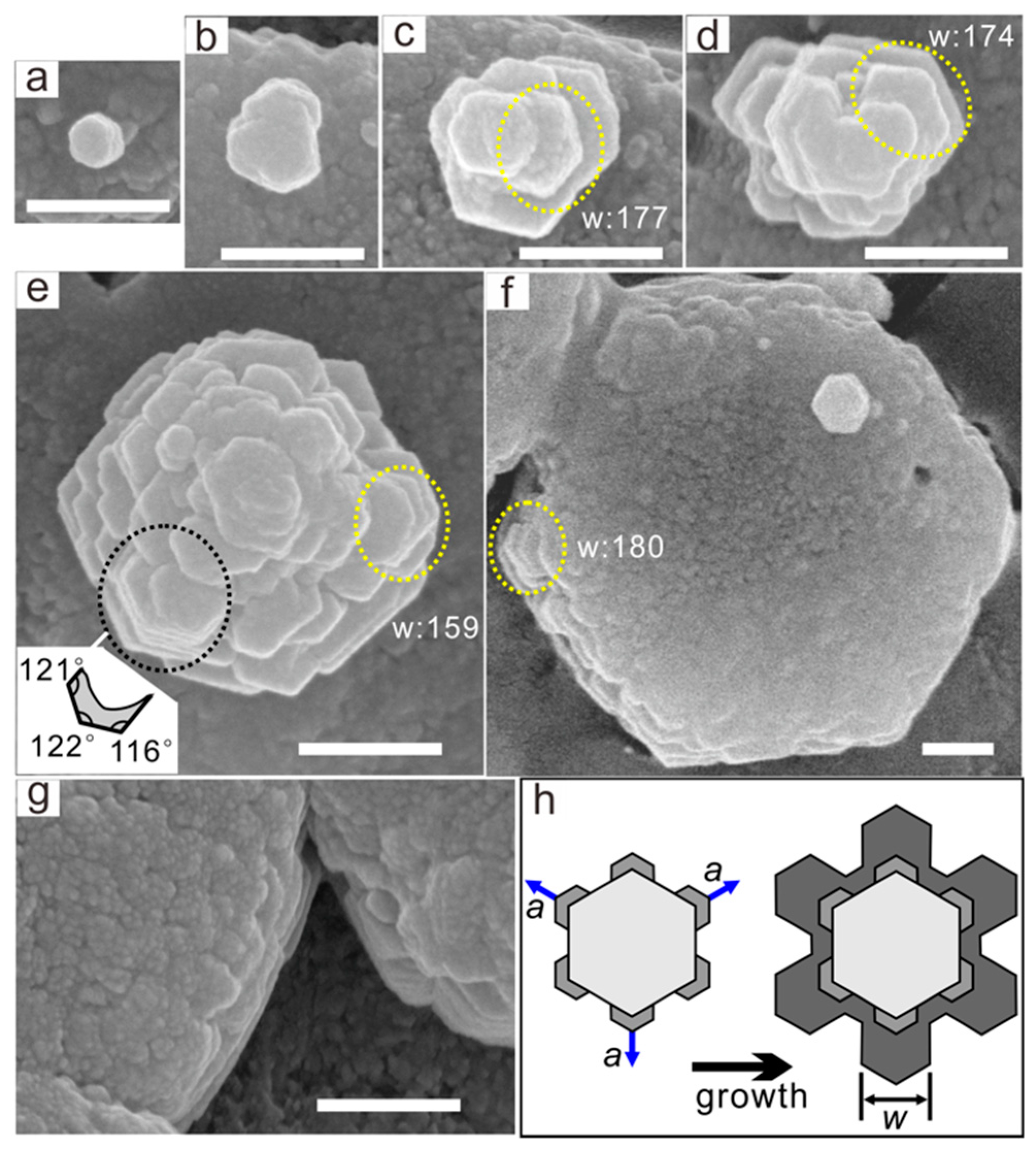
3.2. SEM Observation of Growing Tablets

3.2.1. Nanoparticle Shape and Size

Generally, the growing tablets exhibited a variety of overall shapes depending on their size (corresponding to the growth stages), as shown in Figure 3 and Figure 4. All tablets predominantly consisted of subhedral nanoparticles, except that the initial tablets looked like a rod.
In the top view, nearly all nanoparticles appeared as incomplete pseudo-hexagons (Figure 3b–f) branching out from the core of the tablets. Importantly, they had two sets of the angles, i.e., around 116° and 122°, respectively (Figure 3e, inset). Thus, the side facets of the nanoparticles could be indexed as {010} and {110} (Figure 1c), which implies that these nanoparticles are subhedral aragonite. In addition, the surface of each pseudo-hexagonal nanoparticle displayed a nanogranular structure, and each nanograin was 10–30nm in size, which was about one order of magnitude smaller than the nanoparticle size (Figure 3c and Figure 4d–i).
Surprisingly, in the side or oblique view, the nanoparticles appeared as incomplete short prisms (Figure 4d–f), where free facets (exposed faces) and edges were granular and straight, respectively (Figure 4h, red lines). In contrast, embedded faces, which corresponded to the interfaces with other nanoparticles, were highly irregular, as revealed by their irregular boundaries with other nanoparticles, which may be straight, serrated, or sinuous (Figure 4e, inset, and Figure 4h, blue lines).
In addition, nanoparticle size was independent of that of their parent tablets. For example, nanoparticle width was similar (about 160–180 nm) on tablets of different sizes (ranging from 370 to 1790 nm; Figure 3c–f, yellow circles).
In short, each nanoparticle of the growing tablets was unique in shape and size and adopted a subhedral shape. Finally, note that the shape of each tablet mentioned here and below was unique, that is, there was no inheritance of the growth process between them, because SEM and TEM could not track the entire growth stages of a specific tablet in situ.

3.2.2. Nanoparticle Self-Assembly Process

The first detectable tablet looked like a rod with diameter of 135 nm and length of 274 nm (Figure 4a). In general, these rod-like tablets appeared to be pseudo-hexagonal in the top view (Figure 3a). These rods clearly served as initial cores to which the subsequent nanoparticles were attached (Figure 4b,c). In particular, as the rods grow larger (Figure 4c), they even showed the shape of euhedral aragonite (Figure 1b).
For the subhedral nanoparticles, their arrangement patterns appeared different when viewed from different angles. In the case of the top view, nanoparticles were obviously ordered in orientation. More precisely, their corresponding side facets, such as {110} or {010}, were parallel to each other (Figure 3b–g). As a result, the tablet as a whole looked like a hexagon, albeit with serrated sides. Meanwhile, they branched randomly from the core periphery. Therefore, nanoparticles were arranged into an irregular subhedral pattern. Here, it must be noted that this was not a dendritic crystal, and it could not show six symmetrical arms since it was not hexagonal but orthorhombic [27].
On the other hand, in the case of the side or oblique view, it was easy to observe that nanoparticles were disordered in position. In particular, on the lateral surfaces of the tablets, as shown in Figure 4g–i, nanoparticles were randomly interlocked into a jigsaw-puzzle-like structure. In summary, nanoparticles were ordered in orientation but disordered in position, thus forming a unique subhedral and jigsaw-puzzle-like structure when viewed from different angles.

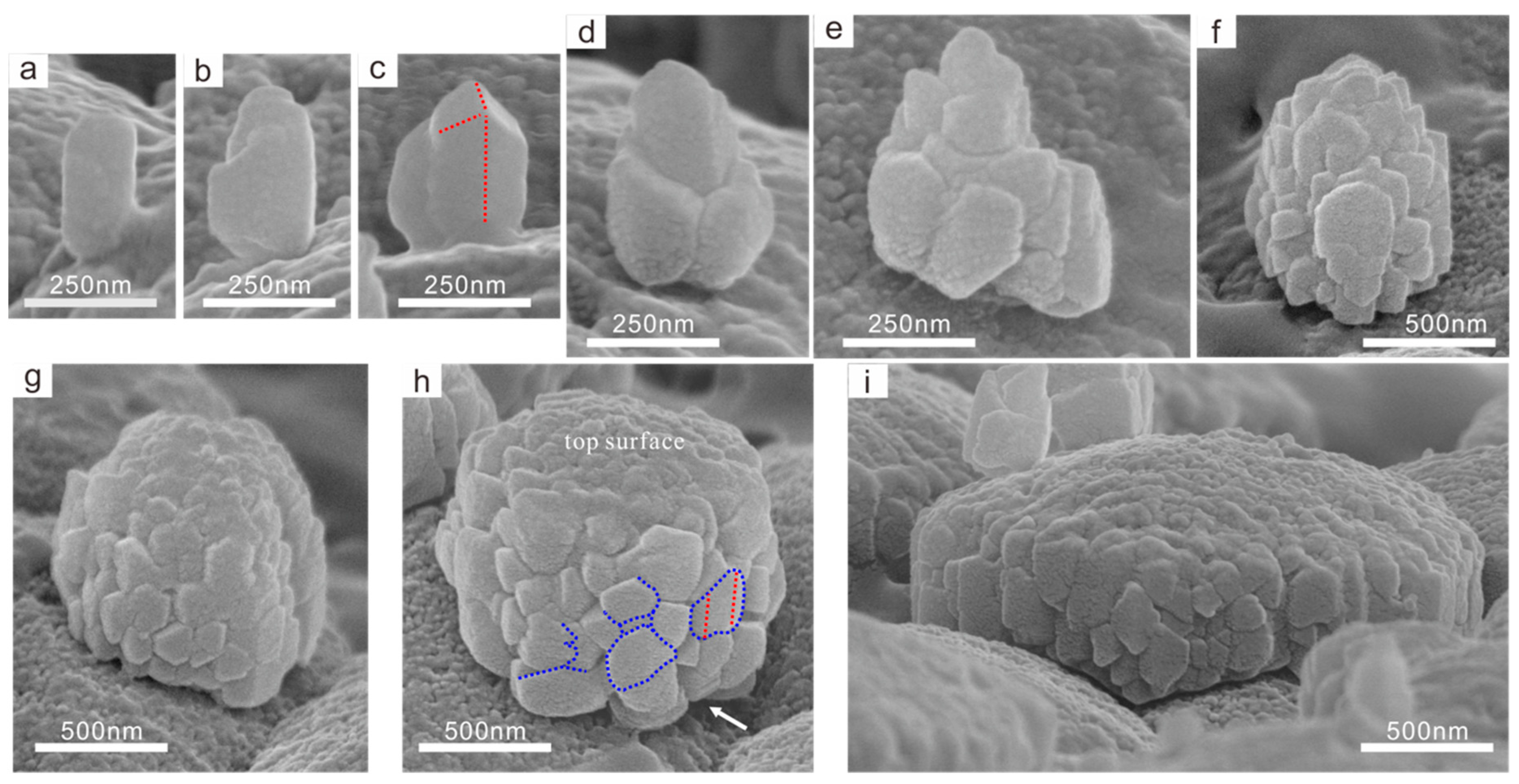
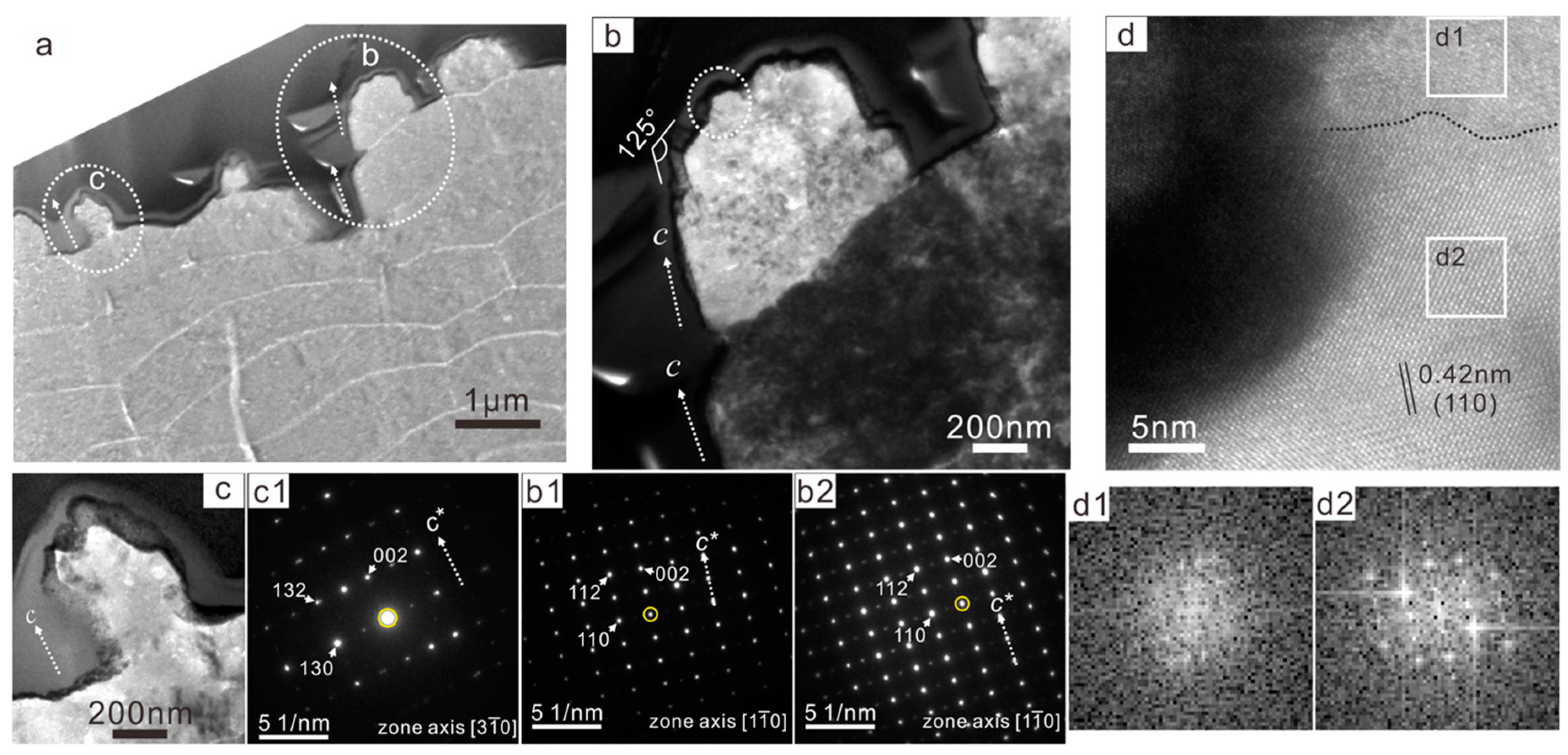
3.3. TEM Observation of the Growing Tablets
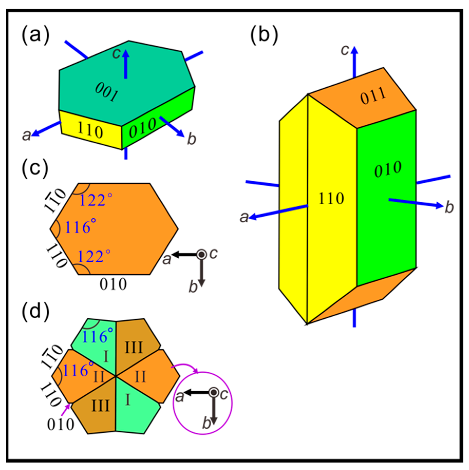
Figure 5a shows the cross-sectional overview of the growing tablets. Close views show that they have clearly stepped or serrated surfaces (Figure 5b,c). Despite this, each growing tablet shows regular diffraction spots in the SAED pattern (Figure 5b1,c1), indicating that it was a mesocrystal, that is, the nanoparticles of the growing tablets were co-oriented. In addition, the c-axes of different growing tablets were nearly parallel to each other with a slight misorientation of <8°. Even in the same zone axes, there was a tilt of 7° between the c-axis of the above and below adjacent tablets, which implied that the crystallographic orientations of the two were not completely identical (Figure 5b1,b2).
Interestingly, the side edge of the tablet was parallel to its c-axis direction, indicating that the nanoparticles were consistent with the c-axis orientation of the tablet (Figure 5a). In particular, on the side of the middle tablet (Figure 5b), the lateral and top surfaces of the nanoparticle could be indexed as (110) and (112) of aragonite crystal, respectively (Figure 5b1). The angle was around 125°, which was identical to the theoretical angle between (110) and (112). That is, the nanoparticles were partly bounded by the crystal faces of aragonite (Figure 1b), which means that they were subhedral aragonite crystals. In other words, they were prismatic crystals with incomplete faces.
Finally, the high-resolution transmission electron microscope (HRTEM) image (Figure 5d) shows that the nanoparticle on the tablet could be divided into two regions with different crystallinity, and their boundary was indicated by the dotted line. Specifically, the top region is weak crystalline, as supported by the fast Fourier transform (FFT) patterns (Figure 5d1), while the rest of the continuous lattice fringes with regular diffraction spots in the corresponding FFT patterns (Figure 5d2).

4. Discussion

Here, we directly observed the morphology and arrangement of nanoparticles that made up the growing tablets. We found that the first detectable tablets were prismatic, which has not been reported in previous studies of bivalves. In addition, they acted as the nuclei for growth of the tablets, which provide a position for subhedral aragonite nanoparticles (SANs) attachment and determine the crystallographic orientation of the attached SANs. The formation mechanism of these rod-like structures is related to the nucleation mechanism of the tablets. The two main hypotheses are matrix templates [28] and mineral bridges [29]. The former proposes that tablets nucleate directly on the organic matrix, while the latter proposes that they do so via the mineral bridges between adjacent layers. There is no agreement on the nucleation mechanism between the two views. However, the focus of this paper are morphology and arrangement of SANs, so the formation mechanism of these rod-like structure is beyond our scope. As shown in Figure 3h, the size of six snowflake arms increased with the growth of the parent plate in classical crystallization. The size of the SANs was independent of that of their parent tablets, indicating that the tablets grew via non-classical crystallization, which is consistent with the conclusions of previous studies [30,31,32].
For subhedral aragonite nanoparticles, we found the following features: (1) each SAN was unique in shape and size; and (2) they contained straight free facets belonging to crystal faces, while their embedded faces were curved and interlocked with adjacent nanoparticles belonging to non-crystal faces. In particular, their free facets belonged to the {010} and {110} of aragonite with low surface energies. Therefore, these facets should form via classical crystallization pathway driven by surface energy minimization [21,33]. In addition, affected by the minimization of surface energy, shown in the top view, the new attached SANs preferentially grow in concave areas between two adjacent SANs [34], so that the subhedral prisms were evenly distributed in all directions around the core of the tablets. In particular, some of the rod-like tablets look like euhedral aragonite crystals, which is because these tablets consisted of only one isolated SAN, and they had enough space to grow into a euhedral shape.
Regarding the formation mechanism of SANs, there are several important questions to answer: (a) Are they formed ex situ, as suggested by Addadi et al. [35], as vesicles of cells, blood cells, and extrapallial fluid are, and then transported to their current deposition location? In other words, are they original building blocks (OBBs)? (b) Are they formed in situ, that is, are they formed by the phase transition of the aggregated and deposited OBBs at the current position? (c) Is their initial phase amorphous calcium carbonate (ACC) or crystalline aragonite? (d) Are they formed via a classical or nonclassical crystallization pathway?
At present, it is difficult to accurately answer these questions. For question (a), if we assume that SANs are OBBs and are transported to the current location after being formed elsewhere, they should be of the same shape and size. It is hard to imagine that creatures intentionally create OBBs of varying shapes and sizes, but can interlock them together accurately to fill a 3D space. Therefore, SANs are unlikely to be OBBs, which means that they are likely to be formed in situ at the current location.
In addition, as by an anonymous reviewer, we inferred that the SANs (160–180 nm in width) observed in this work were likely not OBBs, for of the following reasons: (1) as first revealed by Macías-Sánchez et al. [36], the nanoparticles observed in nacre tablets are different in size from original aggregation nanoparticles (i.e., OBBs). In particular, the former (20–50 nm in diameter) are usually larger than the latter (5–20 nm in diameter). (2) OBBs can cross the pores (20–100 nm in diameter) of interlamellar membranes [37] to reach their mineralization sites during nacre growth. (3) OBBs are probably initially formed in an extrapallial space with a thickness of ca. 100 nm [38], which implies that they should be <100 nm in size. Therefore, we predict that OBBs forming the SANs should be <100 nm in size.
Regarding questions (b) and (c), there is increasing evidence [31,36,39,40,41] showing that the OBBs of nacre tablets are spheroidal ACC nanoparticles (with sizes of 2–20 nm [31,36,40]). Accordingly, we assume that the original ACC particles are transported to the current location, then undergo aggregation and phase transition to form the SANs, which would explain the observations in this study. That is, since the ACC can aggregate and fill space to reproduce any molding structure, they can interlock with each other and completely fill a 3D space, which explains why the embedded faces of the SANs are curved and engage with the other contact nanoparticles. In addition, adjacent spherical ACC particles may not deposit simultaneously, and may be random during aggregation, which is likely to result in the different sizes of the final aggregated SANs.
Regarding question (d), we observed that the surface of each SAN displayed a nanogranular structure, and each nanograin was about one order of magnitude smaller than the nanoparticle size. Therefore, nacre tablets may have the following features: (1) initial nanograins are uniform in size (about 10–30 nm); (2) tablets are hierarchical in structure; and (3) this structure consists of three levels, i.e., nanograins, SANs, and tablets. We conclude that they probably undergo two processes. The first is the non-classical crystallization process, in which ACC particles are stacked together and then aggregated to form ACC clusters of various sizes and shapes. This is followed by a ripening stage, in which these clusters are phase transited into SANs. During the phase transition, the free facets of the SANs are in contact with the extrapallial fluid; thus, they can undergo dissolution–reprecipitation driven by surface energy minimization to eventually form granular and straight crystal faces [21,33]. The same results occur by dissolution–reprecipitation if ACC is hydrated, as proposed by the anonymous reviewers of this work.
In short, on the basis of the above analysis, we concluded that tablets have a hierarchical structure that consists of three levels. SANs belong to the second level, which is different from the anhedral or euhedral shape reported in previous reports, as mentioned in the introduction. SANs were also ordered in orientation but disordered in position, forming a unique subhedral and jigsaw-like structure from the top and side view, respectively. This structure has never been reported in shell nacre, and only Zhou et al. [42] reported the same structure in high-temperature aragonite. In short, detailing the formation mechanism of SANs still needs further work. However, the discovery of such SANs is of great significance and provides a new research model with which to further understand the formation mechanism of nacre.

5. Conclusions

Here, FESEM and FETEM were used to directly observe the morphology and arrangement of nanoparticles in growing nacre tablets. This work firstly found that: (1) the first detectable tablets were rod-like in shape; (2) the nanoparticles of the tablets were subhedral aragonite single crystals with a width of 160–180 nm in different-sized parent tablets; (3) these nanoparticles were oriented and grew along {010} and {110} of the tablet, but were randomly distributed on the tablets, that is, they were ordered in terms of orientation but disordered in terms of position, resulting in a unique subhedral and jigsaw-like pattern from the top and side view, respectively. We directly observed the growth of nacre biocrystals by the self-assembly of aragonite nanoparticles with a novel subhedral morphology. These nanoparticles are different from the anhedral or euhedral units in mature tablets reported in previous reports. This finding provides a new perspective for research in the biomineralization and biomimetic synthesis of nacre.

Supplementary Materials

The following are available online at https://www.mdpi.com/2073-4352/10/1/3/s1; Figure S1: Top-view SEM images of flat tablets growing on shell interior; Table S1: Morphology and organization of nanoparticles in nacre tablets.

Author Contributions

Funding acquisition, G.Z.; investigation, R.G., R.W., and X.F.; supervision, G.Z.; writing—original draft, R.G. All authors have read and agreed to the published version of the manuscript.

Funding

This research was funded by the National Natural Science Foundation of China, grant number 41762004.

Acknowledgments

We gratefully thank Zaiwang Huang and Ning Yan (Central South University, Changsha) for the technical support in TEM observations, as well as Rui Li (Institute of Geochemistry, Chinese Academy of Sciences, Guiyang) for FIB sample preparation. We also appreciate the financial support from the National Natural Science Foundation of China (Grant No. 41762004).

Conflicts of Interest

The authors declare no conflict of interest.

References

  1. Meyers, M.A.; Chen, P.Y.; Lin, A.Y.M.; Seki, Y. Biological materials: Structure and mechanical properties. Prog. Mater. Sci. 2008, 53, 1–206. [Google Scholar] [CrossRef] [Green Version]
  2. Wegst, U.G.K.; Bai, H.; Saiz, E.; Tomsia, A.P.; Ritchie, R.O. Bioinspired structural materials. Nat. Mater. 2015, 14, 23–36. [Google Scholar] [CrossRef] [PubMed]
  3. Ritchie, R.O. The conflicts between strength and toughness. Nat. Mater. 2011, 10, 817–822. [Google Scholar] [CrossRef] [PubMed]
  4. Mao, L.B.; Gao, H.L.; Yao, H.B.; Liu, L.; Colfen, H.; Liu, G.; Chen, S.M.; Li, S.K.; Yan, Y.X.; Liu, Y.Y.; et al. Synthetic nacre by predesigned matrix-directed mineralization. Science 2016, 354, 107–110. [Google Scholar] [CrossRef] [Green Version]
  5. Pan, X.F.; Gao, H.L.; Lu, Y.; Wu, C.Y.; Wu, Y.D.; Wang, X.Y.; Pan, Z.Q.; Dong, L.; Song, Y.H.; Cong, H.P.; et al. Transforming ground mica into high-performance biomimetic polymeric mica film. Nat. Commun. 2018, 9, 2974. [Google Scholar] [CrossRef] [Green Version]
  6. Cao, W.T.; Chen, F.F.; Zhu, Y.J.; Zhang, Y.G.; Jiang, Y.Y.; Ma, M.G.; Chen, F. Binary strengthening and toughening of MXene/Cellulose nanofiber composite paper with nacre-inspired structure and superior electromagnetic interference shielding properties. ACS Nano 2018, 12, 4583–4593. [Google Scholar] [CrossRef]
  7. Yaraghi, N.A.; Kisailus, D. Biomimetic structural materials: Inspiration from design and assembly. Annu. Rev. Phys. Chem. 2018, 69, 23–57. [Google Scholar] [CrossRef] [Green Version]
  8. Taylor, J.D. The shell structure and mineralogy of the Bivalvia. Introduction. Nuculacea-Trigonacea. Bull. Br. Mus. (Nat. Hist.) 1969, 3, 1–125. [Google Scholar]
  9. Wada, K. Nucleation and growth of aragonite crystals in the nacre of some bivalve molluscs. Biomineralisation 1972, 6, 141–159. [Google Scholar]
  10. Rousseau, M.; Rollion-Bard, C. Influence of the depth on the shape and thickness of nacre tablets of pinctada margaritifera pearl oyster, and on oxygen isotopic composition. Minerals 2012, 2, 55–64. [Google Scholar] [CrossRef]
  11. Yao, N.; Epstein, A.K.; Liu, W.W.; Sauer, F.; Yang, N. Organic–inorganic interfaces and spiral growth in nacre. J. R. Soc. Interface 2009, 6, 367–376. [Google Scholar] [CrossRef] [PubMed] [Green Version]
  12. Joan, F.B.; Louis, F.; Gainey, J.; Michael, J.G. Shell ultrastructure in two subspecies of the ribbed mussel. Biol. Bull. 1977, 152, 1–11. [Google Scholar]
  13. Yoshimi, K.; Shoji, M.; Ogawa, T.; Yamauchi, A.; Naganuma, T.; Muramoto, K.; Hanada, S. Microstructure and orientation distribution of aragonite crystals in nacreous layer of pearl shells. Mater. Trans. 2004, 45, 999–1004. [Google Scholar] [CrossRef] [Green Version]
  14. Mukai, H.; Saruwatari, K.; Nagasawa, H.; Kogure, T. Aragonite twinning in gastropod nacre. J. Cryst. Growth 2010, 312, 3014–3019. [Google Scholar] [CrossRef]
  15. Gower, L.B. Biomimetic model systems for investigating the amorphous precursor pathway and its role in biomineralization. Chem. Rev. 2008, 108, 4551–4627. [Google Scholar] [CrossRef] [Green Version]
  16. Feng, Q.L.; Cui, F.Z.; Pu, G.; Wang, R.Z.; Li, H.D. Crystal orientation, toughening mechanisms and a mimic of nacre. Mater. Sci. Eng. C 2000, 11, 19–25. [Google Scholar] [CrossRef]
  17. DiMasi, E.; Sarikaya, M. Synchrotron x-ray microbeam diffraction from abalone shell. J. Mater. Res. 2004, 19, 1471–1476. [Google Scholar] [CrossRef]
  18. De Leeuw, N.H.; Parker, S.C. Surface structure and morphology of calcium carbonate polymorphs calcite, aragonite, and vaterite: An atomistic approach. J. Phys. Chem. B 1998, 102, 2914–2922. [Google Scholar] [CrossRef]
  19. Sekkal, W.; Zaoui, A. Nanoscale analysis of the morphology and surface stability of calcium carbonate polymorphs. Sci. Rep. 2013, 3, 1587. [Google Scholar] [CrossRef] [Green Version]
  20. Sand, K.K.; Rodriguez-Blanco, J.D.; Makovicky, E.; Benning, L.G.; Stipp, S.L.S. Crystallization of CaCO3 in water–alcohol mixtures: Spherulitic growth, polymorph stabilization, and morphology change. Cryst. Growth Des. 2012, 12, 842–853. [Google Scholar] [CrossRef]
  21. De Yoreo, J.J.; Gilbert, P.U.P.A.; Sommerdijk, N.A.J.M.; Penn, R.L.; Whitelam, S.; Joester, D.; Zhang, H.; Rimer, J.D.; Navrotsky, A.; Banfield, J.F.; et al. Crystallization by particle attachment in synthetic, biogenic, and geologic environments. Science 2015, 349, aaa6760. [Google Scholar] [CrossRef] [PubMed]
  22. Oaki, Y.; Imai, H. The hierarchical architecture of nacre and its mimetic material. Angew. Chem. Int. Ed. 2005, 44, 6571–6575. [Google Scholar] [CrossRef] [PubMed]
  23. Zhang, G. Biomineralization on the wavy substrate: Shape transition of nacreous tablets from pyramids of amorphous nanoparticles to dome-capped prisms of single crystals. Acta Biomater. 2016, 36, 277–285. [Google Scholar] [CrossRef] [PubMed]
  24. Power, A.J.; Walker, R.; Payne, K.; Hurley, D. First occurrence of the nonindigenous green mussel, Perna viridis (Linnaeus, 1758) in coastal Georgia, United States. J. Shellfish Res. 2004, 23, 741–744. [Google Scholar]
  25. Wise Sherwood, W., Jr. Microarchitecture and mode of formation of nacre (mother-of-pearl) in pelecypods, gastropods, and cephalopods. Eclogae Geol. Helv. 1970, 63, 775–797. [Google Scholar]
  26. De La Pierre, M.; Carteret, C.; Maschio, L.; André, E.; Orlando, R.; Dovesi, R. The Raman spectrum of CaCO3 polymorphs calcite and aragonite: A combined experimental and computational study. J. Chem. Phys. 2014, 140, 164509. [Google Scholar] [CrossRef] [Green Version]
  27. Libbrecht, K. The physics of snow crystals. Rep. Prog. Phys. 2005, 68, 855–895. [Google Scholar] [CrossRef] [Green Version]
  28. Weiner, S.; Traub, W.; Parker, S.B. Macromolecules in Mollusc Shells and Their Functions in Biomineralization [and Discussion]. Philos. Trans. R. Soc. B Biol. Sci. 1984, 304, 425–434. [Google Scholar] [CrossRef]
  29. Watabe, N. Crystal growth of calcium carbonate in the invertebrates. Prog. Cryst. Growth Charact. 1981, 4, 99–147. [Google Scholar] [CrossRef]
  30. Rousseau, M.; Meibom, A.; Gèze, M.; Bourrat, X.; Angellier, M.; Lopez, E. Dynamics of sheet nacre formation in bivalves. J. Struct. Biol. 2009, 165, 190–195. [Google Scholar] [CrossRef] [Green Version]
  31. Nassif, N.; Pinna, N.; Gehrke, N.; Antonietti, M.; Jager, C.; Colfen, H. Amorphous layer around aragonite platelets in nacre. Proc. Natl. Acad. Sci. USA 2005, 102, 12653–12655. [Google Scholar] [CrossRef] [PubMed] [Green Version]
  32. Wolf, S.E.; Böhm, C.F.; Harris, J.; Demmert, B.; Jacob, D.E.; Mondeshki, M.; Ruiz-Agudo, E.; Rodríguez-Navarro, C. Nonclassical crystallization in vivo et in vitro (I): Process-structure-property relationships of nanogranular biominerals. J. Struct. Biol. 2016, 196, 244–259. [Google Scholar] [CrossRef] [PubMed]
  33. Gal, A.; Weiner, S.; Addadi, L. A perspective on underlying crystal growth mechanisms in biomineralization: Solution mediated growth versus nanosphere particle accretion. CrystEngComm 2015, 17, 2606–2615. [Google Scholar] [CrossRef]
  34. Van Der Merwe, J.H. Theoretical considerations in growing uniform epilayers. Interface Sci. 1993, 1, 77–86. [Google Scholar] [CrossRef]
  35. Addadi, L.; Joester, D.; Nudelman, F.; Weiner, S. Mollusk shell formation: A source of new concepts for understanding biomineralization processes. Chem. A Eur. J. 2006, 12, 980–987. [Google Scholar] [CrossRef] [PubMed]
  36. Macías-Sánchez, E.; Willinger, M.G.; Pina, C.M.; Checa, A.G. Transformation of ACC into aragonite and the origin of the nanogranular structure of nacre. Sci. Rep. 2017, 7, 12728. [Google Scholar] [CrossRef]
  37. Schäffer, T.E.; Ionescu-Zanetti, C.; Proksch, R.; Fritz, M.; Walters, D.A.; Almqvist, N.; Zaremba, C.M.; Belcher, A.M.; Smith, B.L.; Stucky, G.D.; et al. Does abalone nacre form by heteroepitaxial nucleation or by growth through mineral bridges? Chem. Mater. 1997, 9, 1731–1740. [Google Scholar] [CrossRef] [Green Version]
  38. Checa, A.G. Physical and biological determinants of the fabrication of molluscan shell microstructures. Front. Mar. Sci. 2018, 5, 1–21. [Google Scholar] [CrossRef] [Green Version]
  39. Zhang, G.; Xu, J. From colloidal nanoparticles to a single crystal: New insights into the formation of nacre’s aragonite tablets. J. Struct. Biol. 2013, 182, 36–43. [Google Scholar] [CrossRef]
  40. DeVol, R.T.; Sun, C.-Y.; Marcus, M.A.; Coppersmith, S.N.; Myneni, S.C.B.; Gilbert, P.U.P.A. Nanoscale transforming mineral phases in fresh nacre. J. Am. Chem. Soc. 2015, 137, 13325–13333. [Google Scholar] [CrossRef]
  41. Gilbert, P.U.P.A.; Porter, S.M.; Sun, C.Y.; Xiao, S.; Gibson, B.M.; Shenkar, N.; Knoll, A.H. Biomineralization by particle attachment in early animals. Proc. Natl. Acad. Sci. USA 2019, 116, 17659–17665. [Google Scholar] [CrossRef] [PubMed] [Green Version]
  42. Zhou, G.T.; Yao, Q.Z.; Ni, J.; Jin, G. Formation of aragonite mesocrystals and implication for biomineralization. Am. Mineral. 2009, 94, 293–302. [Google Scholar] [CrossRef]
Figure 1. (a) Oblique views of mature flat tablet in nacre (adapted from [11,14]). (b) Oblique view of equilibrium habit of aragonite (orthorhombic system with space group Pmcn) (adapted from [19]). (c) Top views of (a) and (b) (viewing c-axis), where theoretical angles between (110) and (1 1 ¯ 0), and between (110) and (010) are 116° and 122°, respectively. (d) Top views of triple twinning typical of aragonite (adapted from [20]). a, b, c: crystallographic axes of aragonite.
Crystals 10 00003 g001
Figure 2. (a) Top-view scanning electron microscopy (SEM) image of growing tablets on transitional area. GF: growth front of the tablet layer. (b) Powder X-ray diffraction (XRD) patterns of growing tablets and aragonite (JCPDS 05-0453). (c) In the in situ Raman spectrum of growing tablets, insets show detail in 200 and 700 cm−1, respectively.
Crystals 10 00003 g002
Figure 3. Top views of growing tablets with increasing size. (a) Early tablets. (be) Middle tablets. (f) Late tablets. (g) Detailed morphology of lateral surfaces of late tablets. (h) Schematic diagram of horizontal section of newborn snow crystal showing growth process (referred to [27]). All scale bars are 250 nm; w: width (nm) of the nanoparticles.
Crystals 10 00003 g003
Figure 4. Side or oblique views of growing tablets with increasing size. (ac) early tablets with a clear rod-like core. (dg) middle tablets with the general shape of pyramids or frustums. (h,i) late tablets with the shape of dome-caped prisms. Blue lines: boundaries between nanoparticles; red lines: nanoparticle edges.
Crystals 10 00003 g004
Figure 5. Cross-sectional views of growing tablets. (a) Overview. (b) Close view of circled area b in (a); (b1,b2) selected area electron diffraction (SAED) patterns of upper and lower tablet in (b), respectively. (c) Close view of circled area c in (a) with related SAED pattern (c1). (d) High-resolution transmission electron microscope (HRTEM) image of circled area in (b); fast Fourier transform (FFT) of boxed areas d1 and d2 shown in (d1) and (d2), respectively. c and c*: c-axis in space and reciprocal space, respectively.
Crystals 10 00003 g005

