Article

Photocatalytic Decomposition of N2O by Using Nanostructured Graphitic Carbon Nitride/Zinc Oxide Photocatalysts Immobilized on Foam

1
Institute of Environmental Technology, VŠB-Technical University of Ostrava, 17. listopadu 15, 708 00 Ostrava-Poruba, Czech Republic
2
Faculty of Materials Science and Technology, VŠB-Technical University of Ostrava, 17. listopadu 15, 708 00 Ostrava-Poruba, Czech Republic
*
Author to whom correspondence should be addressed.
Catalysts 2019, 9(9), 735; https://doi.org/10.3390/catal9090735
Submission received: 12 August 2019 / Revised: 26 August 2019 / Accepted: 27 August 2019 / Published: 30 August 2019
(This article belongs to the Special Issue Catalytic Decomposition of N2O and NO)

Abstract

:
The aim of this work was to deposit cost-effective g-C3N4/ZnO nanocomposite photocatalysts (weight ratios of g-C3N4:ZnO from 0.05:1 to 3:1) as well as pure ZnO and g-C3N4 on Al2O3 foam and to study their photocatalytic efficiency for the photocatalytic decomposition of N2O, which was studied in a home-made batch photoreactor under ultraviolet A irradiation (λ = 365 nm). Based on the photocatalysis measurements, it was found that photocatalytic decomposition of N2O in the presence of all the prepared samples was significantly higher in comparison with photolysis. The photoactivity of the investigated nanocomposite photocatalysts increased in the following order: g-C3N4/ZnO (3:1) ≈ g-C3N4/ZnO (0.45:1) ≤ g-C3N4/ZnO (2:1) ZnO < g-C3N4 < g-C3N4/ZnO (0.05:1). The g-C3N4/ZnO (0.05:1) nanocomposite showed the best photocatalytic behavior and the most effective separation of photoinduced electron–hole pairs from all nanocomposites. The key roles played in photocatalytic activity were the electron–hole separation and the position and potential of the valence and conduction band. On the other hand, the specific surface area and band gap energy were not the significant factors in N2O photocatalytic decomposition. Immobilization of the photocatalyst on the foam permits facile manipulation after photocatalytic reaction and their repeated application.

Graphical Abstract

1. Introduction

Nitrous oxide is one of the most important greenhouse gases and represents 6.4% of the total global radiative forcing. Its contribution to global warming is important, because it is nearly 300 times higher than that of carbon dioxide [1]. N2O is also a significant contributor to the destruction of the ozone layer in the stratosphere. The increase of N2O emissions in the atmosphere (approximately 0.3% per year) is caused particularly by anthropogenic activities [1]. Synthetic nitrogen fertilizers in agriculture, combustion (fossil fuel, biomass), and the production of adipic acid and nitric acid are among the main contributors to N2O emissions. The decomposition of nitrous oxide into N2 and O2 offers a simple method for its transformation to natural air elements. Decomposition of N2O under UV irradiation on semiconductor materials (Equation (1)) is one possible solution for its removal:
N 2 O h ν ,     c a t a l y s t N 2 + 1 2 O 2 .
Photocatalytic decomposition of nitrous oxide was studied on Cu ions embedded on various oxides, e.g., (SiO2·Al2O3, Al2O3, SiO2) [2], ZnS [3], TiO2 [4], TiO2 doped by Ag [5,6,7], Ce [8,9].
Graphitic carbon nitride (g-C3N4) is an n-type semiconductor with a unique structure and useful optical, electric, and physicochemical properties. Although g-C3N4 has attracted increasing interest worldwide, its photocatalytic performance is still limited due to fast recombination of charge carriers [10,11,12,13]. Enhancing the photoactivity of g-C3N4-based photocatalysts can be achieved by the construction of semiconductors’ heterojunctions [13,14]. The combination of different semiconductor photocatalysts may create new materials with optimal performances. The formation of unique semiconductor–semiconductor junctions can enhance the electron–hole separation for improved photocatalytic efficiency and extend the spectral range for light absorption. Looking into the mechanisms, one can identify that four dissimilar models of semiconductor–semiconductor junctions have been utilized to enhance the charge kinetics, including semiconductor sensitization, Type II, phase junction, and Z-scheme [15,16]. In our previous work, heterojunction photocatalysts (g-C3N4/TiO2 and g-C3N4/WO3 photocatalysts with various weight ratios) were prepared and studied for N2O photocatalytic decomposition [17,18,19]. The formation of heterojunctions contributed to the increase of the N2O conversion.
Recent investigations on semiconductors’ photocatalysis are oriented to photocatalyst immobilization [16]. The immobilization of photocatalysts removes any imperfections with the powder form of the photocatalysts: (i) The difficulties of separating photocatalysts, (ii) the inclination of the nanoparticles of photocatalysts to aggregate, and (iii) the difficulty of applying them to continuous flow reactors [20].
For those reasons, we decided to prepare g-C3N4/ZnO nanocomposites with different weight ratios to immobilize them on Al2O3 foam, and to investigate them for N2O photocatalytic decomposition under UVA (365 nm) irradiation. The g-C3N4/ZnO photocatalysts have not yet been studied for N2O photocatalytic decomposition.

2. Results and Discussion

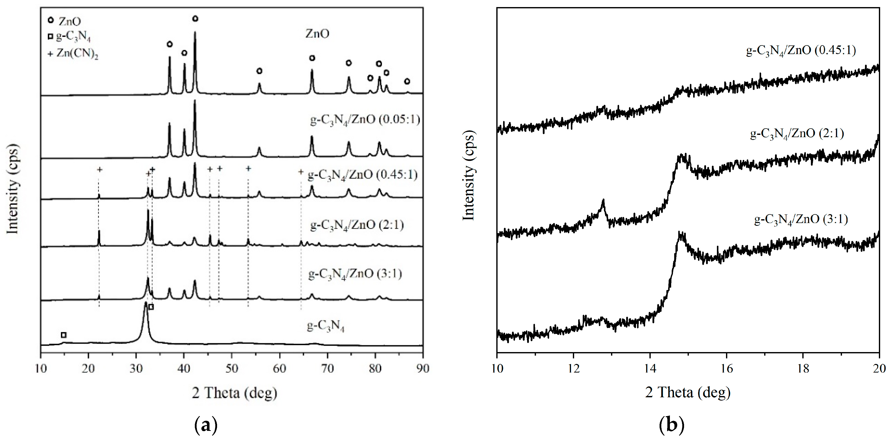
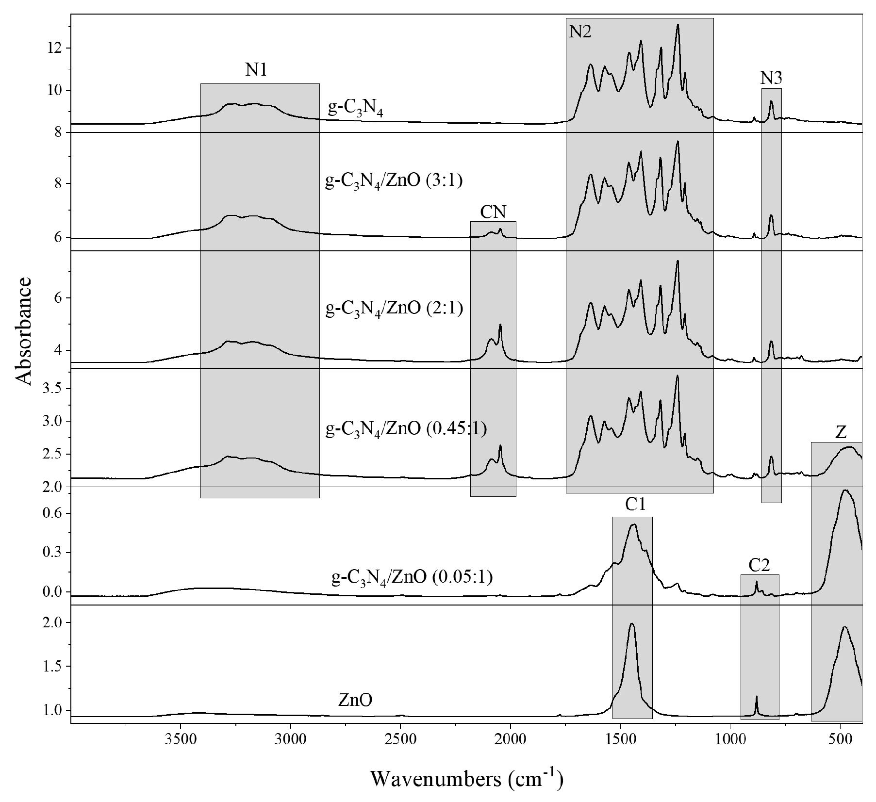
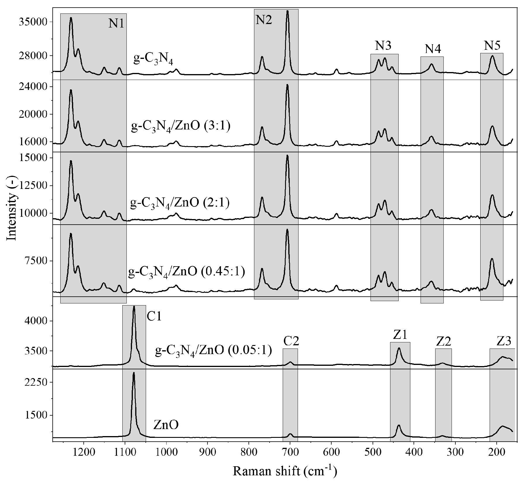
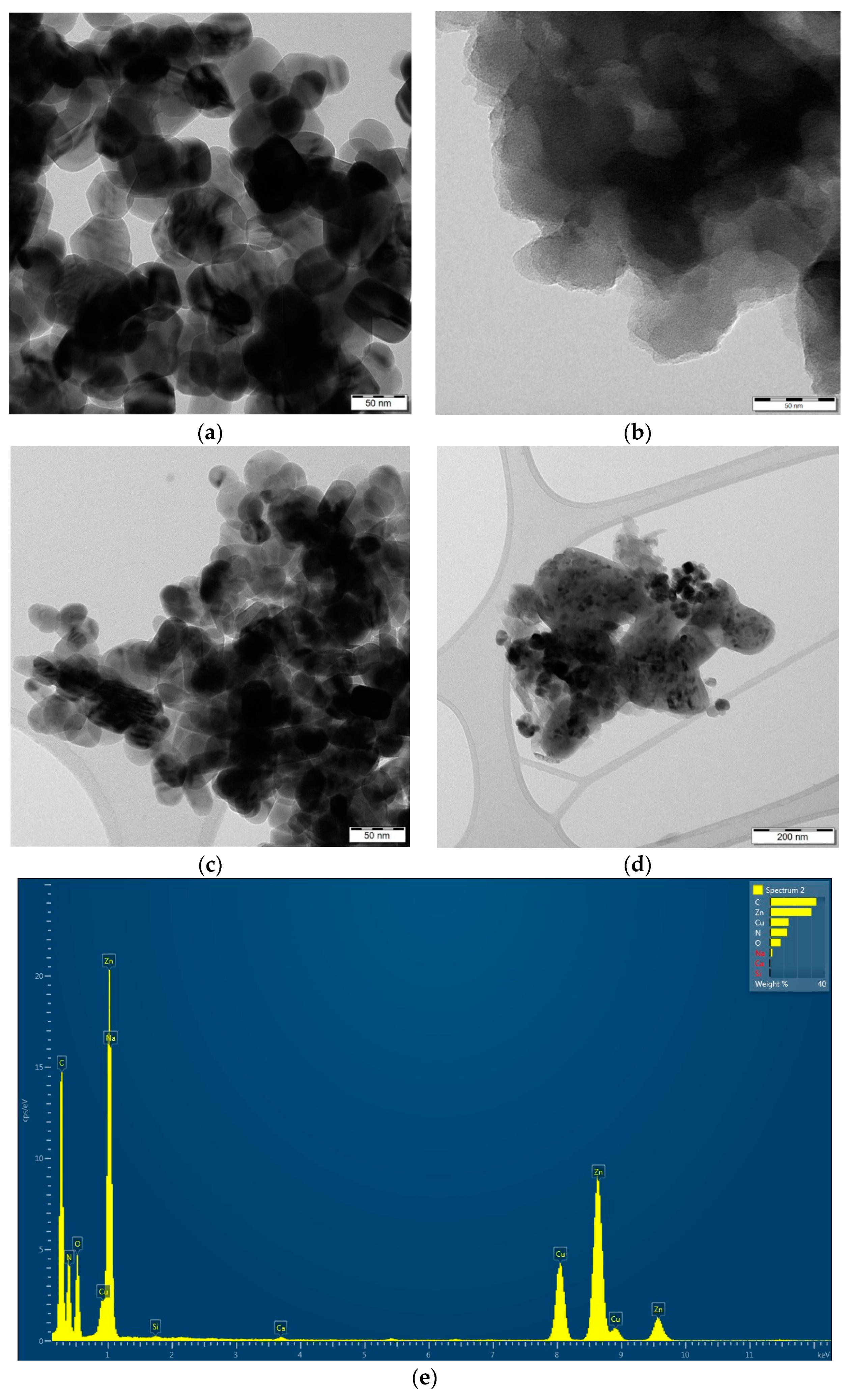
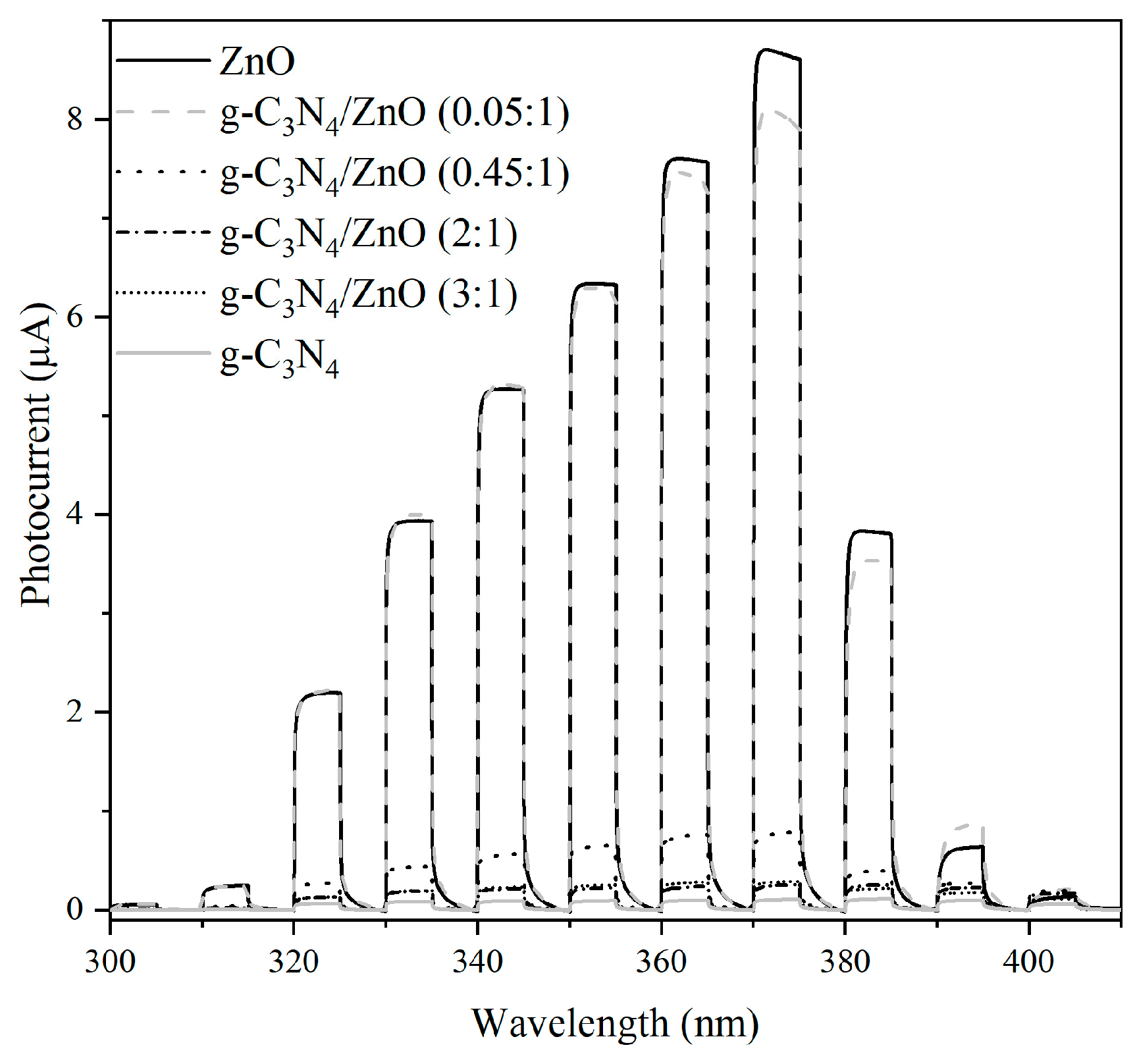
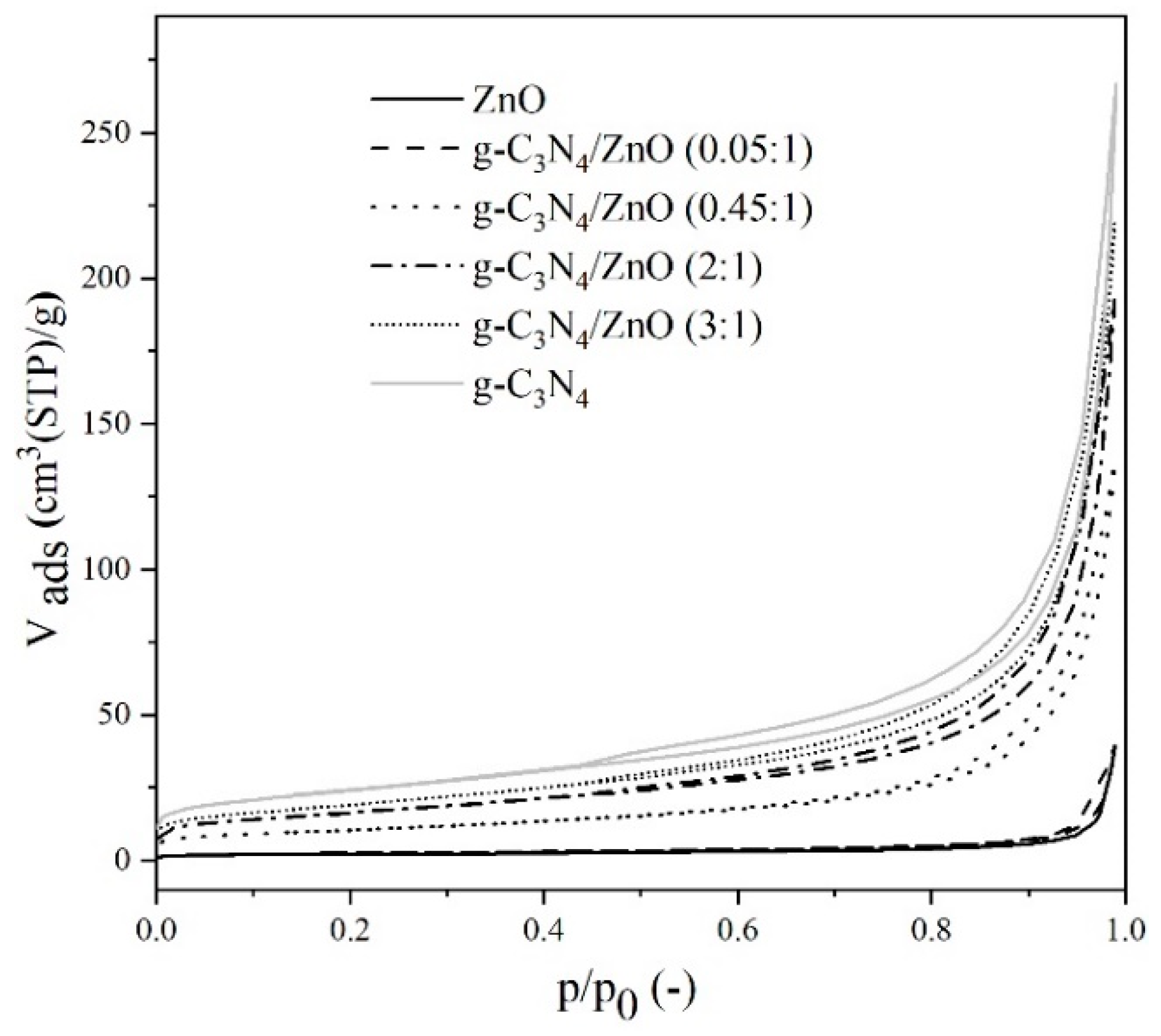
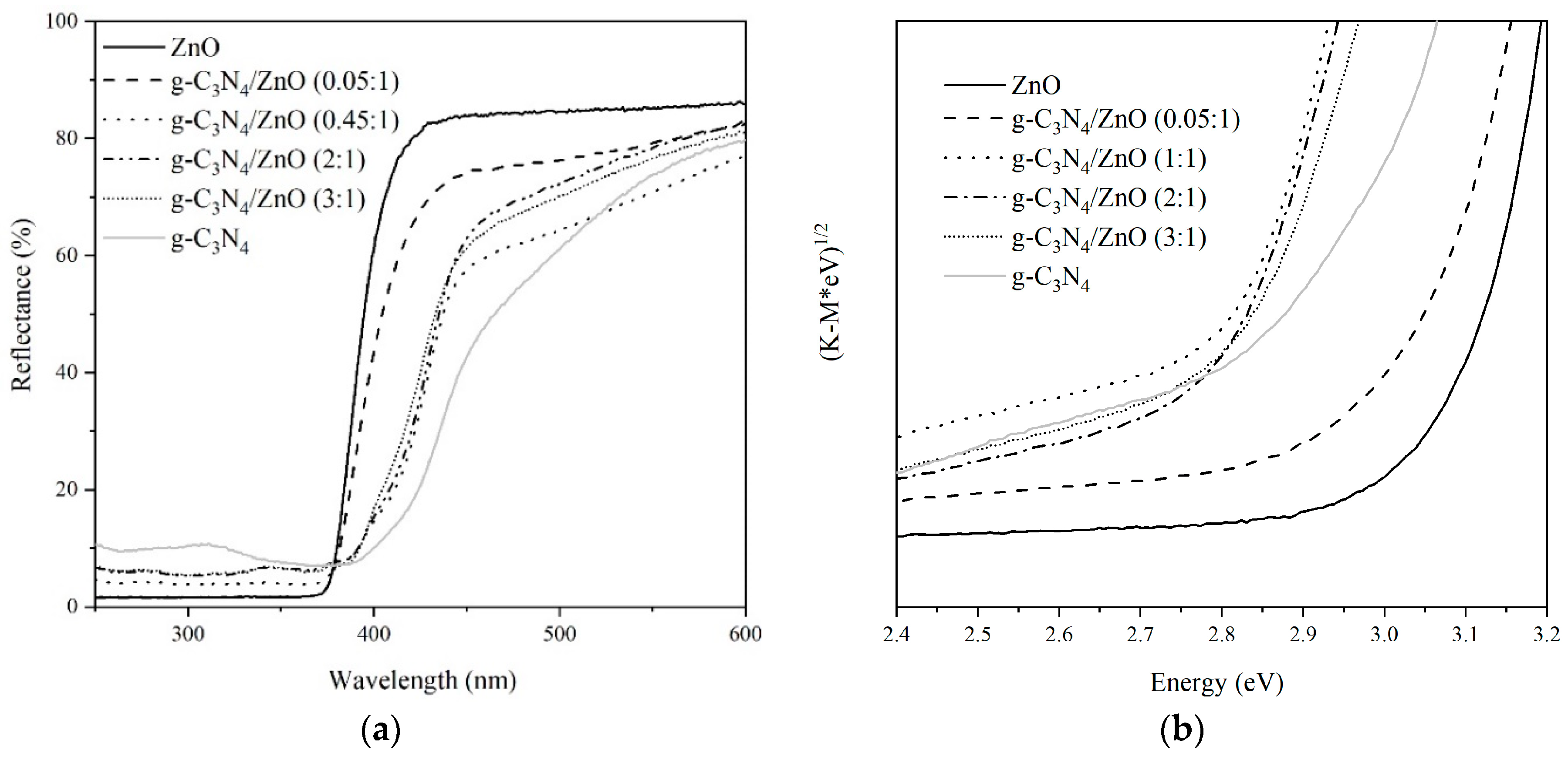
2.1. Photocatalysts Characterization

The real content of ZnO in prepared samples was determined by atomic absorption measurements (Table 1). The nitrogen physisorption characterization technique was used for the specific surface area determination (Figure 1). Each photocatalyst displayed a mesoporous-macroporous character. From the comparison of textural properties of the prepared samples, it is evident that the g-C3N4 addition positively affected the properties of the photocatalyst’s porous structure, significantly enhancing its specific surface area as well as the net pore volume, e.g., pure ZnO showed a specific surface area of 7 m2/g and a net pore volume of 61 mm3liq/g, while g-C3N4 /ZnO (3:1) showed a specific surface area of 68 m2/g and a net pore volume of 339 mm3liq/g (Table 1).
The X-ray powder diffraction (XRD) patterns of all investigated photocatalysts are shown in Figure 2. Two different phases based on zinc, zinc oxide, and zinc cyanamide were observed in all mixtures with higher contents of g-C3N4. The zinc oxide phase was markedly more significant than the zinc cyanide phase. Graphitic carbon nitride was confirmed by the presence of the reflection at position 14.9° 2Theta (d = 0.695 nm) indexed as the (100) plane and attributed to the in-plane tri-s-triazine units forming one-dimension melon strands. The most typical reflection was found around 32.2° 2Theta (d = 0.322 nm) and could be interpreted as long range interplanar stacking of the aromatic systems with the hkl plane (002). Unfortunately, this reflection was overlapped with the zinc cyanamide reflection (211) in all measured samples.
For the obtained g-C3N4/ZnO photocatalysts, nearly all of the characteristic diffraction peaks of zinc oxide were observed in the XRD pattern and confirmed the presence of the ZnO phase. In the case of the g-C3N4 phase, the (002) peak of g-C3N4 was almost not observed. The relatively poor crystallinity of the g-C3N4 phase indicates that simultaneous crystallization of the ZnO phase interferes with the condensation process and interlayer stacking patterns of the g-C3N4 phase, as shown by its weak diffraction intensity [21]. The XRD patterns in g-C3N4/ZnO photocatalysts indicate that these two phases occur separately in the composite and they do not have strong crystal lattice incorporation. This result was also confirmed by transmission electron microscopy (TEM).
All phases in the XRD patterns are marked by different symbols (Figure 2). All phases were identified using the PDF-2 database issued by the international center diffraction data. Zinc oxide corresponds to PDF-2 card no. 01-080-0075 and zinc cyanamide to PDF-2 card no. 01-070-4898. All XRD patterns were recorded using co-radiation with wavelength lambda = 1.78 Å. For this reason, all peaks were shifted to the higher 2Theta angles (based on the Braggs equation) than Cu radiation, which is more common.
The UV–vis diffuse reflectance spectra of g-C3N4, ZnO, and g-C3N4/ZnO photocatalysts are compared in Figure 3a. The band gaps’ energy values of the prepared nanocomposites were estimated by plotting the Kubelka–Munk function, as shown in Figure 3b. The largest band gap was 3.09 eV and it belongs to pure ZnO. On the other hand, pure g-C3N4 had a band gap energy of 2.64 eV. The nanocomposites band gap energies were 3.00, 2.75, 2.75, and 2.75 eV for g-C3N4/ZnO (0.05:1), g-C3N4/ZnO (0.45:1), g-C3N4/ZnO (2:1), and g-C3N4/ZnO (3:1), respectively. The presence of g-C3N4 in the nanocomposites extended the light absorption of the composites into the visible region compared to pure ZnO photocatalyst.
The infrared spectra are seen in Figure 4. The spectra of samples g-C3N4, g-C3N4/ZnO (3:1), g-C3N4/ZnO (2:1), and g-C3N4/ZnO (0.45:1) primarily contain bands that are characteristic for graphitic carbon nitride [22,23,24]. The regions are labelled as N1, N2, and N3. The region N1 contains bands at 3280, 3255, 3160, and 3090 cm−1. These bands can be assigned to the stretching vibration of N-H bonds. The region N2 contains intensive bands at 1680 (shoulder), 1635, 1575, 1545, 1460, 1430 (shoulder), 1410, 1335 (shoulder), 1320, 1280, (shoulder), 1240, and 1210 cm−1. Characteristic bands of the skeletal “breathing” vibration of aromatic rings of nitrogen heterocyclic compounds and stretching vibration bonds between carbon and nitrogen were found in the N2 region. The N3 region contains only a medium intensive band at 815 cm−1, which is due to the breathing mode of the triazine unit. Moreover, the spectra of samples g-C3N4/ZnO (3:1), g-C3N4/ZnO (2:1), and g-C3N4/ZnO (0.45:1) also contain bands of complex cyanide compounds at 2085 and 2045 cm−1 [25]. The bands have a medium or weak intensity (see region CN). The relative red shifts of the complex cyanide compounds compared with the normal wavenumber of 2250 cm−1 are due to the conjugation effect of the extended C-N aromatic system [21]. The spectra of samples g-C3N4/ZnO (0.05:1) and ZnO only contain medium or weak bands of carbonates (see region C1 and C2) and a broad band of zinc oxide (see region Z). The carbonate bands can be assigned to asymmetric stretching vibration of carbonate (1450 cm−1) and symmetric deformation vibration of carbonate (885 cm−1), respectively [25]. The band of vibration of zinc oxide [26] is seen at 480 cm−1. This band is seen also in the spectrum of sample g-C3N4/ZnO (0.45:1).
The Raman spectra are seen in Figure 5. The spectra are in the “full-scale” mode, thus the presented spectra have different scales of intensity axes. The spectra of samples g-C3N4, g-C3N4/ZnO (3:1), g-C3N4/ZnO (2:1), and g-C3N4/ZnO (0.45:1) only contain the bands characteristic for graphitic carbon nitride (i.e., [22,24,27]) in the regions labelled N1, N2, N3, N4, and N5. The N1 region contains strong bands at 1230 and 1215 cm−1 and weak bands at 1150 and 1115 cm−1. The N2 region contains a medium band at 770 cm−1 and a strong band at 705 cm−1. The N3 region contains two medium bands at 485 and 470 cm−1 and one weak band at 455 cm−1. Medium bands at 355 and 210 cm−1 are in the N4 and N5 regions, respectively. The assignment of the above-mentioned Raman bands of graphitic carbon nitride has not been published yet. We can suppose the bands are related to vibration of the tri-s-triazine ring (i.e., [27]). The spectra of samples g-C3N4/ZnO (0.05:1) and ZnO contain two bands of carbonate (see region C1 and C2) and three bands of zinc oxide (see region Z1, Z2, and Z3). The carbonate bands are presented at 1080 cm−1 (symmetric stretching vibration) and at 700 cm−1 (symmetric deformation vibration), respectively [25]. The bands characteristic for zinc oxide are presented at 435, 330, and 185 cm−1.
Figure 6a–d demonstrate the TEM images of pure ZnO, g-C3N4, and g-C3N4/ZnO (3:1) photocatalysts, respectively. The ZnO nanoparticles (Figure 6a) have hexagonal shapes and the particle size is about 50 nm while pure g-C3N4 (Figure 6b) shows a 2D lamellar structure. The TEM micrographs of g-C3N4/ZnO (3:1) (Figure 6c) testify that the ZnO nanoparticles were not supported well on the surface of g-C3N4. It is possible to infer that the salt (from preparation) can be observed in the g-C3N4/ZnO samples (Figure 6d). It is also evident from the results of the EDS analysis (Figure 6e), which confirmed not only the presence of Zn, C, O, and N elements but also Na element. The presence of the salt was also confirmed by Raman and infrared spectroscopy.
Photoelectrochemical measurements of all photocatalysts are shown in Figure 7. It is evident that the magnitude of the generated current decreased with an increasing amount of g-C3N4 presented at the photocatalysts until the g-C3N4 was in the majority. Higher photocurrents measured in the presence of the ZnO sample show that there are significantly more charge carriers produced in ZnO compared to g-C3N4. The photocurrent decreased with an increasing amount of g-C3N4, which formed a significantly smaller photocurrent.

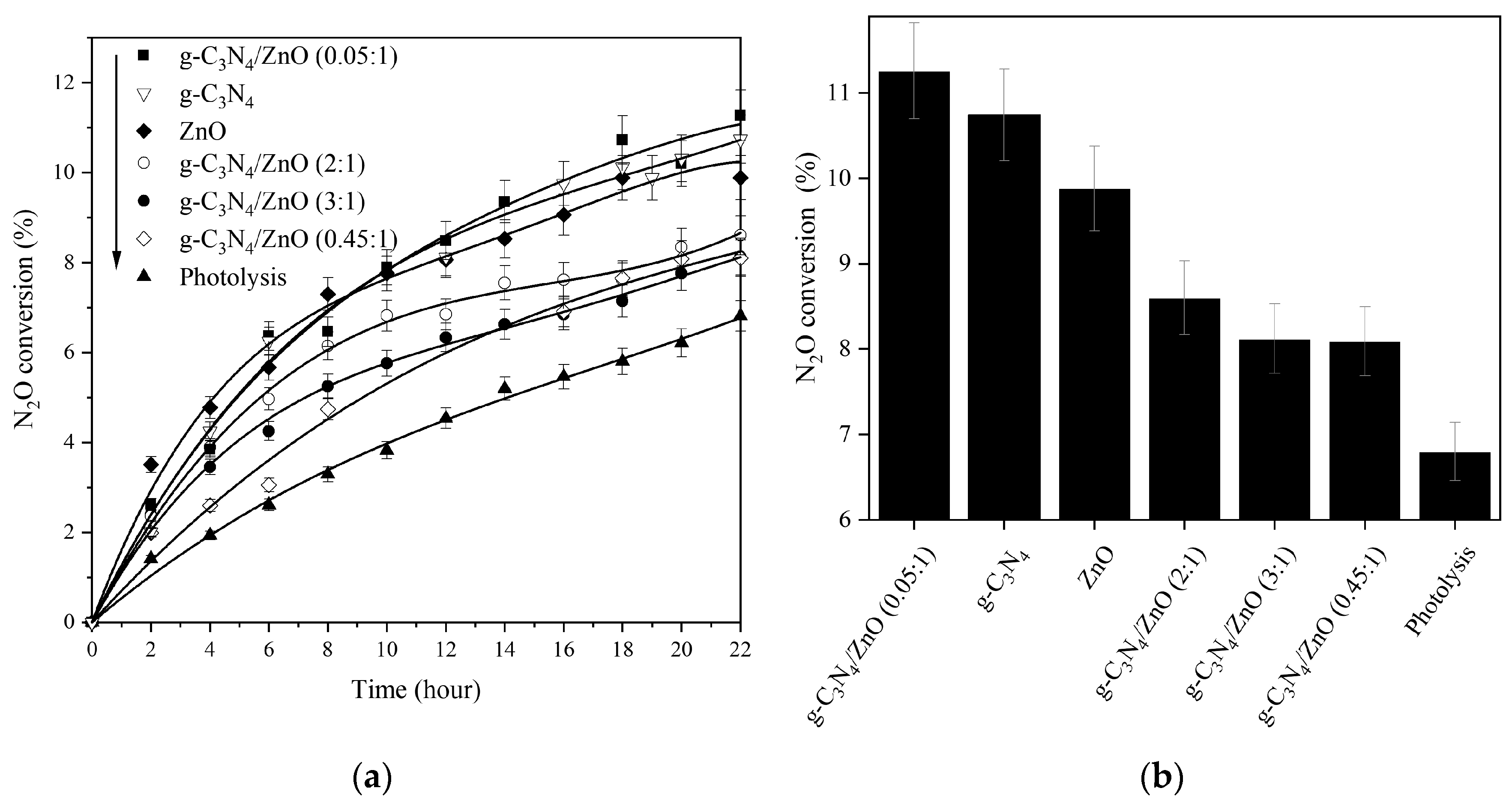
2.2. Photocatalytic Decomposition of N2O

The influence of prepared nanocomposites on the photocatalytic decomposition of nitrous oxide was studied from 0 to 22 h (Figure 8). The photolysis was also conducted in order to confirm the photocatalytic activity of the prepared samples. The time dependences of the nitrous oxide conversion of all g-C3N4/ZnO nanocomposites, including pure ZnO and g-C3N4 photocatalysts, are depicted in Figure 8. It is evident that each prepared photocatalyst proved to be much more effective in comparison with photolysis. The g-C3N4/ZnO (0.05:1) and pure g-C3N4 and ZnO showed a significantly higher efficiency than other photocatalysts.
The highest N2O conversion under UVA irradiation was reached over the g-C3N4/ZnO (0.05:1) photocatalyst. The N2O conversion in the presence of this photocatalyst was 11% after 22 h of UVA irradiation. For comparison, the N2O conversion without photocatalyst was only 6% after 22 h of UVA irradiation. The same foam with photocatalyst was used for repeated tests three times. This confirms the stability and ability to reuse these materials without losing photocatalytic activity.
It is hard to compare the observed photocatalytic results with the literature, as it is usually measured under different reaction conditions (geometry of photoreactor, irradiation source, concentration of N2O, amount of photocatalyst, whether it is immobilized, and also the irradiation time). Each of these conditions influences the photocatalytic decomposition of N2O in a different way and therefore accurate comparison is impossible. Also, not many research groups have investigated the photocatalytic decomposition of N2O. Therefore, the obtained results were compared with our previous works. N2O conversion for the g-C3N4/BiVO4 and g-C3N4/WO3 composites after 20 h of irradiation was 12.5% and 14.5%, respectively. However, both previous sets of photocatalysts were measured in a different photoreactor (different geometry) and therefore, from that point of view, the observed N2O conversion of approximately 10% after 20 h of irradiation seems to be comparable to the previous results.

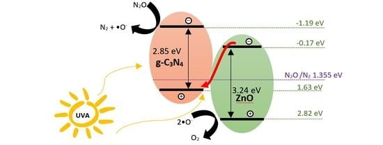
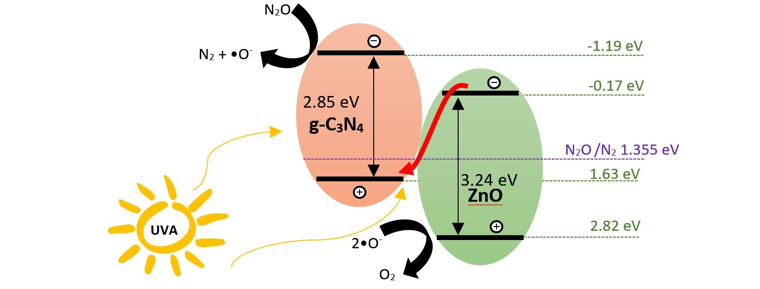
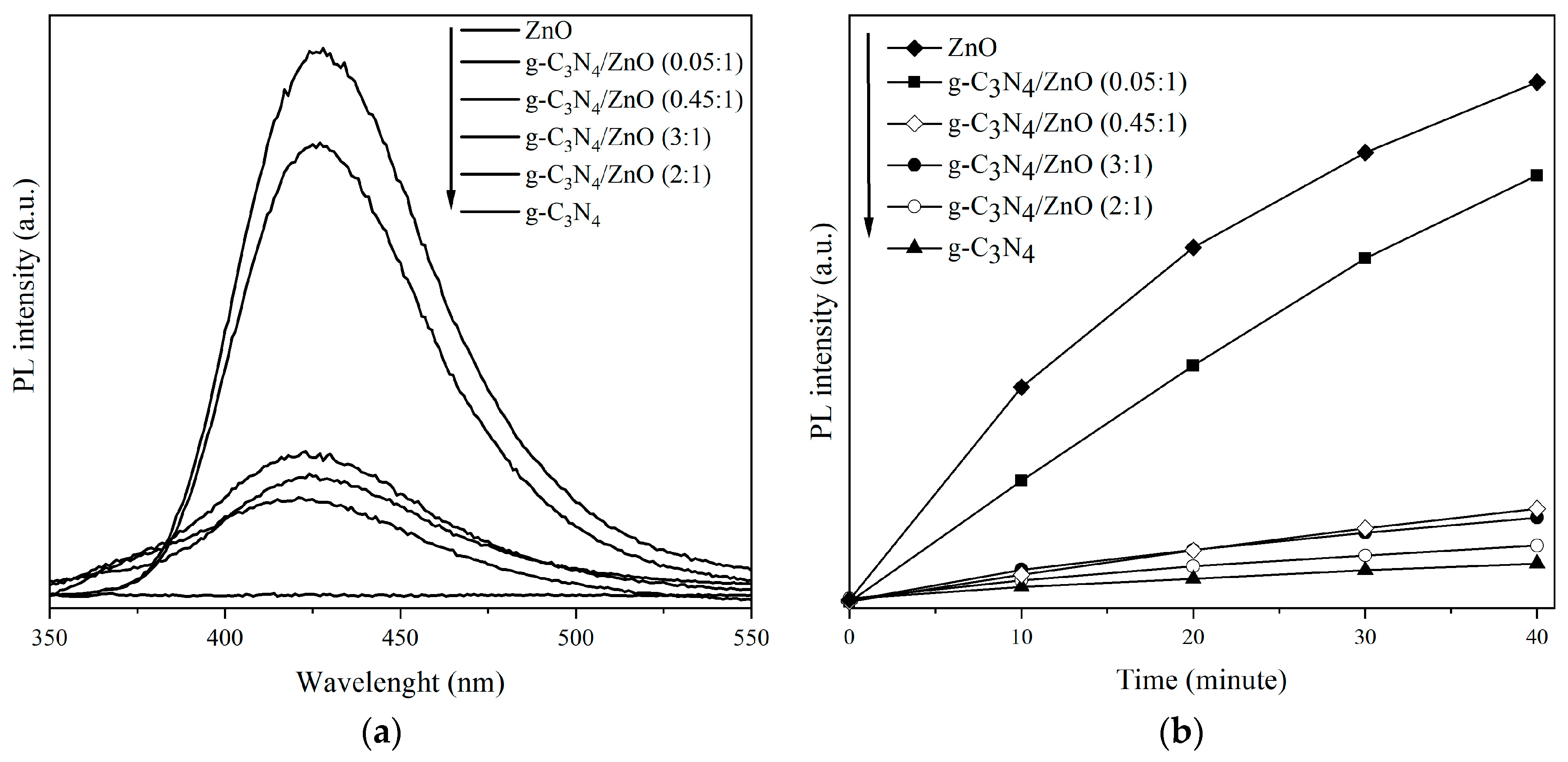
2.3. Proposed Mechanism for Photocatalytic Activity Enhancement

When g-C3N4 is combined with ZnO, the generated electrons (e) and holes (h+) can migrate by two possible mechanisms: Type II heterojunction and the Z-scheme system. In the case of the type II heterojunction of g-C3N4/ZnO, the generated e- in the conduction band (CB) of g-C3N4 can transfer to the CB of ZnO and simultaneously, the h+ in the valence band (VB) of ZnO can migrate to the VB of g-C3N4. In the case of the Z-scheme system of g-C3N4/ZnO, e in the CB of ZnO are directly recombined with the h+ in the VB of g-C3N4 at the heterojunction interface, and so the recombination of e and h+ in ZnO and g-C3N4 is inhibited. In this case, the e- are accumulated in the CB of g-C3N4 and h+ in the VB of ZnO. Due to these two mechanisms, a prolonged lifetime of electrons and holes is gained [14].
The band edge positions of materials were calculated via Equations (2) and (3) [28]:
E V B = χ E 0 + 0.5 E g ,
E C B = E V B E g ,
where χ is the electronegativity of the material, EVB is the VB potential, ECB is the CB potential, Eg is the band gap energy of a material, and E0 is the energy of free electrons (4.5 V vs. NHE). The χ values for g-C3N4 and ZnO are 4.72 and 5.95 eV, respectively [28]. The EVB of g-C3N4 and ZnO were 1.75 and 3.05 eV, respectively. The ECB of g-C3N4 and ZnO were −1.31 and −0.15 V, respectively.
The formation of the Z-scheme system of g-C3N4/ZnO can be confirmed by the hydroxyl radical (•OH) trapping test using modified photoluminescence (PL) measurements [29]. After the photoexcitation, the h+ with sufficient oxidation potential can be either directly involved in the photocatalytic degradation reactions or generate active species. The reaction between terephthalic acid (TA) and hydroxyl radicals (•OH) can generate hydroxyterephthalic acid (HTA), which is detectable by the PL spectrometer (signal with maximal intensity centered at 425 nm) [30]. The PL peak intensity of the HTA is in proportion to the amount of produced •OH.
Since the photoluminescence intensity increased in the presence of each photocatalyst over time, with the exception of pure g-C3N4, the presence of hydroxyl radicals was confirmed (Figure 9a). According to the band structures of ZnO and g-C3N4, the photoinduced h+ on g-C3N4 cannot oxidize the adsorbed H2O molecules to •OH because the VB potential of g-C3N4 (+1.75 V vs. NHE) is less positive than the standard redox potential of •OH/OH (2.4 V vs. NHE). In the case of Z-scheme formation, the photogenerated h+ remain on the ZnO. The h+ accumulation on ZnO is in accordance with the migration mechanism of the direct Z-scheme. The creation of the Z-scheme system was observed by Wang et al. [14], Liu et al. [31], and Yu et. al. [21]. In our case, it was not confirmed that g-C3N4/ZnO nanocomposite had contact with the Z-scheme system because the nanocomposites were not proven to have a higher PL intensity than pure ZnO. The PL intensity decreased with an increasing content of ZnO, which can produce hydroxyl radical.
According to the literature, the standard reduction potential of N2O/N2 is +1.355 V [32]. The conduction bands’ potentials of both pure and prepared photocatalysts (g-C3N4 and ZnO) are more negative (−1.19 and −0.17 V, respectively) than the standard reduction potential of N2O/N2. Therefore, both pure photocatalysts showed similar photocatalytic activity.
The photocatalytic activity of g-C3N4/ZnO, with the exception of g-C3N4/ZnO (0.05:1), was lower with regard to pure photocatalysts. These three nanocomposites (g-C3N4/ZnO (0.45:1), g-C3N4/ZnO (2:1), and g-C3N4/ZnO (3:1)) had not only similar photocatalytic activity but also band gap energy. The photocatalytic activity of nanocomposites (g-C3N4/ZnO (0.45:1), g-C3N4/ZnO (2:1), and g-C3N4/ZnO (3:1)) decreased both by the presence of cyanide compounds formed during preparation (Figure 4) and by the imperfect contact of g-C3N4 and ZnO, which is visible in Figure 6c. The detection of cyanide compounds in g-C3N4/ZnO indicates that simultaneous ZnO crystallization could interfere with the thermal polymerization of melamine to generate a well-defined g-C3N4 phase. Based on the previous discussion about XRD patterns and infrared spectra, such interference in ZnO crystallization can result in relatively poor crystallinity and give rise to defect states within the g-C3N4 phase [21]. These defect states can catch generated charge carriers; however, they are not accessible for the adsorbed molecule of nitrous oxide, which consequently leads to a decrease in photoactivity.
A slightly different situation is the case of g-C3N4/ZnO (0.05:1), which proved to have the highest photocatalytic activity. This nanocomposite contained only 4% of g-C3N4, which means most of the holes in VB of g-C3N4 probably recombined with some of the electrons from the CB of ZnO. In the meantime, the photogenerated electrons in the CB of g-C3N4 and a big part of the electrons in the CB of ZnO, which was not used for recombination with holes of g-C3N4, reduced N2O to N2 and •O. The photoexcited holes in the VB of ZnO oxidized •O to O2 [19]. This result shows that only a small loading of g-C3N4 in g-C3N4/ZnO can increase the effectiveness of photogenerated electron–hole separation.
In heterogeneous photocatalysis, on the contrary to catalysis, the specific surface (SBET) is not the most crucial parameter. It is obvious in this work, where g-C3N4/ZnO (0.05:1) and g-C3N4 had a similarly high photoactivity though the SBET of g-C3N4 was more than 10 times higher than in the case of g-C3N4/ZnO (0.05:1). Many authors have quoted the band gap energy as one of the most important parameters. The photoelectrochemical measurements validated the capability of all nanocomposites to absorb UVA. Moreover, both pure photocatalysts, which had similar photoactivity, have different band gap energy. Consequently, the band gap energy is not the most decisive parameter. The most significant factor, which is valid in photocatalytic reactions generally, is the position and potential of the valence and conduction band.

3. Materials and Methods

3.1. Preparation of ZnO, g-C3N4, and g-C3N4/ZnO Nanocomposites

ZnO was prepared by precipitation [33]. G-C3N4 was formed by heating of melamine [18,34]. In a synthesis procedure of g-C3N4/ZnO, the determined amounts of ZnO and g-C3N4 were dispersed in deionized water and evenly mixed (4 h). Subsequently, the mixture was dried (at 60 °C in air) and calcined [18]. The weight ratios of prepared g-C3N4/ZnO photocatalysts were set at 0.05:1, 0.45:1, 2:1, and 3:1, respectively. The prepared nanocomposites (0.1 g) were anchored on a commercial support VUKOPOR (Lanik Ltd., Boskovice, Czech Republic) [35,36]. For detailed preparation information, see the Supplementary Materials.

3.2. Characterization of g-C3N4/ZnO Nanocomposite and Photocatalytic Decomposition of N2O

The products were characterized in detail by N2 physisorption (Micromeritics), X-ray powder diffraction (XRD, Rigaku, Tokyo, Japan), UV–Vis diffuse reflectance spectra (DRS, IRS-2600Plus), transmission electron microscopy (TEM, JEOL 2100 equipped with an EDS detector, Tokyo, Japan), Fourier transform infrared spectroscopy (FT-IR, Nexus 470 ThermoScientific, Waltham, MA, USA), Raman spectroscopy (DXR SmartRaman (ThermoScientific), photoluminescence (PL) measurements (FLSP920 Series, Edinburgh Instruments, Livingston, UK), and photoelectrochemical measurements (Instytut Fotonowy, Krakow, Poland), and their photocatalytic activity was studied for the photocatalytic decomposition of N2O according to the reported method [18,29]. The photocatalytic inactivity of the bare foam was confirmed via blank tests and compared to the blank test without foam.

4. Conclusions

g-C3N4/ZnO nanocomposites with different ratios of components as well as pure ZnO and g-C3N4 were prepared with success. Each photocatalyst was coated on commercial Al2O3 foam. The prepared nanocomposites were investigated for N2O photocatalytic decomposition for the first time. g-C3N4 addition positively affects the properties of the photocatalysts’ porous structure, significantly enhancing its specific surface area as well as the net pore volume. In all mixtures with higher contents of g-C3N4, two phases based on zinc were observed, -zinc oxide (markedly more significant) and zinc cyanamide. ZnO nanoparticles have hexagonal shapes and pure g-C3N4 shows a 2D lamellar structure. Based on the experimental and characterization results, N2O conversion in the presence of photocatalysts was higher in comparison with photolysis; in the case of the best photocatalysts, g-C3N4/ZnO (0.05:1) was almost twice higher. FT-IR characterization showed that samples of the g-C3N4/ZnO (3:1), g-C3N4/ZnO (2:1), and g-C3N4/ZnO (0.45:1) photocatalysts also contained cyanide compounds, which together with an imperfect contact of g-C3N4 and ZnO caused a decrease of photocatalytic activity. The key role played in photocatalytic activity was the electron–hole separation and the position and potential of the valence and conduction band. On the other hand, the specific surface area and band gap energy were not significant factors in N2O photocatalytic decomposition. The immobilization on commercial Al2O3 foam proved to be promising. A short-term test confirmed the photocatalysts’ stability and reproducible activity. Immobilization might be an efficient way to bring photocatalysis closer to an application, since the powder form is not usable for several reasons, such as filtration from the liquid phase or blowing off by the gas stream. No such drawbacks exist with the immobilized photocatalyst on the foam.

Supplementary Materials

The experiment parts are available online at https://www.mdpi.com/2073-4344/9/9/735/s1.

Author Contributions

Validation, K.K., M.R. and M.Š.; analysis, J.P., M.Š., T.B. and M.R.; preparation of photocatalyst, I.T.; writing—original draft preparation, K.K.; writing—review and editing, M.R.; visualization, M.R.; supervision, P.P.

Funding

This research was funded by ERDF “Institute of Environmental Technology–Excellent Research” (No. CZ.02.1.01/0.0/0.0/16_019/0000853), the Grant Agency of the Czech Republic (No. 16-10527S) and by using Large Research Infrastructure ENREGAT supported by the Ministry of Education, Youth and Sports of the Czech Republic under project No. LM2018098.

Acknowledgments

The work was supported from ERDF “Institute of Environmental Technology—Excellent Research” (No. CZ.02.1.01/0.0/0.0/16_019/0000853), the Grant Agency of the Czech Republic (No. 16-10527S) and by using Large Research Infrastructure ENREGAT supported by the Ministry of Education, Youth and Sports of the Czech Republic under project No. LM2018098“. Authors thank Alexandr Martaus (IET, VŠB-TU Ostrava) for the XRD measurements.

Conflicts of Interest

The authors declare no conflict of interest.

References

  1. Ming, T.; de Richter, R.; Shen, S.; Caillol, S. Fighting global warming by greenhouse gas removal: Destroying atmospheric nitrous oxide thanks to synergies between two breakthrough technologies. Environ. Sci. Pollut. Res. Int. 2016, 23, 6119–6138. [Google Scholar] [CrossRef] [PubMed]
  2. Matsuoka, M.; Ju, W.S.; Takahashi, K.; Yamashita, H.; Anpo, M. Photocatalytic decomposition of N2O into N2 and O2 at 298 K on Cu(I) ion catalysts anchored onto various oxides. The effect of the coordination state of the Cu(I) ions on the photocatalytic reactivity. J. Phys. Chem. B 2000, 104, 4911–4915. [Google Scholar] [CrossRef]
  3. Obalová, L.; Šihor, M.; Praus, P.; Reli, M.; Kočí, K. Photocatalytic and photochemical decomposition of N2O on ZnS-MMT catalyst. Catal. Today 2014, 230, 61–66. [Google Scholar] [CrossRef]
  4. Kočí, K.; Matějová, L.; Obalová, L.; Čapek, L.; Wu, J.C.S. Preparation, characterization and photocatalytic performance of TiO2 prepared by using pressurized fluids in CO2 reduction and N2O decomposition. J. Sol Gel Sci. Technol. 2015, 76, 621–629. [Google Scholar] [CrossRef]
  5. Sano, T.; Negishi, N.; Mas, D.; Takeuchi, K. Photocatalytic Decomposition of N2O on Highly Dispersed Ag+ Ions on TiO2 Prepared by Photodeposition. J. Catal. 2000, 194, 71–79. [Google Scholar] [CrossRef]
  6. Kočí, K.; Krejčíková, S.; Šolcová, O.; Obalová, L. Photocatalytic decomposition of N2O on Ag-TiO2. Catal. Today 2012, 191, 134–137. [Google Scholar] [CrossRef]
  7. Obalová, L.; Reli, M.; Lang, J.; Matějka, V.; Kukutschová, J.; Lacný, Z.; Kočí, K. Photocatalytic decomposition of nitrous oxide using TiO2 and Ag-TiO2 nanocomposite thin films. Catal. Today 2013, 209, 170–175. [Google Scholar] [CrossRef]
  8. Matějová, L.; Šihor, M.; Brunátová, T.; Ambrožová, N.; Reli, M.; Čapek, L.; Obalová, L.; Kočí, K. Microstructure-performance study of cerium-doped TiO2 prepared by using pressurized fluids in photocatalytic mitigation of N2O. Res. Chem. Intermed. 2015, 41, 9217–9231. [Google Scholar] [CrossRef]
  9. Kočí, K.; Matějová, L.; Ambrožová, N.; Šihor, M.; Troppová, I.; Čapek, L.; Kotarba, A.; Kustrowski, P.; Hospodková, A.; Obalová, L. Optimization of cerium doping of TiO2 for photocatalytic reduction of CO2 and photocatalytic decomposition of N2O. J. Sol Gel Sci. Technol. 2016, 78, 550–558. [Google Scholar] [CrossRef]
  10. Prasad, C.; Tang, H.; Bahadur, I. Graphitic carbon nitride based ternary nanocomposites: From synthesis to their applications in photocatalysis: A recent review. J. Mol. Liq. 2019, 281, 634–654. [Google Scholar] [CrossRef]
  11. Yin, S.; Han, J.; Zhou, T.; Xu, R. Recent progress in g-C3N4 based low cost photocatalytic system: Activity enhancement and emerging applications. Catal. Sci. Technol. 2015, 5, 5048–5061. [Google Scholar] [CrossRef]
  12. Masih, D.; Ma, Y.; Rohani, S. Graphitic C3N4 based noble-metal-free photocatalyst systems: A review. Appl. Catal. B Environ. 2017, 206, 556–588. [Google Scholar] [CrossRef]
  13. Wen, J.; Xie, J.; Chen, X.; Li, X. A review on g-C3N4-based photocatalysts. Appl. Surf. Sci. 2017, 391, 72–123. [Google Scholar] [CrossRef]
  14. Wang, J.; Xia, Y.; Zhao, H.; Wang, G.; Xiang, L.; Xu, J.; Komarneni, S. Oxygen defects-mediated Z-scheme charge separation in g-C3N4/ZnO photocatalysts for enhanced visible-light degradation of 4-chlorophenol and hydrogen evolution. Appl. Catal. B 2017, 206, 406–416. [Google Scholar] [CrossRef]
  15. Low, J.; Cheng, B.; Yu, J. Surface modification and enhanced photocatalytic CO2 reduction performance of TiO2: A review. Appl. Surf. Sci. 2017, 392, 658–686. [Google Scholar] [CrossRef]
  16. Bai, S.; Jiang, J.; Zhang, Q.; Xiong, Y. Steering charge kinetics in photocatalysis: Intersection of materials syntheses, characterization techniques and theoretical simulations. Chem. Soc. Rev. 2015, 44, 2893–2939. [Google Scholar] [CrossRef] [PubMed]
  17. Reli, M.; Huo, P.; Sihor, M.; Ambrozova, N.; Troppova, I.; Matejova, L.; Lang, J.; Svoboda, L.; Kustrowski, P.; Ritz, M.; et al. Novel TiO2/C3N4 Photocatalysts for Photocatalytic Reduction of CO2 and for Photocatalytic Decomposition of N2O. J. Phys. Chem. A 2016, 120, 8564–8573. [Google Scholar] [CrossRef] [PubMed]
  18. Kočí, K.; Reli, M.; Troppová, I.; Šihor, M.; Kupková, J.; Kustrowski, P.; Praus, P. Photocatalytic decomposition of N2O over TiO2/g-C3N4 photocatalysts heterojunction. Appl. Surf. Sci. 2017, 396, 1685–1695. [Google Scholar] [CrossRef]
  19. Reli, M.; Svoboda, L.; Šihor, M.; Troppová, I.; Pavlovský, J.; Praus, P.; Kočí, K. Photocatalytic decomposition of N2O over g-C3N4/WO3 photocatalysts. Environ. Sci. Pollut. Res. Int. 2018, 25, 34839–34850. [Google Scholar] [CrossRef]
  20. Alenzi, N.; Liao, W.-S.; Cremer, P.S.; Sanchez-Torres, V.; Wood, T.K.; Ehlig-Economides, C.; Cheng, Z. Photoelectrochemical hydrogen production from water/methanol decomposition using Ag/TiO2 nanocomposite thin films. Int. J. Hydrogen Energy 2010, 35, 11768–11775. [Google Scholar] [CrossRef]
  21. Yu, W.; Xu, D.; Peng, T. Enhanced photocatalytic activity of g-C3N4 for selective CO2 reduction to CH3OH via facile coupling of ZnO: A direct Z-scheme mechanism. J. Mater. Chem. A 2015, 3, 19936–19947. [Google Scholar] [CrossRef]
  22. Komatsu, T. The First Synthesis and Characterization of Cyameluric High Polymers. Macromol. Chem. Phys. 2001, 202, 19–25. [Google Scholar] [CrossRef]
  23. Wu, P.; Wang, J.; Zhao, J.; Guo, L.; Osterloh, F.E. Structure defects in g-C3N4 limit visible light driven hydrogen evolution and photovoltage. J. Mater. Chem. A 2014, 2, 20338–20344. [Google Scholar] [CrossRef]
  24. Papailias, I.; Giannakopoulou, T.; Todorova, N.; Demotikali, D.; Vaimakis, T.; Trapalis, C. Effect of processing temperature on structure and photocatalytic properties of g-C3N4. Appl. Surf. Sci. 2015, 358, 278–286. [Google Scholar] [CrossRef]
  25. Horák, M.; Papoušek, D. Infrared Spectra and Structure of Molecules; Academia: Prague, Czech Republic, 1976. [Google Scholar]
  26. Hariharan, C. Photocatalytic degradation of organic contaminants in water by ZnO nanoparticles: Revisited. Appl. Catal. A 2006, 304, 55–61. [Google Scholar] [CrossRef]
  27. Zinin, P.V.; Ming, L.-C.; Sharma, S.K.; Khabashesku, V.N.; Liu, X.; Hong, S.; Endo, S.; Acosta, T. Ultraviolet and near-infrared Raman spectroscopy of graphitic C3N4 phase. Chem. Phys. Lett. 2009, 472, 69–73. [Google Scholar] [CrossRef]
  28. Kuang, P.-Y.; Su, Y.-Z.; Chen, G.-F.; Luo, Z.; Xing, S.-Y.; Li, N.; Liu, Z.-Q. g-C3N4 decorated ZnO nanorod arrays for enhanced photoelectrocatalytic performance. Appl. Surf. Sci. 2015, 358, 296–303. [Google Scholar] [CrossRef]
  29. Troppová, I.; Šihor, M.; Reli, M.; Ritz, M.; Praus, P.; Kočí, K. Unconventionally prepared TiO2/g-C3N4 photocatalysts for photocatalytic decomposition of nitrous oxide. Appl. Surf. Sci. 2018, 430, 335–347. [Google Scholar] [CrossRef]
  30. Li, J.; Zhang, M.; Li, Q.; Yang, J. Enhanced visible light activity on direct contact Z-scheme g-C3N4-TiO2 photocatalyst. Appl. Surf. Sci. 2017, 391, 184–193. [Google Scholar] [CrossRef]
  31. Liu, Y.; Wang, R.; Yang, Z.; Du, H.; Jiang, Y.; Shen, C.; Liang, K.; Xu, A. Enhanced visible-light photocatalytic activity of Z-scheme graphitic carbon nitride/oxygen vacancy-rich zinc oxide hybrid photocatalysts. Chin. J. Catal. 2015, 36, 2135–2144. [Google Scholar] [CrossRef]
  32. Berks, B.C.; Ferguson, S.J.; Moir, J.W.; Richardson, D.J. Enzymes and associated electron transport systems that catalyse the respiratory reduction of nitrogen oxides and oxyanions. Biochim. Biophys. Acta 1995, 1232, 97–173. [Google Scholar] [CrossRef] [Green Version]
  33. Troppová, I.; Matějová, L.; Sezimová, H.; Matěj, Z.; Peikertová, P.; Lang, J. Nanostructured TiO2 and ZnO prepared by using pressurized hot water and their eco-toxicological evaluation. J. Nanopart. Res. 2017, 19, 198. [Google Scholar] [CrossRef]
  34. Koci, K.; Reli, M.; Svoboda, L.; Praus, P. In Exfoliated nanosheets of graphitic carbon nitride: Study of optical and photoelectrochemical properties. In Proceedings of the 8th International Conference on Nanomaterials—Research & Application (NANOCON 2016), Brno, Czech Republic, 19–21 October 2016; Tanger Ltd.: Brno, Czech Republic, 2017; pp. 98–103. [Google Scholar]
  35. Tasbihi, M.; Kočí, K.; Edelmannová, M.; Troppová, I.; Reli, M.; Schomäcker, R. Pt/TiO2 photocatalysts deposited on commercial support for photocatalytic reduction of CO2. J. Photochem. Photobiol. A 2018, 366, 72–80. [Google Scholar] [CrossRef]
  36. Reli, M.; Troppová, I.; Šihor, M.; Pavlovský, J.; Praus, P.; Kočí, K. Photocatalytic decomposition of N2O over g-C3N4/BiVO4 composite. Appl. Surf. Sci. 2019, 469, 181–191. [Google Scholar] [CrossRef]
Figure 1. Nitrogen adsorption/desorption isotherms of ZnO, g-C3N4, and g-C3N4/ZnO.
Catalysts 09 00735 g001
Figure 2. XRD patterns of ZnO, g-C3N4, and g-C3N4/ZnO (a) and XRD fragment of g-C3N4/ZnO (0.45:1; 2:1, and 3:1) photocatalysts (b).
Catalysts 09 00735 g002
Figure 3. UV-vis diffuse reflectance spectra (a) and Tauc plots (b) of ZnO, g-C3N4, and g-C3N4/ZnO.
Catalysts 09 00735 g003
Figure 4. Infrared spectra of ZnO, g-C3N4, and g-C3N4/ZnO.
Catalysts 09 00735 g004
Figure 5. Raman spectra of ZnO, g-C3N4, and g-C3N4/ZnO.
Catalysts 09 00735 g005
Figure 6. TEM images of ZnO (a), g-C3N4 (b), and g-C3N4/ZnO (3:1) (c,d) and EDS analysis of g-C3N4/ZnO (3:1) (e).
Catalysts 09 00735 g006
Figure 7. Photocurrent generation at electrodes made of ZnO, g-C3N4, and g-C3N4/ZnO recorded at 1 V vs. Ag/AgCl in deoxygenated 0.1 M KNO3.
Catalysts 09 00735 g007
Figure 8. The time dependences of N2O conversion (a) and N2O conversion after 22 h of irradiation (b) over ZnO, g-C3N4 and g-C3N4/ZnO and photolysis under UVA (λ = 365 nm) irradiation.
Catalysts 09 00735 g008
Figure 9. PL spectra in presence of TA in NaOH solution (excitation wave: 365 nm), irradiated for 40 min in the presence of ZnO, g-C3N4, and g-C3N4/ZnO (a) and a comparison of the PL intensity against irradiation time for of g-C3N4/ZnO nanocomposites (b).
Catalysts 09 00735 g009
Table 1. Structural and microstructural properties and content of ZnO ZnO, g-C3N4, and g-C3N4/ZnO.
PhotocatalystsSBET (m2/g)V net (mm3lig/g)Content of Zn2+ (wt.%)
ZnO76180
g-C3N4/ZnO (0.05:1)86277
g-C3N4/ZnO (0.45:1)3721255
g-C3N4/ZnO (2:1)5829826
g-C3N4/ZnO (3:1)6833921
g-C3N4854130

