Article

Carbon Self-Doped Carbon Nitride Nanosheets with Enhanced Visible-Light Photocatalytic Hydrogen Production

1
School of Environmental Science and Engineering, Qingdao University, Qingdao 266071, China
2
School of Environment and Safety Engineering, Qingdao University of Science and Technology, 53 Zhengzhou Road, Qingdao 266042, China
*
Author to whom correspondence should be addressed.
Catalysts 2018, 8(9), 366; https://doi.org/10.3390/catal8090366
Submission received: 29 July 2018 / Revised: 26 August 2018 / Accepted: 27 August 2018 / Published: 29 August 2018
(This article belongs to the Special Issue Heterogeneous Catalysis for Energy Conversion)

Abstract

:
In this study, we prepared carbon self-doped carbon nitride nanosheets through a glucose synergic co-condensation method. In the carbon self-doped structure, the N atoms in the triazine rings were substituted by C atoms, resulting in enhanced visible-light photocatalytic hydrogen production, which is three-times higher than that of bulk carbon nitride. The enhanced photocatalytic hydrogen production was attributed to the higher charge-carrier transfer rate and widened light absorption range of the carbon nitride nanosheets after carbon self-doping. Thus, this work highlights the importance of carbon self-doping for improving the photocatalytic performance. Meanwhile, it provides a feasible method for the preparation of carbon self-doped carbon nitride without destroying the 2D conjugated backbone structures.

Graphical Abstract

1. Introduction

Environmental pollution and energy crises have been seriously threatening the survival of mankind and the sustainable development of society. The photocatalysis technique is considered as an environmentally friendly technology to solve these problems because of its characteristics of being green, its low energy consumption and sustainable recycling [1].
In 2009, Wang et al. [2] reported graphene-like carbon nitride (g-C3N4) as a novel metal-free polymer semiconductor photocatalyst in water splitting for the first time. g-C3N4 has attracted a lot of attention because of its suitable bandgap, abundant composition elements and visible-light response. However, g-C3N4 is limited in practical applications due to its small specific surface area and serious photocarrier recombination. Therefore, it is urgent to increase the specific surface area and the photocarrier transport rate of g-C3N4.
Traditional g-C3N4 generally shows a layered structure that is formed by stacking a large number of two-dimensional triazine ring nanosheets [3]. Modification of the micromorphology can effectively increase the specific surface area and improve the photocatalytic performance. Ultrathin g-C3N4 flake (UF-CN) was prepared through a facile wet mechanical grind method [4]. With a yield rate of 90%, the prepared UF-CN showed higher photocatalytic hydrogen production and photocatalytic degradation rate. Han et al. [5] treated “seaweed” architecture g-C3N4 by direct calcination of the freeze-dried precursor. The as-prepared samples showed increased active sites, improved electron transfer ability and increased visible-light response, which resulted in the enhancement of the photocatalytic performance.
Design and construction of g-C3N4-based heterojunctions can effectively accelerate the transport and transfer of photocarriers, greatly reducing the recombination rate of photocarriers and improving the photocatalytic performance. Various materials, such as CdS [6,7], MoS2 [8], ZnO [9] and TiO2 [10], have been used for constructing g-C3N4-based heterojunctions. However, the heterostructure would reduce the redox ability of the photocarriers, which weakened the energy driving the photocatalytic process. Another commonly used modification method is doping, which can modulate the electronic structure of g-C3N4 to improve the photocatalytic performance. Lan et al. [11] proposed a Br-modified g-C3N4 through thermal polycondensation of the mixture of urea and NH4Br. The introduction of bromine modulated the electronic structure, optical absorption, conductivity and charge-carrier transfer rate of g-C3N4 without changing the polymer architecture of the g-C3N4, leading to enhanced photocatalytic performance. Zhang et al. [12] prepared I-modified g-C3N4 by a simple in-situ surface modification method. The modification enhanced the visible-light absorption, accelerated the transfer rate of photocarriers, and finally increased the photocatalytic hydrogen production rate. Many other dopant atoms, such as S [13], P [14], B [15] and F [16], have been adopted to dope in the triazine structure of g-C3N4. However, the introduction of heterogeneous atoms is likely to cause asymmetric doping or impurities, which can serve as recombination centers for the photogenerated electrons and holes, therefore reducing the photocatalytic performance. Carbon materials such as graphene, graphdiyne, carbon nanotube and carbon quantum dots have been considered as potential materials to couple with photocatalysts to construct effective composite photocatalysts due to their unique physical properties and optical properties [17]. Moreover, the introduction of carbon into g-C3N4 could effectively avoid the generation of impurities. Therefore, morphology control and element doping may effectively improve the photocatalytic performance of g-C3N4.
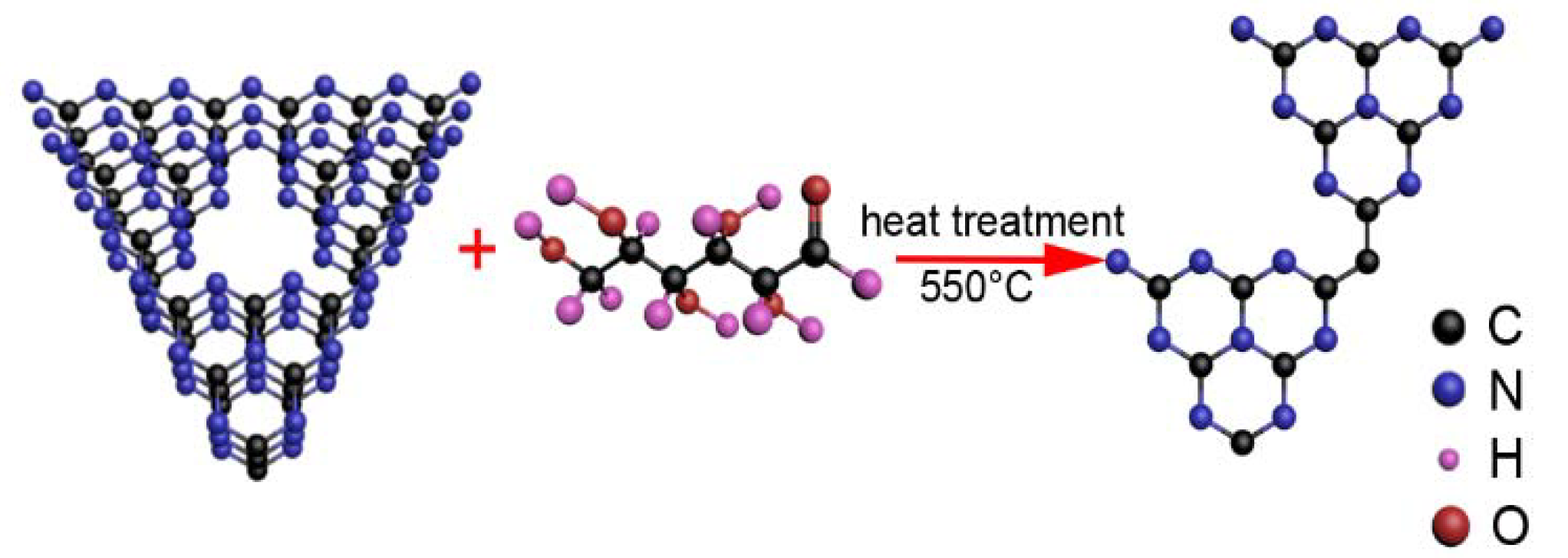
Inspired by these works, we prepared a carbon self-doped g-C3N4 nanosheet (C-C3N4 NS) by heating the mixture of bulk g-C3N4 and glucose, expecting to improve the photocatalytic performance of g-C3N4. The bulk g-C3N4 was obtained by the traditional thermal polymerization, while the secondary heat treatment can effectively exfoliate the layered g-C3N4 into the C3N4 nanosheet (C3N4 NS) and introduce the C element into the triazine structure. Therefore, the obtained C-C3N4 NS showed enhanced visible-light photocatalytic hydrogen production.

2. Results and Discussion

Figure 1A,B presents typical SEM images of C3N4 NS and 0.4 C-C3N4 NS. The C3N4 NS (Figure 1A) exhibits a simple typical layered structure, which is similar to that of bulk C3N4. The layered structure of the C3N4 NS was exfoliated by a thermal oxidation etching process [18].
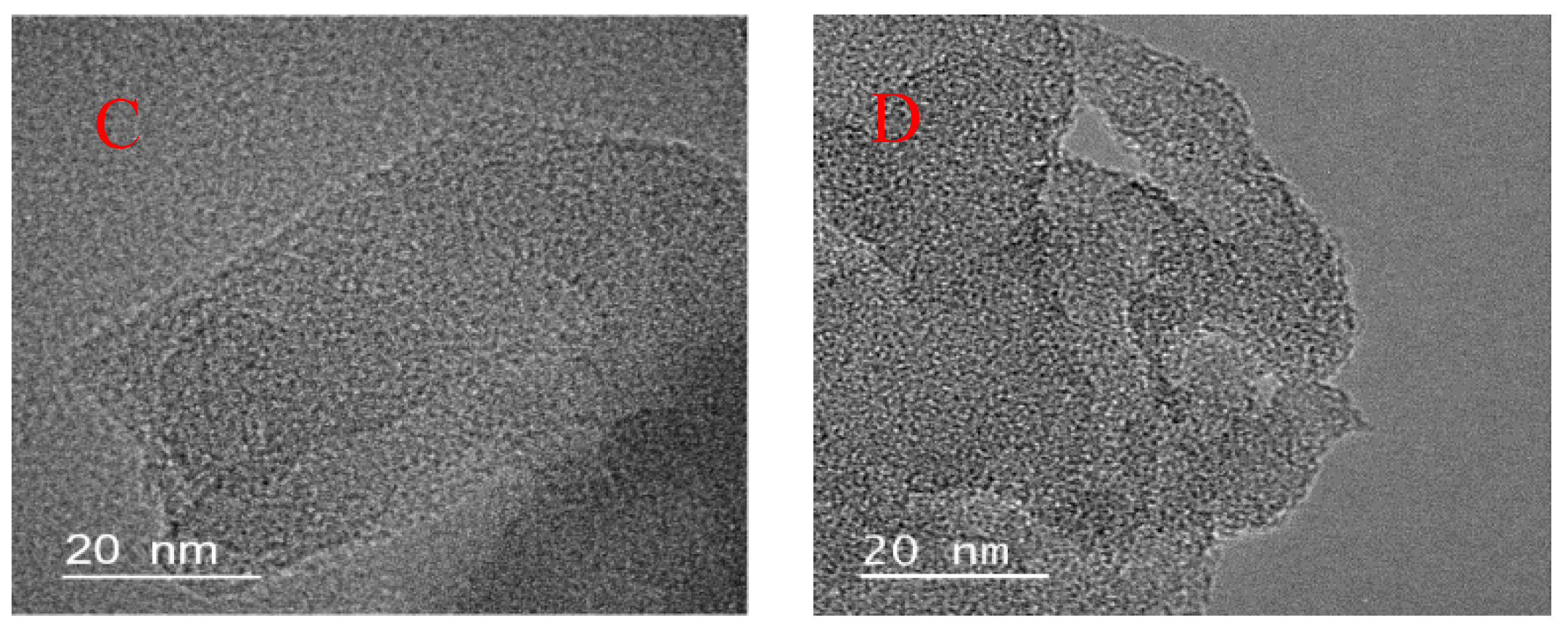
However, the layered structures of 0.4 C-C3N4 NS (Figure 1B) are corroded, which might be ascribed to the introduction of C atoms. To investigate the state of C, high-resolution transmission electron microscope (HRTEM) was tested. As shown in Figure 1C,D, C3N4 NS and 0.4 C-C3N4 NS samples exhibit ultrathin nanoflakes with layered structure, and no obvious carbon particles adhered to the C3N4 NS surface are observed in the 0.4 C-C3N4 NS, indicating that the C might be embedded in the structure of the tri-s-triazine ring.
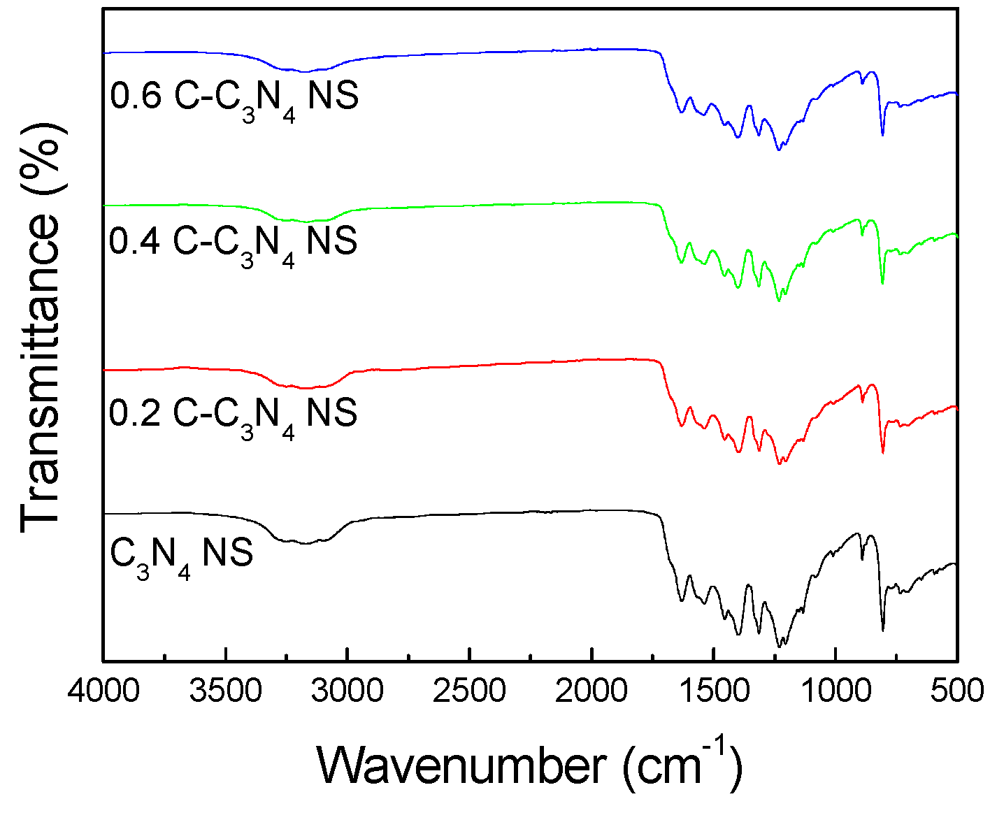
Figure 2A shows the FTIR spectra of C3N4 NS and C-C3N4 NS. The peaks at 1200–1600 and 805 cm−1 are corresponding to the characteristics of C–N heterocycle stretches and tri-s-triazine ring modes, respectively. This phenomenon indicates that the original C3N4 backbones are not damaged after the introduction of carbon doping. The peaks between 3000–3300 cm−1 are assigned to the N–H stretching vibrations, and the peaks for the C-C3N4 NS are weaker than those for the C3N4 NS, which can be ascribed to the decrease of N atoms caused by the doping of C atoms at the N sites [19,20].
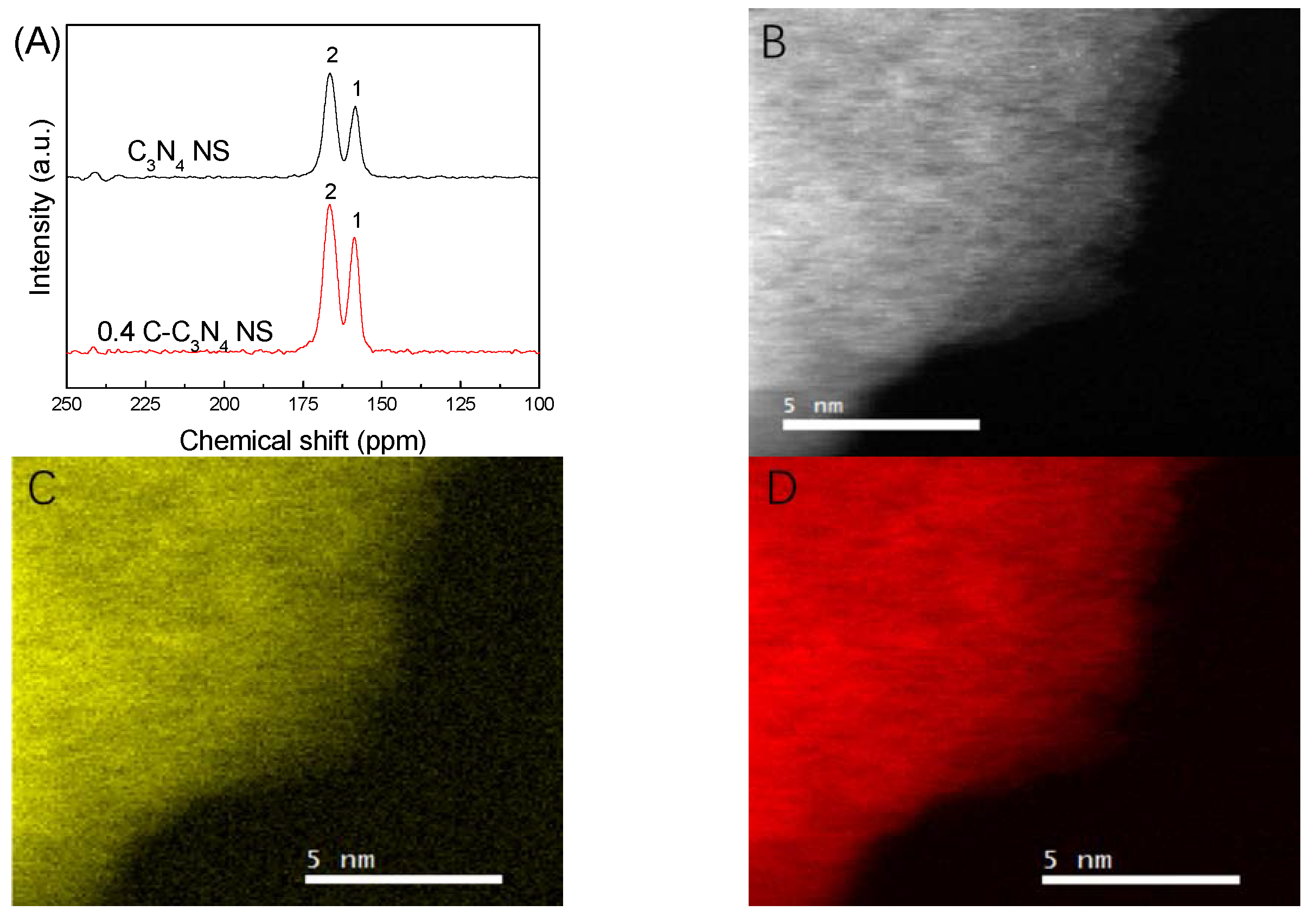
The solid-state 13C-magic-angle spinning (MAS)-NMR spectra of C3N4 NS and 0.4 C-C3N4 NS (Figure 3A) showed two strong peaks at 158.4 and 166.4 ppm, which correspond to C3N (1) and C2N-NHx (2) in the triazine ring units, respectively. No other peak is observed for 0.4 C-C3N4 NS, indicating that the introduction of carbon does not cause the open-loop reaction of the triazine ring, and that the C-C3N4 NS remained in the original C3N4 triazine ring structure, which is similar to the above Fourier-transform infrared (FTIR) results [21]. The maintenance of these 2D conjugated backbone structures provides the prerequisite for the π-delocalized electronic systems [12].
The atomic distributions of C and N are further revealed by the electron-energy loss spectroscopy (EELS) elemental mapping. Figure 3B shows the EELS mapping of 0.4 C-C3N4 NS, the distribution of C and N elements being shown in Figure 3C,D. C and N elements are evenly distributed throughout all the 0.4 C-C3N4 NS material, and no more carbon is observed to be coated on the edge of 0.4 C-C3N4 NS, indicating that the C is not clustered on the C3N4 surface in the form of a coated surface. Moreover, there are no carbon particles adhered to the surface of the 0.4 C-C3N4 NS, as shown in the HRTEM results. Therefore, traces of introduced C atoms might be embedded into the structure of the tri-s-triazine ring. The NMR results exhibit that the introduced C atoms do not cause the open-loop reaction of the triazine ring, and the FTIR results show that the N–H stretching vibrations get weaker after the C doping. It can be concluded that the N atoms in the tri-s-triazine ring were substituted by the introduced C atoms.
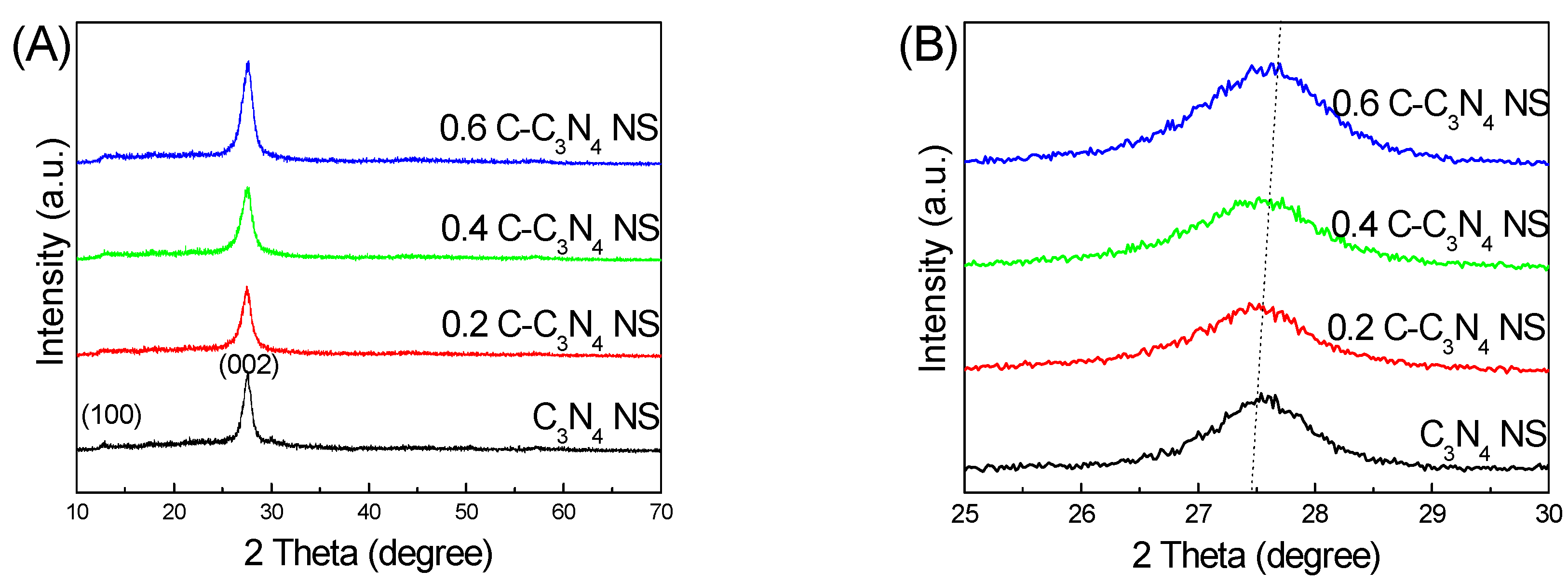
Figure 4A shows the XRD results of the C3N4 NS and C-C3N4 NS. All the samples exhibit two characteristic peaks at 13° and 27.4° corresponding to the (100) in-planar peak and the (002) interlayer-stacking peak of g-C3N4, respectively [2]. Enlarged XRD patterns (Figure 4B) show the gradual shift of the (002) peak to a higher angle with the introduction of carbon, indicating disturbance of the packing and undulations of the single layers. The introduction of the carbon dopant could form delocalized π-bonds that complete the π-delocalized electronic systems [22]. High-resolution C 1s XPS spectra (Figure 4C) shows that the C3N4 NSs were deconvoluted into two peaks at 284.9 eV and 288.4 eV, which can be ascribed to graphitic carbon atoms and sp2-bonded carbon atoms in the heterocycle (N–C=C) of aromatic g-C3N4, respectively [23]. For the 0.4 C-C3N4 NS, the peak corresponding to sp2-bonded carbon slightly shifted to 288.3 eV, which indicated that the binding energies of carbon atoms were affected by the doped carbon. High-resolution N 1s XPS spectra (Figure 4D) shows that all peaks of the 0.4 C-C3N4 NS shifted to lower binding energy compared to those of C3N4 NS. Therefore, the assumption that the N atoms in tri-s-triazine ring were substituted by C atoms, as shown in Figure 5, is further confirmed. The doped C atoms substituting N atoms maintained the original 2D conjugated backbone structures and reduced the number of N–H bonds, which resulted in the abovementioned results [24].
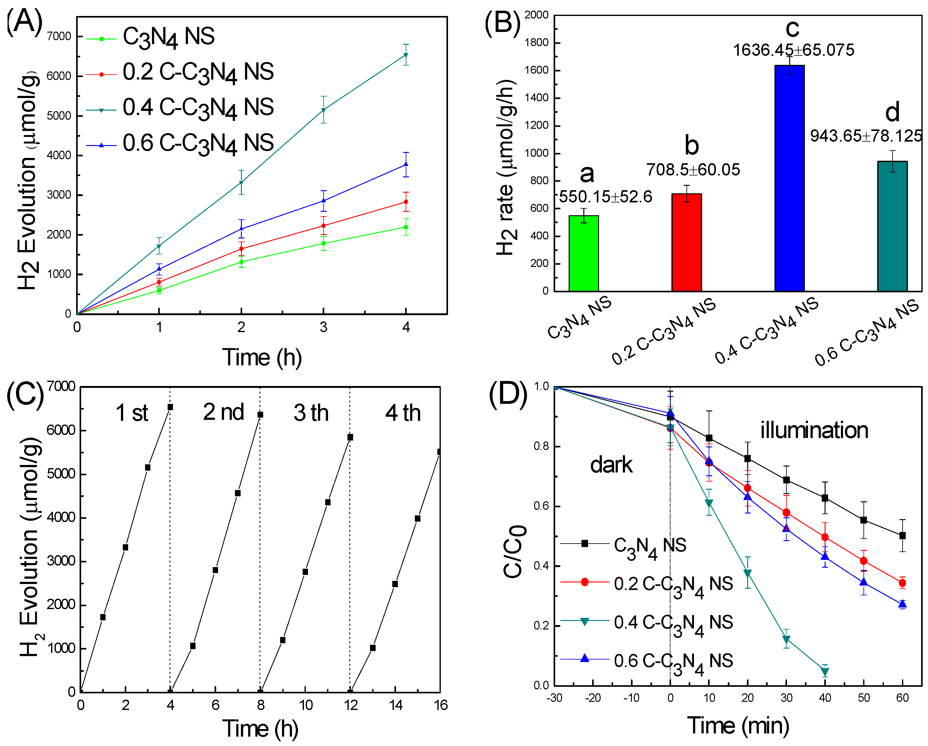
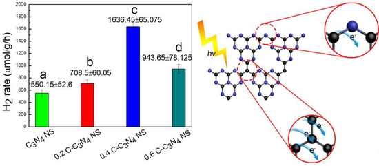
Figure 6A shows the photocatalytic hydrogen production performance of C3N4 NS and C-C3N4 NS. For C3N4 NS, the H2 evolution rate reaches 2200.6 ± 210.4 μmol/g at 4 h, while all the C-C3N4 NS samples exhibit higher evolution rate. Among them, 0.4 C-C3N4 NS reaches the highest H2 evolution rate of 6545.8 ± 260.3 μmol/g at 4 h, which is almost three-times higher than that of C3N4 NS; in order to rule out the contingency of the experiment, we did three sets of parallel experiments for each set of data. Corresponding error bars are given in Figure 6A, and the error ranges of the H2 evolution rates are within reasonable tolerances. The corresponding volume histogram of hydrogen production is shown in Figure 6B, and the 0.4 C-C3N4 NS exhibits the highest hydrogen production performance as well (1636.45 ± 65.075 μmol/g/h). Additionally, the photocatalysis stability of the 0.4 C-C3N4 NS was tested further, as shown in Figure 6C. After four cycles, the hydrogen evolution performance of the 0.4 C-C3N4 NS sample only decreased a little, and stabilized in the third and fourth cycles, indicating the stable photocatalysis performance of 0.4 C-C3N4 NS.
Figure 6D shows the photocatalytic rhodamine B (RhB) degradation results of the series of samples under visible-light illumination. Three sets of parallel experiments were presented to rule out the contingency of the experiment. All data errors are within reasonable tolerances, which might result from the difference in degree of centrifugation. As shown in Figure 6D, after visible-light illumination, for the C3N4 NS photocatalyst, only 50% RhB could be degraded in 60 min. However, all C-C3N4 NS samples exhibited higher photocatalytic degradation rates. Among them, the 0.4 C-C3N4 NS showed the highest degradation performance, which reached approximately 95% RhB degradation in only 40 min.
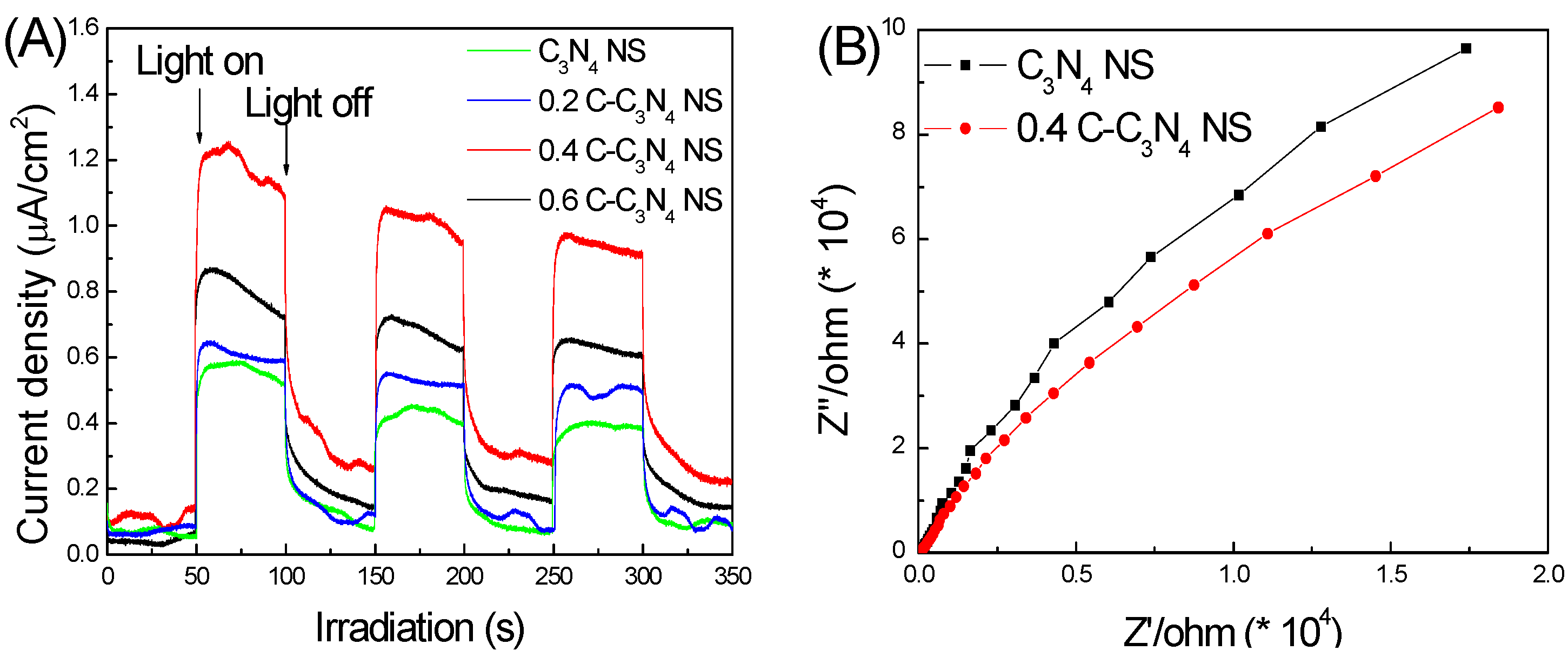
Figure 7A shows the photoinduced current with time i–t curve of the as-prepared photoelectrodes. As shown in Figure 7A, the photoinduced currents of all C-C3N4 NS photoelectrodes are much higher than those of C3N4 NS. Among all C-C3N4 NS samples, 0.4 C-C3N4 NS exhibits the highest photocurrent of 1.2 μA/cm2. The electrochemical impedance spectroscopy (EIS) measurement result is shown in Figure 7B; the resistance arc radius of the 0.4 C-C3N4 NS was smaller than that of the C3N4 NS, suggesting that the introduction of C could facilitate the charge transport of C3N4 NS [25].
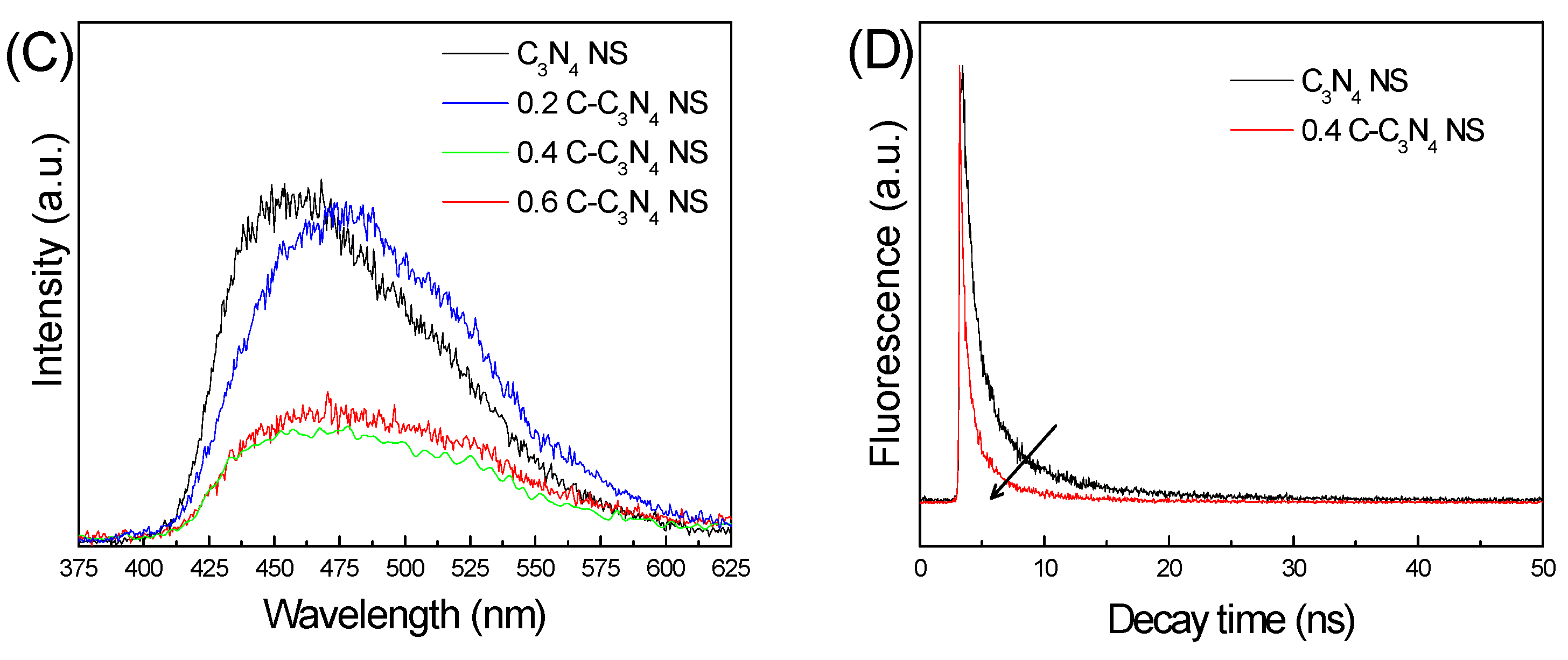
In order to detect the recombination rate of photocarriers in the samples, Photoluminescence (PL) spectra were tested. As shown in Figure 7C, all C-C3N4 NS samples show decreased PL emission intensity in comparison with C3N4 NS ones, indicating that the recombination rate of charge carriers is suppressed by the carbon doping. Photocurrent and PL results show that the introduction of carbon improves the separation of photocarriers and inhibits the recombination rate, which promotes the photocatalytic performance [26]. Time-resolved PL spectra also support the above assumption. It is clear that the fluorescence lifetime of 0.4 C-C3N4 NS samples is shorter than that of C3N4 NS (Figure 7D), and the decreased lifetime manifests more efficient separation of electrons and holes. Therefore, the doped carbon can accelerate the separation and transfer of the photogenerated carriers [27].
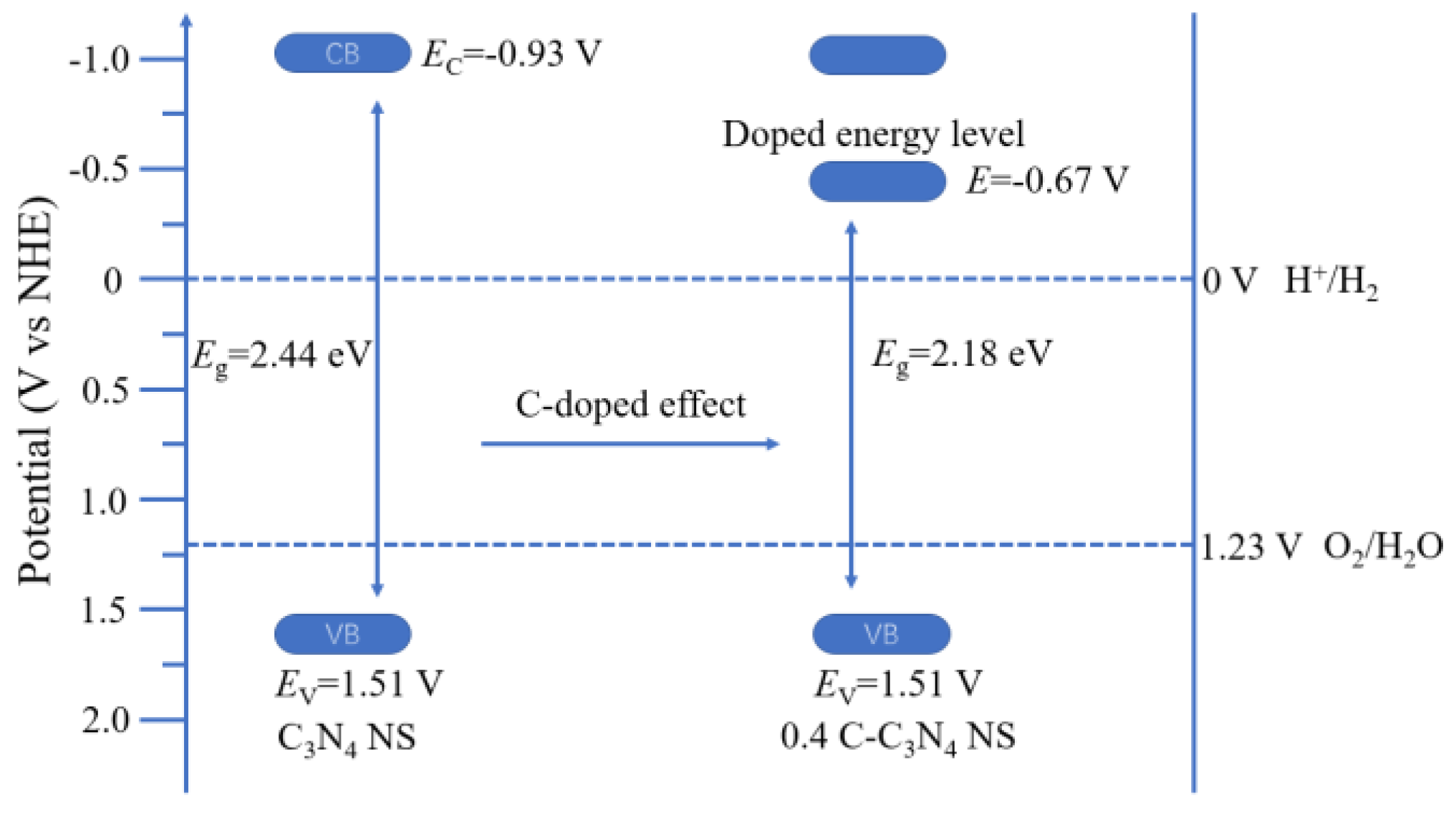
Diffuse reflectance spectra of the series of samples are shown in Figure 8A. It is clear that the C-C3N4 samples show a progressive redshift with increasing C content, confirming that the bandgap can be narrowed by introducing C. The estimated bandgaps (Figure 8B) of the series of samples are 2.04 eV, 2.18 eV, 2.36 eV and 2.44 eV, corresponding to the 0.6 C-C3N4 NS, 0.4 C-C3N4 NS, 0.2 C-C3N4 NS and C3N4 NS, respectively. To further investigate the influence of C on the band position of the C3N4 NS, Valence band X-ray photoelectron spectroscopy (VB-XPS) was tested for the C3N4 NS and 0.4 C-C3N4 NS. As shown in Figure 8C, the VB maximum was similar for both the C3N4 NS and 0.4 C-C3N4 NS located at 2.07 eV, which was estimated to be 1.51 V versus normal hydrogen electrode at pH = 7 through formula: ENHE/V = Ф + 2.07 eV − 4.44 (Ф = 3.88 eV: the electron work function of the analyzer) [28,29].
According to the above analysis, the VB maximum potential of C3N4 NS and 0.4 C-C3N4 NS samples were both located at 1.51 V (vs. Normal Hydrogen Electrode NHE), indicating that the valence band positions of the samples were unaffected by the introduction of C into g-C3N4, and the estimated band gaps of the C3N4 NS and 0.4 C-C3N4 NS were 2.44 eV and 2.18 eV (Figure 8B), respectively. According to Eg = EvEc, the calculated conduction band minimum potentials of C3N4 NS and 0.4 C-C3N4 NS were −0.93 V and −0.67 V (vs. NHE), respectively. The positive shift of the conduction band for the 0.4 C-C3N4 NS was caused by the formation of doped energy level which comes from the C. The estimated band structure schematic is exhibited in Figure 9.
Figure 10 shows the mechanism for the improvement of the photocatalysis performance under visible light. The substitution of C atoms for N atoms in the tri-s-triazine ring maintained the 2D conjugated system of g-C3N4, while the introduction of doped carbon atoms accelerates the charge mobility, thus enhancing the photocatalytic performance.

3. Experimental Section

3.1. Fabrication of g-C3N4

The bulk g-C3N4 was prepared through traditional thermal condensation [4]. 10 g dicyandiamide (Aladdin Co., Shanghai, China) was annealed at 550 °C with a ramping rate of 10 °C·min−1 in air in a muffle furnace for 3 h to obtain a yellow powder.

3.2. Fabrication of C-Modified g-C3N4

In a typical synthesis, 1 g g-C3N4 was dissolved in 50 mL deionized water and sonicated for a few minutes. Then, a different mass of glucose (0.2 g, 0.4 g, 0.6 g, Aladdin Co., Shanghai, China) was added with stirring and then kept stirring for 1 h. The product was centrifuged at 8000 r/min for 2 min and rinsed with water and ethanol for three times, and then dried at 60 °C for 12 h. The mixture was annealed at 550 °C with a ramping rate of 10 °C/min in air in a muffle furnace for 3 h, the samples being denoted as 0.2 C-C3N4 NS, 0.4 C-C3N4 NS, 0.6 C-C3N4 NS, respectively. For comparison, C3N4 NS was prepared at the same condition in the absence of glucose. The procedure is shown as Figure 11.

3.3. Characterization

The crystalline structures of the samples were identified through X-ray diffraction (XRD, D/MAX-2500/PC, Rigaku Co., Tokyo, Japan). The micro-morphologies of the samples were observed using a field-emission scanning electron microscope (SEM, JSM-6700F, JEOL, Tokyo, Japan) and a high-resolution transmission electron microscope (HRTEM, Tecnai G2 F20, FEI, Hillsboro, OR, USA). The atomic distributions of samples were further revealed by the electron energy loss spectroscopy (EELS, GIF Quantum 965, Gatan, Pleasanton, CA, USA) elemental mapping. The C atomic chemical states of samples were conducted by solid-state 13C magic-angle spinning (MAS) NMR (DiscoveryMR750 3.0T, General Electric Company, Boston, MA, USA) measurements at room temperature. The elementary compositions and bonding information of the samples were analyzed by using X-ray photoelectron spectroscopy (XPS, Axis Ultra, Kratos Analytical Ltd., Manchester, England, UK). The optical absorptions of the solid-state samples were tested by a UV-visible diffuse reflectance spectrophotometer (UV-vis DRS, U-41000, HITACHI, Tokyo, Japan). The photoluminescence and fluorescence decay times of the as-prepared materials were characterized using a fluorescence spectrometer (PL, Fluoro Max-4, HORIBA Jobin Yvon, Paris, France) with excitation wavelength at 350 nm. The molecular structures of the samples were identified through Fourier-transform infrared (FT–IR) spectra which used a Nicolet IS10 infrared spectrophotometer (Thermo Fisher Scientific, Waltham, MA, USA) in the transmission mode using a KBr pellet method.

3.4. Photocatalytic Reaction

The photocatalytic activities of the samples were tested by photocatalytic hydrogen production and photocatalytic dye degradation under visible light. In photocatalytic hydrogen production test, 50 mg catalyst powder was dispersed in 100 mL aqueous solution which contained 10 vol % triethanolamine as sacrificial electron donor and 3 wt % H2PtCl6·6H2O (Aladdin Co., Shanghai, China) as a co-catalyst [30]. Before the test, the photoreaction system was vacuumed until the pressure gage was stable. Externally illuminating 150 W Xe lamp (PLS-SXE300, Beijing Changtuo Co., Ltd., Beijing, China) was used as light source, and we added a 420 nm cut-off filter to achieve visible light, and adjusted the photo density to 250 mW/cm2. The illuminated area of the photocatalytic reaction is 28.3 cm2 (light spot diameter is 6 cm). The entire reaction system was kept at a constant 5 °C with condensed water. In photocatalytic dye degradation test, rhodamine B (RhB, Aladdin Co., Shanghai, China) was selected as the target degradant. The specific experimental steps are as follows: 0.05 g photocatalyst was dispersed in 100 mL RhB with a concentration of 10 mg·L−1. Before illumination, the mixture kept stirring for 30 min in the dark to achieve adsorption–desorption equilibrium. The light source and photo density were kept the same with the photocatalytic hydrogen production test. The entire reaction system was maintained at 25 °C with circulating water. The light absorptivity of solutions were decided by the UV-visible absorption spectrophotometer (U-41000, HITACHI, Tokyo, Japan) every 10 min after centrifugation with the speed of 8000 r/min for 2 min.

3.5. Photoelectrode Preparation and Photoelectrochemical Reaction

The photoelectrodes were prepared through electrophoretic deposition method. In total, 20 mg photocatalyst was dispersed in 50 mL water containing 5 mg I2, the mixture was stirred for 30 min to completely dissolve. Then, two Fluorine-doped Tin Oxide (FTO) conductive glasses with the deposition area of 1 cm × 1 cm and a distance 1 cm were immersed into the solution, maintaining a bias voltage of 20 V and a deposition time of 5 min. The obtained photoelectrodes were annealed at 300 °C for 30 min to improve adhesion. The photoelectrochemical systems were measured in a three-electrode system. The as-prepared photoelectrodes, and platinum and Ag/AgCl electrodes, were used as the working, counter and reference electrodes, respectively. 0.1 M Na2SO4 was used as electrolyte. All photoelectrochemical tests were measured using a CHI-660D (Chenhua Instrument Co., Shanghai, China) electrochemical workstation. The photoinduced current density–time (i–t) curves were measured at 1 V (vs. Ag/AgCl) bias under visible light with the photo density at 100 mW/cm2. The electrochemical impedance spectroscopy (EIS) was measured at 0 V (vs. Ag/AgCl) bias with the frequency range from 105–10−1 Hz.

4. Conclusions

In summary, we developed C self-doped g-C3N4 by a simple co-condensation method, and carbon atoms were successfully introduced into the tri-s-triazine structure of g-C3N4. The secondary heat treatment exfoliated the bulk g-C3N4 to nanosheets, which endows materials with higher specific surface area. Compared with pure g-C3N4, the modified C substitutes N atoms in the triazine ring, which broadens the range of light absorption, accelerates the transfer rate of charge carriers as well as increases the photocatalysis performance. Among all the C-C3N4 NS samples, the optimal 0.4 C-C3N4 NS unfolds the superior photocatalytic hydrogen rate of 1636.45 μmol/g/h, which is almost three-times higher than that of pure C3N4 NS. Simultaneously, the 0.4 C-C3N4 NS shows a better photocatalytic RhB degradation rate than that of the C3N4 NS under visible light illumination, the former of which could achieve approximately 95% degradation in 40 min. The photoelectrochemical test results verify that the introduction of C accelerates the transfer rate and inhibits the combination of photo-induced carriers, while also enhancing the photocatalytic performance. This work highlights how carbon modification incorporated into g-C3N4 has improved the photocatalytic performance without destroying the 2D conjugated backbone structures.

Author Contributions

Conceptualization, D.Y., W.L.; Methodology, H.W.; Experiment and Analysis, Z.C., G.H.

Funding

This research received no external funding.

Acknowledgments

This work was financially supported by the Research Fund of State Key Laboratory for Marine Corrosion and Protection of Luoyang Ship Material Research Institute (LSMRI) under the contract No. KF160413.

Conflicts of Interest

The authors declare no conflict of interest.

References

  1. Maeda, K. Photocatalytic water splitting using semiconductor particles: History and recent developments. J. Photochem. Photobiol. C 2011, 12, 237–268. [Google Scholar] [CrossRef]
  2. Wang, X.; Maeda, K.; Thomas, A.; Takanabe, K.; Xin, G.; Carlsson, J.M.; Domen, K.; Antonietti, M. A metal-free polymeric photocatalyst for hydrogen production from water under visible light. Nat. Mater. 2009, 8, 76–80. [Google Scholar] [CrossRef] [PubMed]
  3. Ong, W.; Tan, L.; Ng, Y.; Yong, S.; Chai, S. Graphitic carbon nitride (g-C3N4)-based photocatalysts for artificial photosynthesis and environmental remediation: Are we a step closer to achieving sustainability? Chem. Rev. 2016, 116, 7159–7329. [Google Scholar] [CrossRef] [PubMed]
  4. Bu, Y.; Chen, Z.; Xie, T.; Li, W.; Ao, J. Fabrication of C3N4 ultrathin flakes by mechanical grind method with enhanced photocatalysis and photoelectrochemical performance. RSC Adv. 2016, 6, 47813–47819. [Google Scholar] [CrossRef]
  5. Han, Q.; Wang, B.; Zhao, Y.; Hu, C.; Qu, L. A Graphitic-C3N4 “Seaweed” Architecture for Enhanced Hydrogen Evolution. Angew. Chem. Int. Ed. 2015, 54, 11433–11437. [Google Scholar] [CrossRef] [PubMed]
  6. Fu, J.; Chang, B.; Tian, Y.; Xi, F.; Dong, X. Novel C3N4-CdS composite photocatalysts with organic-inorganic heterojunctions: In situ synthesis, exceptional activity, high stability and photocatalytic mechanism. J. Mater. Chem. A 2013, 1, 3083–3090. [Google Scholar] [CrossRef]
  7. Zhang, J.; Wang, Y.; Jin, J.; Zhang, J.; Lin, Z.; Huang, F.; Yu, J. Efficient visible-light photocatalytic hydrogen evolution and enhanced photostability of core/shell CdS/g-C3N4 nanowires. ACS Appl. Mater. Interfaces 2013, 5, 10317–10324. [Google Scholar] [CrossRef] [PubMed]
  8. Li, Q.; Zhang, N.; Yang, Y.; Wang, G.; Ng, D.H. High efficiency photocatalysis for pollutant degradation with MoS2/C3N4 heterostructures. Langmuir 2014, 30, 8965–8972. [Google Scholar] [CrossRef] [PubMed]
  9. Wang, Y.; Shi, R.; Lin, J.; Zhu, Y. Enhancement of photocurrent and photocatalytic activity of ZnO hybridized with graphite-like C3N4. Energy Environ. Sci. 2011, 4, 2922–2929. [Google Scholar] [CrossRef]
  10. Yan, H.; Yang, H. TiO2-g-C3N4 composite materials for photocatalytic H2 evolution under visible light irradiation. J. Alloys Compd. 2011, 509, L26–L29. [Google Scholar] [CrossRef]
  11. Lan, Z.; Zhang, G.; Wang, X. A facile synthesis of Br-modified g-C3N4 semiconductors for photoredox water splitting. Appl. Catal. B Environ. 2016, 192, 116–125. [Google Scholar] [CrossRef]
  12. Zhang, G.; Zhang, M.; Ye, X.; Qiu, X.; Lin, S.; Wang, X. Iodine modified carbon nitride semiconductors as visible light photocatalysts for hydrogen evolution. Adv. Mater. 2014, 26, 805–809. [Google Scholar] [CrossRef] [PubMed]
  13. Tian, H.; Zhang, X.; Bu, Y. Sulfur-and Carbon-Codoped Carbon Nitride for Photocatalytic Hydrogen Evolution Performance Improvement. ACS Sustain. Chem. Eng. 2018, 6, 7346–7354. [Google Scholar] [CrossRef]
  14. Guo, S.; Tang, Y.; Xie, Y.; Tian, C.; Feng, Q.; Zhou, W.; Jiang, B. P-doped tubular g-C3N4 with surface carbon defects: Universal synthesis and enhanced visible-light photocatalytic hydrogen production. Appl. Catal. B Environ. 2017, 218, 664–671. [Google Scholar] [CrossRef]
  15. Yan, S.; Li, Z.; Zou, Z. Photodegradation of rhodamine B and methyl orange over boron-doped g-C3N4 under visible light irradiation. Langmuir 2010, 26, 3894–3901. [Google Scholar] [CrossRef] [PubMed]
  16. Wang, Y.; Di, Y.; Antonietti, M.; Li, H.; Chen, X.; Wang, X. Excellent visible-light photocatalysis of fluorinated polymeric carbon nitride solids. Chem. Mater. 2010, 22, 5119–5121. [Google Scholar] [CrossRef]
  17. Zhang, H.; Zhao, L.; Geng, F.; Guo, L.; Wan, B.; Yang, Y. Carbon dots decorated graphitic carbon nitride as an efficient metal-free photocatalyst for phenol degradation. Appl. Catal. B Environ. 2016, 180, 656–662. [Google Scholar] [CrossRef] [Green Version]
  18. Niu, P.; Zhang, L.; Liu, G.; Cheng, H. Graphene-like carbon nitride nanosheets for improved photocatalytic activities. Adv. Funct. Mater. 2012, 22, 4763–4770. [Google Scholar] [CrossRef]
  19. Liu, C.; Zhang, Y.; Dong, F.; Reshak, A.H.; Ye, L.; Pinna, N.; Zeng, C.; Zhang, T.; Huang, H. Chlorine intercalation in graphitic carbon nitride for efficient photocatalysis. Appl. Catal. B Environ. 2017, 203, 465–474. [Google Scholar] [CrossRef]
  20. Liu, G.; Zhao, G.; Zhou, W.; Liu, Y.; Pang, H.; Zhang, H.; Hao, D.; Meng, X.; Li, P.; Kako, T.; Ye, J. In situ bond modulation of graphitic carbon nitride to construct p-n homojunctions for enhanced photocatalytic hydrogen production. Adv. Funct. Mater. 2016, 26, 6822–6829. [Google Scholar] [CrossRef]
  21. Tian, N.; Zhang, Y.; Li, X.; Xiao, K.; Du, X.; Dong, F.; Waterhouse, G.I.N.; Zhang, T.; Huang, H. Precursor-reforming protocol to 3D mesoporous g-C3N4 established by ultrathin self-doped nanosheets for superior hydrogen evolution. Nano Energy 2017, 38, 72–81. [Google Scholar] [CrossRef]
  22. Ding, Y.; Tang, Y.; Yang, L.; Zeng, Y.; Yuan, J.; Liu, T.; Zhang, S.; Liu, C.; Luo, S. Porous nitrogen-rich carbon materials from carbon self-repairing g-C3N4 assembled with graphene for high-performance supercapacitor. J. Mater. Chem. A 2016, 4, 14307–14315. [Google Scholar] [CrossRef]
  23. Cao, S.; Low, J.; Yu, J.; Jaroniec, M. Polymeric photocatalysts based on graphitic carbon nitride. Adv. Mater. 2015, 27, 2150–2176. [Google Scholar] [CrossRef] [PubMed]
  24. Li, H.; Li, F.; Wang, Z.; Jiao, Y.; Liu, Y.; Wang, P.; Zhang, X.; Qin, X.; Dai, Y.; Huang, B. Fabrication of carbon bridged g-C3N4 through supramolecular self-assembly for enhanced photocatalytic hydrogen evolution. Appl. Catal. B Environ. 2018, 229, 114–120. [Google Scholar] [CrossRef]
  25. Wang, R.; Xie, T.; Zhang, T.; Pu, T.; Bu, Y.; Ao, J.P. Fabrication of FTO-BiVO4-W-WO3 photoanode for photoelectrochemical performance improving: Based on the Z-scheme electron transfer mechanism. J. Mater. Chem. A 2018, 6, 12956–12961. [Google Scholar] [CrossRef]
  26. Jing, L.; Qu, Y.; Wang, B.; Li, S.; Jiang, B.; Yang, L.; Fu, W.; Fu, H.; Sun, J. Review of photoluminescence performance of nano-sized semiconductor materials and its relationships with photocatalytic activity. Sol. Energy Mater. Sol. Cells 2006, 90, 1773–1787. [Google Scholar]
  27. Li, Y.; Xu, H.; Ouyang, S.; Lu, D.; Wang, X.; Wang, D.; Ye, J. In situ surface alkalinized g-C3N4 toward enhancement of photocatalytic H2 evolution under visible-light irradiation. J. Mater. Chem. A 2016, 4, 2943–2950. [Google Scholar] [CrossRef]
  28. Yu, H.; Shi, R.; Zhao, Y.; Bian, T.; Zhao, Y.; Zhou, C.; Waterhouse, G.I.N.; Wu, L.; Tung, C.; Zhang, T. Alkali-Assisted Synthesis of Nitrogen Deficient Graphitic Carbon Nitride with Tunable Band Structures for Efficient Visible-Light-Driven Hydrogen Evolution. Adv. Mater. 2017, 29, 1605148. [Google Scholar] [CrossRef] [PubMed]
  29. Trasatti, S. The absolute electrode potential: An explanatory note (Recommendations 1986). Pure Appl. Chem. 1986, 58, 955–966. [Google Scholar] [CrossRef]
  30. Iqbal, W.; Qiu, B.; Zhu, Q.; Xing, M.; Zhang, J. Self-modified breaking hydrogen bonds to highly crystalline graphitic carbon nitrides nanosheets for drastically enhanced hydrogen production. Appl. Catal. B Environ. 2018, 232, 306–313. [Google Scholar] [CrossRef]
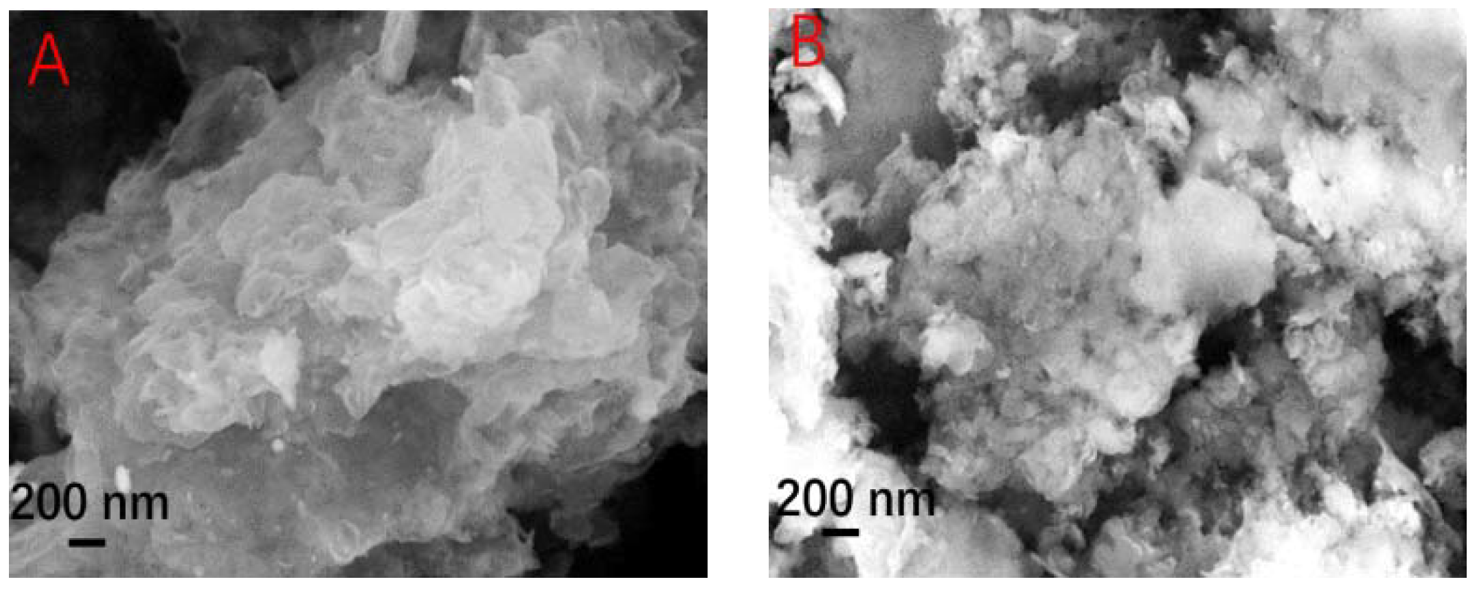
Figure 1. SEM images of (A) C3N4 NS and (B) 0.4 C-C3N4 NS. High-resolution transmission electron microscope (HRTEM) images of (C) C3N4 NS and (D) 0.4 C-C3N4 NS.
Catalysts 08 00366 g001aCatalysts 08 00366 g001b
Figure 2. FTIR spectra of C3N4 NS and C-C3N4 NS samples.
Catalysts 08 00366 g002
Figure 3. (A) Solid-state 13C-magic-angle spinning (MAS)-NMR spectra of C3N4 NS and 0.4 C-C3N4 NS. (B) Electron-energy loss spectroscopy (EELS) mapping of 0.4 C-C3N4 NS for merged mapping, (C) C mapping and (D) N mapping.
Catalysts 08 00366 g003
Figure 4. (A) XRD patterns and (B) enlarged XRD patterns of series samples. High-resolution XPS spectra of (C) C 1s and (D) N 1s of C3N4 NS and 0.4 C-C3N4 NS.
Catalysts 08 00366 g004aCatalysts 08 00366 g004b
Figure 5. Atomic structure of C3N4 NS and C-C3N4 NS.
Catalysts 08 00366 g005
Figure 6. (A) Photocatalysis hydrogen evolution and (B) volume histogram of series of samples. (C) Photocatalysis hydrogen evolution stability test of 0.4 C-C3N4 NS. (D) Photocatalysis rhodamine B (RhB) degradation performances of series of samples.
Catalysts 08 00366 g006
Figure 7. (A) Photoinduced current–time curves and (B) Electrochemical impedance spectroscopy (EIS) results of C3N4 NS and 0.4 C-C3N4 NS electrodes. (C) Photoluminescence spectra of series of samples. (D) Time-resolved fluorescence spectra of C3N4 NS and 0.4 C-C3N4 NS.
Catalysts 08 00366 g007aCatalysts 08 00366 g007b
Figure 8. (A) UV–Vis diffuse reflection and (B) the estimated band gaps of series of samples. (C) VB-XPS spectra for C3N4 NS and 0.4 C-C3N4 NS samples.
Catalysts 08 00366 g008
Figure 9. Band structure of the C3N4 NS and 0.4 C-C3N4 NS.
Catalysts 08 00366 g009
Figure 10. Mechanism of the photocatalysis performance improvement under visible light.
Catalysts 08 00366 g010
Figure 11. Schematic illustration for the formation of C-C3N4 NS.
Catalysts 08 00366 g011