Review

RNA-Cleaving DNAzymes: Old Catalysts with New Tricks for Intracellular and In Vivo Applications

Department of Chemistry, University of Illinois at Urbana–Champaign, 601 S. Mathews Ave., Urbana, IL 61801, USA
Catalysts 2018, 8(11), 550; https://doi.org/10.3390/catal8110550
Submission received: 17 October 2018 / Revised: 12 November 2018 / Accepted: 14 November 2018 / Published: 16 November 2018
(This article belongs to the Section Biocatalysis)

Abstract

:
DNAzymes are catalytically active DNA molecules that are normally isolated through in vitro selection methods, among which RNA-cleaving DNAzymes that catalyze the cleavage of a single RNA linkage embedded within a DNA strand are the most studied group of this DNA enzyme family. Recent advances in DNA nanotechnology and engineering have generated many RNA-cleaving DNAzymes with unique recognition and catalytic properties. Over the past decade, numerous RNA-cleaving, DNAzymes-based functional probes have been introduced into many research areas, such as in vitro diagnostics, intracellular imaging, and in vivo therapeutics. This review focus on the fundamental insight into RNA-Cleaving DNAzymes and technical tricks for their intracellular and in vivo applications, highlighting the recent progress in the clinical trial of RNA-Cleaving DNAzymes with selected examples. The challenges and opportunities for the future translation of RNA-cleaving DNAzymes for biomedicine are also discussed.

1. Introduction

Catalysis in biology has long been considered to be the function of catalytic RNAs (ribozymes) and protein enzymes [1]; however, many studies have reported the discovery of catalytic DNAs (called DNAzymes hereafter) since Breaker and Joyce’s pioneer work on DNAzymes in 1994 [2,3,4]. These DNAzymes are normally isolated through in vitro selection methods from a random DNA library consisting of approximately 1015 DNA sequences [5]. The specific catalytic functionalities of these DNAzymes depend on the sequences of the DNAzymes and in many cases also on the presence of additional cofactors, such as metal ions and amino acids [6]. These DNAzymes have been identified to catalyze many chemical reactions, including RNA cleavage, oxidative or hydrolytic DNA cleavage, DNA/RNA ligation, and DNA phosphorylation [4,5]. These features have been successfully applied in various biomedical applications both in vitro [7,8,9] and in vivo [10,11,12].
Among the abovementioned DNAzymes, RNA-cleaving DNAzymes that catalyze the cleavage of a single RNA (rA) linkage embedded within a DNA strand are the most studied group of this DNA enzyme family. A representative RNA-cleaving DNAzyme is formed through the hybridization of a substrate strand and an enzyme strand, which consists of an active site, an enzymatic region, and two binding arms (Figure 1A). The substrate strand contains an rA linkage that serves as a cleavage site, and in the presence of the cofactor, the enzyme strand forms a defined second structure with the catalytic ability to cleave the rA linkage, resulting in two fragments with a 2′-3′ cyclic phosphate and a 5′-OH terminus, respectively (Figure 1B). This catalytic property of DNAzyme is highly specific to the substrate strand, and even a single base mismatch in the antisense arms could significantly decrease the cleavage efficiency. In contrast, most of the DNAzymes exhibit high flexibility for the design of DNA sequences in the binding arms, and, therefore, the rational design of multifunctional DNAzymes using the same cofactor has been developed for diverse applications. In addition, most of the DNAzymes could efficiently catalyze the cleavage of the substrate strand in a multiple turnover manner that allows for signal amplification for sensitive target detection. These aforementioned features, in conjunction with the good chemical stability, easy modification with signaling molecules, and synthetic accessibility, have made RNA-cleaving DNAzymes a promising candidate in a broad spectrum of applications that range from in vitro diagnosis, intracellular sensing, and intracellular imaging to in vivo imaging and cancer therapy.
In the past decade, the research area of RNA-cleaving DNAzymes has experienced tremendous growth, and from that, been the subject of several reviews [6,10,13,14,15,16,17,18,19]. In this review, I focus on the fundamental insight into RNA-Cleaving DNAzymes and technical tricks for their intracellular and in vivo applications. First, I introduce the fundamentals of RNA-Cleaving DNAzymes, and discuss the recent finding toward the structural characterization of RNA-cleaving DNAzymes. I subsequently summarize the most recent advances in RNA-cleaving DNAzymes for intracellular and in vivo applications. Then, I highlight the recent progress in clinical trials with RNA-Cleaving DNAzymes with selected examples. Finally, I provide insight into the challenges and future perspectives of RNA-cleaving DNAzymes for biomedicine.

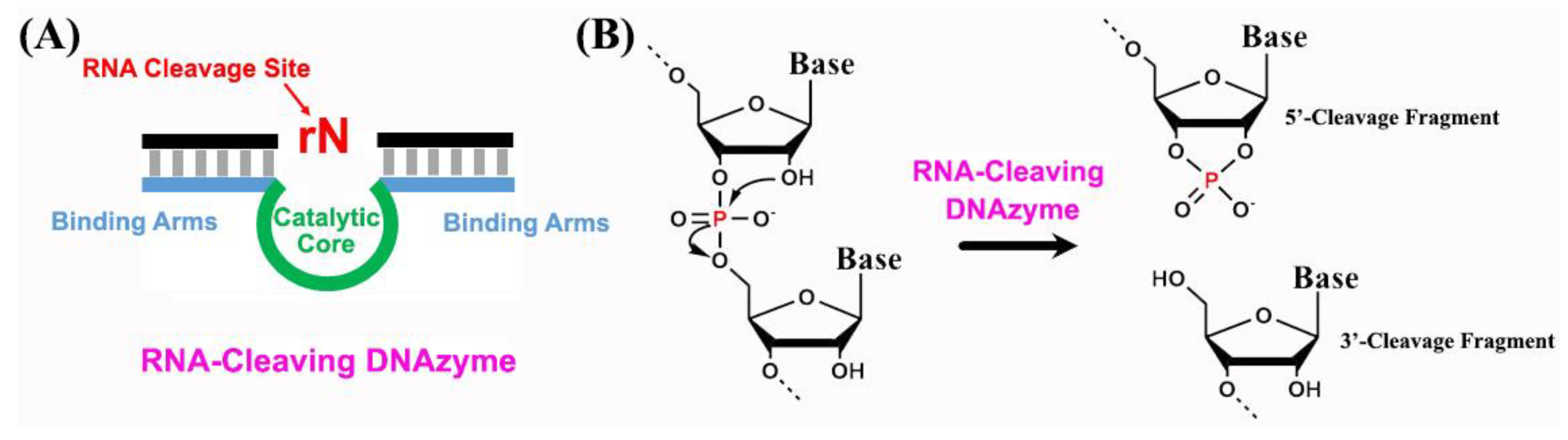
2. Structural Characterization of RNA-Cleaving DNAzymes

The RNA-cleaving DNAzymes are capable of providing diverse structures and functions due to their flexible backbone, unique sugar pucker, and nucleobases [20]. Up to now, many biochemical and spectroscopic methods have been used in the fundamental studies of RNA-cleaving DNAzymes [21]. However, for the chemical and biophysical analysis of a RNA-cleaving DNAzyme, the most important aspect is to understand the three-dimensional structure of the DNAzyme at the atomic level. In other words, obtaining the crystal structure of a DNAzyme is of fundamental importance to understand its catalytic mechanism, its interactions with cofactors and substrates, and the organization of its active site. However, the structural characterization of DNAzymes is severely underexplored because it is difficult to obtain single DNAzyme crystals with high diffraction quality. This difficulty is mainly due to two aspects: (1) the negatively charged surfaces of DNA/RNA result in non-specific contact for crystal packing, which can introduce defects into the crystal lattice; and (2) the structures of DNA/RNA are highly heterogeneous in solution, further contributing to crystalline disorder.
Over the past two decades, many research groups have attempted to investigate the structures of DNAzymes in order to obtain insights into how they perform catalysis, and the role of structure in the function. For instance, Nowakowski et al. reported a crystal structure of the 10–23 RNA-cleaving DNAzyme with X-ray diffraction at a 3.0 Å resolution [22,23]. This DNAzyme consists of a 15-nucleotide catalytic core, flanked by two substrate-recognition domains that bind the target RNA in a four-way junction. However, this stoichiometry and conformation do not reflect the active form of the 10–23 RNA-cleaving DNAzyme under both standard reaction conditions and crystallization conditions. Later, Parkinson et al. reported the crystal structures of both intra- and intermolecular K+-containing quadruplexes formed from four consecutive human telomeric DNA repeats [24]. They have demonstrated that all four DNA strands were parallel, with the three linking trinucleotide loops positioned on the exterior of the quadruplex core in a propeller-like arrangement. Another crystal structure of a B-form DNA duplex containing two consecutive T–HgII–T base pairs was described by Kondo et al. [25], in which they showed that the metallophilic attraction in consecutive T–HgII–T base pairs could stabilize the B-form double helix. Recently, Ponce-Salvatierra et al. have successfully visualized the structure of an RNA-ligating DNAzyme named 9DB1 at a 2.8 Å resolution [26]. They demonstrated that DNA could possess the intrinsic ability to adopt complex tertiary folds that support catalysis, and unveiled the active site of a DNAzyme in the post-catalytic state for the first time. This structural study showed the backbone conformation and intricate three-dimensional organization of deoxyribose in an active DNAzyme catalyst, indicating great potential for a fundamental understanding of the function of DNAzyme catalysts. Despite the above success, none of the aforementioned studies provided information on the structure of RNA-cleaving DNAzymes in an active form.
Very recently, Liu et al. reported the first crystal structure of the 8–17 RNA-cleaving DNAzyme with the highest resolution of 2.55 Å (Figure 2) [27]. In this study, the DNAzyme crystals were grown in the presence of African swine fever virus DNA polymerase X, which could facilitate the molecular packing and crystallization. The obtained crystal structure captured the pre-catalytic state of the RNA cleavage, and the DNAzyme exhibited a V-shaped fold consisting of two helical substrate-recognizing arms and one twisted DNA pseudoknot, and the combination of this compacted pseudoknot with Pb2+ catalysis and three-dimensional interactions thereby made the DNAzyme fold and function as a catalyst. These experimental evidence and findings have had a great impact on the design and application of new RNA-cleaving DNAzymes for a functional need in catalysis [28].

3. The General Concept of Translating RNA-Cleaving DNAzymes into Functional Probes

The attractive superiority and versatility of RNA-cleaving DNAzymes (RCDs) have catalyzed the emergence of RCDs-based functional probes for diverse applications both in vitro and in vivo. However, for diagnostic or therapeutic purposes, and especially for intracellular and in vivo applications, it is essential to illustrate the general concept of translating RNA-cleaving DNAzymes into functional probes. In general, according to the functions of RNA-cleaving DNAzymes, DNAzyme-based probes can be categorized into three types: biorecognition agents, signal amplification agents, and activatable hosting agents as shown in Figure 3.
The biorecognition function of RCDs originates from the in vitro systematic evolution of ligands by exponential enrichment (SELEX), and has been widely used for the detection and imaging of the cofactors, such as metal ions. For this type of probe, the enzyme strand of RCDs hybridizes with the substrate strand to from a DNA duplex, and in the presence of the cofactor (target), the enzyme strand will form a defined three-dimensional (3D) structure, and catalyze the cleavage of the substrate strand to two fragments (Figure 3A). In most cases, the shorter fragment will dissociate from the RCDs duplex due to the decreased melting temperature of the Watson–Crick base pairing after the cleavage. By integration with signal reporter molecules, such as a fluorophore and a quencher, an RCDs-based probe could transform the binding of a target into a target-dependent signal that can be detected through various modalities. The use of RCDs-based probes as biorecognition agents offers many unique features, such as high specificity and sensitivity to the targets, and, most importantly, the advantage of a combinatorial selection process, which would allow for the general design of RCDs-based probes for a multiplex of diagnostic purposes.
In addition to the biorecognition function, RCDs could be also used as a biocatalyst for signal amplification. For this type of probe, the RCDs are often embedded with a biorecognition unit in an inactive form (Figure 3B). In the presence of the target, the biorecognition unit interacts with the target, and subsequently activates the RCDs to cleave the substrate strand with the assistance of a cofactor. In most cases, the cleavage of the substrate strand would either cause the regeneration of the target or the release of the DNAzyme strand to start a new round of catalytic cleavage reactions. In this way, one target input would trigger numerous catalytic cleavage events that amplify the output signals. This function of RCDs is critical for the detection and imaging of trace amounts of targets inside cells or in vivo.
While the abovementioned RCDs-based probes are normally used for diagnostic purposes, the RCDs could be also applied for therapeutic purposes. For this type of probe, the therapeutic reagents, including drugs, miRNAs, and siRNAs, are either labeled or embedded within the RCDs (Figure 3C). As a hosting agent, the RCDs could be activated by internal or external stimuli, resulting in the release of the therapeutic reagents inside the cells or in vivo. The advantage of using RCDs as hosting agents is that they can assemble into various 3D nanostructures through advanced DNA nanotechnology, which could provide a biocompatible matrix for intracellular or in vivo delivery. Additionally, the RCDs could easily be incorporated into many nanomaterials to further improve the efficiency of the delivery, as well as providing a signal transduction property for simultaneous diagnostic and therapeutic purposes.

4. RNA-Cleaving DNAzymes-Based Functional Probes for Intracellular Applications

Cells form the basis of all living organisms, and are sometimes considered to be the “building block of life”. Recent advances in molecular biology and engineering have ignited great interest in visualizing and regulating molecules inside living cells [29]. Compared with extracellular studies, there are three basic criteria of RCDs-based functional probes for meeting the requirements of intracellular applications. First, efficient delivery of probes into the cells is the most important step for intracellular applications; however, the negatively charged phosphate backbones of unmodified RCDs often hinder their penetration through the negatively charged cell membrane, leading to inefficient cellular uptake. In addition, the unmodified RCDs are quite susceptible to endogenous nuclease degradation. Therefore, to address these issues, the self-assembly of RCDs into nanostructures and their integration with nanomaterials have been developed in recent years for efficient intracellular internalization of RCDs-based probes [30]. Second, cytotoxicity is another important aspect, and should be minimized to prevent disturbances in the intracellular process when we design the RCDs-based functional probes. Finally, the ability to induce the activatable delivery of RCDs-based probes into subcellular locations with precise spatiotemporal control should be also taken into consideration before being applied for intracellular studies [31]. Over the past decade, significant effort has been devoted to meet the aforementioned criteria, and many RCDs-based functional probes have been developed for diverse intracellular applications.

4.1. Intracellular Sensing and Imaging of Metal Ions

Metal ions play vital roles in biology, and serve as essential cofactors in regulating many biological processes, including enzymatic reactions and gene transcription [32]. However, these metal ions both at elevated levels and deficient levels can be detrimental to their normal functions [33]. Therefore, understanding the location and the concentration of metal ions in living cells is of significant importance because it can provide essential insights into the molecular basis of the functions of metal ions at the cellular level. Over the past decade, RCDs-based probes have been emerging as a promising platform for the sensing and imaging of metal ions in living cells [34,35,36]. For instance, Wu et al. [34] developed the first RCDs-based probe for metal ions in living cells based on the integration of gold nanoparticles (AuNPs), a uranyl-specific 39E DNAzyme, and a 39S substrate strand labeled with a Cy3 fluorophore and a BHQ-2 quencher, respectively (Figure 4A). In this design, the AuNPs serve as a carrier to deliver the DNAzymes into the cells with enhanced DNA loading efficiency and increased resistance to enzymatic degradation. In the presence of the target metal ions, UO22+, the DNAzyme cleaves the fluorophore-labeled substrate strand, resulting in release of the shorter fluorescent fragment, and thereby showing a Turn-On fluorescent signal, which is related to the concentration of UO22+ inside the cells. Later, Zhang et al. [37] used the dendritic polyethylene–cationic poly(p-phenylene ethynylene) polyvalent nanocarrier as a DNAzyme delivery system, and demonstrated the imaging of Pb2+ in living cells using a Pb2+-dependent DNAzyme. Additionally, Li et al. [38] went a further step to combine two metal-ion-dependent DNAzymes into a single RCDs-based probe, and developed a two-color fluorescence sensor for simultaneous imaging of Zn2+ and Cu2+ in living cells in an on-site manner. To further improve the sensitivity and applicability of the RCDs-based probes, the same group recently reported an amplification strategy based on functional DNA self-assembly [39], in which the cleaved DNA fragment via metal-ion-dependent DNAzyme catalysis can initiate consecutive hybridizations of hairpin probes labeled with a fluorophore, and thereby improved the sensitivity up to 80 pM. Another dual-color-encoded, RCDs-based functional probe for multiplexed detection of intracellular metal ions in living cells was reported by Zhou et al. [40], in which both UO22+- and Pb2+-specific DNAzymes were embedded in tetrahedral DNA nanostructures through hybridization and self-assembly (Figure 4B). Recently, taking advantage of two-photon (TP) fluorescence imaging technology, which allows for an increased penetration depth due to the absorption of two photons, lower tissue autofluorescence, and reduced photodamage, Yang et al. [41] successfully developed an RCDs-based gold nanoprobe for the enhanced TP fluorescence imaging of Zn2+ in living cells and tissue with a deep tissue penetration of up to 160 μm. Recently, Hong et al. [42] reported a new strategy to encapsulate RCDs inside permeable poly(methacrylic acid) nanocapsules, which could improve the cellular uptake, prevent nuclease degradation, and decrease the false positives of RCDs-based sensors. Simultaneous intracellular imaging of Zn2+ and Pb2+ was successfully achieved, paving the way for in situ monitoring of various biological processes in living cells and in vivo.
Despite the above success, there remains a significant challenge to precisely deliver the RCDs to the desired location inside cells, and more importantly prevent the undesirable cleavage of RCDs by extracellular metal ions during the delivery process. To address these issues, some innovative methods have been developed recently, among which the photochemical caging strategy has shown the most promise. As an example, Hwang et al. [43] synthesized a photoactivatable 8–17 DNAzyme through replacing the adenosine ribonucleotide at the scissile position by 2′-O-nitrobenzyl adenosine (Figure 4C). This modification results in the inactivation of DNAzyme and, thus, allows for the protection of nonspecific degradation during the delivery and within cells. After the uptake to the cells, the photocaged DNAzymes could be decaged through UV irradiation at 365 nm, resulting in active DNAzymes for imaging the intracellular metal ions. This strategy enables the temporal control of DNAzymes for intracellular applications, and additionally has been demonstrated to be a general methodology for other metal-ion-dependent DNAzymes. Instead of photocaging at the substrate strand of DNAzymes, Wang et al. [45] recently reported a facile and mild postsynthetic method to incorporate the photolabile groups into the readily available phosphorothioate backbone of the enzyme strand of DNAzymes, and demonstrated a significant “off–on” switch of DNAzyme activity by UV irradiation at 365 nm. Using the above photocaging strategies, numerous RCDs-based probes have been developed for the imaging of intracellular metal ions, including Zn2+ [43,44], Pb2+ [43], and Na+ [46]. In addition to the use of high-energy UV irradiation for photoactivation of RCDs that can harm the cells, Wang et al. [44] recently developed a novel method of near-infrared (NIR) photothermally activated DNAzymes conjugated to gold nanoshells (AuNS) for the detection of metal ions in living cells. In this system, a three-stranded DNAzyme precursor (TSDP) assembled by a linker DNA, an enzyme strand, and a substrate strand was conjugated to AuNS in an inactive form (Figure 4D). Upon NIR irradiation, the local temperature around the AuNS increases to above the melting temperature between the Linker strand and the enzyme strand, and, therefore, releases the DNAzyme complex from the surface of the AuNS, resulting in the activation of DNAzyme for further intracellular imaging. This NIR-activated, DNAzyme-based sensing system broadens the range of applications of DNAzymes in living cells.

4.2. Intracellular Sensing and Imaging of RNAs

Recent advances in biotechnology have enriched our knowledge of the role of intracellular RNAs in a diverse range of cellular processes [47]. The identification and imaging of RNAs in their native physiological environment in living cells has become increasingly important for understanding the spatiotemporal resolution of intracellular RNA dynamics [48]. However, most of the current technologies are generally unsatisfactory to visualize the low abundance of RNAs in a single cell [49]. From a fundamental perspective, an ideal functional probe for intracellular RNAs should: (1) deliver to the desired intracellular compartment in a controlled manner; (2) bind with the target RNA with good affinity and selectivity while not disturbing the RNA’s functions; (3) generate detectable signals for a low level of intracellular RNAs, namely good sensitivity; and (4) allow for the dynamic monitoring of the change in RNAs during cellular processes, such as transport, translation, and degradation. Over the past decade, many research groups have aimed to develop RCDs-based functional probes for the sensing and imaging of RNAs in living cells [50]. As an example, He et al. [51] utilized polydopamine-coated ZnO nanomaterials for the intracellular delivery of four functional hairpin DNA strands (Figure 5A), and demonstrated that the intracellular messenger RNA (mRNA) could trigger the self-assembly of wire-shaped active DNAzyme nanostructures through a hybridization chain reaction (HCR). In this way, the ultrasensitive detection and imaging of a femtomolar level of mRNA was realized due to the intracellular-mRNA-initiated HCR events and DNAzyme cascade catalysis. Later, to improve the delivery efficiency of an RCDs-based probe, Bakshi et al. [52] functionalized DNAzymes with magnetic nanoparticles (Figure 5B), and demonstrated that the external magnetic field could accelerate the delivery of RCDs-based probes by overcoming Brownian diffusion. In addition, the presence of the target mRNA could further assemble the probes to form a biocompartment that is enclosed in a polymeric brush that enables the binary DNAzymes to have a catalytic function with enhanced kinetics, resulting in rapid detection and imaging of intracellular mRNAs.
Another important type of RNA is microRNA (miRNA), which serve as key regulators in gene expression networks [53]. Compared with mRNA, miRNAs are generally much shorter (approximately 19−23 nucleotides), and contain more variants; therefore, the development of sensitive and selective RCDs-based probes for miRNA imaging is highly challenging but desirable [54]. Zhang et al. [55] developed a multicomponent nucleic acid enzyme-based nanodevice consisting of split DNAzymes and mesoporous silica-coated gold nanorods (Figure 5C), which enables stringent recognition and in situ amplification for multiplexed intracellular imaging. The integration of miRNA profiling techniques with nanomaterials provides a general strategy for programmable logic operations, intracellular imaging, and controlled drug release, indicating great promise for the prognosis and diagnosis of disease, as well as the combinational treatment of chemotherapy and gene therapy. Using the RCDs-based functional probes, a series of miRNAs, including miR-21 [55,56,57,58,59], miR-145 [55], miR-141 [60], and miR-10b [61], have been successfully imaged in living cells [62].

4.3. Intracellular Sensing and Imaging of Adenosine 5′-Triphosphate

Adenosine 5′-triphosphate (ATP) is a vital mediator involved in many cellular metabolism and signaling processes [63]. The spatial–temporal visualization of intracellular ATP is of great significance for understanding its dynamics and functions at the cellular level. However, in contrast to the wide intracellular imaging of metal ions and RNAs, there are considerably less studies on RCDs-based functional probes for intracellular ATP imaging. Perhaps a major reason for this difference is that most of the biorecognition elements for ATP are aptamers but not DNAzymes. To address this issue, one elegant strategy is to combine aptamers with DNAzymes to develop aptazymes, which integrates the advantage of specific ATP recognition by aptamers and enzymatic signal amplification by DNAzymes. For instance, Yang et al. [64] developed the first aptazyme sensor for amplified imaging of ATP in living cells using gold nanoparticles functionalized with the Mg2+-dependent 10−23 DNAzyme and an ATP aptamer (Figure 6). In this design, the presence of ATP could promote the formation of the active secondary structure of aptazyme, thus cleaving the fluorophore-labeled substrate strand, and subsequently detaching the shorter fluorescent DNA fragment. This fluorescent “Turn-on” process could be further repeated by cycling and regeneration of the aptazyme, resulting in an amplified fluorescent signal for the sensing and imaging of intracellular ATP. Later, another signal amplification strategy was applied to the RCDs-based platform using a proximity binding assay integrated with graphene oxide (GO) and Pb2+-DNAzyme assistant probe recycling for the intracellular imaging of ATP [65]. These aptazyme-based sensing strategies could be generalized for the imaging of other targets through the use of suitable aptamer partners.

4.4. Intracellular Gene Regulation

Another attractive intracellular application of RCDs-based functional probes is gene regulation, which is of great therapeutic significance for many diseases [66,67]. The key advantage of using RCDs as therapeutic gene regulation agents is that they can catalytically inhibit translation in a manner that is independent of RNA interference, which, therefore, does not require a short interfering RNA to hijack the RNA-induced silencing complex [68]. Although promising, there remain two main barriers for the successful design of RCDs-based functional probes for gene regulation: (1) the intracellular delivery and stability of RCDs are still unsatisfactory; and (2) most of the RCDs require a high concentration of cofactors to maintain their catalytic activities in living cells. To address these issues, several innovative methods have been developed recently [69,70,71]. For example, Fan et al. [72] designed a smart gene-silencing nanosystem by integrating 10–23 DNAzymes with MnO2 nanosheets, which exhibits many unique features, including enhanced intracellular delivery of DNAzymes, an improved resistance to endogenous nuclease digestion, and the in situ self-generation of Mn2+ cofactors in the cytoplasm to maintain the DNAzyme activity for RNA cleavage. Later, He et al. [73] developed a dual-functional probe based on the integration of gold nanoparticles, Zn2+-dependent DNAzyme with an embedded anti-miR-21 DNA sequence, the AS1411 aptamer, doxorubicin, and acid-decomposable ZnO quantum dots (QDs) (Figure 7A). In this nanosystem, the internalization of an RCDs-based probe into the acidic cell organelles could generate abundant Zn2+ ions through dissolving ZnO QDs, and thereby trigger the activation of DNAzyme to cleave the substrate strand. The shorter cleaved DNA fragment was released, and further hybridized with the endogenous miR-21, downgrading its intracellular level, and thereby leading to the inhibition of cell proliferation and activation of cell apoptosis. Another promising strategy for gene regulation was developed by Li et al. [74], in which a multifunctional DNA nano-scorpion nanostructure was self-assembled using tumor targeting aptamers as “scorpion stingers” and DNAzymes as “scorpion pincers” that cleave mRNA into fragments for targeted gene therapy (Figure 7B). This nano-scorption nanostructure showed great promise for targeted discrimination of tumor cells, as well as highly efficient delivery of mRNA therapeutics for gene therapy.

4.5. Other Intracellular Applications

In addition to the abovementioned intracellular applications, RCDs-based functional probes have also been applied for the analysis of intracellular enzyme activities [75,77], imaging of the tumor-associated membrane protein [78], and manipulation of cell behavior [76]. As an example, Chen et al. [75] reported a general method for programming the assembly of enzyme-initiated DNAzyme nanodevices (Figure 7C), which combines the split assembly of two partzymes and a telomerase-triggered intact DNAzyme via toehold exchange displacement assembly. Using MnO2 nanosheets as DzNanos carriers and a source of Mn2+ cofactor, this nanodevice could be efficiently assembled through different conformational states in living cells and operate autonomously with sustained cleavage activity, showing great promise for the monitoring of telomerase catalysis in situ, as well as various diagnostic and therapeutic applications as a genetic circuit. Using a similar design, another intracellular catalytic DNA circuit for intracellular imaging of base-excision repair enzymes was also developed by the same group [77], indicating great opportunities for the intracellular imaging of low-abundance biomarkers and relevant biological pathways. Recently, Li et al. [76] developed a nongenetic method for controlled receptor activation by intracellular small molecules, and consequent manipulation of cell behavior based on DNA-mediated, chemically induced dimerization (D-CID). Using various aptamers and DNAzymes as recognition modules, they demonstrated a versatile D-CID platform to induce c-Met signaling by adding various small molecular or ionic cues, such as ATP, histidine, and Zn2+. In addition, taking advantage of the multi-input properties of D-CID, they have demonstrated the simultaneous manipulation of the behaviors of multiple cell populations in a selective and programmable fashion (Figure 7D).

5. RNA-Cleaving DNAzymes-Based Functional Probes for In Vivo Applications

Compared with the aforementioned intracellular applications for RCDs-based functional probes, in vivo applications based on RCDs have not been extensively exploited until recently, and mainly focus on their therapeutic uses, ranging from cancer therapy to gene regulation.

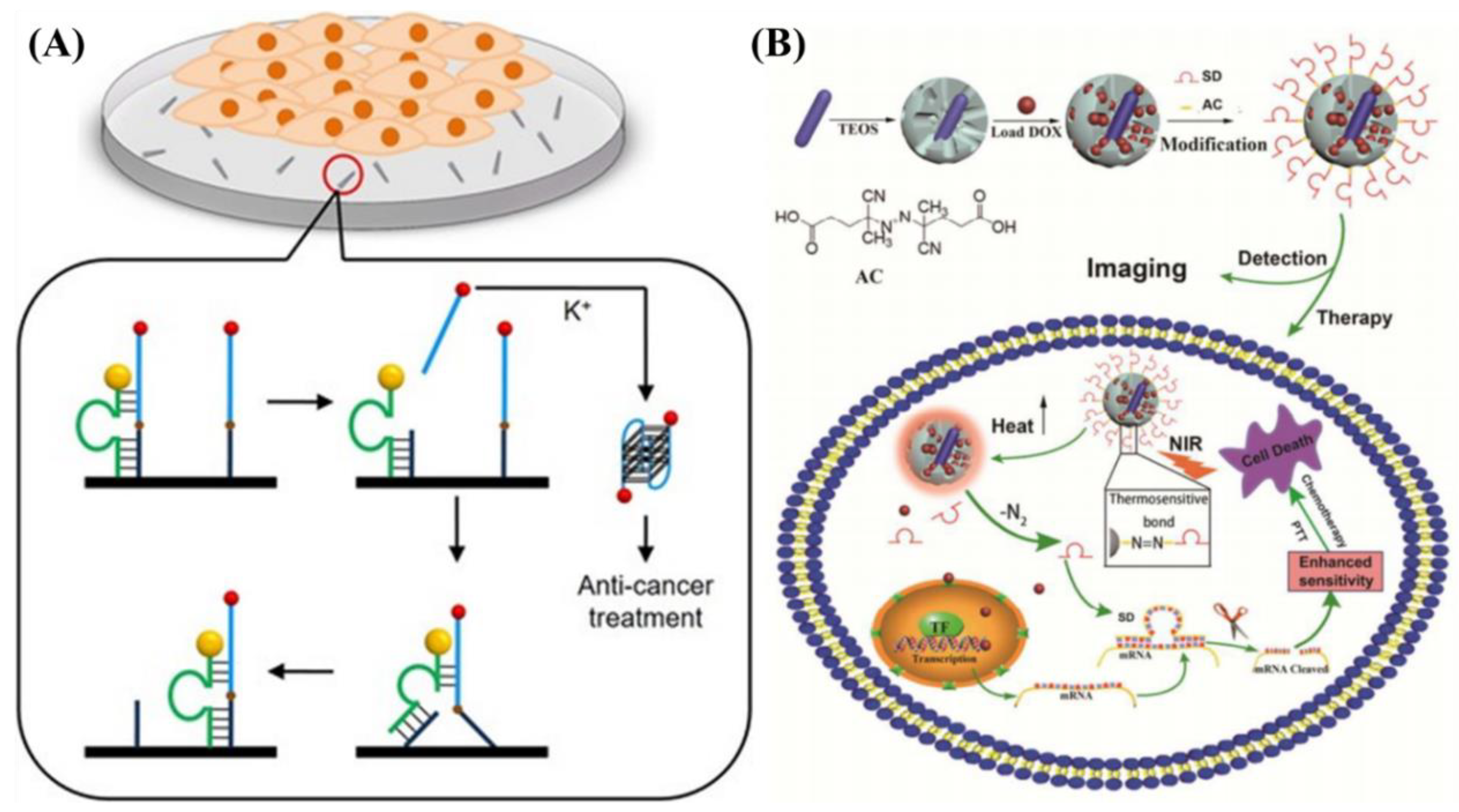
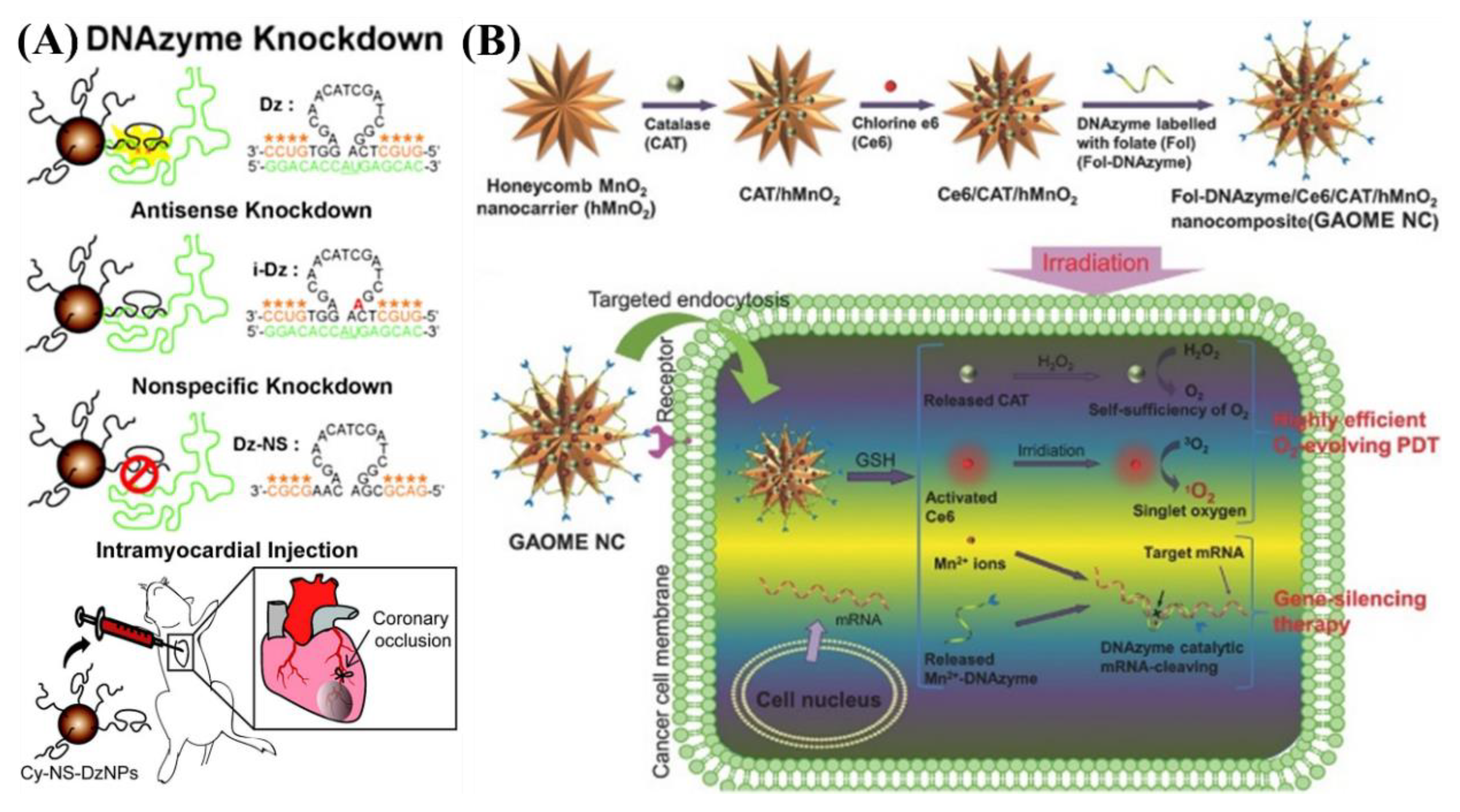
5.1. In Vivo Cancer Therapy

Cancer is one of the most life-threatening diseases in the world, and the development of an efficient therapeutic reagent for cancer therapy is of great importance in cancer research [79,80,81]. In addition to the use of conventional chemotherapeutic drugs for cancer therapy, RCDs have recently been evidentially recognized as a new type of therapeutic agent [82,83,84,85]. Li et al. designed a DNAzyme-based walker system [82] that could control the release of the oligonucleotide drug AS1411 for breast cancer treatment (Figure 8A). In this system, 10–23 DNAzyme (green) labeled with a CdTe/CdS QD (orange) was used as the walker, and moved along the carbon nanotube track (black), leading to the release of anticancer AS1411 from the track’s surface. The released AS1411 could subsequently form a dimeric G-quadruplex structure with the assistance of K+ ions, resulting in the antiproliferation of MCF-7 cells. Later, Jin et al. [83] developed a biodegradable cancer therapeutic system based on a DNA nanoflower (DNF) through the assembly of an aptamer (AS1411) with two DNAzymes (an early growth response-1 DNAzyme and a survivin DNAzyme) on a long single-stranded DNA template. The formed DNF exhibits multiple built-in functions, including targeted cancer cell recognition, dual gene silencing, induction of apoptosis, and inhibition of tumor growth, suggesting great promise for enhanced cancer therapy. Using a similar strategy, the same group further developed a DNAzyme–nanosponge therapeutic platform for the highly efficient photothermal treatment of cancer through catalytically restraining the expression of the survival protein [84].
To overcome therapeutic resistance, an RCDs-based combinational therapeutic strategy was recently reported by Sun et al. [85], in which mesoporous silica-coated gold nanorods were functionalized with a survivin DNAzyme using a thermal-sensitive small molecule to cap the anticancer DOX drug (Figure 8B). This nanosystem showed the NIR-light-activated release of both the anticancer DOX drug and the survivin DNAzyme, resulting in enhanced combinational photothermal therapy, chemotherapy, and gene therapy. Another innovative method for cancer therapy was reported by Yu et al. [86], in which Mg2+-dependent DNAzyme was used as a gatekeeper and trigger for the controlled release of encapsulated therapeutic agents in hollow mesoporous magnesium silicate nanoparticles (HMMSNs). The HMMSNs showed a pH-accelerated biodegradation property, and can be easily excreted via feces and urine, guaranteeing their further clinical translation.

5.2. In Vivo Gene Regulation

Gene regulation is emerging as a promising strategy for the treatment of many diseases, such as cancers, genetic disorders, and virus infections [87]. Up to now, different types of candidates have been used for gene regulation, among which DNAzymes are the most attractive agents due to their inherent advantages of synthetic accessibility, easy modification, and relatively good in vivo stability. Ryoo et al. [87] developed a functional magnetic-nanoparticle-based therapeutic DNAzyme delivery system for the treatment of hepatitis C by inducing knockdown of the hepatitis C virus gene (NS3). In vivo studies showed that the RCDs-based nanoprobes could accumulate in the liver, and more specifically, in hepatocytes for efficient NS3 knockdown. Later, Somasuntharam et al. [88] utilized DNAzyme-conjugated AuNPs to catalytically silence tumor necrosis factor-α (TNF-α) in vivo as a potential therapeutic for myocardial infarction (Figure 9A). This RCDs-based AuNPs probe identifies significant anti-inflammatory effects via suppressing the pro-inflammatory cytokine TNF-α, showing great potential as a viable therapeutic avenue for the treatment of acute cardiac dysfunction. Recently, Li et al. [89] went a step further to develop a novel RCDs-based nanoprobe for multiplexed gene silencing in living cells and in vivo using cobalt oxyhydroxide nanosheets functionalized with three types of DNAzymes. Taking advantage of their good stability, resistance to nuclease cleavage, high selectivity, and excellent biocompatibility, the RCDs-based nanoprobe exhibits a significantly enhanced gene knockdown efficacy and effective in vivo growth inhibition of MCF-7 tumors.
Another promising advantage of RCDs-based gene regulation is its capability to integrate with other therapeutic modalities for synergetic therapy. For instance, He et al. [90] designed a multifunctional hybrid system based on a glutathione (GSH)-activatable and O2/Mn2+-evolving nanocomposite for selective and highly efficient PDT as well as gene-silencing therapy (Figure 9B). The nanocomposite showed enhanced PDT efficacy due to the self-sufficiency of O2 and the consumption of GSH, as well as improved gene-silencing therapy due to the self-generated Mn2+ ions as DNAzyme cofactors.

5.3. Other In Vivo Applications

In addition to the aforementioned in vivo applications, RCDs-based functional probes have also been applied for other therapeutic applications. For instance, Popp et al. [91] developed a RCDs-based probe for specifically blocking the transcription factor GATA-3 and thereby reducing colitis in mice. In this system, the GATA3-specific DNAzyme was delivered directly to the gut and showed efficient distribution throughout the colon in oxazolone colitis, resulting in the suppressed expression of GATA3 mRNA in T cells.

6. Clinical Trials of RNA-Cleaving DNAzymes

As discussed above, RNA-Cleaving DNAzymes represent a promising new class of DNA-based therapeutic agents. During the last decade, several RCDs-based drugs have undergone clinical trials with signs of efficacy and no serious adverse events attributable to the DNAzyme [92,93,94]. Among these RCDs-based drugs, SB010 and DZ13 for the treatment of skin diseases have been extensively studied [95,96]. The active drug substance in SB010 is the GATA-3-specific DNAzyme hgd40, which could specifically bind to and subsequently cleave the messenger RNA of a transcription factor GATA-3. The hgd40 DNAzyme was selected by Sel et al. via in vitro cleavage assays from 70 unmodified DNAzymes containing the catalytic motif of the 10–23 DNAzyme and GATA-3 mRNA as the substrate strand [97]. Up to now, several preclinical studies have demonstrated that hgd40 could efficiently downregulate the Th2 cytokine level via inhibition of GATA-3 mRNA in various animal models [97,98], resulting in significant improvement in asthma symptoms with no observable off-target effect [99,100]. Another preclinical study, performed by Turowska et al., indicated a time- and dose-dependent systemic bioavailability profile of hgd40 with similar pharmacokinetics in mice, rats, and dogs [98]. Built upon the above success in preclinical studies, four phase I/II clinical trials for SB010 have been performed and completed recently (ClinicalTrials.gov number: NCT01743768, NCT01577953, NCT01470911, NCT01554319). For example, Krug et al. (NCT01743768) conducted a randomized, double-blind, placebo-controlled, multicenter clinical trial of SB010, which involved patients who had allergic asthma with sputum eosinophilia and who also had biphasic early and late asthmatic responses after laboratory-based allergen provocation [101]. The results showed that the treatment with SB010 significantly attenuated both late and early asthmatic responses after allergen provocation in patients with allergic asthma. Very recently, Greulich et al. [102] conducted a randomized, double-blind, placebo-controlled, multi-centre clinical trial for SB010 in chronic obstructive pulmonary disease (COPD)-patients with sputum eosinophilia, showing reduced sputum eosinophils through inhalation of SB010, though further long-term studies are required to verify the clinical efficacy. Additionally, the hgd40-based drugs SB011 and SB012 have also been clinically evaluated for lesional skin in patients with atopic eczema (NCT02079688) and active ulcerative colitis patients (NCT02129439), respectively.
Another promising RCDs-based drug under clinical trials is Dz13, which specifically targets and cleaves JUN mRNA, resulting in the inhibition of c-Jun protein expression in human atherosclerotic lesions [92]. The first-in-human study of Dz13 in patients with nodular basal-cell carcinoma was reported by Cho et al. [103], in which they performed a phase I study to assess the safety and tolerability of Dz13 in human beings. All nine recruited patients exhibited no drug-related serious adverse events, indicating that Dz13 was well-tolerated and led to negligible adverse effects in human beings. Although the primary endpoint of this study was the safety and tolerability of Dz13 rather than the clinical response, it takes a pivotal step towards bringing Dz13 from basic research to the clinic. Taking together the above examples of clinical trials, RCDs-based drugs are showing a promising future but are still far from satisfactory in clinical uses.

7. Conclusions and Perspectives

In summary, I have provided an overview of the fundamental and technical tricks for the design of RNA-Cleaving DNAzymes-based functional probes for intracellular and in vivo applications. The recent advance in the structural characterization of RNA-cleaving DNAzymes provides a more fundamental understanding of the catalytic mechanism of the RCDs, which is of significant benefit for the rational design of RCDs-based probes for a functional need in catalysis for diverse applications. Compared with antibodies that often serve as biorecognition and therapeutic agents for in vivo diagnostic and therapeutic purposes, the RCDs could serve as biorecognition agents, signal amplification agents, and activatable hosting agents. Because of these inherent functions, the RCDs have been successfully translated into various functional probes, and demonstrated their promising applications that range from intracellular molecular sensing and imaging to in vivo therapeutic applications. Building upon the success in these preclinical studies, increasing efforts have been made to apply DNAzymes in clinical trials, and I anticipate that this developing research field will have a significant impact on advanced healthcare sciences in the future.
Although great progress has been achieved, there are still some obstacles that need to be addressed before translating RNA-cleaving DNAzymes from basic research to clinical uses. First, the long-term biostability of RCDs is unsatisfactory for a therapeutic purpose. Some strategies have been proposed to improve the long-term biostability of RCDs via their integration with nanomaterials or chemical modifications [50,104,105,106]; however, these methods may introduce other long-term biosafety risks, such as toxicity and inflammation in regards to nanomaterials, requiring further extensive investigation before clinical applications. One elegant strategy is to select the RCDs directly from the biological fluid, such as serum or blood, which could provide more stable RCDs that still maintain their catalytic functions. Second, knowledge of the in vivo pharmacokinetics of RCDs-based drugs remains at the early stage in animal studies, with even less for human beings. Therefore, much work lies ahead to investigate their delivery, in vivo circulation and biodistribution, and metabolism in human beings to offer fundamental insights for further clinical transition [107]. Finally, most of the current therapeutic RCDs-based drugs are designed for skin diseases, and it is important to design more RCDs-based functional probes for other cancer therapies and, more importantly, combinational therapeutics. With further developments and improvements, RNA-Cleaving DNAzymes will provide a breakthrough for diagnostic and therapeutic applications in cancer research.

Funding

This research was funded by the National Institutes of Health (grant numbers 1R43HD092155-01 and 1R43HL142326-01A1).

Acknowledgments

This work was supported by GlucoSentient, Inc. and the Beckman Institute for Advanced Science and Technology at the University of Illinois at Urbana-Champaign.

Conflicts of Interest

The author declares no conflict of interest.

References

  1. Ward, W.L.; Plakos, K.; DeRose, V.J. Nucleic acid catalysis: Metals, nucleobases, and other cofactors. Chem. Rev. 2014, 114, 4318–4342. [Google Scholar] [CrossRef] [PubMed]
  2. Breaker, R.R.; Joyce, G.F. A DNA enzyme that cleaves RNA. Chem. Biol. 1994, 1, 223–229. [Google Scholar] [CrossRef]
  3. Santoro, S.W.; Joyce, G.F. A general purpose RNA-cleaving DNA enzyme. Proc. Natl. Acad. Sci. USA 1997, 94, 4262–4266. [Google Scholar] [CrossRef] [PubMed] [Green Version]
  4. Silverman, S.K. Deoxyribozymes: Selection design and serendipity in the development of DNA catalysts. Acc. Chem. Res. 2009, 42, 1521–1531. [Google Scholar] [CrossRef] [PubMed]
  5. Liu, J.; Cao, Z.; Lu, Y. Functional nucleic acid sensors. Chem. Rev. 2009, 109, 1948–1998. [Google Scholar] [CrossRef] [PubMed]
  6. Silverman, S.K. Catalytic DNA: Scope, Applications, and Biochemistry of Deoxyribozymes. Trends Biochem. Sci. 2016, 41, 595–609. [Google Scholar] [CrossRef] [PubMed] [Green Version]
  7. Zhang, J.; Lu, Y. Biocomputing for Portable, Resettable, and Quantitative Point-of-Care Diagnostics: Making the Glucose Meter a Logic-Gate Responsive Device for Measuring Many Clinically Relevant Targets. Angew. Chem. 2018, 57, 9702–9706. [Google Scholar] [CrossRef] [PubMed]
  8. Zhang, J.J.; Xing, H.; Lu, Y. Translating molecular detections into a simple temperature test using a target-responsive smart thermometer. Chem. Sci. 2018, 9, 3906–3910. [Google Scholar] [CrossRef] [PubMed] [Green Version]
  9. Peng, H.; Newbigging, A.M.; Wang, Z.; Tao, J.; Deng, W.; Le, X.C.; Zhang, H. DNAzyme-Mediated Assays for Amplified Detection of Nucleic Acids and Proteins. Anal. Chem. 2018, 90, 190–207. [Google Scholar] [CrossRef] [PubMed]
  10. Baum, D.A.; Silverman, S.K. Deoxyribozymes: Useful DNA catalysts in vitro and in vivo. Cell. Mol. Life Sci. 2008, 65, 2156–2174. [Google Scholar] [CrossRef] [PubMed]
  11. Zhou, W.; Ding, J.; Liu, J. Theranostic DNAzymes. Theranostics 2017, 7, 1010–1025. [Google Scholar] [CrossRef] [PubMed] [Green Version]
  12. Potaczek, D.P.; Unger, S.D.; Zhang, N.; Taka, S.; Michel, S.; Akdag, N.; Lan, F.; Helfer, M.; Hudemann, C.; Eickmann, M.; et al. Development and characterization of DNAzyme candidates demonstrating significant efficiency against human rhinoviruses. J. Allergy Clin. Immunol. 2018. [Google Scholar] [CrossRef] [PubMed]
  13. Morrison, D.; Rothenbroker, M.; Li, Y.F. DNAzymes: Selected for Applications. Small Methods 2018, 2, 1700319. [Google Scholar] [CrossRef]
  14. Hwang, K.; Hosseinzadeh, P.; Lu, Y. Biochemical and Biophysical Understanding of Metal Ion Selectivity of DNAzymes. Inorg. Chim. Acta 2016, 452, 12–24. [Google Scholar] [CrossRef] [PubMed]
  15. Liu, M.; Chang, D.; Li, Y. Discovery and Biosensing Applications of Diverse RNA-Cleaving DNAzymes. Acc. Chem. Res. 2017, 50, 2273–2283. [Google Scholar] [CrossRef] [PubMed]
  16. Hollenstein, M. DNA Catalysis: The Chemical Repertoire of DNAzymes. Molecules 2015, 20, 20777–20804. [Google Scholar] [CrossRef] [PubMed] [Green Version]
  17. Gong, L.; Zhao, Z.; Lv, Y.F.; Huan, S.Y.; Fu, T.; Zhang, X.B.; Shen, G.L.; Yu, R.Q. DNAzyme-based biosensors and nanodevices. Chem. Commun. 2015, 51, 979–995. [Google Scholar] [CrossRef] [PubMed]
  18. Zhou, W.; Liu, J. Multi-metal-dependent nucleic acid enzymes. Metallomics 2018, 10, 30–48. [Google Scholar] [CrossRef] [PubMed]
  19. Wiraja, C.; Yeo, D.C.; Lio, D.; Zheng, M.; Xu, C. Functional Imaging with Nucleic Acid-based Sensors: Technology, Application and Future Healthcare Prospects. Chembiochem 2018. [Google Scholar] [CrossRef] [PubMed]
  20. Müller, S.; Appel, B.; Balke, D.; Hieronymus, R.; Nübel, C. Thirty-five years of research into ribozymes and nucleic acid catalysis: Where do we stand today? F1000Research 2016, 5, 1511. [Google Scholar] [CrossRef] [PubMed]
  21. Lan, T.; Lu, Y. Metal Ion-Dependent DNAzymes and Their Applications as Biosensors. In Interplay between Metal Ions and Nucleic Acids; Sigel, A., Sigel, H., Sigel, R.K.O., Eds.; Springer: Dordrecht, The Netherlands, 2012; pp. 217–248. [Google Scholar]
  22. Nowakowski, J.; Shim, P.J.; Joyce, G.F.; Stout, C.D. Crystallization of the 10–23 DNA enzyme using a combinatorial screen of paired oligonucleotides. Acta Crystallogr. Sect. D Biol. Crystallogr. 1999, 55, 1885–1892. [Google Scholar] [CrossRef]
  23. Nowakowski, J.; Shim, P.J.; Prasad, G.S.; Stout, C.D.; Joyce, G.F. Crystal structure of an 82-nucleotide RNA-DNA complex formed by the 10–23 DNA enzyme. Nat. Struct. Biol. 1999, 6, 151–156. [Google Scholar] [PubMed]
  24. Parkinson, G.N.; Lee, M.P.H.; Neidle, S. Crystal structure of parallel quadruplexes from human telomeric DNA. Nature 2002, 417, 876–880. [Google Scholar] [CrossRef] [PubMed]
  25. Kondo, J.; Yamada, T.; Hirose, C.; Okamoto, I.; Tanaka, Y.; Ono, A. Crystal Structure of Metallo DNA Duplex Containing Consecutive Watson-Crick-like T-Hg-II-T Base Pairs. Angew. Chem. 2014, 53, 2385–2388. [Google Scholar] [CrossRef] [PubMed]
  26. Ponce-Salvatierra, A.; Wawrzyniak-Turek, K.; Steuerwald, U.; Hobartner, C.; Pena, V. Crystal structure of a DNA catalyst. Nature 2016, 529, 231–234. [Google Scholar] [CrossRef] [PubMed]
  27. Liu, H.H.; Yu, X.; Chen, Y.Q.; Zhang, J.; Wu, B.X.; Zheng, L.N.; Haruehanroengra, P.; Wang, R.; Li, S.H.; Lin, J.Z.; et al. Crystal structure of an RNA-cleaving DNAzyme. Nat. Commun. 2017, 8. [Google Scholar] [CrossRef] [PubMed]
  28. Wirmer-Bartoschek, J.; Schwalbe, H. Understanding How DNA Enzymes Work. Angew. Chem. 2016, 55, 5376–5377. [Google Scholar] [CrossRef] [PubMed]
  29. Zhang, J.J.; Cheng, F.F.; Zheng, T.T.; Zhu, J.J. Versatile aptasensor for electrochemical quantification of cell surface glycan and naked-eye tracking glycolytic inhibition in living cells. Biosens. Bioelectron. 2017, 89, 937–945. [Google Scholar] [CrossRef] [PubMed]
  30. Wang, H.; Li, C.; Liu, X.; Zhou, X.; Wang, F. Construction of an enzyme-free concatenated DNA circuit for signal amplification and intracellular imaging. Chem. Sci. 2018, 9, 5842–5849. [Google Scholar] [CrossRef] [PubMed]
  31. Giannetti, A.; Tombelli, S.; Baldini, F. Oligonucleotide optical switches for intracellular sensing. Anal. Bioanal. Chem. 2013, 405, 6181–6196. [Google Scholar] [CrossRef] [PubMed]
  32. Zhang, J.J.; Cheng, F.F.; Li, J.J.; Zhu, J.J.; Lu, Y. Fluorescent nanoprobes for sensing and imaging of metal ions: Recent advances and future perspectives. Nano Today 2016, 11, 309–329. [Google Scholar] [CrossRef] [PubMed] [Green Version]
  33. Dean, K.M.; Qin, Y.; Palmer, A.E. Visualizing metal ions in cells: An overview of analytical techniques, approaches, and probes. Biochim. Biophys. Acta 2012, 1823, 1406–1415. [Google Scholar] [CrossRef] [PubMed]
  34. Wu, P.; Hwang, K.; Lan, T.; Lu, Y. A DNAzyme-gold nanoparticle probe for uranyl ion in living cells. J. Am. Chem. Soc. 2013, 135, 5254–5257. [Google Scholar] [CrossRef] [PubMed]
  35. Zhai, T.T.; Ye, D.; Shi, Y.; Zhang, Q.W.; Qin, X.; Wang, C.; Xia, X.H. Plasmon Coupling Effect-Enhanced Imaging of Metal Ions in Living Cells Using DNAzyme Assembled Core-Satellite Structures. ACS Appl. Mater. Interfaces 2018, 10, 33966–33975. [Google Scholar] [CrossRef] [PubMed]
  36. Zhu, D.; Zhao, D.X.; Huang, J.X.; Li, J.; Zuo, X.L.; Wang, L.H.; Fan, C.H. Protein-mimicking nanoparticle (Protmin)-based nanosensor for intracellular analysis of metal ions. Nucl. Sci. Technol. 2018, 29, 5. [Google Scholar] [CrossRef]
  37. Zhang, L.; Yin, Q.-H.; Li, J.-M.; Huang, H.-Y.; Wu, Q.; Mao, Z.-W. Functionalization of dendritic polyethylene with cationic poly(p-phenylene ethynylene) enables efficient siRNA delivery for gene silencing. J. Mater. Chem. B 2013, 1, 2245–2251. [Google Scholar] [CrossRef]
  38. Li, L.; Feng, J.; Fan, Y.; Tang, B. Simultaneous imaging of Zn(2+) and Cu(2+) in living cells based on DNAzyme modified gold nanoparticle. Anal. Chem. 2015, 87, 4829–4835. [Google Scholar] [CrossRef] [PubMed]
  39. Si, H.; Sheng, R.; Li, Q.; Feng, J.; Li, L.; Tang, B. Highly Sensitive Fluorescence Imaging of Zn(2+) and Cu(2+) in Living Cells with Signal Amplification Based on Functional DNA Self-Assembly. Anal. Chem. 2018, 90, 8785–8792. [Google Scholar] [CrossRef] [PubMed]
  40. Zhou, W.; Liang, W.; Li, D.; Yuan, R.; Xiang, Y. Dual-color encoded DNAzyme nanostructures for multiplexed detection of intracellular metal ions in living cells. Biosens. Bioelectron. 2016, 85, 573–579. [Google Scholar] [CrossRef] [PubMed]
  41. Yang, C.; Yin, X.; Huan, S.Y.; Chen, L.; Hu, X.X.; Xiong, M.Y.; Chen, K.; Zhang, X.B. Two-Photon DNAzyme-Gold Nanoparticle Probe for Imaging Intracellular Metal Ions. Anal. Chem. 2018, 90, 3118–3123. [Google Scholar] [CrossRef] [PubMed]
  42. Hong, C.Y.; Wu, S.X.; Li, S.H.; Liang, H.; Chen, S.; Li, J.; Yang, H.H.; Tan, W. Semipermeable Functional DNA-Encapsulated Nanocapsules as Protective Bioreactors for Biosensing in Living Cells. Anal. Chem. 2017, 89, 5389–5394. [Google Scholar] [CrossRef] [PubMed]
  43. Hwang, K.; Wu, P.W.; Kim, T.; Lei, L.; Tian, S.L.; Wang, Y.X.; Lu, Y. Photocaged DNAzymes as a General Method for Sensing Metal Ions in Living Cells. Angew. Chem. 2014, 53, 13798–13802. [Google Scholar] [CrossRef] [PubMed] [Green Version]
  44. Wang, W.; Satyavolu, N.S.R.; Wu, Z.; Zhang, J.R.; Zhu, J.J.; Lu, Y. Near-Infrared Photothermally Activated DNAzyme-Gold Nanoshells for Imaging Metal Ions in Living Cells. Angew. Chem. 2017, 56, 6798–6802. [Google Scholar] [CrossRef] [PubMed]
  45. Wang, X.; Feng, M.; Xiao, L.; Tong, A.; Xiang, Y. Postsynthetic Modification of DNA Phosphodiester Backbone for Photocaged DNAzyme. ACS Chem. Biol. 2016, 11, 444–451. [Google Scholar] [CrossRef] [PubMed]
  46. Wu, Z.; Fan, H.; Satyavolu, N.S.R.; Wang, W.; Lake, R.; Jiang, J.H.; Lu, Y. Imaging Endogenous Metal Ions in Living Cells Using a DNAzyme-Catalytic Hairpin Assembly Probe. Angew. Chem. 2017, 56, 8721–8725. [Google Scholar] [CrossRef] [PubMed]
  47. Ma, Z.; Wu, X.; Krueger, C.J.; Chen, A.K. Engineering Novel Molecular Beacon Constructs to Study Intracellular RNA Dynamics and Localization. Genom. Proteom. Bioinform. 2017, 15, 279–286. [Google Scholar] [CrossRef] [PubMed]
  48. Pitchiaya, S.; Heinicke, L.A.; Custer, T.C.; Walter, N.G. Single molecule fluorescence approaches shed light on intracellular RNAs. Chem. Rev. 2014, 114, 3224–3265. [Google Scholar] [CrossRef] [PubMed]
  49. Santangelo, P.J. Molecular beacons and related probes for intracellular RNA imaging. Wiley Interdiscip. Rev. Nanomed. Nanobiotechnol. 2010, 2, 11–19. [Google Scholar] [CrossRef] [PubMed]
  50. Chen, F.; Bai, M.; Zhao, Y.; Cao, K.; Cao, X.; Zhao, Y. MnO2-Nanosheet-Powered Protective Janus DNA Nanomachines Supporting Robust RNA Imaging. Anal. Chem. 2018, 90, 2271–2276. [Google Scholar] [CrossRef] [PubMed]
  51. He, D.; He, X.; Yang, X.; Li, H.W. A smart ZnO@polydopamine-nucleic acid nanosystem for ultrasensitive live cell mRNA imaging by the target-triggered intracellular self-assembly of active DNAzyme nanostructures. Chem. Sci. 2017, 8, 2832–2840. [Google Scholar] [CrossRef] [PubMed] [Green Version]
  52. Bakshi, S.F.; Guz, N.; Zakharchenko, A.; Deng, H.; Tumanov, A.V.; Woodworth, C.D.; Minko, S.; Kolpashchikov, D.M.; Katz, E. Nanoreactors based on DNAzyme-functionalized magnetic nanoparticles activated by magnetic field. Nanoscale 2018, 10, 1356–1365. [Google Scholar] [CrossRef] [PubMed]
  53. Makarova, J.A.; Shkurnikov, M.U.; Wicklein, D.; Lange, T.; Samatov, T.R.; Turchinovich, A.A.; Tonevitsky, A.G. Intracellular and extracellular microRNA: An update on localization and biological role. Prog. Histochem. Cytochem. 2016, 51, 33–49. [Google Scholar] [CrossRef] [PubMed]
  54. Hernandez, R.; Orbay, H.; Cai, W. Molecular Imaging Strategies for In Vivo Tracking of MicroRNAs: A Comprehensive Review. Curr. Med. Chem. 2013, 20, 3594–3603. [Google Scholar] [CrossRef] [PubMed]
  55. Zhang, P.; He, Z.; Wang, C.; Chen, J.; Zhao, J.; Zhu, X.; Li, C.Z.; Min, Q.; Zhu, J.J. In situ amplification of intracellular microRNA with MNAzyme nanodevices for multiplexed imaging, logic operation, and controlled drug release. ACS Nano 2015, 9, 789–798. [Google Scholar] [CrossRef] [PubMed]
  56. Belter, A.; Rolle, K.; Piwecka, M.; Fedoruk-Wyszomirska, A.; Naskret-Barciszewska, M.Z.; Barciszewski, J. Inhibition of miR-21 in glioma cells using catalytic nucleic acids. Sci. Rep. 2016, 6, 24516. [Google Scholar] [CrossRef] [PubMed] [Green Version]
  57. Wu, Y.; Huang, J.; Yang, X.; Yang, Y.; Quan, K.; Xie, N.; Li, J.; Ma, C.; Wang, K. Gold Nanoparticle Loaded Split-DNAzyme Probe for Amplified miRNA Detection in Living Cells. Anal. Chem. 2017, 89, 8377–8383. [Google Scholar] [CrossRef] [PubMed]
  58. Yi, J.T.; Chen, T.T.; Huo, J.; Chu, X. Nanoscale Zeolitic Imidazolate Framework-8 for Ratiometric Fluorescence Imaging of MicroRNA in Living Cells. Anal. Chem. 2017, 89, 12351–12359. [Google Scholar] [CrossRef] [PubMed]
  59. Liu, J.; Cui, M.; Zhou, H.; Yang, W. DNAzyme Based Nanomachine for in Situ Detection of MicroRNA in Living Cells. ACS Sens. 2017, 2, 1847–1853. [Google Scholar] [CrossRef] [PubMed]
  60. Yang, Y.; Huang, J.; Yang, X.; He, X.; Quan, K.; Xie, N.; Ou, M.; Wang, K. Gold Nanoparticle Based Hairpin-Locked-DNAzyme Probe for Amplified miRNA Imaging in Living Cells. Anal. Chem. 2017, 89, 5850–5856. [Google Scholar] [CrossRef] [PubMed]
  61. Peng, H.; Li, X.F.; Zhang, H.; Le, X.C. A microRNA-initiated DNAzyme motor operating in living cells. Nat. Commun. 2017, 8, 14378. [Google Scholar] [CrossRef] [PubMed]
  62. Mahdiannasser, M.; Karami, Z. An innovative paradigm of methods in microRNAs detection: Highlighting DNAzymes, the illuminators. Biosens. Bioelectron. 2018, 107, 123–144. [Google Scholar] [CrossRef] [PubMed]
  63. Dong, J.T.; Zhao, M.P. In-vivo fluorescence imaging of adenosine 5’-triphosphate. TrAC Trensd Anal. Chem. 2016, 80, 190–203. [Google Scholar] [CrossRef]
  64. Yang, Y.; Huang, J.; Yang, X.; Quan, K.; Wang, H.; Ying, L.; Xie, N.; Ou, M.; Wang, K. Aptazyme-Gold Nanoparticle Sensor for Amplified Molecular Probing in Living Cells. Anal. Chem. 2016, 88, 5981–5987. [Google Scholar] [CrossRef] [PubMed]
  65. Gao, F.L.; Wu, J.; Yao, Y.; Zhang, Y.; Liao, X.J.; Geng, D.Q.; Tang, D.Q. Proximity hybridization triggered strand displacement and DNAzyme assisted strand recycling for ATP fluorescence detection in vitro and imaging in living cells. RSC Adv. 2018, 8, 28161–28171. [Google Scholar] [CrossRef]
  66. Fan, H.; Zhang, X.; Lu, Y. Recent advances in DNAzyme-based gene silencing. Sci. China Chem. 2017, 60, 591–601. [Google Scholar] [CrossRef] [Green Version]
  67. Young, D.D.; Lively, M.O.; Deiters, A. Activation and Deactivation of DNAzyme and Antisense Function with Light for the Photochemical Regulation of Gene Expression in Mammalian Cells. J. Am. Chem. Soc. 2010, 132, 6183–6193. [Google Scholar] [CrossRef] [PubMed] [Green Version]
  68. Yehl, K.; Joshi, J.R.; Greene, B.L.; Dyer, R.B.; Nahta, R.; Salaita, K. Catalytic Deoxyribozyme-Modified Nanoparticles for RNAi-Independent Gene Regulation. ACS Nano 2012, 6, 9150–9157. [Google Scholar] [CrossRef] [PubMed] [Green Version]
  69. Hartmann, A.K.; Cairns-Gibson, D.F.; Santiana, J.J.; Tolentino, M.Q.; Barber, H.M.; Rouge, J.L. Enzymatically Ligated DNA-Surfactants: Unmasking Hydrophobically Modified DNA for Intracellular Gene Regulation. ChemBioChem 2018, 19, 1734–1739. [Google Scholar] [CrossRef] [PubMed]
  70. Awino, J.K.; Gudipati, S.; Hartmann, A.K.; Santiana, J.J.; Cairns-Gibson, D.F.; Gomez, N.; Rouge, J.L. Nucleic Acid Nanocapsules for Enzyme-Triggered Drug Release. J. Am. Chem. Soc. 2017, 139, 6278–6281. [Google Scholar] [CrossRef] [PubMed]
  71. Zokaei, E.; Badoei-Dalfrad, A.; Ansari, M.; Karami, Z.; Eslaminejad, T.; Nematollahi-Mahani, S.N. Therapeutic Potential of DNAzyme Loaded on Chitosan/Cyclodextrin Nanoparticle to Recovery of Chemosensitivity in the MCF-7 Cell Line. Appl. Biochem. Biotechnol. 2018. [Google Scholar] [CrossRef] [PubMed]
  72. Fan, H.; Zhao, Z.; Yan, G.; Zhang, X.; Yang, C.; Meng, H.; Chen, Z.; Liu, H.; Tan, W. A smart DNAzyme-MnO(2) nanosystem for efficient gene silencing. Angew. Chem. 2015, 54, 4801–4805. [Google Scholar] [CrossRef] [PubMed]
  73. He, Z.M.; Zhang, P.H.; Li, X.; Zhang, J.R.; Zhu, J.J. A Targeted DNAzyme-Nanocomposite Probe Equipped with Built-in Zn(2+) Arsenal for Combined Treatment of Gene Regulation and Drug Delivery. Sci. Rep. 2016, 6, 22737. [Google Scholar] [CrossRef] [PubMed]
  74. Li, D.; Mo, F.; Wu, J.; Huang, Y.; Zhou, H.; Ding, S.; Chen, W. A multifunctional DNA nano-scorpion for highly efficient targeted delivery of mRNA therapeutics. Sci. Rep. 2018, 8, 10196. [Google Scholar] [CrossRef] [PubMed]
  75. Chen, F.; Bai, M.; Cao, K.; Zhao, Y.; Cao, X.W.; Wei, J.; Wu, N.; Li, J.; Wang, L.H.; Fan, C.H.; et al. Programming Enzyme-Initiated Autonomous DNAzyme Nanodevices in Living Cells. ACS Nano 2017, 11, 11908–11914. [Google Scholar] [CrossRef] [PubMed]
  76. Li, H.; Wang, M.; Shi, T.H.; Yang, S.H.; Zhang, J.H.; Wang, H.H.; Nie, Z. A DNA-Mediated Chemically Induced Dimerization (D-CID) Nanodevice for Nongenetic Receptor Engineering To Control Cell Behavior. Angew. Chem. 2018, 57, 10226–10230. [Google Scholar] [CrossRef] [PubMed]
  77. Chen, F.; Bai, M.; Cao, K.; Zhao, Y.; Wei, J.; Zhao, Y.X. Fabricating MnO2 Nanozymes as Intracellular Catalytic DNA Circuit Generators for Versatile Imaging of Base-Excision Repair in Living Cells. Adv. Funct. Mater. 2017, 27, 1702748. [Google Scholar] [CrossRef]
  78. Chen, X.; Zhao, J.; Chen, T.; Gao, T.; Zhu, X.; Li, G. Nondestructive Analysis of Tumor-Associated Membrane Protein Integrating Imaging and Amplified Detection in situ Based on Dual-Labeled DNAzyme. Theranostics 2018, 8, 1075–1083. [Google Scholar] [CrossRef] [PubMed] [Green Version]
  79. Zhang, J.J.; Zheng, T.T.; Cheng, F.F.; Zhang, J.R.; Zhu, J.J. Toward the Early Evaluation of Therapeutic Effects: An Electrochemical Platform for Ultrasensitive Detection of Apoptotic Cells. Anal. Chem. 2011, 83, 7902–7909. [Google Scholar] [CrossRef] [PubMed]
  80. Liu, K.P.; Zhang, J.J.; Cheng, F.F.; Zheng, T.T.; Wang, C.M.; Zhu, J.J. Green and facile synthesis of highly biocompatible graphene nanosheets and its application for cellular imaging and drug delivery. J. Mater. Chem. 2011, 21, 12034–12040. [Google Scholar] [CrossRef]
  81. Zhang, J.J.; Gu, M.M.; Zheng, T.T.; Zhu, J.J. Synthesis of Gelatin-Stabilized Gold Nanoparticles and Assembly of Carboxylic Single-Walled Carbon Nanotubes/Au Composites for Cytosensing and Drug Uptake. Anal. Chem. 2009, 81, 6641–6648. [Google Scholar] [CrossRef] [PubMed]
  82. Li, F.R.; Cha, T.G.; Pan, J.; Ozcelikkale, A.; Han, B.; Choi, J.H. DNA Walker-Regulated Cancer Cell Growth Inhibition. ChemBioChem 2016, 17, 1138–1141. [Google Scholar] [CrossRef] [PubMed] [Green Version]
  83. Jin, Y.; Li, Z.H.; Liu, H.F.; Chen, S.Z.; Wang, F.; Wang, L.; Li, N.; Ge, K.; Yang, X.J.; Liang, X.J.; et al. Biodegradable, multifunctional DNAzyme nanoflowers for enhanced cancer therapy. NPG Asia Mater. 2017, 9, e365. [Google Scholar] [CrossRef]
  84. Jin, Y.; Liang, L.N.; Sun, X.J.; Yu, G.S.; Chen, S.Z.; Shi, S.T.; Liu, H.F.; Li, Z.H.; Ge, K.; Liu, D.D.; et al. Deoxyribozyme-nanosponges for improved photothermal therapy by overcoming thermoresistance. NPG Asia Mater. 2018, 10, 373–384. [Google Scholar] [CrossRef] [Green Version]
  85. Sun, X.; Jin, Y.; Wang, H.; Feng, N.; Li, Z.; Liu, D.; Ge, K.; Liu, H.; Zhang, J.-C.; Yang, X. A NIR-light activated nanoplatform for sensitizing triple negative breast cancer against therapeutic resistance to enhance treatment effect. J. Mater. Chem. B 2018. [Google Scholar] [CrossRef]
  86. Yu, L.; Chen, Y.; Lin, H.; Gao, S.; Chen, H.; Shi, J. Magnesium-Engineered Silica Framework for pH-Accelerated Biodegradation and DNAzyme-Triggered Chemotherapy. Small 2018, 14, e1800708. [Google Scholar] [CrossRef] [PubMed]
  87. Ryoo, S.R.; Jang, H.; Kim, K.S.; Lee, B.; Kim, K.B.; Kim, Y.K.; Yeo, W.S.; Lee, Y.; Kim, D.E.; Min, D.H. Functional delivery of DNAzyme with iron oxide nanoparticles for hepatitis C virus gene knockdown. Biomaterials 2012, 33, 2754–2761. [Google Scholar] [CrossRef]
  88. Somasuntharam, I.; Yehl, K.; Carroll, S.L.; Maxwell, J.T.; Martinez, M.D.; Che, P.L.; Brown, M.E.; Salaita, K.; Davis, M.E. Knockdown of TNF-alpha by DNAzyme gold nanoparticles as an anti-inflammatory therapy for myocardial infarction. Biomaterials 2016, 83, 12–22. [Google Scholar] [CrossRef] [PubMed]
  89. Li, N.; Li, Y.; Gao, X.; Yu, Z.; Pan, W.; Tang, B. Multiplexed gene silencing in living cells and in vivo using a DNAzymes-CoOOH nanocomposite. Chem. Commun. 2017, 53, 4962–4965. [Google Scholar] [CrossRef] [PubMed]
  90. He, D.; Hai, L.; He, X.; Yang, X.; Li, H.-W. Glutathione-Activatable and O2/Mn2+-Evolving Nanocomposite for Highly Efficient and Selective Photodynamic and Gene-Silencing Dual Therapy. Adv. Funct. Mater. 2017, 27, 1704089. [Google Scholar] [CrossRef]
  91. Popp, V.; Gerlach, K.; Mott, S.; Turowska, A.; Garn, H.; Atreya, R.; Lehr, H.A.; Ho, I.C.; Renz, H.; Weigmann, B.; et al. Rectal Delivery of a DNAzyme That Specifically Blocks the Transcription Factor GATA3 and Reduces Colitis in Mice. Gastroenterology 2017, 152, 176–192. [Google Scholar] [CrossRef] [PubMed]
  92. Cai, H.; Cho, E.A.; Li, Y.; Sockler, J.; Parish, C.R.; Chong, B.H.; Edwards, J.; Dodds, T.J.; Ferguson, P.M.; Wilmott, J.S.; et al. Melanoma protective antitumor immunity activated by catalytic DNA. Oncogene 2018, 37, 5115–5126. [Google Scholar] [CrossRef] [PubMed]
  93. Dass, C.R. Deoxyribozymes: Cleaving a path to clinical trials. Trends Pharmacol. Sci. 2004, 25, 395–397. [Google Scholar] [CrossRef] [PubMed]
  94. Monteleone, G.; Fantini, M.C.; Onali, S.; Zorzi, F.; Sancesario, G.; Bernardini, S.; Calabrese, E.; Viti, F.; Monteleone, I.; Biancone, L.; et al. Phase I Clinical Trial of Smad7 Knockdown Using Antisense Oligonucleotide in Patients With Active Crohn’s Disease. Mol. Ther. 2012, 20, 870–876. [Google Scholar] [CrossRef] [PubMed]
  95. Krug, N.; Hohlfeld, J.M.; Buhl, R.; Renz, J.; Garn, H.; Renz, H. Blood eosinophils predict therapeutic effects of a GATA3-specific DNAzyme in asthma patients. J. Allergy Clin. Immunol. 2017, 140, 625–628. [Google Scholar] [CrossRef] [PubMed]
  96. Homburg, U.; Renz, H.; Timmer, W.; Hohlfeld, J.M.; Seitz, F.; Luer, K.; Mayer, A.; Wacker, A.; Schmidt, O.; Kuhlmann, J.; et al. Safety and tolerability of a novel inhaled GATA3 mRNA targeting DNAzyme in patients with T(H)2-driven asthma. J. Allergy Clin. Immunol. 2015, 136, 797–800. [Google Scholar] [CrossRef] [PubMed]
  97. Sel, S.; Wegmann, M.; Dicke, T.; Sel, S.; Henke, W.; Yildirim, A.O.; Renz, H.; Garn, H. Effective prevention and therapy of experimental allergic asthma using a GATA-3-specific DNAzyme. J. Allergy Clin. Immunol. 2008, 121, 910–916. [Google Scholar] [CrossRef] [PubMed]
  98. Turowska, A.; Librizzi, D.; Baumgartl, N.; Kuhlmann, J.; Dicke, T.; Merkel, O.; Homburg, U.; Hoffken, H.; Renz, H.; Garn, H. Biodistribution of the GATA-3-specific DNAzyme hgd40 after inhalative exposure in mice, rats and dogs. Toxicol. Appl. Pharmacol. 2013, 272, 365–372. [Google Scholar] [CrossRef] [PubMed]
  99. Sulaiman, I.; Lim, J.C.W.; Soo, H.L.; Stanslas, J. Molecularly targeted therapies for asthma: Current development, challenges and potential clinical translation. Pulm. Pharmacol. Ther. 2016, 40, 52–68. [Google Scholar] [CrossRef] [PubMed]
  100. Garn, H.; Renz, H. GATA-3-specific DNAzyme—A novel approach for stratified asthma therapy. Eur. J. Immunol. 2017, 47, 22–30. [Google Scholar] [CrossRef] [PubMed]
  101. Krug, N.; Hohlfeld, J.M.; Kirsten, A.M.; Kornmann, O.; Beeh, K.M.; Kappeler, D.; Korn, S.; Ignatenko, S.; Timmer, W.; Rogon, C.; et al. Allergen-Induced Asthmatic Responses Modified by a GATA3-Specific DNAzyme. N. Engl. J. Med. 2015, 372, 1987–1995. [Google Scholar] [CrossRef] [PubMed] [Green Version]
  102. Greulich, T.; Hohlfeld, J.M.; Neuser, P.; Lueer, K.; Klemmer, A.; Schade-Brittinger, C.; Harnisch, S.; Garn, H.; Renz, H.; Homburg, U.; et al. A GATA3-specific DNAzyme attenuates sputum eosinophilia in eosinophilic COPD patients: A feasibility randomized clinical trial. Respir. Res. 2018, 19, 55. [Google Scholar] [CrossRef] [PubMed]
  103. Cho, E.A.; Moloney, F.J.; Cai, H.; Au-Yeung, A.; China, C.; Scolyer, R.A.; Yosufi, B.; Raftery, M.J.; Deng, J.Z.; Morton, S.W.; et al. Safety and tolerability of an intratumorally injected DNAzyme, Dz13, in patients with nodular basal-cell carcinoma: A phase 1 first-in-human trial (DISCOVER). Lancet 2013, 381, 1835–1843. [Google Scholar] [CrossRef]
  104. Li, Z.H.; Wang, J.; Li, Y.X.; Liu, X.W.; Yuan, Q. Self-assembled DNA nanomaterials with highly programmed structures and functions. Mat. Chem. Front. 2018, 2, 423–436. [Google Scholar] [CrossRef]
  105. Tack, F.; Noppe, M.; Van Dijck, A.; Dekeyzer, N.; Van Der Leede, B.J.; Bakker, A.; Wouters, W.; Janicot, M.; Brewster, M.E. Delivery of a DNAzyme targeting c-myc to HT29 colon carcinoma cells using a gold nanoparticulate approach. Die Pharm. 2008, 63, 221–225. [Google Scholar]
  106. Xing, Z.; Gao, S.; Duan, Y.; Han, H.; Li, L.; Yang, Y.; Li, Q. Delivery of DNAzyme targeting aurora kinase A to inhibit the proliferation and migration of human prostate cancer. Int. J. Nanomed. 2015, 10, 5715–5727. [Google Scholar]
  107. Hu, Q.; Li, H.; Wang, L.; Gu, H.; Fan, C. DNA Nanotechnology-Enabled Drug Delivery Systems. Chem. Rev. 2018. [Google Scholar] [CrossRef] [PubMed]
Figure 1. (A) A representative RNA-cleaving DNAzyme; (B) The common RNA cleavage reaction catalyzed by a RNA-cleaving DNAzyme, resulting in two short nucleic acid fragments.
Catalysts 08 00550 g001
Figure 2. The global architecture of the 8–17 DNAzyme, including the predicted secondary structure, the observed secondary structure, and a cartoon representation showing the overall fold of the DNAzyme/substrate analog complex. Reprinted with permission from reference [27]. Copyright 2017 Nature publishing group.
Catalysts 08 00550 g002
Figure 3. A summary of the general concept of translating RNA-cleaving DNAzymes into functional probes.
Catalysts 08 00550 g003
Figure 4. Representative examples of intracellular sensing and imaging of metal ions based on RNA-cleaving DNAzymes. (A) Design of a DNAzyme-gold nanoparticle probe for imaging UO22+ in living cells. Reprinted with permission from reference [34]. Copyright 2013 American Chemical Society. (B) Multiplexed detection of intracellular UO22+ and Pb2+ with the DNAzyme tetrahedron nanoprobes. FAM = fluorescein, Cy5 = Cyanine-5. Reprinted with permission from reference [40]. Copyright 2016 Elsevier. (C) Design of the caged DNAzyme-based catalytic beacon probe for intracellular imaging of Zn2+. F, Fluorophore; Q, Quencher; PG, protecting group (o-nitrobenzyl). Scale bar: 20 μm. Reprinted with permission from reference [43]. Copyright 2014 Wiley-VCH. (D) Design of near-infrared, photothermal-activated DNAzymes–gold nanoshells for intracellular Zn2+ sensing. Reprinted with permission from reference [44]. TSDP = three-stranded DNAzyme precursor. Copyright 2017 Wiley-VCH.
Catalysts 08 00550 g004
Figure 5. Representative examples for intracellular sensing and imaging of RNAs based on RNA-cleaving DNAzymes. (A) The scheme of the ZnO@PDA-hpDNAs nanosystem for live-cell mRNA imaging via the target-triggered hybridization chain reaction (HCR)-mediated intracellular self-assembly of wire-shaped active DNAzyme nanostructures. Reprinted with permission from reference [51]. Copyright 2017 The Royal Society of Chemistry. (B) The principle of a magnetic-field-activated deoxyribozyme nanoreactor for intracellular mRNA imaging. Reprinted with permission from reference [52]. Copyright 2018 The Royal Society of Chemistry. (C) A schematic illustration of MNAzyme-based nanodevices for multiplexed miRNA intracellular imaging, logic operations, and controlled drug release. Reprinted with permission from reference [55]. Copyright 2015 American Chemical Society.
Catalysts 08 00550 g005
Figure 6. Representative examples for intracellular sensing and imaging of ATP based on RNA-cleaving DNAzymes. (Top): Working mechanism of the aptazyme–gold nanoparticle sensor for amplified ATP imaging in living cells. (Bottom): Real-time fluorescent monitoring of intracellular ATP. Reprinted with permission from reference [64]. Copyright 2016 American Chemical Society.
Catalysts 08 00550 g006
Figure 7. Representative examples for intracellular gene regulation and other applications based on RNA-cleaving DNAzymes. (A) A schematic illustration of the DNAzyme–gold nanoprobe for controlled Dox release and gene regulation in living cells. GNP = gold nanoparticle, ES = DNAzyme and its substrate. Reprinted with permission from reference [73]. Copyright 2016 Nature Publishing Group. (B) Assembly of a multifunctional DNA nano-scorpion nanostructure for targeted, highly efficient gene therapy. Reprinted with permission from reference [74]. Copyright 2018 Nature Publishing Group. (C) Design and programming of telomerase (TE)-activated assembly of autonomous DNAzyme nanodevices operating in living cells. Reprinted with permission from reference [75]. Copyright 2017 American Chemical Society. (D) A DNA-mediated, chemically induced dimerization nanodevice for nongenetic receptor engineering to control cell behavior. Reprinted with permission from reference [76]. Copyright 2018 Wiley-VCH.
Catalysts 08 00550 g007
Figure 8. Representative examples for in vivo cancer therapy based on RNA-cleaving DNAzymes. (A) A working mechanism of a DNA walker-mediated platform for inhibiting cancer cell growth. Reprinted with permission from reference [82]. Copyright 2016 Wiley-VCH. (B) Assembly of the DNAzyme-based nanosystem for near-infrared (NIR)-light-activated combinational cancer therapy. Reprinted with permission from reference [85]. Copyright 2018 The Royal Society of Chemistry.
Catalysts 08 00550 g008
Figure 9. Representative examples of in vivo gene regulation based on RNA-cleaving DNAzymes. (A) In vivo knockdown of TNF-α by DNAzyme–gold nanoparticles as an anti-inflammatory therapy for myocardial infarction. Reprinted with permission from reference [88]. Copyright 2016 Elsevier. (B) Synthesis of the glutathione-activatable and O2/Mn2+-evolving DNAzyme-based nanocomposite for photodynamic therapy and gene silencing inside cancer cells. Reprinted with permission from reference [90]. Copyright 2017 Wiley-VCH.
Catalysts 08 00550 g009

