Article

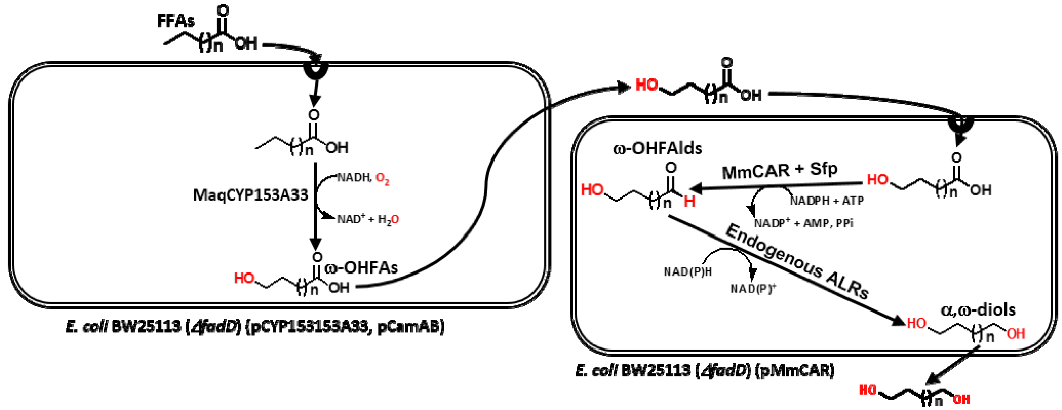
Biosynthesis of Medium- to Long-Chain α,ω-Diols from Free Fatty Acids Using CYP153A Monooxygenase, Carboxylic Acid Reductase, and E. coli Endogenous Aldehyde Reductases

1
School of Biotechnology, Yeungnam University, Gyeongsan 38541, Korea
2
Department of Systems Biotechnology, Konkuk University, Seoul 050-29, Korea
*
Author to whom correspondence should be addressed.
Catalysts 2018, 8(1), 4; https://doi.org/10.3390/catal8010004
Submission received: 27 November 2017 / Revised: 20 December 2017 / Accepted: 22 December 2017 / Published: 24 December 2017
(This article belongs to the Special Issue Biocatalysis and Biotransformations)

Abstract

:
α,ω-Diols are important monomers widely used for the production of polyesters and polyurethanes. Here, biosynthesis of α,ω-diols (C8–C16) from renewable free fatty acids using CYP153A monooxygenase, carboxylic acid reductase, and E. coli endogenous aldehyde reductases is reported. The highest yield of α,ω-diol was achieved for the production of 1,12-dodecanediol. In the nicotinamide adenine dinucleotide phosphate (NADPH) cofactor regeneration system, 5 g/L of 1,12-dodecanediol was synthesized in 24 h reaction from the commercial ω-hydroxy dodecanoic acid. Finally, 1.4 g/L 1,12-dodecanediol was produced in a consecutive approach from dodecanoic acids. The results of this study demonstrated the scope of the potential development of bioprocesses to substitute the petroleum-based products in the polymer industry.

1. Introduction

The utilization of renewable feedstock-derived raw materials and development of bioprocesses thereof to substitute petroleum-based products in polymer industry has shown a steep surge in recent years [1,2,3] due to concern of future shortage of petroleum resources and in consideration of green production [4,5]. Compared to chemical catalysis, biocatalysis has countless advantages including high efficiency, increased stability, high degree of selectivity (regio-, chemo-, and enantio-selectivity), and safe reaction conditions [6,7]. α,ω-Diols are used as monomers for the production of polyesters and polyurethanes [1,2]. At present, industrial-scale preparation has only been confined to short-chain diol biosynthesis, while biosynthesis of medium- to long-chain α,ω-diols (C8–C16) has not been studied extensively [8,9]. Microbial synthesis of α,ω-diol (>C12) has been originally reported in some yeasts species belonging to genus Candida [10,11,12]. Further microbial synthesis α,ω-diols (C5–C12) from alkanes employing cytochrome P450 (CYP5153A) from Acinetobacter sp. OC4 in Escherichia coli with the highest yielded for 1,8-octanediol (722 mg/L) has been reported [10,13]. Similar ability of producing α,ω-diols from alkanes through in vitro biocatalytic reactions using CYP153A16 from Mycobacterium merinum and CYP153A34 from Polaromonas sp. strain JS666 has been reported. CYP153A16 produced C8–C12 α,ω-diols while CYP153A34 produced C8–C9 α,ω-diols. However, the overall yield of α,ω-diols from 1 mM substrate is only 3–12% [14]. Only one study has reported the use of renewable medium-chain fatty alcohols for the production of α,ω3-diols in E. coli by employing engineered P450BM3 [9]. Microbial synthesis of medium- to long-chain α,ω-diols suffers major challenges such as low product yields, low productivities, and narrow substrate range of enzymes used [9,13,15]. Although some preparative-scale reactions have been reported [13], novel approaches are highly desirable to overcome bottlenecks in microbial synthesis of α,ω-diols. In this light, here we report highly efficient and sequential biocatalytic conversion of free fatty acids (FFAs) to medium- to long-chain α,ω-diols.
Vegetable oil derivatives (e.g., FFAs) are important renewable resources [1,2]. Unfortunately, limited attention has been paid to synthesize diols from FFAs that can serve as good starting materials for green processes, including the production of α,ω-diols [13,15]. We describe herein the production of medium- to long-chain α,ω-diols from FFAs using CYP153A monooxygenase, carboxylic acid reductase (CAR) and E. coli endogenous aldehyde reductases (ALRs) (Figure 1). First, hydroxylation of FFAs was carried out using CYP153A33 from Marinobacter aquaeolei (MaqCYP153A33), a promising biocatalyst due to its exceptional regioselectivity (>95%) for the ω-position [16]. Next, a catalytically efficient carboxylic acid reductase (CAR) from Mycobacterium marinum (MmCAR) [17] recently reported was used for selective reduction of carboxylic acid functional group of ω-hydroxy FFAs to corresponding aldehydes. It exhibited high catalytic efficiency (kcat/Km) against benzoic acid and the C6–C16 fatty acids [17]. However, microbial hosts such as E. coli genome encoded for 44 endogenous aldehyde reductases (ALRs), and among them 13 are highly active that converted aldehydes to alcohols ranging from C2 to C18 [18,19]. To efficiently convert ω-hydroxy fatty aldehydes to α,ω-diols, E. coli cells as a source of endogenous ALRs were used.

2. Results and Discussion

2.1. Establishment of MmCAR Reaction System

CARs need to undergo posttranslational activation via phosphopantetheinylation using phosphopantetheinyl transferase (PPTase) [17,20,21,22]. Therefore, MmCAR was co-expressed (Figure S1) with surfactin phosphopantetheinyl transferase (Sfp), a PPTase from Bacillus subtilis. SDS-PAGE analysis confirmed the production of MmCAR and Sfp recombinant proteins in soluble form (Figure S2). Whole-cell (0.3 gCDW/mL) reaction was carried out at 30 °C and 200 rpm using MmCAR with or without co-expressing Sfp cells in potassium phosphate buffer (100 mM, pH 7.5) in the presence of 1% (w/v) glucose and 10 mM MgCl2. Co-expressed cells produced 9.2 mM of benzyl alcohol. However, cells only expressing MmCAR synthesized 0.5 mM of benzyl alcohol (Figure S3). In both cases, only negligible amount of benzylaldehyde was detected (data not shown). This result clearly suggested that Sfp had a pivotal role in the activation of MmCAR and that activities of endogenous ALRs were sufficiently high enough to produce alcohols (Figure S4). MmCAR-Sfp co-expressed cells were then studied further using various FFAs. Whole-cell (0.3 gCDW/mL) reactions were performed using 10 mM octanoic acid (C8), decanoic acid (C10), dodecanoic acid (C12), tetradecanoic acid (C14), and hexadecanoic acid (C16). The amount of produced 1-octanol, 1-decanol, 1-dodecanol, 1-tetradecanol, and 1-hexadecanol was 1.8, 9.8, 9.9, 1.6, and 0.6 mM, respectively (Figure S5). GC analysis revealed that there was no accumulation of aldehydes during the reaction process, implying that endogenous ALRs were sufficient enough to reduce aldehydes to alcohols. This can be explained by the reduction of carboxylic acid functional groups of FFAs to their aldehyde counterparts and concomitant reduction of these aldehydes to alcohols by ALRs (Figure S4). To confirm ALR-mediated reduction of aldehydes, a separate reaction was carried out using E. coli BW25113 (∆fadD, DE3) host cells. Complete conversion of 10 mM dodecanal and benzaldehyde to 1-dodecanol and benzyl alcohol, respectively, was achieved in 6 h, implying that endogenous ALRs from E. coli BW25113 (∆fadD, DE3) host cells had catalytic efficiency (Figure S6). These results were consistent with those of Akhtar et al. (2013) showing that alcohols (C8–C18) could be produced over a short time period (5 h) using MmCAR when natural oils were used as sources of FFAs [17].

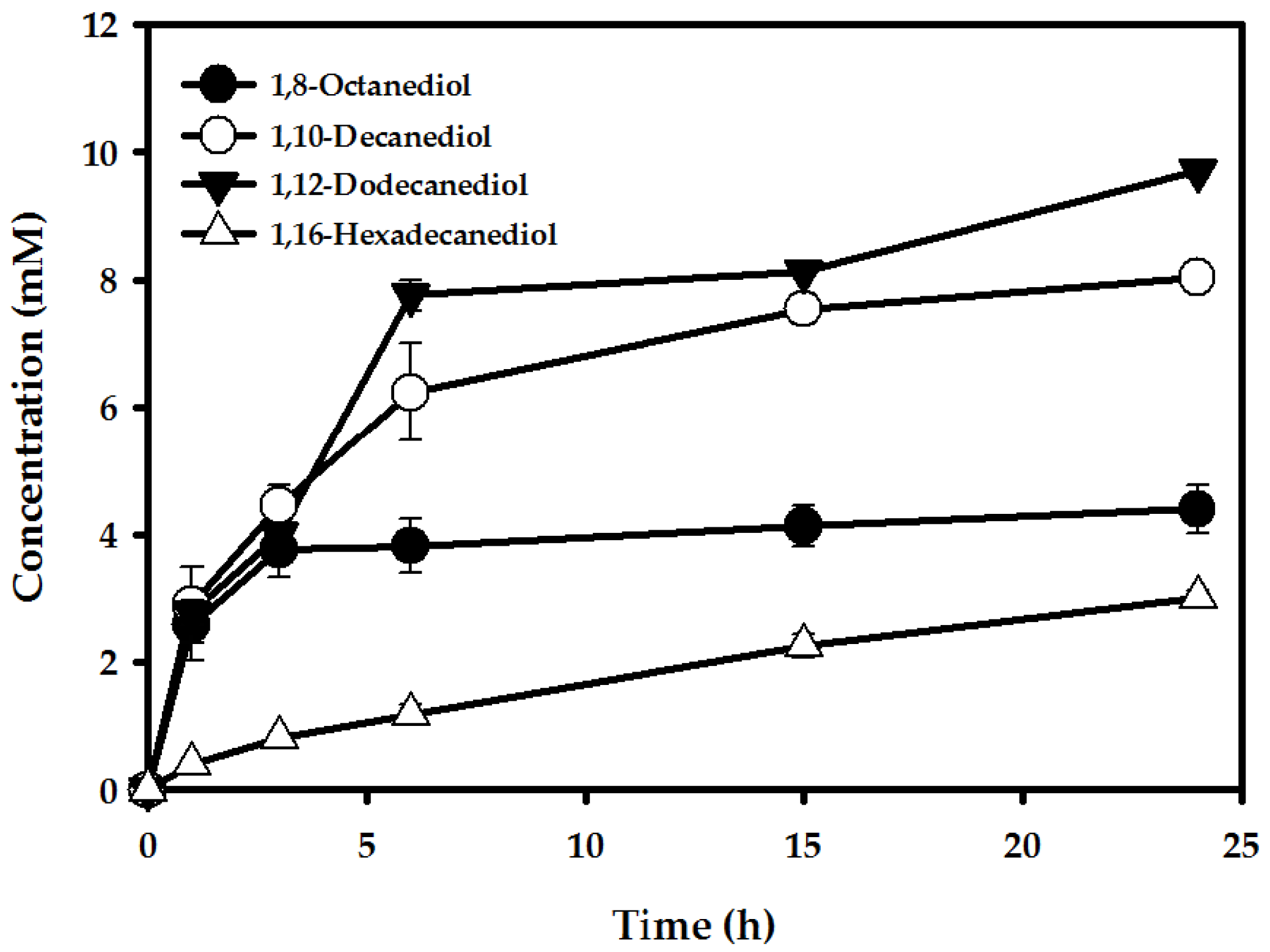
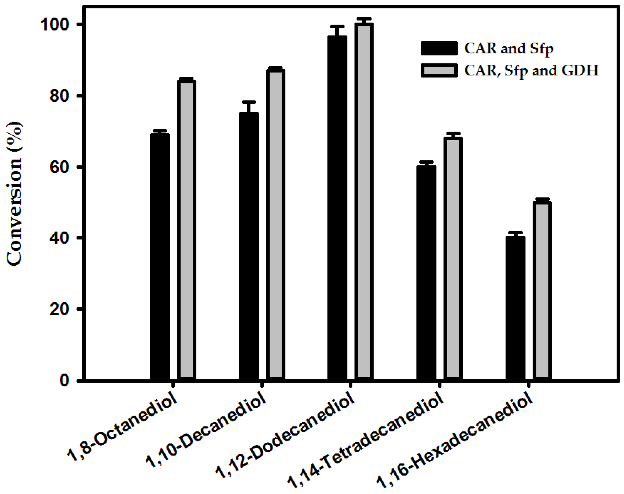
2.2. Production of α,ω-Diols from ω-OHFAs

After successful in vivo synthesis of various alcohols by MmCAR, further biocatalytic reactions were performed using various ω-OHFAs (C8–C16) in order to achieve the final goal of this study (i.e., biosynthesis of medium- to long-chain α,ω-diols). Reaction conditions were the same as those used for MmCAR-catalyzed biotransformation of FFAs to fatty alcohols. Initial specific α,ω-diol biotransformation rates were 0.26, 0.29, 0.28 and 0.04 mM/gCDW/h for ω-hydroxy octanoic acid (ω-OHOA), ω-hydroxy deacanoic acid (ω-OHDA), ω-hydroxydodecanoic acid (ω-OHDDA), and ω-hydroxy hexadecanoic acid (ω-OHHDA), respectively (Figure 2). Although the initial whole-cell reaction rate for ω-OHDAs (C10) was marginally higher, the optimum productivity was observed for the production of 1,12-dodecanediol from ω-OHDDA in 24 h reaction (Figure 2). The highest yield achieved (9.7 mM) from 10 mM of corresponding substrates was found for the production of 1,12-dodecanediol, whereas only 3.01 mM of 1,16-hexadecanediol was produced (Figure 2).
Among renewable FFAs, dodecanoic acid is one of the cheapest and the most abundant substrates while 1,12-dodecanediol is an industrially important monomer for the production of polyesters, polyurethanes, polycarbonates, and epoxy resins [13]. Interestingly, the productivity of 1,12-dodecanediol in the present study was comparatively higher than that of other α,ω-diols. Therefore, further optimization using ω-OHDDA as a substrate was requisite.

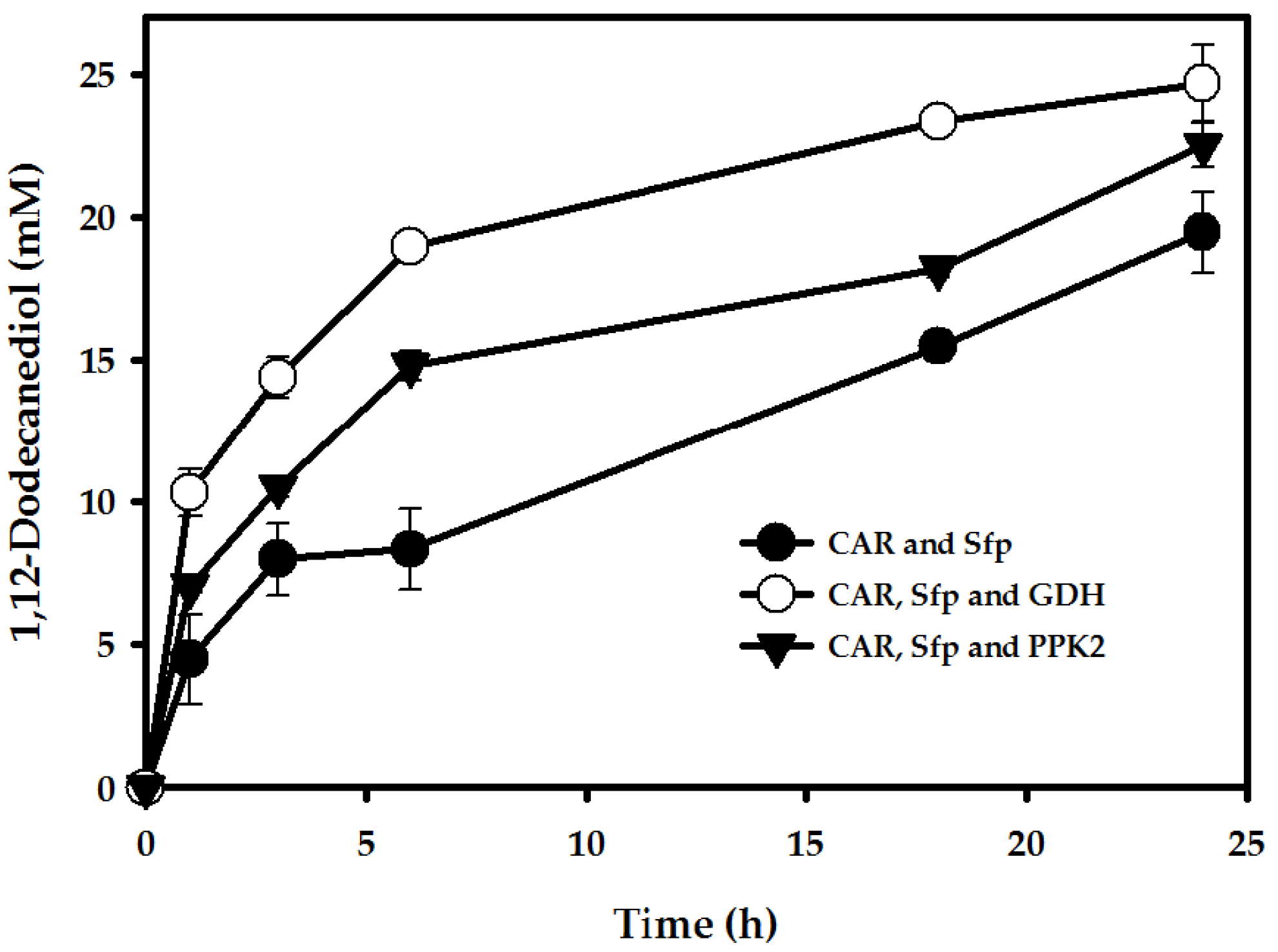
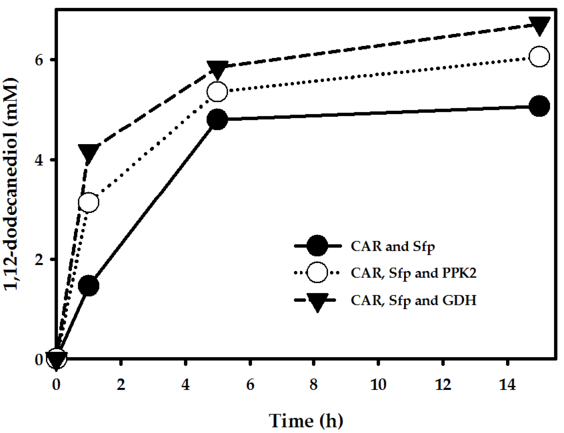
2.3. Enhancement of 1,12-Dodecanediol Production

2.3.1. pH Optimization

To examine the effect of pH on bioconversion of ω-OHDDA to 1,12-dodecanediol using MmCAR, whole-cell reaction was performed at three different pH (6.5, 7.5 and 8.5). Substrate concentration was increased to 30 mM. Optimum activity was measured at pH 7.5, with 19.5 mM product formed in 24 h, whereas only 7.4 and 9.7 mM of products were synthesized at pH 6.5 and 8.5, respectively (Figure S7). Higher concentration of ω-OHDDA (beyond 30 mM) resulted in decreased productivity. This might be due to the toxic effects of higher concentrations of ω-OHDDA to living cells by [23].

2.3.2. Cofactor Regeneration

Regenerations of nicotinamide cofactors (NADPH) and adenosine triphosphate (ATP) are very important in CAR reactions to restore consumed cofactors. For regeneration of ATP, several enzymatic reactions have been reported including PPK2 which mainly catalyzes polyphosphate-dependent phosphorylation [21]. Whole-cell reaction was performed in the above mentioned conditions employing Arthrobacter aurescens PPK2 co-expressing MmCAR and Sfp with an additional use of 2 mM polyP [21]. In 24 h of reaction, produced amount of 1,12-dodecanediol was 22.5 mM (4.6 g/L) (Figure 3).
For regeneration of NADPH in MmCAR-catalyzed reactions, a Bacillus subtilis glucose dehydrogenase (GDH) was co-expressed to ensure constant supply of cofactors the whole-cell biotransformation processes [24]. After 24 h of reaction, 24.7 mM (5.0 g/L) of 1,12-dodecanediol was produced from 30 mM substrate, higher than the yield of MmCAR-PPK2 system (Figure 3). The production of 1,12-dodecanediol showed a substantial improvement (66-fold) compared to that reported by Fujii et al. (2006) who demonstrated 79 mg/L product from 1-dodecanol using E. coli expressing CYP153A from Acinetobacter sp. OC4 [10]. Results of these experiments clearly demonstrated that the carboxylic acid functional group of ω-OHFAs was reduced to the aldehyde functional group by whole-cell catalysts expressing CAR, Sfp and GDH where the aldehyde functional group was further reduced to the alcoholic functional group by E. coli endogenous ALRs.

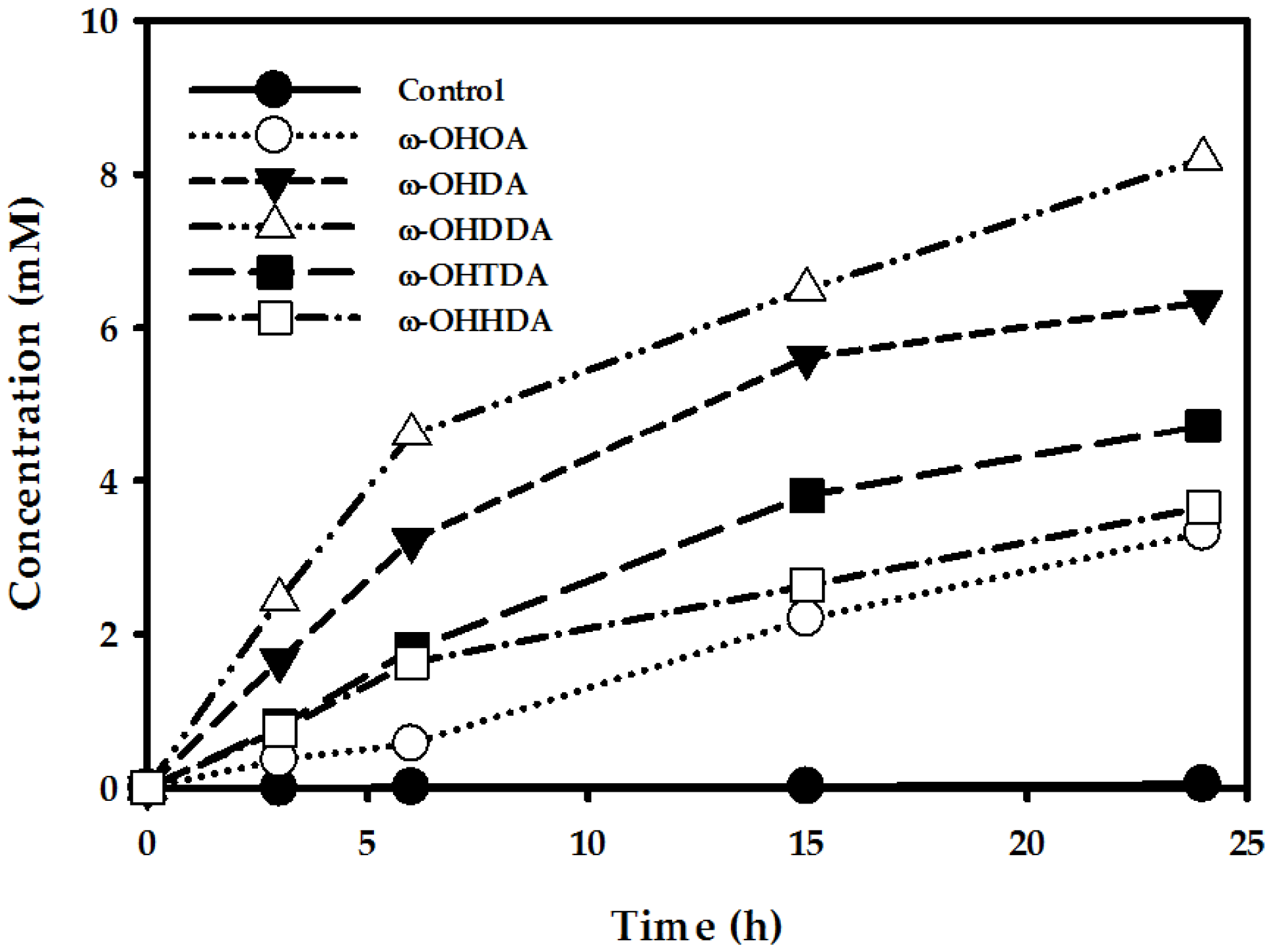
2.4. Production of α,ω-Diols from FFAs

2.4.1. Production of α,ω-Diols by One-Pot, One-Step Reaction

‘One-pot, one step’ reaction is desirable for cascade reaction. To start the one-pot, one-step reaction, E. coli cells expressing MaqCYP153A33/CamAB for ω-hydroxylation of DDA and E. coli cells expressing MmCAR-GDH/Sfp were grown separately. After the expression, both types of cells were harvested, and the cell pellets were washed, and re-suspended in 100 mM potassium phosphate buffer with 1% (w/v) glucose (pH: 7.5). A total of 0.3 gDCW/mL of whole-cell was used in the reaction with 1:1 ratio. The reaction was initiated by adding 10 mM DDA (stock in DMSO, 5% (v/v) final concentration), and 10 mM MgCl2. The reactants were incubated at 30 °C and 200 rpm. After 15 h of whole-cell reaction, 6.8 mM 1-dodecanol and 1.0 mM 1,12-dodecanediol was produced, respectively (Figure 4). These findings suggest that one-pot, one-step reaction of FFAs predominantly produced fatty alcohols rather than α,ω-diols. This is because, FFAs acted as common active substrates for both MmCAR and MaqCYP153A33 enzymes where MmCAR was more active than MaqCYP153A33. Consequently, one-pot reaction was not feasible for the production of α,ω-diols from FFAs. Therefore, next we tried sequential reaction. In first step, ω-OHFAs were produced from FFAs by MaqCYP153A33/CamAB and in second step α,ω-diols were synthesized from biotransformed ω-OHFAs by MmCAR/Sfp.

2.4.2. Production of α,ω-Diols Using Biotransformed ω-OHFAs

Production of Various ω-OHFAs (C8–C16) by CYP153A Monooxygenase

Enzymes belonging to CYP153A sub-family of P450s have natural ability to ω-hydroxylate fatty acid substrates. CYP153As are bacterial class I P450s that require two redox partners for transportation of electrons from NAD(P)H to P450’s active site [14,16,25]. Putidaredoxin reductase (CamA) and putidaredoxin (CamB) from Psedomonas putida ATCC 17453 are well-known redox partners of CYP153As. They are, combined, known as “CamAB system” [14,25]. Enzymes belonging to CYP153A monooxygenases are popularly known to catalyze ω-hydroxylation towards saturated and unsaturated fatty acids. Among them, MaqCYP153A33 has a broad range of substrates with exceptional regioselectivity (>95%) for the ω-position [14,16,25]. To study the utility of FFAs for the synthesis of diols, various FFAs (C8–C16) were utilized for the production of ω-OHFAs using MaqCYP153A33-CamAB system. SDS-PAGE analysis and CO binding spectrum in UV–visible spectroscopy corroborated the active expression of P450 and also confirmed production of MaqCYP153A33 with CamAB (Figure S8). The amount of active P450 was 7.2 nmol/mL. This result was correlated with the earlier findings of Jung et al. (2016) [25]. Whole-cell reaction was carried out using MaqCYP153A33-CamAB co-expressed cells (0.3 gCDW/mL) at 30 °C and 200 rpm in potassium phosphate buffer (100 mM, pH 7.5) containing 1% (w/v) glucose. Twenty-four hour reactions of MaqCYP153A33 resulted in the production of 3.3 mM ω-OHOA, 6.3 mM ω-OHDA, 8.2 mM ω-OHDDA, 4.7 mM ω-OHTDA, and 3.7 mM ω-OHHDA from 10 mM of respective FFA (Figure 5).

Production of α,ω-Diols Using Biotransformed ω-OHFAs

The production of ω-OHFAs ranged from 3.3–8.2 mM. Therefore, 3 mM each of biotransformed and non-purified ω-OHFAs (C8–C16) from previous reactions were utilized for the production of α,ω-diols in MmCAR, and MmCAR-GDH system. Other than Mg2+ cofactors for MmCAR, whole-cell reaction conditions of both MaqCYP153A33 and MmCAR-ALRs were the same (see the Materials and Methods for details). Bioconversion rates of 1,8-octanediol, 1,10-decanediol, 1,12-dodecanediol, 1,14-tetradecanediol, and 1,16-hexadecanediol were 69%, 75%, 96%, 60%, and 40%, respectively, in MmCAR without GDH system (Figure 6). However, in MmCAR-GDH system, these conversion rates were increased by 15%, 12%, 4%, 8%, and 10%, respectively (Figure 6).
Finally, to check the utility of these sequential biocatalytic process, the production of 1,12-dodecanediol was studied using biotransformed ω-OHDDA as a substrate in all three systems (Figure 7). However, owing to lower productivity of ω-OHDDA (8.2 mM) from MaqCYP153A33 reactions, only 7 mM of biotransformed ω-OHDDA was used for subsequent whole-cell reactions involving MmCARs. Nevertheless, the highest bioconversion rate (96%) was achieved in MmCAR-GDH system after 15 h of reaction, with production yield of 1.4 g/L (Figure 7).

3. Materials and Methods

3.1. Chemicals

All chemicals such as fatty acids, ω-hydroxy fatty acids, dimethyl sulfoxide (DMSO), isopropyl-thio-β-d-galactopyranoside (IPTG), 5-aminolevulinic acid (5-ALA), N,O-Bis(trimethylsilyl)-trifluoroacetamide (BSTFA), and sodium polyphosphate crystals were purchased from Sigma-Aldrich (St. Louis, MO, USA). Fatty alcohols, and α,ω-diols were obtained from Tokyo Chemical Industry (Tokyo, Japan). Chloroform was obtained from Junsei (Tokyo, Japan). Bacteriological agar, Luria bertani (LB) broth, and terrific broth (TB) media were purchased from BD Difco (Franklin Lakes, NJ, USA). All chemicals used in this study were of analytical grade.

3.2. Plasmid Construction and Gene Manipulation

All bacterial strains, plasmid vectors and custom designed oligonucleotides used in this study are listed in Table 1. Genomic DNA of Mycobacterium marinum ATCC® BAA535™ was obtained from American Type Culture Collection (ATCC). Gene encoding M. marinum carboxylic acid reductase (CAR, UniProt: B2HN69) was amplified from genomic DNA using a PCR thermocycler (Veriti® 96-Well Thermal Cycler; AB Applied Biosystems, Foster City, CA, USA) and cloned into pETDuet-1 first multiple cloning site (MCS1). A phosphopantetheinyl transferase, Sfp (GI: P39135) from Bacillus subtilis and polyphosphate kinase 2, PPK2 (GI: ABM08865.1) from Arthrobacter aurescens genes were chemically synthesized and codon-optimized for E. coli. Synthesis and codon optimization were performed by Cosmo Genetech (Cosmo Genetech, Seoul, Korea). PPK2 and glucose dehydrogenase (GDH) gene of Bacillus subtilis were cloned into the second multiple cloning site (MCS2) of pETDuet-1 vector separately with CAR. Sfp was cloned into pET24ma vector. All DNA manipulations for cloning were performed following standard protocols [26]. E. coli DH5α was used for cloning purposes. All primers used in this study were purchased from Cosmo Genetech. They are listed in Table 1.

3.3. Protein Expression and Biotransformation

Previously reported E. coli BW25113 (DE3) ∆fadD strains [25] were utilized for biotransformation studies wherein fatty acid degrading β-oxidation pathway was blocked. Recombinant strains were obtained by introducing plasmid DNA into host strains via standard heat shock method of transformation. Transformants were selected based on their antibiotic resistance [26]. Cultivations were carried out at 37 °C. Fresh colonies from agar plates of E. coli BL21 (DE3) transformed with each plasmid vector were cultured in 10 mL of Luria-Bertani (LB) medium containing 50 μg/mL of kanamycin (for pCYP153A33, and pSFP) and/or 100 μg/mL ampicillin (for pCamAB, pMmCAR, pCAR-PK2, and pCAR-GDH) at 37 °C overnight. These overnight pre-cultured cells were then inoculated into larger volume flasks for expression. They were cultured at 37 °C until cell concentration reached an optical density at 600 nm (OD600) of 0.6–0.8 for IPTG induction. MaqCYP153A33 and CamAB protein expression were carried out in 1 L of Terrific-Broth (TB) in a 3 L flask. The induction was performed by adding 0.01 mM IPTG, 0.5 mM 5-ALA as heme precursor, and 0.1 mM FeSO4 at 30 °C for 16 h. MmCAR with or without PPK2/GDH and SFP protein expression were carried out in 1 L of LB medium in 3 L flasks. Induction was performed by adding 0.1 mM IPTG at 20 °C for 16 h.
After expression, cells were harvested by centrifugation (4000 rpm, 20 min, 4 °C), washed with PBS, and resuspended in 100 mM potassium phosphate buffer (pH 7.5) containing 1% (w/v) glucose. Whole-cell reaction (0.3 gCDW/mL) was initiated by adding respective substrates (stock in DMSO, 5% (v/v) final concentration or cell free reaction mixtures containing non-purified ω-OHFAs). These reactants were incubated at 30 °C and 200 rpm. For whole-cell reaction of MmCAR, 10 mM MgCl2 was added during the addition of substrates. Every 6th h, the pH of the reaction mixture was measured with a pH meter and adjusted to pH 7.5 with 5 M KOH. Then, 50 μL of 80% (w/v) glucose was added.
To prepare the non-purified ω-OHFAs substrate for the sequential reaction of MmCAR, and ALRs, the whole-cell reactions of MaqCYP153A33 were stopped after 24 h, and acidified with 6 M HCl. The cells were removed by centrifugation at 16,000 rpm. The clear supernatant thus obtained was used for the further biotransformation without any purification step. The presented amount of produced ω-OHFAs in the reaction mixtures were determined by GC analysis (vide infra). The pH of the reaction mixture was readjusted to 7.5 for the next step bioconversion. Cells containing MmCAR reaction system were resuspended in the reaction mixture, and potassium phosphate buffer (100 mM, pH 7.5) was added again on the basis of ω-OHFAs concentration. To compensate the depleted glucose in earlier reaction, again 1% (w/v) glucose was added, and the whole-cell reaction was carried out as mentioned above.

3.4. Analysis of Compounds by Gas Chromatography

Quantitative analysis was performed using a gas chromatography instrument (GC 2010 plus Series) with a flame ionization detector (GC/FID) fitted with an AOC-20i series auto sampler injector (Shimadzu Scientific Instruments, Kyoto 604-8511, Japan). Two microliters of the sample were injected by split mode (split ratio 20:1) and analyzed using a nonpolar capillary column (5% phenyl methyl siloxane capillary 30 m × 320 μm i.d., 0.25-μm film thickness, HP-5). Oven temperature program for fatty acid analysis was follows: 50 °C for 1 min, increase to 250 °C at 10 °C/min, and hold for 10 min. The inlet temperature was 250 °C while the detector temperature was 280 °C. The flow rate of the carrier gas (He) was at 1 mL/min. Flow rates of H2, air, and He in FID were 45 mL/min, 400 mL/min, and 20 mL/min, respectively. The initial oven temperature was 90 °C. It was then increased to 250 °C by 15 °C/min and held at this temperature for 5 min. Each peak was identified by comparing GC chromatogram with an authentic reference. Internal standards were added to the sample before extraction of the substrate.

4. Conclusions

In summary, this work demonstrated the first gram-scale bioconversion of FFAs to medium- to long-chain α,ω-diols as high-value building blocks for synthesis of polyamides, polyesters, and polyurethanes. The applicability of in vivo cofactor regeneration systems for cost-effective biocatalytic production of α,ω-diols was also demonstrated. This study suggests that further bioconversion of α,ω-diols to corresponding diamines and diacids is possible through green process [10,11,13]. Furthermore, optimization of the MaqCYP153A33 catalyzed reaction for ω-hydroxylation of FFAs could facilitate higher product titres from FFAs to α,ω-diols.

Supplementary Materials

The following are available online at www.mdpi.com/2073-4344/8/1/4/s1, Figure S1, Schematic representation of plasmid system used in this study; Figure S2, SDS PAGE analysis of recombinant E. coli expressing MmCAR (~128 kDa) and Sfp (~27.3 kDa); Figure S3, Effect of Sfp to make active MmCAR; Figure S4, Proposed reaction scheme for the biosynthesis of benzyl alcohol and fatty alcohols (C8–C16); Figure S5, In vivo production of fatty alcohol using E. coli BW25113 (∆fadD, DE3) cells expressing MmCAR, and Sfp; Figure S6, Production of alcohols by E. coli endogenous ALRs using E. coli BW25113 (∆fadD, DE3); Figure S7, Effect of pH for the optimum activity of CAR using E. coli BW25113 (∆fadD, DE3) cells expressing MmCAR, Sfp, and endogenous ALRs; Figure S8, (A) SDS PAGE analysis of recombinant E. coli expressing MaqCYP153A33 (~55.2 kDa), CamA (~47.8 kDa), and CamB (~11.6 kDa). (B) CO binding spectrum of active MaqCYP153A33. Table S1, Retention time of the substrates and products by Gas Chromatography.

Acknowledgments

This work was partly supported by the Ministry of Trade, Industry and Energy of South Korea (MOTIE, Korea) under the Industrial Technology Innovation Program (No. 10062550), entitled “The production of nylon 12 monomer, ω-aminolaruic acid from plant oil by bioconversion” and partly by Basic Science Research Program through the National Research Foundation of Korea (NRF) funded by the Ministry of Science, ICT and future Planning (2016R1A2B2014794).

Author Contributions

Hyungdon Yun and Taeowan Chung designed experiments of the project; Md Murshidul Ahsan carried out research works as part of his PhD project; Hyungdon Yun supervised the whole studies reported in the manuscript; Md Murshidul Ahsan wrote the manuscript; Hyunwoo Jeon and Sihyong Sung assisted in experimental tools; Mahesh D. Patil revised this manuscript.

Conflicts of Interest

The authors declare no financial or commercial conflict of interest related to this study.

References

  1. Kawaguchi, H.; Ogino, C.; Kondo, A. Microbial conversion of biomass into bio-based polymers. Bioresour. Technol. 2017, 245, 1664–1673. [Google Scholar] [CrossRef] [PubMed]
  2. Lligadas, G.; Ronda, J.C.; Galià, M.; Cádiz, V. Oleic and undecylenic acids as renewable feedstocks in the synthesis of polyols and polyurethanes. Polymers 2010, 2, 440–453. [Google Scholar] [CrossRef]
  3. Gandini, A. Polymers from renewable resources: A challenge for the future of macromolecular materials. Macromolecules 2008, 41, 9491–9504. [Google Scholar] [CrossRef]
  4. Lligadas, G.; Ronda, J.C.; Galià, M.; Cádiz, V. Renewable polymeric materials from vegetable oils: A perspective. Mater. Today 2013, 16, 337–343. [Google Scholar] [CrossRef]
  5. Sharma, V.; Kundu, P.P. Condensation polymers from natural oils. Prog. Polym. Sci. 2008, 33, 1199–1215. [Google Scholar] [CrossRef]
  6. Madhavan, A.; Sindhu, R.; Binod, P.; Sukumaran, R.K.; Pandey, A. Strategies for design of improved biocatalysts for industrial applications. Bioresour. Technol. 2017, 245, 1304–1313. [Google Scholar] [CrossRef] [PubMed]
  7. Wang, M.; Si, T.; Zhao, H. Biocatalyst development by directed evolution. Bioresour. Technol. 2012, 115, 117–125. [Google Scholar] [CrossRef] [PubMed]
  8. Jiang, Y.; Liu, W.; Zou, H.; Cheng, T.; Tian, N.; Xian, M. Microbial production of short chain diols. Microb. Cell Fact. 2014, 13, 165. [Google Scholar] [CrossRef] [PubMed]
  9. Zheng, Y.; Li, L.; Liu, Q.; Zhang, H.; Cao, Y.; Xian, M.; Liu, H. High-specificity synthesis of novel monomers by remodeled alcohol hydroxylase. BMC Biotechnol. 2016, 16, 61. [Google Scholar] [CrossRef] [PubMed]
  10. Fujii, T.; Narikawa, T.; Sumisa, F.; Arisawa, A.; Takeda, K.; Kato, J. Production of α,ω-alkanediols using Escherichia coli expressing a cytochrome P450 from Acinetobacter sp. OC4. Biosci. Biotechnol. Biochem. 2006, 70, 1379–1385. [Google Scholar] [CrossRef] [PubMed]
  11. Scheller, U.; Zimmer, T.; Becher, D.; Schauer, F.; Schunck, W.-H. Oxygenation cascade in conversion of n-alkanes to α, ω-dioic acids catalyzed by cytochrome P450 52A3. J. Biol. Chem. 1998, 273, 32528–32534. [Google Scholar] [CrossRef] [PubMed]
  12. Shiio, I.; Uchio, R. Microbial production of long-chain dicarboxylic acids from n-alkanes. Agric. Biol. Chem. 1971, 35, 2033–2042. [Google Scholar] [CrossRef]
  13. Schaffer, S.; Haas, T. Biocatalytic and fermentative production of α,ω-bifunctional polymer precursors. Org. Process. Res. Dev. 2014, 18, 752–766. [Google Scholar] [CrossRef]
  14. Scheps, D.; Honda Malca, S.; Hoffmann, H.; Nestl, B.M.; Hauer, B. Regioselective ω-hydroxylation of medium-chain n-alkanes and primary alcohols by CYP153 enzymes from Mycobacterium marinum and Polaromonas sp. strain JS666. Org. Biomol. Chem. 2011, 9, 6727–6733. [Google Scholar] [CrossRef] [PubMed]
  15. Sabra, W.; Groeger, C.; Zeng, A.-P. Microbial cell factories for diol production. In Bioreactor Engineering Research and Industrial Applications I: Cell Factories; Ye, Q., Bao, J., Zhong, J.-J., Eds.; Springer: Berlin/Heidelberg, Geramny, 2016; pp. 165–197. [Google Scholar]
  16. Honda Malca, S.; Scheps, D.; Kuhnel, L.; Venegas-Venegas, E.; Seifert, A.; Nestl, B.M.; Hauer, B. Bacterial CYP153A monooxygenases for the synthesis of omega-hydroxylated fatty acids. Chem. Commun. 2012, 48, 5115–5117. [Google Scholar] [CrossRef] [PubMed]
  17. Akhtar, M.K.; Turner, N.J.; Jones, P.R. Carboxylic acid reductase is a versatile enzyme for the conversion of fatty acids into fuels and chemical commodities. Proc. Natl. Acad. Sci. USA 2013, 110, 87–92. [Google Scholar] [CrossRef] [PubMed]
  18. Fatma, Z.; Jawed, K.; Mattam, A.J.; Yazdani, S.S. Identification of long chain specific aldehyde reductase and its use in enhanced fatty alcohol production in E. coli. Metab. Eng. 2016, 37, 35–45. [Google Scholar] [CrossRef] [PubMed]
  19. Rodriguez, G.M.; Atsumi, S. Toward aldehyde and alkane production by removing aldehyde reductase activity in Escherichia coli. Metab. Eng. 2014, 25, 227–237. [Google Scholar] [CrossRef] [PubMed]
  20. Finnigan, W.; Thomas, A.; Cromar, H.; Gough, B.; Snajdrova, R.; Adams, J.P.; Littlechild, J.A.; Harmer, N.J. Characterization of carboxylic acid reductases as enzymes in the toolbox for synthetic chemistry. ChemCatChem 2017, 9, 1005–1017. [Google Scholar] [CrossRef] [PubMed]
  21. Khusnutdinova, A.N.; Flick, R.; Popovic, A.; Brown, G.; Tchigvintsev, A.; Nocek, B.; Correia, K.; Joo, J.C.; Mahadevan, R.; Yakunin, A.F. Exploring bacterial carboxylate reductases for the reduction of bifunctional carboxylic acids. Biotechnol. J. 2017. [Google Scholar] [CrossRef] [PubMed]
  22. Venkitasubramanian, P.; Daniels, L.; Rosazza, J.P. Reduction of carboxylic acids by Nocardia aldehyde oxidoreductase requires a phosphopantetheinylated enzyme. J. Biol. Chem. 2007, 282, 478–485. [Google Scholar] [CrossRef] [PubMed]
  23. Lundemo, M.T.; Notonier, S.; Striedner, G.; Hauer, B.; Woodley, J.M. Process limitations of a whole-cell P450 catalyzed reaction using a CYP153A-CPR fusion construct expressed in Escherichia coli. Appl. Microbiol. Biotechnol. 2016, 100, 1197–1208. [Google Scholar] [CrossRef] [PubMed] [Green Version]
  24. Jeon, H.; Durairaj, P.; Lee, D.; Ahsan, M.M.; Yun, H. Improved nadph regeneration for fungal cytochrome P450 monooxygenase by co-expressing bacterial glucose dehydrogenase in resting-cell biotransformation of recombinant yeast. J. Microbiol. Biotechnol. 2016, 26, 2076–2086. [Google Scholar] [CrossRef] [PubMed]
  25. Jung, E.; Park, B.G.; Ahsan, M.M.; Kim, J.; Yun, H.; Choi, K.-Y.; Kim, B.-G. Production of ω-hydroxy palmitic acid using CYP153A35 and comparison of cytochrome P450 electron transfer system in vivo. Appl. Microbiol. Biotechnol. 2016, 100, 10375–10384. [Google Scholar] [CrossRef] [PubMed]
  26. Sambrook, J. Molecular Cloning: A Laboratory Manual/Joseph Sambrook, David W. Russell; Cold Spring Harbor Laboratory: Cold Spring Harbor, NY, USA, 2001. [Google Scholar]
  27. Hanahan, D. Studies on transformation of escherichia coli with plasmids. J. Mol. Biol. 1983, 166, 557–580. [Google Scholar] [CrossRef]
  28. Ahsan, M.M.; Jeon, H.; Nadarajan, S.P.; Chung, T.; Yoo, H.-W.; Kim, B.-G.; Patil, M.D.; Yun, H. Biosynthesis of the Nylon 12 monomer, ω-aminododecanoic acid with novel CYP153A, AlkJ, and ω-TA enzymes. Biotechnol. J. 2017. [Google Scholar] [CrossRef]
Figure 1. Schematic diagram showing the biosynthesis of medium- to long-chain α,ω-diols (C8–C16) from free fatty acids (FFAs). ω-OHFAs, ω-hydroxy fatty acids; ω-OHFAlds, ω-hydroxy fatty aldehydes.
Catalysts 08 00004 g001
Figure 2. In vivo production of α,ω-diols using E. coli BW25113 (∆fadD, DE3) cells expressing MmCAR and Sfp. Reaction Conditions: Substrate, 10 mM ω-OHFAs (C8–C16); Volume, 10 mL in 100 mL flask; Temp, 30 °C; Buffer, 100 mM potassium phosphate buffer (pH: 7.5) with 1% glucose (w/v) and 10 mM MgCl2; Cell OD600, 30.
Catalysts 08 00004 g002
Figure 3. In vivo production of α,ω-diols using E. coli BW25113 (∆fadD, DE3) cells expressing MmCAR and Sfp. Reaction conditions: Substrate, 30 mM ω-OHDDA; Volume, 10 mL in 100 mL flask; Temp, 30 °C; Buffer, 100 mM potassium phosphate buffer (pH: 7.5) with 1% glucose (w/v) and 10 mM MgCl2; Cell OD600, 30.
Catalysts 08 00004 g003
Figure 4. In vivo production of α,ω-diols by one-pot, one-step reaction using two different type of E. coli BW25113 (∆fadD, DE3) cells coexpressing MaqCYP153A33 with CamAB and MmCAR-GDH with Sfp, respectively. Reaction conditions: Substrate, 10 mM (DDA), 10 mM MgCl2, 10 mL in 100 mL flask, 30 °C, 15 OD600/mL of BW25113 (∆fadD, DE3) containing pCYP153A33, pCamAB, 15 OD600/mL of BW25113 (∆fadD, DE3) containing pCAR-GDH and pSFP, 100 mM phosphate buffer (pH 7.5, 1% (w/v) glucose. Values were obtained by triplicate experiment with standard deviations within ≤10%.
Catalysts 08 00004 g004
Figure 5. In vivo production of ω-OHFAs using E. coli BW25113 (∆fadD, DE3) cells expressing MaqCYP153A33 and CamAB. Reaction conditions: Substrate, 10 mM FFAs (C8–C16); Volume, 50 mL in 250 mL flask; Temp, 30 °C; Buffer, 100 mM potassium phosphate buffer (pH: 7.5) with 1% glucose (w/v); and Cell OD600, 30. ω-OHOA, ω-hydroxy octanoic acid; ω-OHDA, ω-hydroxy decanoic acid; ω-OHDDA, ω-hydroxy dodecanoic acid; ω-OHTDA, ω-hydroxy tetradecanoic acid; ω-OHHDA, ω-hydroxy hexadecanoic acid. Control, cells carrying “empty” pET24ma and pETDuet-1 plasmids (without MaqCYP153A33 and CamAB genes). Values were obtained by triplicate experiment with standard deviations within ≤10%.
Catalysts 08 00004 g005
Figure 6. In vivo production of α,ω-diols using E. coli BW25113 (∆fadD, DE3) cells expressing MmCAR and Sfp with/without GDH. Reaction conditions: Substrate, 3 mM biotransformed ω-OHFAs (C8–C16); Reaction time, 24 h; Volume, 10 mL in 100 mL flask; Temp, 30 °C; Buffer, 100 mM potassium phosphate buffer (pH: 7.5) with 1% glucose (w/v) and 10 mM MgCl2; Cell OD600, 30.
Catalysts 08 00004 g006
Figure 7. In vivo production of α,ω-diols using E. coli BW25113 (∆fadD, DE3) cells expressing MmCAR and Sfp. Reaction conditions: Substrate, 7 mM biotransformed ω-OHDDA; Volume, 10 mL in 100 mL flask; Temp, 30 °C; Buffer, 100 mM potassium phosphate buffer (pH: 7.5) with 1% glucose (w/v) and 10 mM MgCl2; Cell OD600, 30. Values were obtained by triplicate experiment with standard deviations within ≤10%.
Catalysts 08 00004 g007
Table 1. Plasmids and strains used in this study *.
Plasmids/StrainsDescriptionReference
Plasmids
pET24maP15A ori lacI T7 promoter, KmR[25]
pCYP153A33pET24ma encoding for MaqCYP153A33[25]
pETDuet-1pBR322 ori lacI T7 promoter, AmpRNovagen
pCamABpETDuet-1 encoding for CamA and CamB[25]
pMmCARpETDuet-1 encoding for MmCARThis study
pCAR-GDHpETDuet-1 encoding for MmCAR and GDHThis study
pCAR-PK2pETDuet-1 encoding for MmCAR and PPK2This study
pSFPpET24ma encoding for SfpThis study
E. coli strains
DH5αF, endA1, glnV44, thi-1, recA1, relA1, gyrA96 deoR, supE44, Φ80dlacZ∆M15 ∆(lacZYA-argF)-U169, hsdR17(rK mK+), λ[27]
BW25113(DE3)rrnB3 ΔlacZ4787 hsdR514 Δ(araBAD)567 Δ(rhaBAD)568 rph-1 λ(DE3)[25]
DLBW25113(DE3) ΔfadD[28]
A33ABDL carrying pCYP153A33 and pCamAB[28]
CARDL carrying pCARThis study
CSDL carrying pCAR and pSFPThis study
CSGDL carrying pCAR-GDH and pSFPThis study
CSKDL carrying pCAR-PK2 and pSFPThis study
PCR oligonucleotidesSequence (5′-3′)
CAR_F (Bam HI)ATTAGGATCCCATGTCGCCAATCACGCGTGCloned into pETDuet1
CAR_R (Hindd III)TAATAAGCTTGAGCAGGCCGAGTAGGCG
SFP_F (Nde I)ATTACATATGAAAATCTACGGCATCTACCloned into pET24ma
SFP_R (Xho I)TAATCTCGAGTTACAGCAGTTCTTCATA
PPK2_F (Nde I)AAGGAGATATACATATGCCGATGGTTGCTGCAGCloned into pETDuet1
PPK2_R (Kpn I)TTACCAGACTCGAGGGTACCTTAAGATTCAACAACCAGAGC
GDH_F (NdeI)AAGGAGATATACATATGTATCCGGATTTAAAAGGACloned into pETDuet1
GDH_R (Kpn I)TTACCAGACTCGAGGGTACCTTAACCGCGGCCTGCCTGGAA
* Restric sites were underlined.

