Design and Application of Magnetic Photocatalysts for Water Treatment. The Effect of Particle Charge on Surface Functionality

 ,
,  ,
,  ,
,
Abstract
:
1. Introduction
2. Results and Discussion
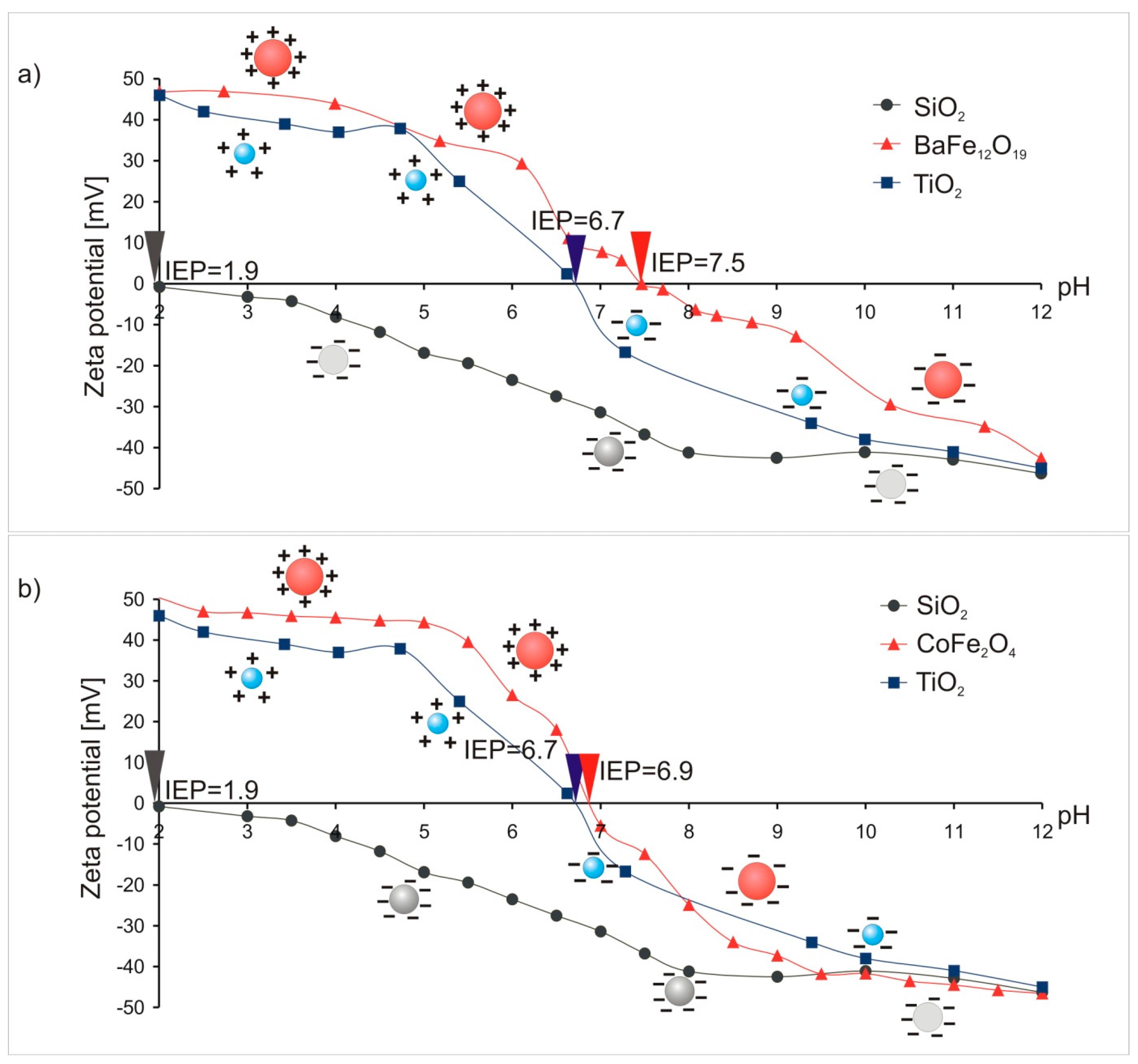
2.1. Preparation of Magnetic Photocatalysts—Measurement of Electrophoretic Mobility in a Function of pH (Zeta Potential Determination)
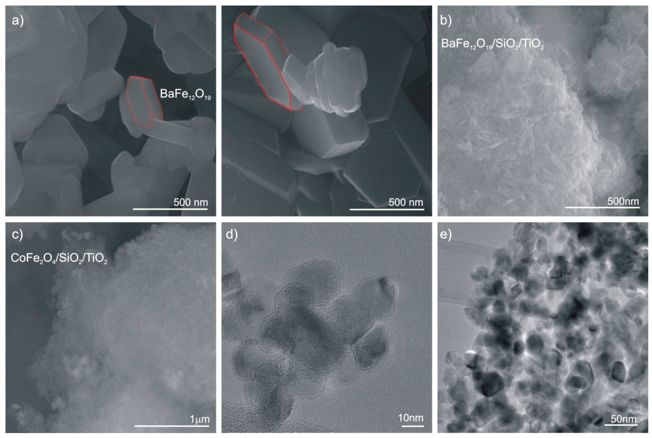
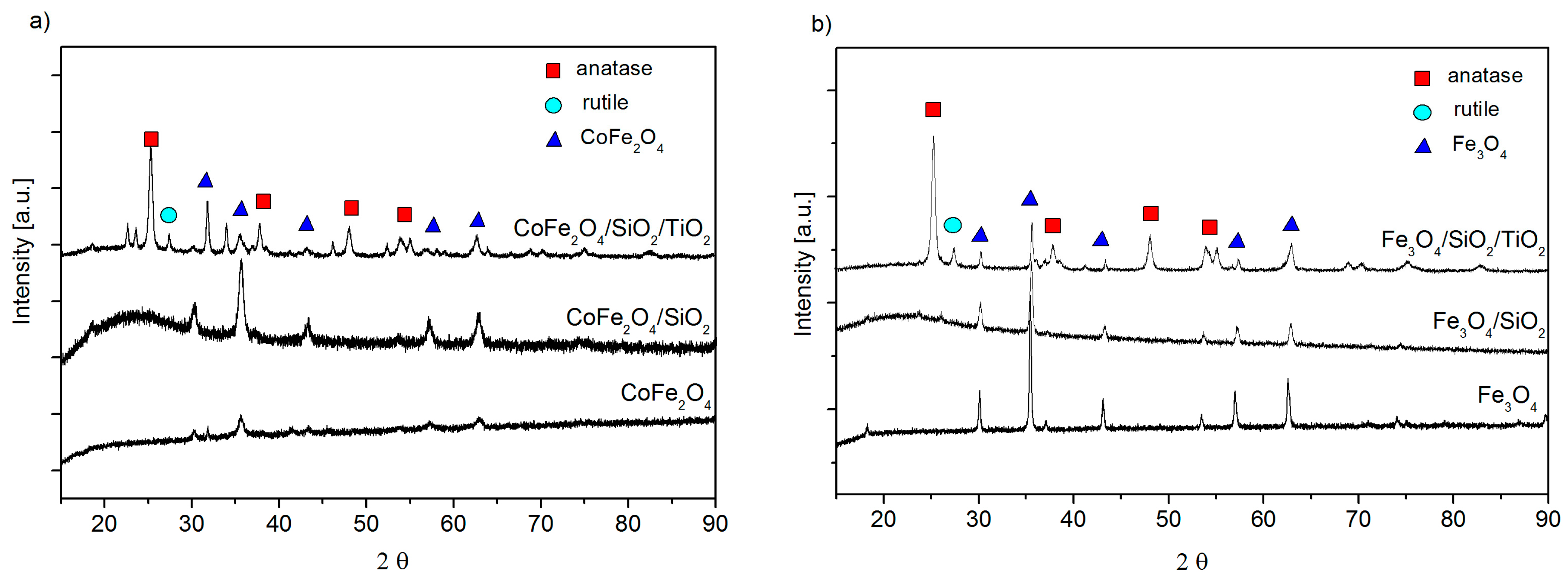
2.2. Characterization of the Nanoparticles
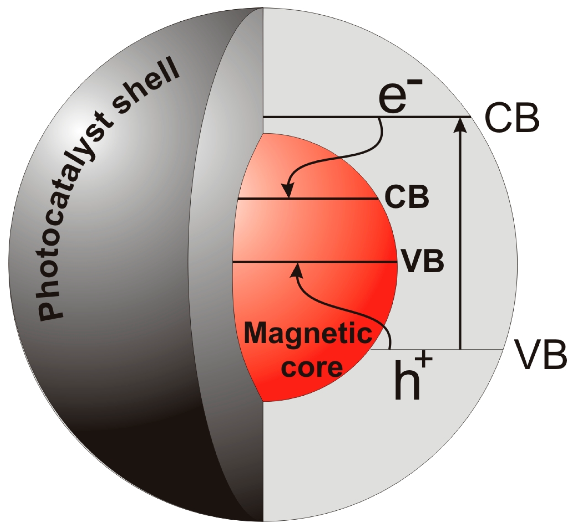
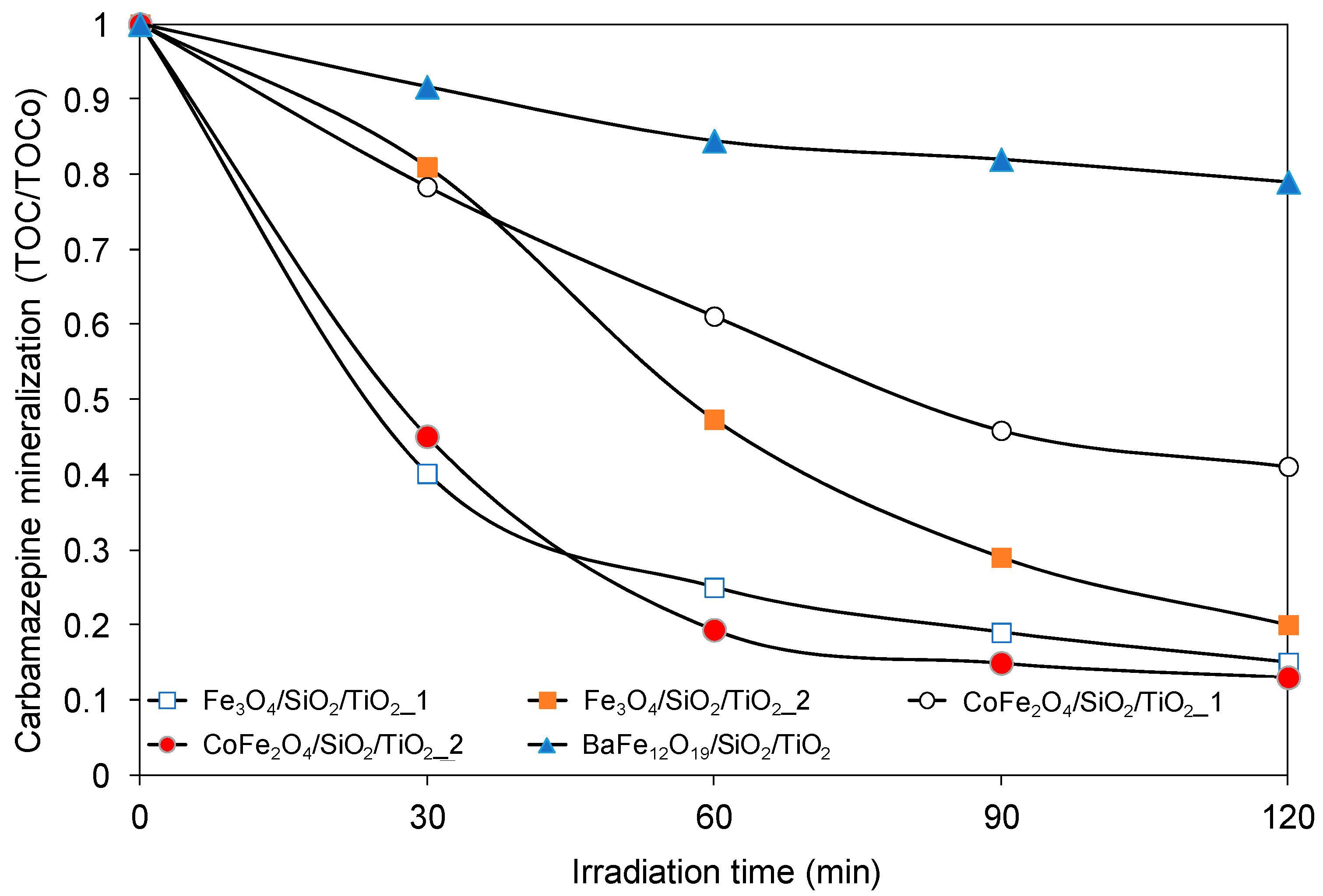
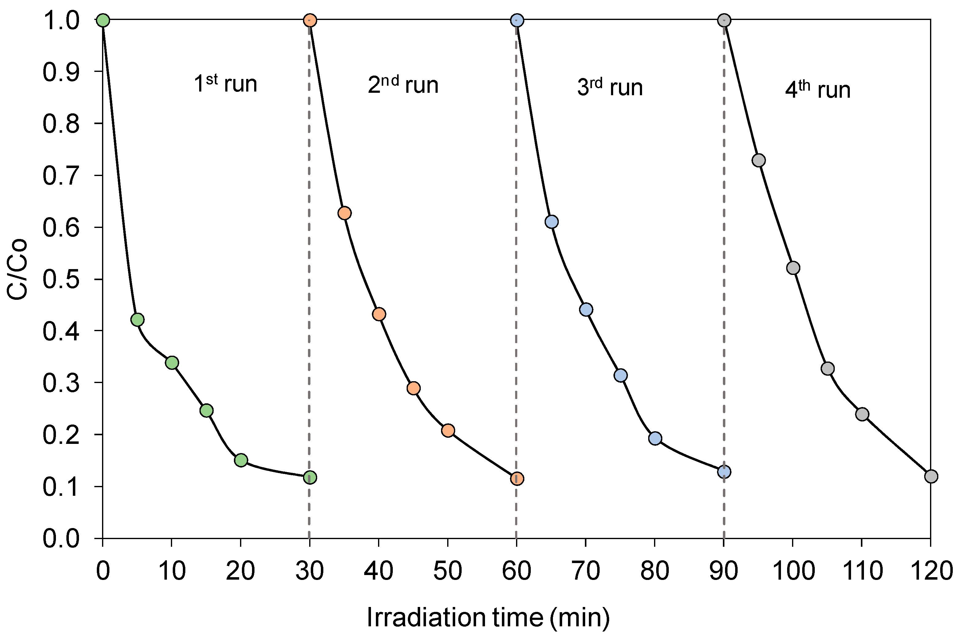
2.3. Photocatalytic Activity of Magnetic Nanocomposites
2.4. Discussion
3. Experimental Part
3.1. Materials
3.2. Preparation of Magnetic Photocatalysts
3.2.1. Preparation of Spinel and Hexagonal Ferrite Particles
3.2.2. Preparation of Spinel and Hexagonal Ferrite Coated with Silica Interlayer and TiO2 Shell
3.3. Characterization of Magnetic Photocatalysts
3.4. Measurements of Photocatalytic Activity
4. Conclusions
Acknowledgments
Author Contributions
Conflicts of Interest
References
- Guo, Z.; Ma, R.; Li, G. Degradation of phenol by nanomaterial TiO2 in wastewater. Chem. Eng. J. 2006, 119, 55–59. [Google Scholar] [CrossRef]
- Carabin, A.; Drogui, P.; Robert, D. Photo-degradation of carbamazepine using TiO2 suspended photocatalysts. J. Taiwan Inst. Chem. Eng. 2015, 54, 109–117. [Google Scholar] [CrossRef]
- He, Y.; Sutton, N.B.; Rijnaarts, H.H.H.; Langenhoff, A.A.M. Degradation of pharmaceuticals in wastewater using immobilized TiO2 photocatalysis under simulated solar irradiation. Appl. Catal. B Environ. 2016, 182, 132–141. [Google Scholar] [CrossRef]
- Zielińska-Jurek, A.; Zaleska, A. Ag/Pt-modified TiO2 nanoparticles for toluene photooxidation in the gas phase. Catal. Today 2014, 230, 104–111. [Google Scholar] [CrossRef]
- Vitiello, G.; Pezzella, A.; Zanfardino, A.; Silvestri, B.; Giudicianni, P.; Constantini, A.; Varcamonti, M.; Branda, F.; Luciani, G. Antimicrobial activity of eumelanin-based hybrids: The role of TiO2 in modulating the structure and biological performance. Mater. Sci. Eng. C 2017, 75, 454–462. [Google Scholar] [CrossRef] [PubMed]
- Jalvo, B.; Faraldos, M.; Bahamonde, A.; Rosal, R. Antimicrobial and antibiofilm efficacy of self-cleaning surfaces functionalized by TiO2 photocatalytic nanoparticles against Staphylococcus aureus and Pseudomonas putida. J. Hazard. Mater. 2017, 340, 160–170. [Google Scholar] [CrossRef] [PubMed]
- Xue, X.-D.; Fu, J.-F.; Zhu, W.-F.; Guo, X.-C. Separation of ultrafine TiO2 from aqueous suspension and its reuse using cross-flow ultrafiltration (CFU). Desalination 2008, 225, 29–40. [Google Scholar] [CrossRef]
- Zielińska-Jurek, A.; Klein, M.; Hupka, J. Enhanced visible light photocatalytic activity of Pt/I-TiO2 in a slurry system and supported on glass packing. Sep. Purif. Technol. 2017, 189, 246–252. [Google Scholar] [CrossRef]
- Dijkstra, M.F.J.; Michorius, A.; Buwalda, H.; Panneman, H.J.; Winkelman, J.G.M.; Beenackers, A.A.C.M. Comparison of the efficiency of immobilized and suspended systems in photocatalytic degradation. Catal. Today 2001, 66, 487–494. [Google Scholar] [CrossRef]
- Yang, L.; Wang, F.; Hakki, A.; Macphee, D.E.; Liu, P.; Hu, S. The influence of zeolites fly ash bead/TiO2 composite material surface morphologies on their adsorption and photocatalytic performance. Appl. Surf. Sci. 2017, 392, 687–696. [Google Scholar] [CrossRef]
- Ilkaeva, M.; Krivtsov, I.; Díaz, E.; Amghouzd, Z.; Pati, E.; Khainakove, S.; Garcíaa, J.S.; Ordónez, S. Photocatalytic degradation of 2-(4-methylphenoxy)ethanol over TiO2 spheres. J. Hazard. Mater. 2017, 332, 59–69. [Google Scholar] [CrossRef] [PubMed]
- Tsai, M.-C.; Yang, M.-H.; Chang, Y.-W.; Tzeng, J.-K.; Lee, C.-Y.; Chiu, H.T.; Chen, H.-C.; Lin, I.-N. Synthesis of porous micro-sized titania cages and their photocatalytic property. Mater. Chem. Phys. 2013, 143, 60–64. [Google Scholar] [CrossRef]
- Dou, L.; Gao, L.; Yang, X.; Song, X. Hierarchical architectures TiO2: Pollen-inducted synthesis, remarkable crystalline-phase stability, tunable size, and reused photo-catalysis. J. Hazard. Mater. 2012, 203–204, 363–369. [Google Scholar] [CrossRef] [PubMed]
- Hiroshi, F.; Yukiko, H.; Michichiro, Y.; Shoichi, A. Magnetic Photocatalyst. Japanese Patent JP6154620, 13 February 1994. [Google Scholar]
- Towata, A.; Sando, M. Photocatalyst Particles Containing Ferromagnetic Metal Particles and Method for Synthesis Thereof. U.S. Patent 5,703,002, 30 August 1997. [Google Scholar]
- Beydoun, D.; Amal, R.; Low, G.; McEvoy, S. A Preliminary Investigation into the Synthesis of Titania-Coated Magnetite as a Novel Photocatalyst. In Proceedings of the Third World Congress on Particle Technology, Brighton, UK, 6–9 July 1998; p. 385. [Google Scholar]
- Abbas, M.; Rao, B.P.; Reddy, V.; Kim, C. Fe3O4/TiO2 core/shell nanocubes: Single-batch surfactantless synthesis, characterization and efficient catalysts for methylene blue degradation. Ceram. Int. 2014, 40, 11177–11186. [Google Scholar] [CrossRef]
- Wei, J. Synthesis and magnetorheological effect of Fe3O4-TiO2 nanocomposite. J. Phys. Conf. Ser. 2009, 149, 25–29. [Google Scholar] [CrossRef]
- Zhang, L.; Wu, Z.; Chen, L.; Zhang, L.; Li, X.; Xu, H.; Wang, H.; Zhu, G. Preparation of magnetic Fe3O4/TiO2/Ag composite microspheres with enhanced photocatalytic activity. Solid State Sci. 2016, 52, 42–48. [Google Scholar] [CrossRef]
- Beydoun, D.; Amal, R.; Low, G.K.-C.; McEvoy, S. Novel Photocatalyst: Titania-Coated Magnetite. Activity and Photodissolution. J. Phys. Chem. B 2000, 104, 4387–4396. [Google Scholar] [CrossRef]
- Fan, Y.; Ma, C.; Li, W.; Yin, Y. Synthesis and properties of Fe3O4/SiO2/TiO2 nanocomposites by hydrothermal synthetic method. Mater. Sci. Semicond. Process. 2012, 15, 582–585. [Google Scholar] [CrossRef]
- Gad-Allah, T.A.; Fujimura, K.; Kato, S.; Satokawa, S.; Kojima, T. Preparation and characterization of magnetically separable photocatalyst (TiO2/SiO2/Fe3O4): Effect of carbon coating and calcination temperature. J. Hazard. Mater. 2008, 154, 572–577. [Google Scholar] [CrossRef] [PubMed]
- Shi, F.; Li, Y.; Zhang, Q.; Wang, H. Synthesis of Fe3O4/C/TiO2 magnetic photocatalyst via vapor phase hydrolysis. Int. J. Photoenergy 2012, 2012, 1–8. [Google Scholar] [CrossRef]
- Yuan, Q.; Li, N.; Geng, W.; Chi, Y.; Li, X. Preparation of magnetically recoverable Fe3O4@SiO2@meso-TiO2 nanocomposites with enhanced photocatalytic ability. Mater. Res. Bull. 2012, 47, 2396–2402. [Google Scholar] [CrossRef]
- Li, R.; Jia, Y.; Bu, N.; Wu, J.; Zhen, Q. Photocatalytic degradation of methyl blue using Fe2O3/TiO2 composite ceramics. J. Alloys Compd. 2015, 643, 88–93. [Google Scholar] [CrossRef]
- Lee, S.; Drwiega, J.; Wu, C.; Mazyck, D.; Sigmund, W.M. Anatase TiO2 Nanoparticle Coating on Barium Ferrite Using Titanium Bis-Ammonium Lactato Dihydroxide and Its Use as a Magnetic Photocatalyst. Chem. Mater. 2004, 12, 1160–1164. [Google Scholar] [CrossRef]
- Li, H.; Zhang, Y.; Wang, S.; Wu, Q.; Liu, C. Study on nanomagnets supported TiO2 photocatalysts prepared by a sol-gel process in reverse microemulsion combining with solvent-thermal technique. J. Hazard. Mater. 2009, 169, 1045–1053. [Google Scholar] [CrossRef] [PubMed]
- Salamat, S.; Younesi, H.; Bahramifar, N. Synthesis of magnetic core–shell Fe3O4@TiO2 nanoparticles from electric arc furnace dust for photocatalytic degradation of steel mill wastewater. RSC Adv. 2017, 7, 19391–19405. [Google Scholar] [CrossRef]
- Prieto-Mahaney, O.O.; Murakami, N.; Abe, R.; Ohtani, B. Correlation between Photocatalytic Activities and Structural and Physical Properties of Titanium(IV) Oxide Powders. Chem. Lett. 2009, 38, 238–239. [Google Scholar] [CrossRef]
- Amano, F.; Nakata, M.; Yamamoto, A.; Tanaka, T. Rutile titanium dioxide prepared by hydrogen reduction of Degussa P25 for highly efficient photocatalytic hydrogen evolution. Catal. Sci. Technol. 2016, 6, 5693–5699. [Google Scholar] [CrossRef]
- Fagerlund, G. Determination of specific surface by the BET method. Mater. Constr. 1973, 6, 239–245. [Google Scholar] [CrossRef]
- Liu, H.; Jia, Z.; Ji, S.; Zheng, Y.; Li, M.; Yang, H. Synthesis of TiO2/SiO2@Fe3O4 magnetic microspheres and their properties of photocatalytic degradation dyestuff. Catal. Today 2011, 175, 293–298. [Google Scholar] [CrossRef]
- Chi, Y.; Yuan, Q.; Li, Y.; Zhao, L.; Li, N.; Li, X.; Yan, W. Magnetically separable Fe3O4@SiO2@TiO2-Ag microspheres with well-designed nanostructure and enhanced photocatalytic activity. J. Hazard. Mater. 2013, 262, 404–411. [Google Scholar] [CrossRef] [PubMed]
- Fu, W.; Yang, H.; Li, M.; Chang, L.; Yu, Q.; Xu, J.; Zou, G. Preparation and photocatalytic characteristics of core-shell structure TiO2/BaFe12O19 nanoparticles. Mater. Lett. 2006, 60, 2723–2727. [Google Scholar] [CrossRef]
- Dubowik, J.; Gościańska, I. Micromagnetic Approach to Exchange Bias. Acta Phys. Pol. A 2015, 127, 147–152. [Google Scholar] [CrossRef]
- Petracic, O. Superparamagnetic nanoparticle ensembles. Superlattices Microstruct. 2010, 47, 569–578. [Google Scholar] [CrossRef]
- Haneda, K.; Morrish, A.H. Magnetic Properties of BaFe12O19 Small Particles. IEEE Trans. Magn. 1989, 25, 2597–2601. [Google Scholar] [CrossRef]
- Torres, T.E.; Lima, E., Jr.; Mayoral, A.; Ibarra, A.; Marquina, C.; Ibarra, M.R.; Goya, G.F. Validity of the Néel-Arrhenius model for highly anisotropic CoxFe3−xO4 nanoparticles. J. Appl. Phys. 2015, 118, 183902. [Google Scholar] [CrossRef]
- Su, R.; Bechstein, R.; Sø, L.; Vang, R.T.; Sillassen, M.; Esbjörnsson, B.; Palmqvist, A.; Besenbacher, F. How the Anatase-to-Rutile Ratio Influences the Photoreactivity of TiO2. J. Phys. Chem. C 2011, 115, 24287–24292. [Google Scholar] [CrossRef]
- Ohno, T.; Sarukawa, K.; Tokieda, K.; Matsumura, M. Morphology of a TiO2 Photocatalyst (Degussa, P-25) Consisting of Anatase and Rutile Crystalline Phases. J. Catal. 2001, 203, 82–86. [Google Scholar] [CrossRef]
- Rui, Z.; Wu, S.; Peng, C.; Ji, H. Comparison of TiO2 Degussa P25 with anatase and rutile crystalline phases for methane combustion. Chem. Eng. J. 2014, 243, 254–264. [Google Scholar] [CrossRef]
- Kawahara, T.; Konishi, Y.; Tada, H.; Tohge, N.; Nishii, J.; Ito, S. A Patterned TiO2(Anatase)/TiO2(Rutile) Bilayer-Type Photocatalyst: Effect of the Anatase/Rutile Junction on the Photocatalytic Activity. Angew. Chem. 2002, 114, 2935–2937. [Google Scholar] [CrossRef]
- Zielińska-Jurek, A.; Wei, Z.; Wysocka, I.; Szweda, P.; Kowalska, E. The effect of nanoparticles size on photocatalytic and antimicrobial properties of Ag-Pt/TiO2 photocatalysts. Appl. Surf. Sci. 2015, 353, 317–325. [Google Scholar] [CrossRef]
- Zielińska-Jurek, A.; Bielan, Z.; Wysocka, I.; Strychalska, J.; Janczarek, M.; Klimczuk, T. Magnetic semiconductor photocatalysts for the degradation of recalcitrant chemicals from flow back water. J. Environ. Manag. 2017, 195, 157–165. [Google Scholar] [CrossRef] [PubMed]
- Zhen, G.; Muir, B.W.; Moffat, B.A.; Harbour, P.; Murray, K.S.; Moubaraki, B.; Suzuki, K.; Madsen, I.; Agron-Olshina, N.; Waddington, L. Comparative study of magnetic behavior of spherical and cubic superparamagnetic iron oxide nanoparticles. J. Phys. Chem. C 2011, 115, 327–334. [Google Scholar] [CrossRef]
- Larumbe, S.; Gómez-Polo, C.; Pérez-Landazábal, J.; Pastor, J.M. Effect of a SiO2 coating on the magnetic properties of Fe3O4 nanoparticles. J. Phys. Condens Matter. 2012, 24, 1–6. [Google Scholar] [CrossRef] [PubMed]


| Sample Label | TiO2 Source | Preparation Conditions | Crystallite Size (nm) | BET Surface Area (m2/g) | Eg (eV) | |||
|---|---|---|---|---|---|---|---|---|
| pH | Surfactant | Anatase | Rutile | Ferrite | ||||
| Fe3O4 | - | 10 | CTAB | - | - | 45 | 9 | 0.5 |
| CoFe2O4 | - | 10 | - | - | - | 13 | 45 | 0.7 |
| BaFe12O19 | - | 10 | CTAB | - | - | 50 | 0.6 | 1.0 |
| Fe3O4/SiO2 | - | 5 | - | - | - | 47 | 169 | 1.4 |
| CoFe2O4/SiO2 | - | 5 | - | - | - | 13 | 124 | 0.9 |
| BaFe12O19/SiO2 | - | 10 | CTAB | - | - | 50 | 170 | 1.0 |
| Fe3O4/SiO2/TiO2_1 | P25 | 5 | TX-100 | 19 | 26 | 47 | 101 | 3.2 |
| Fe3O4/SiO2/TiO2_2 | P25 | 10 | CTAB | 19 | 24 | 45 | 95 | 3.2 |
| CoFe2O4/SiO2/TiO2_1 | TBT | 5 | TX-100 | 5 | - | 13 | 154 | 3.2 |
| CoFe2O4/SiO2/TiO2_2 | P25 | 5 | TX-100 | 18 | 27 | 13 | 69 | 3.2 |
| BaFe12O19/SiO2/TiO2_1 | TBT | 10 | CTAB | 5 | - | 50 | 100 | 3.1 |
| BaFe12O19/SiO2/TiO2_2 | TBT | 5 | TX-100 | 5 | - | 49 | 60 | 3.1 |
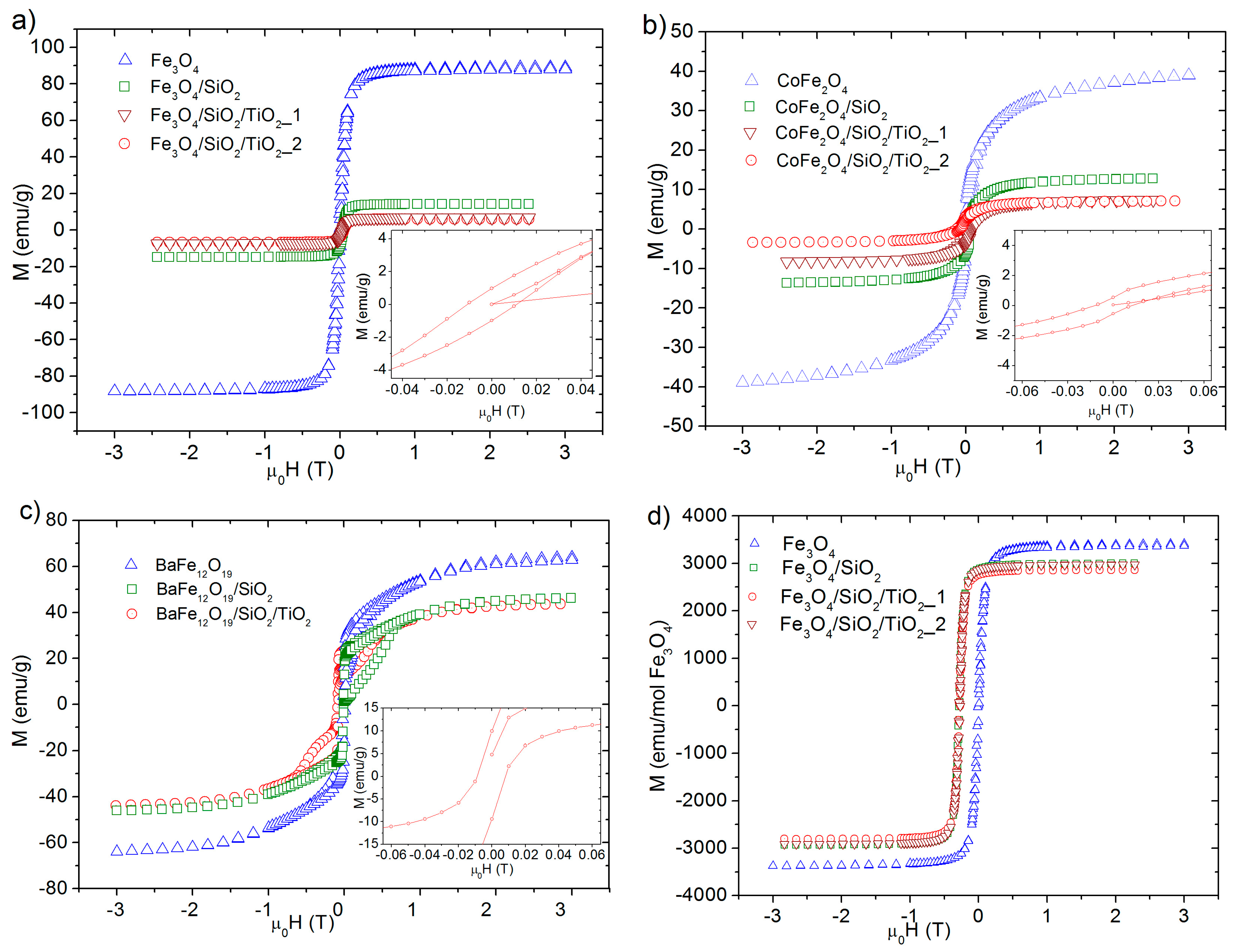
| Sample Label | Ms (emu·g−1) | Hc (Oe) | Mr (emu·g−1) |
|---|---|---|---|
| Fe3O4 | 89 | 100 | 9 |
| CoFe2O4 | 60 | 130 | 2 |
| BaFe12O19 | 64 | 140 | 16 |
| Fe3O4/SiO2 | 17 | 100 | 2 |
| CoFe2O4/SiO2 | 15 | 150 | 0 |
| BaFe12O19/SiO2 | 46 | 80 | 4 |
| Fe3O4/SiO2/TiO2_1 | 8 | 100 | 1 |
| Fe3O4/SiO2/TiO2_2 | 7 | 100 | 1 |
| CoFe2O4/SiO2/TiO2_1 | 9 | 250 | 1 |
| CoFe2O4/SiO2/TiO2_2 | 6 | 150 | 1 |
| BaFe12O19/SiO2/TiO2_1 | 43 | 80 | 9 |
| BaFe12O19/SiO2/TiO2_2 | 33 | 80 | 9 |
© 2017 by the authors. Licensee MDPI, Basel, Switzerland. This article is an open access article distributed under the terms and conditions of the Creative Commons Attribution (CC BY) license (http://creativecommons.org/licenses/by/4.0/).
Share and Cite
Zielińska-Jurek, A.; Bielan, Z.; Dudziak, S.; Wolak, I.; Sobczak, Z.; Klimczuk, T.; Nowaczyk, G.; Hupka, J. Design and Application of Magnetic Photocatalysts for Water Treatment. The Effect of Particle Charge on Surface Functionality. Catalysts 2017, 7, 360. https://doi.org/10.3390/catal7120360
Zielińska-Jurek A, Bielan Z, Dudziak S, Wolak I, Sobczak Z, Klimczuk T, Nowaczyk G, Hupka J. Design and Application of Magnetic Photocatalysts for Water Treatment. The Effect of Particle Charge on Surface Functionality. Catalysts. 2017; 7(12):360. https://doi.org/10.3390/catal7120360
Chicago/Turabian StyleZielińska-Jurek, Anna, Zuzanna Bielan, Szymon Dudziak, Izabela Wolak, Zuzanna Sobczak, Tomasz Klimczuk, Grzegorz Nowaczyk, and Jan Hupka. 2017. "Design and Application of Magnetic Photocatalysts for Water Treatment. The Effect of Particle Charge on Surface Functionality" Catalysts 7, no. 12: 360. https://doi.org/10.3390/catal7120360
APA StyleZielińska-Jurek, A., Bielan, Z., Dudziak, S., Wolak, I., Sobczak, Z., Klimczuk, T., Nowaczyk, G., & Hupka, J. (2017). Design and Application of Magnetic Photocatalysts for Water Treatment. The Effect of Particle Charge on Surface Functionality. Catalysts, 7(12), 360. https://doi.org/10.3390/catal7120360