Article

Polyelectrolyte Complex Beads by Novel Two-Step Process for Improved Performance of Viable Whole-Cell Baeyer-Villiger Monoxygenase by Immobilization

1
Department of Glycobiotechnology, Institute of Chemistry, Slovak Academy of Sciences, Dúbravská cesta 9, SK-845 38 Bratislava, Slovakia
2
Polymer Institute, Slovak Academy of Sciences, Dúbravská cesta 9, SK-845 41 Bratislava, Slovakia
3
Department of Biophotonics, International Laser Centre, Ilkovičova 3, SK-841 04 Bratislava, Slovakia
4
Department of Electron Microscopy, Institute of Scientific Instruments of the ASCR v.v.i., Královopolská 147, CZ-612 64 Brno, Czech Republic
5
Institute of Organic Chemistry and Macromolecular Chemistry, Centre of Excellence for Polysaccharide Research, Friedrich Schiller University of Jena, Humboldtstrasse 10, D-07743 Jena, Germany
*
Author to whom correspondence should be addressed.
Catalysts 2017, 7(11), 353; https://doi.org/10.3390/catal7110353
Submission received: 18 October 2017 / Revised: 10 November 2017 / Accepted: 13 November 2017 / Published: 21 November 2017
(This article belongs to the Special Issue Immobilized Biocatalysts)

Abstract

:
A novel immobilization matrix for the entrapment of viable whole-cell Baeyer–Villiger monooxygenase was developed. Viable recombinant Escherichia coli cells overexpressing cyclohexanone monooxygenase were entrapped in polyelectrolyte complex beads prepared by a two-step reaction of oppositely-charged polymers including highly defined cellulose sulphate. Immobilized cells exhibited higher operational stability than free cells during 10 repeated cycles of Baeyer–Villiger biooxidations of rac-bicyclo[3.2.0]hept-2-en-6-one to the corresponding lactones (1R,5S)-3-oxabicyclo-[3.3.0]oct-6-en-3-one and (1S,5R)-2-oxabicyclo-[3.3.0]oct-6-en-3-one. The morphology of polyelectrolyte complex beads was characterised by environmental scanning electron microscopy; the spatial distribution of polymers in the beads and cell viability were examined using confocal laser scanning microscopy, and the texture was characterised by the mechanical resistance measurements.

1. Introduction

The immobilization of viable whole-cell biocatalysts has long been considered an important method for ensuring their recyclability and stabilisation for the development of the industrial production of chemical specialities and chiral drug precursors [1]. In particular, the combination of viable recombinant cells with overproduced enzymes and their immobilization facilitates recyclability and renders possible a continuous bioreactor arrangement with high application potential [2].
One of the validated techniques for immobilizing viable recombinant cells is their encapsulation in polyelectrolyte complex (PEC) capsules, which afforded promising results in the previous development of encapsulated viable whole-cell biocatalysts [3,4,5]. PEC capsules have been produced by a single-step polyelectrolyte complexation of oppositely-charged polyanion (PA) aqueous solutions consisting of cellulose sulphate (CS), high-viscosity sodium alginate and living bacterial cells with a polycation solution of poly(methylene-co-guanidine) (PMCG), CaCl2 as gelling and NaCl as antigelling agents [6]. PA microdrops have been air-stripped by a coaxial nozzle into a polycation solution flowing in a multiloop chemical reactor for the preparation of spherical and monodispersed PEC capsules [7]. This encapsulation protocol preserved the high viability of the encapsulated recombinant Escherichia coli (E. coli) cells with overproduced ezymes from the group of Baeyer–Villiger monooxygenases (BVMOs) as well as their long-term storage and operational stability [4,8,9,10].
The development of encapsulation techniques, including the PEC capsules, has progressed towards a precise and targeted characterisation of the morphology, physicochemical properties of capsules, biocompatibility and their changes over time, using the latest techniques [11,12] for applications in biotechnology and medicine. Additionally, there is also an increased effort to optimise immobilization by predicting and modelling the bioengineering parameters of potential industrial processes [13]. On the other hand, current practice in industrial applications of immobilized biocatalysts [14] does not entail the consistent characterisation of the immobilization procedure and influence over the geometry, size, stability of composition, mechanical and diffusion properties of immobilized cell particles. This could lead to an incomplete knowledge of the immobilization parameters and methods for their control as well as the unsubstantiated exclusion of promising immobilization techniques for industrial use. The current development and production of tailor-made CS is crucial for further research into innovative and improved PEC capsules [15]. Parameters such as the origin of cellulose, molecular weight (MW), viscosity and degree of substitution (DS) of the CS significantly influence the successful formation of PEC capsules. Hence, the selection of commercial, tailor-made CS by the process described in [15], which, to the best of our knowledge, alone meets the latter scientific requirements, is essential for the encapsulation of cells. Although the production of PEC capsules with semipermeable membranes has been useful for the development of viable whole-cell biocatalysts under laboratory conditions, it entails a relatively complex encapsulation procedure and considerable consumption of PMCG and washing solutions using a continuous, one-step protocol. In addition, for more robust uses, PEC capsules with immobilized whole-cell biocatalysts have proved to be fragile during intensive mixing [10], and it was necessary to use fluidised-bed bioreactors [5] or mini packed-bed reactors with a low flow-rate [9]. Hence, it is desirable to develop protocols for the preparation of PEC beads with enhanced mechanical properties, with a lower consumption of PMCG and washing solutions, and with a simpler cation immobilization procedure.
The present work sought to develop PEC beads with entrapped viable cells complexed in the whole volume of the beads. Another aim was the use of imaging techniques to characterise the morphology of PEC beads and cells in the native state, the spatial distribution of the polymer within PEC beads and the mechanical resistance of the PEC beads. Viable recombinant E. coli (Escherichia coli) cells overproducing cyklohexanone monooxygenase (CHMO) from the group of BVMOs were used as a model biocatalyst in this study. Currently, there is increased interest in research into novel enzymes and the industrial applications of BVMOs, as they catalyse the enantioselective BV biooxidation of a wide range of cyclic ketones to the corresponding lactones as chiral precursors of natural and bioactive compounds as well as potential drugs [16,17,18,19]. The study also sought to determine the operational stability of PEC beads with immobilized recombinant E. coli cells with CHMO during repeated Baeyer–Villiger (BV) biooxidations as well as the influence of repeated biotransformations on the viability of cells.

2. Results and Discussion

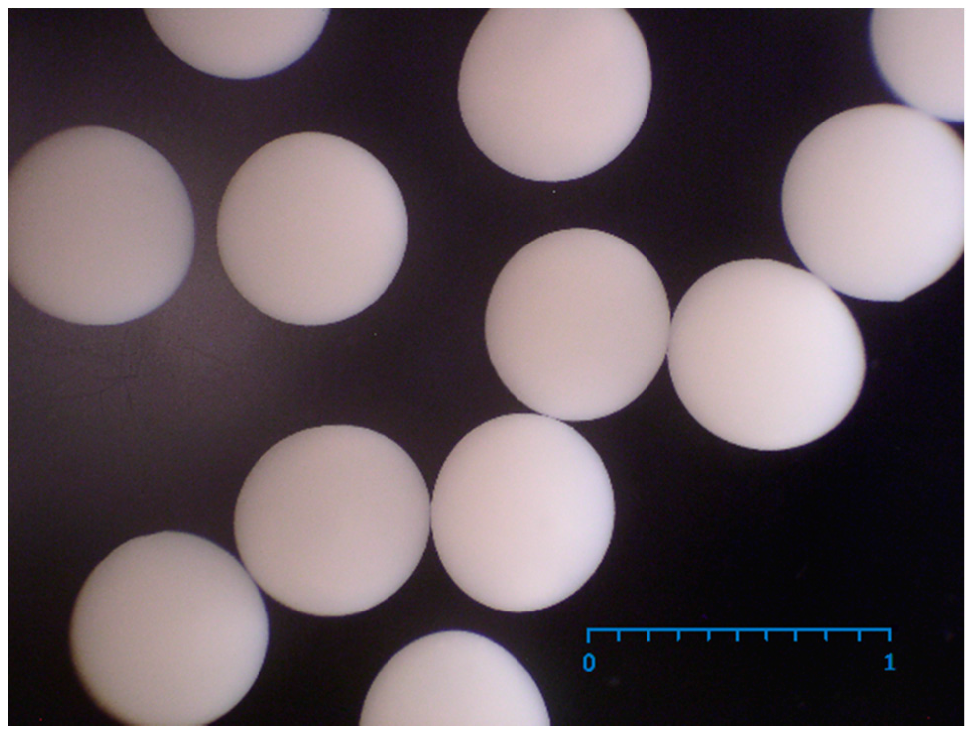
2.1. Production of PEC Bads with Immobilized E. coli Containing CHMO

The development of PEC beads was performed following a modified protocol originally developed for the one-step encapsulation of viable E. coli cells with overproduced BVMO in PEC capsules [4,9]. In the present work, PEC beads were prepared by a two-step procedure. This included the gelation of PA microdrops with cells in a solution of CaCl2 in the first step followed by polyelectrolyte complexation with PMCG in the second step, and finally washing in a physiological saline solution. This immobilization protocol resulted in less stringent and simpler conditions including higher utility and approximately a 2-fold decrease in the consumption of expensive PMCG and washing solutions for the preparation of immobilized viable E. coli with CHMO as compared with the PEC capsules previously used [9]. Use of the two-step procedure for the preparation of PEC beads led to an approximately one-third cost reduction as compared with the previous production of PEC capsules. Irrespective of this, a narrow size distribution of PEC beads of 0.64 ± 0.03 mm (Figure 1), high cell viability as well as higher operational stability of immobilized cells than in free cells was achieved, as detailed subsequently.

2.2. Suitability of Cellulose Sulphate

The selection of suitable CS as the polyanion component is the key to polyelectrolyte complexation with the PMCG polycation, which is crucial for the integrity, mechanical and chemical resistance and biocompatibility of PEC beads. CS with a DS of 1.45 and a molecular weight of 400,000 g/mol–500,000 g/mol was used for the preparation of the PEC beads using a two-stage procedure including gelation and complexation steps. The substitution did not apparently depolymerise the cellulose, because the DS of 1.45 was in good agreement with the increase in MW of the starting cellulose (254,700 g/mol). The crystallinity index of the bleached kraft pulp (BKP) CrI of 50 ± 3%, determined by the method developed by [20] showed a relatively high proportion of the amorphous phase distorted by the supra-molecular structure of the starting cellulose. If a similar crystallinity is exhibited by the spruce sulphite pulp (SSP) used in this work, a relatively high proportion of the amorphous phase should afford a more homogeneous distribution of substitution in the over-molecular cellulose structure. The amorphous phase also affords the availability of the polycations (PMCG) forming the PEC complex as well as better permeability of substrates and products for a biocatalytic reaction in that part of PEC beads in which the CS is incorporated.

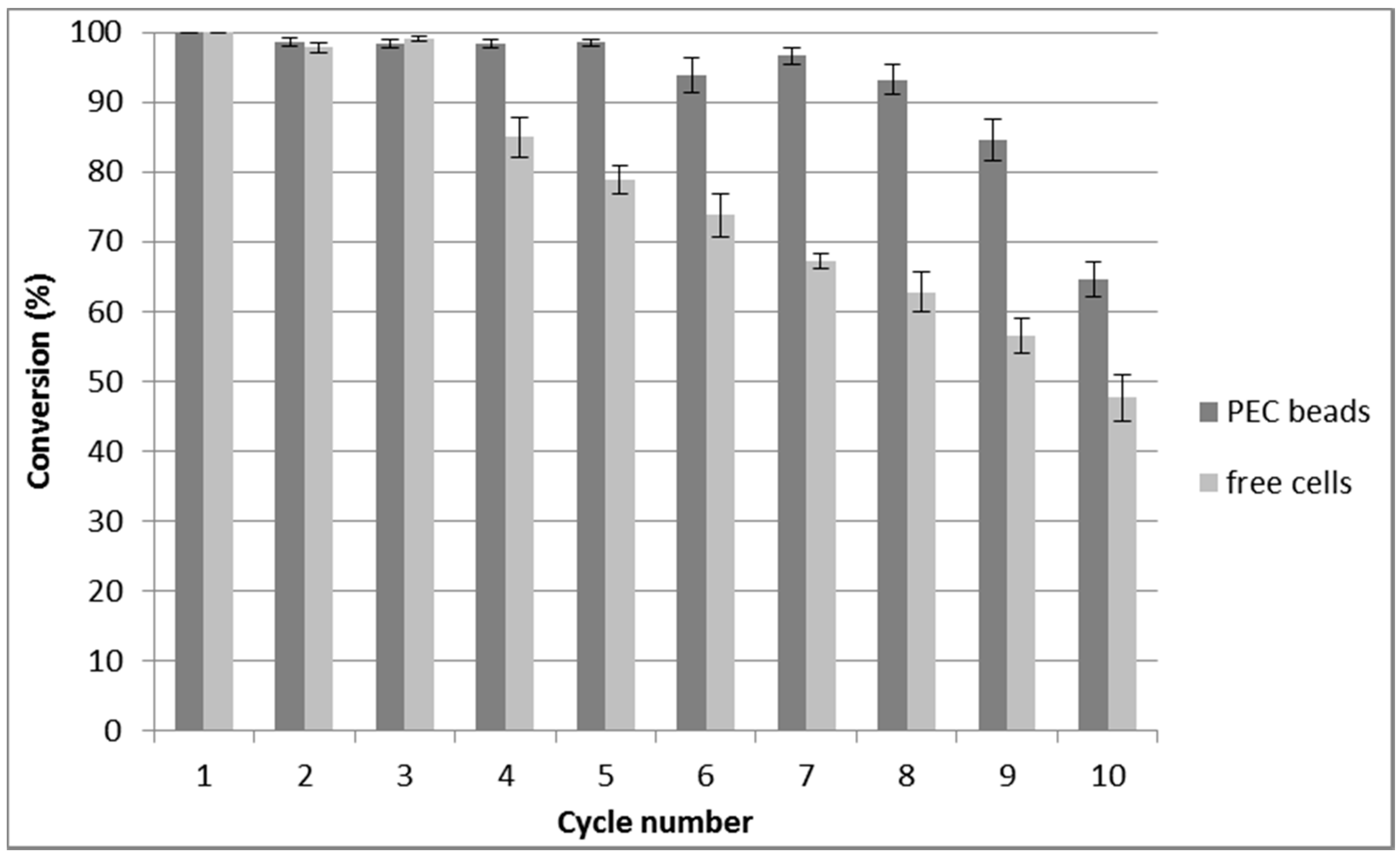
2.3. Repeated Biotransformations Using Immobilized Cells

Stabilisation of the high enzyme activity of cells to achieve the maximum substrate conversion in repeated biotransformation cycles is one of the most important parameters in respect of the profitability of immobilized whole-cell biocatalysts [1]. At the same time, when using live cells, the need to maintain the highest degree of cell viability is also important. Over the course of 10 repeated BV biooxidations of bicyclo[3.2.0]hept-2-ene-6-one to regioisomeric lactones (1R,5S)-3-oxabicyclo-[3.3.0]oct-6-en-3-one and (1S,5R)-2-oxabicyclo-[3.3.0]oct-6-en-3-one, a high and stable conversion rate was observed using E. coli cells with CHMO immobilized in PEC beads (Figure 2). For up to 8 cycles, conversion rates of 100–93 ± 4% were achieved. After the 9th and 10th cycles, decreases to 85 ± 6% and 65 ± 5%, respectively were observed. In contrast with these findings, unprotected free cells exhibited a distinct decrease of 85 ± 6% in substrate conversion of the BV biooxidation in the 4th cycle, which showed a steady decline to 48 ± 6% in the 10th cycle. Cell stabilization within mild and physiological microenvironment of the PEC beads may contribute to higher stability of immobilized cells and delayed their deactivation compared to free cells (Figure 2).

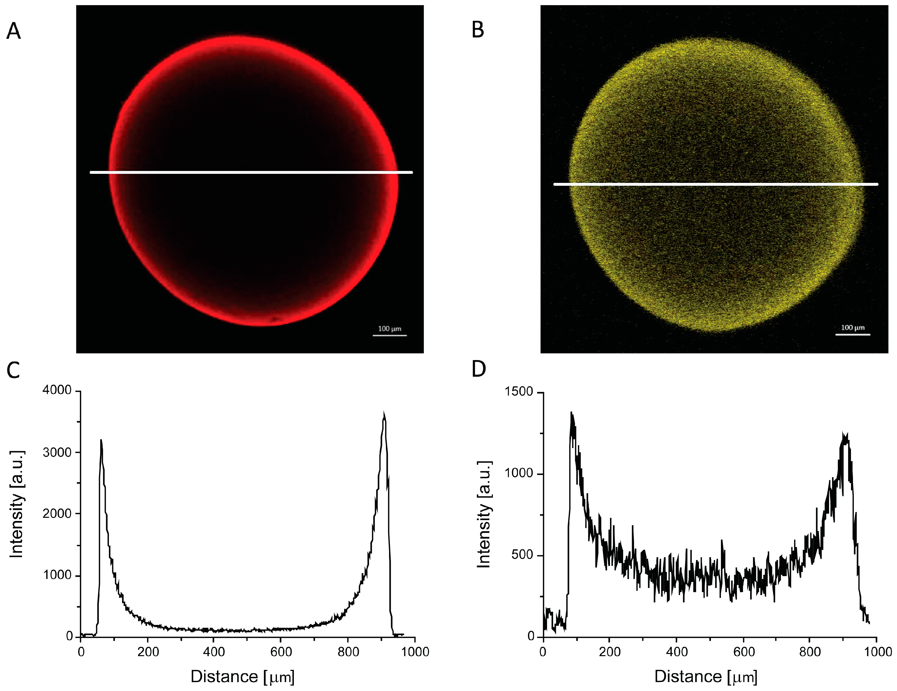
2.4. Morphology of PEC Beads
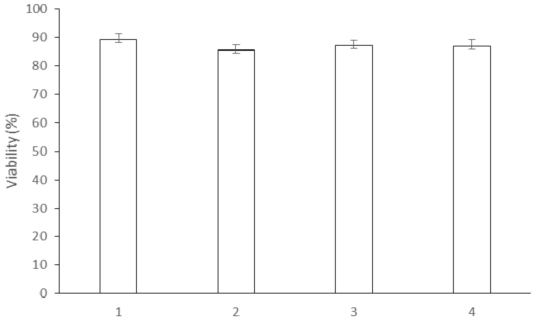
To investigate the morphology of the PEC beads, the confocal laser scanning microscopy arrangement described in the previous section was used (Figure 3). The beads contained Rhodamine 123 and Eosin Y as fluorescent tracer dyes of a concentration of 10−6 mol/L and were incubated for 15−30 min. These two non-covalently bound fluorescent tracer dyes were employed to monitor the distribution of the residual opposite charge within the PEC microcapsule [21]. Anionic Eosin Y (Figure 3A) was predominantly localised in the outer region of the PEC bead, and its concentration dropped sharply towards the bead interior. On the other hand, cationic Rhodamine 123 (Figure 3B) was observed in the entire volume of the PEC bead, with a higher intensity at the outer region and a lower intensity in the centre. Unlike in the previous study on polyelectrolyte capsules [21], no specific layers or internal structures were observed in the present study. It is worth noting that the spatial distribution of the fluorescence reflects the concentration of the dye molecules, corresponding to the residual free-charged groups of the matrix, rather than the spatial distribution of polymer components in the bead.

2.5. Cell Viability

Cell viability prior to and after biotransformation was measured by the intake of PI by the necrotic cells [8]. The viability of immobilized cells of E. coli with CHMO under different conditions and after 10 repeated biotransformation cycles (Figure 4) was evaluated. The viability in all cases remained within an 85−95% range.
The high viability of cells immobilized in PEC beads was identical with that of free cells prior to biotransformations. This demonstrates the excellent biocompatibility of the novel PEC beads and their ability to preserve the physiological microenvironment for immobilized cells. At the same time, the nontoxicity of the PEC complex with CS in capsules [22] was also confirmed in the PEC beads developed in the present work. In addition, the high cell viability which did not vary after 10 biotransformation cycles demonstrates the preservation of the physiological conditions for immobilized cells during biotransformations.

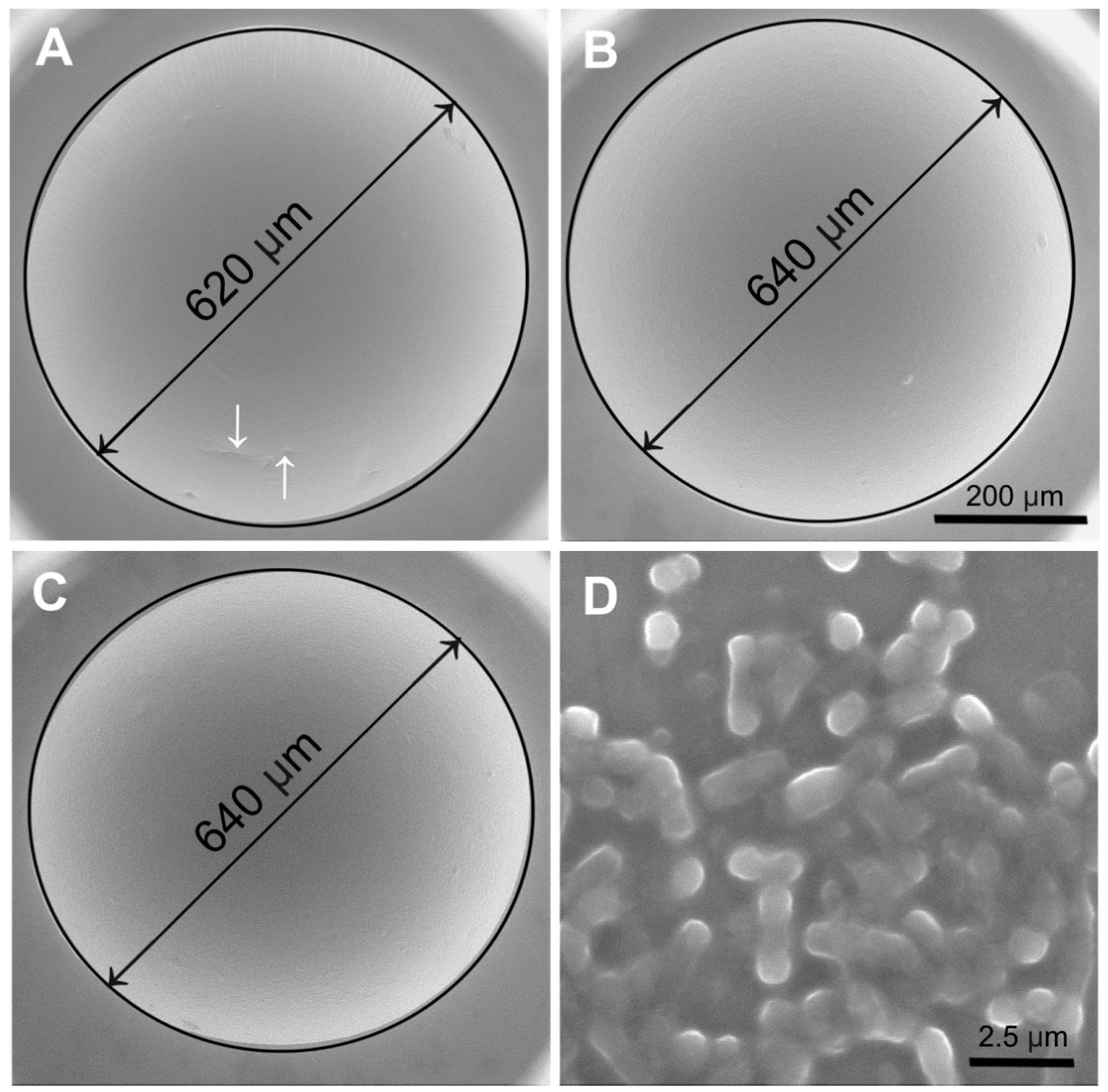
2.6. Environmental Scanning Electron Microscopy (ESEM)

The PEC beads with and without E. coli cells showed a very regular spherical shape with a relatively smooth surface free from cracks and corrugations that was maintained after repeated biotransformations (Figure 5A–C). E. coli cells in their native state covered with a very thin liquid layer of a solution are shown in Figure 5D. The PEC bead microstructure was significantly improved compared with the microstructure described in a previous study, where the roughness of the PEC capsule surface with Gluconobacter oxydans cells was higher [5]. A slight wrinkling of the PEC bead surface without cells is commonly formed during polyelectrolyte complexation (highlighted by white dashed arrows in Figure 5A). In all cases, good roundness and bead regularity as well as low size distribution were demonstrated using ESEM. A slightly blurred image of E. coli cells partially covered by a small layer of the solution exhibited the unique possibility to depict fully wet cells in their native state using the ESEM AQUASEM II (Figure 5D) equipped with custom-built detectors of signal electrons with a very high detection efficiency [23]. A lower quality of this image was caused by the unprocessed state of these live cells, depicted without the use of any preparation technique such as chemical fixation, drying or metal coating [24] as well as their imaging under a very low beam current and a relatively high pressure of water vapour in the specimen chamber of the ESEM.

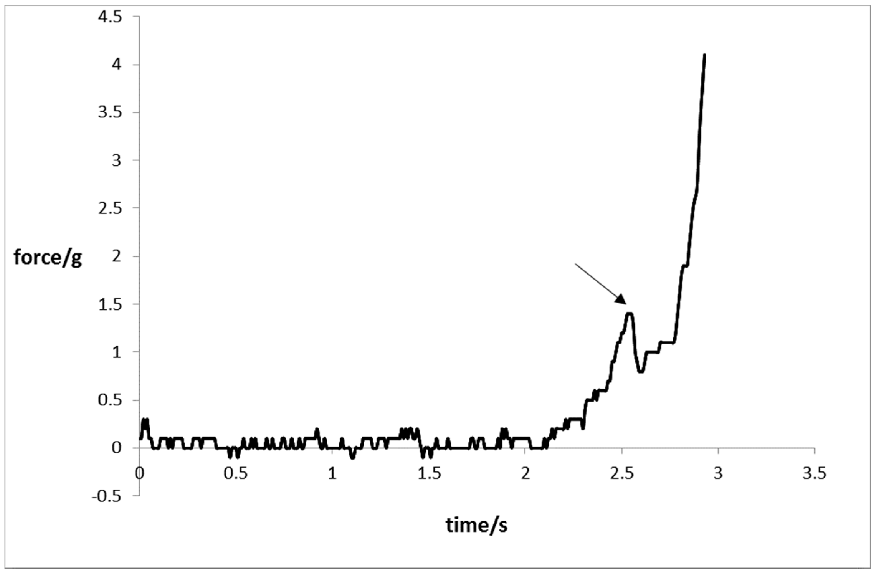
2.7. Mechanical Resistance

Figure 6 depicts a typical course of the force during measurement of the mechanical resistance of PEC beads. The point marked by the arrow represents the bursting force required for mechanical disruption of the PEC bead. The average bursting force of PEC beads with entrapped cells prior to biotransformations was 0.95 ± 0.04 g/bead, which was close to the average bursting force of 1.16 ± 0.26 g/bead obtained for PEC beads after 10 biotransformation cycles. Practically the same mean values of bursting force prior to and after biotransformations showed that the mechanical strength of the PEC complex in the beads was not influenced either by mechanical forces induced by mixing or the sequestering agents such as phosphate buffer present in the reaction media.
It is also important to note that the PEC complex found throughout the volume of PEC beads provides additional mechanical resistance to the cells compared with the previously reported PEC capsule which contains the PEC membrane and the liquid core [25]. This can be seen in the course of the compression force where, after the disruption of a PEC bead at the point indicated by the arrow, a small decrease in the force occurred, followed by an increase in the bead resistance. On the other hand, the bursting of the PEC capsule observed previously [25] was followed by a sharp drop in force caused by the immediate and irreversible PEC capsule collapse.

3. Materials and Methods

3.1. Characterisation Polymers

High-viscosity, high-mannuronic acid sodium alginate from ISP Alginate (Girvan, UK) with the following chemical composition (determined by 1H NMR) was employed: fraction of alginate consisting of guluronic acid FG = 0.41, fraction of alginate consisting of mannuronic acid FM = 0.59; fractions of alginate consisting of dimers and trimers of gluronic acid (G) and mannuronic acid (M): FGG = 0.21, FGM = FMG = 0.20, FMM = 0.39; FGGM = FMGG = 0.03, FMGM = 0.18, FGGG = 0.18, NG>1 = 8. PMCG from Scientific Polymer Products Inc. (Ontario, NY, USA), supplied as a 35% (w/v) aqueous solution, was lyophilised prior to use. The fluorescent tracer dyes Rhodamine 123 and Eosin Y were obtained from Acros Organics (Geel, Belgium). Water-soluble CS with the required DS, dynamic viscosity and MW was provided by Senova Biotechnologieunternehmen (Weimar, Germany). The derivative was prepared in accordance with the literature by the homogeneous conversion of spruce sulphite pulp (MWCuen 254, 700 g/mol from Borregaard ChemCell, Sarpsborg, Norway) with SO3-pyridine complex in the ionic liquid 1-butyl-3-methylimidazolium chloride (BMIMCl) with N,N-dimethylformamide as a dipolar aprotic co-solvent in order to improve the miscibility of the reaction mixture [15]. The final product had a DS of 1.45 and a dynamic viscosity of 15.6 mPa·s in 0.9% NaCl aqueous solution. The viscosity average molecular weight of around 500,000–600,000 g/mol corresponds to the weight average molecular weight of CS in the range from 310,000 to 460,000 g/mol determined by size exclusion chromatography [15]. These values were similar to those of the starting cellulose of 254,700 g/mol [15]. Sharp peaks characteristic of CS originating from sulphate groups were observed at 1256 cm−1 (δ SO2) and 805 cm−1 (δ SO).
A Flash 2000 CHNS/O Analyser (Thermo Fisher Scientific, Waltham, MA, USA) was used for elemental analyses. The DS was calculated from the elemental composition as described in the literature [15]. Size exclusion chromatography (SEC) of CS was performed on a JASCO system (PU-980 isocratic pump, RI-930 refractive index detector, Suprema guard column and columns (100 Ǻ, 1000 Ǻ, 3000 Ǻ, in series) with 1 M Na2HPO4 (pH = 9, 200 mg/L NaN3) as eluent (flow-rate: 1 mL/min, sample, concentration: 0.5 mg/mL). A viscometer (Brookfield Programmable DV-II+ Instrument, Middleboro, MA, USA) equipped with Thermostat Julabo F12 (Seelbach, Germany) was employed to determine the dynamic viscosity of CS solutions in an 0.9% NaCl aqueous solution. The viscosity average molecular weight of CS was determined from the intrinsic viscosity in 0.5 M NaCl at 25 °C (Mark Houwink parameters: α = 0.94, K = 6.37⋅10−6 mL/g) in accordance with the literature [26]. Fourier-transform infra red (FTIR) spectra were measured using a Nicolet 6700 spectrometer (Thermo Fisher Scientific, Waltham, MA, USA) equipped with DTGS detector and Omnic 8.0 software (Thermo Fisher Scientific, Waltham, MA, USA). The spectra were collected in the range from 4000 cm−1 to 400 cm−1 at a resolution of 4 cm−1; the number of scans was 128. Diamond Smart Orbit ATR accessory was applied to solid-state measurements.

3.2. Cultivation of Cells

Cell stocks of E. coli overexpressing CHMO (EC1.14.13.22) from Acinetobacter calcoaceticus NCIMB 9871 were stored at −80 °C. The recombinant E. coli strain was kindly donated by Prof. Marko D. Mihovilovič (TU Vienna, Austria). Cells, streaked at LBamp agar Petri dish containing 10 g/L of peptone, 5 g/L of yeasts extract, 10 g/L of NaCl, 15 g/L of agar and 0.2 g/L of ampiciline, were cultivated in an incubator at 37 °C to afford colonies of approximately 1 mm size. A single colony from LBamp agar was inoculated to a 10 mL of LBamp medium in an Erlenmayer flask and allowed to grow at 37 °C in an orbital shaker at 150 rpm for 12 h. 1% of the inoculum (v/v) was pipetted into a TBamp medium and cultivated at 37 °C in an orbital shaker at 150 rpm for 12 h. The addition of isopropyl β-D-1-thiogalactopyranoside (IPTG) to the final concentration of 0.25 mM to the medium, during shaking at 25 °C in an orbital shaker at 200 rpm for 2 h, induced the synthesis of CHMO enzyme. The biomass was separated from the medium by centrifugation at 15 °C and 4000× g for 15 min and used for biotransformation experiments and immobilization.

3.3. Preparation of Polyelectrolyte Complex Beads

A suspension of cells was prepared in a solution of 1.8% (w/v) of PA consisting of 0.9% (w/v) of sodium alginate and 0.9% of CS in 0.9% (w/v) NaCl at pH = 7 to the final concentration of 10% of wet cells. The PA solution with the cells was added dropwise into a stirred gelation solution of 1.0% (w/v) of CaCl2 using a custom-made coaxial air-stripping extrusion device. The time of gelation was 10 min and the collection time was 1 min. Subsequently, the prepared beads were separated from the gelation medium and transferred into a stirred solution containing 1.8% (w/v) of PMCG and 0.9% (w/v) of NaCl at pH = 7 for 10 min in order to undergo polyelectrolyte complexation. The polyelectrolyte complex beads were washed with 0.9% (w/v) of NaCl and used in biotransformation.

3.4. Repeated Biotransformations

PEC beads with immobilized cells (10% w/w) and free E. coli cells were transferred into a reaction medium containing 50 mM phosphate buffer (pH = 7), 0.2 g/L of ampiciline and 4 g/L of glucose. Model Baeyer–Villiger biooxidation resulting in the production of lactone regioisomers (1S,5R)-2-oxabicyclo[3.3.0]oct-6-en-3-one and (1R,5S)-3-oxabicyclo-[3.3.0]-oct-6-en-2-one [27] was initiated by the addition of substrate rac-bicyclo[3.2.0]hept-2-en-6-one in ethanol (volume ratio 1:9) to the reaction medium to a final concentration of 0.5 g/L. The reaction conditions were 25 °C and 150 rpm in an orbital shaker. Substrate and produced lactones in samples were extracted in dichlormethane with methyl benzoate as internal standard (0.5 mg/mL) and analysed using gas chromatography as reported previously [9]. After each biotransformation, the PEC beads containing cells, and the free cells, were separated from the reaction medium by filtration (encapsulated cells) or centrifugation (free cells), washed with a substrate-free medium and subjected to another biotransformation cycle.

3.5. Confocal Laser Scanning Microscopy

The internal structure of the PEC beads was characterised using confocal laser scanning microscopy (CLSM). Images of the PEC beads were recorded simultaneously in transmission, reflection and fluorescence modes using a laser scanning microscope head LSM 510 META on Axiovert 200M stand (both from Carl Zeiss, Jena, Germany). A Zeiss PlanApochromat 10x/0.45 lens was used to visualise the whole beads. Laser lines of 488 nm for Rhodamine 123 and 543 nm for Eosin Y were used for excitation. Two channels with band-pass filter 500–550 nm for detection of the reflected light and a long-pass filter LP 560 nm were used for fluorescence.

3.6. Cell Viability

The CLSM confocal laser scanning microscopy system described for the investigation of the PEC bead structure was also used for cell viability imaging. Cells necrosis was detected using fluorescence of propidium iodide by LSM 510 META NLO microscope (Carl Zeiss, Jena, Germany) equipped with the C-Apochromat 40xW/corr water immersion objective. Viability of the encapsulated cells was determined after mild disruption of the beads in a test tube. The mixture was carefully stirred so as to ensure that the remains of beads did not affect the measurement. The cells were incubated for 10 min with 1 μM of propidium iodide (PI). Fluorescence was initiated by the 543 nm line of HeNe laser and an LP 560 nm long-pass emission filter was used for PI fluorescence detection. The cell viability was evaluated as a ratio of the number of the surviving bacterial cells to the total number of bacterial cells. The number of viable bacterial cells was calculated as the difference between the total number of bacterial cells and the number of necrotic cells identified by propidium iodide fluorescence. The total number of cells was counted from the respective transmission image in accordance with the literature [8].

3.7. Environmental Scanning Electron Microscopy

Due to the very high sensitivity of PEC beads to electron beam irradiation resulting in radiation damage and a high risk of surface dehydration, images of the beads were recorded under conditions of carefully controlled and very slowly reduced relative humidity (from 100% to 90%) in a specimen chamber of the ESEM AQUASEM II developed by the group of Environmental Electron Microscopy (ISI ASCR in Brno, Czech Republic) in cooperation with TESCAN (Brno, Czech Republic). A sophisticated hydration system with heated needle valve (35 °C) was used as an additional water supply in the microscope. PEC beads and E. coli cells were observed in a small droplet of aqueous solution of 50 mM phosphate buffer (pH = 7), ampicillin 0.2 g/L and glucose 4 g/L, at a reduced beam accelerating voltage of 10 kV and very low beam current of 30 pA. A specially modified ionisation detector of secondary electrons [28] affording a low beam current observation with a large field of view (up to 850 μm) was used.

3.8. Mechanical Strength

The mechanical properties of the PEC beads were determined in compression mode using a Texture Analyser TA-XT2i (Stable Micro Systems, Godalming, UK) equipped with a mobile probe and Texture Expert Exceed 2.64 software (Stable Micro Systems, Godalming, UK). Compression measurements were conducted at a compression speed of 0.5 mm/s to the point at which 95% of 50 PEC beads per batch were deformed. The bursting force is the result of this compression test.

4. Conclusions

This work focused on the development of an immobilization procedure providing PEC beads for the entrapment of viable recombinant E. coli cells with overexpressed cyclohexanone monooxygenase. Uniform and stable PEC beads were successfully produced by a two-step procedure including ionotropic gelation and polyelectrolyte complexation. The PEC beads were prepared under less stringent conditions with a lower consumption of polycation and washing solutions than for the PEC capsules previously used. These characteristics are highly advantageous for a potential production scale-up of Baeyer–Villiger biooxidations using entrapped viable cells. Accordingly, it was important that the performance of cells entrapped in PEC beads for a model Baeyer–Villiger biooxidation, expressed as their operational stability, was enhanced over that of free cells. The PEC beads preserved high cell viability and enabled faster and more precise handling with cells during repeated Baeyer–Villiger biooxidations. Further studies using environmental scanning electron microscopy confirmed the stability of the dimensions and the sphericity of the PEC beads. In addition, both the surface morphology and the mechanical resistance of the PEC beads remained unchanged prior to and after biotransformations.

Acknowledgments

This work was supported by the Slovak Grant Agency for Science VEGA 2/0090/16 and by the Slovak Research and Development Agency under contracts no. APVV-15-0227 and APVV-14-0858. This publication is the result of the project implementation: Applied research in the field of industrial biocatalysis, ITMS code: 26240220079 supported by the Research & Development Operational Programme funded by the ERDF. The authors are indebted to P. Laudeley (Institute of Organic Chemistry and Macromolecular Chemistry, Friedrich Schiller University of Jena) for her assistance with the synthesis of tailored CS.

Author Contributions

Tomáš Krajčovič performed experiments, analyzed data and wrote the paper; Marek Bučko and Peter Gemeiner conceived and designed the experiments, and wrote the paper; Alica Vikartovská contributed reagents/materials/analysis tools; Igor Lacík, Vilém Neděla, Dušan Chorvát and Martin Gericke conceived and designed the experiments, and wrote the paper; Thomas Heinze conceived and designed the experiments and contributed reagents/materials/analysis tools; Lucia Uhelská, Dušan Chorvát, Martin Gericke and Eva Tihlaříková performed experiments and analyzed data.

Conflicts of Interest

The authors declare no conflict of interest.

References

  1. Kadisch, M.; Willrodt, C.; Hillen, M.; Bühler, B.; Schmid, A. Maximizing the stability of metabolic engineering-derived whole-cell biocatalysts. Biotechnol. J. 2017, 12. [Google Scholar] [CrossRef] [PubMed]
  2. Wachtmeister, J.; Rother, D. Recent advances in whole cell biocatalysis techniques bridging from investigative to industrial scale. Curr. Opin. Biotechnol. 2016, 42, 169–177. [Google Scholar] [CrossRef] [PubMed]
  3. Bučko, M.; Vikartovská, A.; Lacík, I.; Kolláriková, G.; Gemeiner, P.; Pätoprstý, V.; Brygin, M. Immobilization of a whole-cell epoxide-hydrolyzing biocatalyst in sodium alginate-cellulose sulfate-poly(methylene-co-guanidine) capsules using a controlled encapsulation process. Enzyme Microb. Technol. 2005, 36, 118–126. [Google Scholar] [CrossRef]
  4. Hucík, M.; Bučko, M.; Gemeiner, P.; Štefuca, V.; Vikartovská, A.; Mihovilovic, M.D.; Rudroff, F.; Iqbal, N.; Chorvát, D., Jr.; Lacík, I. Encapsulation of recombinant E. coli expressing cyclopentanone monooxygenase in polyelectrolyte complex capsules for Baeyer-Villiger biooxidation of 8-oxabicyclo[3.2.1]oct-6-en-3-one. Biotechnol. Lett. 2010, 32, 675–680. [Google Scholar] [CrossRef] [PubMed]
  5. Bertóková, A.; Vikartovská, A.; Bučko, M.; Gemeiner, P.; Tkáč, J.; Chorvát, D.; Štefuca, V.; Neděla, V. Biooxidation of 2-phenylethanol to phenylacetic acid by whole-cell Gluconobacter oxydans biocatalyst immobilized in polyelectrolyte complex capsules. Biocatal. Biotransform. 2015, 33, 111–120. [Google Scholar] [CrossRef]
  6. Lacík, I.; Briššová, M.; Anilkumar, A.V.; Powers, A.C.; Wang, T. New capsule with tailored properties for the encapsulation of living cells. J. Biomed. Mater. Res. 1998, 39, 52–60. [Google Scholar] [CrossRef]
  7. Anilkumar, A.V.; Lacik, I.; Wang, T.G. A novel reactor for making uniform capsules. Biotechnol. Bioeng. 2001, 75, 581–589. [Google Scholar] [CrossRef] [PubMed]
  8. Schenkmayerová, A.; Bučko, M.; Gemeiner, P.; Chorvát, D., Jr.; Lacík, I. Viability of free and encapsulated Escherichia coli overexpressing cyclopentanone monooxygenase monitored during model Baeyer–Villiger biooxidation by confocal laser scanning microscopy. Biotechnol. Lett. 2012, 34, 309–314. [Google Scholar] [CrossRef] [PubMed]
  9. Bučko, M.; Schenkmayerová, A.; Gemeiner, P.; Vikartovská, A.; Mihovilovič, M.D.; Lacík, I. Continuous testing system for Baeyer-Villiger biooxidation using recombinant Escherichia coli expressing cyclohexanone monooxygenase encapsulated in polyelectrolyte complex capsules. Enzyme Microb. Technol. 2011, 49, 284–288. [Google Scholar] [CrossRef] [PubMed]
  10. Schenkmayerová, A.; Bučko, M.; Gemeiner, P.; Treľová, D.; Lacík, I.; Chorvát, D., Jr.; Ačai, P.; Polakovič, M.; Lipták, L.; Rebroš, M.; et al. Physical and bioengineering properties of polyvinyl alcohol lens-shaped particles versus spherical polyelectrolyte complex microcapsules as immobilization matrices for a whole-cell Baeyer-Villiger monooxygenase. Appl. Biochem. Biotechnol. 2014, 174, 1834–1849. [Google Scholar] [CrossRef]
  11. De Vos, P.; Bučko, M.; Gemeiner, P.; Navrátil, M.; Švitel, J.; Faas, M.; Strand, B.L.; Skjak-Braek, G.; Morch, Y.A.; Vikartovská, A.; et al. Multiscale requirements for bioencapsulation in medicine and biotechnology. Biomaterials 2009, 30, 2559–2570. [Google Scholar] [CrossRef] [PubMed]
  12. Rokstad, A.M.A.; Lacík, I.; de Vos, P.; Strand, B.L. Advances in biocompatibility and physico-chemical characterization of micorspheres for cell encapsulation. Adv. Drug Deliv. Rev. 2014, 67–68, 111–130. [Google Scholar] [CrossRef] [PubMed]
  13. Polakovič, M.; Švitel, J.; Bučko, M.; Filip, J.; Neděla, V.; Ansorge-Schumacher, M.B.; Gemeiner, P. Progress in biocatalysis with immobilized viable whole cells: Systems development, reaction engineering and applications. Biotechnol. Lett. 2017, 39, 667–683. [Google Scholar] [CrossRef] [PubMed]
  14. Liese, A.; Seelbach, K.; Buchholz, A.; Haberland, J. Processes. In Industrial Biotransformations, 2nd ed.; Liese, A., Seelbach, K., Wandrey, C., Eds.; Wiley-VCH Verlag GmbH&Co.: Weinheim, Germany, 2006; pp. 147–513. ISBN 3-527-31001-0. [Google Scholar]
  15. Gericke, M.; Liebert, T.; Heinze, T. Interaction of ionic liquids with polysaccharides, 8–synthesis of cellulose sulfates suitable for polyelectrolyte complex formation. Macromol. Biosci. 2009, 9, 343–353. [Google Scholar] [CrossRef] [PubMed]
  16. Bučko, M.; Gemeiner, P.; Schenkmayerová, A.; Krajčovič, T.; Rudroff, F.; Mihovilovič, M.D. Baeyer-Villiger oxidations: Biotechnological approach. Appl. Microbiol. Biotechnol. 2016, 100, 6585–6599. [Google Scholar] [CrossRef] [PubMed]
  17. Leisch, H.; Morley, K.; Lau, C.K. Baeyer-Villiger Monooxygenases: More Than Just Green Chemistry. Chem. Rev. 2011, 111, 4165–4222. [Google Scholar] [CrossRef] [PubMed]
  18. Balke, K.; Kadow, M.; Mallin, H.; Saß, S.; Bornscheuer, U.T. Discovery, application and protein engineering of Baeyer–Villiger monooxygenases for organic synthesis. Org. Biomol. Chem. 2012, 10, 6249–6265. [Google Scholar] [CrossRef] [PubMed]
  19. Rudroff, F.; Fink, M.J.; Pydi, R.; Bornscheuer, U.T.; Mihovilovic, M.D. First chemo-enzymatic synthesis of the (R)-Taniguchi lactone and substrate profiles of CAMO and OTEMO, two new Baeyer-Villiger monooxygenases. Monatsh. Chem. 2017, 148, 157–165. [Google Scholar] [CrossRef] [PubMed]
  20. Ju, X.; Bowden, M.; Brown, E.E.; Zhang, X. An improved X-ray diffraction method for cellulose crystallinity measurement. Carbohyd. Polym. 2015, 123, 476–481. [Google Scholar] [CrossRef] [PubMed]
  21. Podskočová, J.; Chorvát, D., Jr.; Kolláriková, G.; Lacík, I. Characterization of polyelectrolyte microcapsules by confocal laser scanning microscopy and atomic force microscopy. Laser Phys. 2005, 15, 545–551. [Google Scholar]
  22. Zhang, L.Y.; Yao, S.J.; Guan, Y.X. Effects of poly(methylene-co-guanidine) in microbial growth in an alginate/cellulose sulphate-CaCl2/poly(methylene-co-guanidine) capsule system. Process Biochem. 2005, 40, 189–193. [Google Scholar] [CrossRef]
  23. Neděla, V.; Tihlaříková, E.; Runštuk, J.; Hudec, J. High-efficiency detector of secondary and backscattered electrons for low-dose imaging in the ESEM. Ultramicroscopy 2018, 184, 1–11. [Google Scholar] [CrossRef] [PubMed]
  24. Tihlaříková, E.; Neděla, V.; Shiojiri, M. In Situ study of live specimens in an environmental scanning electron microscope. Microsc. Microanal. 2013, 19, 914–918. [Google Scholar] [CrossRef] [PubMed]
  25. Bučko, M.; Vikartovská, A.; Gemeiner, P.; Lacík, I.; Kolláriková, G.; Marison, I.W. Nocardia tartaricans cells immobilized in sodium alginate-cellulose sulfate-poly(methylene-co-guanidine) capsules: Mechanical resistance and operational stability. J. Chem. Technol. Biotechnol. 2006, 81, 500–504. [Google Scholar] [CrossRef]
  26. Kishino, K.; Kawai, T.; Nose, M.; Saitoh, M.; Kamide, K. Dilute Solution Properties of sodium cellulose disulphate. Eur. Polym. J. 1981, 17, 623–630. [Google Scholar] [CrossRef]
  27. Mihovilovic, M.D.; Rudroff, F.; Grötzl, B.; Kapitan, P.; Snajdrova, R.; Rydz, J.; Mach, R. Family clustering of Baeyer–Villiger monooxygenases based on protein sequence and stereopreference. Angew. Chem. Int. Ed. 2005, 44, 3609–3613. [Google Scholar] [CrossRef] [PubMed]
  28. Neděla, V.; Konvalina, I.; Lencová, B.; Zlámal, J. Comparison of calculated, simulated and measured signal amplification in a variable pressure SEM. Nucl. Instrum. Meth. A 2011, 645, 79–83. [Google Scholar] [CrossRef]
Figure 1. Photomicrography of uniform polyelectrolyte complex (PEC) beads with entrapped recombinant E. coli (Escherichia coli) cells with overexpressed cyklohexanone monooxygenase (CHMO). The immobilization yield was 100%. The scale bar is 1 mm.
Catalysts 07 00353 g001
Figure 2. Conversion of bicyclic substrate to enantiomerically pure lactones using free and PEC immobilized E. coli cells containing CHMO enzyme catalysing Baeyer–Villger BV biooxidation. The graph shows the conversion rate in reaction time of 120 min. PEC: polyelectrolyte complex; BV: Baeyer–Villiger.
Catalysts 07 00353 g002
Figure 3. Image of PEC beads obtained by confocal laser scanning microscopy. Visualisation of the spatial distribution of fluorescence of Eosin Y (A) and Rhodamine 123 (B); Profiles of fluorescence intensity distribution, estimated along the white line for the respective tracer dye (C,D).
Catalysts 07 00353 g003
Figure 4. Viability of recombinant E. coli cells with CHMO in different preparations. 1: PEC beads after 10 biotransformation cycles; 2: PEC beads prior to biotransformations; 3: free cells after 10 biotransformation cycles; 4: free cells prior to biotransformations.
Catalysts 07 00353 g004
Figure 5. Microstructure of PEC bead surface free from cells (A), PEC bead with 10% (w/v) of wet E. coli cells prior to biotransformations (B), PEC bead with 10% (w/v) of wet E. coli cells after 10 biotransformation cycles (C), free live E. coli cells (D) in aqueous solution of phosphate buffer recorded by environmental scanning electron microscopy (ESEM) AQUASEM II with highlighted circularity and diameter measurement (black lines). The surface corrugation is due to cells clustering (A, white dashed arrow). The scale bar indicates 200 μm in (AC) and 2.5 μm in (D).
Catalysts 07 00353 g005
Figure 6. Time evolution of compression force applied onto a single PEC bead with immobilized cells after the 10th biotransformation cycle, measured during mechanical resistance test. The arrow indicates the moment of bead disruption.
Catalysts 07 00353 g006

