Article

Characterization of a Novel Thermostable and Alkaliphilic β-Mannanase for Gel-Breaking in Guar Gum Fracturing Fluids

1
Key Laboratory of Molecular Microbiology and Technology, Ministry of Education, College of Life Sciences, Nankai University, Tianjin 300071, China
2
State Key Laboratory of Food Nutrition and Safety, College of Biotechnology, Tianjin University of Science and Technology, Tianjin 300457, China
*
Authors to whom correspondence should be addressed.
Catalysts 2025, 15(9), 905; https://doi.org/10.3390/catal15090905
Submission received: 22 July 2025 / Revised: 11 September 2025 / Accepted: 15 September 2025 / Published: 18 September 2025
(This article belongs to the Section Biocatalysis)

Abstract

The development of robust and efficient β-mannanases is key to advancing environmentally friendly industrial processes, such as guar gum fracturing fluid gel-breaking. Here, we report the identification and characterization of MG4, a novel thermotolerant and alkaliphilic β-mannanase mined from the Earth’s Microbiome database. The recombinant enzyme has a molecular weight of 63 kDa. MG4 displayed maximum activity at 65 °C and pH 9.0, and exhibited remarkable stability across a broad pH range (7.0–10.0). It retained over 80% of its activity after incubation at 50 °C for 1 h, and its activity was enhanced more than 40% by Mg2+ or Ca2+. Moreover, MG4 (20 mg/L) reduced the viscosity of guar gum fracturing fluid to <5 m·PaS within 30 min, outperforming ammonium persulfate (APS, 500 mg/L) which required 1 h, and produced 64.5% less insoluble residue. TEM imaging directly visualized the disruption of the guar gum polymer network by MG4, explaining its efficacy and suggesting reduced formation damage risk compared to chemical breakers. This work characterizes a highly promising biocatalyst whose thermostability, alkaliphily, efficient gel-breaking, low residue yield, and minimal formation damage potential position it as a superior, eco-friendly alternative for petroleum industry applications.

Graphical Abstract

1. Introduction

β-Mannans, major components of plant hemicellulose, are effectively hydrolyzed by β-mannanases—enzymes that degrade them into manno-oligosaccharides (MOS) or mannose units [1]. Mannanases are produced by various organisms, including bacteria, fungi, actinomycetes, plants, and animals [2]. It was widely used in the food, feed and detergent industries, such as clarifying juices, improving livestock intestinal health, and removing clothing stains [3,4,5,6,7,8]. In addition, β-mannanase can hydrolyze a wide range of polysaccharides in a β-mannan matrix for hydrolysis to produce MOS with a variety of physiological activities [9]. In recent years, β-mannanase has been used as a breaker in the gel breaking of fracturing fluids and have demonstrated favorable performance, thereby increasing industrial attention [10].
Guar gum, a galactomannan, has a structure consisting of a polymannose backbone formed by β-1,4-glycosidic linkages and branched chains of α-1,6-glycosidic linkages to galactose residues [11]. In petroleum industry, guar gum is used in corrosion inhibitors, fracturing fluids, viscosity modifiers [12], and is the most commonly used polymer in fracturing fluids [13]. Hydraulic fracturing is a core technology for enhancing oil and gas recovery, involving the injection of high-pressure fluid to fracture the reservoir rock. The fracturing fluid serves as the carrier for proppants and facilitates fracture creation and propagation. However, high molecular weight guar gum polymers can cause solid-phase damage if they cannot be effectively broken after fracturing [14]. Once fracking fluid residues and insoluble materials are trapped and accumulate in the pore and fracture surfaces, they gradually block the tiny pore throats, leading to a severe reduction in permeability near the matrix and fracture [15]. Therefore, the high viscosity must be effectively reduced post-operation to maximize oil recovery [16].
Viscosity reduction relies on breakers, and oxidizers are the most commonly used chemical gum breakers, such as ammonium persulfate [17], potassium persulfate [18], and sodium persulfate [19]. However, since the generation of free radicals is based on the thermal decomposition of persulfate, the reaction is slow at temperatures below 50 °C. On the other hand, high temperatures accelerate the decomposition of the oxidizer, causing its premature depletion. This renders it unable to effectively participate in the fracturing process, ultimately having a negative impact on the fracturing effect [20]. Microbial degradation seems to be the environmentally friendly way [21], but the complex reservoir environment makes it difficult for most microorganisms to survive and function effectively within the reservoir; moreover, introducing exogenous microbes carries risks such as bioclogging or souring [22]. As fracturing fluid breakers, enzymes have significant advantages over oxidizers and microbial degradation due to their polymer-specific catalytic ability and high compatibility with the fracturing system, while the biocatalytic mechanism is non-corrosive to the equipment and also regenerates itself through the catalytic cycle, thus avoiding the problem of catalyst depletion [23,24]. In addition, enzymes can break down guar gum more efficiently and leave less residue than oxidizing agents [10]. Since β-1,4-mannanase can attack the mannan backbone it can be used to reduce the viscosity of mannan thickeners, making it a highly suitable candidate for guar gum fracturing fluids gel breaking [25,26]. However, reservoir environments are typically neutral to alkaline, and temperatures are mesophilic [10]. Therefore, thermotolerant and alkaliphilic β-mannanase need to be tapped to adapt to downhole conditions to achieve a reduction in fracturing fluid viscosity.
The core aim of this study is to identify and characterize a novel β-mannanase that can efficiently break the gel of guar gum fracturing fluids under harsh downhole conditions in the petroleum industry. To access suitable β-mannanase for fracturing fluid gel breaking, metagenomic mining of uncultured microbiomes provides a strategic solution [27]. The Genomes of Earth’s microbiomes (GEM) database contains 10,450 metagenome data from diverse habitats and geographic locations that are underutilized in the field of industrial biocatalyst function [28]. Mining this resource promises to significantly expand the enzyme toolbox for sustainable biotechnology. In this study, we obtained a β-mannanase potential gene from the GEM database, which was identified via sequence homology screening and catalytic domain prediction. The codon-optimized gene (designated MG4) was synthesized, cloned into pET-28a (+), and heterologously expressed in E. coli BL21(DE3). MG4 exhibited maximum activity at 65 °C and pH 9.0, showing dual adaptation to high temperature and alkalinity.

2. Results and Discussion

2.1. Identification and Sequence Analysis of MG4

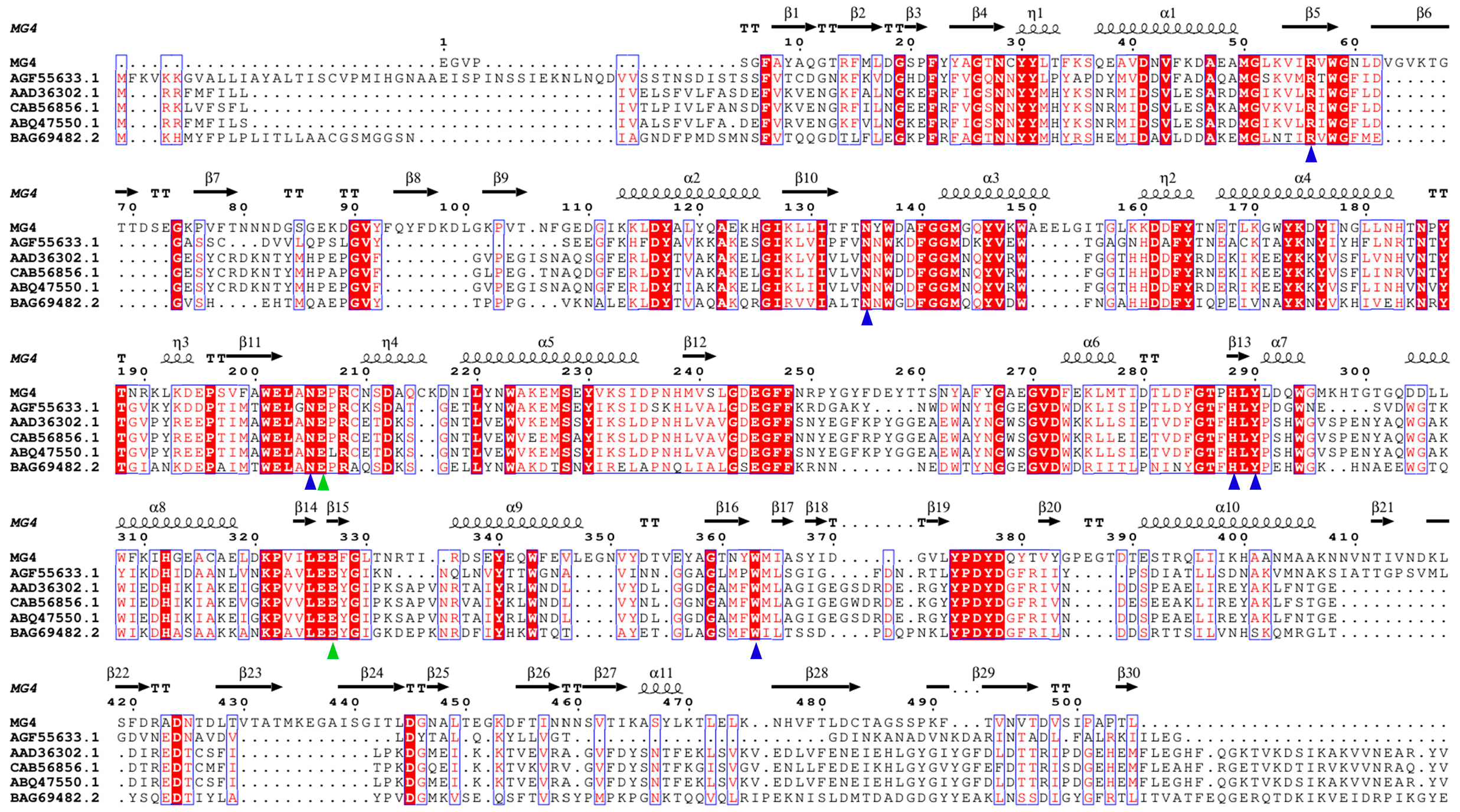
A novel thermostable β-mannanase potential gene, MG4, was mined by BLASTP tool (v 2.9.0) through the thermostable mannanases rPoMan5A and TtMan5A as reference sequences in the GEM database. MG4 contains two mannanase glutamate catalytic residues with six conserved amino acid residues, although it has only 33.2% and 32.0% sequence identity to rPoMan5A and TtMan5A, respectively (Figure S1). MG4 has an open reading frame of 1785 bp, encoding 595 amino acids, and was predicted to have 35 amino acid residues as the signal peptide at the N-terminus by SignalP 6.0 (the following experiments and analysis are based on the protein without the signal peptide). Sequences with high similarity to MG4 were found to be from Eubacterium siraeum sp. and Ruminiclostridium sp. by BLASTP tool of NCBI database, while the annotation suggests that MG4 may be from glycoside hydrolase family 5 (GH5). All amino acid sequences belonging to GH5 from Ruminiclostridium sp. or Eubacterium siraeum sp. were collected to construct the phylogenetic tree (Figure 1). The tree indicates that MG4 belongs to subfamily 7 of GH5 (GH5_7) [29]. MG4 aggregated with CBL33682.1, UWP25311.1, and CBK97477.1, which from Eubacterium siraeum sp. belongings to GH5_7, suggests a potential taxonomic origin in Eubacterium siraeum, though experimental validation is required. However, these proteins mentioned above were merely annotated, so MG4 and characterized β-mannanases from GH5_7 were compared by multiple sequence alignment, which revealed two catalytic residues (Glu206 and Glu327) and six strictly conserved amino acids (Arg56, Asn135, Asn205, His288, Tyr290, and Trp363) are present in MG4 (Figure 2) [30,31,32].

2.2. Functional and Structural Analysis of MG4

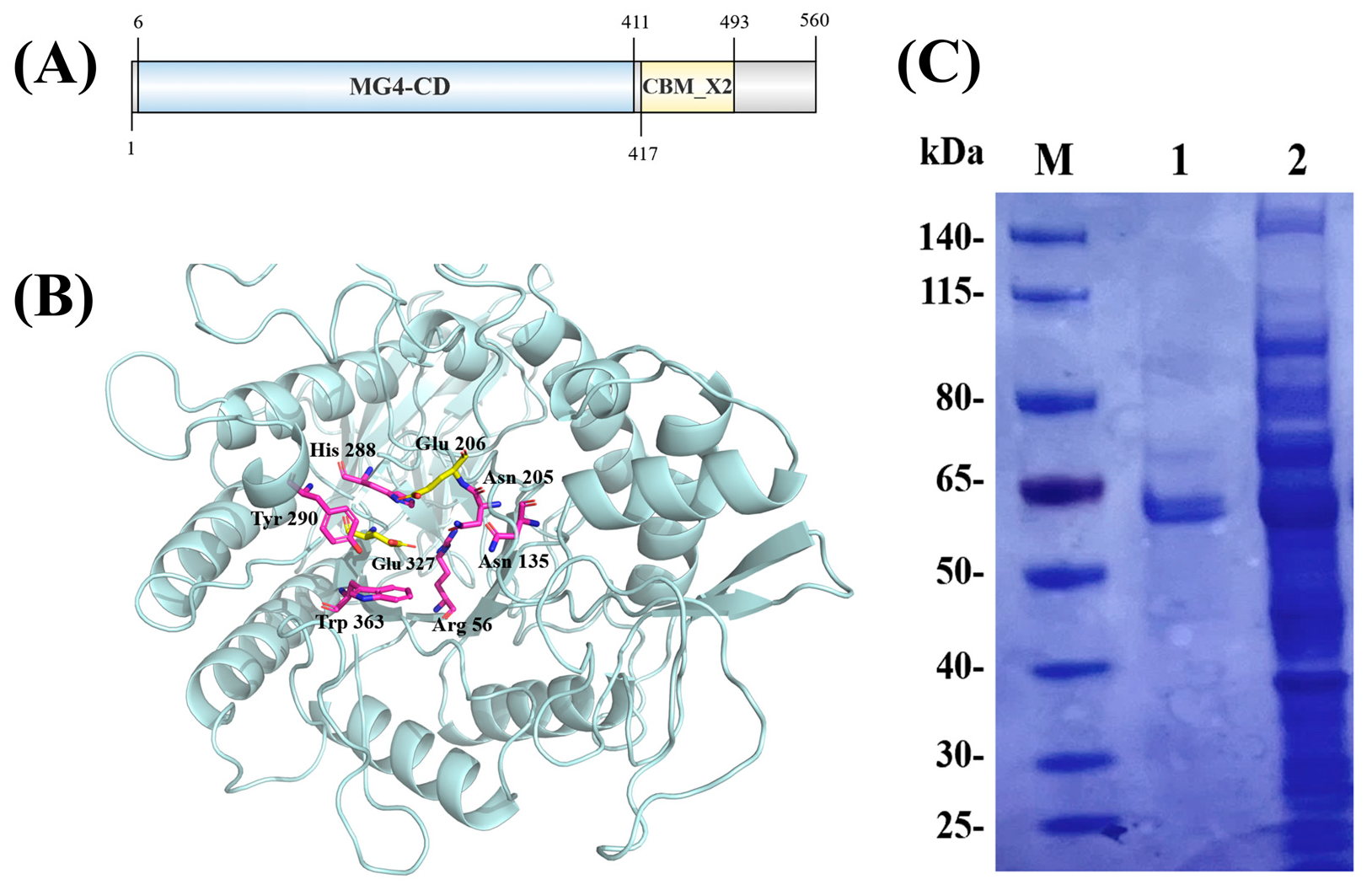
The annotation of InterProScan showed that the C-terminus of MG4 was a X2-like carbohydrate binding module (CBM_X2, 417-493 AA) (Figure 3A). To confirm the classification of the CBM from MG4, a multiple sequence alignment was performed with characterized CBM_X2 modules from Hominimerdicola aceti, Leifsonia shinshuensis, Microbacterium yannicii and Herbiconiux daphne [33]. The CBM from MG4 exhibited significant sequence similarity and featured the characteristic conserved residues with other characterized CBM_X2 (Figure S4), strongly supporting its membership in the CBM_X2 family. The X2 module is widely distributed, belongs to the immunoglobulin superfamily, and is predicted to be implicated in the localization of the cellulosome, cellulose binding, cell wall binding, or the enhancement of free cellulase activity [34,35]. Furthermore, previous studies have suggested that X2 modules might increase the solubility and substrate binding affinity of X2-bearing proteins [36]. However, β-mannanases carrying CBM_X2 have hardly been reported. To investigate the role of CBM_X2 in MG4, we constructed a truncated variant, MG4-CD, in which the CBM_X2 domain was deleted (Figure S2). The removal of CBM_X2 resulted in an 8.8% decrease in enzymatic activity. While this marginal decrease could be influenced by various factors, it is consistent with the role of CBMs in enhancing substrate proximity. Further investigations could include analysis if an activity decrease is connected to potential substrate binding which has been described for other X2-modules [36].
However, it also implied that the catalytic domain of MG4 alone is sufficient for near-optimal hydrolysis. The CBM_X2 likely plays a non-essential, ancillary role under standard assay conditions.
The N-terminus of MG4 is a conserved catalytic structural domain (CD, 6-411 AA) annotated as mannan endo-1,4-beta-mannosidase-like, which exhibited the TIM barrel (also called (β/α)8 barrel) architecture (Figure 3B), a typical structure of β-1,4-endo-mannanase [37]. The TIM barrel is the most frequently occurring folding motif in proteins, with a typical structure consisting of eight α-helices and eight parallel β strands that alternate along the peptide backbone [38,39]. Extremely conserved amino acid residues and catalytic residues (Glu206 and Glu327) were presented inside the barrel (Figure 3B). The results of molecular docking of MG4 with M3 ligand (Figure S3) also confirmed that the two catalytic residues play a crucial role in enzyme catalysis.

2.3. SDS-PAGE Analysis and Enzyme Activity Assay

E.coli-pET-28a(+)-MG4 had been induced to express were collected by centrifugation and broken by ultrasound in an ice bath. The cell breakage supernatant was purified by HyPur T Ni-NTA 6FF (His-Tag) resin in order to clarify the expression efficiency of MG4 in E. coli-BL21 and to determine its enzymatic properties. Purified MG4 was assayed separately from the unpurified cell-broken supernatant using SDS-PAGE, which revealed that the molecular weight of MG4 was consistent with the expected molecular weight of 63.0 kDa, as shown in Lane 1 (Figure 3C).
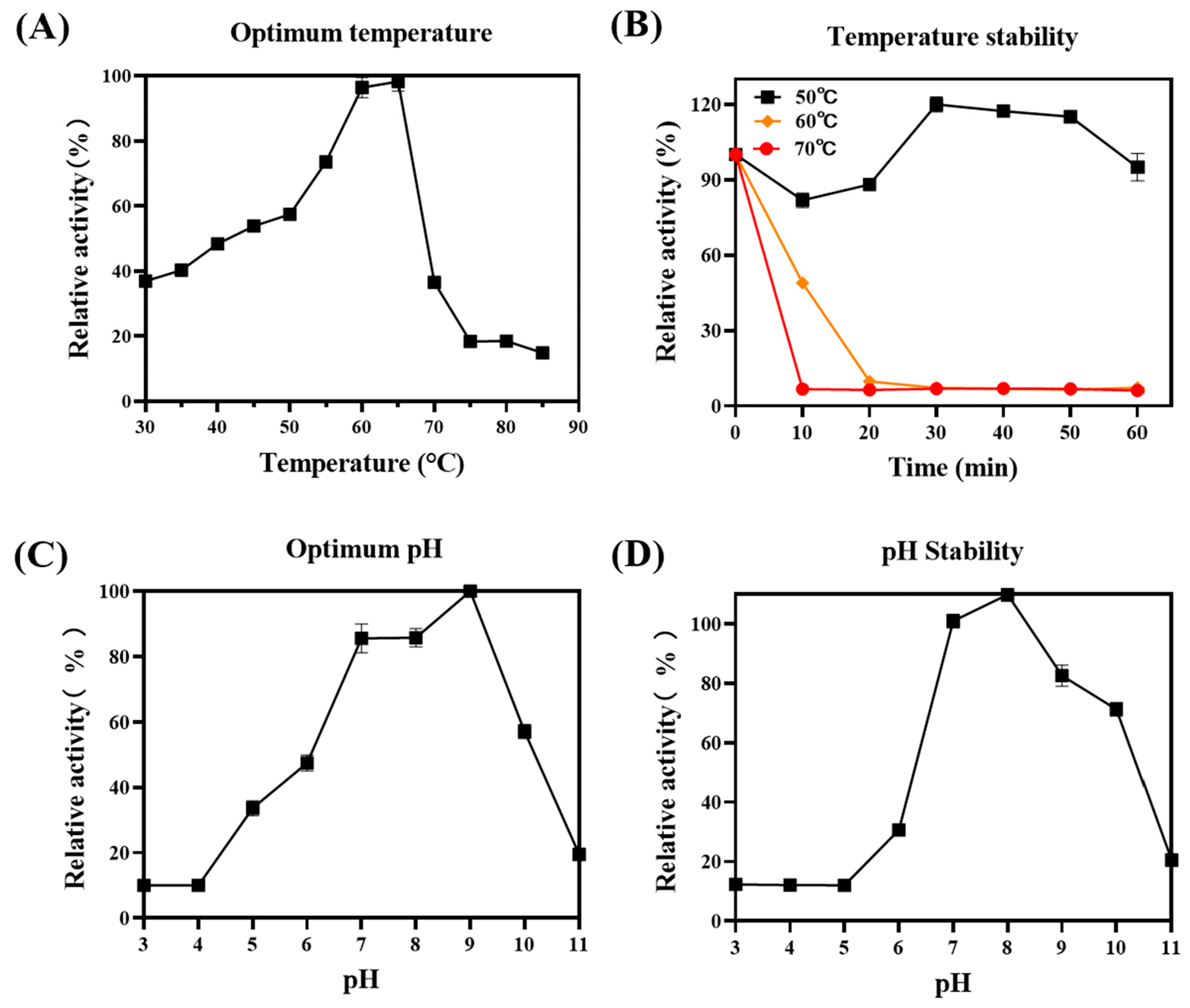
The enzymatic characteristics of MG4 were evaluated by varying temperature and pH conditions. MG4 exhibited maximum activity at 65 °C (Figure 4A). More than 85% of the residual activity was retained after incubation at 50 °C for 1 h, exhibiting remarkable thermal stability. The activity decreased to 40% after 30 min of incubation at 60 °C, which indicated that a possible structural change occurred near the optimal temperature (Figure 4B). MG4 exhibited maximum enzyme activity at pH 9.0 (Figure 4C), furthermore, it maintained more than 50% of its activity between pH 7.0 and 10.0 (Figure 4D), demonstrating that it is an alkaliphilic β-mannanase with broad pH stability adapted to alkaline environments. The vast majority of β-mannanases have an optimal pH in the acidic to neutral range [2], whereas the alkaliphilic nature of MG4 with broad pH stability distinguishes the narrow pH range of action of most β-mannanases. The alkalophilic character of MG4 makes it more suitable for fracturing fluid breakage applications, as in the case of the alkaline β-mannanase DtManB [40], which can reduce the viscosity of guar gum-based fracturing fluids at pH 8.0 and 9.0 at rates of 49.8 and 32.2 mPa s min−1.

2.4. Effect of Metal Ions and EDTA on Enzyme Activity

To assess the effect of various metal ions and EDTA on MG4 enzyme activity, different metal ions were added to the enzyme activity assay system (Table 1). For MG4, Cu2+ and Zn2+ inhibited the enzyme activity most strongly, which is consistent with the results of the mannanase Man/Cel5B [41] from Thermotoga maritima and ManKs_4-555 [42] from Kitasatospora sp. It is speculated that Cu2+ and Zn2+ bind specifically to the catalytic glutamate residues in the catalytic activity center, thus rendering it catalytically inactive [43]. Ni2+, Fe3+ and EDTA exhibited strong inhibition of activity, which is similar to the results of AfMan5A [44] from Aspergillus fumigatus HBFH5. It is presumed that the normal catalytic environment is damaged due to EDTA chelating the metal ions in the system that are critical for enzyme activity [45]. In contrast, Mn2+, K+, Fe2+ promoted the enzyme activity, which is in agreement with the reports of Mn428 [46] and MnSt [47] from Streptomyces sp. Ba2+, Ca2+, Na+, Mg2+ increased the enzyme activity by more than 30%, which is similar to that of ManSS11 from Klebsiella pneumoniae [8]. The results provide a guide for MG4 to avoid potential metal ion inhibitors and select appropriate enzyme activity enhancers in production applications.

2.5. Substrate Specificity and Enzyme Kinetic

The substrate specificity of MG4 was evaluated using different mannose-based polysaccharides as substrates, including LBG, GG, KGM, INM, and XG (Table 2). MG4 exhibited higher activity in hydrolysis of LBG (25.77 ± 0.68 U/mg) and KGM (27.27 ± 0.76 U/mg) and showed slightly lower activity for GG (8.89 ± 0.85 U/mg). Both LBG and GG are galactomannan with a structure consisting of a β-mannan backbone formed by β-1,4-glycosidic bond linkages and a branched structure consisting of α-(1,6)-glycosidic bond linkages to galactose residues; however, the ratio of mannose to glucose usually ranges from 1.6:1 to 1.8:1 [11] in GG and 4:1 [48] in LBG. The lower activity on GG vs. LBG likely stems from higher galactose substitution, which sterically hinders backbone access. It also revealed that MG4 could hydrolyse β-1,4-glycosidic bonds but not α-(1,6)-glycosidic bonds. Surprisingly, MG4 exhibited the highest hydrolytic activity for KGM, which has a glucomannan backbone linked by β-1,4-glycosidic bonds, with a mannose to glucose ratio of 1.6:1–1.4:1 [49], indicating that MG4 has a high degree of specificity for β-1,4-glycosidic bonds. MG4 showed no activity for either INM or CMC. INM is a linear β-mannan almost unbranched; however, its crystal structure creates a steric hindrance that prevents binding of the enzyme to the backbone [50]. The backbone of CMC consists of glucose units linked by β-1,4-glycosidic bonds, which suggests that MG4 only targets β-1,4-glycosidic bonds linked to mannose and not glucose [51]. The inability of XG to hydrolyze also proves the above result. Overall, MG4 was demonstrated to have specificity for β-mannan substrates linked by β-1,4-glycosidic bonds.
The enzymatic kinetic parameters of MG4 were calculated by non-linear regression using LBG (1.0–10.0 mg/mL) as substrate. The Vmax and Km of MG4 were 59.36 mmol/min/mg and 4.99 mg/mL, respectively.

2.6. Hydrolysis Product Analysis

Hydrolysis products of LBG degradation by MG4 were investigated by TLC at different incubation times (Figure 5). At 0 h (Lane 1), there is little to no visible MOS production, indicating that hydrolysis has not yet occurred significantly. At 10 min (Lane 2), MG4 exhibited rapid catalytic activity, producing MOS with different degrees of polymerization (DP). After prolonged incubation (0.5–5 h), the hydrolysis products remained as MOS with DP 2-5. Notably, no further degradation to monosaccharides or smaller fragments was observed, suggesting that MG4 hydrolyzed LBG to MOS rather than completely hydrolyzing the substrate. The composition of the hydrolyzed product demonstrated the typical characteristics of MG4 as a strict endo-type mannanase, which binds to the internal sites of the polysaccharide chain for cleavage, rather than starting from the end of the chain [52]. The pattern consistent with the behavior of other endo-mannanases, such as endo-1,4-β-mannanase from Bacteroides ovatus [53] and Nonomuraea jabiensis ID06-379 [54], which similarly produce MOS with DP 2-6 as end products under prolonged hydrolysis conditions. In recent years, MOS have gained significant interest as a prebiotic in recent years [55], particularly for their potential health benefits [56]. Previous studies have indicated that MOS can promote the proliferation of beneficial bacteria in the digestive tract [57,58], improve immune response [59,60,61], exhibiting anti-cancer activity [62] and lowering blood sugar and lipids [63,64]. While the TLC analysis confirms MOS production with DP 2-5, this method cannot distinguish between unsubstituted MOS and galactose-substituted MOS (GMOS). The prebiotic specificity of these oligosaccharides is strongly influenced by galactose substitution patterns, as different gut bacteria exhibit distinct structural preferences. For instance, Bifidobacterium adolescentis utilizes a broader range of GMOS structures, while Roseburia hominis shows more specific requirements [53]. Furthermore, cooperative cross-feeding interactions between bacterial species are essential for complete MOS/GMOS utilization [65]. Although MG4 effectively generates MOS mixtures, a comprehensive characterization of their prebiotic potential requires detailed structural analysis of their galactosylation patterns, for instance by HPLC or HPAEC-PAD.

2.7. Gel Breaking of Guar Gum Fracturing Fluid

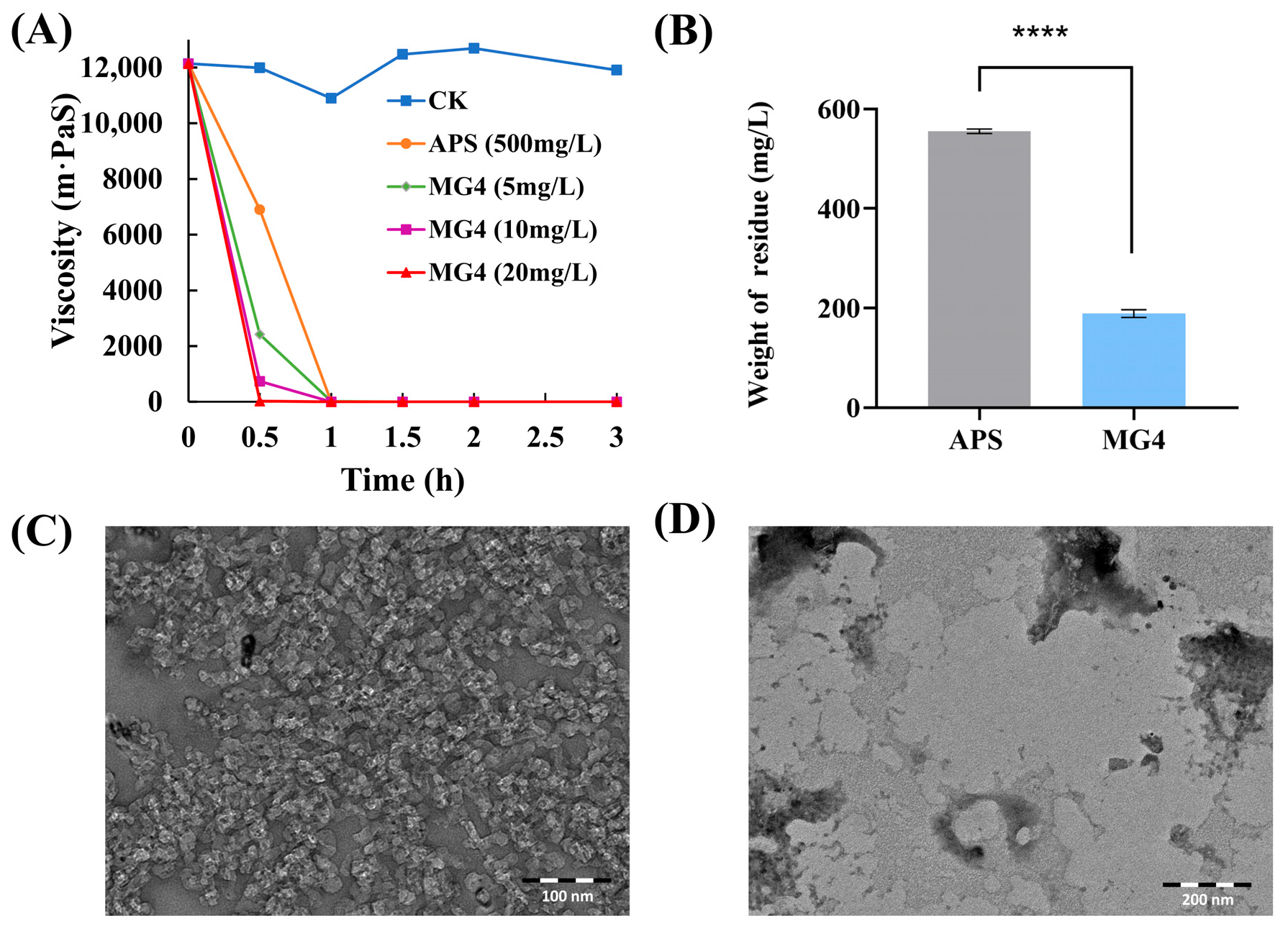
The viscosity of guar gum fracturing fluid with different concentrations of MG4 (5 mg/L, 10 mg/L, 20 mg/L) was examined at 55 °C to evaluate its gel-breaking efficiency (Figure 6A). For comparison, APS (500 mg/L), which is a commonly used chemical breaker in hydraulic fracturing operations [66], is used as a control to judge the gel-breaking ability of MG4. As shown in Figure 6A, MG4 (5 mg/L, 10 mg/L) and APS (500 mg/L) reduced the viscosity to below 5 mPa·S within 60 min. Notably, at 20 mg/L, MG4 achieved complete gel-breaking in 30 min. The decreasing trend of the curves further indicated that the viscosity reduction rate of MG4 is consistently faster than that of APS, and demonstrated a clear concentration-dependent effect. Specifically, the gel breaking time of MG4 (20 mg/L) is half that of APS (500 mg/L), highlighting its superior breaking ability for guar gum fracturing fluid.
The amount of residue is another crucial indicator for evaluating the quality of gel breakers [67]. As depicted in Figure 6B, the weight of residue produced by MG4 after gel-breaking was approximately 35.5% of that resulting from the APS treatment, which implied that MG4 would produce less solid blockage and thus smoother fracturing fluid rejection [68]. Overall, the dual advantages of efficiency and low residue amount establish β-mannanase MG4 as a more suitable candidate for guar gum fracturing fluid gel-breaking compared to traditional chemical breakers like APS.
TEM observation also confirmed the breaking ability of MG4 for guar-based fracturing fluid. A large number of high-density irregular granular structures resided in the guar-based fracturing fluid, with uneven particle size distribution and obvious aggregation between the particles, indicating that the system still retained a relatively intact guar polymerization structure (Figure 6C). In contrast, the guar gum fracturing fluid with MG4 showed significantly different morphological characteristics. Most of the granular structure was degraded, and the high-density region in the system was significantly reduced, leaving only some slightly agglomerated small particles and amorphous materials (Figure 6D). It was revealed that MG4 has a significant effect on degrading the guar gum main chain structure so that its spatial structure is completely interrupted, and the aggregation and viscosity of the system are reduced, thus contributing to the improvement of the fracturing fluid rejection efficiency [14]. In conclusion, the TEM images clearly revealed the effect of β-mannanase on the microstructure of guar fracturing fluid, and the MG4 treatment significantly altered the micro-morphological structure of the fracturing fluid. The structural change helped to optimize the rheological properties of the fracturing fluid, thereby improving the application of fracturing fluid in oil and gas extraction.
In this study, the gel-breaking capacity of β-mannanase MG4 showed advantages in terms of viscosity reduction rate and residue determination. In the future, its transformation towards on-site application in oilfield fracturing operations can be promoted through formation simulation experiments [67] and immobilized enzyme technology [69].

3. Materials and Methods

3.1. Strains and Reagents

The MG4 encoding β-mannanase gene was synthesized by Azenta (Tianjin, China). E. coli BL21 (DE3) receptor cell was purchased from Vazyme (Nanjing, China) for cloning and expression. HyPur T Ni-NTA 6FF (His-Tag) was purchased from Sangon (Shanghai, China) for protein purification. Mannobiose, mannotriose, mannotetraose and mannopentaose were purchased from Megazyme (Bray, Ireland) for thin-layer chromatography (TLC). Locust bean gum (LBG), guar gum (GG) and konjac glucomannan (KGM) were purchased from Meryer (Shanghai, China) and ivory nut mannan (INM) was purchased from Biotoped (Beijing, China) for substrate-specific analysis. All other chemicals were analytical grade unless otherwise stated.

3.2. Synthesis and Expression of Recombinant MG4

Genomes in GEM were annotated using PROKKA. The candidate mannanase-coding genes were identified using blastp in BLAST package v2.9.0 with an e-value threshold of 1 × 10⁻¹⁰. The reference sequences for mannanase were rPoMan5A (GenBank: AGW24296.1) [70] and TtMan5A (Gen Bank: BAP19029.1) [50], with optimal temperatures of 80 °C and 85 °C, and with optimal pH values of 4.0 and 4.5, respectively. Following the initial BLAST search, sequences with lower identity (<70%) to the queries were further scrutinized, rather than highly identical ones, in order to target enzymes with a more alkaline pH optimum. Furthermore, the presence of crucial highly conserved residues definitive of the GH5 family [71], particularly the catalytic glutamates [72], was a mandatory criterion for selecting candidates. We speculated that this degree of identity would conserve the relatively high thermostability characteristic of the query enzymes [50,70]. The candidate mannanase gene was synthesized and named MG4. MG4 expression vector was pET-28a (+), which was synthesized by optimizing synonymous codons to show bias towards E. coli BL21 (DE3).

3.3. Sequence Analysis

Signal peptides were predicted using SignalP 6.0 (https://services.healthtech.dtu.dk/services/SignalP-6.0/ (accessed on 12 January 2025)). Multiple sequence alignment was performed by MAFFT (https://mafft.cbrc.jp/alignment/server/ (accessed on 12 January 2025)) and ESPript 3.0 (https://espript.ibcp.fr/ESPript/ESPript/ (accessed on 12 January 2025)). A phylogenetic tree was constructed by maximum likelihood (ML) in IQ-TREE (version 2.2.5) [73] under the optimal model of LG+F+I+G4 with 1000 rapid bootstrap inferences and subsequently visualized by iTol (https://itol.embl.de/ (accessed on 15 January 2025)). Structural domain prediction by InterProScan (https://www.ebi.ac.uk/interpro/search/sequence/ (accessed on 25 January 2025)) and dbCAN2 (https://bcb.unl.edu/dbCAN2/blast.php (accessed on 25 January 2025)). Structural domain visualization by IBS (http://ibs.biocuckoo.org/online.php (accessed on 28 January 2025)).

3.4. Structural Analysis and Molecular Docking

3D structure of MG4 modeling and analysis performed by AlphaFold3 (https://alphafoldserver.com/ (accessed on 10 February 2025)) and PyMOL 2.5 software (Schrödinger, New York, NY, USA). The structure of mannotriose (M3), the ligand for the effective substrate of mannanase, was obtained from Chemspider (https://www.chemspider.com/ (accessed on 26 February 2025)). Molecular docking was performed by the AutoDock 4.2 program via AutoDockTools 1.5.6 (ADT) [74]. For the docking of MG4 and M3, the XYZ dimensions were defined as a cubic grid box with side lengths of 126 and a grid spacing of 0.375 Å at the centers of 20.955, −1.744, and −8.56, respectively. The Lamarckian Genetic Algorithm (LGA) was chosen as the method to recognize the binding conformations within the above grid boxes.

3.5. Expression and Purification of MG4

For expression of MG4, IPTG was added to a final concentration of 0.5 mM to induce expression when the OD600 of the culture was between 0.6 and 0.8. The culture was incubated at 16 °C for 20–22 h. Bacteria were washed and collected with buffer, cells were broken by ultrasonication in an ice water bath, and the supernatant was obtained by high-speed centrifugation as the crude mannanase solution. The crude mannanase solution was purified by HyPur T Ni-NTA 6FF (His-Tag) resin and eluted at different concentrations of imidazole buffer at a natural flow rate. The purification step was carried out at 4 °C. Molecular weights of pure mannanase were determined by sodium dodecyl sulfate-polyacrylamide gel electrophoresis (SDS-PAGE). The concentration of the proteins was determined by the BCA protein assay kit.

3.6. Enzyme Activity Assay

Mannanase activity was determined by quantifying the amount of reducing sugars released from LBG by the 3,5-dinitrosalicylic acid (DNS) method using mannose as standard [75,76]. A reaction mixture containing 50 μL of appropriately diluted mannanase and 450 μL of 0.5% (w/v) LBG solution was incubated at 55 °C for 10 min. An equivalent volume of inactivated mannanase was used for the blank group. Then, 500 μL of DNS reagent was added to each sample. After thorough mixing, the reaction was carried out by heating in a boiling water bath for 5 min. The amount of reducing sugars released was determined by measuring the absorbance at 540 nm. The optimal pH was determined in the range of 3.0–10.0 (pH 3.0–6.0, citric acid-sodium citrate buffer; pH 7.0–8.0, Tris-HCl buffer; pH 9.0–11.0, glycine-NaOH buffer). All buffers were adjusted to 50 mM ionic strength. The pH stability was determined by incubating the samples in different pH conditions for 1 h. The optimal temperature was determined in the range of 30–85 °C at 5 °C intervals, and temperature stability was assessed by incubation at 50 °C, 60 °C, and 70 °C for different times.
One unit of mannanase activity was defined as the amount of enzyme that formed reducing groups corresponding to 1 μmol of mannose in 1 min under the assay conditions. All enzyme activity measurements were performed in triplicate, and the average of the three experiments is reported.

3.7. Effect of Metal Ions and EDTA on Enzyme Activity

Aliquots of MG4 and substrate were co-incubated with metal ions or EDTA at 55 °C for 10 min to evaluate the effect of metal ions and EDTA on enzyme activity. The following reagents were used: CuSO4, MnSO4, ZnCl2, BaCl2, KCl, CaCl2, FeSO4, NaCl, MgCl2, NiCl2, FeCl3, and EDTA. These reagents were used at a final concentration of 1 mM in the total system. The reaction system without any added compounds served as a control (defined as 100%).

3.8. Substrate Specificity and Enzyme Kinetic Parameters

The specific activity of purified MG4 with different mannose-based polysaccharides, including LBG, KGM, GG, INM, and XG, was determined by incubation at 55 °C for 10 min.
The hydrolysis reaction rate (mmol/min/mg) of MG4 was determined by different locust bean gum concentrations (1.0–10.0 mg/mL) as substrates. The relationship between the reaction rate and substrate concentration was explored to verify that the hydrolysis pattern of MG4 conforms to the Michaelis-Menten equation. Km and Vmax values were determined graphically using non-linear regression (curve fit).

3.9. Hydrolysis Products

Hydrolysis products were analyzed by TLC. The hydrolysis reaction contained 0.5% (w/v) LBG and was incubated with MG4 at a final concentration of 0.1 mg/mL at 55 °C. Aliquots were taken at the indicated time points (0, 10, 30 min, 1, 2, 3, 4, 5 h), boiled for 10 min to inactivate the enzyme, and centrifuged (12,000 rpm/20 min). The supernatants were spotted on the dry silica gel plates. The silica gel plate developed in developing solvent, which consisted of n-butanol, glacial acetic acid and water (2:1:1). Sprayed with aniline-diphenylamine-phosphoric acid color developer, sugar spots were formed after high-temperature color development.

3.10. Guar Gum Fracturing Fluid Breaking

The composition of the guar gum fracturing fluid system included 0.35% (w/v) guar gum, 0.35% saturated borax solution as a cross-linking agent, and the pH was adjusted to 10.0 using NaOH [10]. The breakers included ammonium persulfate (APS, 500 mg/L, control group) and MG4 (5 mg/L, 10 mg/L, and 20 mg/L, experimental group). The system was maintained in a water bath at 25 °C for 4 h to allow its viscosity to stabilize. Subsequently, it formed a gel that could be lifted and hung on a glass rod, indicating successful crosslinking [77]. The fracturing fluid system was immersed in a water bath at 55 °C, and a portion was taken out per 30 min. The viscosity fluctuation curve of the fracturing fluid was measured by rotational viscometer at 25 °C [10].
The precipitate of the thoroughly broken system was collected after washing and centrifugation. It was dried to a constant weight to obtain the remaining residue after complete gum breaking [40]. Viscosity less than 5 mPa s is considered to be completely broken. The morphology of guar gum fracturing fluid and enzyme-treated guar gum fracturing fluid was observed and compared by HITACHI-HT 7800 transmission electron microscopy (TEM) [78].

3.11. Statistical Analysis

All experiments were performed in triplicate. The results are presented as the mean ± standard deviation. Statistical analyses were performed using GraphPad Prism 8.0 software. The independent samples t-test was used for comparisons between two groups. A p-value of less than 0.05 was considered statistically significant.

4. Conclusions

In this study, a β-mannanase gene, MG4, was obtained from the GEM database, which encodes 595 amino acids. Phylogenetic and structural domain analyses indicated that MG4 encodes a novel β-mannanase with the structural domain of the GH 5_7. MG4 exhibited the highest activity at 65 °C and pH 9.0, hydrolyzed substrates with β-1,4-glycosidic mannose, and exhibited enzymatic activity for LBG, GG, and KGM. Critically, it achieved complete guar fracturing fluid gel-breaking within 0.5 h at 20 mg/L while reducing insoluble residue by 64.5% versus APS. MG4 showed excellent viscosity reduction effect on guar gum fracturing fluids, indicating its potential as a promising eco-friendly fracturing fluid breaker. Furthermore, this work demonstrates the power of metagenomic mining from uncultured microbiomes to discover industrially adapted extremozymes, accelerating green alternatives for petroleum operations.

Supplementary Materials

The following supporting information can be downloaded at: https://www.mdpi.com/article/10.3390/catal15090905/s1. Figure S1: Multiple sequence comparison of MG4 with the thermophilic enzyme mannanases rPoMan5A23 (80 °C) and TtMan5A 24 (85 °C). Blue triangles indicate conserved amino acid residues. Green triangles indicate catalytic residues; Figure S2: To investigate the contribution of CBM_X2, the enzyme activity of MG4 (560 aa) and MG4-CD (417 aa, obtained by removing CBM_X2), was determined. (A) SDS-PAGE results from total cellular proteins. lane M: protein molecular weight marker; lane 1: MG4 (63.0 kDa); lane 2: MG4-CD (47.4 kDa); (B) Relative enzyme activity from MG4 and MG4-CD. Figure S3: The molecular docking of MG4 and M3. Figure S4: Multiple sequence comparison of CBM_X2 from MG4 with CBM_X2 modules from Hominimerdicola aceti, Leifsonia shinshuensis, Microbacterium yannicii and Herbiconiux daphne.

Author Contributions

Conceptualization, W.T.; methodology, T.L.; software, S.W.; formal analysis, W.W. and Z.W.; investigation, S.C. and Y.T.; writing—original draft preparation, W.T.; writing—review and editing, Y.Y., G.L. and T.M.; visualization, W.T.; project administration, T.M.; funding acquisition, T.M. All authors have read and agreed to the published version of the manuscript.

Funding

This research was funded by Natural Science Foundation of China (No. 42173079) and Natural Science Foundation of Tianjin (No. 21JCZDJC00210).

Data Availability Statement

The original contributions presented in this study are included in the article. Further inquiries can be directed to the corresponding authors.

Conflicts of Interest

The authors declare no conflicts of interest.

Abbreviations

The following abbreviations are used in this manuscript:
MOSManno-oligosaccharides
GEMGenomes of Earth’s microbiomes
LBGLocust bean gum
GGGuar gum
KGMKonjac glucomannan
INMIvory nut mannan
DNS3,5-dinitrosalicylic acid
XGXanthan gum
CMCCarboxymethyl cellulose
TLCThin-layer chromatography
APSAmmonium persulfate
TEMTransmission electron microscopy
GHGlycoside hydrolase
CBMCarbohydrate binding module
CDCatalytic domain
TIMTriose phosphate isomerase
SDS-PAGESodium dodecyl sulfate polyacrylamide gel electrophoresis
DPDegrees of polymerization

References

  1. Wang, P.; Pei, X.; Zhou, W.; Zhao, Y.; Gu, P.; Li, Y.; Gao, J. Research and Application Progress of Microbial β-Mannanases: A Mini-Review. World J. Microbiol. Biotechnol. 2024, 40, 169. [Google Scholar] [CrossRef]
  2. Chauhan, P.S.; Puri, N.; Sharma, P.; Gupta, N. Mannanases: Microbial Sources, Production, Properties and Potential Biotechnological Applications. Appl. Microbiol. Biotechnol. 2012, 93, 1817–1830. [Google Scholar] [CrossRef] [PubMed]
  3. Li, N.; Han, J.; Zhou, Y.; Zhang, H.; Xu, X.; He, B.; Liu, M.; Wang, J.; Wang, Q. A Rumen-Derived Bifunctional Glucanase/Mannanase Uncanonically Releases Oligosaccharides with a High Degree of Polymerization Preferentially from Branched Substrates. Carbohydr. Polym. 2024, 330, 121828. [Google Scholar] [CrossRef]
  4. Nadaroglu, H.; Adiguzel, G.; Adiguzel, A.; Sonmez, Z. A Thermostable-Endo-β-(1,4)-Mannanase from Pediococcus Acidilactici (M17): Purification, Characterization and Its Application in Fruit Juice Clarification. Eur. Food Res. Technol. 2017, 243, 193–201. [Google Scholar] [CrossRef]
  5. David, A.; Singh Chauhan, P.; Kumar, A.; Angural, S.; Kumar, D.; Puri, N.; Gupta, N. Coproduction of Protease and Mannanase from Bacillus Nealsonii PN-11 in Solid State Fermentation and Their Combined Application as Detergent Additives. Int. J. Biol. Macromol. 2018, 108, 1176–1184. [Google Scholar] [CrossRef] [PubMed]
  6. Baker, J.T.; Deng, Z.; Sokale, A.; Frederick, B.; Kim, S.W. Nutritional and Functional Roles of β-Mannanase on Intestinal Health and Growth of Newly Weaned Pigs Fed Two Different Types of Feeds. J. Anim. Sci. 2024, 102, skae206. [Google Scholar] [CrossRef]
  7. Zhao, D.; Zhang, X.; Wang, Y.; Na, J.; Ping, W.; Ge, J. Purification, Biochemical and Secondary Structural Characterisation of β-Mannanase from Lactobacillus Casei HDS-01 and Juice Clarification Potential. Int. J. Biol. Macromol. 2020, 154, 826–834. [Google Scholar] [CrossRef] [PubMed]
  8. Singh, S.; Singh, G.; Khatri, M.; Kaur, A.; Arya, S.K. Thermo and Alkali Stable β-Mannanase: Characterization and Application for Removal of Food (Mannans Based) Stain. Int. J. Biol. Macromol. 2019, 134, 536–546. [Google Scholar] [CrossRef]
  9. Kumar Suryawanshi, R.; Kango, N. Production of Mannooligosaccharides from Various Mannans and Evaluation of Their Prebiotic Potential. Food Chem. 2021, 334, 127428. [Google Scholar] [CrossRef]
  10. Meng, Y.; Zhao, F.; Jin, X.; Feng, Y.; Sun, G.; Lin, J.; Jia, B.; Li, P. Performance Evaluation of Enzyme Breaker for Fracturing Applications under Simulated Reservoir Conditions. Molecules 2021, 26, 3133. [Google Scholar] [CrossRef]
  11. Wang, T.; Ye, J. Rheological and Fracturing Characteristics of a Cationic Guar Gum. Int. J. Biol. Macromol. 2023, 224, 196–206. [Google Scholar] [CrossRef]
  12. Hasan, A.M.A.; Abdel-Raouf, M.E. Applications of Guar Gum and Its Derivatives in Petroleum Industry: A Review. Egypt. J. Pet. 2018, 27, 1043–1050. [Google Scholar] [CrossRef]
  13. Liu, J.; Wang, S.; Wang, C.; Zhao, F.; Lei, S.; Yi, H.; Guo, J. Influence of Nanomaterial Morphology of Guar-Gum Fracturing Fluid, Physical and Mechanical Properties. Carbohydr. Polym. 2020, 234, 115915. [Google Scholar] [CrossRef] [PubMed]
  14. Chen, X.; Zhang, G.; Ding, R.; Zheng, D.; Yang, Z.; Sun, Z.; Zhou, F.; Wang, D. Research on Water Blocking and Residue Damage Mechanism of Fracturing Fluid in Yongjin Tight Reservoirs. Phys. Fluids 2024, 36, 042018. [Google Scholar] [CrossRef]
  15. Liang, T.; Zhou, F.; Lu, J.; DiCarlo, D.; Nguyen, Q. Evaluation of Wettability Alteration and IFT Reduction on Mitigating Water Blocking for Low-Permeability Oil-Wet Rocks after Hydraulic Fracturing. Fuel 2017, 209, 650–660. [Google Scholar] [CrossRef]
  16. Ma, X.; Song, P.; Liu, L.; Da, Q.; Lei, G.; Yao, C.; Shor, L.M. Low-Temperature pH-Regulable Gel-Breaking of Galactomannan-Based Fracturing Fluids by the Mannanase from Bacillus Aerius. Int. Biodeterior. Biodegrad. 2021, 160, 105226. [Google Scholar] [CrossRef]
  17. Murthy, R.V.V.R.; Chavali, M. A Novel Hydraulic Fracturing Gel Realization for Unconventional Reservoirs. Beni-Suef Univ. J. Basic Appl. Sci. 2020, 9, 37. [Google Scholar] [CrossRef]
  18. Reddy, T.T.; Tammishetti, S. Free Radical Degradation of Guar Gum. Polym. Degrad. Stab. 2004, 86, 455–459. [Google Scholar] [CrossRef]
  19. Trabelsi, S.; Kakadjian, S. Comparative Study between Guar and Carboxymethylcellulose Used as Gelling Systems in Hydraulic Fracturing Application. In Proceedings of the SPE Production and Operations Symposium, Oklahoma City, OK, USA, 23–26 March 2013; p. SPE-164486-MS. [Google Scholar]
  20. Barati, R.; Liang, J. A Review of Fracturing Fluid Systems Used for Hydraulic Fracturing of Oil and Gas Wells. J. Appl. Polym. Sci. 2014, 131, app.40735. [Google Scholar] [CrossRef]
  21. Ma, X.; Wang, Z.; Da, Q.; Cheng, M.; Yao, C.; Lei, G. Application of Guar Gum Degrading Bacteria in Microbial Remediation of Guar-Based Fracturing Fluid Damage. Energy Fuels 2017, 31, 7894–7903. [Google Scholar] [CrossRef]
  22. Chen, X.; Sun, P.; Li, L.; Zhou, X.; Han, C.; Ma, S.; Cai, Y.; Zhang, W.; Li, Y.; Cao, Z. Sustainable Enhancing Oil Recovery in Different Reservoirs via Reservoir Adaptability and Multifunction of Bacillus velezensis. Fuel 2025, 388, 134488. [Google Scholar] [CrossRef]
  23. Abdelrahim, M.A.; Ghosh, D.B.; Belhaj, D.H.; Ghosh, D. High-Temperature Stable Specific Enzyme for Guar Polymer Based Fracturing Fluid Degradation. In Proceedings of the SPE/IATMI Asia Pacific Oil & Gas Conference and Exhibition, Virtual, 12–14 October 2021; p. D012S032R062. [Google Scholar]
  24. Naeem, M.; Khalil, A.B.; Tariq, Z.; Mahmoud, M. A Review of Advanced Molecular Engineering Approaches to Enhance the Thermostability of Enzyme Breakers: From Prospective of Upstream Oil and Gas Industry. Int. J. Mol. Sci. 2022, 23, 1597. [Google Scholar] [CrossRef]
  25. Erkan, S.B.; Ozcan, A.; Yilmazer, C.; Gurler, H.N.; Karahalil, E.; Germec, M.; Yatmaz, E.; Kucukcetin, A.; Turhan, I. The Effects of Mannanase Activity on Viscosity in Different Gums. J. Food Process. Preserv. 2021, 45, e14820. [Google Scholar] [CrossRef]
  26. Lv, L.; Lin, J.; Feng, Y.; Wang, W.; Li, S. Coated Recombinant Escherichia Coli for Delayed Release of β-Mannanase in the Water-Based Fracturing Fluid. Process Biochem. 2021, 107, 121–128. [Google Scholar] [CrossRef]
  27. Glasner, M.E. Finding Enzymes in the Gut Metagenome. Science 2017, 355, 577–578. [Google Scholar] [CrossRef] [PubMed]
  28. Nayfach, S.; Roux, S.; Seshadri, R.; Udwary, D.; Varghese, N.; Schulz, F.; Wu, D.; Paez-Espino, D.; Chen, I.-M.; Huntemann, M.; et al. A Genomic Catalog of Earth’s Microbiomes. Nat. Biotechnol. 2021, 39, 499–509. [Google Scholar] [CrossRef] [PubMed]
  29. Aspeborg, H.; Coutinho, P.M.; Wang, Y.; Brumer, H.; Henrissat, B. Evolution, Substrate Specificity and Subfamily Classification of Glycoside Hydrolase Family 5 (GH5). BMC Evol. Biol. 2012, 12, 186. [Google Scholar] [CrossRef]
  30. Parker, K.N.; Chhabra, S.R.; Lam, D.; Callen, W.; Duffaud, G.D.; Snead, M.A.; Short, J.M.; Mathur, E.J.; Kelly, R.M. Galactomannanases Man2 and Man5 from Thermotoga Species: Growth Physiology on Galactomannans, Gene Sequence Analysis, and Biochemical Properties of Recombinant Enzymes. Biotechnol. Bioeng. 2001, 75, 322–333. [Google Scholar] [CrossRef]
  31. Santos, C.R.; Squina, F.M.; Navarro, A.M.; Ruller, R.; Prade, R.; Murakami, M.T. Cloning, Expression, Purification, Crystallization and Preliminary X-Ray Diffraction Studies of the Catalytic Domain of a Hyperthermostable Endo-1,4-β-D-Mannanase from Thermotoga petrophila RKU-1. Acta Crystallogr. Sect. F 2010, 66, 1078–1081. [Google Scholar] [CrossRef]
  32. Tanaka, M.; Umemoto, Y.; Okamura, H.; Nakano, D.; Tamaru, Y.; Araki, T. Cloning and Characterization of a β-1,4-Mannanase 5C Possessing a Family 27 Carbohydrate-Binding Module from a Marine Bacterium, Vibrio Sp. Strain MA-138. Biosci. Biotechnol. Biochem. 2009, 73, 109–116. [Google Scholar] [CrossRef][Green Version]
  33. Kosugi, A.; Amano, Y.; Murashima, K.; Doi, R.H. Hydrophilic Domains of Scaffolding Protein CbpA Promote Glycosyl Hydrolase Activity and Localization of Cellulosomes to the Cell Surface of Clostridium cellulovorans. J. Bacteriol. 2004, 186, 6351–6359. [Google Scholar] [CrossRef]
  34. Chanal, A.; Mingardon, F.; Bauzan, M.; Tardif, C.; Fierobe, H.-P. Scaffoldin Modules Serving as “Cargo” Domains to Promote the Secretion of Heterologous Cellulosomal Cellulases by Clostridium Acetobutylicum. Appl. Environ. Microbiol. 2011, 77, 6277–6280. [Google Scholar] [CrossRef]
  35. Pasari, N.; Adlakha, N.; Gupta, M.; Bashir, Z.; Rajacharya, G.H.; Verma, G.; Munde, M.; Bhatnagar, R.; Yazdani, S.S. Impact of Module-X2 and Carbohydrate Binding Module-3 on the Catalytic Activity of Associated Glycoside Hydrolases towards Plant Biomass. Sci. Rep. 2017, 7, 3700. [Google Scholar] [CrossRef]
  36. Tao, X.; Liu, J.; Kempher, M.L.; Xu, T.; Zhou, J. In Vivo Functional Characterization of Hydrophilic X2 Modules in the Cellulosomal Scaffolding Protein. Front. Microbiol. 2022, 13, 861549. [Google Scholar] [CrossRef] [PubMed]
  37. St John, F.J.; González, J.M.; Pozharski, E. Consolidation of Glycosyl Hydrolase Family 30: A Dual Domain 4/7 Hydrolase Family Consisting of Two Structurally Distinct Groups. FEBS Lett. 2010, 584, 4435–4441. [Google Scholar] [CrossRef]
  38. Wang, C.-H.; Lu, L.-H.; Huang, C.; He, B.-F.; Huang, R.-B. Simultaneously Improved Thermostability and Hydrolytic Pattern of Alpha-Amylase by Engineering Central Beta Strands of TIM Barrel. Appl. Biochem. Biotechnol. 2020, 192, 57–70. [Google Scholar] [CrossRef] [PubMed]
  39. Höcker, B.; Jürgens, C.; Wilmanns, M.; Sterner, R. Stability, Catalytic Versatility and Evolution of the (Βα)8-Barrel Fold. Curr. Opin. Biotechnol. 2001, 12, 376–381. [Google Scholar] [CrossRef] [PubMed]
  40. Hu, K.; Li, C.-X.; Pan, J.; Ni, Y.; Zhang, X.-Y.; Xu, J.-H. Performance of a New Thermostable Mannanase in Breaking Guar-Based Fracturing Fluids at High Temperatures with Little Premature Degradation. Appl. Biochem. Biotechnol. 2014, 172, 1215–1226. [Google Scholar] [CrossRef]
  41. Sadaqat, B.; Sha, C.; Rupani, P.F.; Wang, H.; Zuo, W.; Shao, W. Man/Cel5B, a Bifunctional Enzyme Having the Highest Mannanase Activity in the Hyperthermic Environment. Front. Bioeng. Biotechnol. 2021, 9, 637649. [Google Scholar] [CrossRef]
  42. Rahmani, N.; Kashiwagi, N.; Lee, J.; Niimi-Nakamura, S.; Matsumoto, H.; Kahar, P.; Lisdiyanti, P.; Yopi; Prasetya, B.; Ogino, C.; et al. Mannan Endo-1,4-β-Mannosidase from Kitasatospora sp. Isolated in Indonesia and Its Potential for Production of Mannooligosaccharides from Mannan Polymers. AMB Express 2017, 7, 100. [Google Scholar] [CrossRef]
  43. Li, H. An Alternative Amino Acid Leaching of Base Metals from Waste Printed Circuit Boards Using Alkaline Glutamate Solutions: A Comparative Study with Glycine. Sep. Purif. Technol. 2025, 356, 129953. [Google Scholar] [CrossRef]
  44. Gu, X.; Lu, H.; Chen, W.; Meng, X. Characterization of a Novel Thermophilic Mannanase and Synergistic Hydrolysis of Galactomannan Combined with Swollenin. Catalysts 2021, 11, 254. [Google Scholar] [CrossRef]
  45. Guo, X.; Zhao, G.; Zhang, G.; He, Q.; Wei, Z.; Zheng, W.; Qian, T.; Wu, Q. Effect of Mixed Chelators of EDTA, GLDA, and Citric Acid on Bioavailability of Residual Heavy Metals in Soils and Soil Properties. Chemosphere 2018, 209, 776–782. [Google Scholar] [CrossRef] [PubMed]
  46. Pradeep, G.C.; Cho, S.S.; Choi, Y.H.; Choi, Y.S.; Jee, J.-P.; Seong, C.N.; Yoo, J.C. An Extremely Alkaline Mannanase from Streptomyces sp. CS428 Hydrolyzes Galactomannan Producing Series of Mannooligosaccharides. World J. Microbiol. Biotechnol. 2016, 32, 84. [Google Scholar] [CrossRef] [PubMed]
  47. Yoo, H.Y.; Pradeep, G.C.; Kim, S.W.; Park, D.H.; Choi, Y.H.; Suh, J.W.; Yoo, J.C. A Novel Low-Molecular Weight Alkaline Mannanase from Streptomyces Tendae. Biotechnol. Bioprocess Eng. 2015, 20, 453–461. [Google Scholar] [CrossRef]
  48. O’Connell, A. The Structure and Dynamics of Locust Bean Gum in Aqueous Solution. Food Hydrocoll. 2023, 138, 108446. [Google Scholar] [CrossRef]
  49. Zhu, F. Modifications of Konjac Glucomannan for Diverse Applications. Food Chem. 2018, 256, 419–426. [Google Scholar] [CrossRef]
  50. Suzuki, K.; Michikawa, M.; Sato, H.; Yuki, M.; Kamino, K.; Ogasawara, W.; Fushinobu, S.; Kaneko, S. Purification, Cloning, Functional Expression, Structure, and Characterization of a Thermostable β-Mannanase from Talaromyces trachyspermus B168 and Its Efficiency in Production of Mannooligosaccharides from Coffee Wastes. J. Appl. Glycosci. 2018, 65, 13–21. [Google Scholar] [CrossRef]
  51. Mirzaei, M.; Movahhed, S.; Asadollahzadeh, M.J.; Ahmadi Chenarbon, H. Effect of Carboxymethylcellulose and Locust Bean Gums on Some of Physicochemical, Mechanical, and Textural Properties of Extruded Rice. J. Texture Stud. 2021, 52, 91–100. [Google Scholar] [CrossRef]
  52. Moreira, L.R.S.; Filho, E.X.F. An Overview of Mannan Structure and Mannan-Degrading Enzyme Systems. Appl. Microbiol. Biotechnol. 2008, 79, 165–178. [Google Scholar] [CrossRef]
  53. Bhattacharya, A.; Wiemann, M.; Stålbrand, H. β-Mannanase BoMan26B from Bacteroides Ovatus Produces Mannan-Oligosaccharides with Prebiotic Potential from Galactomannan and Softwood β-Mannans. LWT 2021, 151, 112215. [Google Scholar] [CrossRef]
  54. Ratnakomala, S.; Kahar, P.; Kashiwagi, N.; Lee, J.; Kudou, M.; Matsumoto, H.; Apriliana, P.; Yopi, Y.; Prasetya, B.; Ogino, C.; et al. Manno-Oligosaccharide Production from Biomass Hydrolysis by Using Endo-1,4-β-Mannanase (ManNj6-379) from Nonomuraea Jabiensis ID06-379. Processes 2022, 10, 269. [Google Scholar] [CrossRef]
  55. Gibson, G.R.; Hutkins, R.; Sanders, M.E.; Prescott, S.L.; Reimer, R.A.; Salminen, S.J.; Scott, K.; Stanton, C.; Swanson, K.S.; Cani, P.D.; et al. Expert Consensus Document: The International Scientific Association for Probiotics and Prebiotics (ISAPP) Consensus Statement on the Definition and Scope of Prebiotics. Nat. Rev. Gastroenterol. Hepatol. 2017, 14, 491–502. [Google Scholar] [CrossRef]
  56. Liao, J.; Yan, B.; Lai, C.; Zeng, K.; Yang, Y.; Zhu, Y.; Yin, B.; Huang, C. The State-of-the-Art Preparation, Purification and Biological Activities of Mannan Oligosaccharides. Ind. Crops Prod. 2025, 225, 120594. [Google Scholar] [CrossRef]
  57. Wang, J.; Ke, S.; Strappe, P.; Ning, M.; Zhou, Z. Structurally Orientated Rheological and Gut Microbiota Fermentation Property of Mannans Polysaccharides and Oligosaccharides. Foods 2023, 12, 4002. [Google Scholar] [CrossRef]
  58. Tao, Y.; Wang, T.; Huang, C.; Lai, C.; Ling, Z.; Zhou, Y.; Yong, Q. Incomplete Degradation Products of Galactomannan from Sesbania cannabina Modulated the Cecal Microbial Community of Laying Hens. J. Anim. Sci. 2022, 100, skac087. [Google Scholar] [CrossRef] [PubMed]
  59. Cheng, T.-Y.; Lin, Y.-J.; Saburi, W.; Vieths, S.; Scheurer, S.; Schülke, S.; Toda, M. β-(1→4)-Mannobiose Acts as an Immunostimulatory Molecule in Murine Dendritic Cells by Binding the TLR4/MD-2 Complex. Cells 2021, 10, 1774. [Google Scholar] [CrossRef] [PubMed]
  60. Sharma, N. Exploring the Potential of Mannan Oligosaccharides in Enhancing Animal Growth, Immunity, and Overall Health: A Review. Carbohydr. Polym. Technol. Appl. 2025, 9, 100603. [Google Scholar] [CrossRef]
  61. Jana, U.K.; Suryawanshi, R.K.; Prajapati, B.P.; Kango, N. Prebiotic Mannooligosaccharides: Synthesis, Characterization and Bioactive Properties. Food Chem. 2021, 342, 128328. [Google Scholar] [CrossRef]
  62. Pason, P.; Tachaapaikoon, C.; Suyama, W.; Waeonukul, R.; Shao, R.; Wongwattanakul, M.; Limpaiboon, T.; Chonanant, C.; Ngernyuang, N. Anticancer and Anti-Angiogenic Activities of Mannooligosaccharides Extracted from Coconut Meal on Colorectal Carcinoma Cells in Vitro. Toxicol. Rep. 2024, 12, 82–90. [Google Scholar] [CrossRef]
  63. Zheng, J.; Li, H.; Zhang, X.; Jiang, M.; Luo, C.; Lu, Z.; Xu, Z.; Shi, J. Prebiotic Mannan-Oligosaccharides Augment the Hypoglycemic Effects of Metformin in Correlation with Modulating Gut Microbiota. J. Agric. Food Chem. 2018, 66, 5821–5831. [Google Scholar] [CrossRef]
  64. Wang, H.; Zhang, X.; Wang, S.; Li, H.; Lu, Z.; Shi, J.; Xu, Z. Mannan-Oligosaccharide Modulates the Obesity and Gut Microbiota in High-Fat Diet-Fed Mice. Food Funct. 2018, 9, 3916–3929. [Google Scholar] [CrossRef] [PubMed]
  65. Bhattacharya, A.; Majtorp, L.; Birgersson, S.; Wiemann, M.; Sreenivas, K.; Verbrugghe, P.; Van Aken, O.; Van Niel, E.; Stålbrand, H. Cross-Feeding and Enzymatic Catabolism for Mannan-Oligosaccharide Utilization by the Butyrate-Producing Gut Bacterium Roseburia Hominis A2-183. Microorganisms 2022, 10, 2496. [Google Scholar] [CrossRef]
  66. Lin, X.; Zhang, S.; Wang, Q.; Feng, Y.; Shuai, Y. Improving the Fracturing Fluid Loss Control for Multistage Fracturing by the Precise Gel Breaking Time Design. J. Nat. Gas Sci. Eng. 2015, 25, 367–370. [Google Scholar] [CrossRef]
  67. Xu, Z.; Zhao, M.; Liu, J.; Zhang, Y.; Gao, M.; Song, X.; Sun, N.; Li, L.; Wu, Y.; Dai, C. Study on Formation Process and Reservoir Damage Mechanism of Blockages Caused by Polyacrylamide Fracturing Fluid in Production Wells. Fuel 2024, 358, 130154. [Google Scholar] [CrossRef]
  68. Huang, Q.; Li, J.; Liu, S.; Wang, G. Experimental Study on the Adverse Effect of Gel Fracturing Fluid on Gas Sorption Behavior for Illinois Coal. Int. J. Coal Sci. Technol. 2021, 8, 1250–1261. [Google Scholar] [CrossRef]
  69. DiCosimo, R.; McAuliffe, J.; Poulose, A.J.; Bohlmann, G. Industrial Use of Immobilized Enzymes. Chem. Soc. Rev. 2013, 42, 6437–6474. [Google Scholar] [CrossRef] [PubMed]
  70. Liao, H.; Li, S.; Zheng, H.; Wei, Z.; Liu, D.; Raza, W.; Shen, Q.; Xu, Y. A New Acidophilic Thermostable Endo-1,4-β-Mannanase from Penicillium Oxalicum GZ-2: Cloning, Characterization and Functional Expression in Pichia Pastoris. BMC Biotechnol. 2014, 14, 90. [Google Scholar] [CrossRef]
  71. Liberato, M.V.; Silveira, R.L.; Prates, É.T.; De Araujo, E.A.; Pellegrini, V.O.A.; Camilo, C.M.; Kadowaki, M.A.; Neto, M.D.O.; Popov, A.; Skaf, M.S.; et al. Molecular Characterization of a Family 5 Glycoside Hydrolase Suggests an Induced-Fit Enzymatic Mechanism. Sci. Rep. 2016, 6, 23473. [Google Scholar] [CrossRef]
  72. Sanjaya, R.E.; Putri, K.D.A.; Kurniati, A.; Rohman, A.; Puspaningsih, N.N.T. In Silico Characterization of the GH5-Cellulase Family from Uncultured Microorganisms: Physicochemical and Structural Studies. J. Genet. Eng. Biotechnol. 2021, 19, 143. [Google Scholar] [CrossRef]
  73. Minh, B.Q.; Schmidt, H.A.; Chernomor, O.; Schrempf, D.; Woodhams, M.D.; Von Haeseler, A.; Lanfear, R. IQ-TREE 2: New Models and Efficient Methods for Phylogenetic Inference in the Genomic Era. Mol. Biol. Evol. 2020, 37, 1530–1534. [Google Scholar] [CrossRef] [PubMed]
  74. Morris, G.M.; Huey, R.; Lindstrom, W.; Sanner, M.F.; Belew, R.K.; Goodsell, D.S.; Olson, A.J. AutoDock4 and AutoDockTools4: Automated Docking with Selective Receptor Flexibility. J. Comput. Chem. 2009, 30, 2785–2791. [Google Scholar] [CrossRef]
  75. Jiang, Z.; Wei, Y.; Li, D.; Li, L.; Chai, P.; Kusakabe, I. High-Level Production, Purification and Characterization of a Thermostable β-Mannanase from the Newly Isolated Bacillus Subtilis WY34. Carbohydr. Polym. 2006, 66, 88–96. [Google Scholar] [CrossRef]
  76. Miller, G.L. Use of Dinitrosalicylic Acid Reagent for Determination of Reducing Sugar. Anal. Chem. 1959, 31, 426–428. [Google Scholar] [CrossRef]
  77. SYT 7627-2021; Technical Requirements of Water-Based Fracturing Fluid. Petroleum Industry Press: Beijing, China, 2021.
  78. Ma, Y.-X.; Du, Y.-R.; Zou, C.-H.; Lai, J.; Ma, L.-Y.; Guo, J.-C. A High-Temperature-Resistant and Metallic-Crosslinker-Free Fracturing Fluid Based on Guar Gum/Montmorillonite Nanocomposite. J. Nat. Gas Sci. Eng. 2022, 105, 104712. [Google Scholar] [CrossRef]
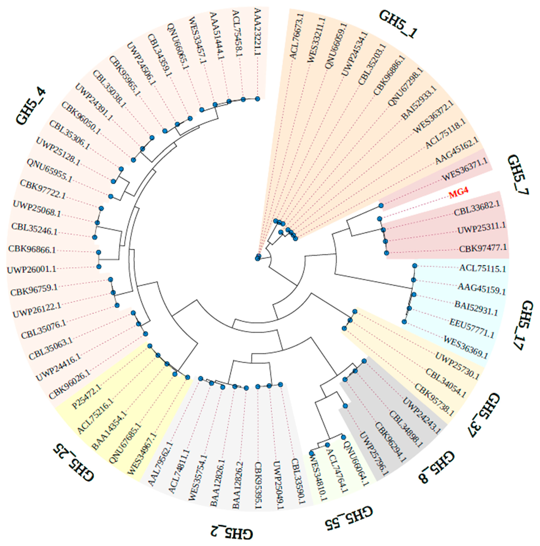
Figure 1. Phylogenetic analysis of MG4. The tree was generated by the maximum likelihood method based on the amino acid sequences of 68 enzymes from Ruminiclostridium sp. or Eubacterium siraeum sp., belonging to the GH5 family. The branch color represents different subfamilies, and the inner circle blue dots indicate nodes with bootstrap values > 80.
Catalysts 15 00905 g001
Figure 2. Multiple sequence alignment of MG4 and mannanases from GH5_7. The two catalytic residues are labeled by green triangles, and the conserved amino acid residues are labeled by blue triangles.
Catalysts 15 00905 g002
Figure 3. (A) Annotation of MG4 by InterProScan. (B) 3D structure of MG4 with two glutamate catalytic residues (Glu206, Glu327) labeled in yellow and six highly conserved amino acid residues, including (Arg56, Asn135, Asn205, His288, Tyr290, and Trp363) labeled in purple. (C) SDS-PAGE result of recombinant proteins. Lane M: protein molecular weight marker; Lane 1: purified MG4 (63.0 kDa); Lane 2: total cellular proteins.
Catalysts 15 00905 g003
Figure 4. Temperature and pH characteristics of MG4. (A) The optimum temperature. (B) The temperature stability. (C) The optimum pH. (D) The pH stability. Maximum activity was defined as 100% in the optimum conditions assay. Initial activity was defined as 100% in the temperature and pH stability assays. Data represent the mean ± SD (n = 3).
Catalysts 15 00905 g004
Figure 5. TLC analysis of MOS products from hydrolysis of LBG by MG4. Reaction conditions: 0.5% (w/v) LBG, 0.1 mg/mL MG4, 55 °C. Lane S, MOS standards (M1–M5); Lanes 1–8; Hydrolyzed products at different reaction times (0, 10, 30 min, 1, 2, 3, 4 and 5 h). TLC analysis of MOS products from hydrolysis of LBG by MG4.
Catalysts 15 00905 g005
Figure 6. (A) Viscosity curves of guar gum fracturing fluid by different breaker treatments. (B) Residual residue content of guar gum fracturing fluid after complete breaking. Gray columns and blue columns indicate APS treatment and enzyme treatment, respectively. Data are presented as mean ± SD. Asterisks denote significant differences compared to the control group (**** p < 0.0001, by t-test). (C) TEM photograph of guar gum fracturing fluid without any treatment. (D) TEM photograph of guar gum fracturing fluid after it was thoroughly broken by MG4.
Catalysts 15 00905 g006
Table 1. The effects of metal ions and EDTA on the mannanase activity of MG4.
Metal IonRelative Activity (%) *
None100.00
Cu2+6.3 ± 0.7
Zn2+10.6 ± 0.3
Fe3+26.3 ± 0.04
EDTA53.5 ± 1.9
Ni2+75.8 ± 1.1
K+114.2 ± 1.6
Mn2+121.1 ± 0.8
Fe2+122.6 ± 1.3
Na+130.4 ± 0.6
Ba2+132.1 ± 1.4
Mg2+143.2 ± 2.1
Ca2+200.4 ± 4.6
* The results presented are the averages of three separate determinations (n = 3) ± standard deviations.
Table 2. Substrate specificity of MG4 towards polysaccharides.
SubstrateSpecific Activity (U/mg) 1
LBG25.77 ± 0.68
KGM27.27 ± 0.76
GG8.89 ± 0.85
INMND 2
XGND 2
CMCND 2
1 The results presented are the averages of three separate determinations (n = 3) ± standard deviations. 2 ND, enzyme is not active towards the substrate under the assayed conditions.
Disclaimer/Publisher’s Note: The statements, opinions and data contained in all publications are solely those of the individual author(s) and contributor(s) and not of MDPI and/or the editor(s). MDPI and/or the editor(s) disclaim responsibility for any injury to people or property resulting from any ideas, methods, instructions or products referred to in the content.