Article

Engineering Malic Enzyme CO2 Fixation Activity via a Structure–Sequence–SCANNER (3S) Co-Evolution Strategy

MOE Key Laboratory of Bio-Intelligent Manufacturing, School of Bioengineering, Dalian University of Technology, Dalian 116024, China
*
Authors to whom correspondence should be addressed.
These authors contributed equally to this work.
Catalysts 2025, 15(8), 789; https://doi.org/10.3390/catal15080789
Submission received: 14 July 2025 / Revised: 15 August 2025 / Accepted: 16 August 2025 / Published: 18 August 2025
(This article belongs to the Section Biocatalysis)

Abstract

Enzymatic CO2 fixation offers great potential for the sustainable synthesis of value-added compounds. Malic enzyme (ME) catalyzes the reverse carboxylation of pyruvate to malate, enabling direct CO2 conversion into C4 compounds with broad biosynthetic applications. However, the reverse carboxylation activity of wild-type ME is insufficient, and conventional enzyme engineering strategies remain limited by the complexity of identifying distal functional sites. Here, we present a Structure–Sequence–SCANNER (3S) co-evolution strategy that integrates protein structural analysis, sequence conservation profiling, and co-evolutionary network analysis to enable systematic identification of functionally relevant hotspot residues. Using this approach, we engineered Escherichia coli ME (EcME) variants with enhanced CO2 fixation activities. In total, 106 single-point variants were constructed and screened. Among these, variants A464S and D97E exhibited significantly improved reverse carboxylation activities, with 1.7-fold and 1.6-fold increases in catalytic activity and 1.5-fold and 1.8-fold improvements in catalytic efficiency (kcat/Km), respectively, compared to wild-type EcME. Their catalytic efficiencies (kcat/Km) improved by 1.5-fold and 1.8-fold, increasing from 80 mM−1·min−1 for the wild-type enzyme to 120 and 130 mM−1·min−1, respectively. Mechanistic analyses revealed that A464S introduces a stabilizing hydrogen bond with N462, enhancing NADPH binding, while D97E forms a new salt bridge network with K513, resulting in contraction of the substrate pocket entrance and increased pyruvate affinity. These findings demonstrate the effectiveness of the 3S strategy in reprogramming enzyme functions and highlight its potential for constructing efficient artificial CO2 fixation systems.

1. Introduction

Aligned with global carbon neutrality targets, the development of efficient and sustainable carbon dioxide fixation technologies has emerged as a research frontier in the fields of carbon resource utilization [1]. In nature, several CO2 utilization pathways have been established, such as the canonical Calvin–Benson–Bassham (CBB) cycle and the Wood–Ljungdahl pathway [2,3]. In addition, artificial CO2 fixation routes such as the CETCH (Crotonyl-CoA/Ethylmalonyl-CoA/Hydroxybutyryl-CoA) cycle and the ASAP (Artificial Starch Anabolic Pathway) have been developed [4,5]. However, these natural and artificial pathways generally suffer from low carbon fixation rates and complex metabolic networks, limiting their industrial applicability [6]. In contrast, enzymatic carboxylation reactions offer a promising alternative strategy for constructing novel carbon assimilation routes due to their simplified reaction pathways, high degree of modularity, and superior theoretical energy efficiency [7,8].
Among known CO2-transforming enzymes, certain natural decarboxylases are capable of catalyzing the reverse carboxylation reaction under specific conditions, providing a biochemical basis for CO2 fixation [9]. Malic enzyme (ME), for instance, is an NAD(P)+-dependent oxidative decarboxylase that catalyzes the conversion of malate to pyruvate with the release of CO2 in its canonical reaction. From both thermodynamic and mechanistic perspectives, ME also possesses the potential to catalyze the reverse reaction, whereby pyruvate is carboxylated to form malate using reducing power supplied by NAD(P)H [10,11]. However, the reverse carboxylation activity of wild-type ME is low and falls far short of meeting the demands for efficient CO2 fixation in synthetic biology applications. Therefore, functional reprogramming of ME through molecular engineering is urgently needed.
Notably, malic enzyme is capable of catalyzing the one-step conversion of pyruvate to malate in vitro. As a C4 compound containing both hydroxyl and carboxyl functional groups, malate serves as a key biosynthetic intermediate for the production of various high-value chemicals [12,13], including succinic acid, oxaloacetate, and aspartate, with wide applications across the food, pharmaceutical, and personal care industries [14,15]. Currently, industrial malate production predominantly relies on microbial metabolic engineering approaches [16], which often involve complex metabolic pathways, high energy consumption, and challenging regulatory control. Thus, employing malic enzyme to achieve the direct carboxylation of pyruvate to malate in vitro offers an attractive strategy for developing efficient and low-cost CO2 utilization platforms [17].
In recent years, efforts have been made to enhance the carboxylation activity of malic enzyme from various sources through enzyme engineering approaches. For example, site-directed mutagenesis of the NADP(H)-dependent ME from Thermococcus kodakarensis not only altered its cofactor preference but also increased malate yield from 7.7% to 10.3% [18]. Similarly, introducing a C490S mutation into the ME from Arabidopsis thaliana resulted in an increased intracellular malate production [19], reaching 3.62 g·L−1. However, conventional strategies such as saturation mutagenesis and rational design often face significant limitations in multi-site combinatorial optimization, including low efficiency, insufficient structural information, difficulty in identifying distal functional sites, and the combinatorial explosion of potential mutations [20,21]. Although recent advances in high-throughput screening and machine learning-assisted library design have improved enzyme engineering efficiency [22,23], a systematic strategy that can identify functional sites and elucidate their cooperative structure–activity relationships remains to be developed.
To address the challenge of reprogramming enzymes for novel catalytic functions, we aimed to develop an evolution strategy that integrates structural features and evolutionary constraints to guide the identification of functionally important residues. Our goal was to identify and engineer key functional residues in malic enzyme to enhance its CO2 fixation capacity, thereby establishing a generalizable framework for artificial carbon-fixing enzyme design.

2. Results and Discussion

2.1. Structure-Guided Construction of ME Variant Libraries

We first analyzed the structure of E. coli malic enzyme (EcME). To date, only the apo-form structure of the EcME L310R/Q401C mutant has been reported [24], which lacks ligand-binding information. Therefore, we used AlphaFold3 to predict the structure of EcME in complex with NADPH. Compared with the reported EcME structure (PDB ID: 6AGS), the AlphaFold3-calculated model includes NADPH-binding information and shows a low RMSD of 0.29 Å relative to the experimental structure. Based on this model, we further performed molecular docking using AutoDock Vina to obtain the complex structure with the carboxylation substrate pyruvate (Figure 1A). The carboxyl group of pyruvate forms a hydrogen bond (2.5 Å) with the OG1 atom of Thr105, while its carbonyl group forms a hydrogen bond (2.9 Å) with the NZ atom of Lys175 (Figure 1B). In addition, several residues located within 5 Å of the pyruvate substrate were identified, including E246, D269, D270, T274, Y104, N463, and L59. These residues are completely conserved among multiple NADPH-dependent malic enzymes (Figure S1) and are likely directly involved in substrate binding. To construct a high-activity ME variant library, we expanded the target sites to residues within 10 Å of the active site, identifying 25 non-conserved candidates such as G109, A464, F465, and I466 (Figure 1C).

2.2. Sequence-Guided Construction of ME Variant Libraries

Following the selection of target sites based on structural analysis, we performed multiple sequence alignment of homologous malic enzymes, which revealed 25 non-conserved residues among the selected sites. To avoid perturbing residues essential for enzyme activity, we chose these non-conserved residues as mutation targets. These residues were subjected to virtual saturation mutagenesis, and the Gibbs free energy changes (ΔΔG) associated with each mutation were calculated using FoldX (Figure 2). Although ΔΔG values do not directly reflect the catalytic activity of mutants, they provide useful insights into the impact of mutations on protein stability. Variants exhibiting large absolute ΔΔG values are likely to disrupt protein structure, leading to loss of enzyme activity. Based on statistical thresholds commonly applied in protein engineering studies and informed by the sequence alignment results [25,26], we selected mutations with absolute ΔΔG values less than 2 kcal·mol−1 for library construction. Ultimately, by integrating sequence alignment with FoldX-based virtual screening, we designed a library comprising 32 variants across 18 target sites.
In addition, we performed sequence and structural comparisons between EcME and Arabidopsis thaliana malic enzyme (AtME) to guide further library design and screening [19]. Sequence alignment revealed that mutations enhancing carboxylation activity in AtME corresponded to residues D97, S177, C461, and A464 in EcME. Accordingly, we designed seven EcME variants by substituting these four residues with the corresponding amino acids from AtME or with amino acids shown to enhance AtME activity. These variants included D97E, D97V, S177V, C461A, C461Y, A464C, and A464S. Furthermore, we expanded the design based on the physicochemical properties of amino acid side chains at these positions. Combined with ΔΔG calculations to evaluate the effects of mutations on protein stability, an additional 16 variants were constructed at these sites.

2.3. SCANNER-Guided Construction of ME Variant Libraries

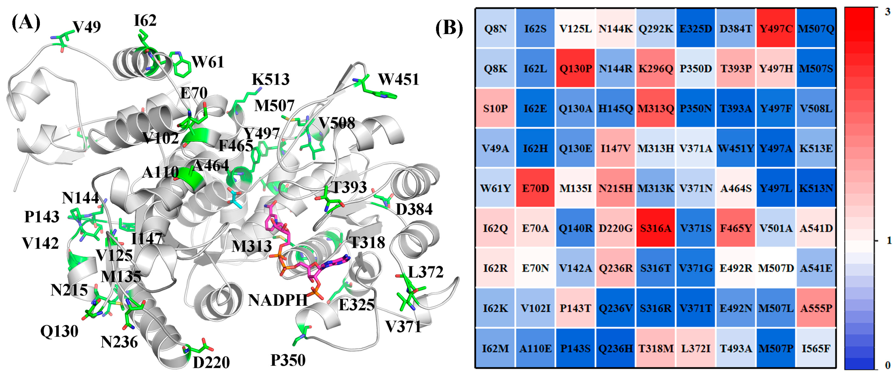
SCANNER is an online database that enables multiple sequence alignment. By inputting the amino acid sequence of the target protein, the system performs comprehensive alignment and outputs the sequence co-evolution index (SCI) for each amino acid residue. A higher SCI value indicates a greater proportion of the mutated residue among homologs, suggesting a higher likelihood that such mutations are beneficial. For SCANNER-guided library construction, analysis identified 81 variants with SCI values greater than zero (Figure 3). Subsequent evaluation of these variants using FoldX revealed that 66 had absolute ΔΔG values less than 2 kcal·mol−1, indicating that these mutations were unlikely to destabilize the protein structure. After excluding three variants (A464S, F465L, and F464Y) that had already been studied in Section 2.1, a total of 63 variants remained for library construction.

2.4. Preliminary Screening of ME Variant Libraries

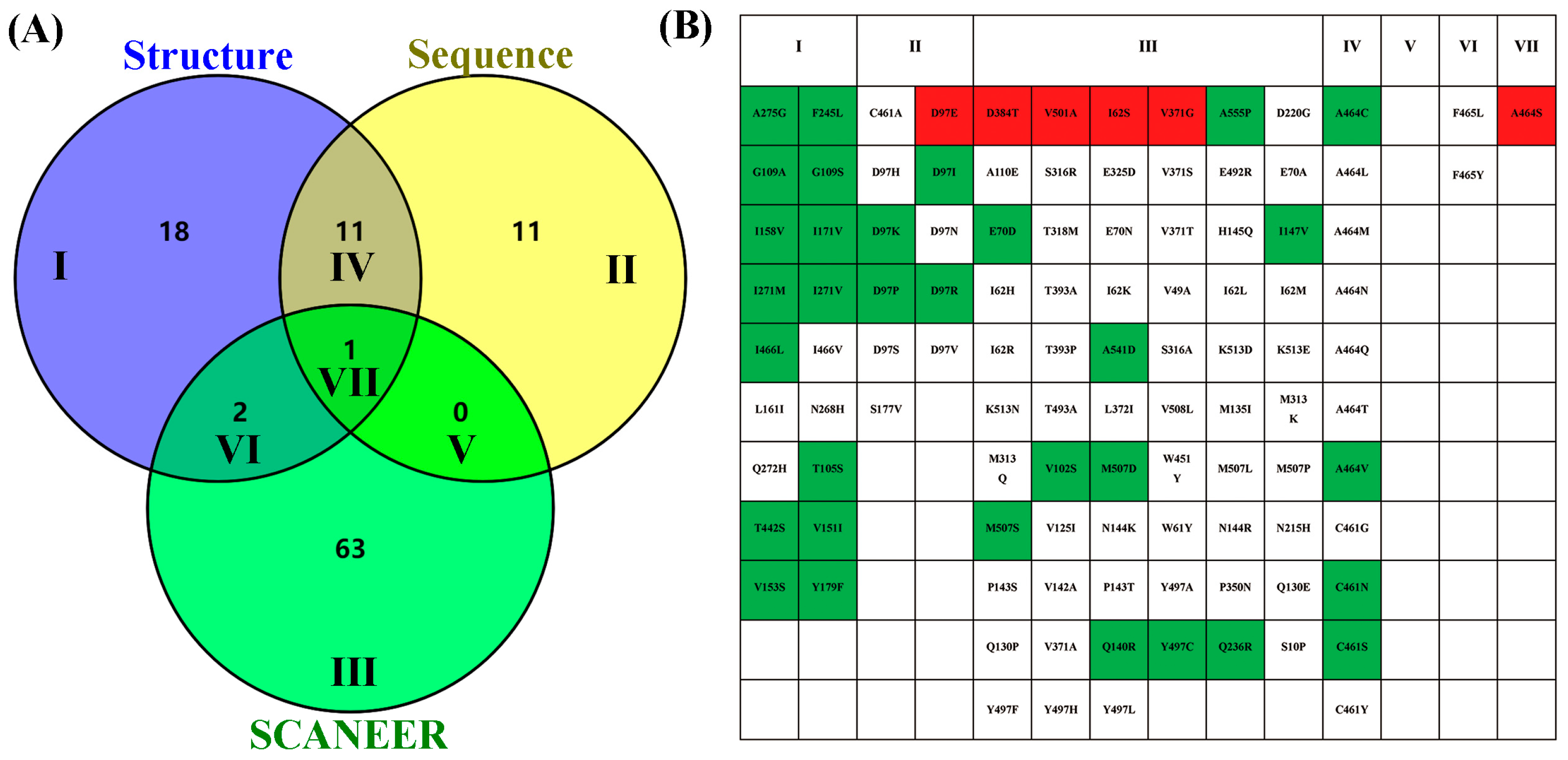
Based on the integrated design strategies described above, a total of 106 single-point variants were constructed: 32 variants were designed using the structure-guided approach, 23 variants were identified via sequence-guided design, and 66 variants were obtained from the SCANNER-guided strategy (Figure 4A). Venn diagram analysis revealed that 18 variants were uniquely identified by the structure-based strategy (Region I), 11 variants were unique to the sequence-based strategy (Region II), and 63 variants were unique to the SCANNER-based strategy (Region III). Additionally, 11 variants were identified by both the structure- and sequence-guided strategies (Region IV), 2 variants by both the structure- and SCANNER-guided strategies (Region VI), and an A464S variant was identified by all three strategies (Region VII) (Figure 4A). All variants were individually constructed and expressed in E. coli BL21(DE3). Whole-cell catalytic assays were performed to evaluate their carboxylation activity, using NADH consumption as the activity readout. Among the 106 variants screened, six variants—D97E, A464S, I62S, V371G, V501A, and D384T—showed enhanced activity, exhibiting at least a 1.2-fold increase compared to the wild-type enzyme. In contrast, 34 variants showed significantly reduced activity (less than 0.8-fold relative to wild type), while 66 variants exhibited no significant change (0.8–1.2-fold relative to wild type). Notably, A464S was the only variant identified by the intersection of all three “3S” design strategies, highlighting the effectiveness of the integrated approach (Figure 4B).

2.5. Purified Enzyme Re-Screening and Kinetic Characterization

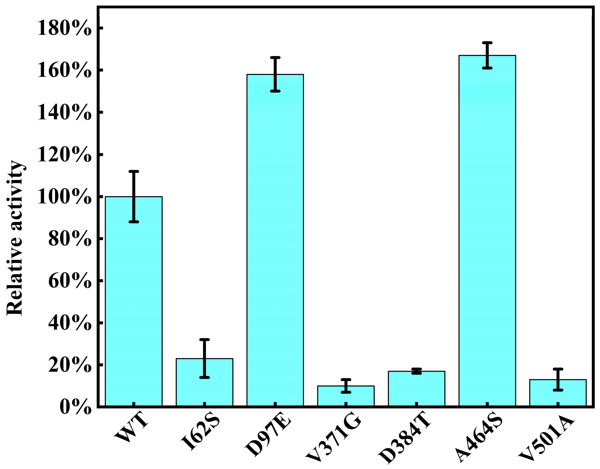
The six variants that exhibited greater than 1.2-fold carboxylation activity compared to wild type (WT) in the initial crude enzyme screening were subjected to further analysis. These variants were further purified using affinity chromatography followed by size-exlcusion chromatography, yielding soluble target proteins for all variants (Figure S2). Protein concentrations were quantified using the Bradford assay, revealing that variants I62S and D97E exhibited slightly lower expression levels (~5 mg·L−1) compared to WT (~10 mg·L−1), while the remaining four variants showed expression levels comparable to WT. The purified enzymes were then evaluated for their carboxylation activities. As shown in Figure 5, variants D97E and A464S exhibited significantly enhanced activities, with relative carboxylation activities of 1.6-fold and 1.7-fold compared to WT, respectively. In contrast, the remaining four variants displayed activities lower than that of WT.
Then, the kinetic parameters of variants A464S and D97E for the carboxylation reaction were determined, and the fitted Michaelis–Menten curves are shown in Table 1. The kcat values of A464S and D97E were 16.3 ± 0.6 min−1 and 16.0 ± 0.5 min−1, respectively, comparable to that of the WT enzyme (17.7 ± 0.6 min−1). Notably, the catalytic efficiencies (kcat/Km) of A464S and D97E were markedly improved to 123 mM−1·min−1 and 132 mM−1·min−1, representing 1.5-fold and 1.8-fold increases relative to WT (80 mM−1·min−1). This enhancement primarily resulted from decreased Km values, indicating an increased affinity towards pyruvate and thereby improved carboxylation activities. For comparison, the Arabidopsis thaliana malic enzyme (AtME) has been reported to exhibit a relatively high carboxylation activity, approximately 91% that of EcME [19]. Directed evolution of AtME previously yielded mutants with up to a 56% increase in carboxylation activity. In contrast, variant A464S of EcME developed in this study showed an 87% improvement in carboxylation activity compared to AtME, representing the highest carboxylation activity reported to date for malic enzyme variants.
Additionally, we have measured the kinetic parameters for the decarboxylation of the enzyme to L-malate. The wild-type malic enzyme exhibited a Km of 513 ± 58 μM, a kcat of 199 ± 10 min−1, and a catalytic efficiency (kcat/Km) of 0.39 ± 0.02 μM−1min−1. In contrast, the A464S mutant showed significantly improved parameters, with a Km of 229 ± 23 μM, a kcat of 338 ± 12 min−1, and a catalytic efficiency of 1.48 ± 0.09 μM−1min−1. This corresponds to a 3.8-fold increase in kcat/Km for the decarboxylation reaction relative to WT, indicating that the mutation enhances catalytic efficiency in both the carboxylation and decarboxylation directions, with a more pronounced effect on decarboxylation.

2.6. Mechanistic Analysis of the Enhanced Catalytic Activity of the Variants

Both A464S and D97E mutations are located beyond 10 Å from the substrate-binding pocket, suggesting they do not directly interact with the pyruvate substrate. To investigate their potential mechanistic roles, we constructed models of EcME and its variants complexed with NADPH using AlphaFold3 and performed two independent molecular dynamics (MD) simulations for each system.
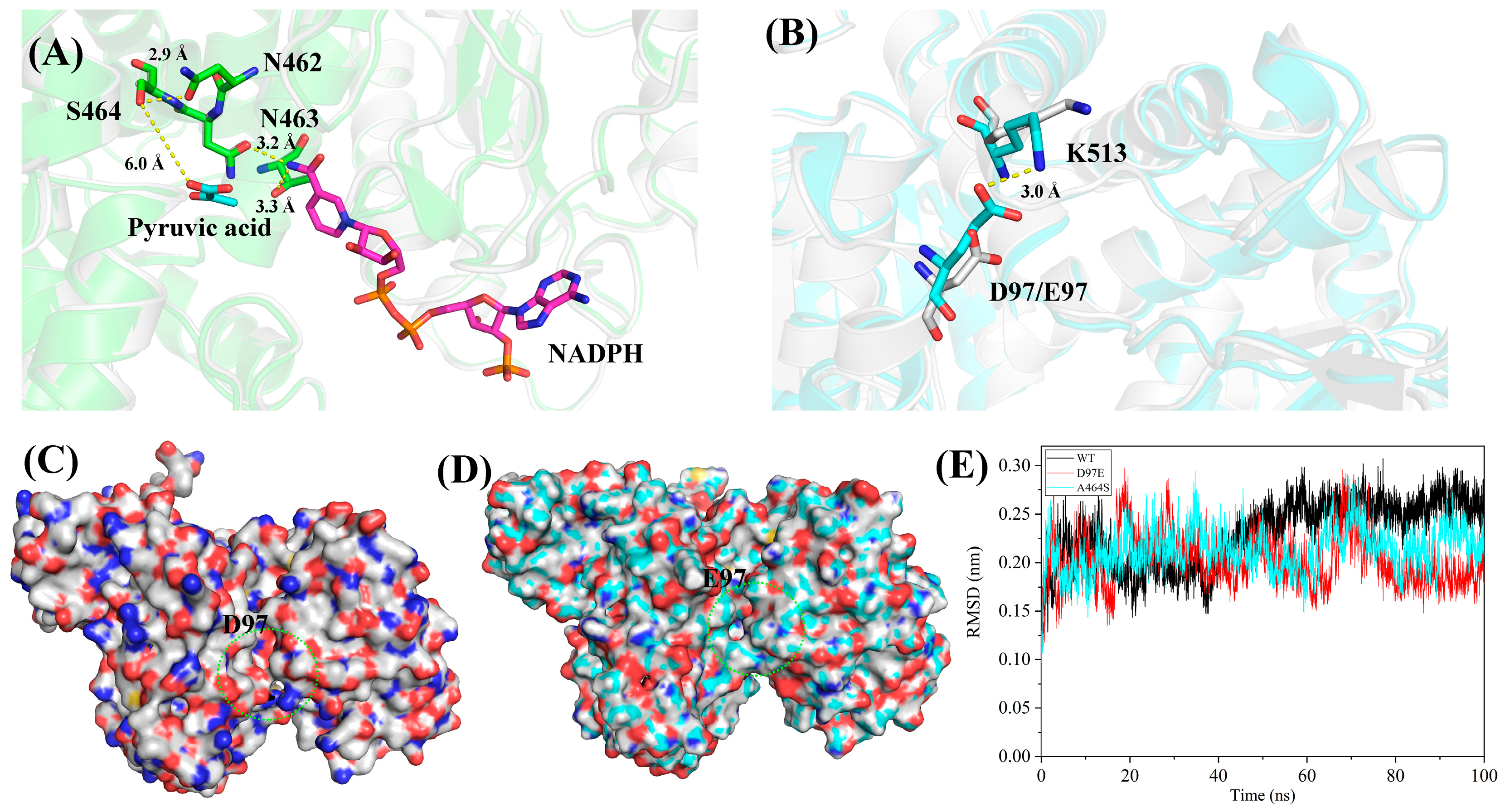
As shown in Figure 6A, the hydroxyl group introduced by the A464S mutation forms a new hydrogen bond (2.9 Å) with the carbonyl oxygen of N462. This interaction likely stabilizes the local conformation of the 462–464 region, indirectly enhancing the binding of NADPH, as N463 is positioned to form a hydrogen bond with the nicotinamide moiety of NADPH, despite S464 being ~6 Å away from pyruvate. Similarly, residue D97 is located on the protein surface and does not interact with other residues in the wild-type enzyme. Upon mutation to D97E, the side chain is extended. MD simulations of A464S and D97E over 100 ns revealed, as shown in Figure 6B, that E97 in the D97E variant forms a potential hydrogen bond with the adjacent K513 residue—a bond not observed in the wild type due to the shorter side chain of aspartate. Intriguingly, structural analysis indicates that this newly formed salt bridge network with K513 results in a local contraction at the entrance of the substrate pocket, which could enhance the binding affinity towards the small molecule substrate pyruvate (Figure 6C,D). This is consistent with the observed decrease in Km for pyruvate in D97E.
Further RMSD analysis demonstrated that both variants remained structurally stable throughout the MD trajectories. Specifically, during the equilibrated last 20 ns, the wild-type EcME exhibited an average RMSD of 2.6 ± 0.1 Å, whereas D97E and A464S showed reduced RMSD values of 1.8 ± 0.1 Å and 2.2 ± 0.2 Å, representing decreases of 29% and 16%, respectively (Figure 6E). These results suggest enhanced structural stability in both variants, which, combined with the additional NADPH interaction in A464S and the reduced substrate pocket volume in D97E, likely underpins their increased carboxylation and CO2 fixation activities. Additionally, the RMSD values of NADPH were comparable across all variants (Figure S3). However, for the pyruvate molecule, especially after approximately 20 ns of simulation, the wild type exhibited a higher RMSD of 0.36 nm, whereas both D97E and A464S mutants showed lower RMSD values around 0.32 nm. This suggests that the pyruvate substrate is more stable in the mutant complexes. However, these hypotheses have not yet been directly validated by mutagenesis controls such as K513A or D97A mutants. Nonetheless, the enhanced catalytic efficiencies (kcat/Km) observed for the D97E and A464S variants provide functional evidence supporting their positive effects on enzyme activity.

3. Materials and Methods

3.1. Materials

The expression hosts E. coli BL21 (DE3) and E. coli DH5α were obtained from TransGen Biotech (Beijing, China). The pET-28a-EcME plasmid was synthesized by GENEWIZ (Suzhou, China) with cloning sites NdeI and XhoI. PrimerStar Max premix and the restriction enzyme DpnI were purchased from Takara Bio (Beijing, China). Isopropyl β-D-1-thiogalactopyranoside (IPTG), lysozyme, kanamycin, sodium pyruvate and other chemicals were purchased from Sangon Biotech (Shanghai, China).

3.2. Structural Prediction and Molecular Docking

While both NADH and NADPH can serve as cofactors for malic enzymes, structural modeling using AlphaFold3 was currently only feasible with NADPH due to limitations in NADH ligand prediction on the available online servers. Thus, the three-dimensional structures of wild-type EcME and its mutants A464S and D97E in complex with NADPH were predicted using the AlphaFold3 server (https://alphafold.com). Docking simulations were performed using AutoDock Vina (version 1.1.2) with default parameters unless otherwise specified [29]. The three-dimensional structure of pyruvate was retrieved from PubChem and geometrically optimized using the Gaussian 16 software suite. Geometry optimization was carried out employing the B3LYP functional and the 6-31G(d,p) basis set to accurately model the electronic distribution of the molecule. The coordinates of the Mg2+ ion were derived from the crystal structure of 1EFK and incorporated into our model following structural alignment. Following optimization, the electrostatic potential was computed and used to derive partial atomic charges via the restrained electrostatic potential method. For molecular docking, AutoDock Vina was employed. The docking grid was centered on the nicotinamide moiety of the cofactor and key active site residues Y104 and K175, with a grid box size of 22 Å × 22 Å × 22 Å. Docking poses were ranked according to their binding affinity scores, with the highest-scoring conformations selected for further analysis. All structural models and molecular graphics were visualized using PyMOL.

3.3. Multiple Sequence Alignment

Multiple sequence alignment was performed using Clustal Omega 1.2.4 [30] for 27 malic enzyme sequences derived from diverse sources, including E. coli and A. thaliana. The resulting alignment was subsequently visualized and annotated to indicate conserved residues using ESPript 3.0 [31].

3.4. SCANNER-Based Multiple Sequence Alignment and Co-Evolution Analysis

The amino acid sequence of the target protein was submitted to the SCANNER for multiple sequence alignment and co-evolutionary analysis [32]. The SCANNER tool employed evaluates residue–residue co-evolution, which scans the evolution of protein sequences and direct mutation strategy to improve enzyme activity. The system automatically aligned the input sequence with homologous sequences and calculated the sequence co-evolution index (SCI) for each amino acid residue position upon mutation. A higher SCI value indicates a greater proportion of the mutated residue within the homologous sequence set.

3.5. FoldX Calculation of Gibbs Free Energy Changes

ΔΔG calculations were performed using the FoldX plugin integrated in YASARA. Briefly, the calculated structure of malic enzyme was imported into YASARA [33], with temperature set at 298 K, ionic strength at 0.05 M, and pH at 7.0, applying the default system parameters. All calculations were performed using default FoldX parameters, and each mutation was computed three times to obtain an averaged ΔΔG value with improved accuracy.

3.6. Construction of Mutant Libraries

Site-directed mutagenesis was performed using PCR-based techniques to introduce specific mutations into the target gene. Primers were designed as listed in Table S1. PCR amplification was carried out following conditions described in previous studies. The resulting PCR products were verified by agarose gel electrophoresis. To eliminate the parental template, the reaction mixtures were treated with DpnI at 37 °C for 1 h, followed by transformation into E. coli DH5α competent cells. Positive clones were confirmed by DNA sequencing.

3.7. Expression of Wild-Type and Mutant Enzymes

E. coli BL21 (DE3) was used as the host for protein expression. Cells harboring the recombinant plasmids were inoculated into Luria–Bertani medium supplemented with 50 μg·mL−1 kanamycin and incubated overnight at 37 °C with shaking at 220 rpm. The overnight cultures were then diluted into fresh LB medium containing the kanamycin and grown at 37 °C until the optical density at 600 nm reached 0.6–0.8. Protein expression was induced by the addition of 0.2 mM IPTG, followed by incubation at 16 °C with shaking at 220 rpm for 16 h.

3.8. Purification of the Enzymes

For protein purification, induced cells were harvested by centrifugation and lysed by sonication in an appropriate lysis buffer as previously described. The lysate was clarified by centrifugation, and the supernatant was subjected to purification. Final purification was performed by size-exclusion chromatography using a Superdex 200 column (GE Healthcare, Uppsala, Sweden) equilibrated with a buffer containing 50 mM Tris-HCl (pH 8.0), 300 mM NaCl, 5% (v/v) glycerol, and 1 mM dithiothreitol. Protein concentration was determined using the Bradford assay with bovine serum albumin as the standard.

3.9. Library Screening

For the first round of enzymatic screening of the library, induced cells were harvested by centrifugation at 3500 rpm for 10 min and resuspended in 1 mL of lysis buffer containing 100 mM Tris-HCl (pH 8.0), 100 mM MgCl2, and 1 mg·mL−1 lysozyme. Cell lysis was performed at 37 °C with shaking at 220 rpm for 30 min. The lysate was clarified by centrifugation, and the supernatant was used for the catalytic assay. For activity screening, 5 μL of the supernatant was transferred to a 96-well microplate containing a reaction mixture with 0.15 mM pyruvate and 0.15 mM NADH. After the 10 min reaction, the absorbance at 340 nm was measured using a microplate reader to quantify NADH consumption. The carboxylation activity was evaluated based on the amount of NADH consumed during the reaction.

3.10. Determination of Kinetic Parameters

Enzyme kinetic assays were performed to determine the carboxylation activity using varying concentrations of pyruvate ranging from 50 μM to 2000 μM in the reaction system described above. Initial reaction rates were measured, and the Michaelis–Menten kinetic parameters were calculated by fitting the data to the Michaelis–Menten equation using GraphPad Prism 8 (GraphPad Software, San Diego, CA, USA). The Km values were obtained from the fitted curves. The decarboxylation activity of EcME was measured in 200 μL reaction systems containing 100 mM HEPES (pH 6.5), 0.15 mM L-malate, 0.15 mM NAD+, and 10 mM MgCl2, initiated by appropriate amount EcME. After 10 min incubation at 30 °C, the reaction was terminated and NADH production was quantified spectrophotometrically at 340 nm to determine enzymatic activity.

3.11. Molecular Dynamics Simulations

Molecular dynamics (MD) simulations were performed using GROMACS version 2022.02 with the AMBER99SB-ILDN force field. Force field parameters for NADPH and pyruvate were generated using ACPYPE [34]. The protein–ligand complexes were solvated in a cubic box of TIP3P water molecules. Counter ions were added by replacing water molecules to neutralize the overall charge of the system. Temperature and pressure were maintained at 300 K and 1 bar, respectively, using the velocity-rescale modified Berendsen thermostat and the Parrinello–Rahman barostat. Following 100 ps of energy minimization and equilibration, production MD simulations were conducted for 100 ns with a time step of 2 fs.

4. Conclusions

In this study, we developed a Structure–Sequence–SCANNER (3S) co-evolution strategy to systematically engineer the reverse carboxylation activity of malic enzyme for CO2 fixation. Our 3S framework integrates three layers of information—protein structure, evolutionary sequence conservation, and residue co-evolution networks—to enable a more targeted and mechanistically interpretable residue selection. Through this multi-dimensional strategy, we constructed and screened a comprehensive malic enzyme variant library, leading to the identification of distal mutations A464S and D97E, which enhanced carboxylation efficiency by 1.5-fold and 1.8-fold, respectively, relative to the wild-type enzyme. These findings demonstrate the effectiveness of the 3S strategy in identifying functional hotspots and optimizing their contributions to catalytic activity, thereby providing a framework for advancing the development of novel biocatalytic platforms for sustainable CO2 utilization and the biosynthesis of value-added chemicals.

Supplementary Materials

The following supporting information can be downloaded at: https://www.mdpi.com/article/10.3390/catal15080789/s1, Figure S1: Multiple sequence alignment of malic enzymes from 27 different sources; Figure S2 Expression and purification of the EcME; Figure S3. RMSD analysis of the NADPH and pyruvate ligand in EcME molecular dynamics, and Table S1: Primers used in this study.

Author Contributions

Conceptualization, J.S., Y.F. and S.X.; methodology J.S., M.W., X.L. and T.F.; writing—review and editing, J.S., M.W., Y.F. and S.X. All authors have read and agreed to the published version of the manuscript.

Funding

This research was funded by the National Key Research and Development Program, grant number: 2022YFC2105602; Department of Science and Technology of Liaoning Province (grant number 2024JH2/102600031), and Fundamental Research Funds for the Central Universities (grant number DUT24LAB104) for their financial support.

Data Availability Statement

Data of this study are available in the article.

Conflicts of Interest

The authors declare no conflicts of interest.

References

  1. Yang, C.; Mao, L.; Chen, Y.X.; Zhou, Y.H.; Zhang, R.F.; Yi, Z.W.; Zhang, D.C.; Zhang, G.Y. Ancestral carbonic anhydrase with significantly enhanced stability and activity for CO2 capture and utilization. Bioresour. Technol. 2025, 419, 132054. [Google Scholar] [CrossRef]
  2. Karishma, S.; Kamalesh, R.; Saravanan, A.; Deivayanai, V.C.; Yaashikaa, P.R.; Vickram, A.S. A review on recent advancements in biochemical fixation and transformation of CO2 into constructive products. Biochem. Eng. J. 2024, 208, 109366. [Google Scholar] [CrossRef]
  3. Lee, J.; Yu, H.E.; Lee, S.Y. Metabolic engineering of microorganisms for carbon dioxide utilization. Curr. Opin. Biotech. 2025, 91, 103244. [Google Scholar] [CrossRef]
  4. Schwander, T.; von Borzyskowski, L.S.; Burgener, S.; Cortina, N.S.; Erb, T.J. A synthetic pathway for the fixation of carbon dioxide in vitro. Science 2016, 354, 900–904. [Google Scholar] [CrossRef]
  5. Cai, T.; Sun, H.B.; Qiao, J.; Zhu, L.L.; Zhang, F.; Zhang, J.; Tang, Z.J.; Wei, X.L.; Yang, J.G.; Yuan, Q.Q.; et al. Cell-free chemoenzymatic starch synthesis from carbon dioxide. Science 2021, 373, 1523–1527. [Google Scholar] [CrossRef]
  6. Bierbaumer, S.; Nattermann, M.; Schulz, L.; Zschoche, R.; Erb, T.J.; Winkler, C.K.; Tinzl, M.; Glueck, S.M. Enzymatic Conversion of CO2: From Natural to Artificial Utilization. Chem. Rev. 2023, 123, 5702–5754. [Google Scholar] [CrossRef]
  7. Chen, H.; Huang, Y.; Sha, C.; Moradian, J.M.; Yong, Y.C.; Fang, Z. Enzymatic carbon dioxide to formate: Mechanisms, challenges and opportunities. Renew. Sust. Energ. Rev. 2023, 178, 113271. [Google Scholar] [CrossRef]
  8. Pu, X.; Han, Y.J. Promotion of Carbon Dioxide Biofixation through Metabolic and Enzyme Engineering. Catalysts 2022, 12, 399. [Google Scholar] [CrossRef]
  9. Chen, J.; Wang, S.R.; Zhou, J.R.; Xu, J.X.; Wu, B.; Gao, Z.; He, B.F. Phenolic Acid Decarboxylase for Carbon Dioxide Fixation: Mining, Biochemical Characterization, and Regioselective Enzymatic β-carboxylation of -hydroxystyrene Derivatives. Catalysts 2025, 15, 210. [Google Scholar] [CrossRef]
  10. Wu, N.; Zhang, J.H.; Chen, Y.R.; Xu, Q.; Song, P.; Li, Y.F.; Li, K.; Liu, H. Recent advances in microbial production of L-malic acid. Appl. Microbiol. Biot. 2022, 106, 7973–7992. [Google Scholar] [CrossRef] [PubMed]
  11. Are, K.R.A.; Ohshima, S.; Koike, Y.; Asanuma, Y.; Kashikura, S.; Tamura, M.; Matsuda, T. Enzymatic direct carboxylation under supercritical CO2. Biochem. Eng. J. 2021, 171, 108004. [Google Scholar] [CrossRef]
  12. Zhang, L.C.; Jin, H.Z.; Jiao, D.N.; Lin, W.Q.; Liu, J.; Liu, H. High-yield bioconversion of glycerol to L-malic acid in Aspergillus niger via systematic engineering of glycerol catabolism and minimal glucose supplementation. Chem. Eng. J. 2025, 515, 163687. [Google Scholar] [CrossRef]
  13. Hu, G.P.; Zhou, J.; Chen, X.L.; Qian, Y.Y.; Gao, C.; Guo, L.; Xu, P.; Chen, W.; Chen, J.; Li, Y.; et al. Engineering synergetic CO2-fixing pathways for malate production. Metab. Eng. 2018, 47, 496–504. [Google Scholar] [CrossRef]
  14. Liu, X.T.; Zhao, G.; Sun, S.J.; Fan, C.L.; Feng, X.J.; Xiong, P. Biosynthetic Pathway and Metabolic Engineering of Succinic Acid. Front. Bioeng. Biotech. 2022, 10, 843887. [Google Scholar] [CrossRef] [PubMed]
  15. Katagiri, T.; Amao, Y. Trivalent metal ions promote the malic enzyme-catalyzed building of carbon-carbon bonds from CO2 and pyruvate. New J. Chem. 2020, 44, 17208–17214. [Google Scholar] [CrossRef]
  16. Ding, Q.; Ye, C. Recent advances in producing food additive L-malate: Chassis, substrate, pathway, fermentation regulation and application. Microb. Biotechnol. 2023, 16, 709–725. [Google Scholar] [CrossRef]
  17. Zelle, R.M.; de Hulster, E.; van Winden, W.A.; de Waard, P.; Dijkema, C.; Winkler, A.A.; Geertman, J.M.A.; van Dijken, J.P.; Pronk, J.T.; van Maris, A.J.A. Malic acid production by Engineering of pyruvate carboxylation, oxaloacetate reduction, and malate export. Appl. Environ. Microb. 2008, 74, 2766–2777. [Google Scholar] [CrossRef]
  18. Morimoto, Y.; Honda, K.; Ye, X.T.; Okano, K.; Ohtake, H. Directed evolution of thermotolerant malic enzyme for improved malate production. J. Biosci. Bioeng. 2014, 117, 147–152. [Google Scholar] [CrossRef]
  19. Dong, X.X.; Chen, X.L.; Qian, Y.Y.; Wang, Y.C.; Wang, L.; Qiao, W.H.; Liu, L.M. Metabolic engineering of W3110 to produce L-malate. Biotechnol. Bioeng. 2017, 114, 656–664. [Google Scholar] [CrossRef] [PubMed]
  20. Li, Y.; Li, S.F.; Fu, S.Y.; Ren, X.D.; Zhou, Y.C.; Xue, Y.P.; Zheng, Y.G. Design of a Distal Site Saturation Test-Iterative Parallel Mutagenesis for Engineering Hydroxysteroid Dehydrogenase. J. Agric. Food Chem. 2025, 73, 9701–9713. [Google Scholar] [CrossRef] [PubMed]
  21. Amatto, I.V.D.; da Rosa-Garzon, N.G.; Simoes, F.A.D.; Santiago, F.; Leite, N.P.D.; Martins, J.R.; Cabral, H. Enzyme engineering and its industrial applications. Biotechnol. Appl. Biochem. 2022, 69, 389–409. [Google Scholar] [CrossRef]
  22. Jang, W.D.; Kim, G.B.; Kim, Y.; Lee, S.Y. Applications of artificial intelligence to enzyme and pathway design for metabolic engineering. Curr. Opin. Biotechnol. 2022, 73, 101–107. [Google Scholar] [CrossRef] [PubMed]
  23. Buller, R.; Lutz, S.; Kazlauskas, R.J.; Snajdrova, R.; Moore, J.C.; Bornscheuer, U.T. From nature to industry: Harnessing enzymes for biocatalysis. Science 2023, 382, adh8615. [Google Scholar] [CrossRef]
  24. Liu, Y.X.; Guo, X.J.; Liu, W.J.; Wang, J.T.; Zhao, Z.K. Structural Insights into Malic Enzyme Variants Favoring an Unnatural Redox Cofactor. Chembiochem 2021, 22, 1765–17684. [Google Scholar] [CrossRef]
  25. Valanciute, A.; Nygaard, L.; Zschach, H.; Jepsen, M.M.; Lindorff-Larsen, K.; Stein, A. Accurate protein stability predictions from homology models. Comput. Struct. Biotechnol. J. 2023, 21, 66–73. [Google Scholar] [CrossRef]
  26. Feng, T.; Huang, H.X.; Xu, Y.; Che, X.Y.; Wang, M.D.; Tao, X.Y.; Feng, Y.B.; Xue, S. Multisite synergistic evolution of oleate hydratase via DCCM and the orthogonal location of distal sites. Int. J. Biol. Macromol. 2025, 312, 144001. [Google Scholar] [CrossRef]
  27. Crooks, G.E.; Hon, G.; Chandonia, J.M.; Brenner, S.E. WebLogo: A sequence logo generator. Genome Res. 2004, 14, 1188–1190. [Google Scholar] [CrossRef] [PubMed]
  28. Shi, J.P.; Fan, Y.; Jiang, X.S.; Li, X.L.; Li, S.; Feng, Y.B.; Xue, S. Efficient synthesis of L-malic acid by malic enzyme biocatalysis with CO2 fixation. Bioresour. Technol. 2024, 403, 130843. [Google Scholar] [CrossRef]
  29. Eberhardt, J.; Santos-Martins, D.; Tillack, A.F.; Forli, S. AutoDock Vina 1.2.0: New Docking Methods, Expanded Force Field, and Python Bindings. J. Chem. Inf. Model. 2021, 61, 3891–3898. [Google Scholar] [CrossRef] [PubMed]
  30. Madeira, F.; Madhusoodanan, N.; Lee, J.H.; Eusebi, A.; Niewielska, A.; Tivey, A.R.N.; Lopez, R.; Butcher, S. The EMBL-EBI Job Dispatcher sequence analysis tools framework in 2024. Nucleic Acids Res. 2024, 52, W521–W525. [Google Scholar] [CrossRef]
  31. Robert, X.; Gouet, P. Deciphering key features in protein structures with the new ENDscript server. Nucleic Acids Res. 2014, 42, W320–W324. [Google Scholar] [CrossRef] [PubMed]
  32. Kim, D.; Noh, M.H.; Park, M.; Kim, I.; Ahn, H.; Ye, D.Y.; Jung, G.Y.; Kim, S. Enzyme activity engineering based on sequence co-evolution analysis. Metab. Eng. 2022, 74, 49–60. [Google Scholar] [CrossRef] [PubMed]
  33. Van Durme, J.; Delgado, J.; Stricher, F.; Serrano, L.; Schymkowitz, J.; Rousseau, F. A graphical interface for the FoldX forcefield. Bioinformatics 2011, 27, 1711–1712. [Google Scholar] [CrossRef]
  34. Kagami, L.; Wilter, A.; Diaz, A.; Vranken, W. The ACPYPE web server for small-molecule MD topology generation. Bioinformatics 2023, 39, btad350. [Google Scholar] [CrossRef] [PubMed]
Figure 1. Structural analysis of the active site pocket of EcME. (A) Molecular docking of pyruvate into EcME, the models highlighted in cyan and magenta represent pyruvate and NADPH, respectively. (B) Amino acid residues within 5 Å of the substrate-binding pocket. (C) Amino acid residues within 5–10 Å of the substrate-binding pocket.
Catalysts 15 00789 g001
Figure 2. Analysis of 25 non-conserved mutation sites. (A) Gibbs free energy changes (ΔΔG) upon virtual saturation mutagenesis at the 25 mutation sites. (B) Sequence conservation analysis of the 25 non-conserved residues, presented by WebLogo [27].
Catalysts 15 00789 g002
Figure 3. SCANNER-guided construction of malic enzyme variant libraries. (A) Mutation sites are shown in green and displayed as stick models. (B) Variants designed based on SCI values higher than zero. Blue indicates variants with an SCI value < 1, while red indicates variants with an SCI value > 1.
Catalysts 15 00789 g003
Figure 4. Venn diagram of malic enzyme variant libraries designed using the Structure–Sequence–SCANNER approach. (A) Regions I, II, and III indicate variants designed based on structure only, sequence only, and SCANNER only, respectively. Region IV represents variants designed by combining structure and sequence information, Region V represents variants designed by combining sequence and SCANNER, Region VI represents variants designed by combining structure and SCANNER, and Region VII represents variants designed by integrating structure, sequence, and SCANNER approaches. (B) Screening results of the mutants. Variants highlighted in red exhibit ≥ 1.2-fold activity relative to the wild type, those in green exhibit ≤ 0.8-fold activity, and those in white show comparable activity to the wild type (0.8–1.2-fold).
Catalysts 15 00789 g004
Figure 5. Carboxylation activities of six purified malic enzyme variants.
Catalysts 15 00789 g005
Figure 6. Molecular mechanisms underlying the effects of D97E and A464S mutations on EcME carboxylation activity. (A) Predicted structures of EcME wild type (grey) and the A464S variant (green) complexed with NADPH (purple) and pyruvate (cyan), as calculated by AlphaFold3. (B) Structural comparison between wild-type EcME (grey) and the D97E variant (cyan) after 100 ns of molecular dynamics equilibrium, highlighting conformational differences. (C) Surface view of wild-type EcME showing the substrate pocket entrance. (D) Surface view of the D97E variant, illustrating the reduced substrate pocket entrance size induced by the mutation (indicated by green dashed circles). (E) RMSD analysis of wild-type EcME (black), D97E (red), and A464S (cyan) during 100 ns molecular dynamics simulations. The x-axis represents simulation time (ns), and the y-axis represents RMSD values (Å).
Catalysts 15 00789 g006
Table 1. Enzyme kinetics of the carboxylation reactions catalyzed by EcME, A464S, and D97E.
kcat (min−1)Km (μM−1)kcat/Km (mM−1·min−1)
WT [28]17.7 ± 0.6221 ± 3180 ± 12
D97E16.0 ± 0.5121 ± 17132 ± 19
A464S16.3 ± 0.6132 ± 23123 ± 22
Disclaimer/Publisher’s Note: The statements, opinions and data contained in all publications are solely those of the individual author(s) and contributor(s) and not of MDPI and/or the editor(s). MDPI and/or the editor(s) disclaim responsibility for any injury to people or property resulting from any ideas, methods, instructions or products referred to in the content.