Article

Role of Cation Nature in FAU Zeolite in Both Liquid-Phase and Gas-Phase Adsorption

1
Energy and Fuels for Sustainable Environment Team, ICPEES-UMR 7515, CNRS, Université de Strasbourg, 25 Rue Becquerel, F-67087 Strasbourg, France
2
Department of Chemistry, Azerbaijan State Oil and Industry University—ASOIU, 16/21 Azadliq Avenue, Baku AZ1010, Azerbaijan
3
Centre National de la Recherche Scientifique—CNRS, Chemistry of Complex Matter (CMC) UMR 7140, Université de Strasbourg, 4 rue Blaise Pascal, F-67000 Strasbourg, France
4
Department of Chemical Engineering, Baku Engineering University—BEU, Khirdalan, 120 AZ0101 Hasan Aliyev Street, Baku AZ0101, Azerbaijan
*
Author to whom correspondence should be addressed.
Catalysts 2025, 15(8), 734; https://doi.org/10.3390/catal15080734 (registering DOI)
Submission received: 20 June 2025 / Revised: 24 July 2025 / Accepted: 29 July 2025 / Published: 1 August 2025

Abstract

This study focuses on the exchange of mono- and divalent metal cations in FAU-type zeolite and their behavior in gas-phase CO2 adsorption measurements and liquid-phase methylene blue (MB) adsorption in the absence of oxidizing agents under dark conditions. Firstly, zeolites exchanged with different cations were characterized by several techniques, such as XRD, SEM, XRF, XPS, and N2 adsorption–desorption, to reveal the impact of the cations on the zeolite texture and structure. The adsorption studies revealed a positive effect of cation exchange on the adsorption capacity of the zeolite, particularly for silver-loaded FAU zeolite. In liquid-phase experiments, Ag-Y zeolite also demonstrated the highest MB removal, with a value of 79 mg/g. Kinetic studies highlighted that Ag-Y could reach the MB adsorption equilibrium within 1 h, with its highest rate of adsorption occurring during the first 5 min. In gas-phase adsorption studies, the highest CO2 adsorption capacity was also achieved over Ag-Y, yielding 10.4 µmol/m2 of CO2 captured.

Graphical Abstract

1. Introduction

Porous materials, such as carbons [1] and silica [2], have been extensively utilized in numerous applications, particularly in the field of adsorption. Among them, microporous materials exhibiting ordered channels or cages, such as Metal–Organic Frameworks (MOFs) [3] and zeolites [4], are regarded as more promising due to the presence of an ordered microporous structure and tunable properties, helping to reach a higher adsorbent selectivity. Zeolites are classified as porous aluminosilicate materials exhibiting a high specific surface area and elevated cation exchange capacity, yielding a good-to-excellent adsorption performance [5]. The large-scale synthesis of zeolites has facilitated their application in the removal of contaminants in both liquid and gaseous phases. In liquid-phase applications, zeolites have mostly been used for the removal of heavy metals and organic contaminants from polluted water sources [6]. They have also demonstrated the successful adsorption of gases, such as CO2, H2S, and VOCs [7]. However, it was found that the pristine Na form of zeolites could not yield sufficient selectivity or adsorption capacity towards several pollutants under varying environmental conditions. To tackle this problem, several strategies were undertaken to modify the zeolites, such as cation exchange, surface functionalization, and dealumination, to enhance their adsorption and catalytic properties [8].
MB is a cationic dye recognized as one toxic contaminant found in wastewater from textile industries. Since it presents a significant threat to both aquatic animals and humans, its removal from water sources has gained popularity over the past century. Natural and synthetic zeolites have already been used in both laboratory and at the industry scale to efficiently remove MB molecules [9]. Asefa et al. showed that FAU zeolites synthesized with different routes successfully reached a 60% MB removal efficiency [10]. Likewise, the study by Le et al. revealed that the development of zeolites from waste sources, such as rice husk, could also increase their adsorption efficiency to 93% towards MB molecules [11]. Besides using zeolites in their as-synthesized Na form, the modification of their cation nature and the formation of composites showed a promising effect for the adsorption of dye molecules. The modification of FAU zeolites with magnetic nanoparticles yielded the highest removal efficiency of 99.8% for MB over magnetic HY zeolite [12]. In another study, Zhu et al. developed an LTA zeolite and reduced graphene oxide (rGO)-based composite possessing an adsorption capacity of 53 mg/g for MB adsorption studies [13]. As zeolites possess a cation exchange ability, the presence of different exchangeable cations within their structure enhances their adsorption and catalytic properties [7,14,15,16]. Considering these intrinsic properties of zeolites, González-Crisostomo et al. developed Ti-, Cu-, Zn-, and Ag-exchanged CHA and studied their properties in MB degradation in the presence of H2O2 [16]. They found that zeolite doped with Cu2+ showed the highest MB removal efficiency at 98.9% compared to zeolites exchanged with other cations, thus proving the impact of cations on dye removal studies. Likewise, Chowdhury et al. showed that Cu2+-exchanged Y zeolite enhanced MB degradation from 50 to 70% compared to the pristine Na form [17]. Colar et al. showed that the reduction of silver cations in Ag-exchanged zeolites could also significantly increase MB removal to 70% in the presence of oxidizing agents and UV irradiation [18]. Despite numerous investigations in the field of MB removal, there is still a scarcity of studies dealing with cation-exchanged zeolites used for MB removal in dark conditions and/or in the absence of oxidizing agents.
Zeolites have also been recognized as effective adsorbents for CO2, particularly in post-combustion capture processes. Among them, 13X zeolite (FAU structure) stands out as the most promising in terms of CO2 capacity [19]. However, its sensitivity to water vapor poses a limitation to its industrial application [20]. Functionalization of the zeolite surface, a decrease in crystal size, the substitution of cations, and the preparation of composites have also been attempted to improve CO2 adsorption by zeolites. Clatworthy et al. also conducted a study demonstrating that variations in zeolite particle size can enhance selectivity towards CO2 adsorption due to changes in diffusion pathways and surface area [21]. Former studies have also examined the influence of the cations in zeolites on CO2 adsorption. This influence typically depends on factors such as the electronegativity of the cation (charge/size ratio) and the pore size of the zeolite. For FAU-type zeolites, it has been demonstrated that exchanging with alkali metals with a smaller radius can lead to higher CO2 adsorption values [22]. Additionally, Yang et al. showed that multivalent ions could block the pores, thereby preventing CO2 from entering. For BEA zeolites, adsorption decreased in the following order: K+ > Na+ > Li+ > Ba2+ > Ca2+; Cs+ > Mg2+ [23]. However, this trend did not hold for LTA, where higher CO2 adsorption was observed with Ca-A compared to Na-A. This discrepancy was attributed to the increase in pore size resulting from the insertion of multivalent cations, allowing CO2 to enter the pores more readily [24].
In light of prior research, it has been observed that the exchange of zeolites with mono- or divalent cations remains inadequately explored in the context of both liquid-phase and gas-phase adsorption studies. Furthermore, the removal of MB at different concentrations in the absence of light and oxidizing agents using silver-exchanged zeolites has not been extensively investigated. Additionally, selecting a versatile zeolite that works efficiently in both the liquid and gas phases could be advantageous for large-scale implementation. Considering all these requirements, our study focuses on the development of Na, Ca, Mg, and Ag cation-exchanged zeolites and their contaminant adsorption properties in both the liquid and gas phases.

2. Results and Discussion

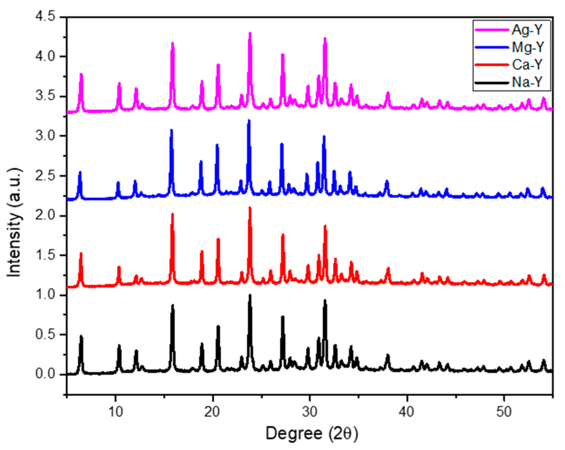
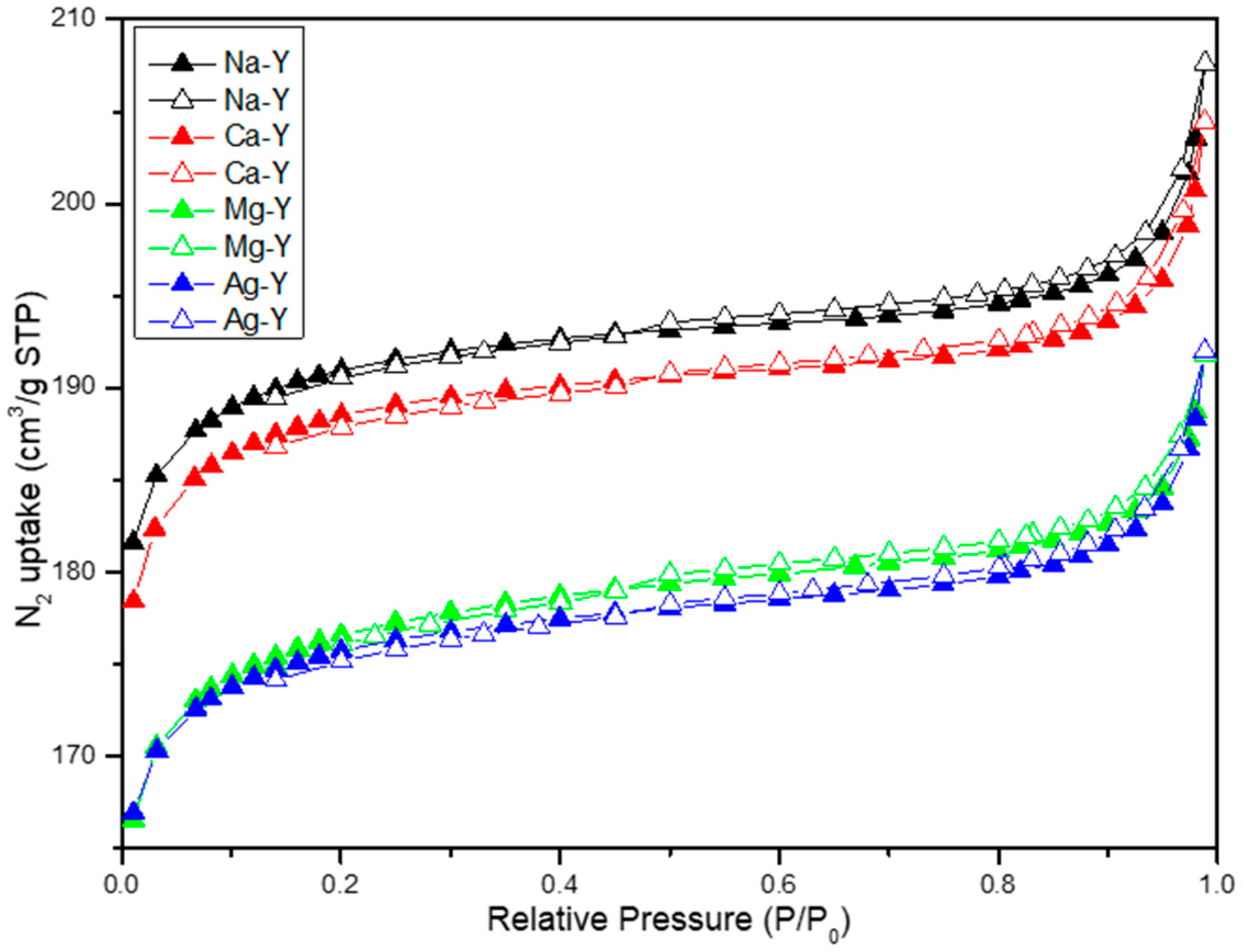
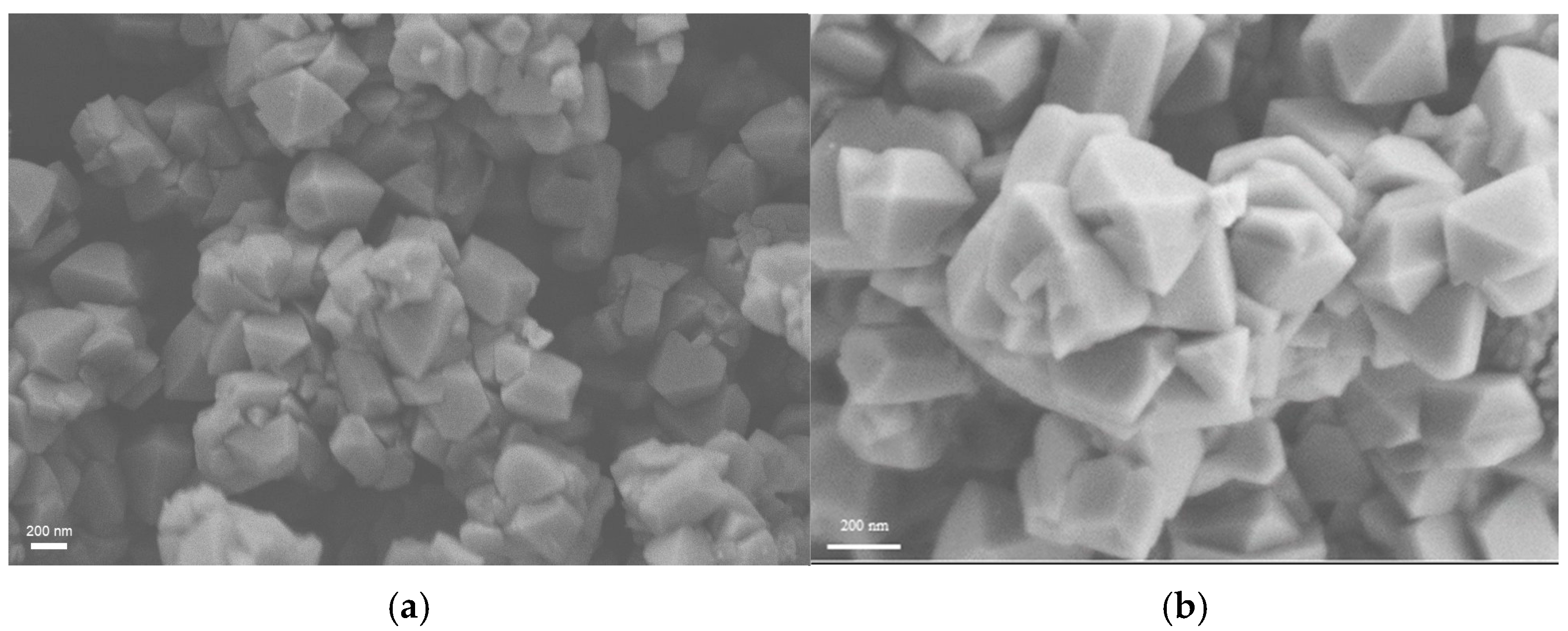
2.1. Zeolite Characterization

The XRD technique was used to determine the phase purity of as-synthesized zeolites. The diffraction patterns of cation-exchanged zeolites (Figure 1) did not exhibit significant differences with respect to pristine zeolite. This could be explained by the mild conditions of the cation exchange procedure, specifically for silver exchange, where the agglomeration of nanoparticles can be prevented by a low-temperature exchange process.
The XRF elemental analysis of exchanged zeolites confirms a Si/Al ratio of 2.2, which is a characteristic value for Y zeolites [25,26]. In comparison to our previous study, a higher Ca content of 6.9 wt% is achieved in the exchanged zeolite (Table 1), which can be explained by a lower Si/Al ratio [26]. The lower amount of Mg in the exchanged zeolite compared to Ca likely results from the high solvation of Mg2+ ions, creating large complexes that cannot diffuse within the zeolite channels. Additionally, the higher charge density of Mg2+ may hinder the exchange of cations within the zeolite framework, as shown by Dyer [27,28]. The lower amount of Ag exchange (3.2 wt.% in Table 1) can be attributed to the low concentration of AgNO3 solution used for the exchange medium to avoid the agglomeration of silver clusters on the zeolite surface. The silver quantity is also quantified by SEM-EDX analysis, which shows a good correlation with the XRF results (Figure S1).
The surface area and porosity of the zeolites were characterized by N2 adsorption–desorption analysis. Prior analysis, it was found that the degassing temperature had a considerable effect on Ag-Y zeolite. The zeolites degassed at 250 °C changed their colors from white to black (Figure S13), which is considered as an indicator for the reduction of silver cations into their metallic state [29,30]. However, it was observed that Ag-Y zeolites degassed at 150 °C for 5 h did not induce color change. The characterization of Ag-Y zeolite by a UV–Visible spectrophotometer after degassing at 250 °C exhibited absorbance between 300 and 400 nm (Figure S14), corresponding to the plasmonic resonance of silver nanoclusters where silver exists in its zero oxidation state [31]. However, the zeolite degassed at 150 °C did not exhibit the same absorbance in the same spectral range. This behavior of silver-exchanged FAU zeolites could be explained by the temperature sensitivity of silver. It was demonstrated by Yu et al. that heating zeolites at 300 °C for 2 h is sufficient to reduce silver from Ag+ to Ag0 [32]. The study by Chen et al. also demonstrated that degassing silver-exchanged FAU-X zeolites at 300 °C can cause silver reduction [33]. Another study by Min et al. showed that silver-exchanged zeolites may contain reduced silver particles depending on the treatment conditions. They found that treatment at high temperatures can result in the formation of silver nanoclusters in vacuum conditions [34]. Considering the possibility of the reduction of silver cations within the zeolite structure at elevated temperatures, all zeolites were degassed at 150 °C for 5 h before CO2 adsorption and N2 adsorption–desorption studies.
Zeolites with different cation types showed a decrease in their BET surface area values from 636 to 557 m2/g after the cation exchange of pristine zeolite (Table 2). This trend can be attributed to the limited pore-blocking effect of cations, which has also been observed in former studies [15,35]. As seen from Table 2, the micropore volume of zeolites decreased slightly, in the same order as the BET surface area. Similar results were observed by Puchalska et al., who demonstrated that the cation exchange of FAU-X zeolite can lead to a decrease in micropore volume from 0.30 cm3/g to 0.20 cm3/g [35]. Additionally, the distribution of cations at different sites within the zeolite structure may also be a contributing factor. Studies have found that the cation exchange of Na-Y zeolites with Ca2+ leads to the majority of Ca2+ cations being placed in site I in the dehydrated FAU zeolite form, where they create an octahedral configuration with oxygen atoms. This octahedral configuration and the bonding of oxygen atoms with Ca2+ fill the zeolite pores, which is not the case for Na-containing zeolites. Similar observations have been made for silver-exchanged zeolites. It has been suggested that silver cations can form Ag3-type clusters with oxygen atoms within sites I and I’ of the zeolite. These clusters are not considered as reduced nanoparticles, but their coordination with oxygen atoms can still occupy the pores [36].
The N2 adsorption–desorption isotherms of zeolites before and after cation exchange are given in Figure 2, where it is possible to observe a Type I isotherm, with a shape remaining unchanged after cation exchange. This explains that the microporous structure of zeolite is not affected by cation exchange, which could be related to the mild cation exchange conditions [37]. However, it is observed that the amount of N2 adsorbed for Ag-Y and Mg-Y is lower than that of Na-Y and Ca-Y, which is in line with the BET surface area and micropore volume values presented in Table 2. This can be explained by the occupation of cations at different exchangeable sites within the zeolite.
The characterization of the real density of the zeolites using He Pycnometry revealed that the zeolites had a density of around 2.6 g/cm3, which is a characteristic value for synthetic zeolites. Indeed, similar results were obtained by Nguyen et al., who reported a density of 2.53 g/cm3 for FAU-Y zeolites using pycnometry analysis [38].
The crystal morphology of the zeolite samples, characterized by Scanning Electron Microscopy, revealed the presence of homogeneous octahedral crystals (Figure 3). It is noteworthy that there was no change in the crystal morphology, nor was there the agglomeration of Ag nanoclusters on the zeolite crystals, which has been the case in other studies where Ag nanoclusters have been detected [39]. The EDX mapping also revealed that the exchanged cations were uniformly distributed across the surface of the zeolite, rather than forming clusters or segregated oxide nanoparticles in some regions of the zeolite crystal (Figure S10).
The oxidation state of silver cations was examined using XPS analysis. The XPS analysis was conducted in fast scan mode, as the zeolite changed color from white to black during prolonged analysis, thus indicating the formation of reduced silver—Ag0. It is not trivial to differentiate the oxidation states of Ag+ and Ag0 based on their binding energies due to a difference of only 0.3 eV. However, differences in kinetic energies in the Auger region render possible the determination of silver cations’ oxidation state after calculating the Auger parameter, which is the sum of binding energies at the Ag 3d5/2 and Ag M4N45N45 regions. According to the literature, Auger parameter values at 726.0 eV and higher correspond to the formation of reduced silver—Ag0. In our case, the Auger parameter was 722.0 eV (Figure S11), corresponding to Ag+, which has also been reported by Aspromonte et al. [40].
UV–Vis spectroscopy has also been used to validate the presence of Ag+ in silver-exchanged zeolite. This method is primarily used to differentiate Ag0 from Ag+ [41,42]. Ag0 shows absorbance between 400 nm and 500 nm, corresponding to the plasmonic band, while Ag+ exhibits absorbance in the range of 210–260 nm, corresponding to the charge transfer (CT) between 4d0 and 4d95s1 [31]. In our case, it can be observed from Figure S12 that a strong absorbance peak for Ag-Y was detected between 210 and 250 nm, correlating with the presence of Ag+ within the zeolite structure. Similar results were also reported by Lopes et al., who prepared Ag-RHO zeolite that exhibited absorbance at 220 nm [43].

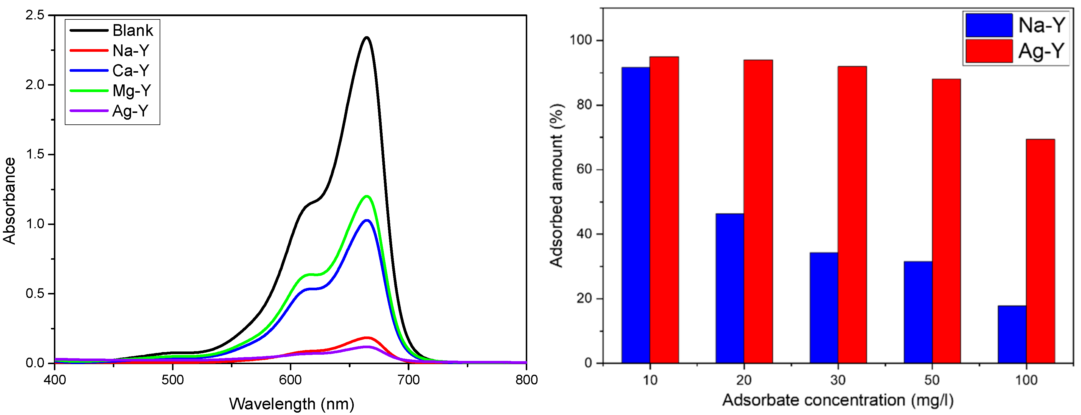
2.2. Liquid-Phase Adsorption Studies

The MB adsorption by zeolites loaded with different cations showed the following order: Mg-Y < Ca-Y < Na-Y < Ag-Y (Figure 4). It has been observed that divalent ion-exchanged zeolites exhibit lower adsorption capacities for cationic dye molecules compared to monovalent ion-exchanged zeolites. This phenomenon can be explained through multiple factors: Firstly, Ag+ ions, being smaller in atomic radius (except Mg), potentially occupy less space within the zeolite structure, thus leaving more space for MB cations on the zeolite surface. Secondly, since MB is a cationic dye, it may be more strongly repelled by divalent ions, which possess a higher positive charge. Moreover, since Ag is catalytically active even in dark conditions, it may also decompose MB molecules [44]. The nitrogen and sulfur present in the MB molecule may enhance interactions with Ag, which is considered a soft acid, in contrast to alkali and alkaline, earth metals that are rather classified as hard acids [45]. This could lead to a greater decrease in concentration during the adsorption process. However, since MB must be adsorbed onto the zeolite before decomposition or coordination occurs, our study focuses solely on adsorption properties, excluding an investigation of its degradation compounds and coordination complexes. As the best results for adsorption were obtained for Na- and Ag-containing zeolites, further kinetic and isothermal studies were conducted on these two zeolites. The MB adsorption on Na-Y and Ag-Y demonstrated that both zeolites adsorbed similar amounts of MB when immersed in a 10 mg/L dye solution, yielding values of 91.7% for Na-Y and 95.0% for Ag-Y, respectively. However, the increase in the dye concentration of the initial solution created a significant difference between the adsorption behavior of Ag-Y and Na-Y. This difference could be observed more clearly when the zeolites were immersed in a 100 mg/L dye solution, where the removal efficiencies were 69.4% and 17.8% for Ag-Y and Na-Y, respectively. Similar results were observed by Zharylkan et al., who used Ag NPs doped Na-X zeolites for MB adsorption. They showed that the presence of Ag NPs on zeolites could increase the adsorption efficiency of the zeolite from 30% to 76% [46]. Another study by González-Crisostomo et al. demonstrated that silver-exchanged zeolites could degrade 71.4% MB in the presence of UV irradiation and H2O2 [16]. The study by Colar et al. also indicated that natural zeolite containing Ag0 particles showed 70% MB removal in the presence of visible light [18]. In comparison to previous studies, our study demonstrated that the presence of Ag+ cations in zeolite can significantly decrease the concentration of MB in aqueous medium in the absence of light or reducing agents, which has not been observed previously to the best of our knowledge.
By analyzing the adsorption kinetics, it is noteworthy that Ag-Y and Na-Y zeolites exhibit the highest MB removal during the first 5 min of the process, with Ag-Y demonstrating a greater removal efficiency than Na-Y (Table 3). This phenomenon can be explained by the rapid exchange between cationic dye molecules and cations present on the surface of the zeolites. The fast adsorption of MB onto the zeolite surface was also observed by Awala et al., who studied the adsorption of MB onto different nanozeolites. They showed that 80% of the dye molecules were adsorbed onto the zeolite surface within 9 min [47]. Likewise, Salimi et al. demonstrated that nanosized natrolite zeolite and its copper-exchanged form could remove 90% MB from aqueous media with a 20 mg/L dye concentration within 5 min when the zeolite concentration was 8 mg/L [48]. It can also be observed from Figure S5 that Ag-Y reaches its equilibrium in 5 min when immersed in a 10 mg/L dye solution, whereas Na-Y requires more time to reach its equilibrium (Figure S4). This demonstrates that the presence of silver cations significantly increases the rate of MB adsorption. A similar trend is also observed for adsorption studies with higher initial dye concentrations, particularly for 100 mg/L, where Ag-Y shows a higher MB removal efficiency than Na-Y during the first 5 min. However, it can be seen that the MB removal of Na-Y slows down after 5 min, while Ag-Y continues moderately to remove the dye molecules until reaching its equilibrium at 60 min. The nearly complete MB molecule removal from the solution indicates that the silver-exchanged zeolites exhibit high adsorption efficiencies of 69–95% after 24 h within the 10–100 mg/L MB concentration range (Table 3).
Considering the fast adsorption profile and the limited number of data points at lower adsorbate concentrations, the kinetics of adsorption were calculated for Ag-Y immersed in 30 mg/L, 50 mg/L, and 100 mg/L of MB solution, as well as for Na-Y immersed in 10 mg/L and 20 mg/L of MB. Kinetic modeling was achieved by fitting the data to pseudo-first-order and pseudo-second-order kinetic models (Figures S6 and S7). As Ag-Y zeolites reached an equilibrium after 60 min of immersion in an MB solution with a higher initial concentration, the data points corresponding to both 1440 min and 60 min are distinctly observable in Figure S6. It was found that both Na-Y and Ag-Y zeolites showed a higher correlation with the PSO model (Table 4), indicating that the MB adsorption on zeolite was governed by the chemisorption or ion exchange process. Similar results were obtained by Asefa et al., who developed zeolites using different synthesis routes for the adsorption of methylene blue [10]. They also observed that the zeolites exhibited a higher correlation with the PSO model than with the PFO models. The K2 values obtained from the PSO model also revealed that increasing the initial dye concentration resulted in a decrease in the adsorption rate for silver-exchanged zeolite. This can be explained by the ease of the dye molecules in reaching the active site at the surface of the zeolite. In the first 5 min, most of the active sites were quickly occupied by MB molecules. After this period, the adsorption rate slowed down, since numerous active sites were occupied.
To better understand the MB removal from the solution by zeolites, the determination of the reaction order was conducted to evaluate the effect of MB concentration on the kinetics of adsorption. It was found that while Na-Y yielded a reaction order of 0, Ag-Y allowed for reaching an order 1 dependence. The zero-order reaction kinetics obtained for Na-Y suggest that the adsorption rate of zeolite was considerably independent of the initial dye concentration. This phenomenon is associated with the rapid saturation of active sites with dye molecules, where most of the active sites are quickly occupied during the first minutes of contact. This explains that Na-Y has a limited number of active sites on its surface. In contrast, Ag-Y exhibited first-order kinetics with an adsorption rate proportional to the initial dye concentration. This behavior suggests that Ag-Y zeolite possesses more active sites where more dye molecules can accommodate. This supports the former observations, where Na-Y quickly reached, after 5 min, its equilibrium, whereas Ag-Y continued to remove MB from the solution even at high dye molecule concentrations.
The adsorption studies were conducted by fitting the adsorption data of Ag-Y and Na-Y to the Langmuir and the Freundlich isothermal models to gain further insights into the adsorption mechanism of dye molecules on the zeolite surface. The modeling results demonstrated that both zeolites showed a high correlation with the Langmuir model, indicating that the adsorption of dye molecules on the zeolite surface followed a monolayer adsorption pattern (Figure S8). It was observed that the maximum adsorption capacity of Ag-Y (Qm = 79.4 mg/g) was 4.8 times higher than that of Na-Y (Qm = 16.6 mg/g) (Table 5), which is also supported by the PSO modeling results and experimental calculations. The study by Aya et al. also demonstrated that FAU-X zeolites exhibit a higher correlation with the Langmuir model, with a Qm value of 21 mg/g [49]. Additionally, a study by Nyankson et al. demonstrated that LTA zeolites show a strong correlation with the Langmuir model due to the monolayer adsorption of MB on the zeolite surface [50]. Our study demonstrated that the silver exchange of FAU zeolites exhibited considerably increased MB removal from the solution, even without oxidizing agents and under dark conditions.
It is well-known that FAU zeolites cannot adsorb anionic dyes due to their negative surface charge. However, to investigate the degradation potential of anionic dyes by silver-exchanged zeolites and the possible coordination of cations with dye molecules, the cation-exchanged zeolites were immersed in an anionic dye (Alizarin Yellow R) [51]. As a result, it was demonstrated that silver-exchanged zeolites, along with other cation-exchanged FAU, were not effective for anionic dyes, as theoretically expected (Figure S3).
Overall, zeolites containing monovalent cations demonstrated the highest MB removal in aqueous phase with an initial concentration of 10 mg/L. Subsequent investigations revealed that Ag-Y zeolites were the most effective for removing contaminants in the liquid phase without any oxidizing agent under dark conditions. Additionally, it was shown that both zeolites exhibited rapid adsorption behavior towards cationic dye molecules. On the other hand, modeling studies indicated that the adsorption was driven by chemisorption, with dye molecules adsorbed on the zeolite surface through monolayer adsorption. These findings suggest that Ag-Y zeolite may warrant further investigation regarding other cationic dye molecules under various conditions.

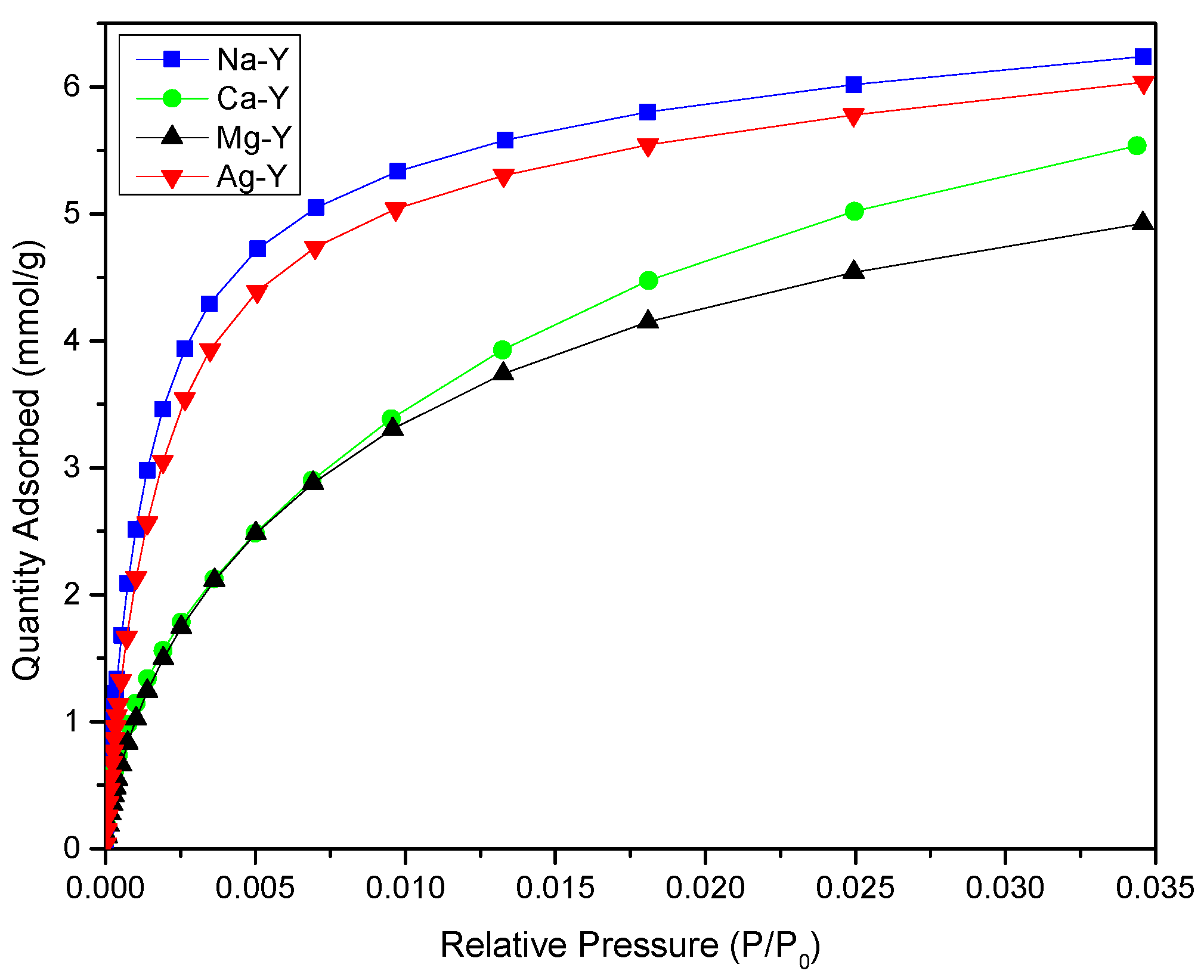
2.3. Gas-Phase Adsorption Studies

The CO2 adsorption results revealed that the amount of CO2 adsorbed per gram of zeolite diminished from Na to Mg with the following given order: Na-Y > Ag-Y > Ca-Y > Mg-Y (Figure 5). This decrease in CO2 adsorption can be explained by an increase in the radius for Ca and Mg, which was also observed by Walton et al. [22]. They demonstrated that an increase in the radius of exchanged cations inhibits the adsorption of CO2 molecules within the zeolite framework. Another possible explanation could be, again, the site occupancies of cations, which were discussed previously in the nitrogen adsorption profiles of zeolites. As Ca, Mg, and Ag cations possess different coordination with oxygen atoms, they tend to form clusters or other structures, which is not observed for Na-Y zeolites [36]. This explanation for CO2 adsorption also shows a correlation with the BET surface area of zeolites. The adsorption profile indicated a Type I adsorption isotherm for CO2 molecules, a characteristic adsorption isotherm for zeolites [22]. The decrease in CO2 adsorption per gram of material also showed a correlation with micropore volumes (Table 2). This can be explained by the reduction in pore accessibility for CO2 molecules after cation exchange. A similar trend was also observed by Puchalska et al., who demonstrated a decline in CO2 adsorption from 5.55 mmol/g to 3.58 mmol/g and 3.19 mmol/g for Ca2+- and Mg2+-exchanged FAU-X zeolites, respectively [35]. As zeolites were degassed at 150 °C, water molecules might have remained in the sodalite cages from the FAU structure, thus affecting the CO2 adsorption behavior of the material and possibly cation localization. This effect should be particularly more pronounced for zeolites exchanged with divalent cations, as they have a higher affinity towards water molecules compared to monovalent cations [52]. Similar results were recently observed by Chaib Draa et al., who demonstrated that degassing the zeolites at 150 °C did not yield complete dehydration and affect the localization of K+ cations [53]. It is worth mentioning that the highest adsorption was observed for Na-Y, with the value of 6.2 mmol/g at 1.2 bar. A similar value (5.98 mmol/g) was also obtained by Benariac-Doumal et al., who conducted CO2 adsorption in a low pressure range (0–1 bar) [54]. Although the highest CO2 adsorption per gram of material was observed for Na-Y, an interesting correlation was also obtained by calculating the adsorbed amount of CO2 per specific surface area of the zeolite samples. It was found that the Ag-loaded Y zeolites exhibited the highest value among the zeolites, even though they exhibited a lower surface area (Table 2). This could be explained by the more polarizable properties of Ag+ cations compared to Na+, Mg2+, and Ca2+, which were also observed for the adsorption of propylene by Xiong et al. [55], but this phenomenon is complex and requires careful consideration of various factors, including the pore size of the zeolite, the ionic radius and charge of the cations, the pressure of the gas, and other variables.

3. Materials and Methods

3.1. Characterization

X-ray diffraction patterns were recorded on a Bruker (Bruker AXS GmbH, Karlsruhe, Germany) D8 Advance diffractometer equipped with a Ni detector side filtered Cu Kα radiation (1.5406 Å) over a 2θ range of 5–55°.
SEM images were obtained with a Zeiss (Oberkochen, Germany) Gemini SEM 500 microscope with an FEG Schottky source and an Everhart–Thornley detector. The microscope was operated at different electron high-tension (EHT) voltages ranging from 2 to 6 kV. The EDX analyses of zeolite samples were conducted using the energy-dispersive X-ray spectroscopy (EDS) detector (EDAX Inc., Mahwah, NJ, USA).
X-ray photoelectron spectroscopy was performed using an ultrahigh vacuum (UHV) spectrometer with a VSW SCIENTIFIC (Manchester, UK) hemispherical electron analyzer. An Al Kα hν = 1486.6 eV dual-anode X-ray source served as the incident radiation. The constant pass energy mode was used to obtain both survey and high-resolution spectra, with the pass energy set at 50 eV.
UV–Vis measurements in liquid state were performed with a Perkin-Elmer (Waltham, MA, USA) Lambda 900 spectrophotometer. The solution was added in 1 mm quartz cuvettes and absorption spectra were recorded in the 400–800 nm range. The same device was used for solid zeolite samples to determine the oxidation state of Ag in exchanged zeolite. The characterization was realized by preparing the pellets of zeolite with BaSO4, which was also used as a reference sample for calibration.
The elemental composition of zeolites was characterized by X-ray Fluorescence Spectroscopy (XRF) using Epsilon 3XL (Malvern Panalytical B.V., Almelo, The Netherlands) equipped with a Ag tube. The characterization was realized by mixing 0.114 g of sample with 1.231 g of melting agent (LiBO2) and 0.019 of non-melting agent (LiBr), which was followed by melting this mixture at 1323 K to obtain a homogeneous melting.
Nitrogen adsorption–desorption isotherms were measured at 77 K using ASAP2420 equipment (Micromeritics, Norcross, GA, USA). Prior to analysis, the zeolite samples (100 mg) were outgassed at 150 °C for 5 h to remove water molecules present within the pores.
The real density of the zeolites was obtained using AccuPy (Tokyo, Japan) II Helium Pycnometer (Micromeritics). Prior to the measurements, samples were outgassed ex situ in the degassing unit of ASAP 2420 at 150 °C for 5 h. The samples were quickly transferred into 1 cm sample holders, which were capped with a fritted cap. The density analysis was conducted at 20 °C with 10 cycles of purging and 10 cycles of measurement.

3.2. Synthesis and Cation Exchange of Y Zeolite

All chemicals utilized in this study were used as received from Merck (Boston, MA, USA), TCI (Tokyo, Japan), and ThermoFischer (Waltham, MA, USA).
The synthesis of Y zeolite was performed using a modified conventional method developed by Bernardon et al. [25]. Initially, a seed gel was prepared by dissolving 2.035 g of NaOH and 1.045 g of NaAlO2 in 10 mL of distilled water (DW) in a Teflon bottle. Upon complete dissolution of the solutes, 8.17 mL of sodium silicate solution was added dropwise and stirred at 1000 rpm for 10 min. The bottle was then capped, and the solution was allowed to age at room temperature for 24 h.
Subsequently, the feedstock gel was prepared in a new Teflon-lined reactor by dissolving 23 mg of NaOH and 2.182 g of NaAlO2 in 22 mL of DW. Once the solution became transparent, 17 mL of Na2SiO3 solution was added dropwise while stirring vigorously. After homogenizing the feedstock, 2.75 g of seed gel was slowly added, and the mixture was stirred vigorously for 20 min before allowing the solution to incubate at room temperature for 24 h.
The overall gel was then heated at 100 °C for 405 min in an oven. The resulting product was filtered, washed until reaching pH 7, and dried overnight at 100 °C.
Calcium- and magnesium-exchanged zeolites were prepared according to the following procedure: 1.5 g of Na-Y zeolite was suspended in 200 mL of 1M CaCl2 × 2 H2O and MgCl2 × 6 H2O solutions, and stirred for 2 h at 80 °C [26]. The solid was washed and dried at 80 °C for 48 h. This process was repeated twice to enhance the amount of Ca and Mg loading in the zeolite structure.
For silver ion exchange, 1 g of Na-Y zeolite was mixed with 25 mL of 0.05 M AgNO3 solution, and the suspension was stirred for 24 h at 60 °C. The product was then dried at 50 °C for 12 h. In this procedure, the cation exchange was not repeated twice to avoid the formation of silver nanoclusters [33].

3.3. Adsorption of Dye Molecules

The MB adsorption of zeolites was realized at room temperature under dark conditions. The adsorption studies were realized by suspending 10 mg of zeolite in 10 mL of MB solution, which was followed by sonication for 30 s. Then, the suspension was left at room temperature in dark conditions for 24 h. The suspensions were centrifuged at 3000 rpm for 3 min at a given time, and the supernatant was analyzed by a UV–Visible spectrophotometer. The adsorption studies were also conducted for anionic dye—Alizarin Yellow R—by suspending 10 mg of zeolite in 10 mg/L of dye solution.
The kinetic studies were conducted by immersing 10 mg of zeolite in 10 mL of MB solution at the corresponding concentrations. The concentration of the solution was monitored by measuring the absorbance of the sample after separating the zeolites by centrifugation (3000 rpm for 3 min), and data were collected at various time intervals over 24 h. Pseudo-first order and pseudo-second order models were used for kinetic modeling.
The isothermal modeling was realized using the adsorption data obtained after a 24 h contact time. The data were fitted with the Langmuir and Freundlich models. The reaction order was calculated by fitting the adsorption data with the reaction rate law. The calculations for the kinetic and isothermal modelling and reaction order, as well as the fitting graphs, are given in the Supplementary Information.

3.4. CO2 Adsorption Studies

CO2 adsorption studies were conducted using ASAP 2020 Micromeritics equipment. Before analysis, the samples were outgassed at 150 °C for 5 h. The analysis was performed at 0 °C in a pressure range between 0 and 1.2 bar.

4. Conclusions

In this study, Y zeolite modified with various cations, including Na+, Mg2+, Ca2+, and Ag+, was characterized using several techniques, and its adsorption properties were assessed both in the liquid and gas phases. Interestingly, for MB adsorption in aqueous media, Ag-exchanged zeolite demonstrated the best result in the absence of oxidizing agents under dark conditions. The kinetics and isothermal models revealed that Ag-Y zeolites showed the highest adsorption capacities and fast adsorption kinetics compared to Na-Y zeolite. This promising result was also followed in the gas-phase CO2 adsorption, where Ag-Y zeolite showed the highest adsorption value per m2 of the samples. It is worth mentioning that these fast adsorption kinetics were obtained with a small amount of silver cations in zeolites, which creates interest in zeolites for dye degradation. Except for Ag-Y, Na-Y showed the highest results in both MB adsorption in aqueous media and CO2 adsorption compared to zeolites loaded with divalent cations. This study, along with others, underscores the importance of the cation exchange process in zeolites. In particular, the high MB removal capacity of Ag-Y without oxidizing agents and light opens new horizons for dispersing other active metals within the surface of zeolites for dye removal studies.

Supplementary Materials

The following supporting information can be downloaded at: https://www.mdpi.com/article/10.3390/catal15080734/s1, Figure S1: SEM-EDX results of silver-exchanged zeolite; Figure S2: Calibration curves of cationic and anionic dye molecules; Figure S3: The absorption spectra of zeolites in anionic dye solution; Figure S4: The adsorption spectra of Na-Y suspended in MB with different concentrations; Figure S5: The absorption spectra of methylene blue at different concentrations by Ag-Y; Figure S6: (a) PFO and (b) PSO ≤60 min and (c) PSO ≤ 1440 min models for Ag-Y; Figure S7: (a) PFO and (b) PSO models for Na-Y; Figure S8: Langmuir and Freundlich modeling of MB adsorption on Na-Y and Ag-Y; Figure S9: Log (Initial Rate) versus log (Initial concentration, C0) plot for (a) Na-Y and (b) Ag-Y. The slope of the reaction corresponds to the reaction order of MB removal; Figure S10: EDX Mapping of (a) Ca; (b) Mg; (c) Na; and (d) Ag on corresponding zeolites; Figure S11: XPS results of Ag-Y zeolite at (a) 3d region and (b) Auger region; Figure S12: UV–Visible absorption spectra of Ag-Y and Na-Y zeolites; Figure S13: The photographic illustration of Ag-Y before and after degassing at 250 °C for 5 h under vacuum condition. The color of the sample turns into black, indicating the reduction of silver within zeolite structure; Figure S14: UV–Visible absorption spectra of Ag-Y after degassing at 150 °C and 250 °C; Table S1: The true density of zeolites obtained by Helium Pycnometer.

Author Contributions

Conceptualization, B.Z. and B.L.; methodology, B.Z.; validation, B.Z. and B.L.; formal analysis, B.Z.; investigation, B.Z.; resources, B.Z., S.F., and N.I.; data curation, B.Z.; writing—original draft preparation, B.Z.; writing—review and editing, B.Z., B.L., and N.I.; visualization, B.Z.; supervision, B.L., N.G., and G.L.; project administration, B.L.; funding acquisition, B.Z. All authors have read and agreed to the published version of the manuscript.

Funding

The project was funded by the University of Strasbourg and the Azerbaijan State Oil and Industry University through a grant of B. Zarbaliyev for his doctoral studies.

Data Availability Statement

All data including calculations are available in main text and Supplementary Materials.

Acknowledgments

B. Zarbaliyev is grateful for funding from the Azerbaijan State Oil and Industry University and the University of Strasbourg for the grant. The authors are thankful to Thierry Romero and Vasiliki Papaefthimiou for their technical assistance.

Conflicts of Interest

The authors declare no conflicts of interest.

Abbreviations

The following abbreviations are used in this manuscript:
MBMethylene Blue
XRDX-ray Diffraction
BETBrunauer–Emmett–Teller
SEMScanning Electron Microscopy

References

  1. El-Bery, H.M.; Saleh, M.; El-Gendy, R.A.; Saleh, M.R.; Thabet, S.M. High Adsorption Capacity of Phenol and Methylene Blue Using Activated Carbon Derived from Lignocellulosic Agriculture Wastes. Sci. Rep. 2022, 12, 5499. [Google Scholar] [CrossRef]
  2. Li, Z.; Sellaoui, L.; Gueddida, S.; Dotto, G.L.; Ben Lamine, A.; Bonilla-Petriciolet, A.; Badawi, M. Adsorption of Methylene Blue on Silica Nanoparticles: Modelling Analysis of the Adsorption Mechanism via a Double Layer Model. J. Mol. Liq. 2020, 319, 114348. [Google Scholar] [CrossRef]
  3. Israfilov, N.; Soukup, K.; Louis, B.; Planeix, J.-M. MOF Side Chains as Sources of Supramolecular Interactions: Organic Pollutant Extraction from Water. New J. Chem. 2022, 46, 8967–8970. [Google Scholar] [CrossRef]
  4. Becker, A.; Israfilov, N.; Ehrstein, E.; Lara-Ibeas, I.; Planeix, J.-M.; Louis, B.; Le Calvé, S. Adsorption of Gaseous Formaldehyde on Y Zeolites and on Metal-Organic Frameworks. Microporous Mesoporous Mater. 2022, 343, 112136. [Google Scholar] [CrossRef]
  5. Deshpande, S.; Kheur, S.; Kheur, M.; Eyüboğlu, T.F.; Özcan, M. A Review on Zeolites and Their Applications in Dentistry. Curr. Oral Health Rep. 2023, 10, 36–42. [Google Scholar] [CrossRef]
  6. Tufail, M.K.; Ifrahim, M.; Rashid, M.; Ul Haq, I.; Asghar, R.; Uthappa, U.T.; Selvaraj, M.; Kurkuri, M. Chemistry of Zeolites and Zeolite Based Composite Membranes as a Cutting-Edge Candidate for Removal of Organic Dyes & Heavy Metal Ions: Progress and Future Directions. Sep. Purif. Technol. 2025, 354, 128739. [Google Scholar] [CrossRef]
  7. Tao, Z.; Tian, Y.; Hanif, A.; Chan, V.; Gu, Q.; Shang, J. Metal Cation-Exchanged LTA Zeolites for CO2/N2 and CO2/CH4 Separation: The Roles of Gas-Framework and Gas-Cation Interactions. Carbon Capture Sci. Technol. 2023, 8, 100126. [Google Scholar] [CrossRef]
  8. Derbe, T.; Temesgen, S.; Bitew, M. A Short Review on Synthesis, Characterization, and Applications of Zeolites. Adv. Mater. Sci. Eng. 2021, 2021, 6637898. [Google Scholar] [CrossRef]
  9. Oladoye, P.O.; Ajiboye, T.O.; Omotola, E.O.; Oyewola, O.J. Methylene Blue Dye: Toxicity and Potential Elimination Technology from Wastewater. Results Eng. 2022, 16, 100678. [Google Scholar] [CrossRef]
  10. Asefa, M.T.; Feyisa, G.B. Comparative Investigation on Two Synthesizing Methods of Zeolites for Removal of Methylene Blue from Aqueous Solution. Int. J. Chem. Eng. 2022, 2022, 9378712. [Google Scholar] [CrossRef]
  11. Le, T.P.; Luong, H.V.T.; Nguyen, H.N.; Pham, T.K.T.; Trinh Le, T.L.; Tran, T.B.Q.; Ngo, T.N.M. Insight into Adsorption-Desorption of Methylene Blue in Water Using Zeolite NaY: Kinetic, Isotherm and Thermodynamic Approaches. Results Surf. Interfaces 2024, 16, 100281. [Google Scholar] [CrossRef]
  12. Majid, Z.; AbdulRazak, A.A.; Noori, W.A.H. Modification of Zeolite by Magnetic Nanoparticles for Organic Dye Removal. Arab. J. Sci. Eng. 2019, 44, 5457–5474. [Google Scholar] [CrossRef]
  13. Zhu, J.; Wang, Y.; Liu, J.; Zhang, Y. Facile One-Pot Synthesis of Novel Spherical Zeolite–Reduced Graphene Oxide Composites for Cationic Dye Adsorption. Ind. Eng. Chem. Res. 2014, 53, 13711–13717. [Google Scholar] [CrossRef]
  14. Sheemol, V.N.; Tyagi, B.; Jasra, R.V. Acylation of Toluene Using Rare Earth Cation Exchanged Zeolite β as Solid Acid Catalyst. J. Mol. Catal. A: Chem. 2004, 215, 201–208. [Google Scholar] [CrossRef]
  15. Sun, Y.; Tang, J.; Li, G.; Hua, Y.; Sun, Y.; Hu, S.; Wen, X. Adsorption, Separation and Regeneration of Cation-Exchanged X Zeolites for LNG Purification: Li+, K+, Mg2+ and Ca2+. Microporous Mesoporous Mater. 2022, 340, 112032. [Google Scholar] [CrossRef]
  16. González-Crisostomo, J.C.; López-Juárez, R.; Yocupicio-Gaxiola, R.I.; Villanueva, E.; Zavala-Flores, E.; Petranovskii, V. Chabazite Synthesis and Its Exchange with Ti, Zn, Cu, Ag and Au for Efficient Photocatalytic Degradation of Methylene Blue Dye. Int. J. Mol. Sci. 2022, 23, 1730. [Google Scholar] [CrossRef]
  17. Chowdhury, S.; Bhattacharyya, K.G. Use of Cu(II)-Incorporated Zeolite Y for Decolourization of Dyes in Water: A Case Study with Aqueous Methylene Blue and Congo Red. SN Appl. Sci. 2019, 1, 87. [Google Scholar] [CrossRef]
  18. Colar, L.A.; Jakab, A.; Manea, F.; Pode, R.; Orha, C. Photocatalytic Performance of Ag-Modified Natural Zeolite Catalyst for Photocatalysis Degradation of Methylene Blue (MB) Under VIS Irradiation; WIT Press: Southampton, UK, 2012; pp. 335–344. [Google Scholar]
  19. Boer, D.G.; Langerak, J.; Pescarmona, P.P. Zeolites as Selective Adsorbents for CO2 Separation. ACS Appl. Energy Mater. 2023, 6, 2634–2656. [Google Scholar] [CrossRef]
  20. Castillo, J.M.; Silvestre-Albero, J.; Rodriguez-Reinoso, F.; Vlugt, T.J.H.; Calero, S. Water Adsorption in Hydrophilic Zeolites: Experiment and Simulation. Phys. Chem. Chem. Phys. 2013, 15, 17374. [Google Scholar] [CrossRef] [PubMed]
  21. Clatworthy, E.B.; Ghojavand, S.; Guillet-Nicolas, R.; Gilson, J.-P.; Llewellyn, P.L.; Nesterenko, N.; Mintova, S. Dynamic Adsorption of CO2 by CHA Zeolites—Size Matters. Chem. Eng. J. 2023, 471, 144557. [Google Scholar] [CrossRef]
  22. Walton, K.S.; Abney, M.B.; Douglas LeVan, M. CO2 Adsorption in Y and X Zeolites Modified by Alkali Metal Cation Exchange. Microporous Mesoporous Mater. 2006, 91, 78–84. [Google Scholar] [CrossRef]
  23. Yang, S.-T.; Kim, J.; Ahn, W.-S. CO2 Adsorption over Ion-Exchanged Zeolite Beta with Alkali and Alkaline Earth Metal Ions. Microporous Mesoporous Mater. 2010, 135, 90–94. [Google Scholar] [CrossRef]
  24. Tao, Z.; Tian, Y.; Ou, S.Y.; Gu, Q.; Shang, J. Direct Air Capture of CO2 by Metal Cation-exchanged LTA Zeolites: Effect of the Charge-to-size Ratio of Cations. AIChE J. 2023, 69, e18139. [Google Scholar] [CrossRef]
  25. Bernardon, C.; Louis, B.; Bénéteau, V.; Pale, P. Diels-Alder Reaction between Isoprene and Methyl Acrylate over Different Zeolites: Influence of Pore Topology and Acidity. ChemPlusChem 2013, 78, 1134–1141. [Google Scholar] [CrossRef] [PubMed]
  26. Lutzweiler, G.; Zhang, Y.; Gens, F.; Echalard, A.; Ladam, G.; Hochart, J.; Janicot, T.; Mofaddel, N.; Louis, B. Deciphering the Role of Faujasite-Type Zeolites as a Cation Delivery Platform to Sustain the Functions of MC3T3-E1 Pre-Osteoblastic Cells. Mater. Adv. 2022, 3, 8616–8628. [Google Scholar] [CrossRef]
  27. Dyer, A. Ion-Exchange Properties of Zeolites and Related Materials. In Studies in Surface Science and Catalysis; Elsevier: Amsterdam, The Netherlands, 2007; Volume 168, pp. 525–553. ISBN 978-0-444-53063-9. [Google Scholar]
  28. Dyer, A. Ion Exchange Capacity. In Verified Syntheses of Zeolitic Materials; Elsevier: Amsterdam, The Netherlands, 2001; pp. 67–68. ISBN 978-0-444-50703-7. [Google Scholar]
  29. Gellens, L.R.; Mortier, W.J.; Uytterhoeven, J.B. Oxidation and Reduction of Silver in Zeolite Y: A Structural Study. Zeolites 1981, 1, 85–90. [Google Scholar] [CrossRef]
  30. Shameli, K.; Mansor Bin Ahmad, M.; Mohsen, Z.; Yunis, W.Z.; Ibrahim, N.A. Fabrication of Silver Nanoparticles Doped in the Zeolite Framework and Antibacterial Activity. Int. J. Nanomed. 2011, 6, 331–341. [Google Scholar] [CrossRef]
  31. Min, J.G.; Kemp, K.C.; Hong, S.B. Silver ZK-5 Zeolites for Selective Ethylene/Ethane Separation. Sep. Purif. Technol. 2020, 250, 117146. [Google Scholar] [CrossRef]
  32. Yu, J.; Ye, S.; Xv, X.; Pan, L.; Lin, P.; Liao, H.; Wang, D. Thermal-Driven Formation of Silver Clusters Inside Na/Li FAUY Zeolites for Formaldehyde Detection. Nanomaterials 2022, 12, 3215. [Google Scholar] [CrossRef] [PubMed]
  33. Chen, S.; Popovich, J.; Iannuzo, N.; Haydel, S.E.; Seo, D.-K. Silver-Ion-Exchanged Nanostructured Zeolite X as Antibacterial Agent with Superior Ion Release Kinetics and Efficacy against Methicillin-Resistant Staphylococcus aureus. ACS Appl. Mater. Interfaces 2017, 9, 39271–39282. [Google Scholar] [CrossRef]
  34. Min, J.G.; Kemp, K.C.; Kencana, K.S.; Hong, S.B. Silver-Exchanged CHA Zeolite as a CO2-Resistant Adsorbent for N2/O2 Separation. Microporous Mesoporous Mater. 2021, 323, 111239. [Google Scholar] [CrossRef]
  35. Gęsikiewicz-Puchalska, A.; Zgrzebnicki, M.; Michalkiewicz, B.; Kałamaga, A.; Narkiewicz, U.; Morawski, A.W.; Wrobel, R. Changes in Porous Parameters of the Ion Exchanged X Zeolite and Their Effect on CO2 Adsorption. Molecules 2021, 26, 7520. [Google Scholar] [CrossRef]
  36. Frising, T.; Leflaive, P. Extraframework Cation Distributions in X and Y Faujasite Zeolites: A Review. Microporous Mesoporous Mater. 2008, 114, 27–63. [Google Scholar] [CrossRef]
  37. Pérez-Botella, E.; Valencia, S.; Rey, F. Zeolites in Adsorption Processes: State of the Art and Future Prospects. Chem. Rev. 2022, 122, 17647–17695. [Google Scholar] [CrossRef] [PubMed]
  38. Nguyen, H.G.T.; Tao, R.; Van Zee, R.D. Porosity, Powder X-Ray Diffraction Patterns, Skeletal Density, and Thermal Stability of NIST Zeolitic Reference Materials RM 8850, RM 8851, and RM 8852. J. Res. Natl. Inst. Stan. 2021, 126, 126047. [Google Scholar] [CrossRef]
  39. Pan, L.; Ye, S.; Xv, X.; Lin, P.; Huang, R.; Wang, D. Zeolite-Encaged Luminescent Silver Nanoclusters. Materials 2023, 16, 3736. [Google Scholar] [CrossRef]
  40. Aspromonte, S.G.; Romero, A.; Boix, A.V.; Alonso, E. Hydrolysis of Cellulose to Glucose by Supercritical Water and Silver Mesoporous Zeolite Catalysts. Cellulose 2019, 26, 2471–2485. [Google Scholar] [CrossRef]
  41. Wang, L.; Lin, C.; Boldog, I.; Yang, J.; Janiak, C.; Li, J. Inverse Adsorption Separation of N2 O/CO2 in AgZK-5 Zeolite. Angew. Chem. Int. Ed. 2024, 63, e202317435. [Google Scholar] [CrossRef]
  42. Zheng, Y.; Vidal-Moya, A.; Hernández-Garrido, J.C.; Mon, M.; Leyva-Pérez, A. Silver-Exchanged Zeolite Y Catalyzes a Selective Insertion of Carbenes into C–H and O–H Bonds. J. Am. Chem. Soc. 2023, 145, 24736–24745. [Google Scholar] [CrossRef] [PubMed]
  43. Lopes, A.C.; Silva, M.P.; Gonçalves, R.; Pereira, M.F.R.; Botelho, G.; Fonseca, A.M.; Lanceros-Mendez, S.; Neves, I.C. Enhancement of the Dielectric Constant and Thermal Properties of α-Poly(Vinylidene Fluoride)/Zeolite Nanocomposites. J. Phys. Chem. C 2010, 114, 14446–14452. [Google Scholar] [CrossRef]
  44. Vanaja, M.; Paulkumar, K.; Baburaja, M.; Rajeshkumar, S.; Gnanajobitha, G.; Malarkodi, C.; Sivakavinesan, M.; Annadurai, G. Degradation of Methylene Blue Using Biologically Synthesized Silver Nanoparticles. Bioinorg. Chem. Appl. 2014, 2014, 742346. [Google Scholar] [CrossRef]
  45. Pearson, R.G. Hard and Soft Acids and Bases. J. Am. Chem. Soc. 1963, 85, 3533–3539. [Google Scholar] [CrossRef]
  46. Zharylkan, S.; Sultakhan, S.; Suleimenova, M.; Seitkhan, A.; Sailaukhanuly, Y.; Tulepov, M.; Tauanov, Z. Enhanced Adsorption of Mercury and Methylene Blue Using Silver and Surface Modified Zeolite-NaX Derived from Rice Husk. Eng. Sci. 2024, 31, 1241. [Google Scholar] [CrossRef]
  47. Awala, H.; Leite, E.; Saint-Marcel, L.; Clet, G.; Retoux, R.; Naydenova, I.; Mintova, S. Properties of Methylene Blue in the Presence of Zeolite Nanoparticles. New J. Chem. 2016, 40, 4277–4284. [Google Scholar] [CrossRef]
  48. Salimi, F.; Eskandari, M.; Karami, C. Investigation of Methylene Blue Adsorption in Wastewater Using Nano-Zeolite Modified with Copper. Desalin. Water Treat. 2017, 85, 206–214. [Google Scholar] [CrossRef]
  49. Hadda Aya, H.; Djamel, N.; Samira, A.; Otero, M.; Ali Khan, M. Optimizing Methylene Blue Adsorption Conditions on Hydrothermally Synthesized NaX Zeolite through a Full Two-Level Factorial Design. RSC Adv. 2024, 14, 23816–23827. [Google Scholar] [CrossRef]
  50. Nyankson, E.; Adjasoo, J.; Efavi, J.K.; Amedalor, R.; Yaya, A.; Manu, G.P.; Asare, K.; Amartey, N.A. Characterization and Evaluation of Zeolite A/Fe3O4 Nanocomposite as a Potential Adsorbent for Removal of Organic Molecules from Wastewater. J. Chem. 2019, 2019, 8090756. [Google Scholar] [CrossRef]
  51. Boudjema, L.; Assaf, M.; Salles, F.; Gassin, P.-M.; Martin-Gassin, G.; Zajac, J. Renewing Interest in Zeolites as Adsorbents for Capture of Cationic Dyes from Aqueous and Ethanolic Solutions: A Simulation-Based Insight into the Efficiency of Dye Adsorption in View of Wastewater Treatment and Valorization of Post-Sorption Materials. Molecules 2024, 29, 2952. [Google Scholar] [CrossRef]
  52. Jiao, D.; King, C.; Grossfield, A.; Darden, T.A.; Ren, P. Simulation of Ca2+ and Mg2+ Solvation Using Polarizable Atomic Multipole Potential. J. Phys. Chem. B 2006, 110, 18553–18559. [Google Scholar] [CrossRef] [PubMed]
  53. Chaib Draa, Y.M.; Ors, T.; Deroche, I.; Dejoie, C.; Paillaud, J.-L. 3D ED for the Localization of Cations in Potassium Exchanged and Partially Dehydrated Nano Y Zeolite†. CrystEngComm 2025, 27, 5095–5099. [Google Scholar] [CrossRef]
  54. Bénariac-Doumal, L.; Deroche, I.; Nouali, H.; Paillaud, J.-L.; Ors, T.; Fitch, A.N.; Dejoie, C. A Comparative Study of CO2 Adsorption in a Series of Zeolites. Phys. Chem. Chem. Phys. 2025, 27, 10621–10634. [Google Scholar] [CrossRef] [PubMed]
  55. Xiong, Y.; Tian, T.; L’Hermitte, A.; Méndez, A.S.J.; Danaci, D.; Platero-Prats, A.E.; Petit, C. Using Silver Exchange to Achieve High Uptake and Selectivity for Propylene/Propane Separation in Zeolite Y. Chem. Eng. J. 2022, 446, 137104. [Google Scholar] [CrossRef]
Figure 1. XRD patterns of cation-exchanged zeolites.
Catalysts 15 00734 g001
Figure 2. N2 adsorption–desorption isotherms of zeolites loaded with different cations.
Catalysts 15 00734 g002
Figure 3. SEM images of (a) Na-Y and (b) Ag-Y zeolites showing neither a change in crystal morphology nor an agglomeration of silver nanocrystals.
Catalysts 15 00734 g003
Figure 4. MB adsorption on cation-exchanged zeolites (left) after 24 h and the effect of Na-Y and Ag-Y on MB removal from the solution (right).
Catalysts 15 00734 g004
Figure 5. CO2 adsorption isotherms over cation-exchanged zeolites at 0 °C up to 1.2 bar.
Catalysts 15 00734 g005
Table 1. Elemental analysis of zeolites.
ZeoliteSi/Al RatioCation Content (wt%)Exchange Degree (%)
Na-Y2.29.6-
Ca-Y2.16.980
Mg-Y2.23.671
Ag-Y2.23.27
Table 2. Surface area and CO2 adsorption values on cation-exchanged zeolites.
ZeolitesSurface Area (m2/g)Vtotal (cm3/g)Vmicro (cm3/g)Adsorbed CO2 Quantity (mmol/g)CO2 Adsorption Per m2 (µmol/m2)
Na-Y6360.310.296.29.61
Ca-Y5980.300.285.38.86
Mg-Y5600.280.274.68.21
Ag-Y5570.280.265.810.4
Table 3. MB removal percentages of Na-Y and Ag-Y in 5 min and in 24 h.
MB ConcentrationNa-Y 5 Min./24 hAg-Y 5 Min./24 h
10 mg/L66.3%/91.7%94.7%/95.0%
20 mg/L34.1%/46.4%91.1%/94.0%
30 mg/L27.2%/34.3%88.2%/92.0%
50 mg/L25.4%/31.5%59.9%/88.1%
100 mg/L12.8%/17.8%39.6%/69.4%
Table 4. Kinetic modeling parameters of MB adsorption on Na-Y and Ag-Y.
Kinetic ModelConstantsNa-Y
10 mg/L
Na-Y
20 mg/L
Ag-Y
30 mg/L
Ag-Y 50 mg/LAg-Y 100 mg/L
Pseudo-first-orderK1 (L/min)0.0041770.0027910.0708280.0692250.096555
Qe (mg/g)3.4846033.4586843.70193720.95728564.414176
R20.5919320.5489210.6446840.8923140.994152
Pseudo-second-orderK2 (g/mg·min)0.0054480.0035730.1212230.0149240.004202
Qe (mg/g)9.8320539.96465930.22912746.26858771.133793
R20.9990170.9974450.9999940.9999990.999991
Table 5. Isotherm modeling parameters of MB adsorption on Na-Y and Ag-Y.
Isotherm ModelsConstantsNa-Y
10 mg/L
Ag-Y
30 mg/L
Langmuir ModelQm (mg/g)17.9379.38
KL (L/mg)0.3244760.24768
RL0.25670.551
Freundlich ModelR20.9944980.999307
KF (mg/g)9.64598716.62
1/n0.1359090.469567
R20.7595770.945006
Disclaimer/Publisher’s Note: The statements, opinions and data contained in all publications are solely those of the individual author(s) and contributor(s) and not of MDPI and/or the editor(s). MDPI and/or the editor(s) disclaim responsibility for any injury to people or property resulting from any ideas, methods, instructions or products referred to in the content.

