Article

Ultraviolet Photocatalytic Performance of ZnO Nanorods Selectively Deposited with Bi2O3 Quantum Dots

1
Engineering Research Center for New Industrial Water Pollution, Hangzhou 310000, China
2
Zhejiang Energy R&D Institute Co., Ltd., Hangzhou 310000, China
3
Zhejiang Zheneng Co., Ltd., Xiaoshan Power Plants, Hangzhou 311251, China
4
Institute of Corrosion Science and Technology, Guangzhou 510530, China
5
Research Center of Advanced Metastable Metallic Materials, Metallurgy of School, Northeastern University, Shenyang 110819, China
*
Author to whom correspondence should be addressed.
Catalysts 2025, 15(7), 695; https://doi.org/10.3390/catal15070695
Submission received: 17 June 2025 / Revised: 10 July 2025 / Accepted: 17 July 2025 / Published: 21 July 2025
(This article belongs to the Special Issue Advanced Catalytic Processes for Wastewater Treatment)

Abstract

A strong interaction between Bi3+ and ZnO was used to successfully sensitize ZnO nanorods with quantum dots (QDs) of Bi2O3 through three different strategies. Although the Bi2O3 QDs had similar particle size distributions, their photocatalytic performance varied significantly, prompting the investigation of factors beyond particle size. The study revealed that the photochemical method selectively deposited Bi2O3 QDs onto electron-rich ZnO sites, providing a favorable pathway for efficient electron–hole separation and transfer. Consequently, abundant h+ and ·OH radicals were generated, which effectively degraded Rhodamine B (RhB). As demonstrated in the RhB degradation experiments, the Bi2O3/ZnO nanorod catalyst achieved an 89.3% degradation rate within 120 min, significantly outperforming catalysts with other morphologies. The photoluminescence (PL) and time-resolved photoluminescence (TRPL) results indicated that the Bi2O3/ZnO heterostructure constructed an effective interface to facilitate the spatial separation of photogenerated charge carriers, which effectively prolonged their lifetime. The electron paramagnetic resonance (EPR) results confirmed that the ·OH radicals played a key role in the degradation process.

Graphical Abstract

1. Introduction

Under light irradiation, the photogenerated electron–hole pairs in photocatalysts can drive redox reactions, converting organic pollutants into harmless CO2 and H2O [1]. However, the poor visible-light response and rapid recombination of photogenerated charge carriers in current photocatalysts limit their practical applications, resulting in low photocatalytic efficiency. Owing to its low cost [2], low toxicity, unique electronic structure, and excellent physicochemical properties, Bi2O3 has emerged as a promising photocatalyst among various semiconductor materials.
Bi2O3 has a polymorph-dependent bandgap ranging from about 2.1 to 3.66 eV, which allows for light absorption across both the visible and ultraviolet (UV) regions depending on its crystal phase [3]. Nevertheless, it has a significantly reduced number of effective charge carriers and limited photocatalytic performance owing to the rapid recombination of the photogenerated electron–hole pairs. The sensitization of quantum dots (QDs) is a promising strategy for enhancing their photocatalytic activity and addressing this issue [4]. The loading of narrow-bandgap QDs (such as CdS or PbS) onto ZnO surfaces can broaden the light absorption range and form effective heterojunctions between the two materials, which promotes charge separation and substantially improves the photocatalytic activity [5]. Therefore, QD sensitization coupled with structural optimization of the material is a key approach to enhance the photocatalytic performance of Bi2O3- and ZnO-based systems.
As a versatile and efficient strategy, QD sensitization has received increasing attention for increasing the photocatalytic activity of ZnO [6]. Controlling the composition and size of QDs can finely tune their bandgaps, enabling ZnO to absorb a broader light spectrum and improving its photocatalytic efficiency. Furthermore, the high extinction coefficients and prominent intrinsic dipole moments of QDs contribute to the efficient charge separation and enhanced photoelectrochemical activity of ZnO [7]. Among the extensive studies on ZnO sensitized with various QDs, most have focused on chalcogenide- and metal-based QDs [8]. In contrast, owing primarily to the synthetic challenges associated with metal oxide QDs, reports on their use to sensitize ZnO are scarce. QD size is often regarded as a critical factor affecting photocatalytic performance, but other parameters, such as surface chemistry, crystal structure, and interfacial properties with ZnO, have not been fully investigated [9]. Therefore, these findings highlight the need to comprehensively consider multiple factors when designing QD-sensitized ZnO photocatalysts to achieve optimal performance.
This study showcases three distinct in situ strategies to grow Bi2O3 QDs on ZnO nanorods. Interestingly, the resulting Bi2O3 QDs exhibited similar size distributions but significantly different photocatalytic performances. In-depth analysis revealed that an effective heterojunction formed between the Bi2O3 QDs and ZnO, which enabled efficient separation of the photoexcited electrons (e) and holes (h+) via the deposition method. Bi2O3 was loaded onto ZnO nanorods via different deposition techniques to synthesize Bi2O3/ZnO composite photocatalysts. The structural and optical properties of the materials were thoroughly characterized by scanning electron microscopy (SEM), transmission electron microscopy (TEM), scanning transmission electron microscopy (STEM), X-ray diffraction (XRD), photoluminescence (PL), and electron paramagnetic resonance (EPR). The photocatalytic activities of the composites were evaluated through degradation experiments with Rhodamine B (RhB), and a photocatalytic mechanism for RhB degradation over the Bi2O3/ZnO heterostructure was proposed.

2. Results

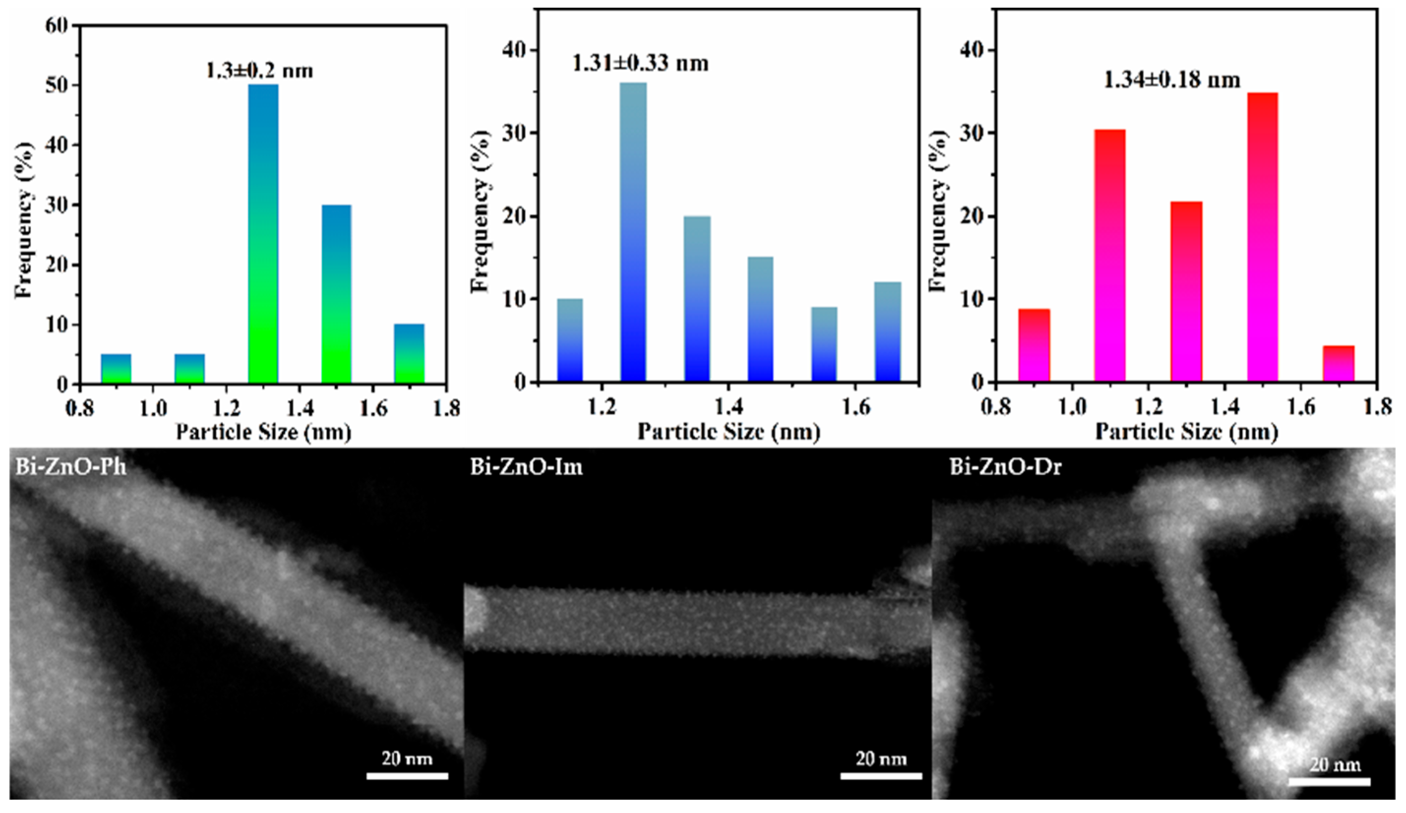
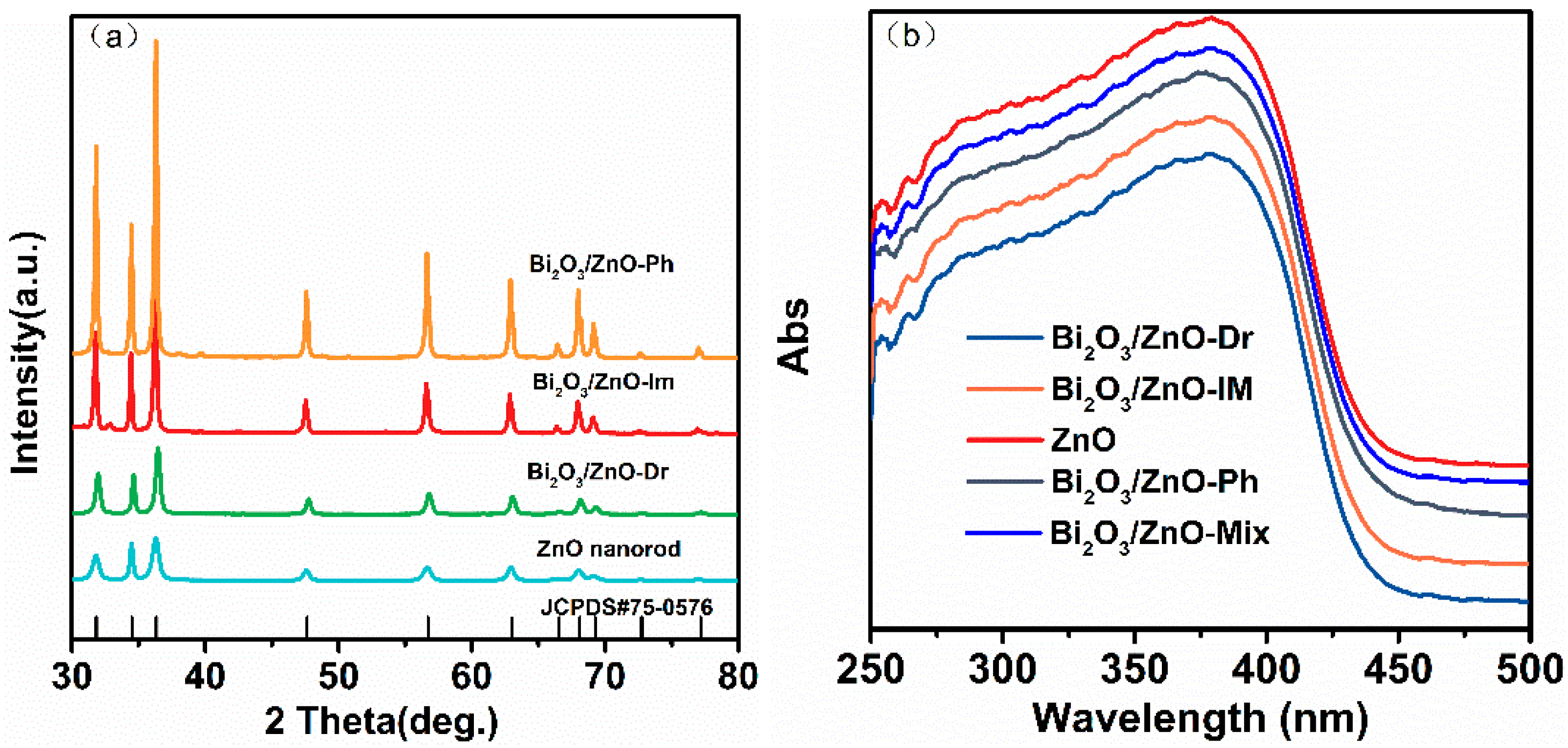
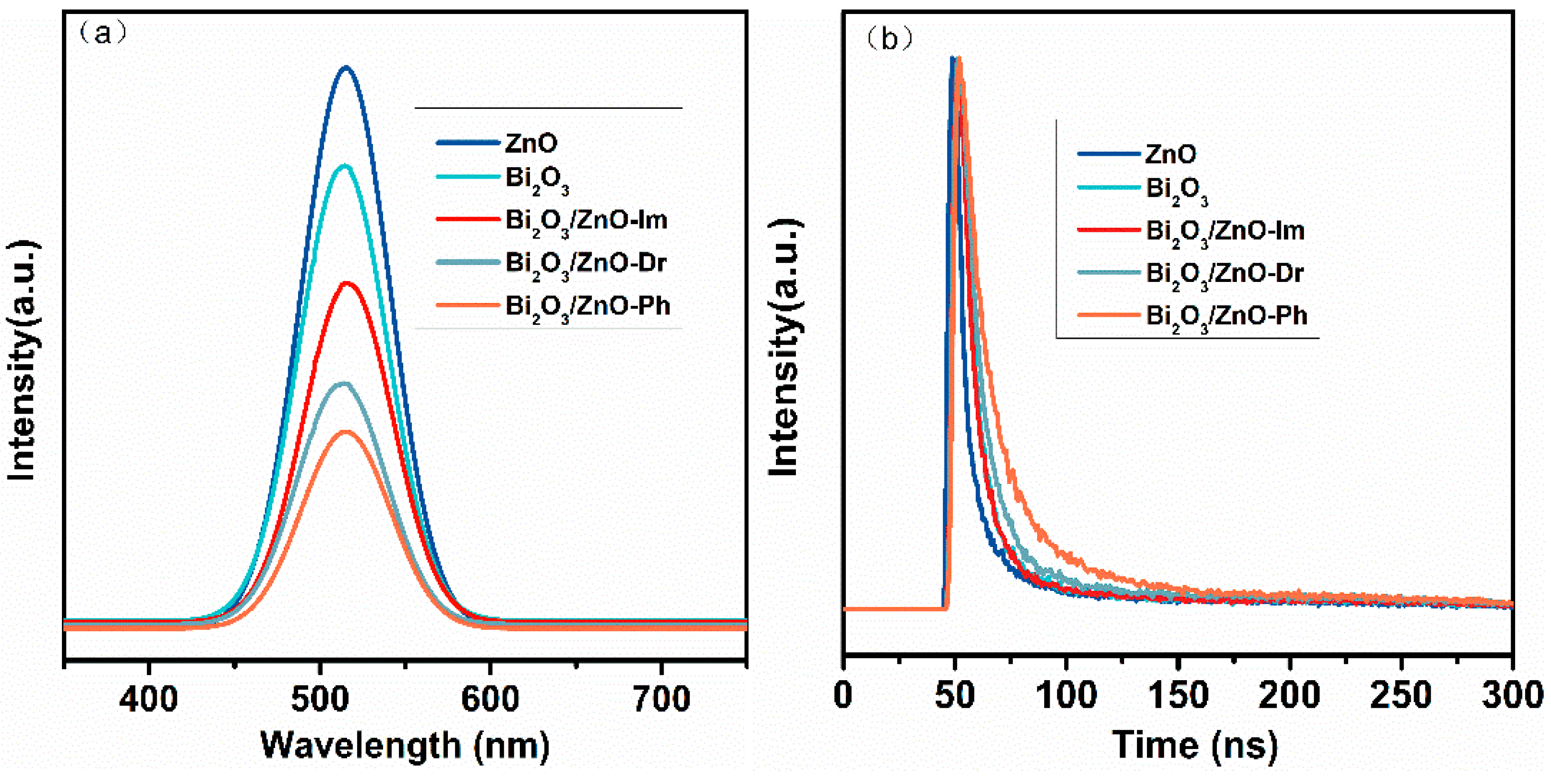
2.1. Structural Characterization

The microscopic morphology of the ZnO nanorods was characterized via TEM. As shown in Figure S1, the ZnO nanorods synthesized via a slightly modified literature-reported method had well-defined and uniform structures. The ZnO nanorods had lengths and diameters in the approximate ranges of 500–1000 nm and 30–50 nm, respectively. These results demonstrated the effectiveness and controllability of the adopted synthesis approach, which enabled the stable formation of the desired nanostructures.
Furthermore, SEM images of the Bi2O3/ZnO composite catalysts synthesized via different routes are presented in Figure 1. The results indicated that the strong interactions between Bi(NO3)3 and the ZnO support promoted the uniform dispersion of bismuth (Bi) on the ZnO surface, resulting in highly dispersed Bi particles. The high degree of dispersion was beneficial for improving the catalytic performance of the Bi2O3–ZnO composites. Statistical particle size analysis revealed that the Bi2O3 particles loaded on ZnO via different synthetic methods had an average size of about 1.3 nm, which fell within the quantum confinement regime defined by an exciton Bohr radius of about 2–10 nm and satisfied the size criteria for QDs.
Additionally, the Bi content of each composite catalyst was quantified via inductively coupled plasma-mass spectrometry (ICP-MS). The Bi loadings for the composites with nanoparticle, nanorod, and nanorod supports were 4.6, 4.8, and 4.7 wt.%, respectively. These results indicated that the different synthesis methods yielded nearly identical Bi contents and particle distributions, which closely matched the theoretical loading values. Moreover, this consistency underscores the high reproducibility and reliability of the employed synthetic strategies. Collectively, these characterization results provide a solid experimental foundation for subsequent photocatalytic performance evaluations and further confirm the effectiveness of the synthesis methods and structural stability of the resulting catalysts.
The chemical compositions and crystal structures of the samples were investigated via XRD. As shown in Figure 2a, the XRD pattern of the ZnO sample had characteristic diffraction peaks, which corresponded to the hexagonal wurtzite structure. The prominent diffraction peaks were at 2θ values of 31.7°, 34.4°, 36.2°, 47.5°, 56.6°, 62.8°, 66.3°, 68.0°, and 69.1° and could be indexed to the (100), (002), (101), (102), (110), (103), (200), (112), and (201) crystal planes, respectively. The (002) plane at 34.4° had the highest intensity, indicating that the ZnO crystallites had a preferred orientation along the c-axis direction. The Bi-loaded ZnO samples with ZnO nanorods as the support showed no distinct diffraction peaks corresponding to Bi species in the XRD patterns, which could be attributed to the high dispersion and ultrasmall size of the Bi2O3 nanoparticles, which likely fell below the XRD detection limit. This observation was consistent with the particle size distribution results obtained via electron microscopy, which further supported the formation of well-dispersed Bi QDs on the ZnO surface.
To further evaluate the crystalline characteristics, the crystallite sizes of the ZnO and Bi2O3/ZnO composites were estimated via the Scherrer equation on the basis of the (101) diffraction peak of ZnO at 2θ ≈ 36.2°:
D = K λ β C O S θ
where K is the shape factor (0.9), λ is the X-ray wavelength (0.15406 nm), β is the full width at half maximum (FWHM), and θ is the Bragg angle. The calculated crystallite sizes are about 24.5 nm for ZnO, 25.2 nm for Bi2O3/ZnO-Dr, 27.6 nm for Bi2O3/ZnO-Im, and 29.1 nm for Bi2O3/ZnO-Ph. Compared with those of the pure ZnO and Bi2O3/ZnO-Dr samples, the significantly increased diffraction peak intensities observed in the Bi2O3/ZnO-Im and Bi2O3/ZnO-Ph samples can be attributed to an increase in the crystallite size and improved crystallinity. The immersion and photochemical deposition methods likely promote partial recrystallization or better alignment of the ZnO nanorods, reducing structural defects and enhancing the diffraction signals. A noticeable shift in the (100) reflection near 2θ ≈ 31.7° is observed for the -Im and -Ph samples. This shift may arise from lattice distortion or stress induced by the Bi2O3 loading process. Partial substitution of Zn2+ by larger Bi3+ ions (ionic radii: Bi3+ = 1.03 Å, Zn2+ = 0.74 Å) or interfacial strain at the ZnO-Bi2O3 interface could lead to lattice expansion and a corresponding shift to lower diffraction angles. Moreover, the Bi2O3/ZnO-Im and Bi2O3/ZnO-Ph XRD patterns exhibit weak reflections at about 32.5°, 37°, and 40°, which are not present in pure ZnO. These peaks may correspond to minor crystalline Bi2O3 phases or related derivatives. Specifically, the peak at 32.5° may be assigned to the (201) plane of monoclinic α-Bi2O3 (PDF#41–1449), whereas the reflections near 37° and 40° might originate from Bi-rich subphases such as Bi12O15. The low intensity of these peaks suggests that Bi2O3 is highly dispersed in the composite, likely existing in nanoscale domains or semicrystalline states [10].
As shown in the ultraviolet-visible (UV-vis) absorption spectra in Figure 2b, compared with the physical mixture of commercial Bi2O3 and ZnO, the synthesized composite catalysts exhibited a pronounced blueshift in the absorption edge. The blueshift is commonly regarded as a signature of quantum confinement effects, confirming the presence of Bi2O3 QDs in the composites [11]. The pure ZnO samples had an absorption edge at about 377 nm [12]. In contrast, the absorption edge for the Bi2O3-modified ZnO samples notably shifted toward the UV region, indicating that Bi2O3 QD sensitization significantly enhanced the photosensitivity of ZnO. The enhancement trend was as follows: the Bi2O3/ZnO composite catalyst was synthesized via photochemical deposition (Bi2O3/ZnO-Ph) > the Bi2O3/ZnO composite catalyst was synthesized via impregnation (Bi2O3/ZnO-Im) > the Bi2O3/ZnO composite catalyst was synthesized via deposition-reduction (Bi2O3/ZnO-Dr), suggesting that the interfacial interaction between Bi2O3 and ZnO was strongly influenced by the method used to generate Bi2O3 QDs. Furthermore, these interfacial and structural differences determine the degree of sensitization achieved [13]. Therefore, these findings offer critical insights for optimizing synthesis strategies and improving visible-light harvesting efficiency in future photocatalyst designs.
In contrast to the previous assumption of a blueshift based solely on visual comparison, a more rigorous analysis shows that the absorption edges of the Bi2O3/ZnO composites do not significantly shift along the x-axis (wavelength) but instead exhibit enhanced absorption intensities along the y-axis. This suggests that the introduction of Bi2O3 QDs enhances the overall light-harvesting capacity rather than shifting the band edge.
To clarify the electronic structure, the optical bandgap energy (Eg) of the samples was calculated via Tauc plots derived from the UV-vis diffuse reflectance spectra. For direct bandgap semiconductors, the Tauc relation is given as follows:
(αhν)2 = A( − Eg)
where α is the absorption coefficient, hν is the photon energy, and A is a constant. The extrapolation of the linear portion of the Tauc plot to the x-axis (where (αhν)2 = 0) yields the bandgap value. The estimated bandgap energies are about 3.23 eV for ZnO, 3.21 eV for Bi2O3/ZnO-Dr, 3.20 eV for Bi2O3/ZnO-Im, and 3.18 eV for Bi2O3/ZnO-Ph. These results confirm that the incorporation of Bi2O3 QDs has a minimal effect on the fundamental bandgap of ZnO. Instead, the observed increase in light absorption intensity is attributed to improved charge separation and additional sub-bandgap transitions at the Bi2O3-ZnO heterojunction interface. Therefore, the improvement in photocatalytic performance is not driven by bandgap narrowing but rather by interfacial charge dynamics.

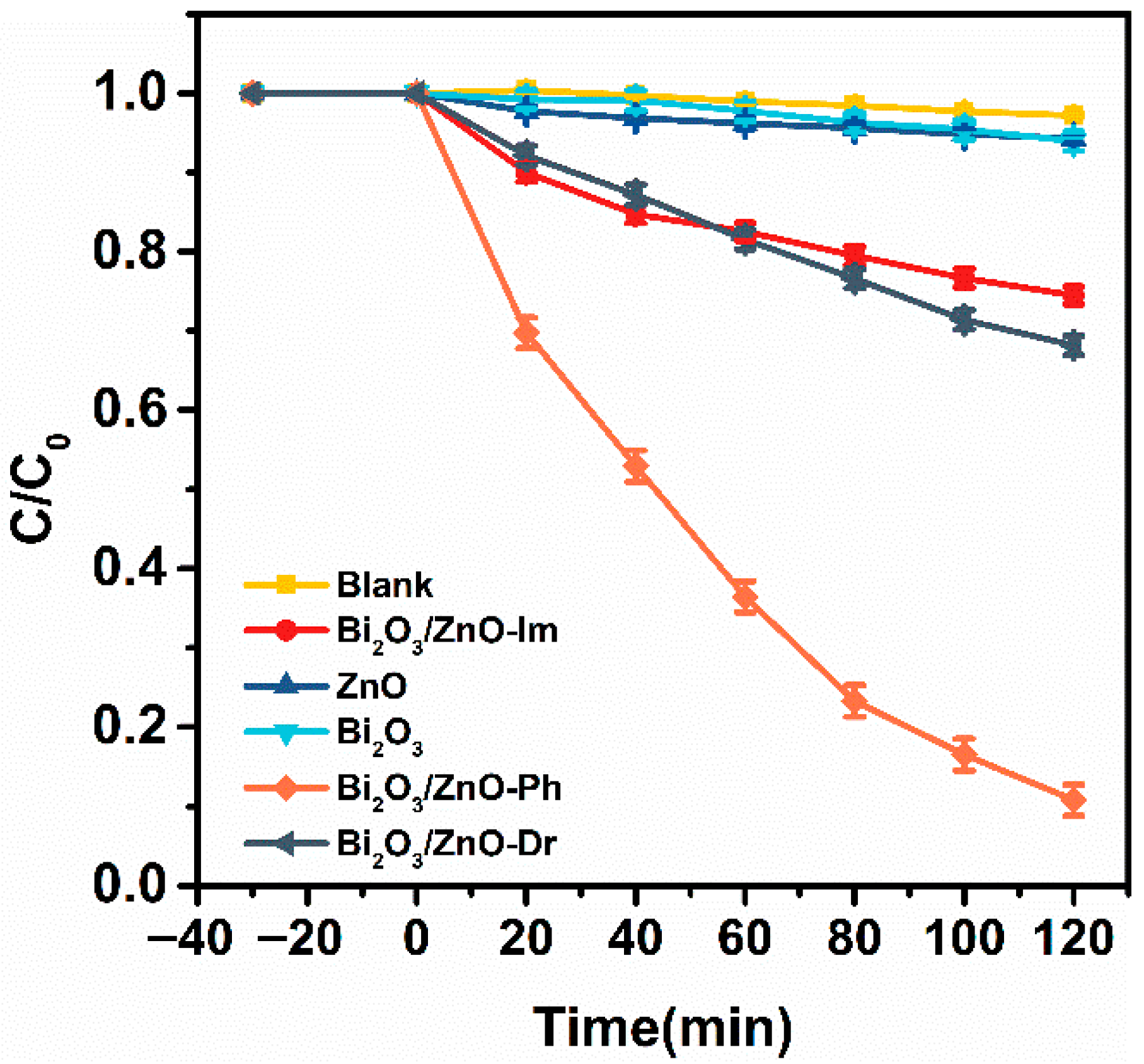
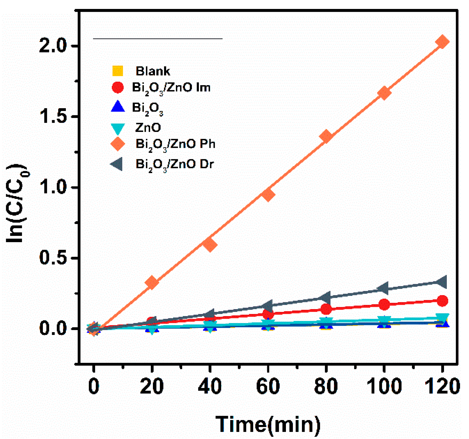
2.2. Photocatalytic Degradation Performance Analysis of RhB

The photocatalytic degradation performance of RhB under UV irradiation was evaluated using UV-vis absorption spectroscopy [14]. As illustrated in Figure 3, the RhB solution remained stable during the 2 h dark stirring phase before illumination, indicating that the adsorption–desorption equilibrium was established without any significant degradation. Upon exposure to UV light, RhB was negligibly degraded within the first 30 min in the absence of a catalyst, confirming that RhB did not undergo photolysis under UV light alone. After 120 min of UV illumination, pure ZnO and Bi2O3 showed limited photocatalytic activity and only 4.1% and 6.1% RhB degradation efficiencies, respectively. In contrast, the 5 wt.% Bi2O3/ZnO composite catalysts synthesized via impregnation and deposition-reduction methods exhibited enhanced performance and achieved 26.6% and 31.9% RhB degradation, respectively. Therefore, these results demonstrated that Bi2O3 loading effectively improved the photocatalytic activity of ZnO.
Notably, the Bi2O3/ZnO-Ph composite exhibited superior performance, achieving substantially greater RhB degradation of 89.3% within the same duration compared to Bi2O3/ZnO-Im and Bi2O3/ZnO-Dr. Furthermore, a pseudo-first-order model was used to fit the degradation kinetics, and the apparent reaction rate constants (k) of the different catalysts were compared, as shown in Figure 4. The Bi2O3/ZnO-Ph catalyst exhibited the highest k value of 1.02 h−1, which was 9.4 and 6.8 times greater than those of Bi2O3/ZnO-Im (0.109 h−1) and Bi2O3/ZnO-Dr (0.15 h−1), respectively. Therefore, these findings further highlighted that the photocatalytic efficiency was highly dependent on the synthesis method employed.

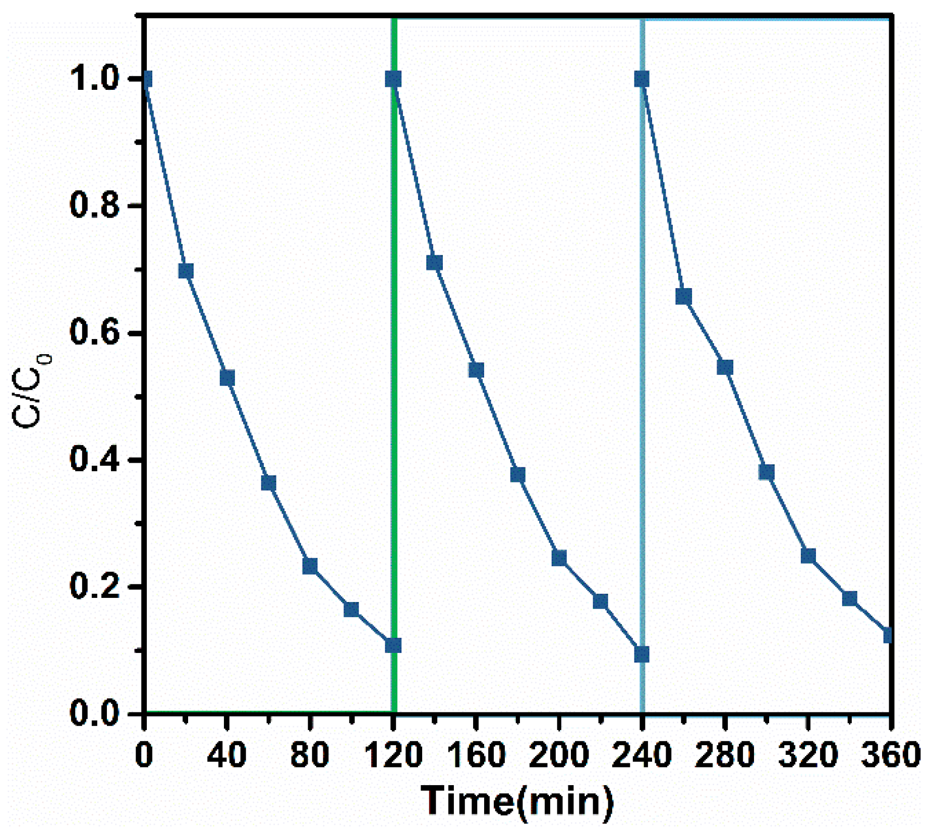
2.3. Photocatalyst Stability Evaluation
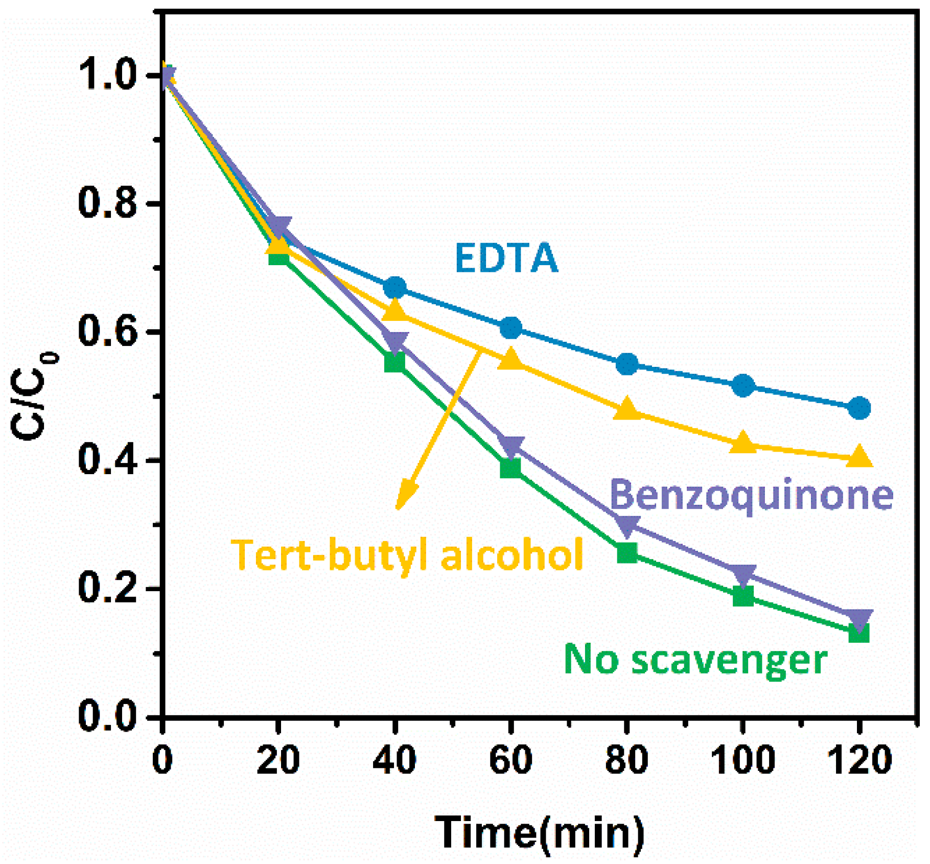
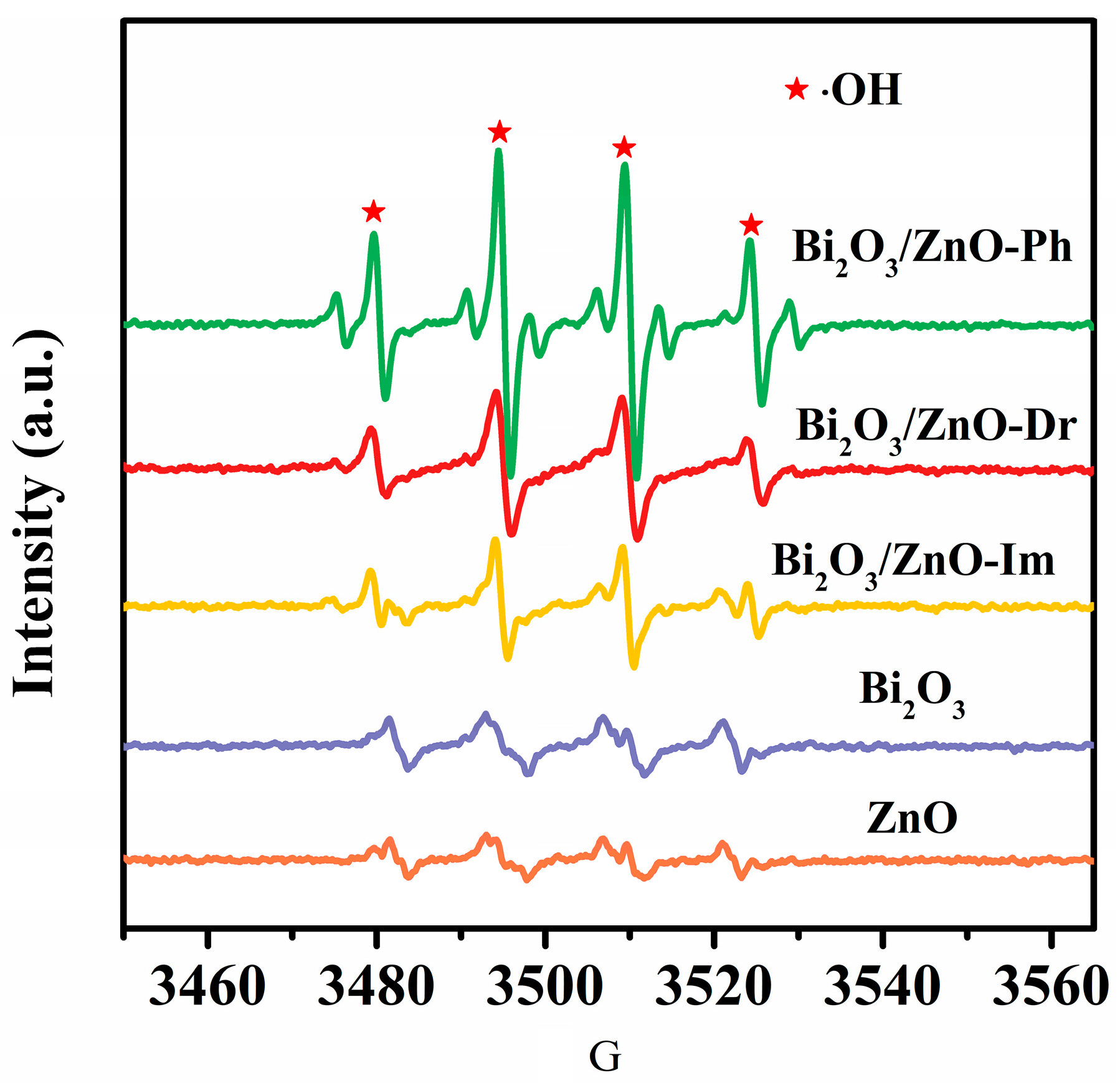
The reusability and long-term stability of the Bi2O3/ZnO-Ph photocatalyst were systematically evaluated through repeated photocatalytic degradation experiments of RhB under UV irradiation. The experimental conditions were consistent with the optimized parameters, as described in Section 2.2. After each photocatalytic cycle, the catalyst was separated via centrifugation and thoroughly washed with a mixed solution of ethanol and deionized water to remove any residual reactants or degradation products. The cleaned catalyst was subsequently dried and reused in subsequent cycles under identical reaction conditions.
As illustrated in Figure 5, the Bi2O3/ZnO-Ph photocatalyst maintained high photocatalytic activity over three consecutive cycles, with only negligible changes in the RhB degradation efficiency. The RhB removal rate minimally varied across the repeated runs, indicating that no significant photocatalyst deactivation occurred during the UV-driven photocatalytic process. This stable performance confirmed that the composite structure and active sites of the photocatalyst were largely intact throughout the cycling tests. The consistent degradation efficiency over multiple uses highlighted the structural robustness and chemical stability of the Bi2O3/ZnO-Ph photocatalyst. These findings suggested that the photochemically deposited Bi2O3 QDs were strongly anchored to the ZnO nanorod surface, contributing to the durability of the composite. Additionally, the results implied that the material could effectively withstand repetitive photocatalytic conditions without substantial performance loss, which demonstrated strong potential for practical applications in water treatment and other UV-light-assisted catalytic processes.

3. Discussion

The underlying reasons for the varying photocatalytic performances of the Bi2O3/ZnO composites synthesized via different methods were elucidated via PL and time-resolved photoluminescence (TRPL) [15,16], as shown in Figure 6. PL analysis serves as an effective tool for investigating the separation, migration, and transfer efficiency of charge carriers in photocatalysts since PL emission arises from the recombination of photogenerated electron–hole pairs [17]. These measurements offered insights into the recombination dynamics and lifetimes of photogenerated carriers within the composite system [18]. The PL spectra of all the Bi2O3/ZnO samples were recorded under 325 nm excitation. A lower PL intensity indicates a reduced recombination rate of photogenerated electrons and holes, reflecting more efficient charge separation [19]. As illustrated in Figure 6a, the PL emission intensities followed the order ZnO > Bi2O3/ZnO-Im > Bi2O3/ZnO-Dr > Bi2O3/ZnO-Ph. Although a reduction in the PL intensity was observed for Bi2O3/ZnO-Ph compared with the other samples, the change in the signal intensity was relatively modest and did not reach a two-order magnitude shift. Thus, while this trend suggests improved charge carrier separation, further studies are needed to conclusively attribute this to a significant reduction in recombination. Moreover, further insights were obtained from the TRPL decay profiles, which quantified the average lifetime of the photogenerated charge carriers [20]. The results indicated that Bi2O3/ZnO-Ph had the longest charge carrier lifetime, followed by Bi2O3/ZnO-Dr, Bi2O3/ZnO-Im, and ZnO. Specifically, the average lifetime of the photogenerated carriers increased from 0.43 ns for pure ZnO to 1.26 ns for Bi2O3/ZnO-Ph, suggesting that the heterojunction formed between ZnO and the Bi2O3 QDs effectively inhibited rapid electron–hole recombination. This enhancement was due to the formation of a well-defined heterojunction interface between the Bi2O3 QDs and ZnO, which promoted directional electron transfer from Bi2O3 to ZnO. This spatial electron–hole separation prolonged carrier lifetimes and improved their migration efficiency [21]. The superior performance of Bi2O3/ZnO-Ph can be attributed to the unique interfacial characteristics formed during the photochemical deposition process. Unlike impregnation or reduction methods, photochemical deposition selectively deposits Bi2O3 QDs onto electron-rich (defect-rich) sites of ZnO nanorods, forming a highly coupled heterojunction. This site-specific anchoring enables more intimate interfacial contact and directional charge transfer from Bi2O3 to ZnO. This enhanced heterojunction quality not only improves the spatial separation of charge carriers but also increases the lifetime and migration distance of electrons and holes, ultimately improving the generation efficiency of reactive species such as (·OH) radicals. The strong correlation between long-lived charge carriers and superior photocatalytic performance further confirmed the benefit of extended carrier lifetimes for surface redox reactions. Consequently, Bi2O3 QD sensitization effectively facilitated charge separation, and the synthesis method strongly influenced this beneficial effect. The combination of the PL and TRPL results provided compelling evidence that the enhanced photocatalytic activity of Bi2O3/ZnO-Ph stemmed from its superior charge separation and long carrier lifetimes [22], emphasizing the critical role of heterojunction engineering and preparation techniques in tuning the photocatalyst performance.
The investigation of the effects of different scavengers on photocatalytic degradation provided valuable insights into the reaction mechanism of the catalyst in photocatalytic reactions [23]. The scavengers selectively captured specific reactive species, determining the contribution of each species in the degradation process. These studies are crucial for optimizing catalyst design and enhancing photocatalytic performance. In photocatalytic reactions, the degradation of organic pollutants relies on in situ-generated reactive oxygen species (ROS) [24], such as hydroxyl radicals (·OH) and superoxide anions (O2), along with photogenerated holes (h+). ROS promote the degradation of organic pollutants through strong oxidation [25]. The identification of these active species aided in a deeper understanding of the photocatalytic reaction mechanism, providing a theoretical basis for improving catalyst efficiency and stability. A systematic study of the active species could reveal the reaction mechanism of the catalyst, supporting the development of more efficient photocatalytic materials. Furthermore, such studies have important implications for the application of photocatalytic materials in environmental remediation and energy conversion. Understanding the contributions of different active species to the photocatalytic process enhances the ability to design better catalysts with optimized performance for practical applications in pollution treatment and sustainable energy solutions.
Furthermore, various scavengers were employed to selectively capture specific active species to investigate the roles of different active species in the photocatalytic degradation of RhB via the Bi2O3/ZnO-Ph catalyst. Specifically, tert-butanol was used as a scavenger for ·OH radicals, and p-benzoquinone and disodium ethylenediaminetetraacetate (Na2-EDTA) were used to capture O2 and h+ [26], respectively. As shown in Figure 7, the Bi2O3/ZnO-Ph catalyst exhibited high stability and good repeatability in the photocatalytic degradation of RhB without the addition of any scavenger under UV light, with similar degradation efficiencies, indicating the reliability and consistency of the experimental setup [27]. However, the reaction rate showed little to no change when p-benzoquinone was added, suggesting that O2 was not the main active species involved in RhB degradation. In contrast, the degradation efficiency of RhB significantly decreased when Na2-EDTA and tert-butanol were added, indicating that h+ and ·OH radicals were the key active species in the photocatalytic degradation of RhB [28]. The addition of p-benzoquinone (O2 scavenger) had little impact, indicating that O2 was not the dominant reactive species. In contrast, both Na2-EDTA (h+ scavenger) and tert-butanol (·OH scavenger) significantly suppressed the degradation process. Notably, EDTA caused a greater reduction in degradation efficiency than tert-butanol did, even though ·OH is considered the dominant oxidant. This apparent contradiction is explained by the fact that h+ is a key upstream precursor for ·OH generation, particularly via the following reaction:
h+ + OH → OH
Therefore, scavenging h+ not only eliminates its direct oxidative contribution but also suppresses ·OH formation at the source. In contrast, tert-butanol only neutralizes already-formed ·OH radicals. This explains the stronger inhibitory effect of EDTA and highlights the crucial role of H+ in both the direct oxidation and ·OH generation pathways. This mechanistic insight is supported by the EPR results (Figure 8), which revealed the strongest DMPO-OH signal in Bi2O3/ZnO-Ph, confirming its enhanced radical generation capacity under UV light.
Furthermore, the reaction mechanism of the catalyst was verified via EPR spectroscopy to capture the reaction characteristics of different Bi2O3/ZnO catalysts, with DMPO as the trapping agent [29]. As shown in the EPR spectra in Figure 8, all catalyst samples had the characteristic signal of DMPO-OH, which presented four asymmetric peaks with a 1:2:2:1 intensity ratio and confirmed that ·OH radicals were the primary active species involved in the catalytic reaction [30,31]. The characteristic signal validated the key role of ·OH radicals in RhB degradation. Notably, the Bi2O3/ZnO-Ph catalyst displayed significantly stronger EPR signals than Bi2O3/ZnO-Im and Bi2O3/ZnO-Dr. This phenomenon indicated that the Bi2O3/ZnO-Ph catalyst generated more ·OH radicals under UV light exposure, further enhancing its photocatalytic activity [32]. Therefore, these findings provide important experimental evidence for optimizing catalyst preparation methods for improved photocatalytic performance.
In summary, the exceptional activity of Bi2O3/ZnO-Ph originates from the synergistic effects of selective quantum dot deposition, efficient interfacial charge separation, extended carrier lifetimes, and robust ·OH radical generation. These findings underscore the importance of heterojunction engineering and preparation strategies in tuning photocatalytic performance.

4. Materials and Methods

4.1. Synthesis of Catalysts

4.1.1. Synthesis of the ZnO Nanorods

First, different concentrations of zinc acetate dihydrate (Zn(Ac)2·2H2O) and sodium hydroxide (NaOH) solutions were prepared, with ethanol as the solvent for both. The prepared solutions were stored for future use. Then, 10 mL of 0.1 M Zn(Ac)2·2H2O ethanol solution was mixed with 20 mL of 0.5 M NaOH-ethanol solution to yield a clear solution. This solution was subsequently transferred into a stainless-steel autoclave lined with Teflon and heated at 150 °C for 24 h. Finally, a white precipitate was collected after the reaction, followed by washing repeatedly with water and ethanol and drying in a vacuum oven at 60 °C.

4.1.2. Preparation of the 5 wt.% Bi2O3/ZnO-Ph Composite Catalyst

The light source used was a 300 W high-pressure xenon lamp. Typically, 100 mg of ZnO nanorods and 11.6 mg of Bi(NO3)3·5H2O (99%, National Pharmaceutical Group Chemical Reagents Co., Ltd., Beijing, China.) were dispersed in 4 mL of ethylene glycol in a Pyrex glass reactor. The suspension was bubbled with Ar gas for 30 min to remove the dissolved O2, followed by UV irradiation. After the reaction, the precipitate was collected, washed twice with water and ethanol and dried in a vacuum oven at 60 °C.

4.1.3. Preparation of the 5 wt.% Bi2O3/ZnO-Dr Composite Catalyst

The Bi2O3/ZnO-Dr composite catalyst was synthesized via a deposition-reduction method with NaBH4 as the reducing agent. First, 23.2 mg of Bi(NO3)3·5H2O was dissolved in 4 mL of ethylene glycol, followed by the addition of 4 mL of deionized water and 200 mg of ZnO nanorods. The mixture was subsequently stirred at room temperature for 90 min to facilitate interactions between the Bi3+ ions and the ZnO surface. Subsequently, 2 mL of a 5 M NaBH4 aqueous solution was added dropwise to the mixture, and NaBH4 was precooled at 4 °C for 30 min to prevent rapid reactions. The addition of NaBH4 initiated the reduction of Bi3+ to Bi2O3, and Bi2O3 nanoparticles were deposited on the surfaces of the ZnO nanorods. The mixture was stirred for 2 h to ensure complete reduction and uniform deposition of Bi2O3.

4.1.4. Preparation of the 5 wt.% Bi2O3/ZnO-Im Composite Catalyst

The Bi2O3/ZnO-Im composite catalyst was prepared using impregnation. First, 23.2 mg of Bi(NO3)3·5H2O was dissolved in 4 mL of ethylene glycol, followed by the addition of 4 mL of deionized water and 200 mg of ZnO nanorods to ensure uniform dispersion of ZnO in the solution. The mixture was stirred for 12 h at room temperature to promote interactions between the Bi3+ ions and the ZnO surface. Finally, the precipitate was collected, washed twice with water and ethanol, and then calcined in air at 350 °C for 3 h to obtain the composite catalyst.

4.2. Material Characterization Methods

The powder XRD patterns were recorded via a diffractometer (Rigaku Ultimate IV, Rigaku Corporation, Tokyo, Japan) with Cu Kα radiation and operated at a 40 mA current and a 40 kV voltage. The morphologies of the samples were examined by scanning electron microscopy (SEM, Hitachi S4800, Hitachi High-Technologies Corporation, Tokyo, Japan). High-angle annular dark-field scanning transmission electron microscopy (HAADF-STEM, FEI Company, Hillsboro, OR, USA) images were obtained at an acceleration voltage of 200 kV. The UV-vis absorption spectra were recorded via a UV-vis spectrophotometer (Shimadzu UV-3600, Shimadzu Corporation, Kyoto, Japan). The fluorescence spectra were measured via a fluorescence spectrometer (Horiba Fluorolog-3–22, HORIBA Scientific, Kyoto, Japan) operating at a 200 mA current.

4.3. Catalytic Performance Evaluation

The RhB degradation experiment was conducted using a 350 W xenon lamp photochemical reactor (Nanjing Xujing Co., Ltd., Nanjing, China). A quartz tube was used as the reaction vessel, and a circulating water system ensured stable reaction conditions and kept the reaction system at room temperature. The experimental procedure was as follows: First, 30 mL of a 10 mg/L RhB aqueous solution was thoroughly mixed with 30 mg of catalyst, followed by stirring the mixture for 2 h in the dark to ensure adsorption equilibrium. Subsequently, 0.5 mL of 30 wt.% H2O2 was added to the tube. The xenon lamp, a UV light source, was then turned on to initiate the photocatalytic degradation reaction. The samples were collected every 20 min during the reaction. After centrifugation to separate the catalyst, RhB degradation was monitored via UV-vis absorption spectra analysis of the samples via a spectrophotometer (Shimadzu UV-1800, Shimadzu Corporation, Kyoto, Japan). This method effectively tracked the progress of the photocatalytic reaction and determined the RhB degradation efficiency through spectral analysis.

5. Conclusions

This study developed and compared three distinct methods for the in situ deposition of Bi2O3 quantum dots (QDs) onto ZnO nanorods to construct heterostructured photocatalysts. Among them, the photocatalyst synthesized via photochemical deposition (Bi2O3/ZnO-Ph) exhibited the best UV light-driven photocatalytic performance, achieving an 89.3% Rhodamine B (RhB) degradation efficiency within 120 min, significantly outperforming the other two methods. Comprehensive characterization revealed that the QDs in all the samples had similar particle size distributions; however, their photocatalytic activities varied markedly depending on the synthesis method. The superior performance of Bi2O3/ZnO-Ph was attributed to the selective anchoring of Bi2O3 QDs onto the electron-rich defect sites of ZnO, forming highly coupled heterojunctions that enabled efficient separation and transfer of photogenerated charge carriers. This structural feature was supported by PL and TRPL analyses, which confirmed reduced recombination rates and significantly prolonged carrier lifetimes. Moreover, scavenger experiments and EPR spectra demonstrated that ·OH radicals and h+ were the dominant active species during the degradation process, with h+ playing a dual role: contributing directly to pollutant oxidation and promoting ·OH generation. The stronger inhibitory effect of EDTA than tert-butanol further validated this mechanism. Overall, the findings emphasize that the heterojunction quality and interfacial electron dynamics governed by the synthesis strategy are more critical than the quantum dot size alone in determining the photocatalytic efficiency. The photochemical deposition approach offers an effective pathway for enhancing charge separation and ROS generation, providing valuable design insights for next-generation semiconductor-based photocatalysts for environmental remediation.

Supplementary Materials

The following supporting information can be downloaded at: https://www.mdpi.com/article/10.3390/catal15070695/s1, Figure S1: Microstructure of ZnO Nanorods.

Author Contributions

Conceptualization, B.L. and Z.Z.; methodology, B.L. and C.Z.; formal analysis, X.W., F.H. and B.Z.; investigation, B.L., Y.L. and B.Z.; resources, B.L. and X.F.; data curation, B.L. and B.Z.; writing—original draft preparation, Z.Z.; project administration, Z.Z.; funding acquisition, Z.Z. All authors have read and agreed to the published version of the manuscript.

Funding

This work was funded by the Independent Scientific Research Project of Zhejiang Energy R&D Institute (ZERD-KJ-2024-001), National Natural Science Foundation of China (52301222), Natural Science Foundation of Inner Mongolia Autonomous Region of China (20230N05015).

Data Availability Statement

Data are contained within the article and Supplementary Materials.

Conflicts of Interest

Authors Baohui Lou, Chi Zhang, Xianhao Wu, Xiangdong Feng, Feipeng Huang were was employed by Zhejiang Energy R&D Institute Co., Ltd. and Ying Liu was employed by the company Zhejiang Zheneng Co., Ltd. The remaining authors declare that the research was conducted in the absence of any commercial or financial relationships that could be construed as a potential conflict of interest.

References

  1. Pavel, M.; Anastasescu, C.; State, R.-N.; Vasile, A.; Papa, F.; Balint, I. Photocatalytic degradation of organic and inorganic pollutants to harmless end products: Assessment of practical application potential for water and air cleaning. Catalysts 2023, 13, 380. [Google Scholar] [CrossRef]
  2. Riente, P.; Adams, A.M.; Albero, J.; Palomares, E.; Pericàs, M.A. Light-driven organocatalysis using inexpensive, nontoxic Bi2O3 as the photocatalyst. Angew. Chem. Int. Ed. 2014, 53, 9613–9616. [Google Scholar] [CrossRef] [PubMed]
  3. Zahid, A.H.; Han, Q. A review on the preparation, microstructure, and photocatalytic performance of Bi2O3 in polymorphs. Nanoscale 2021, 13, 17687–17724. [Google Scholar] [CrossRef] [PubMed]
  4. Chen, Y.; Bai, X. A review on quantum dots modified g-C3N4-based photocatalysts with improved photocatalytic activity. Catalysts 2020, 10, 142. [Google Scholar] [CrossRef]
  5. Low, J.; Yu, J.; Jaroniec, M.; Wageh, S.; Al-Ghamdi, A.A. Heterojunction photocatalysts. Adv. Mater. 2017, 29, 1601694. [Google Scholar] [CrossRef] [PubMed]
  6. Gao, G.; Xi, Q.; Zhou, H.; Zhao, Y.; Wu, C.; Wang, L.; Guo, P.; Xu, J. Selectivity of quantum dot sensitized ZnO nanotube arrays for improved photocatalytic activity. Phys. Chem. Chem. Phys. 2017, 19, 11366–11372. [Google Scholar] [CrossRef] [PubMed]
  7. Inamdar, S.R.; Pujar, G.; Sannaikar, M. FRET from ZnSe/ZnS QDs to coumarin dyes: Role of acceptor dipole moment and QD surface states on FRET efficiency. J. Lumin. 2018, 203, 67–73. [Google Scholar] [CrossRef]
  8. Munonde, T.S.; Nomngongo, P.N. Review on metal chalcogenides and metal chalcogenide-based nanocomposites in photocatalytic applications. Chem. Afr. 2023, 6, 1127–1143. [Google Scholar] [CrossRef]
  9. Sun, P.; Xing, Z.; Li, Z.; Zhou, W. Recent advances in quantum dots photocatalysts. Chem. Eng. J. 2023, 458, 141399. [Google Scholar] [CrossRef]
  10. Hartmann, F.; Etter, M.; Cibin, G.; Groß, H.; Kienle, L.; Bensch, W. Understanding sodium storage properties of ultra-small Fe 3 S 4 nanoparticles–a combined XRD, PDF, XAS and electrokinetic study. Nanoscale 2022, 14, 2696–2710. [Google Scholar] [CrossRef] [PubMed]
  11. Fitzpatrick, J.A.J.; Andreko, S.K.; Ernst, L.A.; Waggoner, A.S.; Ballou, B.; Bruchez, M.P. Long-term persistence and spectral blue shifting of quantum dots in vivo. Nano Lett. 2009, 9, 2736–2741. [Google Scholar] [CrossRef] [PubMed]
  12. Singla, M.L.; Kumar, M. Optical characterization of ZnO nanoparticles capped with various surfactants. J. Lumin. 2009, 129, 434–438. [Google Scholar] [CrossRef]
  13. Fu, J.; Zhao, C.; Zhang, J.; Peng, Y.; Xie, E. Enhanced gas sensing performance of electrospun Pt-functionalized NiO nanotubes with chemical and electronic sensitization. ACS Appl. Mater. Interfaces 2013, 5, 7410–7416. [Google Scholar] [CrossRef] [PubMed]
  14. Xing, B.; Shi, C.; Zhang, C.; Yi, G.; Chen, L.; Guo, H.; Huang, G.; Cao, J. Preparation of TiO2/activated carbon composites for photocatalytic degradation of RhB under UV light irradiation. J. Nanomater. 2016, 2016, 8393648. [Google Scholar] [CrossRef]
  15. Baloch, A.A.B.; Alharbi, F.H.; Grancini, G.; Hossain, M.I.; Nazeeruddin, K.; Tabet, N. Analysis of photocarrier dynamics at interfaces in perovskite solar cells by time-resolved photoluminescence. J. Phys. Chem. C 2018, 122, 26805–26815. [Google Scholar] [CrossRef]
  16. Chen, Y.; Wang, T.; Li, Z.; Li, H.; Ye, T.; Wetzel, C.; Li, H.; Shi, S.-F. Communicating two states in perovskite revealed by time-resolved photoluminescence spectroscopy. Sci. Rep. 2018, 8, 16482. [Google Scholar] [CrossRef] [PubMed]
  17. Sarkar, A.; Karmakar, K.; Khan, G.G. Designing Co-Pi modified one-dimensional n–p TiO2/ZnCo2O4 nanoheterostructure photoanode with reduced electron–hole pair recombination and excellent photoconversion efficiency (>3%). J. Phys. Chem. C 2017, 121, 25705–25717. [Google Scholar] [CrossRef]
  18. Ma, J.; Miao, T.J.; Tang, J. Charge carrier dynamics and reaction intermediates in heterogeneous photocatalysis by time-resolved spectroscopies. Chem. Soc. Rev. 2022, 51, 5777–5794. [Google Scholar] [CrossRef] [PubMed]
  19. Bi, L.; Gao, X.; Zhang, L.; Wang, D.; Zou, X.; Xie, T. Enhanced photocatalytic hydrogen evolution of NiCoP/g-C3N4 with improved separation efficiency and charge transfer efficiency. ChemSusChem 2018, 11, 276–284. [Google Scholar] [CrossRef] [PubMed]
  20. Singh, M.K.; Mehata, M.S. Temperature-dependent photoluminescence and decay times of different phases of grown TiO2 nanoparticles: Carrier dynamics and trap states. Ceram. Int. 2021, 47, 32534–32544. [Google Scholar] [CrossRef]
  21. Žerjav, G.; Arshad, M.S.; Djinović, P.; Junkar, I.; Kovač, J.; Zavašnik, J.; Pintar, A. Improved electron–hole separation and migration in anatase TiO2 nanorod/reduced graphene oxide composites and their influence on photocatalytic performance. Nanoscale 2017, 9, 4578–4592. [Google Scholar] [CrossRef] [PubMed]
  22. Wang, C.; Wu, B.; Wang, C. Rational construction and efficient regulation of stable and long-lived charge-separation state in fullerene materials. Acc. Mater. Res. 2024, 5, 426–437. [Google Scholar] [CrossRef]
  23. Das, A.; Adak, M.K.; Mahata, N.; Biswas, B. Wastewater treatment with the advent of TiO2 endowed photocatalysts and their reaction kinetics with scavenger effect. J. Mol. Liq. 2021, 338, 116479. [Google Scholar] [CrossRef]
  24. Hossain, S.; Mollah, M.Y.A.; Susan, A.B.H.; Islam, M. Role of in situ electrogenerated reactive oxygen species towards degradation of organic dye in aqueous solution. Electrochim. Acta 2020, 344, 136146. [Google Scholar] [CrossRef]
  25. Xie, Z.H.; He, C.S.; Zhou, H.Y.; Li, L.L.; Liu, Y.; Du, Y.; Liu, W.; Mu, Y.; Lai, B. Effects of molecular structure on organic contaminants’ degradation efficiency and dominant ROS in the advanced oxidation process with multiple ROS. Environ. Sci. Technol. 2022, 56, 8784–8795. [Google Scholar] [CrossRef] [PubMed]
  26. Zuo, D.; Yin, Y.; Jiang, L.; Xia, Z.; Tan, C.; Li, J.; Zuo, J.; Wang, Y. Synthesis of Ag/ZnO/BiOCl composite material and its photodegradation performance on ciprofloxacin. Coatings 2024, 14, 192. [Google Scholar] [CrossRef]
  27. Dubey, S.; Thakur, A.S.; Vaish, R.; Algarni, Z.; Ismail, M.A.; Amari, A. Impact of rotational speed and pH on tribocatalytic dye degradation activity of 0.5 Ba (Zr0.2Ti0.8) O3–0.5 (Ba0.7Sr0.3) TiO3. Int. J. Appl. Ceram. Technol. 2025, 22, e15015. [Google Scholar] [CrossRef]
  28. Shi, W.; Fang, W.-X.; Wang, J.-C.; Qiao, X.; Wang, B.; Guo, X. pH-controlled mechanism of photocatalytic RhB degradation over gC3N4 under sunlight irradiation. Photochem. Photobiol. Sci. 2021, 20, 303–313. [Google Scholar] [CrossRef] [PubMed]
  29. Han, S.K.; Hwang, T.M.; Yoon, Y.; Kang, J.W. Evidence of singlet oxygen and hydroxyl radical formation in aqueous goethite suspension using spin-trapping electron paramagnetic resonance (EPR). Chemosphere 2011, 84, 1095–1101. [Google Scholar] [CrossRef] [PubMed]
  30. Wang, J.; Wang, S. Reactive species in advanced oxidation processes: Formation, identification and reaction mechanism. Chem. Eng. J. 2020, 401, 126158. [Google Scholar] [CrossRef]
  31. Yu, G.; Wang, Y.; Cao, H.; Zhao, H.; Xie, Y. Reactive oxygen species and catalytic active sites in heterogeneous catalytic ozonation for water purification. Environ. Sci. Technol. 2020, 54, 5931–5946. [Google Scholar] [CrossRef] [PubMed]
  32. Nagarajan, S.; Skillen, N.C.; Fina, F.; Zhang, G.; Randorn, C.; Lawton, L.A.; Irvine, J.T.; Robertson, P.K. Comparative assessment of visible light and UV active photocatalysts by hydroxyl radical quantification. J. Photochem. Photobiol. A Chem. 2017, 334, 13–19. [Google Scholar] [CrossRef]
Figure 1. Microscopic structures and particle size distributions of Bi2O3/ZnO synthesized via different methods.
Catalysts 15 00695 g001
Figure 2. (a) XRD patterns and (b) UV-vis spectra of commercial ZnO and Bi2O3/ZnO synthesized via different methods.
Catalysts 15 00695 g002
Figure 3. UV photocatalytic degradation performance of RhB using different catalysts.
Catalysts 15 00695 g003
Figure 4. Kinetic curves of RhB degradation using different catalysts.
Catalysts 15 00695 g004
Figure 5. Reusability test of Bi2O3/ZnO Ph photocatalysts synthesized using different carriers for the UV degradation of RhB.
Catalysts 15 00695 g005
Figure 6. PL spectra (a) and TRPL decay spectra (b) of Bi2O3/ZnO photocatalysts synthesized using different carriers.
Catalysts 15 00695 g006
Figure 7. Effect of scavengers on the photocatalytic activity of catalysts.
Catalysts 15 00695 g007
Figure 8. EPR spectra of the DMPO-·OH adducts of various samples.
Catalysts 15 00695 g008
Disclaimer/Publisher’s Note: The statements, opinions and data contained in all publications are solely those of the individual author(s) and contributor(s) and not of MDPI and/or the editor(s). MDPI and/or the editor(s) disclaim responsibility for any injury to people or property resulting from any ideas, methods, instructions or products referred to in the content.

