Article

Biosynthesis of Zinc Oxide Nanostructures Using Leaf Extract of Azadirachta indica: Characterizations and In Silico and Nematicidal Potentials

1
Department of Pharmaceutical Chemistry and Pharmacognosy, College of Pharmacy, Jazan University, Jazan 45142, Saudi Arabia
2
Department of Biology, College of Science, Imam Mohammad Ibn Saud Islamic University (IMSIU), Riyadh 11623, Saudi Arabia
3
Department of Biology, College of Science, Jazan University, Jazan 45142, Saudi Arabia
4
Department of Physical Sciences, Chemistry Division, College of Science, Jazan University, P.O. Box 114, Jazan 45142, Saudi Arabia
5
Nanotechnology Research Unit, Jazan University, P.O. Box 114, Jazan 45142, Saudi Arabia
6
Department of Pharmaceutics, College of Pharmacy, Jazan University, Jazan 45142, Saudi Arabia
7
Department of Chemistry, College of Science, Imam Mohammad Ibn Saud Islamic University (IMSIU), Riyadh 11623, Saudi Arabia
8
Department of Chemical Engineering, College of Engineering and Computer Sciences, Jazan University, Jazan 45142, Saudi Arabia
*
Author to whom correspondence should be addressed.
Catalysts 2025, 15(7), 693; https://doi.org/10.3390/catal15070693
Submission received: 3 May 2025 / Revised: 1 July 2025 / Accepted: 16 July 2025 / Published: 21 July 2025
(This article belongs to the Special Issue (Bio)nanomaterials in Catalysis)

Abstract

Biosynthesized ZnO nanostructures were characterized by X-ray diffraction (XRD), scanning electron microscopy (SEM), transmission electron microscopy (TEM), ultraviolet–visible (UV-vis) spectroscopy, and Fourier transform–infrared (FT-IR) spectroscopy. XRD confirmed a hexagonal wurtzite phase with an average crystallite size of 36.44 nm, while UV-vis spectroscopy showed a distinct absorption peak at 321 nm. The Zeta potential of the ZnO nanostructures was −24.28 mV, indicating high stability in suspension, which is essential for their dispersion and functionality in biological and environmental applications. The nematicidal activity of ZnO was evaluated in vitro at concentrations of 150, 300, 450, and 600 ppm, with the highest concentration achieving 75.71% mortality of second-stage juveniles (J2s) after 72 h. The calculated LC50 values for the treatments were 270.33 ppm at 72 h. Additionally, molecular docking studies indicated significant interactions between the ZnO nanostructures and nematode proteins, HSP-90 and ODR1, supporting their potential nematicidal mechanism. This research highlights the effectiveness of neem leaf extract-mediated ZnO nanostructures as an eco-friendly, sustainable alternative for nematode control, presenting a promising solution for agricultural pest management.

1. Introduction

The global agricultural sector faces significant challenges due to plant diseases, with plant parasitic nematodes (PPNs) like Meloidogyne incognita causing severe crop damage [1,2]. While several control strategies have been employed, including the use of chemical nematicides, they often present environmental and health risks. Recent research has explored the preparation of ZnO nanoparticles (ZnO NPs) for controlling such nematodes [3]. However, the synthesis methods have typically relied on chemical processes that are not sustainable. This study presents a novel approach by utilizing Azadirachta indica leaf extract for the biosynthesis of ZnO nanostructures (ZnO NSs), which offers a sustainable, low-cost, and eco-friendly alternative [4,5]. Furthermore, the nematicidal activity of these biosynthesized ZnO NSs has not been extensively studied, particularly in comparison to traditional synthetic nematicides. This study underscores the potential of ZnO NSs as a viable solution for nematode control in agriculture, providing an environmentally friendly alternative to harmful chemical agents [6,7]. The ZnO NSs have been selected for this study due to their unique properties, including their antimicrobial activity, sustainability, and cost-effectiveness [8,9]. ZnO has shown significant nematicidal activity, outperforming other metal oxide nanomaterials like TiO2 and CuO in pest control [10,11]. Furthermore, ZnO offers an eco-friendly synthesis route, such as biosynthesis from neem leaf extract, which aligns with the principles of sustainable agriculture [12,13]. The use of ZnO in pest management presents a safer alternative to conventional chemical nematicides and other nanoparticles, such as silver, which pose environmental and health risks [14]. Additionally, ZnO’s low toxicity to plants and its broad-spectrum activity against nematodes make it a suitable choice for this investigation [15]. Mohammad et al. [16] investigated the surface chemical properties of ZnO NPs coated with three different surfactant biomolecules, namely polyethylene glycol (PEG), cetyltrimethylammonium bromide (CTAB), and sodium dodecyl sulfate (SDS), to control the toxicity-induced potentials.
ZnO NSs have been of particular interest in agricultural research owing to their distinctive physical and chemical properties, such as their immense surface area, antimicrobial activity, and capacity to promote plant growth [17,18,19]. ZnO NPs possess a high surface area and active sites that allow them to adsorb heavy metals such as lead (Pb), mercury (Hg), and arsenic (As) from contaminated water. Research conducted between 2021 and 2023 has shown that ZnO NPs can achieve significant removal efficiencies, making them suitable for water purification applications [20]. ZnO NPs have shown great promise as nanocarriers in targeted drug delivery, particularly in cancer therapy, where their surfaces can be functionalized with targeting ligands like peptides to selectively direct drugs to tumor cells, as demonstrated in prostate cancer models [21]. The morphology and specific surface area affect the antibacterial activity of ZnO NPs; therefore, it is necessary to synthesize a material with a novel morphology with increased adsorption sites and surface area [22]. Biosynthesized ZnO NPs are more effective on Gram-positive bacteria than Gram-negative bacteria based on their cell structure, metabolic activities, and degree of contact of the bacteria, as reported in study [23]. ZnO NPs effectively eliminate Staphylococcus aureus, Escherichia coli, Pseudomonas aeruginosa, and Bacillus cereus, underscoring their potential as alternative antimicrobial agents [24,25]. Mishra et al. [26] reported that ZnO NPs synthesized with Trichoderma harzianum culture filtrate have increased antifungal activity against Alternaria brassicae, even at lower doses, and can be used as an alternative to traditional fungicides without affecting the environment. Welch et al. [27] reported roles of the iron storage protein ferritin as both a source of iron for iron-mediated oxidation and as a mechanism to safely store iron in cells.
ZnO NPs were produced using Nigella sativa seed extract and characterized through UV-Vis spectroscopy, FT-IR, X-ray diffraction, and scanning electron microscopy (SEM). They have shown antifungal properties against Cercospora canescens, the causative agent of Cercospora leaf spot in mung bean [28]. Copper oxide (CuO) and Zinc oxide (ZnO), obtained using the spin-coating technique, exhibit excellent antimicrobial activity against Staphylococcus aureus, as well as outstanding antiviral activity against the HSV-2 virus [29]. ZnO NPs biosynthesized from Achillea millefolium leaf aqueous extract show dose-dependent juvenile mortality and hatching inhibition, as well as enhanced growth parameters of tomato plants infected with M. incognita [14]. ZnO NPs reduced salt stress by promoting physiological growth parameters; they also enhanced plant growth and reduced reactive oxygen species (ROS) generation by regulating plant nutrient homeostasis and chlorophyll fluorescence activity [30,31,32]. Raza et al. [33] reported that ZnO NPs emerged as a potential drought alleviator and yield-oriented safe nano-fertilizer for wheat in semi-arid regions facing irrigation challenges. Chen et al. [34] reported that ZnO NPs have a positive impact on the photosynthesis of tea plants, the sprouting of new shoots, and the community of phyllosphere microorganisms, which can improve the growth conditions of tea plants. Four powders of ZnO were tested and revealed antimicrobial activity in the dark; different amounts and kinds of ROS were emitted from each ZnO powder, and their sustainability depends significantly on the contents of interstitial Zn contained in the ZnO particles [35].
Generally, nanotechnology offers precise and controlled distribution of agrochemicals, which reduces the need for excessive pesticide use and minimizes environmental contamination. Recent investigations have also revealed the role of nanoparticles as plant growth promoters, stimulating seed germination and seedling development, thereby improving overall crop yields [36,37]. Moreover, nanoparticle biosynthesis using plant extracts, bacteria, fungi, or other biological materials provides an eco-friendly, low-cost replacement to chemical methods of nanoparticle production. Tomatoes, a key crop in many parts of the world, are particularly susceptible to RKN infestations, which can lead to stunted growth, reduced yields, and even total crop loss. By testing the efficacy of ZnO NS on M. incognita, this research adds to the expanding corpus of knowledge on nanotechnology in agriculture and its role in developing sustainable pest control solutions. The incorporation of nanotechnology in pest management not only provides an innovative approach to reducing the impact of harmful chemical nematicides but also supports environmentally sustainable agricultural practices. Molecular docking is a method for generating and supplying information about ligand–receptor communications that helps in predicting how ligands will bind to their target proteins or DNA [38,39]. Furthermore, this approach facilitates systemic learning by delivering a molecule in a non-covalent manner to the binding site of the targeted macromolecule, primarily to a specific binding at each ligand’s active site [40]. Because of extensive H-bonding and hydrophobic and alkyl interactions, Kundu et al. [41] discovered that in silico molecular interaction assessments of the seven nematode target proteins and major essential oil constituents revealed the highest binding affinity of the geraniol–odorant response gene complex.
As agriculture faces increasing pressures from climate change, soil degradation, and pest infestations, sustainable pest control strategies are more important than ever. This research highlights the efficacy of biosynthesized ZnO NSs as a viable and environmentally friendly substitute for chemical nematicides, paving the way for more sustainable agricultural production systems. The application of nanotechnology in agriculture has the potential to enhance long-term sustainability and food security by reducing reliance on harmful chemicals and promoting environmentally beneficial practices. Although previous studies have explored the synthesis of ZnO NSs using neem leaf extract [12], this research presents a unique contribution by combining biosynthesis, molecular docking, and in-depth nematicidal analysis. The study provides a comprehensive view of the time-dependent and concentration-dependent nematicidal effects, with an emphasis on the LC50 values over 72 h. Additionally, molecular docking studies offer insights into the interaction of ZnO NSs with nematode proteins (HSP-90 and ODR1), advancing the understanding of its nematicidal mechanism. Furthermore, this work highlights the eco-friendly aspects of biosynthesized ZnO NSs, presenting a viable alternative to conventional nematicides and aligning with global efforts to minimize the environmental impact of agriculture.

2. Results

2.1. Analysis of Biosynthesized-ZnO NSs

The biosynthesized ZnO NSs were characterized using multiple techniques to gain insights into their structural and chemical properties. X-ray diffraction (XRD) analysis was conducted with a Rigaku Smart Lab diffractometer, scanning over the 2θ range of 30° to 70° using Cu-Kα radiation (1.54 Å). The scan rate was set at 0.02 steps per second, with a step size of 0.01°, to determine the crystallinity of the nanostructures. The scanning electron microscopy (SEM) analysis was performed using a JEOL (Peabody, MA, USA) JSM-6510 LV SEM equipped with an Oxford EDS system at 200 kV to study the surface morphology and elemental composition of the ZnO NS. For a more detailed structural investigation, transmission electron microscopy (TEM) was employed at magnifications ranging from 10 to 50 nm to observe the fine details and size distribution of the nanostructures. To investigate the surface functional groups, Fourier transform–infrared spectroscopy (FT-IR) was carried out using a Thermo Scientific Nicolet iS10 FT-IR spectrometer (Waltham, MA, USA), providing insights into the chemical bonds present in the material. Finally, the optical properties of the ZnO NSs were analyzed using UV-visible spectroscopy (UV-vis) with a Perkin–Elmer spectrophotometer, which provided valuable information about the absorption characteristics of the nanostructures.
X-rays were produced with a voltage of 40 kilovolts (kV) and a current of 40 kiloamperes (kA). The size of the nanocrystallites (D) was determined from the sharpest peak (101) in the X-ray diffraction pattern using the Scherrer equation. The following comparison of average crystalline diameters was made using the Williamson–Hall Equation (1) and the Scherrer Equation (2) to confirm this assessment [42,43].
β c o s θ = 4 ε s i n θ + k λ D
D = k λ β c o s θ
The variable “β” represents the whole width at half-maximum for the discussed diffraction peaks, measured in radians. The constant “K” has a value of 0.9. The variable “θ” represents the diffraction angle, measured in degrees. Finally, “D” represents the size of the crystallite, measured in nanometers. The wavelength of the monochromatic X-ray beam, namely CuKα radiation, is denoted by the symbol “λ” and measures 0.154056 nm.
The crystallite size was determined using both the Scherrer equation and the Williamson–Hall plot. The crystallite size calculated from the Scherrer equation was compared to that obtained from the Williamson–Hall plot, which also accounted for strain. The crystallite size was calculated using the three most intense peaks, (100), (101), and (102), to ensure accuracy. Additionally, crystallinity percentage, porosity, and phase percentages were estimated using Rietveld refinement of the XRD data. The refinement analysis revealed a hexagonal wurtzite phase with a crystallinity of 92% and a porosity of 12%. These results support the high purity and structural integrity of the ZnO NS. The crystallite size of the biosynthesized ZnO NS was determined using the Scherrer equation, which was applied to the three most intense peaks: (100), (101), and (102). The calculated average crystallite size was 36.44 nm, confirming the high crystallinity of the synthesized ZnO NSs. The crystallization pattern and hexagonal wurtzite phase of ZnO NSs were confirmed based on standard reference data from the International Centre for Diffraction Data (ICDD). The XRD spectrum (Figure 1) exhibited the characteristic peaks for the wurtzite phase of ZnO, with prominent peaks at 31°, 34°, and 36° corresponding to the (100), (002), and (101) planes, respectively. These findings are in alignment with the expected diffraction pattern for ZnO. The absence of the (102) plane around 47° is likely due to factors such as particle size, crystal defects, or the preferential orientation of the nanostructures during synthesis, which can cause the peak to be weak or not visible. However, the key diffraction peaks confirmed the presence of the wurtzite-phase ZnO NSs, as expected.
An in-depth understanding of the surface topography along with the morphology of the substance may be obtained via the use of SEM assessment. SEM analysis provided detailed insights into the material’s surface texture and morphology. As depicted in Figure 2, the images confirmed the successful formation of green-synthesized ZnO NS. Additionally, the elemental composition of the biosynthesized ZnO NS was further confirmed through energy-dispersive X-ray spectroscopy (EDAX). The EDAX spectrum revealed the presence of zinc (Zn) and oxygen (O) as the major elements, with atomic percentages of 67.2% for zinc and 32.8% for oxygen. These results confirm the successful synthesis of pure ZnO NS, with no significant impurities, thus validating the material’s high purity and composition (Figure 3). According to Figure 4, the investigation performed using transmission electron microscopy (TEM) demonstrated the existence of distinct ZnO NS within the 28–94 nm range. Hence, the biosynthesized ZnO NS predominantly exhibited a rod-like morphology, as observed in the SEM and TEM images. The SEM images showed uniform prismatic-shaped structures, while TEM analysis confirmed the presence of well-defined nanorods, with an average size ranging from 28 nm to 94 nm. The crystalline structure of these nanorods was consistent with the hexagonal wurtzite phase, as confirmed by XRD and further supported by the high crystallinity observed in the TEM images. To provide further insight into the morphology and size distribution of the biosynthesized ZnO NS, SEM images were captured at various magnifications, ranging from 12,000× to 35,000×, to observe the uniformity and morphology of the nanorods. TEM images were also obtained at different magnifications, revealing well-defined nanorods. A particle size distribution histogram was generated based on the TEM images, showing a narrow size distribution with a peak at approximately 80 nm, confirming the uniformity of the nanostructures (Figure 4). The UV-Vis absorption spectrum (Figure 5) showed a notable absorption band (λmax) at about 321 nm, with no additional peaks observed, confirming the high quality and uniformity of the ZnO NS. The optical bandgap of the biosynthesized ZnO NS was determined using the Tauc plot method. The bandgap energy (Eg) was found to be 3.12 eV (Figure 6), indicating the semiconducting nature of the material. This value is consistent with the observed high crystallinity and the well-defined nanorod morphology of the ZnO NS, as evidenced by the SEM and TEM results. The enhanced crystallinity of the nanostructures facilitates efficient electronic transitions, contributing to the bandgap value. The rod-like morphology further supports the electronic properties by providing a larger surface area for potential applications. The objective of characterizing the produced nanostructures was to further understand their optical, structural, and morphological characteristics. The crystalline structure seen in Figure 1 (XRD investigation) is compatible with the hexagonal wurtzite phase of the ZnO NS. Furthermore, FT-IR was employed to investigate the performance and chemical characteristics of the sample, generating an image that revealed the vibrational and rotational modes of prevailing molecules. This, in turn, facilitated the identification of functional groups and potential phytochemical molecules responsible for the reduction and stabilization of the biosynthesized ZnO NS (Figure 7). The FTIR spectrum was acquired using a Thermo Scientific Nicolet iS10 FT-IR spectrometer. Notable absorption bands were observed between 3400–3600 cm−1 and around 1620 cm−1, corresponding to O-H stretching vibrations and C-H bending vibrations, respectively, likely originating from adsorbed water and organic contaminants. Although the characteristic ZnO peak near 500 cm−1 was not detected—presumably due to surface hydroxylation or the presence of organic capping agents—the Zn-O stretching vibration appeared distinctly at 483 cm−1, thereby confirming the successful formation of the ZnO NS. Moreover, the Zeta potential of the ZnO NS (Figure 8) was found to be −24.28 mV, which indicates a high level of stability in the colloidal dispersion. This negative value suggests that the particles possess sufficient repulsion forces to prevent aggregation, ensuring long-term stability of the nanoparticle suspension.

2.2. Nematicidal Effect of Biosynthesized-ZnO NS on J2 Mortality of M. incognita In Vitro

This study demonstrated that the four concentrations of ZnO NS (150, 300, 450, and 600 ppm) had a significant impact on J2 mortality at 12, 24, 48, and 72 h under incubation (p < 0.05). ZnO NSs at varying doses were harmful to M. incognita J2s as compared to DDW (control). The reference concentration (600 ppm) had the highest death rate when compared to the other concentrations (150, 300, and 450 ppm). Each dose showed the most significant J2 mortality after 72 h of incubation, outperforming the results observed at 12, 24, and 48 h. At 72 h of incubation, the 600 ppm concentration was shown to be very harmful to J2s, with a substantial difference from the remaining concentrations. In a similar way, the incubation duration had a substantial impact on J2 mortality, which peaked at 72 h compared to 12, 24, and 48 h. Once the concentration of ZnO NS increases from 150 to 600 ppm, so does the mortality of J2s (Table 1, Figure 9). After 72 h of exposure, LC50 showed higher J2 mortality (270.33) than it did after 48 h (LC50—397.68), 24 h (LC50—585.72), and 12 h (LC50—842.15) of contact (Table 2). Our study found that the treatment’s toxicity to M. incognita J2s was higher with lower LC50 values and that treatment with higher LC50 values was less toxic to J2s. The LC50 values decreased over time, highlighting the concentration- and time-dependent efficacy of the extracts. Results of LC50 values of various botanical extracts at 12, 24, 48, and 72 h time intervals specified the progressive reduction in LC50 values over time, reflecting the increasing mortality of J2s with prolonged exposure to ZnO NS treatment (Table 2). Among the exposure periods, 72 h exhibited the lowest LC50 value, with 270.33, showing the highest nematicidal efficacy. The results of the study highlight a significant time-dependent decrease in LC50 values for all tested concentrations, suggesting that prolonged exposure enhances their nematicidal effectiveness. The progressive reduction in LC50 values from 12 to 72 h suggests a prolonged nematicidal activity of ZnO NS, enhancing their effectiveness over time. A lower LC50 value indicates higher toxicity and, consequently, greater efficacy of the ZnO NS. The results demonstrated that at LC50, there was significant mortality of J2s within the exposure period, suggesting a strong nematicidal effect. This supports the potential of the ZnO NS as a viable option for RKN management. According to the findings, all concentrations were exceedingly toxic to J2s, with fatality rates varying from 11% to 75% (Table 1). The toxic effects of different ZnO NS concentrations on J2s are detailed in Table 1.

2.3. Nematicidal Effect of Biosynthesized ZnO NS on Egg Hatching of M. incognita In Vitro

The bioassay demonstrated considerable variation among various concentrations of ZnO NS regarding J2 hatching inhibition (Figure 10). Employing a direct contact approach, all concentrations (150, 300, 450, 200, and 600 ppm) markedly decreased J2 hatching in comparison to the control (DDW). The level of inhibition escalated with increasing doses, with 600 ppm exhibiting the most significant impact, followed by 450 ppm and 300 ppm. The minimal inhibition was recorded at 150 ppm. J2s’ hatching inhibition improved simultaneously as the concentration grew progressively from 150 to 600 ppm. Different ZnO NS concentrations (150, 300, 450, 200, and 600 ppm) had varied inhibitory effects on J2 hatching, as shown in Figure 10.

2.4. Effect of Biosynthesized ZnO NS on J2 Penetration in Roots of Tomato Seedlings

Following 5 days of inoculation, various concentrations of ZnO NS (150, 300, 450, and 600 ppm) were observed to diminish J2 penetration in the roots of tomato seedlings to variable degrees when compared to J2 alone. The 600 ppm concentration inhibited J2 penetration the most, while 150 ppm inhibited J2 penetration the least in tomato plant roots. Other doses, such as 300 and 450 ppm, demonstrated considerable suppression of J2 penetration. Concentrations increased from 150 to 600 ppm, resulting in increased inhibition of J2 penetration. The most significant J2 penetration was reported in tomato seedlings that had only been inoculated with J2. In general, greater doses inhibited J2 penetration in tomato roots, while a lower concentration resulted in minimal penetration five days post-inoculation in comparison to seedlings infected just with J2. Based on the findings, it was determined that ZnO NSs have nematicidal qualities and might be utilized to regulate M. incognita in order to enhance sustainability. The suppression of J2 penetration in tomato roots and consequent toxicity is influenced by the time frame of exposure length and concentration of ZnO NS. This suggests that a reduced penetration was attained at the greatest concentration, but at lower concentrations, a higher penetration was seen. Table 3 shows the individual inhibiting impacts of various ZnO NS concentrations on J2 penetration.

2.5. Molecular Docking of Hsp90-ZnO Complex

The molecular docking analysis of Hsp90 with ZnO NS revealed a binding energy of −6.156 kcal/mol, suggesting a moderate binding affinity (Figure 11). The docking pose and interaction map demonstrate that ZnO NSs establish multiple interactions with key residues within the binding pocket of Hsp90. Table 4 shows that ZnO NSs form conventional hydrogen bonds with ILE87 (3.124 Å) and pi-donor hydrogen bonds with TRP90 (3.266 Å and 3.760 Å). Additionally, pi-alkyl interactions were observed between ZnO and TYR57 (5.008 Å), PHE67 (5.036 Å), and PHE130 (4.752 Å). These interactions suggest that the ZnO NS is stabilized within the binding cavity through a combination of hydrogen bonding and hydrophobic interactions. The significant involvement of aromatic residues (TRP90, TYR57, PHE67, PHE130) indicates that π-electron interactions contribute to the stabilization of the complex. The presence of ILE87 in hydrogen bonding suggests that polar interactions also play a role in binding. The moderately strong binding energy suggests that the ZnO NS could interfere with the function of Hsp90, potentially modulating its role in protein folding and stress response pathways.

2.6. Molecular Docking of ODR1-ZnO Complex

Docking simulations of ODR1 with a ZnO NS resulted in a binding energy of −6.33 kcal/mol, slightly higher in affinity than Hsp90-ZnO binding (Figure 12). The interaction analysis, summarized in Table 5, indicates the formation of conventional hydrogen bonds with PHE123 (3.226 Å) and HIS121 (3.277 Å). Moreover, a pi-donor hydrogen bond was observed between PHE123 and ZnO (3.728 Å), further stabilizing the interaction. Pi-alkyl interactions were identified between HIS111 and ZnO (5.069 Å). These findings suggest that ZnO NSs establish a stable interaction network through both hydrogen bonding and hydrophobic interactions, which may play a role in disrupting or altering the function of ODR1. The presence of histidine and phenylalanine residues in the interaction profile suggests an essential role of aromaticity and hydrophobicity in ZnO binding. The slightly stronger binding affinity compared to Hsp90 suggests that the ZnO NS may preferentially interact with ODR1, potentially affecting its functional pathways involved in cellular signaling.

3. Discussion

3.1. Structural Properties of Biosynthesized ZnO NS

The XRD investigation identified prominent wide peaks at 2θ values of 31°, 34°, 36°, 57°, 67°, and 69°, which correspond to the diffraction planes (100), (002), (101), (110), (112), and (201) of the ZnO NS, respectively. The observed peaks aligned with conventional reference data from the International Centre for Diffraction Data, validating the nanostructure’s structural identification. The significant peaks at (100), (002), and (101) confirmed the establishment of a pure wurtzite crystal structure, with no impurities seen, thereby affirming the excellent purity of the synthesized material. The pronounced peak at 31° indicated that crystallization mostly took place along this favored direction. The average crystallite size was determined to be 36.44 nm using the Scherrer equation, which relies on the full width at half maximum (FWHM) of the (100) plane, illustrating the inverse relationship between peak width and crystallite size.

3.2. Morphological Nature of Biosynthesized ZnO NS

The morphology of ZnO NS was analyzed through SEM. The SEM demonstrates the characteristic organization of ZnO NS into hexagonal arrays, which coalesce to form bundles. The observations aligned with the XRD results, which validated the size/shape of the nanostructures. EDAX was used to analyze the periodic elemental composition of the ZnO NS. The EDAX analysis verified the presence of zinc (Zn) and oxygen (O) in the samples. The additional peaks observed in the EDAX spectrum were ascribed to the substrate utilized during SEM imaging. SEM analysis indicated that the synthesized ZnO NS displayed elongated, rod-like structures, indicating the formation of a distinct 2D lattice morphology. The anticipated elemental composition was validated through EDAX analysis.
TEM was employed for comprehensive structural characterization, corroborating the findings obtained from SEM. This complementary approach offered a thorough understanding of the structural properties of ZnO NSs. TEM displayed clusters of 2D nanostructures, primarily composed of thin, needle-like components. The elongated, rod-like structures in the ZnO NS indicated the formation of a distinct 2D lattice. By extending the incubation period post-synthesis, while maintaining a constant temperature, finer ZnO NSs with reduced particle sizes were produced. This TEM analysis revealed that ZnO NSs tend to form bundled, needle-like aggregates, further refining their structural properties through extended processing times. In simpler terms, TEM imaging confirmed that the ZnO NS developed as thin, needle-like rods with a distinct crystallographic order. Extending the post-synthesis incubation period allowed the particles to become finer while preserving their 2D geometry. Various synthesis techniques, such as solution precipitation, sol-gel, hydrothermal, and microwave-assisted methods, have been used to produce ZnO NSs with different shapes and sizes, including nanotowers, nanoflowers, and nanorods. Each technique offers distinct advantages depending on the desired physical properties. For example, solution precipitation is simple and scalable, but more advanced techniques like hydrothermal methods offer better control over the nanostructures’ morphology [44].
The different parts of neem, such as leaves, flowers, seeds, fruits, roots, and bark, have all been used for their therapeutic properties [45,46], cancer [47], fever, and skin diseases [48]. Limonoid is a furanolactone found in neem and known for its inhibitory properties in the production of inflammatory mediators; it is also known as a pain and an anesthetizer [49]. Parida et al. [50] reported the inhibitory potential of neem leaves on Dengue virus type-2 replication. The limonoids (meldenin, isomeldenin, nimocinol, and nimbandiol) isolated from the ethanolic extract of fresh neem leaves have been found to demonstrate antimalarial activity against chloroquine-resistant Plasmodium falciparum strain K1 [51]. Products generated from neem trees are used as effective insect growth regulators (IGRs) and can also help manage nematodes and fungi [52]. Neem and its constituents are effective at inhibiting the growth of a wide range of microorganisms, including viruses, bacteria, and fungi [53]. Pseudomonas syringae pv. syringae, Xanthomonas arboricola pv. corylina and Agrobacterium tumefaciens [53], and Alternaria alternata [54] were all shown to be highly suppressed by neem seed extract.
The GC-MS analysis of the neem leaf extract ethyl acetate fraction indicated the presence of 21 bioactive compounds, as reported by El-Beltagi et al. [12]: alpha terpinolene; citronellyl propionate; hexadecanoic acid; ethyl ester; palmitic acid; TMS derivative; phytol; distearyl phosphate; alpha linolenic acid; dicyclohexyl phthalate; heptacosane; tetracosane;pent-4-enal; Vitamin E; ethyl 2-cyano-3-Ethylpentanoate; stigmasterol; gamma sitosterol; beta-sitosterol; 2-diazocyclooctanone; 3-butenamide; 4-(4-chlorophenyl)-N-(1,1-dimethylethyl)-3 methyl-4-phenyl-,(Z)-; rographolide; and (2S,3R)-2,3-epoxy-5-methyl-5-hexene-1-oland stigmasteryl tosylate. The neem leaf extracts, which contain functional substances, are thought to play an important role in the bio-reduction and stabilization of nanoparticles [55]. Neem leaf extract contains phytochemicals such as flavones, organic acids, ketones, amides, and aldehydes; out of these substances, flavones and organic acids, which are water-soluble, function as bioreductants and reduce zinc ions to form zinc nanoparticles [56]. According to the reduction of the zinc nitrate using A. indica leaves, extracts such as a green synthesis of ZnO-NPs and the compounds containing (OH, -NH, and NH2) can reduce the (Zn2+) ion and exhibit appropriate reducing effects in addition to high stabilizing properties during ZnO NP preparation [57,58]. Neem leaf aqueous extract was utilized as a reducing and stabilizing agent to synthesize ZnO NPs. Plant-based synthesis methods have several benefits, such as being easy to deal with, affordable, and possible without the use of chemical solvents or harmful chemicals [59,60].
Furthermore, understanding the surface morphology of ZnO NSs is essential for determining their interaction with nematodes. The observed elongated, rod-like structures in the ZnO NS indicate the formation of a distinct 2D lattice morphology of the biosynthesized ZnO nanostructures, characterized through SEM and TEM analysis, which enhances the material’s surface area and facilitates greater interaction with the nematode cells. The increased surface area allows for more active sites, promoting stronger binding with nematode membranes. Moreover, the sharp edges of the elongated, rod-like ZnO NS may physically damage the nematode’s protective cell membranes, leading to increased toxicity. Such a morphological characteristic likely contributes to the enhanced nematicidal properties observed in this study. In particular, the size and shape of the nanostructures play a significant role in their ability to penetrate and disrupt the nematode’s cellular structure. The increased surface area of the ZnO nanorods, compared to spherical nanoparticles, likely facilitates improved contact with the nematode surface, thus enhancing the material’s nematicidal activity. Therefore, surface morphology can significantly influence the effectiveness of ZnO NSs in controlling nematode populations, providing valuable insights for the design of more efficient nanomaterials in pest management.

3.3. Optical Characteristics of Biosynthesized ZnO NS

The optical properties of the ZnO NS were assessed using a double-beam UV-visible spectrophotometer. The measurements were taken immediately after sample preparation, aligning with previous findings by Guan et al. The UV-vis absorption spectrum displayed peaks within the UV-A range (320–400 nm), with the highest absorbance at a wavelength maximum (λmax) of 321 nm, consistent with the reported values for ZnO NS. The UV-visible spectrum exhibited no additional peaks, signifying the very high quality of the ZnO nanostructures that were manufactured. The lack of extraneous peaks highlights the material’s quality and uniformity. The findings indicate that ZnO NSs possess considerable potential as efficient UV-A absorbers. This property renders them appropriate for diverse practical applications, such as sunscreens, photovoltaic devices, and sensors. Further research is needed to explore how factors such as temperature, synthesis method, and sample aging may affect the optical characteristics of ZnO NSs. The optical bandgap of the biosynthesized ZnO NS was determined using the Tauc plot method. The bandgap energy (Eg) was found to be 3.12 eV (Figure 7), indicating the semiconducting nature of the material. This value is consistent with the observed high crystallinity and the well-defined nanorod morphology of the ZnO NS, as evidenced by the SEM and TEM results. The enhanced crystallinity of the nanostructures facilitates efficient electronic transitions, contributing to the bandgap value. The rod-like morphology further supports the electronic properties by providing a larger surface area for potential applications. Zhang et al. [61] 2025 fabricated the ZnO@NPC/PDS system and explored the interaction between nitrogen-rich porous carbon layers and Ov to regulate the occurrence of non-radical pathways, which could provide a strategy to control the PDS reaction pathway.

3.4. Chemical Bonding and Functional Groups in ZnO NS

The FTIR spectrum of the ZnO NS displays several key bands that provide insight into the material’s structure and surface characteristics. The most prominent feature is a broad band between 3400 and 3600 cm−1, which is attributed to O-H stretching vibrations from surface hydroxyl groups or water molecules adsorbed on the surface of the nanostructures. This suggests that the ZnO NSs are likely exposed to moisture or possess hydroxylated surfaces, which is common in nanostructures due to their high surface area. Another notable feature is the band around 1620 cm−1, which typically corresponds to C-H bending vibrations from organic contamination, possibly originating from residual organic capping agents or environmental contamination. The peak around 483 cm−1 is a clear indicator of the Zn-O stretching vibration, a characteristic and distinctive band for zinc oxide, confirming the presence of ZnO in the sample. The position and intensity of this peak are consistent with the expected ZnO signature, indicating that the nanostructures are primarily composed of zinc oxide. Additionally, smaller peaks between 1000 and 1200 cm−1 are observed, which are usually attributed to C-O stretching vibrations, likely originating from organic groups that may have been used during synthesis or are present as surface contaminants. Overall, the FTIR spectrum confirms the presence of ZnO NSs with surface hydroxylation and possible organic capping or contamination, which is common in synthesized nanoparticle materials.

3.5. Zeta Potential and Stability in ZnO NS

The Zeta potential of −24.28 mV observed in the ZnO NS is significantly higher than the typical threshold, indicating that these nanostructures exhibit excellent colloidal stability. Such a negative Zeta potential ensures that the nanostructures remain well-dispersed in aqueous media, preventing aggregation that could lead to a loss of functionality, particularly in biomedical and environmental applications. The stability is crucial for ensuring that the nanostructures maintain their size and reactivity in different dispersions and environments, making them suitable for long-term applications.

3.6. Aspect Format of Biosynthesized ZnO NS

Earlier studies by Yogamalar et al. found a link among the physical characteristics of the produced two-dimensional ZnO NSs. They demonstrated that the overall length (l) and width (w) of the average rod frames and the blunt shape of these rods of ZnO NS crystals were linked to the parameters of the synthesis technique. This morphological dependency was demonstrated using SEM imaging. Later, Khan et al. demonstrated that increasing the post-synthesis incubation period in an established environment resulted in enhanced relative dimension properties of the resultant ZnO NS [62,63].
The perceived improvement in the dimension ratio can be attributed to the environmental temperature and time of incubation variations during syntheses. Changing these parameters simultaneously reduces the total length and width of the produced ZnO NS. However, the drop in width (w) is significantly greater than that in length (l). This eventually leads to thin, elongated, rod-like frames in ZnO NS morphologies, which are especially apparent when the incubation time is extended at a uniform temperature.
In this regard, this previous study emphasizes the role of temperature and incubation period in altering the shape of ZnO NS. By carefully regulating the synthesis conditions, researchers may modify the aspect ratio of ZnO NS crystals to produce elongated, rod-like structures in the ZnO nanostructures, which indicate the formation of a distinct 2D lattice. These findings contribute to our comprehension of the ZnO NS development mechanism and provide advice for the intentional synthesis of customized two-dimensional nanostructures for a variety of applications in nanotechnologies.

3.7. Nematicidal Efficacy of Biosynthesized ZnO NS

In this study, the application of varying concentrations of ZnO NS as a bio-nematicide significantly reduced egg hatching and increased the mortality of M. incognita J2s. The data indicated that both egg-hatching inhibition and J2 mortality progressively increased with higher concentrations of ZnO NSs. Previous research has demonstrated that different metal nanoparticles, such as silver, silica oxide, and silicon nanoparticles, exert different inhibitory effects on M. incognita [64]. The mechanism of action for nanoparticles (NPs) appears to involve disrupting cellular processes, which allows them to penetrate the cell walls of nematode eggs [65]. Nguyen et al. highlighted that metal-based nanoparticles exhibit antimicrobial activity through the release of toxic ions, alterations in proton gradients, and damage to cell walls [66]. Although earlier studies have not fully explained the exact mode of action of NPs, it is thought to involve ATP production, membrane permeability, and oxidative stress responses in both prokaryotic and eukaryotic cells [67]. The inhibitory effects of biosynthesized nanoparticles may be attributed to their physical properties (e.g., size, shape, and homogeneity), which likely enhance their ability to penetrate nematode egg cell walls, leading to cellular malfunction [65]. The broad-spectrum effects of NPs are not limited to specific species, making them effective against various plant parasitic nematodes (PPNs).
A comparison of the present biosynthesis approach for ZnO NSs with existing methods for nematode control is presented in Table 6. This comparison illustrates the advantages of the green synthesis method, which utilizes neem leaf extract to produce ZnO NS in an eco-friendly and cost-effective manner. In contrast, conventional chemical nematicides are associated with high environmental toxicity, and other nanomaterials often face challenges related to synthesis complexity or cost. The ZnO NSs synthesized via green methods provide an innovative, sustainable alternative with proven efficacy against RKNs. The results of this investigation confirmed that the examined treatments presented potent nematicidal activity, significantly reducing the final population of M. incognita. The concentrations used in the study had a detrimental effect on J2 development and reproduction. The mode of action of nanoparticles, which involves the malfunction of cellular systems, allows them to penetrate nematode egg cell walls [65]. Moreover, the reduction in the depth of J2 penetration could be attributed to decreased attraction of J2s to plant roots and limited root diffusion, which may deter nematodes [68]. The decline in M. incognita population may be linked to J2s’ diminished ability to invade eggplant roots [69]. Additionally, root infestation may be affected by the repellent activity of nematicidal compounds absorbed by the roots or the activation of plant defense mechanisms [69,70].

3.8. Comparative Discussion of ZnO NS Binding to Hsp90 and ODR1

The comparative docking results indicate that the ZnO NS exhibits slightly stronger binding to ODR1 (−6.33 kcal/mol) compared to Hsp90 (−6.156 kcal/mol). This difference may be attributed to the higher involvement of histidine residues in ODR1, which can enhance coordination interactions with ZnO NS. Both proteins exhibit a combination of hydrogen bonding and hydrophobic interactions, with pi-alkyl interactions playing a crucial role in stabilizing the complexes. The presence of aromatic residues in both proteins further suggests that π-electron interactions significantly contribute to ZnO NS binding.
From a functional perspective, ZnO NS binding to Hsp90 could impact its chaperone activity, which is crucial for protein homeostasis and stress response. In contrast, ZnO binding to ODR1 might alter its signaling properties, potentially affecting cellular communication pathways. These interactions indicate the potential of ZnO NSs to modulate protein function, which may be further explored in experimental and computational studies to assess their biomedical applications or toxicological effects.

4. Materials and Techniques

4.1. Materials and Instrumentations

Neem leaves (Azadirachta indica) were collected from Samtah in Jazan, Saudi Arabia. The following chemicals were used in the experiment: zinc nitrate hexahydrate (Sigma-Aldrich, Saint Louis, MO, USA, analytical grade), sodium hydroxide (NaOH, Sigma-Aldrich, USA, analytical grade), and double-distilled ethanol (analytical grade). All chemicals were used without further modification to maintain the integrity and reproducibility of the experimental results. The glassware required for scientific procedures was supplied by Borosil® Glass Works Limited (Mumbai, India), a company located in Mumbai, India. The double-distilled water (DDW) was utilized consistently throughout the biosynthesis process to ensure purity. The reagents, as well as the required chemicals used in the experiment, were analytical grade and were applied without modification, assuring the reliability and repeatability of the results. RKN, Meloidogyne incognita, was selected as the objective pathogen, and the test plant was the tomato (Lycopersicon lycopersicum L. H. Karst).

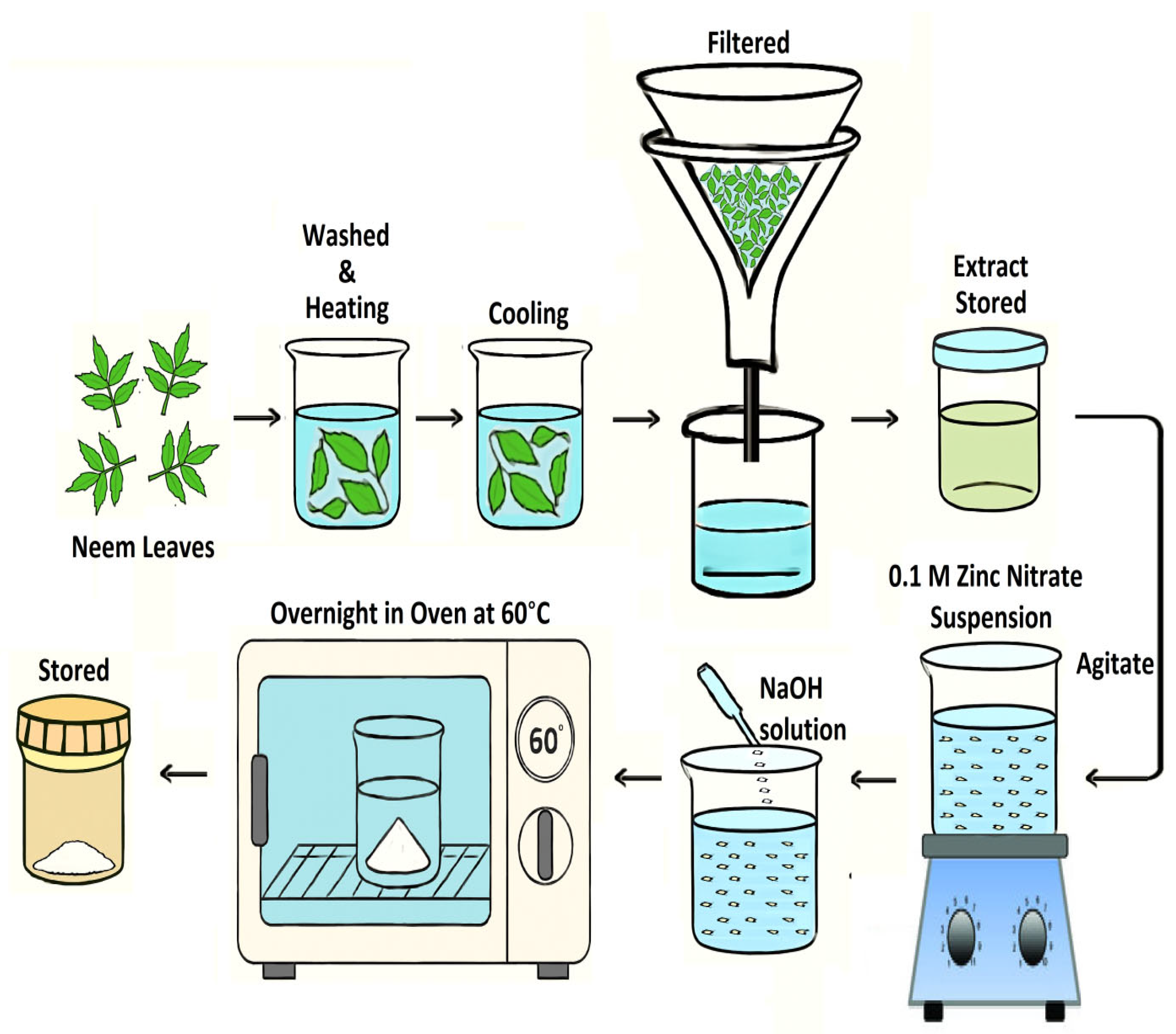
4.2. Extraction of Leaf Extract and Green Synthesis of ZnO NS

Neem (CJS 16628) [76] is a tree 40–50 feet or higher, with a straight trunk and long spreading branches forming a broad, round crown; it has rough, dark brown bark with wide longitudinal fissures separated by flat ridges. The leaves are compound, imparipinnate, each comprising 5–15 leaflets. The compound leaves are themselves alternating with one another. It bears many flowered panicles, mostly in the leaf axils. The sepals are ovate and about 1 cm long, with sweet-scented white oblancolate petals. It produces yellow drupes that are ellipsoid and glabrous, 12–20 mm long. Fruits are green, turning yellow on ripening, aromatic with garlic like odor [Figure 13] [77].
The neem (A. indica) leaves were freshly picked, diligently cleaned with regular water to get rid of dust and chemical contaminants, treated with DDW, cut into long, thin strips, placed in a beaker filled with DDW, and then dried in the air at room temperature. After a half-hour boiling session, the liquid underwent cooling and was subsequently filtered using Whatman No. 1 paper. For later use, the obtained extracted neem was stored at 4 °C in a covered beaker. The ZnO NSs were synthesized by adding 10 mL of neem extract to 90 mL of 0.1 M zinc nitrate suspension, followed by agitation at room temperature for 4 h. After 2 h of continuous agitation, 50 mL of 2.0 M NaOH solution was added to the mixture. The procedure yielded a white solid, which was subsequently filtered and washed repeatedly with DDW as well as ethanol to eliminate pollutants. The settled precipitate was subsequently stored in the oven through the night at around 60 °C [Scheme 1]. The technique for synthesizing ZnO NS using neem leaf extract as nanostructures acquired them as a fine white powder for analysis. In a previous study, ZnO NPs were synthesized using neem leaf aqueous extracts and characterized using transmission electron microscopy (TEM), ultraviolet visible spectroscopy (UV-Vis), dynamic light scattering (DLS), and antibacterial activity against Gram-positive and Gram-negative bacteria [12]. In the present study, A. indica is used in the production of ZnO NSs to enhance nematicidal effectiveness against root-knot nematodes (RKNs) (M. incognita), known for inflicting harm on tomato plants.

4.3. Characterization of Biosynthesized-ZnO NS

The biosynthesized ZnO NS underwent comprehensive characterization employing a range of advanced analytical techniques, including SEM, FT-IR, TEM, UV-vis, EDAX, and XRD. Characterization of the ZnO NS was performed using various techniques. X-ray diffraction (XRD) analysis was conducted using a Rigaku (Tokyo, Japan) SmartLab X-ray diffractometer with Cu-Kα radiation (λ = 1.5406 Å) in the 2θ range of 30–70°, offering detailed insights into its structural characteristics. The crystalline structure and stages of the ZnO NS were analyzed using an X-ray diffractometer. The scan covered the 2θ range of 30–70° using an X-ray beam (monochromatic) filtered through nickel to emit Cu-Kα radiation. The scanning rate was set at 0.02 steps per second with a step size of 0.01°. X-rays were produced with a voltage of 40 kilovolts (kV) and a current of 40 kiloamperes (kA). Scanning electron microscopy (SEM) was carried out using a JEOL JSM-6510 LV SEM (Tokyo, Japan) at 200 kV with magnifications ranging from 12,000× to 35,000×, providing extensive information on surface morphology. Transmission electron microscopy (TEM) was performed using a JEOL JEM-2100F (Tokyo, Japan), revealing the morphology and dimensions of the ZnO NS UV-vis absorption spectra were recorded using a PerkinElmer (Waltham, MA, USA) Lambda 950 UV-Vis spectrophotometer, covering a wavelength range of 200–800 nm. Fourier-transform infrared (FTIR) spectroscopy was conducted with a Thermo Scientific Nicolet iS10 FT-IR spectrometer in the range of 4000–400 cm−1, enabling chemical characterization of the nanostructures. The crystallinity of the material was further assessed through XRD, providing key information about the crystalline phase and structural orientation. Zeta potential measurements were conducted individually using a ZetaSizer-HT from Malvern, UK. Collectively, these techniques provided a thorough and multifaceted understanding of the structural, chemical, and morphological properties of the ZnO NSs, which are crucial for evaluating their potential applications.

4.4. Gathering and Multiplying the Inoculum (J2s) of M. incognita

Meloidogyne spp.-infested roots were taken from a field in the Samtah, Jazan. According to Eisenback and Hunt’s instructions, the perineal pattern was examined to validate the identification of M. incognita [78]. The juveniles in their second stage (J2s) were raised on studied plants kept in a greenhouse. The plants were carefully removed to ensure the egg masses stayed connected to the roots. After that, all dirt was removed from the roots by carefully rinsing them with DDW. The egg masses were extracted from the infected roots using sterilized scissors. After that, they were cleaned with DDW and put on 25 μm mesh sieves with crossed layers of tissue paper. To encourage J2 hatching, these sieves were positioned in Petri dishes with just enough water to cover the egg masses. After that, a BOD incubator was used to hold the Petri dishes in order to speed up the hatching process. Because of the mesh sieves, the hatched J2s could traverse the mesh and settle at the bottom of the Petri dishes. Daily collections of the hatching J2 suspension were made, and DDW was added. The freshly hatched J2s concentration was standardized as required and preserved for further research.

4.5. Mortality Test

Various ZnO NS concentrations (150, 300, 450, and 600 ppm) were estimated for nematicidal action against J2s of M. incognita, and LC50 values generated for each of the treatments. To determine how ZnO NS affected J2 mortality, 8 mL of ZnO NS at several concentrations were administered to petri dishes containing 70 newly hatched J2s and 2 mL of DDW each. Petri dishes with only DDW are used as controls. A dissecting microscope was used to measure J2 mortality after 12, 24, 48, and 72 h of incubation. To prevent evaporation, petri dishes are coated with parafilm. There were six replicas of every treatment. If the J2s moved or formed a zigzag shape, they were regarded as alive [79]. However, when examined with a very fine needle after being moved to tap water, dead J2s showed no motility [80]. The LC50 values for all treatments were calculated using probit analysis [81,82]. The subsequent calculation was employed to calculate percent mortality for every treatment.
M o r t a l i t y   %   o f   J 2 s = I n i t i a l   J 2 s S u r v i v i n g   J 2 s I n i t i a l   J 2 s × 100

4.6. Hatching Bioassay

Employing the egg mass dipping method, the inhibitory effect of ZnO NS on J2 hatching was tested at different concentrations (150, 300, 450, and 600 ppm). Five egg masses of comparable size were carefully removed from infected roots, and the ZnO NS solutions, at their designated concentrations, were carefully dispensed into Petri plates (10 mL per plate). The petri dishes were sealed with parafilm and maintained at 28 °C for incubation to reduce evaporation. As the control, five more egg masses were added to Petri plates containing DDW. To ensure statistical reliability, every treatment was replicated six times. The inhibition of J2 hatching was subsequently calculated (%) using the formula proposed by Almutairi et al. [83].
H a t c h i n g   i n h i b i t i o n   % = C 0 T α C 0 × 100
where
  • C0 = Count of J2s Emerged in DDW (Control);
  • Tα = Count of Emerged J2s in each concentration of synthesized ZnO NS.

4.7. J2s Infection Bioassay

J2s’ rate of penetration into tomato plant roots was measured in a lab setting. To develop tomato seedlings, sensitive tomato cultivar seeds planted in pots and kept in a greenhouse. Three-week-old seedlings were subsequently transplanted into individual disposable 7 cm tea cups, each containing 40 g of thoroughly cleaned river sand. Then, 50 J2s per pot and 10 mL of ZnO NS at different concentrations (150, 300, 450, and 600 ppm), organized in a randomized full block design, were applied to each seedling. To assess the degree of J2 penetration, the tomato plants that had been infected with J2s were gently pulled after five days. After giving the root systems a thorough washing under running water to get rid of any last bits of sand, they were dried off with paper towels. To see the J2s, the roots were divided into 2 mm slices and stained with a fuchsin–lactoglycerol solution. Lactoglycerol was then used to clean the roots [84]. To determine the percentage of penetration, the dried roots were placed on glass slides and examined under a stereomicroscope. The number of J2s that pierced was counted.

4.8. In Silico Analysis

Molecular docking studies were performed to investigate the interaction of Heat Shock Protein 90 (Hsp90) (PDB ID: 2XJX) and Odorant Response 1 protein (ODR1) (PDB ID: 3K1E) with ZnO NS using AutoDock 4.2. The three-dimensional crystal structures of Hsp90 and ODR1 were retrieved from the Protein Data Bank (PDB), while the structural information for ZnO NS was obtained from the literature or modelled computationally [85,86]. The target proteins were pre-processed using Discovery Studio and PyMOL, where water molecules and heteroatoms were removed to eliminate non-relevant interactions. Hydrogen atoms were added to maintain the correct protonation state. The AutoDock Tools (ADT) module was used to assign Gasteiger charges to the proteins and ZnO NS. The ZnO NS were prepared as ligands by optimizing their geometry and ensuring their stability before docking.
AutoDock 4.2 was employed for docking simulations, with Lamarckian Genetic Algorithm (LGA) used as the search algorithm. The docking grid was defined with dimensions 120 × 120 × 120 Å to encompass the entire active site and allow flexible binding conformations. The exhaustiveness was set to 100 runs to ensure optimal binding pose selection. Other docking parameters included a population size of 150, maximum evaluations of 2,500,000, and energy minimization of 10,000 iterations [87,88]. After docking, the best binding conformations were selected based on the lowest binding energy (kcal/mol) and their molecular interactions. The AutoDock results were visualized in Discovery Studio and PyMOL to analyse hydrogen bonding, hydrophobic interactions, and electrostatic forces between ZnO NS and the target proteins. The interaction details were tabulated, including binding affinities and amino acid residues involved in key interactions [89,90].

4.9. Statistical Analysis

The gathered data, which encompassed a wide variety of features in inquiry, were statistically analyzed by R software (version 2.14.1). Duncan’s Multiple Range Test (DMRT) was used to evaluate whether the analyzed attributes predicted significant differences (p = 0.05). OPSTAT [91], was used to do an ANOVA. The LC50 values corresponding to each treatment were determined through analysis using the OPSTAT software (http://14.139.232.166/opstat/) (Access date: 12 February 2025), ensuring precise statistical computation [91].

5. Conclusions

This study validates that biosynthesized ZnO NS, derived from neem leaf extract, offer a promising eco-friendly alternative for controlling M. incognita infestations. The results indicate significant nematicidal activity with high mortality rates of J2s and effective suppression of egg hatching, with ZnO NS outperforming traditional chemical nematicides. The green synthesis method employed aligns with sustainable agricultural practices, reducing reliance on harmful chemicals. Future research should focus on optimizing the application techniques, assessing long-term effects in field conditions, and exploring the broader applicability of ZnO NS in integrated pest management systems. Furthermore, molecular docking studies suggest that ZnO NS interacts with key proteins such as Hsp90 and ODR1, which may influence nematode behavior and cellular function, warranting further computational and experimental investigations. Compared to other nanomaterials, ZnO NS offers a safer, more environmentally friendly solution, making it a viable candidate for future pest control strategies in sustainable agriculture.

Author Contributions

G.K., A.A.C., M.I., K.A. and S.K.A. Conceptualization, Methodology, Supervision, Funding Acquisition, Writing original draft, Writing review and editing. A.M.M., A.A.A., F.A. and S.K.A. Formal analysis, Investigations, Resource, Writing review and editing. G.K., M.S.A., M.I.A., N.R. and M.A.M.A. Visualization, Data curation, Writing review and editing. All authors have read and agreed to the published version of the manuscript.

Funding

This research was funded by the Deanship of Scientific Research at Imam Mohammad Ibn Saud Islamic University (IMSIU) grant number IMSIU-DDRSP2501.

Data Availability Statement

The authors confirm that all the experimental data are available and accessible via the main text.

Acknowledgments

This work was supported and funded by the Deanship of Scientific Research at Imam Mohammad Ibn Saud Islamic University (IMSIU) (grant number IMSIU-DDRSP2501).

Conflicts of Interest

The authors declare no conflict of interest.

References

  1. Abdelkhalek, A.A.; Al-Askar, A.A.; Behiry, S.I. Bacillus licheniformis strain POT1 mediated poly-phenol biosynthetic pathways genes activation and systemic resistance in potato plants against Alfalfa mosaic virus. Sci. Rep. 2020, 10, 16120. [Google Scholar] [CrossRef] [PubMed]
  2. Abo-Zaid, G.A.; Matar, S.M.; Abdelkhalek, A. Induction of plant resistance against tobacco mosaic virus using the biocontrol agent Streptomyces cellulosae isolate Actino 48. Agronomy 2020, 10, 1620. [Google Scholar] [CrossRef]
  3. Hussain, M.A.; Mukhtar, T.; Kayani, M.Z. Reproduction of Meloidogyne incognita on resistant and susceptible okra cultivars. Pak. J. Agric. Sci. 2016, 53, 371–375. [Google Scholar] [CrossRef]
  4. El-Saadony, M.T.; Khalil, O.S.; Osman, A.; Alshilawi, M.S.; Taha, A.E.; Aboelenin, S.M.; Saad, A.M. Bioactive peptides supplemented raw buffalo milk: Biological activity, shelf life and quality properties during cold preservation. Saudi J. Biol. Sci. 2021, 28, 4581–4591. [Google Scholar] [CrossRef] [PubMed]
  5. Saad, A.M.; El-Saadony, M.T.; El-Tahan, A.M.; Sayed, S.; Moustafa, M.A.; Taha, A.E.; Ramadan, M.M. Polyphenolic extracts from pomegranate and watermelon wastes as substrate to fabricate sustainable silver nanoparticles with larvicidal effect against Spodopteralittoralis. Saudi J. Biol. Sci. 2021, 28, 5674–5683. [Google Scholar] [CrossRef] [PubMed]
  6. Cromwell, W.; Yang, J.; Starr, J.; Jo, Y.K. Nematicidal effects of silver nanoparticles on root-knot nematode in bermudagrass. J. Nematol. 2014, 46, 261–266. [Google Scholar] [PubMed]
  7. Abdelkhalek, A.; Salem, M.Z.M.; Hafez, E.; Behiry, S.I.; Qari, S.H. The phytochemical, antifungal, and first report of the antiviral properties of Egyptian Haplophyllumtuberculatum extract. Biology 2020, 9, 248. [Google Scholar] [CrossRef] [PubMed]
  8. Kaliyamoorthy, T.S.; Subramaniyan, V.; Renganathan, S.; Elavarasan, V.; Ravi, J.; Prabhakaran Kala, P.; Vijayakumar, S. Sustainable Environmental-Based ZnO Nanoparticles Derived from Pisonia grandis for Future Biological and Environmental Applications. Sustainability 2022, 14, 17009. [Google Scholar] [CrossRef]
  9. Gulab, H.; Fatima, N.; Tariq, U.; Gohar, O.; Irshad, M.; Khan, M.Z.; Hanif, M.B. Advancements in Zinc Oxide Nanomaterials: Synthesis, Properties, and Diverse Applications. Nano Struct. Nano Objects 2024, 39, 101271. [Google Scholar] [CrossRef]
  10. Tortella, G.; Rubilar, O.; Pieretti, J.C.; Fincheira, P.; de Melo Santana, B.; Fernández-Baldo, M.A.; Seabra, A.B. Nanoparticles as a Promising Strategy to Mitigate Biotic Stress in Agriculture. Antibiotics 2023, 12, 338. [Google Scholar] [CrossRef] [PubMed]
  11. Danish, M.; Shahid, M.; Farah, M.A.; Al-Anazi, K.M.; Ahmed, S.M.; Mohamed, H.I.; Ahamad, L. CuO–ZnO Nanocomposites Mitigate Root-Knot Nematode Stress in Vigna radiata by Enhancing Physiological and Antioxidant Defense Responses. Physiol. Mol. Plant Pathol. 2025, 139, 102776. [Google Scholar] [CrossRef]
  12. El-Beltagi, H.S.; Ragab, M.; Osman, A.; El-Masry, R.A.; Alwutayd, K.M.; Althagafi, H.; El-Saber, M.M. Biosynthesis of Zinc Oxide Nanoparticles via Neem Extract and Their Anticancer and Antibacterial Activities. PeerJ 2024, 12, e17588. [Google Scholar] [CrossRef] [PubMed]
  13. Sahoo, S.K.; Dwivedi, G.K.; Dey, P.; Praharaj, S. Green Synthesized ZnO Nanoparticles for Sustainable Production and Nutritional Biofortification of Green Gram. Environ. Technol. Innov. 2021, 24, 101957. [Google Scholar] [CrossRef]
  14. Jindal, A.; Bhat, A.H.; Raja, V.; Ahmad, S.S.; Hussain, M.A.; Ataya, F.S.; Fouad, D. Eco-Friendly Synthesis of Zinc Oxide Nanoparticles from Achillea millefolium: Multifunctional Applications in Plant Growth and Nematode Control. Physiol. Mol. Plant Pathol. 2025, 136, 102590. [Google Scholar] [CrossRef]
  15. Hussain, M.A.; Parveen, G.; Bhat, A.H.; Reshi, Z.A.; Ataya, F.S.; Handoo, Z.A. Harnessing Walnut-Based Zinc Oxide Nanoparticles: A Sustainable Approach to Combat the Disease Complex of Meloidogyne arenaria and Macrophomina phaseolina in Cowpea. Plants 2024, 13, 1743. [Google Scholar] [CrossRef] [PubMed]
  16. Mohammad, F.; Bwatanglang, I.B.; Al-Lohedan, H.A.; Shaik, J.P.; Al-Tilasi, H.H.; Soleiman, A.A. Influence of Surface Coating towards the Controlled Toxicity of ZnO Nanoparticles In Vitro. Coatings 2023, 13, 172. [Google Scholar] [CrossRef]
  17. Ansari, A.; Ahmed, S.; Rehman, B.; Ali, S.K.; Azooz, R.E.; Hassan, K.F.; Khan, A.; Ranjan, P.; Negi, D.S. A synergistic effect of ZnO low dimensional rods/PEDOT:PSS hybrid structure for UV radiation detection. Appl. Phys. A 2024, 130, 7849. [Google Scholar] [CrossRef]
  18. Ahmed, S.; Ansari, A.; Bishwanathan, S.; Siddiqui, M.A.; Tailor, S.; Gupta, P.K.; Negi, D.S.; Ranjan, P. Electronic tongue based on ZnO/ITO@glass for electrochemical monitoring of spiciness levels. Langmuir 2024, 40, 4434–4446. [Google Scholar] [CrossRef] [PubMed]
  19. Abutaleb, A.; Ahmed, S.; Imran, M. Synergistic photocatalysis: Harnessing WSe2-ZnOnanocomposites for efficient malachite green dye degradation. Eur. Phys. J. Plus 2023, 138, 1046. [Google Scholar] [CrossRef]
  20. Beydaghdari, M.; Saboor, F.H.; Babapoor, A.; Karve, V.V.; Asgari, M. Recent Advances in MOF-Based Adsorbents for Dye Removal from the Aquatic Environment. Energies 2022, 15, 2023. [Google Scholar] [CrossRef]
  21. Lebaka, V.R.; Ravi, P.; Reddy, M.C.; Thummala, C.; Mandal, T.K. Zinc Oxide Nanoparticles in Modern Science and Technology: Multifunctional Roles in Healthcare, Environmental Remediation, and Industry. Nanomaterials 2025, 15, 754. [Google Scholar] [CrossRef] [PubMed]
  22. Anandhan, K.; Harish, S.; Tilak Kumar, R. Effect of Morphology on the Formation of CdO Nanostructures for Antibacterial and Hemolytic Studies. Appl. Surf. Sci. 2019, 489, 262–268. [Google Scholar] [CrossRef]
  23. Priyadharsini, N.; Bhuvaneswari, N.; Joshy, J. Plant Mediated Synthesis of ZnO and Mn Doped ZnO Nanoparticles Using Carica papaya Leaf Extract for Antibacterial Application. Asian J. Appl. Sci. Technol. 2021, 5, 69–81. [Google Scholar] [CrossRef]
  24. Ali, S.S.; Sonbol, F.I.; Sun, J.; Hussein, M.A.; Hafez, A.-E.E.; Abdelkarim, E.A.; Kornaros, M.; Ali, A.; Azab, M. Molecular Characterization of Virulence and Drug Resistance Genes-Producing Escherichia coli Isolated from Chicken Meat: Metal Oxide Nanoparticles as Novel Antibacterial Agents. Microb. Pathog. 2020, 143, 104164. [Google Scholar] [CrossRef] [PubMed]
  25. Ali, S.S.; Moawad, M.S.; Hussein, M.A.; Azab, M.; Abdelkarim, E.A.; Badr, A.; Sun, J.; Khalil, M. Efficacy of Metal Oxide Nanoparticles as Novel Antimicrobial Agents against Multi-Drug and Multi-Virulent Staphylococcus aureus Isolates from Retail Raw Chicken Meat and Giblets. Int. J. Food Microbiol. 2021, 344, 109116. [Google Scholar] [CrossRef] [PubMed]
  26. Mishra, D.N.; Prasad, L.; Suyal, U. Synthesis of Zinc Oxide Nanoparticles Using Trichoderma harzianum and Its Bio-Efficacy on Alternaria brassicae. Front. Microbiol. 2025, 16, 1506695. [Google Scholar] [CrossRef] [PubMed]
  27. Welch, K.D.; Davis, T.Z.; Van Eden, M.E.; Aust, S.D. Deleterious iron-mediated oxidation of biomolecules. Free. Radic. Biol. Med. 2002, 32, 577–583. [Google Scholar] [CrossRef] [PubMed]
  28. Aftab, Z.-e.-H.; Mirza, F.S.; Anjum, T.; Rizwana, H.; Akram, W.; Aftab, M.; Ali, M.D.; Li, G. Antifungal Potential of Biogenic Zinc Oxide Nanoparticles for Controlling Cercospora Leaf Spot in Mung Bean. Nanomaterials 2025, 15, 143. [Google Scholar] [CrossRef] [PubMed]
  29. Mannai, Z.; Bouslama, W.; Karkouch, I.; Bouslama, L.; Khlifi, K.; Aouadi, K.; Nouira, F. Synthesis and Characterization of ZnO and CuO Coatings for Antibacterial and Antiviral Applications. Mater. Chem. Phys. 2025, 329, 130071. [Google Scholar] [CrossRef]
  30. Gupta, A.; Bharati, R.; Kubes, J.; Popelkova, D.; Praus, L.; Yang, X.; Severova, L.; Skalicky, M.; Brestic, M. Zinc Oxide Nanoparticles Application Alleviates Salinity Stress by Modulating Plant Growth, Biochemical Attributes and Nutrient Homeostasis in Phaseolus vulgaris L. Front. Plant Sci. 2024, 15, 1432258. [Google Scholar] [CrossRef] [PubMed]
  31. Freinbichler, W.; Colivicchi, M.A.; Stefanini, C.; Bianchi, L.; Ballini, C.; Misini, B.; Weinberger, P.; Linert, W.; Varešlija, D.; Tipton, K.F.; et al. Highly reactive oxygen species: Detection, formation, and possible functions. Cell Mol. Life Sci. 2011, 68, 2067–2079. [Google Scholar] [CrossRef] [PubMed] [PubMed Central]
  32. Zhang, C.; Ding, N.; Pan, Y.; Fu, L.; Zhang, Y. The degradation pathways of contaminants by reactive oxygen species generated in the Fenton/Fenton-like systems. Chin. Chem. Lett. 2024, 35, 109579. [Google Scholar] [CrossRef]
  33. Raza, M.A.S.; Muhammad, F.; Farooq, M.; Aslam, M.U.; Akhter, N.; Toleikienė, M.; Iqbal, R. ZnO-Nanoparticles and Stage-Based Drought Tolerance in Wheat (Triticum aestivum L.): Effect on Morpho-Physiology, Nutrient Uptake, Grain Yield and Quality. Sci. Rep. 2025, 15, 5309. [Google Scholar] [CrossRef] [PubMed]
  34. Chen, H.; Song, Y.; Wang, Y.; Wang, H.; Ding, Z.; Fan, K. ZnO Nanoparticles: Improving Photosynthesis, Shoot Development, and Phyllosphere Microbiome Composition in Tea Plants. J. Nanobiotechnol. 2024, 22, 389. [Google Scholar] [CrossRef] [PubMed]
  35. Nguyen, T.M.P.; Hirota, K.; Kato, M.; Tsukagoshi, K.; Yamada, H.; Terabe, A.; Mizutani, H. Addition Dependence of Antibacterial Activity of ZnO Powders on Their Physico-chemical Properties. J. Jpn. Soc. Powder Powder Metall. 2019, 66, 434–441. [Google Scholar] [CrossRef]
  36. Tryfon, P.; Antonoglou, O.; Vourlias, G.; Mourdikoudis, S.; Menkissoglu-Spiroudi, U.; Dendrinou-Samara, C. Tailoring Ca-based nanoparticles by polyol process for use as nematicidals and pH adjusters in agriculture. ACS Appl. Nano Mater. 2019, 2, 3870–3881. [Google Scholar] [CrossRef]
  37. El-Argawy, E.; Rahhal, M.; El-Korany, A.; Elshabrawy, E.; Eltahan, R. Efficacy of some nanoparticles to control damping-off and root rot of sugar beet in El-Behiera Governorate. Asian J. Plant Pathol. 2017, 11, 35–47. [Google Scholar] [CrossRef]
  38. Lee, A.; Kim, D. CRDS: Consensus reverse docking system for target fishing. Bioinformatics 2020, 36, 959–960. [Google Scholar] [CrossRef] [PubMed]
  39. Shibata, T.; Uchida, K. Protein adductomics: A comprehensive analysis of protein modifications by electrophiles. Free. Radic. Biol. Med. 2019, 144, 218–222. [Google Scholar] [CrossRef] [PubMed]
  40. Bharathi, A.; Roopan, S.M.; Vasavi, C.S.; Munusami, P.; Gayathri, G.A.; Gayathri, M. In silico molecular docking and in vitro antidiabetic studies of dihydropyrimido [4,5-a]acridin-2-amines. Biomed. Res. Int. 2014, 2014, 971569. [Google Scholar] [CrossRef] [PubMed]
  41. Kundu, A.; Dutta, A.; Mandal, A.; Negi, L.; Malik, M.; Puramchatwad, R.; Antil, J.; Singh, A.; Rao, U.; Saha, S.; et al. A comprehensive in vitro and in silico analysis of nematicidal action of essential oils. Front. Plant Sci. 2021, 11, 614143. [Google Scholar] [CrossRef] [PubMed]
  42. Bhadwal, N.; Ben Mrad, R.; Behdinan, K. Review of Zinc Oxide Piezoelectric Nanogenerators: Piezoelectric Properties, Composite Structures and Power Output. Sensors 2023, 23, 3859. [Google Scholar] [CrossRef] [PubMed]
  43. Le, A.T.; Ahmadipour, M.; Pung, S.Y. A Review on ZnO-Based Piezoelectric Nanogenerators: Synthesis, Characterization Techniques, Performance Enhancement and Applications. J. Alloys Compd. 2020, 844, 156172. [Google Scholar] [CrossRef]
  44. Abdelmohsen, A.H.; Rouby, W.M.E.; Ismail, N.; Farghali, A.A. Morphology transition engineering of ZnO nanorods to nanoplatelets grafted Mo8O23-MoO2 by polyoxometalates: Mechanism and possible applicability to other oxides. Sci. Rep. 2017, 7, 5946. [Google Scholar] [CrossRef] [PubMed]
  45. Ramadan, K.M.; El-Beltagi, H.S.; Bendary, E.S.; Ali, H.M. Experimental Evaluation of the Antioxidant and Antitumor Activities of Thyme and Basil Essential Oils and Their Phenolic Constituents: Theoretical Antioxidant Evaluation. Chem. Biol. Technol. Agric. 2022, 9, 102. [Google Scholar] [CrossRef]
  46. Ramadan, K.M.A.; El-Beltagi, H.S.; Mohamed, H.I.; Shalaby, T.A.; Galal, A.; Mansour, A.T.; Aboul Fotouh, M.M.; Bendary, E.S.A. Antioxidant, Anti-Cancer Activity and Phytochemicals Profiling of Kigelia pinnata Fruits. Separations 2022, 9, 379. [Google Scholar] [CrossRef]
  47. Paul, R.; Prasad, M.; Sah, N.K. Anticancer Biology of Azadirachta indica L. (Neem): A Mini Review. Cancer Biol. Ther. 2011, 12, 467–476. [Google Scholar] [CrossRef] [PubMed]
  48. Al Saiqali, M.; Tangutur, A.D.; Banoth, C.; Bhukya, B. Antimicrobial and Anticancer Potential of Low Molecular Weight Polypeptides Extracted and Characterized from Leaves of Azadirachta indica. Int. J. Biol. Macromol. 2018, 114, 906–921. [Google Scholar] [CrossRef] [PubMed]
  49. Soares, D.G.; Godin, A.M.; Menezes, R.R.; Nogueira, R.D.; Brito, A.M.S.; Melo, I.S.F.; Machado, R.R. Anti-Inflammatory and Antinociceptive Activities of Azadirachtin in Mice. Planta Med. 2014, 80, 630–636. [Google Scholar] [CrossRef] [PubMed]
  50. Parida, M.M.; Upadhyay, C.; Pandya, G.; Jana, A.M. Inhibitory Potential of Neem (Azadirachta indica Juss) Leaves on Dengue Virus Type-2 Replication. J. Ethnopharmacol. 2002, 79, 273. [Google Scholar] [CrossRef] [PubMed]
  51. Vasanth, S.; Hamsaveni Gopal, R.; Rao, R.B. Plant Anti-Malarial Agents. J. Sci. Ind. Res. 1990, 49, 68–77. [Google Scholar]
  52. Lokanadhan, S.; Muthukrishnan, P.; Jeyaraman, S. Neem Products and Their Agricultural Applications. J. Biopestic. 2012, 5, 72. [Google Scholar] [CrossRef]
  53. Goel, N.; Anukrati, K.; Paul, P.K. Anti-Phytopathogenic and SAR Inducing Properties of Neem: A Review. J. Chem. Pharm. Sci. 2016, 9, 2547–2555. [Google Scholar]
  54. Chaudhary, R.F.; Patel, R.L.; Chaudhari, S.M.; Pandey, S.K.; Brajesh, S. In Vitro Evaluation of Different Plant Extracts against Alternaria alternata Causing Early Blight of Potato. J. Indian Potato Assoc. 2003, 30, 141–142. [Google Scholar]
  55. Narde, J.; Ahmed, N.; Maiti, S.; Rajeshkumar, S.; Ganapathy, D. Green Synthesis of Silver Nanoparticles from Aloe vera and Neem Leaf Extract and Their Cytotoxic Effect Evaluation. J. Popul. Ther. Clin. Pharmacol. 2023, 30, 174–180. [Google Scholar] [CrossRef]
  56. Sangeetha, G.; Rajeshwari, S.; Venckatesh, R. Green Synthesis of Zinc Oxide Nanoparticles by Aloe barbadensis Miller Leaf Extract: Structure and Optical Properties. Mater. Res. Bull. 2011, 46, 2560–2566. [Google Scholar] [CrossRef]
  57. Alharbi, F.N.; Abaker, Z.M.; Makawi, S.Z.A. Phytochemical Substances Mediated Synthesis of Zinc Oxide Nanoparticles (ZnO NPs). Inorganics 2023, 11, 328. [Google Scholar] [CrossRef]
  58. Eltak, N.A.; Gniedy, N.A.; Abdel-Haleem, D.R.; Farag, S.M. Based on GC-MS Analysis: An Evaluation Activity of Some Algal Extracts against Culex pipiens L. (Diptera: Culicidae). Egypt. J. Aquat. Biol. Fish. 2023, 27, 461–489. [Google Scholar] [CrossRef]
  59. Sohail, M.F.; Rehman, M.; Hussain, S.Z.; Huma, Z.E.; Shahnaz, G.; Qureshi, O.S.; Qandeel, K.; Shaper, M.; Irshad, H.; Webster, T.J. Green Synthesis of Zinc Oxide Nanoparticles by Neem Extract as Multi-Facet Therapeutic Agents. J. Drug Deliv. Sci. Technol. 2020, 59, 101911. [Google Scholar] [CrossRef]
  60. Fouda, F. Synthesis and Characterization of Zinc Oxide Nanoparticles (ZnO-NPs) from Leaves of Some Plants. Ann. Agric. Sci. Moshtohor 2023, 61, 97–110. [Google Scholar] [CrossRef]
  61. Zhang, Z.; Xiong, T.; Peng, H.; Zhang, H.; He, S.; Liu, X.; Liu, Y.; Feng, W.; Yang, Z.; Xiong, W. Optimization of Fenton-like reaction pathways using O v-containing ZnO@ nitrogen-rich porous carbon: The electron transfer and 1O2 triggered non-radical process. Environ. Sci. Nano 2025, 12, 936–947. [Google Scholar] [CrossRef]
  62. Khan, M.R.; Rizvi, T.F. Nanotechnology: Scope and Application in Plant Disease Management. Plant Pathol. J. 2014, 13, 214–231. [Google Scholar] [CrossRef]
  63. Yogamalar, N.R.; Bose, A.C. Absorption–Emission Study of Hydrothermally Grown Al:ZnO Nanostructures. J. Alloys Compd. 2011, 509, 8493–8500. [Google Scholar] [CrossRef]
  64. El-Ashry, R.M.; El-Saadony, M.T.; El-Sobki, A.E.; El-Tahan, A.M.; Al-Otaibi, S.; El-Shehawi, A.M.; Saad, A.M.; Elshaer, N. Biological Silicon Nanoparticles Maximize the Efficiency of Nematicides Against Biotic Stress Induced by Meloidogyne incognita in Eggplant. Saudi J. Biol. Sci. 2021, 29, 920–932. [Google Scholar] [CrossRef] [PubMed]
  65. Sharon, M.; Choudhary, A.K.; Kumar, R. Nanotechnology in Agricultural Diseases and Food Safety. J. Phytol. 2010, 2, 83–92. [Google Scholar]
  66. Nguyen, V.T.; Vu, V.T.; Nguyen, T.A.; Tran, V.K.; Nguyen-Tri, P. Antibacterial Activity of TiO2- and ZnO-Decorated with Silver Nanoparticles. J. Compos. Sci. 2019, 3, 61. [Google Scholar] [CrossRef]
  67. Ahamed, M.; Posgai, R.; Gorey, T.J.; Nielsen, M.; Hussain, S.M.; Rowe, J.J. Silver Nanoparticles Induced Heat Shock Protein 70, Oxidative Stress and Apoptosis in Drosophila melanogaster. Toxicol. Appl. Pharmacol. 2010, 242, 263–269. [Google Scholar] [CrossRef] [PubMed]
  68. Dammini-Premachandra, W.T.C.; Mampitiyarachchi, H.; Ebssa, L. Nemato-Toxic Potential of Betel (Piper betle L.) (Piperaceae) Leaf. J. Crop Prot. 2014, 65, 1–5. [Google Scholar] [CrossRef]
  69. Sharma, M.; Saini, I.; Kaushik, P.; Aldawsari, M.M.; Al Balawi, T.; Alam, P. Mycorrhizal fungi and Pseudomonas fluorescens application reduces root-knot nematode (Meloidogyne javanica) infestation in eggplant. Saudi J. Biol. Sci. 2021, 28, 3685–3691. [Google Scholar] [CrossRef] [PubMed]
  70. Gao, H.; Qi, G.; Yin, R.; Zhang, H.; Li, C.; Zhao, X. Bacillus cereus Strain S2 Shows High Nematicidal Activity Against Meloidogyne incognita by Producing Sphingosine. Sci. Rep. 2016, 6, 28756. [Google Scholar] [CrossRef] [PubMed]
  71. Mohamed, E.A.; Elsharabasy, S.F.; Abdulsamad, D. Evaluation of In Vitro Nematicidal Efficiency of Copper Nanoparticles against Root-Knot Nematode Meloidogyne incognita. S. Asian J. Parasitol. 2019, 2, 13–18. [Google Scholar]
  72. Esposito, S. “Traditional” Sol-Gel Chemistry as a Powerful Tool for the Preparation of Supported Metal and Metal Oxide Catalysts. Materials 2019, 12, 668. [Google Scholar] [CrossRef] [PubMed]
  73. Bruna, T.; Maldonado-Bravo, F.; Jara, P.; Caro, N. Silver Nanoparticles and Their Antibacterial Applications. Int. J. Mol. Sci. 2021, 22, 7202. [Google Scholar] [CrossRef] [PubMed]
  74. Serov, D.A.; Gritsaeva, A.V.; Yanbaev, F.M.; Simakin, A.V.; Gudkov, S.V. Review of Antimicrobial Properties of Titanium Dioxide Nanoparticles. Int. J. Mol. Sci. 2024, 25, 10519. [Google Scholar] [CrossRef] [PubMed]
  75. Enyiukwu, D.N.; Ononuju, C.C.; Okeke, C.; Chukwu, L.A. Plant Parasitic Nematodes, Serious but Most Trivialized Biotic Challenge against Food Security: A Spotlight on Their Management for Sustainable Agriculture and Public Health. Direct Res. J. Public Health Environ. Technol. 2021, 6, 104–112. [Google Scholar]
  76. Mohammad, I.; Khan, M.S.; Ansari, M.R.; Bari, M.N. Exploring the potential of Neem (Azadirachta indica A. Juss.) as a sustainable biopesticide: Opportunities in Sudan. Ann. Phytomed. 2024, 13, 418–424. [Google Scholar] [CrossRef]
  77. Imam, H.; Hussain, A.; Ahmed, A. Neem (Azadirachta indica A. Juss)—A Nature’s Drugstore: An Overview. Res. J. Pharm. Biol. Chem. Sci. 2012, 3, 76–79. [Google Scholar]
  78. Eisenback, J.D.; Hunt, D.J. General Morphology. In Root-Knot Nematodes; Starr, J.L., Moens, M., Perry, R.N., Eds.; CABI: Wallingford, UK, 2009; pp. 18–54. [Google Scholar]
  79. El-Rokiek, K.G.; El-Nagdi, W.M. Dual Effects of Leaf Extracts of Eucalyptus citriodora on Controlling Purslane and Root-Knot Nematode in Sunflower. J. Plant Prot. Res. 2011, 51, 121–129. [Google Scholar] [CrossRef]
  80. Aissani, N.; Urgeghe, P.P.; Oplos, C.; Saba, M.; Tocco, G.; Petretto, G.L. Nematicidal Activity of the Volatilome of Eruca sativa on Meloidogyne incognita. J. Agric. Food Chem. 2015, 63, 6120–6125. [Google Scholar] [CrossRef] [PubMed]
  81. Sakuma, M. Probit Analysis of Preference Data. Appl. Entomol. Zool. 1998, 33, 339–347. [Google Scholar] [CrossRef]
  82. Behreus, A.S.; Karbeur, L. Determination of LD50. Arch. Exp. Pathol. Pharmakol. 1953, 28, 177–183. [Google Scholar]
  83. Almutairi, F.M.; Khan, A.; Ajmal, M.R.; Khan, R.H.; Khan, M.F.; Lal, H.; Ullah, M.F.; Ahmad, F.; Ahamad, L.; Khan, A. Phytochemical Analysis and Binding Interaction of Cotton Seed Cake Derived Compounds with Target Protein of Meloidogyne incognita for Nematicidal Evaluation. Life 2022, 12, 2109. [Google Scholar] [CrossRef] [PubMed]
  84. Bridge, J.; Page, S.; Jordan, S. An Improved Method for Staining Nematodes in Roots. Rothamsted Exp. Stn. Annu. Rep. 1982, 1, 171. [Google Scholar]
  85. Tameem, M.; Amir, M.; Muslim, M.; Ahmed, R.; Khan, M.A.; Ahmad, M.; Ali, F.; Javed, S. Multispectral Analysis and Molecular Docking of a Zinc (II) Complex Interaction with Bovine Serum Albumin and Studies on Antibacterial Properties, and Catecholase Mimicry of the Complex. Biophys. Chem. 2025, 317, 107355. [Google Scholar] [CrossRef] [PubMed]
  86. Amir, M.; Nabi, F.; Zaheer, S.M.F.; Khan, R.H.; Javed, S. Exploring the Molecular Basis of Tucatinib Interaction with Human Serum Albumin: A Spectroscopic and Computational Analysis. J. Mol. Liq. 2024, 401, 124642. [Google Scholar] [CrossRef]
  87. Amir, M.; Qureshi, M.A.; Khan, A.; Nayeem, S.M.; Malik, W.A.; Javed, S. Exploring the Interaction of Tepotinib with Calf Thymus DNA Using Molecular Dynamics Simulation and Multispectroscopic Techniques. Spectrochim. Acta A Mol. Biomol. Spectrosc. 2024, 308, 123678. [Google Scholar] [CrossRef] [PubMed]
  88. Qureshi, M.A.; Akbar, M.; Amir, M.; Javed, S. Molecular Interactions of Esculin with Bovine Serum Albumin and Recognition of Binding Sites with Spectroscopy and Molecular Docking. J. Biomol. Struct. Dyn. 2023, 41, 2630–2644. [Google Scholar] [CrossRef] [PubMed]
  89. Amir, M.; Javed, S. Elucidation of Binding Dynamics of Tyrosine Kinase Inhibitor Tepotinib to Human Serum Albumin Using Spectroscopic and Computational Approach. Int. J. Biol. Macromol. 2023, 241, 124656. [Google Scholar] [CrossRef] [PubMed]
  90. Qureshi, M.A.; Amir, M.; Khan, R.H.; Musarrat, J.; Javed, S. Glycation Reduces the Binding Dynamics of Aflatoxin B1 to Human Serum Albumin: A Comprehensive Spectroscopic and Computational Investigation. J. Biomol. Struct. Dyn. 2023, 41, 14797–14811. [Google Scholar] [CrossRef] [PubMed]
  91. Sheoran, O.P.; Tonk, D.S.; Kaushik, L.S.; Hasija, R.C.; Pannu, R.S. Statistical Software Package for Agricultural Research Workers. In Recent Advances in Information Theory, Statistics & Computer Applications; Hooda, D.S., Hasija, R.C., Eds.; Department of Mathematics Statistics, CCS HAU: Hisar, India, 1998; pp. 139–143. [Google Scholar]
Figure 1. XRD pattern of synthesized ZnO NS.
Catalysts 15 00693 g001
Figure 2. SEM images of biosynthesized ZnO NS at different magnifications. Panel (a): SEM image at 12,000× magnification; Panel (b): SEM image at 35,000× magnification.
Catalysts 15 00693 g002
Figure 3. EDAX spectrum of the biosynthesized ZnO NS, showing prominent peaks for zinc (Zn) and oxygen (O), confirming the successful synthesis of pure ZnO.
Catalysts 15 00693 g003
Figure 4. TEM images of biosynthesized ZnO NS at different magnifications. Panel (A): TEM image at 10 nm magnification showing the nanorod structure. Panel (B,C): TEM image at 20 nm, 50 nm magnification showing the fine details of the nanorod morphology. Particle size distribution histogram of the biosynthesized ZnO NS derived from TEM images. (D) The histogram shows a narrow distribution with a peak at ~80 nm, confirming the uniformity of the nanostructures.
Catalysts 15 00693 g004
Figure 5. UV–visible absorption spectrum of the ZnO NS.
Catalysts 15 00693 g005
Figure 6. Tauc plot of the biosynthesized ZnO NS showing the optical bandgap of 3.12 eV, which is indicative of the material’s semiconducting properties.
Catalysts 15 00693 g006
Figure 7. FTIR spectrum of the biosynthesized ZnO NS.
Catalysts 15 00693 g007
Figure 8. Zeta potential (D) of biosynthesized ZnO NS.
Catalysts 15 00693 g008
Figure 9. Regression lines showing linear relationship between various concentrations of ZnO NSs and J2s mortality of M. incognita. [DW: distilled water (Control); ppm: parts per million].
Catalysts 15 00693 g009
Figure 10. Impact of various ZnO NS concentrations on M. incognita J2 hatching over a 5-day in vitro incubation period. An average of six replicates makes up each value. Duncan’s multiple-range test indicates that there is no significant difference (p ≤ 0.05) between any bar that is followed by the same letter. [J2s: juveniles in their second stage; ppm: parts per million; DW: distilled water (Control)].
Catalysts 15 00693 g010
Figure 11. Molecular docking interaction of Hsp90 with ZnO NS. The binding pocket and interacting residues of Hsp90 (PDB ID: 2XJX) are illustrated, highlighting hydrogen bonds (green), π-donor hydrogen bonds (purple), and π-alkyl interactions (pink). The secondary structure of Hsp90 is depicted in ribbon format, with α-helices (red), β-sheets (cyan), and loops (grey). The ZnO nanostructure interaction involves key residues such as ILE87, TRP90, TYR57, PHE67, and PHE130, with a binding energy of −6.156 kcal/mol.
Catalysts 15 00693 g011
Figure 12. Molecular docking interaction of ODR1 with ZnO NS. The binding site and key interacting residues of ODR1 (PDB ID: 3K1E) are shown, indicating hydrogen bonding (green), π-donor hydrogen bonding (purple), and π-alkyl interactions (pink). The protein structure is represented with α-helices (red), β-sheets (cyan), and loops (grey). ZnO NS binding involves residues PHE123, HIS121, and HIS111, with a binding energy of −6.33 kcal/mol.
Catalysts 15 00693 g012
Figure 13. The neem tree, showing different parts.
Catalysts 15 00693 g013
Scheme 1. Schematic representation of the green synthesis process for ZnO NSs using neem leaf extract. The process begins with the preparation of neem leaf extract, followed by the reaction with zinc nitrate solution, the addition of NaOH to form ZnO precipitates, and the subsequent drying process to obtain the ZnO NSs.
Catalysts 15 00693 sch001
Table 1. Impact of varying ZnO NS concentrations on M. incognita J2s mortality during 12, 24, 48, and 72 h of in vitro incubation.
TreatmentConcentrations
(ppm)
Average Number of Dead J2s at Various Time Intervals (Hours)
(Mean ± SE)
12244872
ZnO NS1508 ± 1.15
(11.42%)
13 ± 1
(18.57%)
20 ± 1.15
(28.57%)
26 ± 1.73
(37.14%)
30015 ± 1
(21.42%)
21 ± 1.15
(30%)
28 ± 1.52
(40%)
34 ± 1.73
(48.57%)
45021 ± 1.52
(30%)
29 ± 1.52
(41.42%)
35 ± 1.73
(50%)
42 ± 2.08
(60%)
60030 ± 1.52
(45.71%)
37 ± 1.73
(52.85%)
45 ± 2.08
(64.28%)
53 ± 1.73
(75.71%)
DW0 ± 0
(0%)
0 ± 0
(%)
0 ± 0
(%)
0 ± 0
(%)
Degrees of freedom4444
Sum of Squares78396010141196.25
Mean Squares261320338398.75
F-Calculated49.7155.6540.9733.93
Significance0.000020.000010.000030.00007
Six replicates were averaged to obtain each value. DDW used as control. SE: standard error. J2s: second-stage juveniles. Parentheses around values indicate the percentage over control of J2 mortality. The numbers without parentheses indicate the number of dead J2s of M. incognita at various ZnO NS concentrations.
Table 2. Nematicidal activity of ZnO NS against J2s of M. incognita.
TreatmentExposure
Period (Hours)
LC50 Value in ppm
(95% CL)
12842.15
24585.723
48397.683
72270.33
LC50: 50% of deaths occurred after 12, 24, 48, and 72 h at 95% confidence intervals due to the lethal concentration. CL—confidence limit.
Table 3. Impact of varying ZnO NS concentrations on the J2s of M. incognita’s penetration into roots during a 5-day period.
TreatmentConcentrations (ppm)Number of Penetrated J2s (Mean ± SE) in Roots After
5 Days of Inoculation
5 days
ZnO NS15033 ± 1.52
(37.77%)
30027 ± 1.52
(51.11%)
45023 ± 1.73
(60%)
60018 ± 1.15
(71.11%)
DW45 ± 1.52
(11.11%)
Degrees of freedom4
Sum of Squares362.25
Mean Squares120.75
F-Calculated17.88
Significance0.00066
Six replicates were averaged to obtain each value. SE: standard error. J2s: second-stage juveniles. The values in parentheses indicate the percentage of inhibition of J2 penetration in tomato roots. The values that are provided without parentheses indicate how many J2s can enter the tomato roots.
Table 4. Types of bonds involved in the interaction of Hsp90 protein with ZnO NS.
S.N.Amino Acids of Hsp90 ProteinAtoms of ZnO NSType of Bonds InvolvedDistance (Å)
1.ILE87OxygenConventional H-Bond3.12449
2.TRP90OxygenPi-Donor H-Bond3.26606
3.TRP90OxygenPi-Donor H-Bond3.76018
4.TYR57ZincPi-Alkyl5.00853
5.PHE67ZincPi-Alkyl5.03625
6.PHE130ZincPi-Alkyl4.75283
Table 5. Types of bonds involved in the interaction of ODR1 protein with ZnO NS.
S.N.Amino Acid of ODR1 ProteinAtoms of ZnO NSType of Bonds InvolvedDistance (Å)
1.PHE123OxygenConventional H-Bond3.22606
2.HIS121OxygenConventional H-Bond3.27704
3.PHE123OxygenPi-Donor H-Bond3.72806
4.HIS111ZincPi-Alkyl5.06967
Table 6. Comparison of biosynthesized ZnO NSs with other nematicidal methods, highlighting their advantages and limitations.
MethodSynthesis ApproachAdvantagesLimitationsEffectivenessReferences
Chemical SynthesisTraditional chemical methods
(e.g., precipitation, sol-gel)
High purity, controlled particle sizeExpensive, hazardous, complex processModerate efficacy, environmental concerns[71,72]
Other Nanomaterials
(e.g., AgNPs, TiO2)
Nanoparticles of silver, titanium dioxideBroad antimicrobial activity, well-studiedHigh cost, environmental risk (e.g., silver)Effective, but can be toxic and expensive[73,74]
Synthetic NematicidesChemical pesticides
(e.g., carbofuran)
Fast actingToxic to environment, expensiveHigh efficacy but harmful to non-target species[75]
Green Synthesis
(Neem Extract)
Biosynthesis using neem leaf extractEco-friendly, low cost, sustainableLimited scalabilityHigh nematicidal efficacy, low toxicityPresent Study
Disclaimer/Publisher’s Note: The statements, opinions and data contained in all publications are solely those of the individual author(s) and contributor(s) and not of MDPI and/or the editor(s). MDPI and/or the editor(s) disclaim responsibility for any injury to people or property resulting from any ideas, methods, instructions or products referred to in the content.

