Article

Inhibition Mechanism of Lecithin-Dependent Hemolysin from Vibrio parahaemolyticus by Flavonoids: An Enzyme Kinetic and Structural Approach

by
Francisco J. Vazquez-Armenta
1,†,
Andres Alvarez-Armenta
2,†,
Rocio Sugich-Miranda
1,
Fernando Ayala-Zavala
3,
Adriana Morales-Ortega
4,
Aldo A. Arvizu-Flores
1 and
Alonso A. Lopez-Zavala
1,*
1
Departamento de Ciencias Químico-Biológicas, Universidad de Sonora, Encinas y Rosales s/n Col. Centro, Hermosillo 83000, Sonora, Mexico
2
Departamento de Investigación y Posgrado en Alimentos, Universidad de Sonora, Blvd. Luis Encinas y Rosales S/N, Col. Centro, Hermosillo 83000, Sonora, Mexico
3
Coordinación de Tecnología de Alimentos de Origen Vegetal, Centro de Investigación en Alimentación y Desarrollo, A.C. Carretera Gustavo Enrique Astiazarán Rosas, No. 46, Col. La Victoria, Hermosillo 83304, Sonora, Mexico
4
Ingeniería Industrial en Manufactura, Univesidad Estatal de Sonora, Ley Federal del Trabajo s/n. Col. Apolo, Hermosillo 83100, Sonora, Mexico
*
Author to whom correspondence should be addressed.
These authors contributed equally to this work.
Catalysts 2025, 15(3), 257; https://doi.org/10.3390/catal15030257
Submission received: 29 January 2025 / Revised: 19 February 2025 / Accepted: 5 March 2025 / Published: 7 March 2025
(This article belongs to the Section Biocatalysis)

Abstract

:
Vibrio parahaemolyticus is a pathogenic bacterium that threatens food safety by infecting humans and marine organisms. Among its virulence factors, lecithin-dependent hemolysin (vpLDH) has been identified as a promising target for attenuating its pathogenicity. This study explores the inhibitory mechanisms of three natural flavonoids—quercetin, morin, and EGCG—on LDH using enzyme kinetics, fluorescence quenching, and molecular dynamics simulations. The flavonoids quercetin, morin, and EGCG inhibited vpLDH phospholipase A2 activity via a competitive mechanism with inhibition constants of 17.1, 17.27, and 24.24 µM, respectively. Fluorescence quenching experiments confirmed that the evaluated flavonoids formed a stable, non-fluorescent complex (1:1 stoichiometry) with vpLDH. Also, via Stern–Volmer plots, the dissociation constant was calculated (Kd); quercetin showed the highest affinity, followed by morin and EGCG. Molecular dynamics simulations revealed that all evaluated ligands bind to the vpLDH active site vicinity with oscillations < 1.7 nm (100 ns), indicating that both the flavonoids and substrate vpLDH complexes are stable. These findings demonstrate that quercetin, morin, and EGCG are stable competitive inhibitors, highlighting their potential as natural anti-virulence agents against V. parahaemolyticus.

1. Introduction

Vibrio parahaemolyticus, a Gram-negative and facultatively anaerobic bacterium, is a prominent member of the Vibrionaceae family known for its pathogenic potential in humans and marine organisms. Characterized by its curved, rod-shaped morphology and motile behavior, V. parahaemolyticus thrives in marine environments, particularly in coastal regions with warm temperatures [1]. This bacterium is a pathogen significantly affecting white shrimp (Litopenaeus vannamei) farming, causing various forms of vibriosis that can occur in both hatchery and growing-out ponds [2], such as atypical vibriosis, named acute hepatopancreatic necrosis disease [3,4], with severe economic losses in aquaculture [5].
The widespread prevalence of V. parahaemolyticus worldwide exacerbates the problem, as strains are often resistant to multiple antibiotics [6,7]. To address this challenge, researchers are focusing on discovering alternative antimicrobial agents. For instance, the use of natural compounds with antibacterial properties sourced from plants, such as phenolic compounds, organic acids, essential oils, and terpene components, has been extensively documented against V. parahaemolyticus and related species [8,9,10]. However, recent reports also indicate that these compounds can modulate the virulence traits of pathogenic bacteria. This approach is called anti-virulence therapy and is crucial in combating bacterial resistance. It offers an innovative approach to tackling infections without directly killing bacteria, thus reducing the selective pressure for developing resistance [11].
Unlike traditional antibiotics, which target essential bacterial processes and often lead to the emergence of resistant strains, anti-virulence compounds disarm pathogenic bacteria by inhibiting their ability to produce toxins, adhere to host cells, or evade the immune system [12,13]. For example, in one study, natural flavonoids such as quercetin and morin, at sub-lethal concentrations, reduced biofilm formation and the swimming motility of V. parahaemolyticus [14]. Regarding quercetin, this effect is associated with the downregulation of genes related to flagella motility (flaA and flgL), biofilm formation (vp0952 and vp0962), and quorum sensing (luxS and aphA) [15]. Similarly, it has been reported that the flavonoid derivatives 6-aminoflavone, 3,2-dihydroxyflavone, and 2,2-dihydroxy-4-methoxybenzophenone are active biofilm inhibitors and impaired iron acquisition mechanisms and hemolysin production at sub-lethal doses [16]. These key virulence factors are linked to the pathogen’s persistence, supporting the potential use of these flavonoids as V. parahaemolyticus anti-virulence agents. Also, this strategy extends the efficacy of existing antibiotics and provides a sustainable pathway to manage infectious diseases in an era where antibiotic resistance poses a significant global health threat.
The pathogenesis of V. parahaemolyticus has several stages, beginning with the bacterium’s entry into the host followed by the secretion of a variety of virulence factors, such as hemolysins, proteases, and other toxins, which damage host tissues and disrupt normal cellular functions [17,18,19]. This bacterium produces thermostable direct hemolysin (TDH) and TDH-related hemolysin (TRH), both of which contribute to its pathogenicity in clinical and environmental settings. However, strains that lack one or both toxins can still be pathogenic [20,21]. Additionally, two novel hemolysins have been identified in V. parahaemolyticus: an alpha-hemolysin (hemolysin A) and a pore-forming hemolysin (hemolysin III). Mutant strains lacking these hemolysins (ΔhlyA or ΔhlyIII) exhibited a decrease in hemolytic activity of only 31.4% and 24.9%, respectively, compared to the wild-type (WT) strain [22]. These findings collectively suggest the presence of alternative virulence mechanisms.
V. parahaemolyticus lecithin-dependent hemolysin (vpLDH), also known as thermolabile hemolysin (TLH), is encoded by the tlh gene (VP0226) and secreted via the Type III Secretion System 1 (T3SS1) [23]. The tlh gene is present in all V. parahaemolyticus isolates, and it is used as a species-specific molecular marker [24,25]. Although the role of vpLDH in the pathogenesis of V. parahaemolyticus has not been completely clarified, various studies suggest that it could be more active in this species’ infection processes. Chimalapati, de Souza Santos [26] demonstrated that, unlike other bacterial lipases, cytoplasmic vpLDH esterifies cholesterol using host polyunsaturated fatty acids (PUFAs), leading to a weakened plasma membrane that allows V. parahaemolyticus to escape to host cells. Additionally, it was demonstrated that the V. parahaemolyticus strain deleted for tlh could not escape from invaded HeLa and Caco-2 cells [26]. This underscores the involvement of vpLDH in the dissemination of V. parahaemolyticus in host tissues, making it a potential anti-virulence target.
vpLDH exhibits complex structural characteristics that underpin its enzymatic activity and pathogenicity. This toxin belongs to the serine protease family, featuring a highly conserved GDSL motif and a catalytic triad of serine (Ser153), aspartic acid (Asp154), and histidine (His393). These residues are embedded within the SGNH hydrolase domain, a hallmark of the SGNH hydrolase superfamily [27,28]. This domain comprises four conserved blocks (I, II, III, and V), which coordinate catalytic activity. The GXSXG motif in Block I provides the nucleophilic Ser153, while glycine (Gly204) and asparagine (Asn248) in Blocks II and III act as proton donors in the oxyanion cavity. His393 in Block V activates Ser153 and, together with Asp390, stabilizes the tetrahedral intermediate during catalysis [29,30].
Another crucial structural characteristic is the presence of two flexible loops: loopβ3−β4 (81WWSSVSFKNM90) and loopβ8−α5 (202VGGAAGENQYIALT215) located at the vicinity of the substrate binding pocket. Wang, Liu [31] demonstrated that in dual lipase transferase from V. algyniolyticus (homolog to vpLDH), both loops undergo conformational changes upon ligand binding. The flexibility of these regions is responsible for binding various substrates and catalyzing different reactions, such as lipase or transferase activities. These structural features are widely conserved among LDH Vibrio species, and thus, understanding these structural and functional attributes is critical for elucidating the mechanisms of bacterial toxin catalytic activity and for selecting and/or designing effective inhibitors.
In a previous study by our research group, we conducted a biochemical characterization of recombinant vpLDH and investigated the effects of phenolic compounds on phospholipase and hemolytic activity [32]. Quercetin, epigallocatechin gallate (EGCG), and morin showed the best vpLDH phospholipase inhibitory activity, with IC50 values of 4.51, 6.29, and 9.91 µM, respectively. Quercetin and EGCG are the most extensively studied flavonoids, recognized for their antioxidant, antimicrobial, and anti-inflammatory properties due to their chemical structure and functional groups. Additionally, they can modulate the pathogenic traits of several bacteria, including Listeria monocytogenes, Pseudomonas aeruginosa, Escherichia coli, and Vibrio species like V. parahaemolyticus [15,33,34]. Morin, despite it being less studied, shares a similar backbone structure with quercetin differing in the hydroxyl (-OH) substitution patterns in the B-ring and also showed antibacterial and anti-virulence properties [14,35]. All these compounds have shown effectiveness as vpLDH inhibitors within the micromolar range [32]. Given their lower cytotoxicity and reduced risk of resistance development, these flavonoids are promising candidates for anti-virulence therapy [36,37,38]. However, improving their bioavailability and stability is crucial for this application.
On the other hand, one molecular docking analysis suggested that these compounds potentially bind to the active site of vpLDH [32]; however, these interactions have not been experimentally validated. Understanding the binding mode is essential for elucidating the molecular mechanisms underlying the interactions and guiding drug discovery efforts. Meanwhile, measuring binding affinity provides valuable information about a drug’s potential efficacy, as compounds with higher affinity are more likely to effectively modulate the target protein’s activity. Therefore, in the present study, the inhibition mechanism of quercetin, morin, and EGCG to vpLDH was studied in detail using enzyme kinetics, fluorescence quenching, and in silico analysis by molecular dynamics approaches to determine the binding mechanism of these natural-compound inhibitors. This knowledge is pivotal in designing novel anti-virulence compounds and/or strategies to combat spreading V. parahaemolyticus infection.

2. Results and Discussion

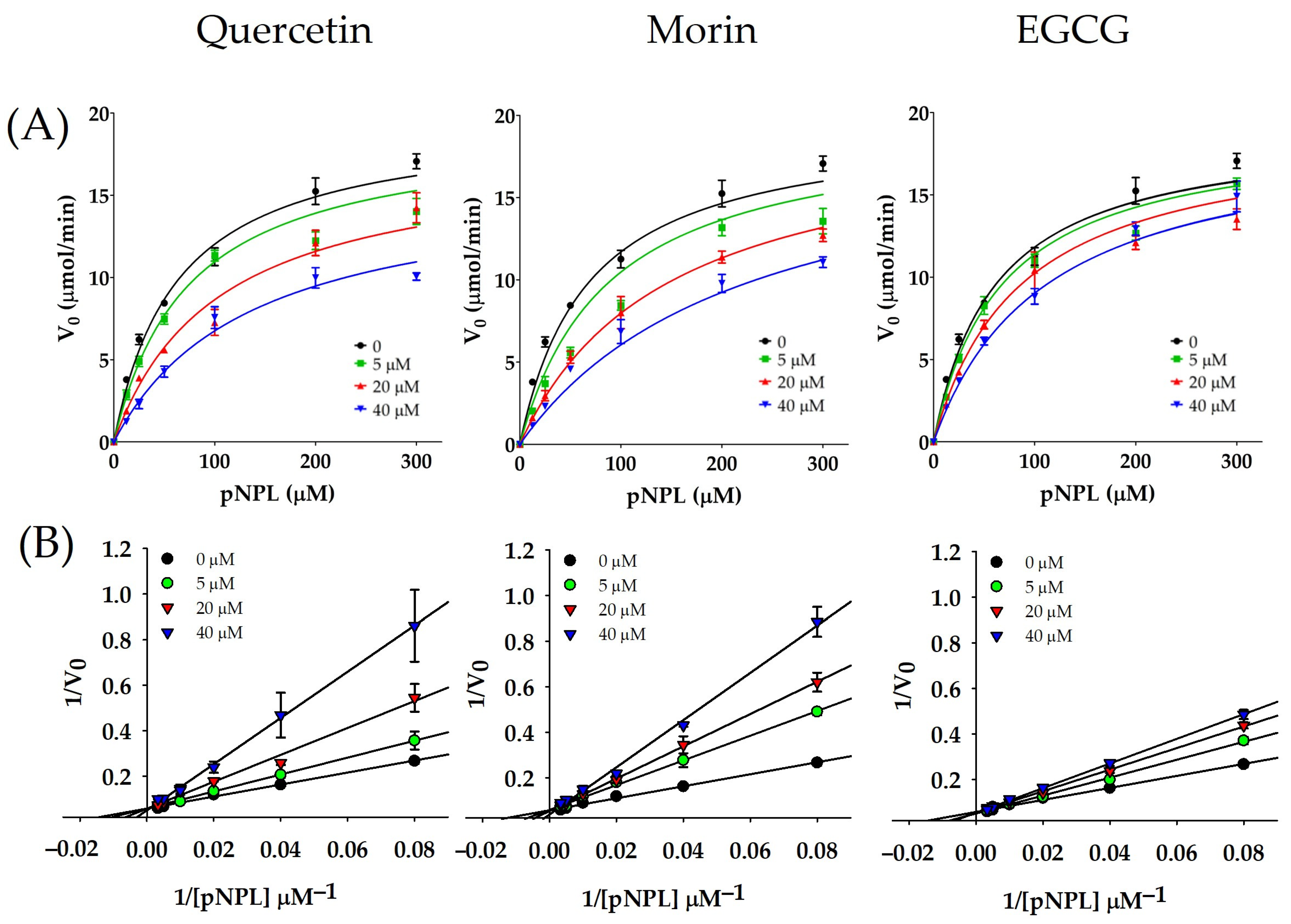
2.1. Inhibition Kinetics of vpLDH by Flavonoids

The inhibitory action of quercetin, morin, and EGCG against the phospholipase A2 activity of recombinant vpLDH was previously demonstrated [32]. Hence, in this study, the interaction of these flavonoids with vpLDH was investigated using enzyme kinetics. The flavonoid-mediated inhibition of vpLDH was evaluated by varying the ligand (flavonoids) and substrate analog 4-nitrophenyl laurate (pNPL) concentration (Figure 1). The effect of flavonoids in the vpLDH kinetic parameters was evaluated by a non-linear regression analysis (NLR) of the Michaelis–Menten equation, as shown in Figure 1 and Table S1.
The presence of different concentrations of the flavonoids causes changes in vpLDH kinetic behavior, indicating that the vpLDH Km constant was more affected than Vmax. In addition, the inhibition constants (Ki) were calculated by the same global fitting, being 17.1, 17.2, and 24.4 µM for quercetin, morin, and EGCG, respectively (Table S1). These results suggest that the tested flavonoids affect the vpLDH catalytic activity. Also, global fitting was used to determine the inhibition mechanism to different inhibition models (competitive, non-competitive, and uncompetitive) and the statistical Akaike Information Criterium (AIC) to compare all fit inhibitions models. Based on the NLR and the statistical criterion of the inhibition model comparison (AIC), morin and EGCG showed a better fitting to a competitive model compared to other inhibition models, while the comparison of competitive and mixed inhibition models for quercetin suggested a mixed inhibition mechanism with a positive value in the difference of the AIC = 1.499. However, considering the alpha value for this comparison (alpha = 6.87), which determines the degree to which the binding of the inhibitor changes the affinity of the enzyme for the substrate according to the GraphPad manual [39], for alpha values greater than 1, the ligand (inhibitor) preferentially binds to the free enzyme. Based on this, we propose that vpLDH is inhibited by quercetin via a competitive mechanism.
Additionally, all inhibition data were analyzed by a linear regression method using the Lineweaver–Burk equation model (Equation (3)) and a subsequent analysis of the plots. As seen in Figure 1 (panel B), increasing concentrations of quercetin, morin, and EGCG promoted an increment of linearized data slopes. These changes were made without altering the y-axis intercept (1/Vmax) but with varying the x-axis intercepts (−1/Km). Thus, this suggests that the evaluated flavonoids inhibit the vpLDH phospholipase A2 activity through a competitive mechanism, indicating they likely interact with or near the vpLDH active site and interfere with substrate binding.
LDH from several Vibrio species has been biochemically characterized [30,40]. In this sense, other studies have focused on LDH as a molecular target for inhibition by metal ions and synthetic compounds as reducing or chelating agents (2-mercaptoethanol and EDTA) [41,42]. However, the effect of natural compounds (such as phenolic acids and flavonoids) as inhibitors of LDH is not widely analyzed with kinetic and mechanistic approaches. PLA2 from Crotalus durissus cumanensis snake venom, which shares a similar catalytic triad (serine, histidine, and glutamic/aspartic acid), was successfully inhibited by phenolic compounds (gallic, ferulic, and caffeic acids) and the flavonoid EGCG [43]. In our previous research, we observed that the Michaelis–Menten parameters Vmax and catalytic turnover of vpLDH were higher than those of LDH from V. vulnificus and similar to values observed in other GDSL-related proteins, such as the cold-adapted lipase from Pseudomonas spp. [30,32,44]. Additionally, the inhibition profile of phenolic compounds on vpLDH was analyzed; it was found that the enzyme was not inhibited by single phenolic acids, while the flavonoid quercetin was a better inhibitor than EGCG and morin (IC50 values of 4.5 μM, 6.3 μM, and 9.9 μM, respectively) against vpLDH phospholipase activity. These results are consistent with our enzyme kinetics analysis, with all three flavonoids showing the inhibition constant in the same order of magnitude (μM).

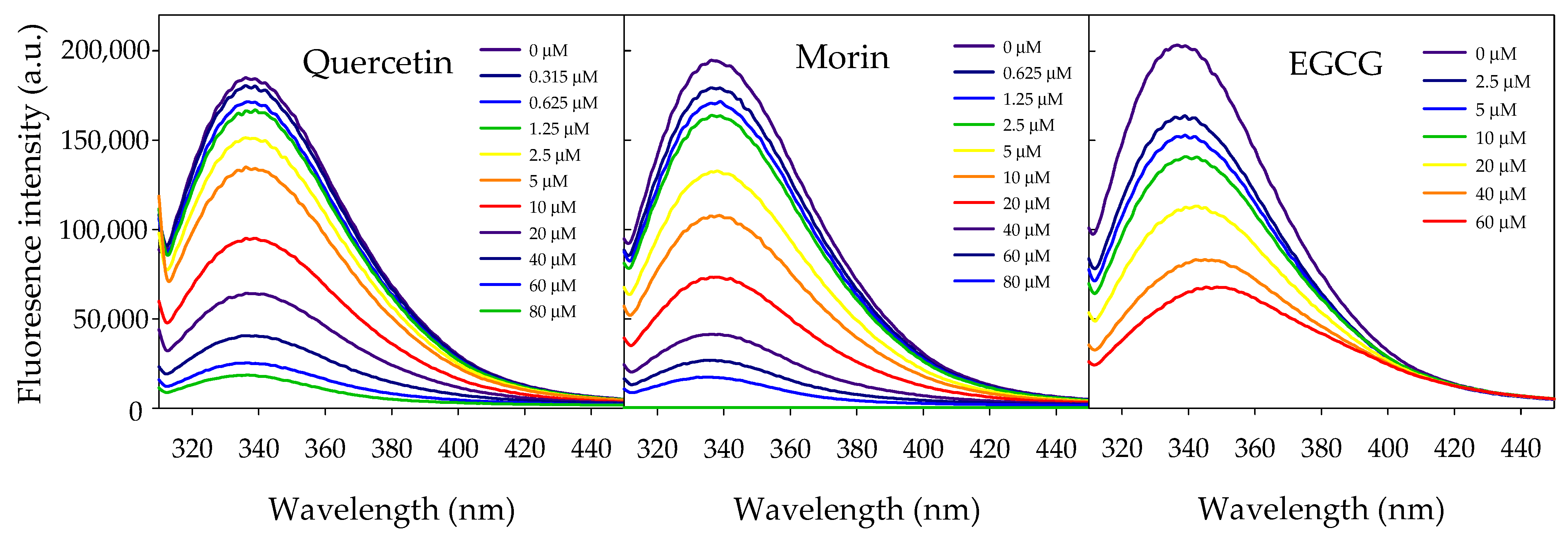
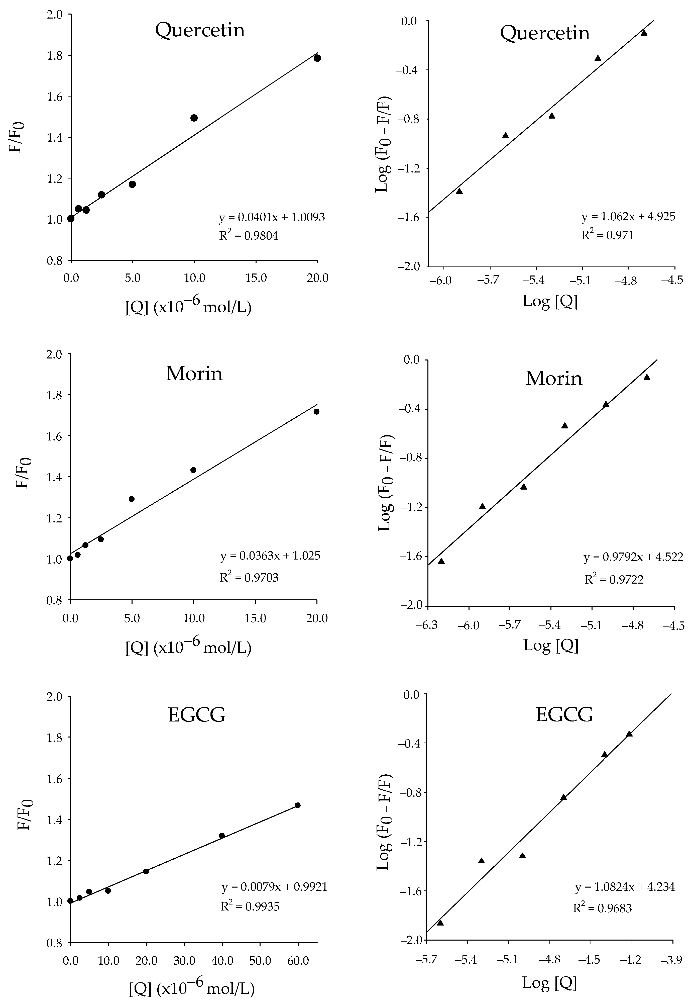
2.2. vpLDH–Flavonoid Interaction by Quenching Fluorescence

To gain further insights into how flavonoids interact with vpLDH, a fluorescence spectroscopy experiment was conducted to investigate the binding mechanism and determine the binding affinity of all three flavonoids. Figure 2 presents the fluorescence emission spectra of vpLDH in the presence of quercetin, morin, or EGCG. The maximum emission of vpLDH was observed at λ = 336 nm after being excited at 295 nm. A decay of fluorescence emission was observed as the concentration of all three flavonoids increased. Interestingly, a red shift of 14 nm was observed at the higher concentration of EGCG, moving the maximum emission at 350 nm. Meanwhile, quercetin and morin did not change the maximum emission at the evaluated concentrations.
The change in the maximum emission spectra is related to the photophysical properties of tryptophan residues, whose emission originates from two electronic excited states (1La and 1Lb) of the indole group, which are highly sensitive to changes in the polarity of their surroundings [45]. In particular, the transition to lower energy levels is related to the large excited-state dipole moment of the 1La state and its higher sensitivity towards hydrogen bonding with respect to the excited 1Lb [46]. Therefore, a red shift in the maximum emission spectra suggests that the microenvironment of Trp residues becomes more polar or hydrophilic after EGCG binding. vpLDH contains 11 Trp residues, Trp50, Trp64, Trp66, Trp81, Trp82, Trp133, Trp166, Trp174, Trp185, Trp200, and Trp389, all of them contributing to total intrinsic fluorescence. Because interpreting the intrinsic fluorescence of a protein with multiple tryptophan residues is complex, it remains uncertain whether all tryptophan residues contribute equally to the total fluorescence emitted by the protein [47]. Nevertheless, according to our results from the enzyme-inhibition kinetic experiment, all three flavonoids showed a competitive inhibition mechanism. Thus, the fluorescence of Trp174 residue, located near the vpLDH acyl-binding site, could likely be quenched by quercetin, morin, and EGCG.
To investigate the fluorescence quenching mechanism involved in the flavonoids–vpLDH interaction, fluorescence emission data were examined according to the Stern–Volmer equation (Equation (5)). As shown in Figure 3 (left panel), the Stern–Volmer plots of quercetin, morin, and EGCG showed a good linear model fitting (R2 ≥ 0.97) at the evaluated concentrations, suggesting that a single quenching process is occurring. The Stern–Volmer quenching constants (KSV) were calculated from the slopes of these plots, and the resulting values are presented in Table 1. Quercetin and morin exhibited similar KSV values within the same order of magnitude at 4.01 × 10⁴ mol−1 and 3.63 × 10⁴ mol−1, respectively, while EGCG showed a KSV of 7.9 × 10³ mol−1. KSV values can be used to compare quenching rates between quencher molecules, where a higher KSV reflects better quenching efficacy [48,49]. Additionally, the bimolecular rate constant Kq was calculated for each compound using Equation (6). The Kq value reflects the efficiency of the interaction between the fluorophore and the quencher, indicating how rapidly the quencher deactivates the excited state of the fluorophore per unit of concentration and time. Based on the average fluorescence lifetime of the biomolecules in the absence of a quencher, reported as 1 × 10⁻⁸ s−1 [50], quercetin, morin, and EGCG (Table 1) exhibited Kq values (1012 M−1 s−1) exceeding the diffusion-controlled limit, which are typically found in the range of 109 to 1010 M−1 s−1 [51]. This suggests that the quenching mechanism could not be solely dynamic (collisional) but probably involves static quenching, which is related to the formation of a stable, non-fluorescent complex between flavonoids and the vpLDH.
On the other hand, modified Stern–Volmer plots (Figure 3, right panel) were used to calculate the binding constant (Kd) and the number of binding sites (n) from the inverse of the Y-axis intercept and the slope, respectively, according to Equation (7). Quercetin, morin, and EGCG Kd values were found in the same order of magnitude: 7.91 ± 0.55, 25.05 ± 1.63, and 44.96 ± 2.88 μM, respectively (Table 1). Also, it may be observed that the number of binding sites between vpLDH and flavonoids is close to 1.0, indicating a flavonoid–vpLDH ratio of 1:1.
In the development of anti-virulent therapy, the first step is the identification of molecules able to modulate the virulence traits of pathogenic bacteria, followed by validating the interaction between the molecules and their target [52,53]. In this context, in the present study, the interaction between vpLDH and quercetin, morin, and EGCG was analyzed using fluorescence quenching experiments. According to our results, the studied flavonoids bind to a single site of vpLDH with binding affinities in the micromolar range (Kd = 7.91–44.96 μM), forming a stable non-fluorescent complex, with quercetin showing the higher affinity (p < 0.05). These results agree with vpLDH enzyme kinetic data and support the proposed flavonoid’s competitive inhibitory mechanism.
The fluorescence quenching properties of flavonoids, including those analyzed in this study, have been extensively documented in the literature; these properties have been utilized to evaluate and validate the interaction of flavonoids with proteins of several sources such as food, human, and bacteria [54,55,56,57,58,59,60,61,62]. However, in the context of anti-virulence therapy, there are few reports, and only a limited number of studies have investigated the interaction of flavonoids with bacterial toxins or virulence factors. For instance, Wang et al. [61] reported that myricetin, morin, baicalein, chrysin, and naringenin bind to the pore-forming toxin listeriolysin O (LLO) o L. monocytogenes, with Kd values ranging from 2.37 to 3.18 μM. These flavonoids also inhibit LLO’s hemolytic activity, with IC50 values between 0.46 μg/mL and 186.57 μg/mL. Additionally, the binding of the flavonol glycoside hibifolin and punicalagin (an ellagitannin) to sortase A, a virulence factor of Staphylococcus aureus, has been confirmed using fluorescence quenching methods, yielding Kd values of 58.14 μM and 159 nM, respectively [59,60]. However, the quenching mechanisms were not elucidated in these studies. In this context, our findings further support the ability of flavonoids to bind to and inhibit bacterial virulence factors. In addition, measuring binding affinity provides valuable information about the potential efficacy of an anti-virulence agent, as compounds with higher affinity (lower Kd values) are more likely to modulate the target protein’s activity.

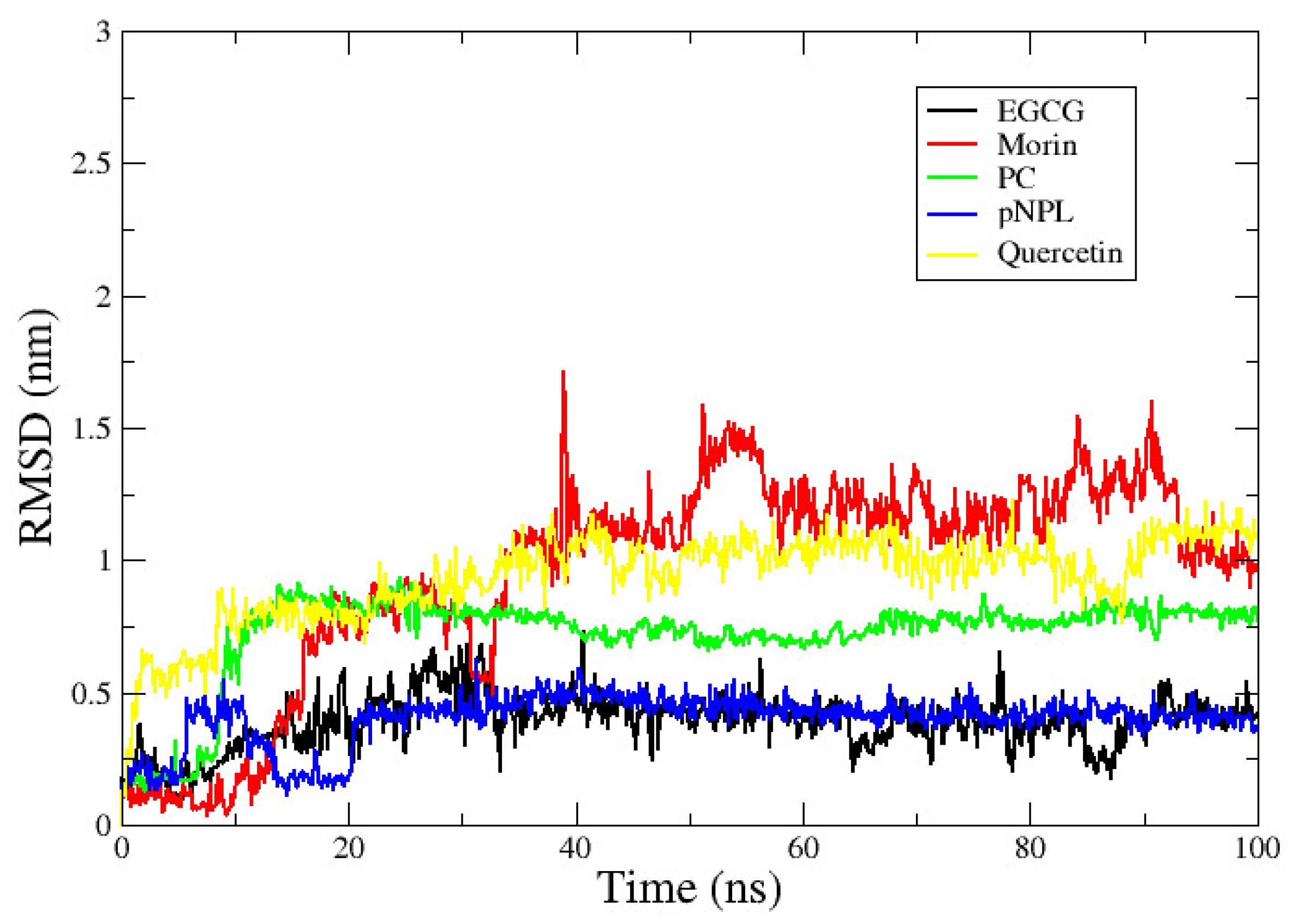
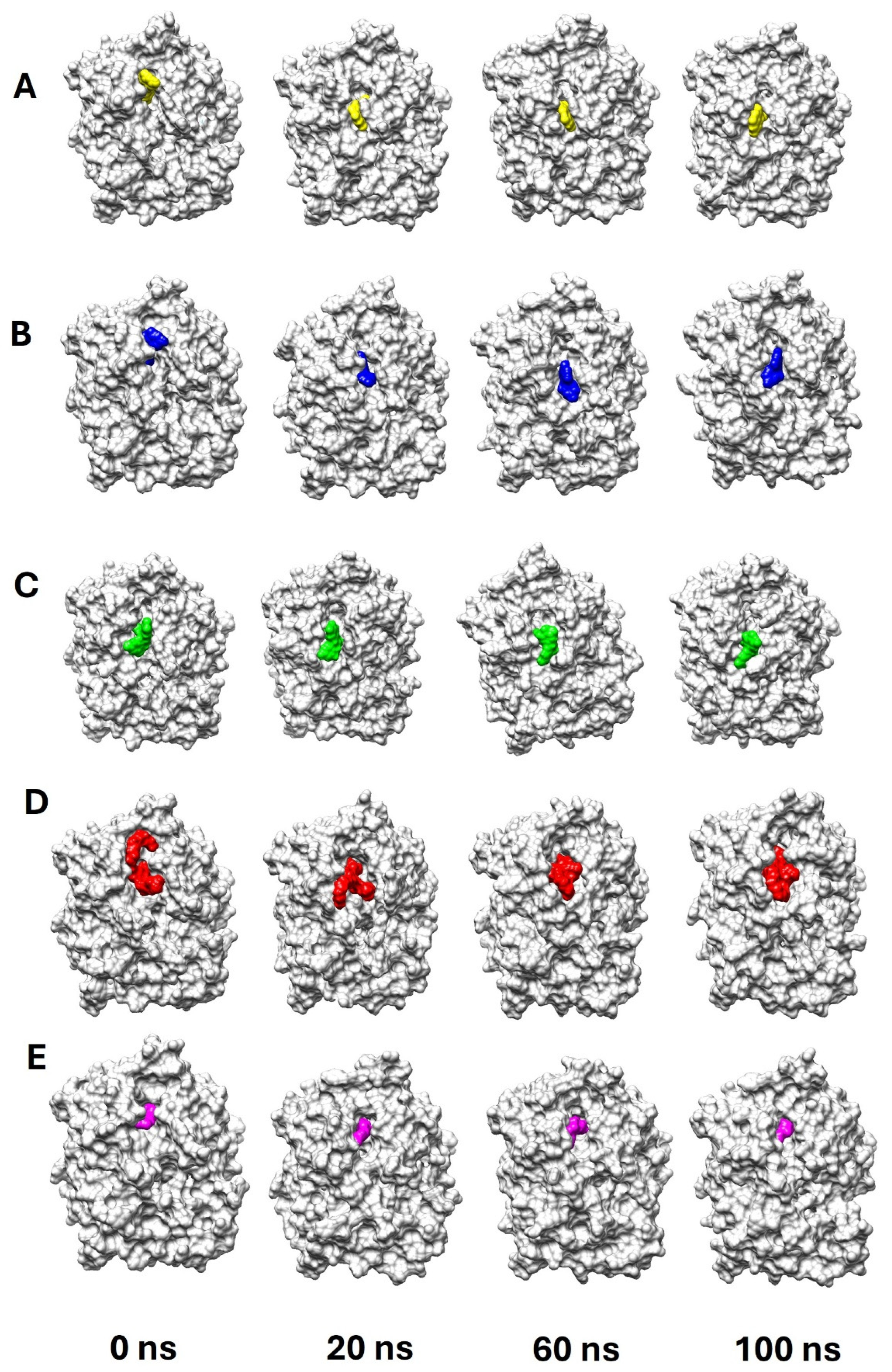
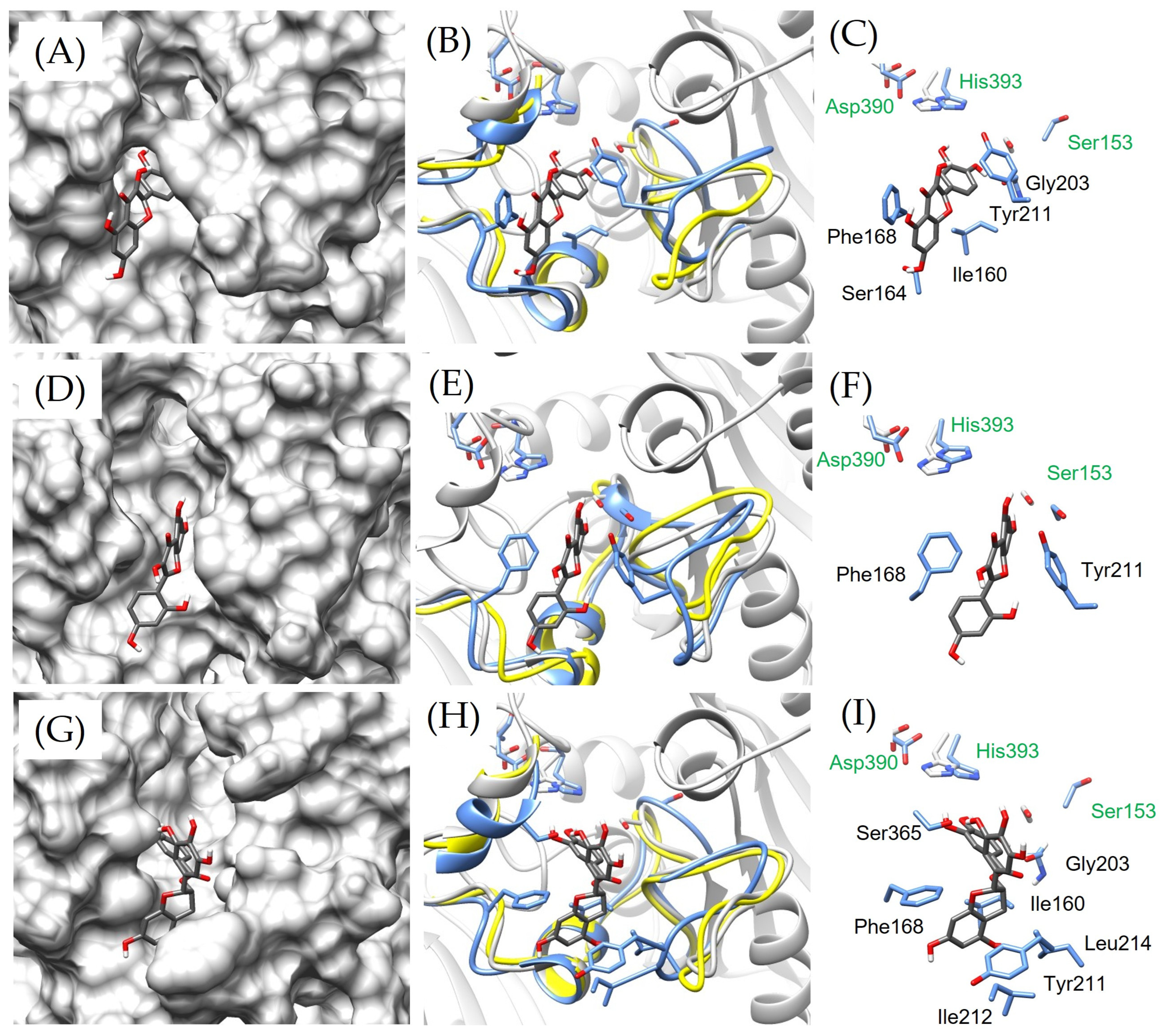
2.3. vpLDH–Flavonoid Structural–Binding Mode by MD Simulations

We used MD simulations to determine the stability and binding mode of the vpLDH–flavonoid complex. The binding mode refers to the specific way in which a molecule interacts with its target and also describes the interaction, orientation, and position of the molecule within the binding site of the target. Understanding the binding mode is important for determining how the molecule exerts its effects, such as inhibiting or activating the target’s function. For this purpose, the molecules pNPL and phosphatidylcholine (PC) were used as interaction controls because they are vpLDH substrates: analog and cognate, respectively. In this sense, the root mean square deviation (RMSD) between protein and ligands was the parameter used to describe the stability of the interaction in terms of distance. In particular, the trajectories obtained by MD simulations revealed that the RMSD (Figure 4) between vpLDH and flavonoids (morin, quercetin, and EGCG) and substrates (pNPL and PC) exhibited oscillations of <1.7 nm over time (100 ns), indicating that all ligands are not dissociated and stay near the vpLDH active site. Additionally, snapshots of complexes were taken at different time intervals to visualize the poses of flavonoids and substrates during the MD simulation. All selected compounds stayed in the protein’s binding pocket throughout the 100 ns MD simulation (Figure 5).
Moreover, it is important to mention that all ligands employed in this study present a different grade of interaction. Nevertheless, morin and quercetin, the flavonoids with major protein–ligand RMSD, show more vpLDH inhibition activity (Figure 1). Moreover, the energetic decomposition (Table 2) found by the MMPBSA analysis describes that morin and quercetin interact with catalytic residues, Ser153 and His393. Similar behavior was observed with PC and pNPL. Furthermore, the high stability of EGCG during the simulation trajectories (protein–ligand RMSD < 0.6 nm), in comparison with morin and quercetin, suggests that the experimentally low inhibition activity is related to limited specific interaction with catalytic residue. However, the inhibition activity observed in the experimental results for this compound could be attributed to the interaction with loopβ8−α5 (202VGGAAGENQYIALT215), since the decomposition MMPBSA analysis indicates that EGCG interacts more frequently with Gly203, Gly204, Tyr211, and Leu214 (Table 2).
Crystal structural studies revealed that loopβ8−α5 and loopβ3−β4 are located in the vicinity of the V. algynolyticus LDH active site, which was named as a moonlight enzyme showing dual lipase–transferase activity (valDLT). Also, loopβ8−α5 undergoes conformational changes upon ligand binding, and this flexibility was crucial by stabilizing substrates during both V. algynolyticus LDH lipase and transferase activity [31]. These structural features are widely conserved among LDH Vibrio species, and thus, understanding these structural and functional attributes is critical for elucidating the mechanisms of Vibrio toxins’ catalytic activity. Additionally, the behavior observed in the protein–ligand RMSD levels of ECGC is compared to pNPL (Figure 4), a vpLDH substrate, during the 100 ns of simulation.
On the other hand, the final poses of quercetin, morin, and EGCG after 100 ns of simulation are shown in Figure 6. The basic chemical structure of flavonoids consists of a 15-carbon skeleton arranged in a C6-C3-C6 configuration. This structure comprises two aromatic rings (A- and B-rings) connected by a three-carbon bridge, forming an oxygenated heterocyclic ring (C-ring) [63]. Quercetin, morin, and EGCG are flavonoids that differ in their structural arrangements and functional groups. Quercetin is a flavonol characterized by a hydroxyl group at positions 3, 5, 7, 3′, and 4′. However, morin differs from quercetin by the placement of the hydroxyl group in the B-ring, having it at positions 2′ and 4′ instead of 3′ and 4′, which affects its polarity and interaction with biological targets [64,65]. While, EGCG belongs to the flavanol subclass, which differs from the other two flavonoids, EGCG lacks the ketone group in the C-ring but contains an esterified gallate group at position 3 of the C-ring, making it bulkier and increasing its potential for hydrogen bonding [66]. These structural variations influence their solubility, stability, and biological activity.
In this sense, after a visual inspection of the binding poses of the flavonoids during the MD simulation, it was observed that, in the case of morin, the B-ring was oriented towards the inside of the vpLDH active site, interacting with the residues Tyr368 (hydrophobic interaction) and Asn252, Gln292, and Thr392, with a hydrogen bond at the beginning of the simulation (Figure 5 and Figure S1). Still, after 20 ns and until 100 ns, the morin was partially displaced from the active site (Figure 4), and at the end of the simulation trajectory, the A-ring attached to the three-carbon pyran ring (C6-C3 moiety) occupied this pocket, forming a van der Waals interaction with Tyr253 (a residue near the catalytic triad) (Figure S1). Moreover, the B-ring formed van der Waals interactions with Val366, Ser164, and Arg167. Evidently, the morin pose and position change to interactions more favorable (Figure S1 and Table 2).
Meanwhile, quercetin maintained its initial pose with the B-ring oriented inside the active site during 100 ns of simulation (Figure S2). Specifically, the A- and B-rings from quercetin conserved favorable interactions with Phe168 (van der Waals and hydrophobic interactions) and Ser365 (van der Waals), respectively (Table 2 and Figure S2). Regarding EGCG, the gallate group is the part oriented inside the binding pocket during the entire simulation (Figure 6). Additionally, the map interactions (Figure S3) at 0 and 100 ns show that Phe168 (hydrophobic interaction) and Tyr211 (hydrogen bond) are very important in their contributions to interaction stability (Table 2). Moreover, EGCG has the capacity to interact via a hydrogen bond and van der Waals interactions with Ser153 and Hsd393, respectively (Figure S3). This behavior indicates that the three flavonoids have different binding modes in the vpLDH active site. Evidence that will contribute to the search or design of new molecules with better capacity for the inhibition of the vpLDH enzymatic and hemolytic activity could contribute to the reduction in the V. parahaemolyticus pathogenicity. For the latter, it is essential to explore the impact of these compounds on the pathogenicity and virulence of this bacteria through further in vitro and in vivo studies.

3. Materials and Methods

3.1. vpLDH Protein Overexpression and Purification

Recombinant LDH from V. parahaemolyticus was produced following the method outlined by Vazquez-Morado [32]. A synthetic gene encoding vpLDH (Accession number BAA25328.1) with a C-terminal 6x-His tag was inserted into the pET-28b (+) plasmid. This vector was then introduced into chemically competent Escherichia coli Rosetta 2 strains using heat shock, followed by incubation in SOC media (containing 2% w/v tryptone, 0.5% w/v yeast extract, 10 mmol/L NaCl, 2.5 mmol/L KCl, 10 mmol/L MgCl2, 10 mmol/L MgSO4, and 20 mmol/L glucose) at 37 °C for 4 h. For selection, the transformed cells were plated on Luria–Bertani agar containing kanamycin (25 µg/mL) and incubated overnight at 37 °C.
The transformed bacteria were cultured in LB broth with kanamycin (50 µg/mL) at 37 °C, with continuous shaking at 220 rpm in a MaxQ 4000 orbital shaker (Thermo-Scientific, Waltham, MA, USA). Once the culture reached an optical density of O.D. = 0.6 (measured at 600 nm), IPTG (Isopropyl β-D-1-thiogalactopyranoside) was added to a final concentration of 1 mmol/L to induce vpLDH overexpression (16 h at 25 °C, 220 rpm). After induction, the biomass was collected by centrifugation (7000 rpm for 20 min at 4 °C), pelleted, and stored at −80 °C until further use.
For vpLDH purification, 1 g of biomass was suspended in lysis buffer (50 mmol/L Tris base, 5 mmol/L EDTA, 100 mmol/L NaCl, 6 mmol/L benzamidine, and 1 mmol/L DTT) at a ratio of 1:4. Bacterial cells were disrupted using sonication on ice, with six cycles of 10 s pulses followed by 40 s rest periods at 30% amplitude. The resulting mixture was centrifuged at 12,000 rpm for 20 min at 4 °C to separate the lysate. Protein expression in both the soluble and insoluble fractions was then evaluated using SDS-PAGE (12%) with Coomassie blue staining.
Inclusion bodies were obtained from the insoluble cellular debris, which was resuspended by sonication (as previously described) using buffer 1 (50 mmol/L Tris base, 1 mmol/L DTT, 5 mmol/L EDTA, 2% Triton X-100; pH 7.0) at a 1:8 (w/v) ratio. The mixture was then centrifuged at 12,000 rpm for 20 min. The precipitate was collected, and this centrifugation step was repeated three times. The pellet was then washed twice using buffer 2 (which was identical to buffer 1 but without Triton X-100) under the same conditions. To solubilize the recovered inclusion bodies, they were treated with urea (50 mmol/L Tris base, 1 mmol/L DTT, 8 M urea, pH 7.0) by sonication and incubated overnight at 4 °C with constant stirring for 12 h. The homogenate was clarified by centrifugation (12,000 rpm at 4 °C for 30 min), and the soluble fraction containing LDH in 8 M urea was recovered. Protein concentration was determined at 280 nm using nanodrop equipment, accounting for the molar extinction coefficient (ϵ ≈ 96,510 M−1 cm−1).
Finally, vpLDH was purified by Affinity Chromatography (IMAC) in the presence of 8 M urea within a 1 mL HiTrap™ column; the entire procedure was conducted in an Äkta Prime Plus system (GE Healthcare, Chicago, IL, USA). The enzyme was refolded, dialyzing against buffer 50 mM Tris-HCl pH 7.4, 150 mM NaCl using a 12 kDa membrane to gradually remove the urea. The enzyme’s purity and concentration were assessed by SDS-PAGE and the Bradford method, respectively.

3.2. Enzymatic Activity

Enzymatic activity was measured as described previously by Vazquez-Morado [32] with some modifications. Each reaction was conducted in a 300 µL reaction containing 50 mM Tris-HCl pH 7.4, 150 mM NaCl, egg yolk lecithin (0.0001%), and refolded vpLDH (3 µg/mL). The reaction was started by adding pNPL as the substrate (200 μM, final concentration). Substrate hydrolysis was monitored at 410 nm for 120 s. The reaction was carried out in a 96-well microplate (Costar 96) in a microplate reader (FluoSTAR Omega, BMG Labtech, Ortenberg, Germany). The negative control assay was performed in the absence of vpLDH. One unit of lipase activity is defined as the amount of enzyme required to hydrolyze 1 µmol of pNPL per minute. Specific activity was calculated using the following equation:
U/(mg of protein) = (m·V)/(ε·p·l)
where m is the slope of the reaction, V is the reaction volume, p is the protein concentration (mg/mL), and l is the cell path length in cm.

3.3. vpLDH Inhibition Kinetics

Initial velocities of vpLDH in the presence of quercetin, morin, or EGCG were recorded to determine the constants of the Michaelis–Menten equation and the inhibition mechanism of each compound. All experiments were conducted under the same conditions as described above, with varying substrate concentrations by maintaining a fixed inhibitor concentration. In brief, initial velocities of vpLDH in the absence and presence of flavonoids were determined using 12.5, 25, 50, 100, 200, and 300 μM of the pNPL substrate and fixed final concentrations as follows: quercetin and morin (0, 5, 20, and 40 μM) or EGCG (0, 2.5, 10, and 20 μM). The vpLDH kinetic parameters Vmax Km and the inhibition constant (Ki) were calculated by global fitting of the initial reaction rates to the Michaelis–Menten equation model (Equation (2)) using non-linear regression analysis. The inhibition mechanism was determined by fitting the data to competitive, uncompetitive, non-competitive, and mixed inhibition models using non-linear regression with GraphPad Prism software (Version 5.01). Also, the inhibition models were compared using the Akaike Information Criterion to identify the best-fitting model to obtain Km, Vmax, and Ki kinetic parameters. Additionally, the Michaelis–Menten kinetic parameters were determined by linearizing kinetic data using the Lineweaver–Burk equation model (Equation (3)) and subsequent analysis of the plots. Assays were performed at least in triplicate.
v = V m a x   [ S ] K m + [ S ]
1 v = K m V m a x   [ S ] + 1 V m a x
where v is the initial velocity, Vmax is the maximum velocity, Km is the Michaelis–Menten constant, and [S] is the substrate concentration.

3.4. Determination of Binding Constants Between Flavonoids and vpLDH by Intrinsic Tryptophan Fluorescence

Intrinsic tryptophan (Trp) fluorescence quenching was used to analyze the interactions between quercetin, morin, and EGCG with vpLDH. Fluorescence measurements were conducted with a Shimadzu RF-6000 spectrofluorophotometer (Shimadzu, Tokyo, Japan) equipped with a 150 W xenon lamp. The excitation and emission slits were set at 10 nm with an excitation wavelength of 295 nm. Emission spectra were recorded at room temperature in the 300–450 nm wavelength range.
Fluorescence measurements were recorded in triplicate for vpLDH (60 µg/mL) titrated with increased concentrations of quercetin, morin, or EGCG (0–80 µM) in an activity buffer (50 mM Tris-HCl, 7.4, 150 mM NaCl, and egg yolk lecithin (0.0001%)).
The inner filter effect correction was carried out using the following equation (Equation (4)):
F c o r r = F o b s   × 10 ( A e x + A e m ) 2
Fcor is the corrected fluorescence intensity, and Fobs is the measured fluorescence intensity. Aex and Aem is the absorbance at excitation and emission wavelengths, respectively.
The possible fluorescence quenching mechanism of quercetin, morin, and EGCG was analyzed using the Stern–Volmer equation (Equation (5)).
F 0 F = 1 + K S V Q = 1 + K q τ 0 Q
k q = K s v τ 0
where F0 and F are the steady-state fluorescence intensities in the absence and presence of a quencher, respectively, [Q] is defined as the quencher (flavonoid) concentration, and Ksv is the Stern–Volmer quenching constant. Kq (Equation (6)) is the bimolecular rate constant, and τ0 is the average lifetime of the biomolecule in the absence of the quencher (~10−8 s−1) [50]. The binding constants and binding sites were analyzed by using the modified Stern–Volmer equation (Equation (7)):
log F 0 F 1 = log k a + n   log Q
where ka is the binding constant and n is the number of binding sites.

3.5. Molecular Dynamics Simulation and Surface Area

The stability of interaction between inhibitors and substrates was evaluated by molecular dynamics (MD) simulations and correlated its effect to vpLDH inhibition. For this objective, the topologies of the docked vpLDH–ligand complex obtained by Chimera (v. 1.17.1) were prepared using the CHARMM-GUI webserver (“https://www.charmm-gui.org/ (accessed on 31 August 2024)”) and further parameterized using the CHARMM36m force field. The TIP3 water model was used to solvate the simulation system and was neutralized with Na+ and Cl ions at 0.15 mM NaCl. Then, the system energy was minimized by the steepest descending algorithm (5000 descending steps) and equilibrated using NVT/NPT ensembles (200 ps) at 303.15 °K. Parriello–Rahman pressure and Nose–Hoover controller systems were used to maintain a constant pressure of 1 bar throughout the equilibration temperature. MD simulations were carried out using GROMACS (Version 2023.4) software at 100 ns (each run) by duplicate. Finally, complete trajectories were conducted to obtain the binding parameters of the vpLDH–ligand interactions. Moreover, the Molecular Mechanic Poisson–Boltzmann Surface Area (MM/PBSA) method was employed to analyze the interactions between ligands and vpLDH using the gmx_MMPBSA program (Version 1.6.4) [67]. The binding free energy and decomposition analysis by residue (within 4 Å) were calculated using the complete MD simulation trajectory.

3.6. Statistical Analysis

All experiments utilized a completely randomized experimental design. In the enzyme kinetics and quenching experiments, the factors included the concentration of each flavonoid, while the response variables were the kinetic parameters (Vmax, Km, and Ki) and the quenching parameters (Ksv, Kq, Kd, and n), respectively. The kinetic parameters Vmax and Km were calculated by non-linear regression of the experimental data fitted to the Michaelis–Menten equation by using GraphPad Prism 5 software (Version 5.01). Subsequently, the fits to competitive, non-competitive, uncompetitive, and mixed inhibition models were compared by non-linear regression analysis with equations specific to each type of inhibition. The best fitting of the inhibition models was evaluated using the coefficient of determination (R2) and the difference of Akaike Information Criterion (AIC). Additionally, the NCCS (Version 2007) package was used for analysis of variance (ANOVA) followed by Tukey’s post hoc test to compare fluorescence quenching parameters obtained in the presence and absence of flavonoids, and differences were considered statistically significant when p < 0.05.

4. Conclusions

In this study, we analyzed the inhibition mechanism of the lecithin-dependent hemolysin of V. parahaemolyticus by quercetin, morin, and EGCG using enzyme kinetics, fluorescence quenching, and in silico analysis through molecular dynamics (MD) simulation approaches. All three flavonoids inhibited the vpLDH phospholipase A2 activity by competitive mechanisms, forming a 1:1 flavonoid–enzyme complex, exhibiting both an inhibition constant and binding affinities in the micromolar range. The in silico analysis supports our experimental findings, such as forming a stable complex between vpLDH and the flavonoids. It also revealed that the three studied flavonoids showed different interaction patterns and molecular orientations during the MD simulations in the vicinity of the vpLDH active site. Moreover, the decomposition by residue in the MMPBSA analysis describes the principal residues that contribute to the interaction stability in energetic terms, such as Phe198. This finding provides valuable information about the potential of flavonoids as anti-virulence compounds that could aid in designing strategies to combat the spread of V. parahaemolyticus.

Supplementary Materials

The following supporting information can be downloaded at https://www.mdpi.com/article/10.3390/catal15030257/s1, Table S1. Kinetic parameters of vpLDH inhibition by flavonoids. Figure S1: Details of vpLDH–morin interactions observed during molecular dynamics simulations. Left panel shows the interaction map at 0 and 100 ns and right panel shows the morin position on the active site at 0 (yellow structure) and 100 (green structure) ns; Figure S2: Details of vpLDH–quercetin interactions observed during molecular dynamics simulations. Left panel shows the interaction map at 0 and 100 ns and right panel shows the morin position on the active site at 0 (yellow structure) and 100 (green structure) ns; Figure S3: Details of vpLDH–EGCG interactions observed during molecular dynamic simulations. Left panel shows the interaction map at 0 and 100 ns and right panel shows the morin position on the active site at 0 (yellow structure) and 100 (green structure) ns.

Author Contributions

Conceptualization, F.J.V.-A., A.A.-A., R.S.-M. and A.A.L.-Z.; methodology, F.J.V.-A., A.A.A.-F. and A.A.L.-Z.; software, F.J.V.-A., A.M.-O. and A.A.A.-F.; validation, R.S.-M., F.A.-Z. and A.A.L.-Z.; formal analysis, R.S.-M., F.A.-Z., A.A.A.-F. and A.A.L.-Z.; investigation, F.J.V.-A., A.M.-O., A.A.-A. and A.A.L.-Z.; resources, R.S.-M. and F.A.-Z.; data curation, F.J.V.-A., A.A.-A. and A.A.L.-Z.; writing—original draft preparation, F.J.V.-A., A.A.-A. and A.A.L.-Z.; writing—review and editing, A.A.L.-Z., R.S.-M., A.M.-O., A.A.A.-F. and F.A.-Z.; visualization, F.J.V.-A., A.M.-O. and A.A.-A.; supervision, A.A.L.-Z.; project administration, A.A.L.-Z. and F.J.V.-A.; funding acquisition, A.A.L.-Z. All authors have read and agreed to the published version of the manuscript.

Funding

This research was funded by Universidad de Sonora (UNISON) grant #USO313007854; to SECIHTI for the postdoctoral scholarship grant ESTANCIAS POSDOCTORALES POR MÉXICO 2022 (1); to UNAM-CIC-UNISON-2023; and the APC was funded by Facultad Interdisciplinaria de Ciencias Biológicas y de la Salud y Dirección de Apoyo a Docentes, Investigación y Posgrado from the UNISON.

Data Availability Statement

Datasets are available on request to the authors.

Acknowledgments

We thank the Departamento de Ciencias Químico Biológicas-UNISON for sustained support and infrastructure. Also, we appreciate Cesar B. Otero-León and Diego M. Monge-Zazueta for technical support. We would like to acknowledge UNISON for the use of the Acarus Computational Laboratory for providing the necessary computing resources to carry out the simulations reported in this paper.

Conflicts of Interest

The authors declare no conflicts of interest. The funders had no role in the design of the study; in the collection, analyses, or interpretation of data; in the writing of the manuscript; or in the decision to publish the results.

References

  1. Namadi, P.; Deng, Z. Optimum environmental conditions controlling prevalence of Vibrio parahaemolyticus in marine environment. Mar. Environ. Res. 2023, 183, 105828. [Google Scholar] [CrossRef]
  2. Yang, F.; Xu, L.; Huang, W.; Li, F. Highly lethal Vibrio parahaemolyticus strains cause acute mortality in Penaeus vannamei post-larvae. Aquaculture 2022, 548, 737605. [Google Scholar] [CrossRef]
  3. Han, J.E.; Tang, K.F.J.; Lightner, D.V. Genotyping of virulence plasmid from Vibrio parahaemolyticus isolates causing acute hepatopancreatic necrosis disease in shrimp. Dis. Aquat. Org. 2015, 115, 245–251. [Google Scholar] [CrossRef] [PubMed]
  4. Han, J.E.; Tang, K.F.J.; Tran, L.H.; Lightner, D.V. Photorhabdus insect-related (Pir) toxin-like genes in a plasmid of Vibrio parahaemolyticus, the causative agent of acute hepatopancreatic necrosis disease (AHPND) of shrimp. Dis. Aquat. Org. 2015, 113, 33–40. [Google Scholar] [CrossRef]
  5. de Souza Valente, C.; Wan, A.H.L. Vibrio and major commercially important vibriosis diseases in decapod crustaceans. J. Invertebr. Pathol. 2021, 181, 107527. [Google Scholar] [CrossRef]
  6. Fang, G.-Y.; Liu, X.-Q.; Mu, X.-J.; Huang, B.-W.; Jiang, Y.-J. Distinct increase in antimicrobial resistance genes among Vibrio parahaemolyticus in recent decades worldwide. Chemosphere 2023, 340, 139905. [Google Scholar] [CrossRef]
  7. Wang, D.; Flint, S.H.; Palmer, J.S.; Gagic, D.; Fletcher, G.C.; On, S.L.W. Global expansion of Vibrio parahaemolyticus threatens the seafood industry: Perspective on controlling its biofilm formation. LWT 2022, 158, 113182. [Google Scholar] [CrossRef]
  8. Balta, I.; Linton, M.; Pinkerton, L.; Kelly, C.; Stef, L.; Pet, I.; Stef, D.; Criste, A.; Gundogdu, O.; Corcionivoschi, N. The effect of natural antimicrobials against Campylobacter spp. and its similarities to Salmonella spp, Listeria spp., Escherichia coli, Vibrio spp., Clostridium spp. and Staphylococcus spp. Food Control 2021, 121, 107745. [Google Scholar] [CrossRef]
  9. Li, A.; Shi, C.; Qian, S.; Wang, Z.; Zhao, S.; Liu, Y.; Xue, Z. Evaluation of antibiotic combination of Litsea cubeba essential oil on Vibrio parahaemolyticus inhibition mechanism and anti-biofilm ability. Microb. Pathog. 2022, 168, 105574. [Google Scholar] [CrossRef]
  10. Yu, H.; Pei, J.; Qiu, W.; Mei, J.; Xie, J. The Antimicrobial Effect of Melissa officinalis L. Essential Oil on Vibrio parahaemolyticus: Insights Based on the Cell Membrane and External Structure. Front. Microbiol. 2022, 13, 812792. [Google Scholar] [CrossRef]
  11. Dehbanipour, R.; Ghalavand, Z. Anti-virulence therapeutic strategies against bacterial infections: Recent advances. Germs 2022, 12, 262–275. [Google Scholar] [CrossRef]
  12. Brannon, J.R.; Hadjifrangiskou, M. The arsenal of pathogens and antivirulence therapeutic strategies for disarming them. Drug Des. Dev. Ther. 2016, 10, 1795–1806. [Google Scholar] [CrossRef]
  13. Calvert, M.B.; Jumde, V.R.; Titz, A. Pathoblockers or antivirulence drugs as a new option for the treatment of bacterial infections. Beilstein J. Org. Chem. 2018, 14, 2607–2617. [Google Scholar] [CrossRef]
  14. Vazquez-Armenta, F.J.; Aros-Corrales, M.O.; Alvarez-Ainza, M.L.; Bernal-Mercado, A.T.; Ayala-Zavala, J.F.; Ochoa-Leyva, A.; Lopez-Zavala, A.A. Antibacterial and anti-virulence potential of plant phenolic compounds against Vibrio parahaemolyticus. F1000Res 2023, 12, 1256. [Google Scholar] [CrossRef] [PubMed]
  15. Roy, P.K.; Park, S.-H.; Song, M.G.; Park, S.Y. Antimicrobial Efficacy of Quercetin against Vibrio parahaemolyticus Biofilm on Food Surfaces and Downregulation of Virulence Genes. Polymers 2022, 14, 3847. [Google Scholar] [CrossRef] [PubMed]
  16. Faleye, O.S.; Lee, J.-H.; Lee, J. Selected flavonoids exhibit antibiofilm and antibacterial effects against Vibrio by disrupting membrane integrity, virulence and metabolic activities. Biofilm 2023, 6, 100165. [Google Scholar] [CrossRef]
  17. Aguirre-Guzmán, G.; Sánchez-Martínez, J.G.; Pérez-Castañeda, R.; Palacios-Monzón, A.; Trujillo-Rodríguez, T.; De La Cruz-Hernández, N.I. Pathogenicity and Infection Route of Vibrio parahaemolyticus in American White Shrimp, LitoPenaeus vannamei. J. World Aquac. Soc. 2010, 41, 464–470. [Google Scholar] [CrossRef]
  18. Khimmakthong, U.; Sukkarun, P. The spread of Vibrio parahaemolyticus in tissues of the Pacific white shrimp LitoPenaeus vannamei analyzed by PCR and histopathology. Microb. Pathog. 2017, 113, 107–112. [Google Scholar] [CrossRef]
  19. Tang, K.F.; Bondad-Reantaso, M.G.; Arthur, J.R.; MacKinnon, B.; Hao, B.; Alday-Sanz, V.; Liang, Y.; Dong, X. Shrimp Acute Hepatopancreatic Necrosis Disease Strategy Manual; FAO Fisheries and Aquaculture Circular; FAO: Rome, Italy, 2020. [Google Scholar]
  20. Paria, P.; Kunal, S.P.; Behera, B.K.; Mohapatra, P.K.D.; Das, A.; Parida, P.K.; Das, B.K. Molecular characterization and genetic diversity study of Vibrio parahaemolyticus isolated from aquaculture farms in India. Aquaculture 2019, 509, 104–111. [Google Scholar] [CrossRef]
  21. Beshiru, A.; Igbinosa, E.O. Surveillance of Vibrio parahaemolyticus pathogens recovered from ready-to-eat foods. Sci. Rep. 2023, 13, 4186. [Google Scholar] [CrossRef]
  22. Che, J.; Hu, S.; Fang, Q.; Liu, B.; Liu, Z.; Hu, C.; Wang, L.; Li, L.; Bao, B. Construction and characterization of different hemolysin gene deletion strains in Vibrio parahaemolyticus (ΔhlyA, ΔhlyIII) and evaluation of their virulence. J. Invertebr. Pathol. 2024, 207, 108210. [Google Scholar] [CrossRef] [PubMed]
  23. Lian, L.; Xue, J.; Li, W.; Ren, J.; Tang, F.; Liu, Y.; Xue, F.; Dai, J. VscF in T3SS1 Helps to Translocate VPA0226 in Vibrio parahaemolyticus. Front. Cell. Infect. Microbiol. 2021, 11, 652432. [Google Scholar] [CrossRef] [PubMed]
  24. Ananda Raja, R.; Sridhar, R.; Balachandran, C.; Palanisammi, A.; Ramesh, S.; Nagarajan, K.; Paulpandi, S. Identification of Vibrio parahaemolyticus from farmed penaeid shrimp, Penaeus vannamei (Boone, 1931) by multiplex PCR targeting toxR and tlh genes. Indian J. Fish. 2023, 70, 162–167. [Google Scholar] [CrossRef]
  25. Paria, P.; Behera, B.K.; Mohapatra, P.K.D.; Parida, P.K. Virulence factor genes and comparative pathogenicity study of tdh, trh and tlh positive Vibrio parahaemolyticus strains isolated from Whiteleg shrimp, LitoPenaeus vannamei (Boone, 1931) in India. Infect. Genet. Evol. 2021, 95, 105083. [Google Scholar] [CrossRef]
  26. Chimalapati, S.; de Souza Santos, M.; Lafrance, A.E.; Ray, A.; Lee, W.-R.; Rivera-Cancel, G.; Vale, G.; Pawlowski, K.; Mitsche, M.A.; McDonald, J.G.; et al. Vibrio deploys type 2 secreted lipase to esterify cholesterol with host fatty acids and mediate cell egress. eLife 2020, 9, e58057. [Google Scholar] [CrossRef]
  27. Carter, P.; Wells, J.A. Dissecting the catalytic triad of a serine protease. Nature 1988, 332, 564–568. [Google Scholar] [CrossRef]
  28. Taniguchi, H.; Hirano, H.; Kubomura, S.; Higashi, K.; Mizuguchi, Y. Comparison of the nucleotide sequences of the genes for the thermostable direct hemolysin and the thermolabile hemolysin from Vibrio parahaemolyticus. Microb. Pathog. 1986, 1, 425–432. [Google Scholar] [CrossRef]
  29. Akoh, C.C.; Lee, G.-C.; Liaw, Y.-C.; Huang, T.-H.; Shaw, J.-F. GDSL family of serine esterases/lipases. Prog. Lipid Res. 2004, 43, 534–552. [Google Scholar] [CrossRef]
  30. Wan, Y.; Liu, C.; Ma, Q. Structural analysis of a Vibrio phospholipase reveals an unusual Ser–His–chloride catalytic triad. J. Biol. Chem. 2019, 294, 11391–11401. [Google Scholar] [CrossRef]
  31. Wang, C.; Liu, C.; Zhu, X.; Peng, Q.; Ma, Q. Catalytic site flexibility facilitates the substrate and catalytic promiscuity of Vibrio dual lipase/transferase. Nat. Commun. 2023, 14, 4795. [Google Scholar] [CrossRef]
  32. Vazquez-Morado, L.E.; Robles-Zepeda, R.E.; Ochoa-Leyva, A.; Arvizu-Flores, A.A.; Garibay-Escobar, A.; Castillo-Yañez, F.; Lopez-Zavala, A.A. Biochemical characterization and inhibition of thermolabile hemolysin from Vibrio parahaemolyticus by phenolic compounds. PeerJ 2021, 9, e10506. [Google Scholar] [CrossRef] [PubMed]
  33. Roy, P.K.; Song, M.G.; Park, S.Y. The Inhibitory Effect of Quercetin on Biofilm Formation of Listeria monocytogenes Mixed Culture and Repression of Virulence. Antioxidants 2022, 11, 1733. [Google Scholar] [CrossRef]
  34. Wu, Y.; Jiang, L.; Ran, W.; Zhong, K.; Zhao, Y.; Gao, H. Antimicrobial activities of natural flavonoids against foodborne pathogens and their application in food industry. Food Chem. 2024, 460, 140476. [Google Scholar] [CrossRef] [PubMed]
  35. Nag, D.; Dastidar, D.G.; Chakrabarti, G. Natural flavonoid morin showed anti-bacterial activity against Vibrio cholera after binding with cell division protein FtsA near ATP binding site. Biochim. Biophys. Acta (BBA)—Gen. Subj. 2021, 1865, 129931. [Google Scholar] [CrossRef] [PubMed]
  36. Mottaghi, S.; Abbaszadeh, H. The anticarcinogenic and anticancer effects of the dietary flavonoid, morin: Current status, challenges, and future perspectives. Phytother. Res. 2021, 35, 6843–6861. [Google Scholar] [CrossRef]
  37. Palei, N.N.; Mounika, G.; Mohanta, B.C.; Rajangam, J. Quercetin and Morin dual drug loaded nanostructured lipid carriers: Formulation and in vitro cytotoxicity study on MCF7 breast cancer cells. J. Dispers. Sci. Technol. 2024, 45, 2146–2154. [Google Scholar] [CrossRef]
  38. Chen, X.; Liu, B.; Tong, R.; Ding, S.; Wu, J.; Lei, Q.; Fang, W. Improved stability and targeted cytotoxicity of epigallocatechin-3-gallate palmitate for anticancer therapy. Langmuir 2021, 37, 969–977. [Google Scholar] [CrossRef]
  39. Copeland, R.A. Evaluation of Enzyme Inhibitors in Drug Discovery: A guide for Medicinal Chemists and Pharmacologists; John Wiley & Sons: Hoboken, NJ, USA, 2013. [Google Scholar]
  40. Jia, A.; Woo, N.Y.; Zhang, X.-H. Expression, purification, and characterization of thermolabile hemolysin (TLH) from Vibrio alginolyticus. Dis. Aquat. Org. 2010, 90, 121–127. [Google Scholar] [CrossRef]
  41. Sakatoku, A.; Hatano, K.; Takada, K.; Shimizu, R.; Suzuki, T.; Seki, M.; Suzuki, N.; Tanaka, D.; Nakamura, S.; Isshiki, T. Purification and Characterization of the Lecithin-Dependent Thermolabile Hemolysin Vhe1 from the Vibrio sp. Strain MA3 Associated with Mass Mortality of Pearl Oyster (Pinctada fucata). Curr. Microbiol. 2023, 80, 288. [Google Scholar] [CrossRef]
  42. Vazquez-Armenta, F.J.; Valdez-Olmos, U.F.; Arvizu-Flores, A.A.; Ayala-Zavala, J.F.; Ochoa-Leyva, A.; Lopez-Zavala, A.A. Metal Ions and Chemical Modification Reagents Inhibit the Enzymatic Activity of Lecithin-Dependent Hemolysin from Vibrio parahaemolyticus. Toxins 2022, 14, 609. [Google Scholar] [CrossRef]
  43. Pereañez, J.A.; Núñez, V.; Patiño, A.C.; Londoño, M.; Quintana, J.C. Inhibitory Effects Of Plant Phenolic Compounds On Enzymatic And Cytotoxic Activities Induced By A Snake Venom Phospholipase A2. Vitae 2011, 18, 295–304. [Google Scholar] [CrossRef]
  44. Wicka, M.; Wanarska, M.; Krajewska, E.; Pawlak-Szukalska, A.; Kur, J.; Cieśliński, H. Cloning, expression, and biochemical characterization of a cold-active GDSL-esterase of a Pseudomonas sp. S9 isolated from Spitsbergen island soil. Acta Biochim. Pol. 2016, 63, 117–125. [Google Scholar] [CrossRef]
  45. Slater, L.S.; Callis, P.R. Molecular Orbital Theory of the 1La and 1Lb States of Indole. 2. An ab Initio Study. J. Phys. Chem. 1995, 99, 8572–8581. [Google Scholar] [CrossRef]
  46. Ghisaidoobe, A.B.; Chung, S.J. Intrinsic tryptophan fluorescence in the detection and analysis of proteins: A focus on Förster resonance energy transfer techniques. Int. J. Mol. Sci. 2014, 15, 22518–22538. [Google Scholar] [CrossRef]
  47. Stachurska, K.; Antosiewicz, J.M. Contribution of Each Tryptophan to the Total Fluorescence Emitted by a Protein with Multiple Tryptophan Residues Depends on the Energy of the Excitation Radiation Quantum. ACS Omega 2024, 9, 43998–44004. [Google Scholar] [CrossRef]
  48. Motz, R.N.; Sun, A.C.; Lehnherr, D.; Ruccolo, S. High-Throughput Determination of Stern–Volmer Quenching Constants for Common Photocatalysts and Quenchers. ACS Org. Inorg. Au 2023, 3, 266–273. [Google Scholar] [CrossRef]
  49. Gayer, A.V.; Yakimov, B.P.; Budylin, G.S.; Shirshin, E.A. Evaluating the number of ligand binding sites on protein from tryptophan fluorescence quenching under typical experimental conditions. J. Biomed. Photonics Eng. 2020, 6, 020303. [Google Scholar] [CrossRef]
  50. Muñoz-Bacasehua, C.; Rosas-Rodríguez, J.A.; López-Zavala, A.A.; Valenzuela-Soto, E.M. Spectroscopic analysis of coenzyme binding to betaine aldehyde dehydrogenase dependent on potassium. Luminescence 2021, 36, 1733–1742. [Google Scholar] [CrossRef]
  51. Berg, O.G.; von Hippel, P.H. Diffusion-controlled macromolecular interactions. Annu. Rev. Biophys. Biophys. Chem. 1985, 14, 131–158. [Google Scholar] [CrossRef]
  52. Lau, W.Y.V.; Taylor, P.K.; Brinkman, F.S.; Lee, A.H. Pathogen-associated gene discovery workflows for novel antivirulence therapeutic development. eBioMedicine 2023, 88, 104429. [Google Scholar] [CrossRef]
  53. Ogawara, H. Possible drugs for the treatment of bacterial infections in the future: Anti-virulence drugs. J. Antibiot. 2021, 74, 24–41. [Google Scholar] [CrossRef]
  54. Al-Shabib, N.A.; Khan, J.M.; Malik, A.; Alsenaidy, M.A.; Rehman, M.T.; AlAjmi, M.F.; Alsenaidy, A.M.; Husain, F.M.; Khan, R.H. Molecular insight into binding behavior of polyphenol (rutin) with beta lactoglobulin: Spectroscopic, molecular docking and MD simulation studies. J. Mol. Liq. 2018, 269, 511–520. [Google Scholar] [CrossRef]
  55. Siddiqui, G.A.; Siddiqi, M.K.; Khan, R.H.; Naeem, A. Probing the binding of phenolic aldehyde vanillin with bovine serum albumin: Evidence from spectroscopic and docking approach. Spectrochim. Acta Part A Mol. Biomol. Spectrosc. 2018, 203, 40–47. [Google Scholar] [CrossRef] [PubMed]
  56. Li, X.; Duan, H.; Song, Z.; Xu, R. Comparative study on the interaction between fibrinogen and flavonoids. J. Mol. Struct. 2022, 1262, 132963. [Google Scholar] [CrossRef]
  57. Li, R.; Huang, L.; Zhang, Z.; Chen, J.; Tang, H. Integrated multispectroscopic analysis and molecular docking analyses of the structure-affinity relationship and mechanism of the interaction of flavonoids with zein. Food Chem. 2022, 386, 132839. [Google Scholar] [CrossRef]
  58. Sandu, N.; Chilom, C.G.; Popescu, A.I. Structural and molecular aspects of flavonoids as ligands for serum transferrin. Spectrochim. Acta Part A Mol. Biomol. Spectrosc. 2021, 254, 119600. [Google Scholar] [CrossRef]
  59. Song, W.; Wang, L.; Zhao, Y.; Lanzi, G.; Wang, X.; Zhang, C.; Guan, J.; Wang, W.; Guo, X.; Meng, Y. Hibifolin, a natural sortase a inhibitor, attenuates the pathogenicity of Staphylococcus aureus and enhances the antibacterial activity of cefotaxime. Microbiol. Spectr. 2022, 10, e00950-22. [Google Scholar] [CrossRef]
  60. Song, W.; Wang, L.; Jin, M.; Guo, X.; Wang, X.; Guan, J.; Zhao, Y. Punicalagin, an Inhibitor of Sortase A, Is a Promising Therapeutic Drug to Combat Methicillin-Resistant Staphylococcus aureus Infections. Antimicrob. Agents Chemother. 2022, 66, e00224-22. [Google Scholar] [CrossRef]
  61. Wang, J.; Zhou, X.; Liu, S.; Li, G.; Zhang, B.; Deng, X.; Niu, X. Novel inhibitor discovery and the conformational analysis of inhibitors of listeriolysin O via protein-ligand modeling. Sci. Rep. 2015, 5, 8864. [Google Scholar] [CrossRef]
  62. Moreno-Córdova, E.N.; Arvizu-Flores, A.A.; Valenzuela-Soto, E.M.; García-Orozco, K.D.; Wall-Medrano, A.; Alvarez-Parrilla, E.; Ayala-Zavala, J.F.; Domínguez-Avila, J.A.; González-Aguilar, G.A. Gallotannins are uncompetitive inhibitors of pancreatic lipase activity. Biophys. Chem. 2020, 264, 106409. [Google Scholar] [CrossRef]
  63. Billowria, K.; Ali, R.; Rangra, N.K.; Kumar, R.; Chawla, P.A. Bioactive Flavonoids: A Comprehensive Review on Pharmacokinetics and Analytical Aspects. Crit. Rev. Anal. Chem. 2024, 54, 1002–1016. [Google Scholar] [CrossRef] [PubMed]
  64. Rossi, M.; Rickles, L.F.; Halpin, W.A. The crystal and molecular structure of quercetin: A biologically active and naturally occurring flavonoid. Bioorganic Chem. 1986, 14, 55–69. [Google Scholar] [CrossRef]
  65. Cody, V.; Luft, J.R. Conformational analysis of flavonoids: Crystal and molecular structures of morin hydrate and myricetin (1:2) triphenylphosphine oxide complex. J. Mol. Struct. 1994, 317, 89–97. [Google Scholar] [CrossRef]
  66. Mehmood, S.; Maqsood, M.; Mahtab, N.; Khan, M.I.; Sahar, A.; Zaib, S.; Gul, S. Epigallocatechin gallate: Phytochemistry, bioavailability, utilization challenges, and strategies. J. Food Biochem. 2022, 46, e14189. [Google Scholar] [CrossRef] [PubMed]
  67. Valdés-Tresanco, M.S.; Valdés-Tresanco, M.E.; Valiente, P.A.; Moreno, E. gmx_MMPBSA: A New Tool to Perform End-State Free Energy Calculations with GROMACS. J. Chem. Theory Comput. 2021, 17, 6281–6291. [Google Scholar] [CrossRef]
Figure 1. Initial velocity data of pNPL hydrolysis by vpLDH in the absence or presence of flavonoids fitted to the Michaelis–Menten equation (panel A) and Lineweaver–Burk plots (panel B). Data are the mean ± S.E. of at least 3 replicates per evaluated concentration.
Catalysts 15 00257 g001
Figure 2. Intrinsic emission fluorescence spectra of vpLDH titrated with variable concentrations of flavonoids: quercetin, morin, and EGCG.
Catalysts 15 00257 g002
Figure 3. Stern–Volmer plot (left panel) and modified double reciprocal Stern–Volmer plot (right panel) of flavonoids and vpLDH interaction. [Q], concentration of the evaluated flavonoid. Data are the mean of three independent experiments.
Catalysts 15 00257 g003
Figure 4. Root mean square deviation (RMSD) between ligands and vpLDH binding site interaction. EGCG, epigallocatechin gallate; PC, phosphatidylcholine; pNPL, p-nitrophenyl laurate.
Catalysts 15 00257 g004
Figure 5. Binding poses of evaluated compounds during 100 ns MD simulation. The surface representation of the vpLDH structure is shown in a light gray color with (A) quercetin (yellow), (B) morin (blue), (C) EGCG (green), (D) PC (red), and (E) pNPL (magenta).
Catalysts 15 00257 g005
Figure 6. Binding poses of (AC) quercetin, (DF) morin, and (GI) EGCG at the end of the MD simulations (100 ns). Surface representation (left panel), ribbons (middle panel), and the interacting residues (right panel). In the middle panel, apo-vpLDH is shown in white, while flavonoid-vpLDH complexes after 0 ns and 100 ns of MD simulations are shown in yellow and light blue, respectively. Catalytic triads (Ser153, Asp390, and His393) are shown for orientation purposes.
Catalysts 15 00257 g006
Table 1. Binding parameters of the interaction of vpLDH and flavonoids 1.
FlavonoidsKsv (104 mol−1)Kq (1012 mol−1 s−1)Kd (μM)n
Quercetin4.01 ± 0.054.01 ± 0.057.91 ± 0.55 a1.06
Morin3.63 ± 0.073.63 ± 0.0725.05 ± 1.63 b0.98
EGCG0.79 ± 0.020.79 ± 0.0244.69 ± 2.88 c1.08
1 Data are mean ± SD (n = 3). Different letters in Kd values indicate differences (p < 0.05) among the flavonoids.
Table 2. Binding free energy (Kcal/mol) between ligands and vpLDH residues.
ResidueEGCGMorinPCpNPLQuercetin
Asp152 1.10 ± 0.580.88 ± 0.32
Ser153 −0.16 ± 0.27−1.02 ± 0.61−0.60 ± 0.35−0.16 ± 0.52
Leu154 −1.27 ± 0.41−0.43 ± 0.24
Ile160−1.28 ± 0.62 −0.91 ± 0.38
Ser164−0.61 ± 0.40
Phe168−2.05 ± 0.56 −1.61 ± 0.88−2.11 ± 0.59−0.86 ± 0.33−3.06 ± 1.02
Phe179−0.55 ± 0.29−0.29 ± 0.37−0.84 ± 0.43 −0.53 ± 0.35
Trp185 −0.43 ± 0.32
Gly203−0.99 ± 0.66 −2.12 ± 0.75
Gly204−0.55 ± 0.31 −1.76 ± 0.63
Tyr211−2.16 ± 0.91 −0.64 ± 0.50
Leu214 −0.45 ± 0.36
Leu247 −1.05 ± 0.32−1.05 ± 0.30
Asn248 −0.11 ± 0.24−1.32 ± 1.29−0.91 ± 0.38−0.10 ± 0.44
Met251 −0.98 ± 0.38−1.06 ± 0.39
Asn252−0.22 ± 0.57−0.44 ± 0.96−1.02 ± 0.95−0.83 ± 0.40
Tyr253−0.19 ± 0.44 0.33 ± 0.34
Met282 −0.44 ± 0.23
Pro285 −0.92 ± 0.36−0.34 ± 0.27
Ala287 −0.80 ± 0.25−0.76 ± 0.25
Ala290 −0.45 ± 0.20−0.28 ± 0.18
Gln292 −0.10 ± 0.31−0.26 ± 0.39−0.19 ± 0.22
Thr334 −0.66 ± 0.23
Phe338 −0.37 ± 0.31−0.14 ± 0.18
Ser363 −0.18 ± 0.52
Ser364 −0.14 ± 0.31−0.14 ± 0.22
Ser365−1.27 ± 0.80−0.69 ± 0.56−0.46 ± 0.47−0.74 ± 0.33−1.29 ± 0.39
Tyr368−0.66 ± 0.30−0.62 ± 0.81−1.50 ± 0.43−1.53 ± 0.54−0.45 ± 0.34
Met369−0.34 ± 0.38
Phe388 −0.16 ± 0.15−0.17 ± 0.13
Val391 −0.25 ± 0.17
Thr392 −0.31 ± 0.77−1.59 ± 0.37−1.52 ± 0.40−0.10 ± 0.19
His393 −0.35 ± 0.86−0.24 ± 0.570.30 ± 0.41
Pro394 −0.69 ± 0.31−0.34 ± 0.23
Thr398 −0.78 ± 0.22−0.15 ± 0.13
His399 0.63 ± 0.310.22 ± 0.08
Val402 −0.72 ± 0.26
These results were obtained from the residue decomposition analysis of vpLDH using the MMPBSA.
Disclaimer/Publisher’s Note: The statements, opinions and data contained in all publications are solely those of the individual author(s) and contributor(s) and not of MDPI and/or the editor(s). MDPI and/or the editor(s) disclaim responsibility for any injury to people or property resulting from any ideas, methods, instructions or products referred to in the content.

