Article

Artificial Visible Light-Driven Photodegradation of Orange G Dye Using Cu-Ti-Oxide (Cu3TiO5) Deposited Bentonite Nanocomposites

by
Abdulrahman Al-Ameri
1,2,
Kahina Bentaleb
1,2,
Zohra Bouberka
1,2,
Nesrine Dalila Touaa
1,2 and
Ulrich Maschke
2,*
1
Laboratoire Physico-Chimie des Matériaux-Catalyse et Environnement (LPCM-CE), Université des Sciences et de la Technologie d’Oran-Mohamed Boudiaf (USTOMB), Oran 31000, Algeria
2
Unité Matériaux et Transformations (UMET), UMR 8207, University of Lille, CNRS, INRAE, Centrale Lille, 59000 Lille, France
*
Author to whom correspondence should be addressed.
Catalysts 2025, 15(1), 88; https://doi.org/10.3390/catal15010088
Submission received: 5 November 2024 / Revised: 25 December 2024 / Accepted: 10 January 2025 / Published: 18 January 2025
(This article belongs to the Special Issue Commemorative Special Issue for Prof. Dr. Dion Dionysiou)

Abstract

:
Bentonite-supported TiO2 (Montmorillonite (MMT)-TiO2) and Cu3TiO5 oxides (MMT-Cu3TiO5) nanomaterials were synthesized via a facile and sustainable sol–gel synthesis approach. The XRD results indicate the presence of mixed phases, namely, TiO2 anatase and a new semiconductor, Cu3TiO5, in the material. The specific surface area (SBET) exhibits a notable increase with the incorporation of TiO2 and Cu3TiO5, rising from 85 m2/g for pure montmorillonite to 245 m2/g for MMT-TiO2 and 279 m2/g for MMT-Cu3TiO5. The lower gap energy of MMT-Cu3TiO5 (2.15 eV) in comparison to MMT-TiO2 (2.7 eV) indicates that MMT-Cu3TiO5 is capable of more efficient absorption of visible light with longer wavelengths. The immobilization of TiO2 and Cu3TiO5 on bentonite not only enhances the textural properties of the samples but also augments their visible light absorption capabilities, rendering them potentially more efficacious for adsorption and photocatalytic applications. The photocatalytic efficacy of both MMT-TiO2 and MMT-Cu3TiO5 was evaluated through the monitoring of the degradation of Orange G, an anionic azo dye. The MMT-Cu3TiO5 photocatalyst was observed to induce complete degradation (100%) of the Orange G dye in 120 min when tested in an optimized reaction medium with a pH of 3 and a catalyst concentration of 2 g/L. MMT-Cu3TiO5 was demonstrated to be an exceptionally effective catalyst for the degradation of Orange G. Following the synthesis of the catalyst, it can be simply washed with the same recovered solution and reused multiple times for the photocatalytic process without the need for any chemical additives.

1. Introduction

The environmental impact of dyes is considerable, largely due to their extensive use and improper disposal by industries such as textiles, paper, leather, and food production [1]. These persistent pollutants have been demonstrated to disrupt aquatic ecosystems by reducing sunlight penetration and harming organisms [2,3]. Moreover, a number of dyes, particularly azo dyes, have been identified as toxic and carcinogenic [4,5,6]. It is imperative that stricter regulations and sustainable practices be implemented in order to adequately address this issue. Fortunately, a number of techniques exist for the removal of dyes, including biodegradation, coagulation/flocculation, and advanced oxidation techniques [7,8,9]. Among these, adsorption and photocatalysis are particularly promising due to their speed, durability, and environmental friendliness [10]. This research presents a notable advancement: bifunctional adsorbent photocatalysts, which combine the adsorption of dyes onto a material’s surface with their subsequent degradation via photocatalysis under light irradiation. The evidence supporting this claim is evident in the materials’ capacity to integrate these two processes seamlessly, as demonstrated by their high adsorption capacity and efficient photocatalytic degradation rates [11]. The optimization of these materials and reduction in production costs have the potential to revolutionize wastewater treatment.
Bentonite, a natural clay, is emerging as a powerful tool for dye removal due to its exceptional adsorption capacity, as evidenced by research in references [12,13]. This abundant and inexpensive material, particularly in its fine particle form, offers a large surface area ideal for the capture of pollutants [14]. It is noteworthy that the versatility of bentonite extends beyond its inherent attraction [15,16]. It can be modified to target a wider range of dyes, including those with a negative charge. These modifications address the initial repulsion between bentonite and certain dyes, thereby enhancing its effectiveness. Scientists are developing functionalized bentonite-based semiconductor photocatalysts, which represent a highly promising approach for wastewater treatment. By modifying the clay’s surface, researchers have enhanced the material’s conductivity, which is vital for photocatalysis, and its surface area, which allows for improved capture of pollutants. The functionalization of the clay provides additional space for the trapping of contaminants, thereby rendering these materials highly promising for the treatment of wastewater [17,18].
Titanium dioxide (TiO2) has become a significant component in innovative wastewater dye treatment technologies, as evidenced by references [19,20,21,22]. As a semiconductor, TiO2 plays a pivotal role in these transformative technologies. Its exceptional photocatalytic activity, stability under light and chemical conditions, and availability contribute to its popularity as a choice material. Nevertheless, the extensive implementation of this technology is constrained by its restricted light absorption capacity within the ultraviolet (UV) spectrum and the difficulty in separating and recovering TiO2 particles following the treatment process [23,24]. These limitations are being addressed by scientists with innovative solutions. One promising approach involves the coupling of TiO2’s unique structure with visible light sensitizers, which significantly enhances its ability to capture light [25]. Furthermore, the development of more efficient and cost-effective methods for the recovery of particles is crucial for the economic viability of this process.
Copper oxide (CuOx) is emerging as a particularly promising visible-light sensitizer for coupling with TiO2. It is noteworthy that when these two materials are brought into close proximity, they can interact under specific conditions to form a new material: copper titanium oxide (Cu3TiO5). This intriguing phenomenon is one of the reasons this combination is being investigated for its effectiveness in the degradation of dyes during wastewater treatment [26,27]. Cu3TiO5 itself is non-toxic and does not present any environmental or health hazards [28]. Moreover, the production process does not entail the use of harmful chemicals, thereby reducing the environmental impact. The narrow bandgap energy of Cu3TiO5 is a significant advantage, as it enables activation by visible light, in contrast to the widely used photocatalyst titanium dioxide (TiO2) [29]. Moreover, copper oxide displays intrinsic antimicrobial characteristics, thus offering an additional purification stage during wastewater treatment [30].
The strategic combination of TiO2 and CuOx nanoparticles immobilized on bentonite clay generates a powerful photocatalytic system, wherein the synergistic effect between TiO2 and CuOx markedly enhances the system’s efficiency in dye degradation. This system also paves the way for the removal of a wider range of pollutants. In the presence of light, titanium dioxide (TiO2) generates electron–hole pairs, while the oxidized copper (CuOx) acts as an electron acceptor, promoting charge separation and inhibiting recombination. This enhanced process results in an increased production of reactive oxygen species (ROS), which are the primary agents responsible for dye degradation [31,32]. Bentonite not only provides a substantial surface area for effective dye adsorption but also extends the light absorption range of the composite to include visible light [33]. This process maximizes the number of active sites and prevents nanoparticle agglomeration, thereby ensuring long-term stability and consistent photocatalytic performance. It is of particular significance that the interaction between TiO2 and CuOx on the bentonite surface facilitates the formation of Cu3TiO5, a distinctive semiconductor material within the composite. The formation of Cu3TiO5 plays a pivotal role in the enhancement of photocatalytic activity, facilitating charge transfer and augmenting the production of ROS. Moreover, Cu3TiO5 displays the biocidal characteristics of CuO, thus broadening the scope of this system’s capacity to address a more diverse array of pollutants. The composite is capable of effectively degrading not only organic dyes but also microbial contaminants, rendering it a promising candidate for wastewater treatment applications [34]. In conclusion, the synergistic system formed by Cu3TiO5 and bentonite provides a robust and versatile approach to dye degradation and the removal of a diverse range of pollutants. The system effectively combines pollutant adsorption with catalysis through the in situ formation of Cu3TiO5 on bentonite.
This work presents a novel approach to the synthesis of an active Cu3TiO5/bentonite nanocomposite. The results demonstrate the adsorption and photocatalytic degradation of Orange G, a representative azo dye commonly used commercially.

2. Results and Discussion

2.1. Chemical Analysis and Cationic Exchange Capacity

Table 1 presents a comprehensive analysis of the chemical composition of MMT, MMT-TiO2, and MMT-Cu3TiO5. As the primary clay mineral, MMT is primarily composed of silica (SiO2) and alumina (Al2O3), comprising approximately 77.44% of its total weight. This suggests that it should be classified as an aluminosilicate material. These oxides are present within the structure of montmorillonite, which is the most prevalent clay mineral in bentonite. As reported by Ghrair et al. [35], montmorillonite is the dominant mineral in bentonite, a type of clay rock. The high alumina (Al2O3) content suggests the possibility of substitution within the tetrahedral or octahedral sheets of the montmorillonite structure. Conversely, elevated levels of magnesium oxide (MgO), calcium oxide (CaO), potassium oxide (K2O), and sodium oxide (Na2O) indicate their presence as readily exchangeable cations within bentonite. Furthermore, the SiO2/Al2O3 ratio, which is 4.39 for bentonite, offers valuable insights. This ratio falls within the 2–5 range, which is characteristic of smectite clays, thus further suggesting a high cation exchange capacity (CEC). The XRF results indicate that the bentonite composition contains insignificant quantities of TiO2 and CuO. In order to differentiate between initial bentonite and modified bentonite (MMT-TiO2 and MMT-Cu3TiO5) composites, it is necessary to identify the elemental composition present in each. This is presented in Table 1.
The observed disappearance of Ca2+, Na+, and K+ in the treated samples, despite the absence of direct cation exchange in the preparation procedure, can be explained by several interconnected factors. The highly acidic environment generated during TiCl4 hydrolysis likely promotes partial cation exchange, where protons (H+) replace exchangeable cations in the clay’s interlayer spaces, leading to their removal during washing. Furthermore, the occurrence of minor structural changes in the clay, such as edge dissolution due to the presence of acidic conditions, can render certain cations more accessible and susceptible to displacement. In addition, multivalent cations, such as Ca2+, can react with hydrolyzed titanium species or chloride ions to form insoluble precipitates that are subsequently removed during the washing process. The deposition of TiO2 or CuO nanoparticles has been observed to further alter the clay structure by obstructing ionic exchange sites, thereby reducing cation retention. The combined effects of these processes, along with the slight leaching observed in the reduction in major oxides (SiO2, Al2O3, Fe2O3), collectively explain the disappearance of these cations in the treated samples. During the synthesis process, titanium species are primarily deposited onto the surface or interlayer spaces of bentonite rather than integrating into its crystalline framework. This deposition leads to a composite material with titanium species distributed across the clay, influenced by the synthesis conditions and interactions between titanium precursors and the clay matrix. This interaction, in conjunction with the heterogeneous and porous nature of bentonite, may account for the observed TiO2 content of approximately 48.79% and 52.14% in MMT-TiO2 and MMT-Cu3TiO5, respectively, despite the initial 2:1 Ti/clay ratio. The results demonstrate the successful integration of TiO2 nanoparticles into the bentonite matrix.
Additionally, the presence of CuO is observed exclusively in MMT-Cu3TiO5, with a content of 0.02%. This confirms the targeted introduction of CuO alongside TiO2 during the modification process, resulting in a change in the chemical formula for MMT-TiO2: (Si2.28Al0.61Fe0.17Mg0.34Ti2.42)O10(OH)2 and for MMT-Cu3TiO5: (Si2.17Al0.56 Fe0.17Mg0.25Ti2.61Cu0.001)O10(OH)2. These observations underscore the substantial influence of TiO2 and TiO2-CuO modification on the original bentonite composition. Such modifications have the potential to induce novel functional properties, including photocatalytic activity and antibacterial behavior.

2.2. Powder X-Ray Diffraction (PXRD) Analysis

The structural characteristics of MMT, MMT-TiO2, and MMT-Cu3TiO5 were investigated using X-ray diffraction (XRD), as illustrated in Figure 1. The diffraction patterns for all three samples exhibited the characteristic features of layered structures. The diffractograms of the three catalyst materials exhibited distinct peaks corresponding to hkl reflections of type (100), (105), (210), and (300), located at 2θ angles of 19.89°, 35.02°, 54.23°, and 62.02°, respectively. A comparison of these data with the JCPDS Card N0 00-029-1498 crystallographic card allows for the standard structure of Na-montmorillonite to be identified [36,37]. The most prominent diffraction peak for bentonite was observed at a 2θ value of 6.49°, which is characteristic of the 001 plane of montmorillonite. The corresponding interlayer spacing, d001, was determined to be approximately 13.60 Å. The presence of quartz and cristobalite as the main impurities is confirmed by the observation of characteristic reflections at 2θ = 26.56°, 29.19°, and 73.18°, which align with the standard JCPDS cards for cristobalite (No. 00-039-1425) and quartz (No. 00-033-1161) [38,39]. A comparison of the DRX analysis of unmodified bentonite with that of MMT-TiO2 and MMT-Cu3TiO5 photocatalysts reveals the presence of higher background peaks, which can be attributed to the semi-crystalline nature of these materials.
However, the d001 value remains largely unaltered following impregnation with TiO2 or TiO2-CuO heterojunction oxides, exhibiting 2θ values of 6.61° and 6.01°, respectively. The introduction of Ti4 and Cu2+ ions, which have larger ionic radii (R (Ti4+) = 0.61 Å and R (Cu2+) = 0.73 Å) compared to Al3+(octahedral layer) and Si4+(tetrahedral layer) ions in montmorillonite, does not directly result in the substitution of these ions. Rather, it leads to the formation of the TiO2 and Cu3TiO5 phases. The interaction of these cations with the clay structure, particularly in the interlayer spaces and on the surface, may influence the overall structure and ordering of the montmorillonite. This interaction may result in a reduction in the intensity of diffraction peaks, indicating that the layered structure may undergo some degree of distortion or disorder along the C axis during the synthesis process via the sol–gel method. The MMT-Cu3TiO5 composite displays the most substantial structural impact, likely due to the synergistic interaction between the materials. Additional reflections observed in the X-ray diffraction (XRD) patterns indicate the crystallization of the Cu3TiO5 phase. Despite the low copper content (0.02%), the detection of Cu3TiO5 is feasible, as the formation of this phase is not solely dependent on the absolute concentration of copper.
The particular synthesis conditions, such as the sol–gel method, may facilitate the coordination of copper ions with titanium species, even at low concentrations. XRD analysis, indexed with the ASTM reference card (JCPDS: 00-018-0461), revealed the presence of peaks corresponding to the Cu3TiO5 phase at 2θ = 35.02°, 37.60°, 59.76°, and 61.54°, which can be attributed to the (310), (311), (511), and (204) planes, respectively. The presence of these peaks thus corroborates the identification of the Cu3TiO5 phase despite the relatively low concentration of copper. The high crystallinity of Cu3TiO5 and the sensitivity of XRD provide further evidence that this phase can be detected at trace concentrations. Furthermore, the TiO2 present in the MMT-TiO2 structure is predominantly in the anatase phase, as indicated by peaks at 2θ = 25.3° (101), 36.9° (103), 37.73° (004), 48.8° (200), and 53.8° (105), which align with the standard (JCPDS: 01-083-5916). Additionally, the rutile phase of TiO2 was observed at 2θ = 27.50° (110), 36.04° (101), and 54.23° (211), though with lower intensity. The dominance of the anatase phase can be attributed to the confinement of TiO2 nanoparticles within the silica-based matrix, which restricts their growth and results in smaller particle sizes, thereby enhancing their photocatalytic performance [40].
The stabilization of the anatase phase is attributed to the formation of Si–O–Ti bonds and the high dispersion of TiO2 particles across the silica-rich clay matrix, which effectively prevents the transformation of anatase to the rutile phase [18].
The X-ray diffraction analysis of Cu-doped TiO2-modified bentonite, as illustrated in Figure 2, indicates the existence of mixed phases. The presence of the anatase phase of TiO2 is confirmed by the observation of characteristic peaks at 25.31° and 48.07°, which correspond to the (101) and (200) planes, respectively. The relative intensity of these peaks indicates that the anatase tetragonal structure is the predominant phase.
New peaks emerge at 2θ = 35.02°, 37.60°, 59.76°, and 61.54°, which are attributed to the (310), (311), (511), and (204) planes, respectively. These peaks corroborate the formation of the tetragonal structure of the Cu3TiO5 semiconductor in accordance with the JCPDS file 00-018-0461.
As previously stated, a reduction in crystallinity can result in an elevated number of defects in the material relative to a perfectly crystalline material. The average crystallite size of the phases can be calculated from the XRD data using the following formula [41,42] D = kλ/βcosθ, where k is a constant approximately equal to 0.9; λ is the wavelength of the XRD (λ = 1.5406Å); β represents the full width at half maximum (FWHM) of the reflection in radians, and θ is the angle between the incident and diffracted beams.
The average crystallite sizes of the MMT, MMT-TiO2, and MMT-Cu3TiO5 nanocomposites are approximately 5.53 µm, 0.52 µm, and 0.14 µm, respectively. The crystallite size of MMT-TiO2 is markedly smaller than that of pure MMT, indicating a substantial reduction resulting from the interaction between TiO2 particles and the bentonite surface, which impedes crystallite growth. This reduction is further amplified in MMT-Cu3TiO5, potentially due to the formation of a distinct crystal structure with Cu3TiO5, which involves more robust chemical interactions and more constrained crystal growth. Such alterations in crystallite size may impact the physicochemical properties of the materials.

2.3. N2 Adsorption/Desorption

To correlate the textural characteristics of the MMT-TiO2 and MMT-Cu3TiO5 catalysts with their catalytic performance, a BET N2 adsorption–desorption isotherm was utilized (Figure 2), and the corresponding textural parameters are presented in 
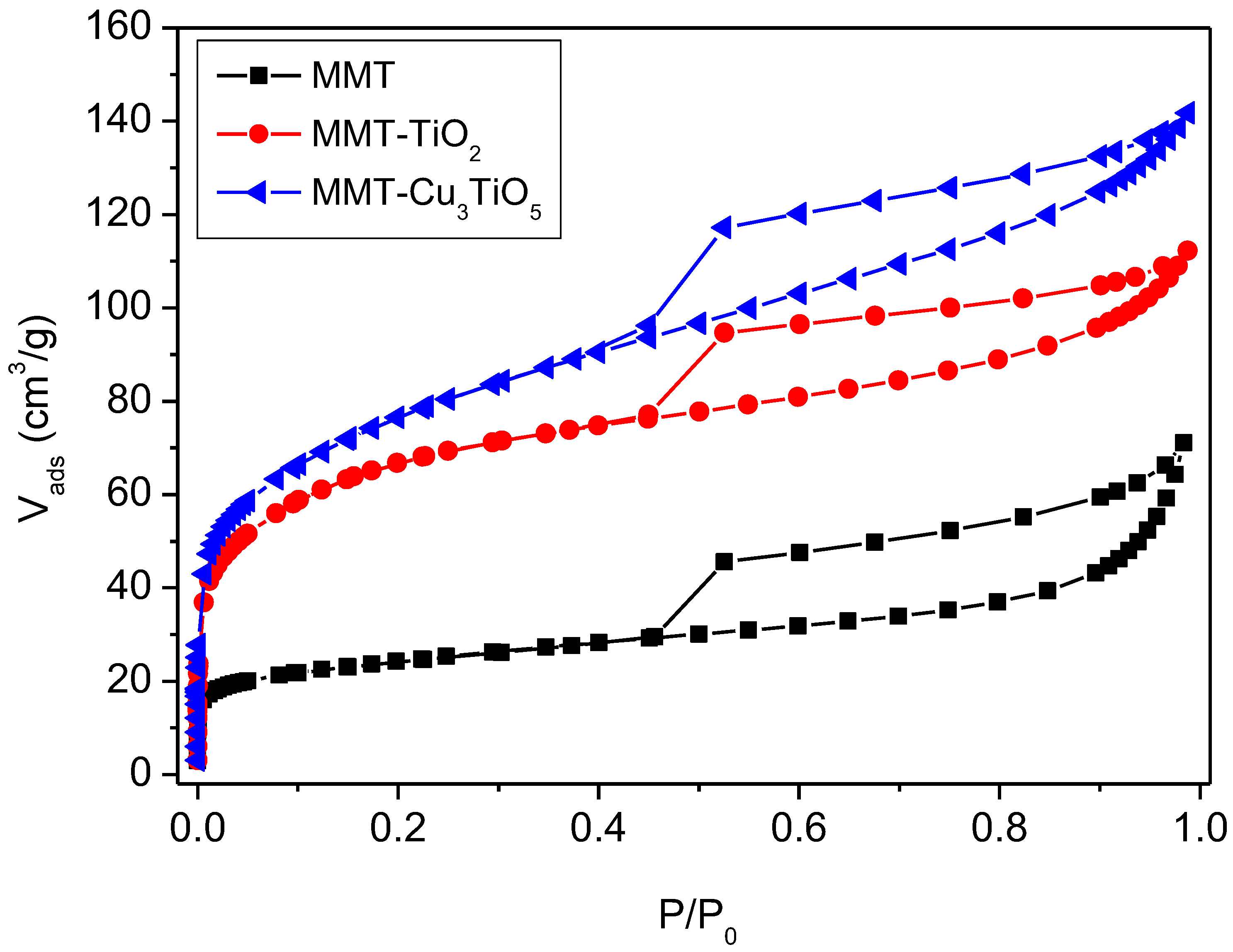
Table 2. In accordance with the classification system established by the International Union of Pure and Applied Chemistry (IUPAC), all three isotherms exhibit a pronounced H3 hysteresis loop, a characteristic feature of porous materials with a mesoporous structure. This aligns with the Type IV isotherm classification, frequently associated with multilayer adsorption followed by capillary condensation [43,44].
The modified MMT-TiO2 and MMT-Cu3TiO5 samples display a wider hysteresis loop, indicative of a broader pore size distribution and more pronounced mesoporous porosity. The hysteresis loop is indicative of plate particle aggregates or slit pores [45]. At low pressures (P/P0 < 0.2), rapid initial adsorption occurs due to adsorption on the external surface and in the micropores. At higher relative pressures (P/P0 > 0.4), the adsorption in the mesopores becomes more significant, as indicated by the continuous increase in the adsorbed volume. The hysteresis loop initiates at P/P0 = 0.45 and culminates near P/P0, indicative of the presence of moderate mesoporous pores.
A comparative analysis of the textural parameters of the samples presented in Table 2 reveals notable disparities between the pure montmorillonite, MMT-TiO2, and MMT-Cu3TiO5. The nitrogen adsorption results indicate a notable enhancement in the specific surface area (SBET), total pore volume (Vtot), and micropore volume (Vmicro) of the modified materials (MMT-TiO2 and MMT-Cu3TiO5) in comparison to pure montmorillonite. The SBET increases significantly with the addition of TiO2 and Cu3TiO5, rising from 85 m2/g for pure MMT to 245 m2/g for MMT-TiO2 and 279 m2/g for MMT-Cu3TiO5. The mean pore diameter (D) exhibited a notable increase from 5.84 Å for the MMT sample to 6.76 Å for the MMT-TiO2 sample and 6.74 Å for the MMT-Cu3TiO5 sample. This suggests that the modified materials possess a more developed pore structure. This phenomenon has been previously documented in the literature for catalysts obtained via impregnation of active phases [46]. This increase can be attributed to the formation of novel adsorption sites and the expansion of the pore size distribution, particularly within the mesoporous range. The modified materials exhibit an elevated average pore diameter and a more extensive pore distribution, indicating an enhancement in their textural characteristics. The micropore volume represents 19% and 11% of the total pore volume, respectively, while the external surface area contributes 67% and 78% to the total specific surface area of the MMT-TiO2 and MMT-Cu3TiO5 samples. The mesoporosity, which can be described as a “house of cards” type porous organization, indicates that the external surface is the primary site of interaction with the adsorbed molecules.
These findings suggest that the incorporation of TiO2 and Cu3TiO5 into the montmorillonite structure results in the formation of more porous materials, which exhibit enhanced potential for applications in adsorption and catalysis.

2.4. Morphological Characterization by SEM

Figure 3 depicts SEM images of MMT, MMT-TiO2, and MMT-Cu3TiO5. Bentonite particles manifest as irregular aggregates with considerable variation in size, as illustrated in Figure 3a. The surface is observed to be rough and porous, a quality also exhibited by clays such as bentonite [47]. Micrometer-sized filler clusters form agglomerates, as can be observed in Figure 3b,c. It is evident that MMT-TiO2 also exhibits irregularly shaped particles but with a notable difference from those observed in pure bentonite. The particles appear more compact and have more defined structures, which is likely due to the presence of titanium dioxide. The surface displays a reduction in cracking and an increase in uniformity when compared to bentonite alone [48]. This phenomenon may be attributed to the adsorption or coverage of TiO2 particles on the bentonite surface. TiO2 particles have the potential to act as a protective layer, modifying the bentonite surface.
The MMT-Cu3TiO5 particles (Figure 3c) are not discrete entities; rather, they form irregular aggregates of micrometric dimensions. The surface of the aggregates is characterized by a high degree of roughness and the presence of numerous pores. This phenomenon can be attributed to the lamellar structure of the bentonite and the incorporation of the semiconductor Cu3TiO5. It is challenging to discern individual Cu3TiO5 particles in this image. This indicates the presence of either a highly dispersed TiO2 phase within the bentonite matrix or a strong interaction between the two phases, which obscures the visibility of the TiO2 particles. The particles and aggregates appear to be micrometric in size, which is consistent with the nature of the bentonite. Nevertheless, a certain particle size distribution is evident, with the presence of larger and smaller aggregates.

2.5. FTIR Characterization

FTIR spectroscopy provides valuable insight into the molecular structure and intermolecular interactions of materials. In the present case, a comparative analysis of the spectra of MMT, MMT-TiO2, and MMT-Cu3TiO5 reveals the modifications induced by the incorporation of TiO2 and Cu3TiO5 into the bentonite structure. Figure 4 depicts the FTIR spectra of MMT, MMT-TiO2, and MMT-Cu3TiO5, which collectively demonstrate that all bentonite types exhibit absorption peaks that are characteristic of montmorillonite. The spectra display the characteristic bands of MMT at 3613, 3400, and 3229 cm−1. These broad, intense bands are attributed to the valence vibrations of the O–H bonds of adsorbed water molecules and the structural hydroxyl groups of MMT [49,50].
The peak at 1631 cm−1 is indicative of the angular deformation of adsorbed water molecules. The bands at 1106, 1003, 973, 916, 840, and 786 cm−1 are indicative of the valence and strain vibrations of Si–O and Al–O bonds within the tetrahedra and octahedra of the MMT structure, as previously documented in references [51,52,53]. In comparison to MMT, the spectra of MMT-TiO2 and MMT-Cu3TiO5 exhibit notable alterations. The O–H bands exhibit a shift and broadening. This indicates the presence of an interaction between water molecules and the TiO2 and Cu3TiO5 species introduced into the montmorillonite structure. The appearance of new bands in the low wavenumber regions (below 1000 cm−1) can be attributed to vibrations that are characteristic of Ti–O and Cu–O bonds. This evidence corroborates the incorporation of TiO2 and Cu3TiO5 into the bentonite matrix. Furthermore, alterations in the relative intensity of the Si–O and Al–O bands suggest a disruption in the bentonite structure following the introduction of the metal species. These findings indicate that the integration of TiO2 and Cu3TiO5 into the bentonite matrix alters its local environment by forming new bonds and disrupting existing ones. These modifications have the potential to significantly impact the physico-chemical properties of bentonite, including its CEC. As previously observed, there has been a notable enhancement in the cation exchange capacity and external specific surface area of bentonite. The external surface area of MMT-TiO2 exhibited a 300% increase, while that of MMT-Cu3TiO5 demonstrated a 403% increase in comparison to the initial external surface area of MMT. This increase had a significant impact on the catalytic properties.

2.6. Thermogravimetric Analysis

The TGA thermograms of the materials illustrated in Figure S1 demonstrate distinct thermal behaviors. For MMT, a 1% mass loss is observed between 30 and 150 °C, which is attributed to the desorption of physically adsorbed water. A further loss of 5.72% is observed between 200 and 500 °C, extending up to 700 °C, corresponding to the dehydroxylation of structural hydroxyl groups. The profiles of MMT-TiO2 and MMT-Cu3TiO5 are similar but show slight discrepancies. A mass loss of 3% is observed between 30 and 200 °C, which is attributed to the desorption of water and the elimination of hydroxyl groups.
A 7% loss is observed between 200 and 800 °C, which is attributed to phase transformations and dehydroxylation. Although these processes result in mass loss, they do not indicate a loss of thermal stability. The curve for MMT-Cu3TiO5 is slightly below that for MMT-TiO2, indicating a stronger interaction between Cu3TiO5 and MMT. This interaction enhances thermal stability by preserving structural integrity during phase transformations and dehydroxylation.
In conclusion, the materials MMT-TiO2 and MMT-Cu3TiO5 exhibit improved thermal stability compared to pure MMT, with MMT-Cu3TiO5 demonstrating the most favorable thermal stability, likely due to optimal interactions between Cu3TiO5 and the MMT matrix. The observed thermal stability beyond 500 °C for all samples indicates their potential for high-temperature applications. Additionally, the combined properties of montmorillonite, TiO2, and Cu3TiO5 may offer promising prospects for catalytic or sorption applications.

2.7. Zeta Potential

In the Orange G (OG) solution (Figure 5a), the zeta potential of the bentonite decreases with increasing pH, indicating that the bentonite surface becomes increasingly negative. This phenomenon may be attributed to the deprotonation of the adsorption sites on the bentonite. In comparison, the zeta potential values of the bentonite in distilled water (Figure 5b) exhibit minimal variation, indicating that the adsorption sites remain predominantly protonated. The lack of interaction between bentonite and OG dye molecules, as evidenced by the decrease in the zeta potential of MMT-TiO2 and MMT-Cu3TiO5 with increasing pH, also suggests that the surface of MMT-TiO2 and MMT-Cu3TiO5 becomes increasingly negative. However, this reduction is less pronounced compared to that observed in bentonite. This phenomenon may be attributed to the deprotonation of adsorption sites. The acid activation of bentonite and the presence of TiO2 may also result in the creation of new adsorption sites that exhibit differing reactivity with the dye compared to pure bentonite.
This is evidenced by the observed change in zeta potential with the presence of MMT-TiO2 and MMT-Cu3TiO5 on OG, with the peak change occurring at pH = 3, followed by a gradual decrease as the pH value increases and then decreases. The zeta potentials of MMT-Cu3TiO5 are less negative than those of pure bentonite and MMT-TiO2 at low pHs, indicating the potential for interaction between the copper in the composite and the dye, which could affect the surface charge. Zeta potential measurements demonstrate that the interactions between Orange G and the different materials are complex and are strongly influenced by pH. The addition of TiO2 and Cu to the composites appears to modulate the effect of pH on the surface charge, which could have a significant impact on the adsorption capacity of the dye.

2.8. UV Spectroscopy of Photocatalysts

As illustrated in Figure S2, the UV–visible spectrum provides the evaluated values of the band gap energy (Eg) for the prepared materials. These values were determined by plotting (αhν)0.5 as a function of photon energy (hν), where α represents the absorption coefficient; h is the Planck constant (4.14 × 10⁻¹⁵ eV·s), and ν is the photon frequency (Hz). The resulting UV–visible absorbance spectrum was obtained by measuring the absorbance (A) as a function of wavelength. The absorption coefficient (α) can be calculated from a spectrum [54] using the following equation:
α = 2.303A/t
where t is film thickness; α varies with the bandgap length of the semiconductor (Eg) and with the energy of the absorbed photon (hν) according to the Tauc equation:
αhν ∝ (hν − Eg)n
The experimental band gap of sample E can be obtained by identifying the point of intersection between the tangent to the linear portion of the curve and the abscissa axis (αhν)1/n = 0. The value of n is contingent upon the type of electronic transition involved (e.g., n = 1/2 for a direct allowed transition).
The gap energy for MMT-TiO2 and MMT-Cu3TiO5 was determined to be 2.7 eV and 2.15 eV, respectively, corresponding to the absorbance wavelengths of 459.2 nm and 568.8 nm.

2.9. Adsorption Kinetics

In order to investigate the photocatalytic degradation of OG, it was first necessary to determine the adsorption kinetics of the dye onto MMT-TiO2 and MMT-Cu3TiO
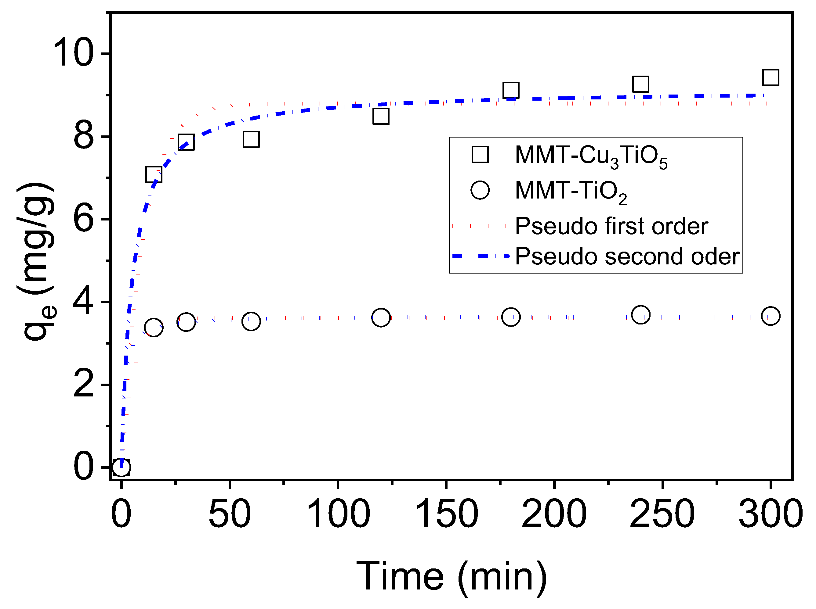
5 catalysts. The adsorption process, which precedes photodegradation, is of critical importance in establishing the efficiency of the catalyst. Batch adsorption experiments were conducted at 25 °C and pH 7, with an initial OG concentration of 40 mg/L and an adsorbent concentration of 2 g/L. The adsorption kinetics were monitored in the absence of light for a period of 300 min to ascertain the time required to reach equilibrium. The results depicted in Figure 6 provide valuable insights into the adsorptive properties of these catalysts and their potential for effective OG removal. The graph illustrates the temporal evolution of the quantity of OG dye adsorbed (in mg/g) for the two adsorbent materials as a function of time (in minutes). The materials under consideration are MMT-Cu3TiO5 and MMT-TiO2. The quantity of dye adsorbed exhibited a rapid increase within the initial 15 min of contact. This indicates that the most accessible adsorption sites are rapidly occupied by dye molecules. This rapid adsorption can be attributed to physisorption, a process whereby dye molecules are attracted to the material’s adsorption sites by weak forces, such as van der Waals forces. Subsequently, the quantity of dye adsorbed reaches a plateau, indicating that equilibrium has been reached with regard to the adsorption process. The number of available adsorption sites is reached, and the amount of adsorbed dye no longer varies significantly. The equilibrium uptake of dye on MMT-TiO2 is reached in 30 min with 3.60 mg/g (37%) of dye removal, while on MMT-Cu3TiO5, equilibrium is reached in 120 min with 9.11 mg/g (41.50%) of dye removal. The MMT-Cu3TiO5 material exhibits a greater adsorption capacity than the MMT-TiO2 material, attaining a higher maximum adsorption amount.
The findings indicate that the integration of copper into the MMT-Cu3TiO5 structure markedly enhances its adsorption capacity. This can be attributed to the increase in specific surface area and total pore volume (279 m2/g and 0.11 cm3/g versus 245 m2/g and 0.10 cm3/g for MMT-TiO2), which results in the creation of additional active sites for dye adsorption. Furthermore, the presence of copper introduces new specific adsorption sites and promotes coordination interactions between metal ions and dye molecules. While MMT-TiO2 exhibits promising adsorption capacity, the nature of its active sites, predominantly associated with titanium dioxide, appears less conducive to interactions with the dye under investigation. The surface coverage rate, which reflects the efficiency of an adsorbent, was evaluated for the materials MMT-TiO2 and MMT-Cu3TiO5. In accordance with the principle of proportionality between the surface area occupied by a molecule and its molecular surface area [55,56,57], it was estimated that OG used as a model dye occupies a surface area of 169.39 Å. The results demonstrate that MMT-Cu3TiO5 achieves a coverage rate of 6.87%, which is significantly higher than that of MMT-TiO2 (3.23%). Moreover, the enhanced pore structure and pore size, as demonstrated by BET analysis, could also facilitate the adsorption process. Therefore, it can be postulated that the interactions between the dye and the MMT-Cu3TiO5 surface may be more robust, thereby enhancing the adsorption process.
In order to investigate the adsorption mechanism, the nonlinear forms of the pseudo-first-order [58] and the pseudo-second-order [59] kinetic rate equations are provided as Equations (3) and (4), respectively.
q t = q e 1 e k 1 t
q t = k 2 q e 2 1 + k 2 q e t t
where qt and qe represent the amount of dyes adsorbed at equilibrium (mg/g) and at time t (min), and k1 (min−1) and k2 (g/mg min) are the pseudo-first-order and pseudo-second-order rate constants, respectively. The rate constants k1, k2, and qe(cal) were obtained from plots of Equations (3) and (4), as well as from qe(exp) (Figure 6). The resulting values are gathered in Table 3, along with the correlation coefficients R2 and the chi-square Test (χ2).
The pseudo-first-order and pseudo-second-order kinetic models demonstrated satisfactory fits to the experimental data. However, the rate constants (k1 = 0.180 min−1 and k2 = 0.206 g/mg.min) for MMT-TiO2 are significantly higher than those for MMT-Cu3TiO5. The rate constants (k1 = 0.099 min−1 and k2 = 0.021 g/mg. min) indicate that the adsorption kinetics of OG dye on MMT-Cu3TiO5 follows a relatively slow process. With regard to the correlation coefficient, the value of 0.999 for MMT-TiO2 is slightly higher than that of 0.997, indicating that the pseudo-second-order model provides a superior description of the adsorption kinetics for this material. In contrast, for MMT-Cu3TiO5, the value of 0.939 is less than 0.968, indicating that the first-order model may be more applicable to this material, although the fit is slightly inferior to that of MMT-TiO2. The chi-square test (χ2) indicates a discrepancy between the models and the experimental data, with low values for the pseudo-second-order model. Nevertheless, the pseudo-second-order model demonstrates an excellent correlation with the experimental data, suggesting that this model more accurately describes the adsorption kinetics of the OG dye for both materials. These results indicate that the adsorption of the dye likely follows a more intricate mechanism, consistent with the assumptions of the pseudo-second-order model. This suggests that specific interactions, such as multilayer formation or chemical reactions at the active sites, play a pivotal role in the adsorption process. This finding reflects the heterogeneous nature of adsorption on MMT-TiO2 and MMT-Cu3TiO5 materials, with the pseudo-second-order model offering the most accurate description of the observed process.

2.10. Study of the pH Effect on the Adsorption and Photodegradation of Orange G onto MMT-TiO2 and MMT-Cu3TiO5

The adsorption process is significantly influenced by pH, as it affects the surface charge of the adsorbents (MMT-TiO2 and MMT-Cu3TiO5) and the ionic form of the anionic dye OG (Figure S3). The adsorption of the anionic dye OG on MMT is found to be negligible under all pH conditions, indicating that pure montmorillonite is not an effective adsorbent for the dye at any pH. The highest adsorption is observed at pH 3, reaching approximately 52.48% on MMT-Cu3TiO5 and 31.01% on MMT-TiO2, demonstrating excellent efficiency under acidic conditions. The zeta potential was found to be approximately +2.81 mV for MMT-Cu3TiO5 and +1.05 mV for MMT-TiO2, indicating a significant positive charge. The anionic form of OG was the predominant species at pH values greater than 1, resulting from the deprotonation of its two SO3H groups (pKa = 1) [60]. This finding is consistent with the high adsorption of the dye at this pH, which suggests a favorable electrostatic interaction between the anionic dye and the positively charged material. At higher pH values, above pH = 7, the zeta potential becomes slightly negative (approximately −0.49 mV and −11.6 mV for MMT-Cu3TiO5 and MMT-TiO2, respectively), which may account for the slight decrease in adsorption observed. The efficiency of the MMT-Cu3TiO5 material declines, though it remains relatively stable at approximately 35% between pH 4 and 7 and between 24.28% and 19.43% between pH 4 and 9 for MMT-TiO2. The decline in efficiency with rising pH levels suggests that the reaction mechanism is more complex than a straightforward electrostatic interaction between oppositely charged species. Nevertheless, the rate of adsorption declines precipitously at pH values exceeding 9. At elevated pH levels, the repulsion between the negatively charged surfaces of the materials and the anionic dye (OG) increases, resulting in a reduction in adsorption efficiency. This is due to a reduction in favorable electrostatic interactions, which results in a decline in the efficacy of the adsorption process.
The adsorption process of the anionic azo dye OG on the nanocomposites MMT-TiO2 and MMT-Cu3TiO5 appears to be primarily regulated by physisorption forces. At acidic and slightly acidic pH levels, electrostatic interactions are established between the sulfonate groups (SO3−) of the anionic dye and the positive surface charges of the nanocomposites. Moreover, polar forces generated by an electric field within the micropores of the nanocomposites, as well as potential hydrogen bonds with the silanol (-SiOH) and aluminol (-AlOH) groups present on some nanocomposites, may also play a pivotal role in the adsorption process [61]. Therefore, the capacity of bentonite to adsorb anionic azo dye molecules can be markedly augmented through the immobilization of Cu3TiO5 nanoparticles on its surface. The efficiency of OG degradation using MMT-Cu3TiO5 at pH 3 was 99% after 120 min.

2.11. Photolytic Behavior of Orange G Dye

Figure 7 illustrates the photolysis kinetics of the degradation of OG in an aqueous solution with an initial concentration of 70 mg/L at pH 3 under LC8 lamp irradiation. As the duration of irradiation increases, the degradation of OG initially rises, reaching a maximum of 1% at 30 min. Further exposure beyond this point has a negligible impact. This indicates that the optimal concentration of hydroxyl radicals is reached within 30 min, thereby limiting further degradation of the dye’s functional groups [62]. In the absence of a catalyst, the degradation of OG remains low, irrespective of the duration of irradiation.
The results presented in Figure 7 illustrate the substantial influence of the MMT-TiO2 and MMT-Cu3TiO5 catalysts on the photocatalytic degradation of OG. The MMT-TiO2 catalyst markedly enhances the degradation process compared to simple photolysis, although complete degradation is not attained within a 300-minute period, with a rate of 96%. The addition of montmorillonite serves to stabilize the TiO2, thereby optimizing its photocatalytic performance. Nevertheless, the incomplete degradation may be attributed to saturation of the active sites or diffusion limitations. The MMT-Cu3TiO5 catalyst demonstrated significantly enhanced performance. Its structure, with a band gap of 2.15 eV in comparison to 2.7 eV for MMT-TiO2, allows for greater absorption of visible light. The immobilization of Cu3TiO5 to the montmorillonite system also appears to facilitate the generation of reactive radicals (OH•) and enhance selectivity in the attack on dye molecules, resulting in nearly complete degradation (100%) of the dye within 150 min. This distinctive MMT-Cu3TiO5 combination may be attributed to the enhanced separation of electron–hole pairs, which reduces recombination and enhances the efficiency of the photocatalytic reaction.
In order to ascertain the order of the OG degradation reaction, the experimental data were fitted to nonlinear kinetic models of pseudo-first-order (PFO) in accordance with Equation (5) and pseudo-second-order (PSO) in accordance with Equation (6). The best fit was identified through evaluation of the coefficient of determination (R2) and the chi-square test (χ2), which revealed that the predominant reaction order was as follows:
C C 0 = e k 1 t
C C 0 = 1 1 + k 2   C 0 t
where C0 represents the initial dye concentration (in mg/L); C denotes the concentration at time t (in mg/L); k1 signifies the rate constant of pseudo-first-order degradation (in units of minutes/L), and k2 denotes the rate constant of pseudo-second-order degradation (in units of L mg−1 min−1).
The experimental results were analyzed using pseudo-first- and pseudo-second-order kinetic models. Figure 7 presents a comparison of the two models, thereby enabling the identification of the model that most accurately describes the kinetics of the studied reaction. The regression coefficients and the chi-square (χ2) test (Table 4) demonstrated that the pseudo-first-order model provided the most accurate representation of the experimental data. This observation is consistent with prior research on the photodegradation kinetics of OG dye on diverse catalyst surfaces [63].
The photodegradation of OG dye using the MMT-Cu3TiO5 catalyst was found to be markedly more efficient than that achieved with the MMT-TiO2 material. This discrepancy in performance can be attributed to the enhanced specific surface area and positive surface charge of MMT-Cu3TiO5, which facilitate superior photocatalytic activity. The MMT-Cu3TiO5 nanocomposite was tested at varying catalyst concentrations (2, 2.66, 3.33, 4, and 4.66 g/L) to ascertain the optimal concentration for the photodegradation of OG. The results are presented in Figure 8. In contrast with conventional methodologies, the reaction medium was not maintained in a dark environment prior to irradiation; instead, it was directly exposed to the UV–Visible lamp. Time point t0 = 0 corresponds to the initiation of the solution’s exposure to the light, thereby marking the commencement of the photocatalysis experiment. However, the photolysis of OG dye in the absence of any photocatalyst resulted in minimal dye degradation, indicating that the degradation process was exclusively photocatalytic, as OG was not susceptible to UV radiation in the absence of the photocatalyst. This result is consistent with those reported by Derya et al. [64], wherein the photolysis of OG resulted in minimal dye degradation. The results demonstrate a rapid reduction in the concentration of OG, reaching 100% after only 180 min of irradiation for catalyst concentrations of 2.66, 3.33, 4, and 4.66 g/L of MMT-Cu3TiO5. Furthermore, additional experiments were conducted at a catalyst concentration of 2 g/L of MMT-Cu3TiO5, as higher concentrations resulted in an excessively rapid degradation process, yielding a limited number of exploitable data points. In the study of the rate at which a reaction occurs on the surface of a catalyst (heterogeneous catalysis), the Langmuir–Hinshelwood model is a commonly employed model. The model posits that reactant molecules initially adsorb onto the catalyst surface before undergoing a reaction with one another. The interaction on the catalyst surface directly influences the reaction rate, which can be quantified using Equation (7).
  V 0 = k K L C 1 + k K L C
In this equation, k represents the reaction rate constant; KL is the adsorption equilibrium constant that characterizes the affinity between the reactant and the catalyst surface, and C denotes the instantaneous concentration of the reactant in the solution. Daneshvar and colleagues [65] proposed an adaptation of the Langmuir–Hinshelwood model for the specific purpose of investigating the impact of the quantity of TiO2 employed as a catalyst Equation (8).
  V 0 = k K T i O 2 = k a p p T i O 2
In this context, kapp represents the apparent first-order rate constant. The experimental data are presented in Figure S4 for purposes of illustration.
Figure S4 illustrates that an increase in the concentration of MMT-Cu3TiO5 is accompanied by an enhancement in the initial reaction rate. This evidence substantiates the hypothesis that MMT-Cu3TiO5 plays a catalytic role in the degradation of the OG dye. The relationship between V0 and catalyst concentration appears to be approximately linear over the range of catalyst concentrations studied. This figure demonstrates that Equation (8) is verified, thereby validating the Langmuir–Hinshelwood model within the specified range of catalyst concentration. This study allows us to conclude that the photodegradation of OG dye in the presence of MMT-Cu3TiO5 can occur via two simultaneous processes: adsorption and photocatalysis, with a rate constant of 0.1646 min−1.

2.12. Regeneration of MMT-Cu3TiO5 Catalyst After Orange G Adsorption and Photodegradation

The potential for repurposing the MMT-Cu3TiO5 photocatalyst is of significant economic and practical value, necessitating comprehensive life cycle assessment and the identification of optimal treatment methodologies.
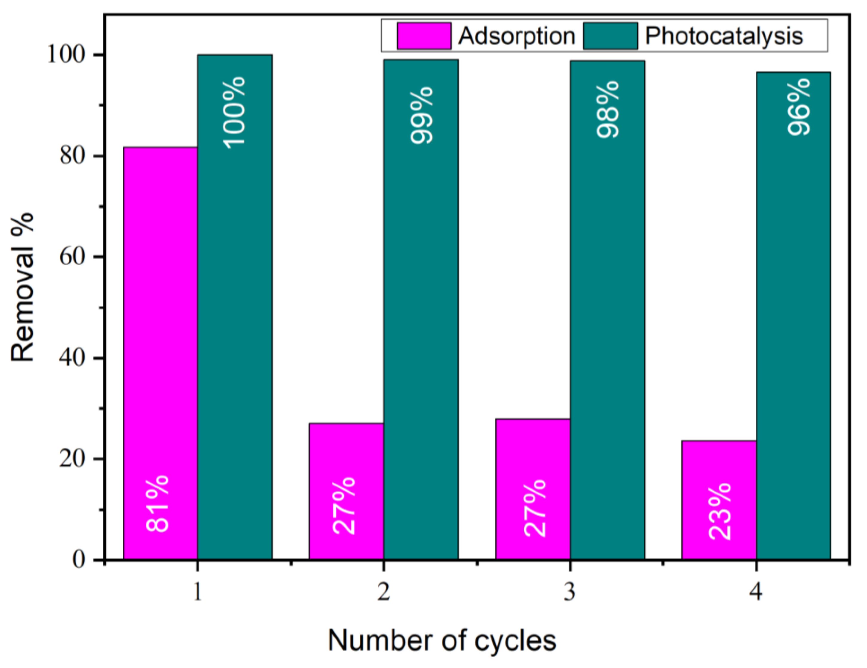
The potential for reusing MMT-Cu3TiO5 was investigated for up to four cycles of OG degradation under the following experimental conditions: an OG concentration of 70 mg/L and a catalyst concentration of 2 g/L, comprising MMT-TiO2 and MMT-Cu3TiO5. The adsorption process was monitored for a duration of 30 min, and the photocatalytic process was monitored for a duration of 120 min at a pH of 3 and a temperature of 25 °C. Subsequent to the completion of each process, the catalyst was extracted from the reaction mixture via centrifugation and subjected to a wash with the same solution that was recovered following the catalyst synthesis process, thereby maintaining the same acidity. Subsequently, the material was washed with distilled water, recovered by centrifugation, and dried at 80 °C. The catalyst’s weight was measured both before and after the regeneration process, and no appreciable loss of the catalyst was observed during this procedure.
As illustrated in Figure 9, the adsorption capacity of the catalyst exhibited a decline over time. However, the catalyst demonstrated the capacity to sustain the degradation of the dye throughout the same period, which substantiates its ability to maintain the production rate of and the same dye degradation rates. The findings demonstrate the capacity of the MMT- Cu3TiO5 catalyst to sustain its photocatalytic efficacy following four successive degradation cycles, exhibiting a modest decline in the extent of OG degradation.
Initially, the capacity of the catalyst to purify itself from adsorbed materials was observed, as evidenced by the DRX and FTIR results (Figure 10 and Figure 11, respectively). The absence of new materials after use indicates that the catalyst utilized the photocatalytic properties of the dye to achieve complete degradation.

3. Materials and Methods

3.1. Catalysts Preparation

In this study, the support material was untreated bentonite from Sigma-Aldrich, an affiliate of Merck KGaA, Darmstadt, Germany. The bentonite utilized is predominantly composed of montmorillonite (MMT). Moreover, the composition is validated by mineralogical and chemical analyses. The particle size is ≤25 µm, and the bentonite in question exhibits a high cation exchange capacity (CEC) of 213 meq/100 g. The general structural formula is (Si3.89Al0.10)IV(Al0.93Fe0.32Mg0.61Ti0.02)VIO10Na0.33Ca0.23K0.02(OH)2,nH2O. The basic chemical composition is presented in Table 1.
The sol–gel method was employed to synthesize two catalysts, designated as MMT-TiO2 and MMT-Cu3TiO5 (Figure 12). The synthesis of MMT-TiO2 was initiated with the preparation of a solution of TiCl4. To this end, 2.8 mL of TiCl4 (99.9%, Fluka/Honeywell, Charlotte, NC, USA) was mixed with ethanol (99.8%, Fluka/Honeywell) and distilled water (2 mL, at 5 °C). This solution was then added, under continuous stirring, to a suspension of montmorillonite clay at a concentration of 1% (w/w), maintaining a Ti/clay mass ratio of 2:1. This ratio was selected to ensure adequate titanium loading for effective immobilization of TiO2 on the clay surface. After a 24-h aging period, the resulting material was subjected to a thorough washing process to remove any residual unreacted precursors. It is important to note that, rather than achieving a completely homogeneous coverage of the clay surface, the final material consists of a composite of TiO2 and montmorillonite. The titanium species involved include both TiO2 and potential residual titanium compounds (such as titanium hydroxides), which are not uniformly distributed on the clay surface. The composite was then dried at 80 °C for 24 h and sieved in order to obtain the MMT-TiO2 catalyst. In the case of MMT-Cu3TiO5, copper was introduced as a modifying agent in a controlled amount. A specific quantity of CuCl2 was dissolved in 2 mL of distilled water and subsequently added to the TiO2 precursor solution. The quantity of CuCl2 was calculated in order to achieve a Cu/Ti mass ratio of 0.02/2. This low ratio was deliberately selected to optimize the synergistic interaction between Cu and TiO2; excessive Cu loading, which could lead to particle agglomeration or interference with photocatalytic activity, was, thus, avoided. Subsequently, the Cu-modified TiO2 solution was immobilized onto montmorillonite in accordance with the same procedure employed for MMT-TiO2 synthesis. Following immobilization, the MMT-Cu3TiO5 catalyst was subjected to the same washing, drying (80 °C for 24 h), and sieving steps as MMT-TiO2 synthesis. Although the low Cu/Ti ratio may result in the limited detectability of Cu-containing phases by XRD, the sol–gel synthesis ensures the homogeneous distribution of Cu species. Furthermore, the incorporation of Cu enhances the visible light absorption and photocatalytic performance of the final material.

3.2. Characterization of Catalysts

A variety of physicochemical techniques were utilized to characterize the catalyst materials. The chemical composition was determined using an Agilent Technologies Inductively Coupled Plasma Emission Spectrometer (ICP-AES 5110, dual view, Santa Clara, CA, USA). The structure and crystallinity were analyzed by PXRD using a Siemens D-5000 diffractometer with CuKα radiation (1.5406 Å). Patterns were recorded over the 5–80° 2θ range in steps of 0.04° with a counting time per step of 8 s. The infrared absorption (IR) spectra were determined between 4000 and 400 cm−1 using a PHILIPS PU 9714 spectrophotometer.
Thermogravimetric analysis (TGA) was performed with a NETZSCH TG 209F1 Libra (Selb, Germany) with a mass resolution of 3 µg and using HT platinum plates. The analysis of the samples with an average weight of 8 mg was conducted under a nitrogen atmosphere, with a flow rate of 20 mL·min−1. The samples were subjected to a heating ramp of 10 °C·min−1 within the temperature range of 40 to 800 °C. The textural properties of the prepared samples were determined through the analysis of nitrogen adsorption/desorption isotherms, which were measured with a Micromeritics ASAP 2020 analyzer (Norcross, GA, USA). The specific surface area was determined by the Brunauer–Emmett–Teller (BET) multi-point analysis method from nitrogen adsorption at 77 K. The total pore volume was calculated from the amount of gas adsorbed at (P/P0) = 0.99, and the micropore volume was calculated using the Barrett–Joyner–Halenda (BJH) model on the desorption branch.
Scanning electron microscopy (SEM) observations were conducted using a JEOL JSM-7800F apparatus (Tokyo, Japan), operating from 0.5 to 30 kV, which was equipped with an energy dispersive spectrometer (EDX) for chemical analysis. The powder material was dispersed on a support with a double-sided conductive carbon tape. To prevent electronic charging, a nanometer-thick chrome deposit was applied to the preparation.
Zeta-potential measurements were conducted at room temperature using a Zetasizer (Nano-ZS model, Malvern Instruments, Malvern, UK). To investigate the impact of pH on the surface charge of MMT, MMT-TiO2, and MMT-Cu3TiO5, a series of aqueous solutions with varying pH values were prepared by adjusting the pH of the MMT/water mixtures with 0.1 N HCl or NaOH solutions. The material was dissolved in a solution of 2 g·L−1 at a volume of 10 mL. Each zeta-potential value was determined in triplicate using three distinct samples.
The concentration of the dye in the solution was determined using a Cary 100 spectrophotometer (Agilent, Santa Clara, CA, USA), which was operated in absorbance mode. The bandgap energy of the photocatalyst material was calculated using the Tauc equation. In order to select an appropriate light source for photodegradation experiments, a Hamamatsu LC8 light source was chosen, as it emits light with energy greater than or equal to the bandgap energy of the materials, thereby generating the (e, hole) couple and allowing for a series of photodegradation processes to be performed.

3.3. Photoreactor and Analytical Methods

Irradiation experiments were conducted in a photocatalytic oxidation reactor connected to a thermostatic bath equipped with a water-cooling circuit to absorb infrared radiation, thus preventing the solution from heating (Figure S5). At the core of the cylindrical reactor is a 150-watt xenon lamp (Hamamatsu LC8) coupled to an optical fiber positioned perpendicular to the UV–visible light source. The lamp emits a broad-spectrum irradiation, as illustrated in Figure S6, which depicts a peak in the visible range. The optical fiber tip was maintained at a distance of 3 cm from the reactor, and the reaction mixture was continuously agitated using a magnetic stirrer.
A solution of Orange G dye at a concentration of 0.5 g/L was prepared using distilled water as the solvent. The requisite concentrations for the photocatalysis experiments were subsequently derived from this stock solution. In particular, the impact of solution acidity on the adsorption process was examined using a dye concentration of 70 mg/L in 30 mL of solution. The pH of the solution was maintained through the addition of 0.1 N NaOH or 0.1 N HCl. Subsequently, the impact of varying the catalyst quantity on the photocatalytic degradation of the dye was investigated, with the optimal pH being identified as 3. This was performed to identify the appropriate catalyst dosage that maximizes the photocatalytic degradation efficiency and minimizes the treatment time. The degradation of OG (Orange G) was monitored by measuring absorbance using a UV–vis spectrophotometer at a wavelength of 478 nm. The absorbance measurements were converted to concentrations using a standard calibration method (r = 0.9999).
In order to prevent photodegradation, which could otherwise occur during the course of an adsorption experiment, tests are conducted in the dark. This ensures that only adsorption occurs. Once equilibrium has been reached, the catalyst is separated from the solution, and the concentration of the dye is then measured using visible spectroscopy in order to determine the amount adsorbed. In photodegradation experiments, the lamp (LC8) is activated to initiate the photodegradation process. In this instance, the catalyst is subjected to illumination, and any diminution in the concentration of the dye is attributable to degradation rather than adsorption. The presence or absence of light serves as a clear distinction between the two processes. When the material is exposed to light (hν), an electron is excited from a valence band to a conduction band, thereby creating an electron–hole pair.
M M T C u 3 T i O 5 + h ν     M M T C u 3 T i O 5 h + + e
A positive hole (h+) can react with water adsorbed on the catalyst surface to form hydroxyl radicals (OH), which are highly reactive oxidizing agents.
M M T C u 3 T i O 5 h + + H 2 O   M M T C u 3 T i O 5 O H + H +
The electron (e) is capable of reacting with molecular oxygen that has been adsorbed on the catalyst surface, resulting in the formation of superoxide anions (O2−•). These species are also powerful oxidizers.
M M T C u 3 T i O 5 e + O 2   M M T C u 3 T i O 5 O 2
Hydroxyl radicals ( O H ) can directly attack dye molecules, resulting in the oxidation and degradation of these molecules into inorganic products.
M M T C u 3 T i O 5 O H + d y e   d e g r a d a t i o n   p r o d u c t + M M T C u 3 T i O 5
Additionally, superoxide anions O 2 have been demonstrated to facilitate the degradation of dyes, either directly or by the generation of other reactive species.
M M T C u 3 T i O 5 O 2 + d y e   d e g r a d a t i o n   p r o d u c t + M M T C u 3 T i O 5
This type of mechanism is frequently employed to elucidate the phenomenon of heterogeneous photocatalysis, which has a multitude of applications in the remediation of water pollution.

4. Conclusions

This study successfully synthesized and characterized MMT-TiO2 and MMT-Cu3TiO5 nanocomposites using a sustainable sol–gel method, thereby demonstrating notable enhancements in their structural and optical properties. The specific surface area exhibited a notable increase, with XRD analysis confirming the presence of TiO2 anatase and Cu3TiO5. The optical analysis demonstrated that MMT-Cu3TiO5, exhibiting a lower bandgap energy of 2.15 eV, demonstrated enhanced absorption of longer wavelengths of visible light in comparison to MMT-TiO2. The MMT-Cu3TiO5 catalyst demonstrated complete degradation of Orange G dye within 120 min under optimal conditions (pH 3, catalyst concentration 2 g/L), outperforming the MMT-TiO2 catalyst. Furthermore, the MMT-Cu3TiO5 catalyst can be washed and reused multiple times without the addition of chemical agents, thus demonstrating its sustainability. In conclusion, the MMT-Cu3TiO5 nanocomposites display enhanced structural, optical, and photocatalytic properties, rendering them highly promising for wastewater treatment applications, particularly in the degradation of organic contaminants such as Orange G dye.

Supplementary Materials

The following supporting information can be downloaded at https://www.mdpi.com/article/10.3390/catal15010088/s1, Figure S1: TGA of MMT, MMT-TiO2 and MMT-Cu3TiO5; Figure S2: UV-visible spectra of the absorbance of materials (MMT-TiO2, MMT- Cu3TiO5) as a function of wavelength and their corresponding gap energies; Figure S3: Effect of initial pH on (a) adsorption and (b) photodegradation of Orange G dye onto MMT, MMT-TiO2 and MMT-Cu3TiO5: The conditions for each experiment: OG concentration = 70 mg/L; MMT-Cu3TiO5 catalyst concentration = 2g/L; exposure time t = 30 min (MMT) and 120 min (MMT-Cu3TiO5) and T = 25 °C; Figure S4: Validation of the linear Langmuir-Hinshelwood model for Orange G photodegradation catalyzed by MMT-Cu3TiO5; Figure S5: Setup of the irradiation device; Figure S6: Emission spectrum of the Xenon Lamp LC8.

Author Contributions

Conceptualization, Z.B. and U.M.; methodology, Z.B. and U.M.; investigation, A.A.-A. and K.B.; formal analysis, A.A.-A., K.B. and N.D.T.; data curation, A.A.-A., K.B. and N.D.T.; writing-original draft preparation, Z.B.; writing-review and editing, U.M.; visualization, Z.B. and U.M.; supervision, Z.B. and U.M.; project administration, Z.B. and U.M. All authors have read and agreed to the published version of the manuscript.

Funding

This research received no external funding and the APC was funded by MDPI.

Data Availability Statement

Data are contained within the article or supplementary material.

Acknowledgments

The authors express their gratitude to Christel Pierlot, Christophe Volkringer, and Alexandre Fadel for their technical assistance. This work is the result of a close collaboration between the LPCMCE laboratory at USTOMB and the UMET laboratory at the University of Lille. The present project was carried out within the framework of the Hubert Curien Tassili (PHC) research program, titled 24MDU102 (2024–2026). The first author of this study is a beneficiary of the EIFFEL Excellence PhD program (CampusFrance, 2023–2025). The authors would like to express their profound gratitude to all individuals who contributed to the realization of these projects. Additionally, the authors wish to acknowledge the support of the Algerian Ministry of Higher Education and Scientific Research (MESRS), the General Directorate of Scientific Research and Technological Development (DGRSDT) of Algeria, the University of Sciences and Technology of Oran (USTOMB)/Algeria, the French Ministry of Higher Education and Research (MENESR), CampusFrance, CNRS, MDPI, and the University and CROUS of Lille/France.

Conflicts of Interest

The authors declare no conflict of interest.

References

  1. Tkaczyk, A.; Mitrowska, K.; Posyniak, A. Synthetic organic dyes as contaminants of the aquatic environment and their implications for ecosystems: A review. Sci. Total Environ. 2020, 717, 137222. [Google Scholar] [CrossRef]
  2. Abdulmohsen, K.; Alsukaibi, D. Various Approaches for the Detoxification of Toxic Dyes in Wastewater. Processes 2022, 10, 1968. [Google Scholar] [CrossRef]
  3. Carneiro, P.A.; Umbuzeiro, G.A.; Oliveira, D.P.; Zanoni, M.V. Assessment of water contamination caused by a mutagenic textile effluent/dyehouse effluent bearing disperse dyes. J. Hazard. Mater. 2010, 174, 694–699. [Google Scholar] [CrossRef]
  4. Kishor, R.; Purchase, D.; Saratale, G.D.; Saratale, R.G.; Ferreira, L.F.R.; Bilal, M.; Chandra, R.; Bharagava, R.N. Ecotoxicological and health concerns of persistent coloring pollutants of textile industry wastewater and treatment approaches for environmental safety. J. Environ. Chem. Eng. 2021, 9, 105012. [Google Scholar] [CrossRef]
  5. Parmar, S.; Daki, S.; Bhattacharya, S.; Shrivastav, A. Chapter 8—Microorganism: An ecofriendly tool for waste management and environmental safety. In Development in Wastewater Treatment Research and Processes; Elsevier: Amsterdam, The Netherlands, 2022; pp. 175–193. [Google Scholar] [CrossRef]
  6. Al-Tohamy, R.; Ali, S.S.; Li, F.; Okasha, K.M.; Mahmoud, Y.A.G.; Elsamahy, T.; Jiao, H.; Fu, Y.; Sun, J. A critical review on the treatment of dye-containing wastewater: Ecotoxicological and health concerns of textile dyes and possible remediation approaches for environmental safety. Ecotoxicol. Environ. Saf. 2022, 231, 113160. [Google Scholar] [CrossRef] [PubMed]
  7. Svetozarević, M.; Šekuljica, N.; Onjia, A.; Barać, N.; Mihajlović, M.; Knežević-Jugović, Z.; Mijin, D. Biodegradation of synthetic dyes by free and cross-linked peroxidase in microfluidic reactor. Environ. Technol. Innov. 2022, 26, 102373. [Google Scholar] [CrossRef]
  8. Nnaji, P.C.; Anadebe, V.C.; Ezemagu, I.G.; Onukwuli, O.D. Potential of Luffa cylindrica seed as coagulation-flocculation (CF) agent for the treatment of dye wastewater: Kinetic, mass transfer, optimization and CF adsorption studies. Arab. J. Chem. 2022, 15, 103629. [Google Scholar] [CrossRef]
  9. Attri, P.; Garg, S.; Ratan, J.K.; Giri, A.S. Comparative study using advanced oxidation processes for the degradation of model dyes mixture: Reaction kinetics and biodegradability assay. Mater. Today Proc. 2022, 57, 1533–1538. [Google Scholar] [CrossRef]
  10. Huang, X.; Tian, C.; Qin, H.; Guo, W.; Gao, P.; Xiao, H. Preparation and characterization of Al3+-doped TiO2 tight ultrafiltration membrane for efficient dye removal. Ceram. Int. 2020, 46, 4679–4689. [Google Scholar] [CrossRef]
  11. Türk, F.N.; Çiftçi, H.; Arslanoğlu, H. Removal of Basic Yellow 51 Dye by Using Ion Exchange Resin Obtained by Modification of Byproduct Sugar Beet Pulp. Sugar Tech 2023, 25, 569–579. [Google Scholar] [CrossRef]
  12. Cotillas, S.; Llanos, J.; Cañizares, P.; Clematis, D.; Cerisola, G.; Rodrigo, M.A.; Panizza, M. Removal of Procion Red MX-5B dye from wastewater by conductive-diamond electrochemical oxidation. Electrochim. Acta 2018, 263, 1–7. [Google Scholar] [CrossRef]
  13. Hajiali, M.; Farhadian, M.; Tangestaninejad, S.; Khosravi, M. Synthesis and characterization of Bi2MoO6/MIL-101(Fe) as a novel composite with enhanced photocatalytic performance: Effect of water matrix and reaction mechanism. Adv. Powder Technol. 2022, 33, 103546. [Google Scholar] [CrossRef]
  14. Suhag, M.H.; Khatun, A.; Tateishi, I.; Furukawa, M.; Katsumata, H.; Kaneco, S. Purification of aqueous orange II solution through adsorption and visible-light-induced photodegradation using ZnO-modified g-C3N4 composites. RSC Adv. 2024, 14, 17888–17900. [Google Scholar] [CrossRef]
  15. Azha, S.F.; Shahadat, M.; Ismail, S.; Ali, S.W.; Ahammad, S.Z. Prospect of clay-based flexible adsorbent coatings as cleaner production technique in wastewater treatment, challenges, and issues: A review. J. Taiwan Inst. Chem. Eng. 2021, 120, 178–206. [Google Scholar] [CrossRef]
  16. Khan, S.; Ajmal, S.; Hussain, T.; Ur Rahman, M. Clay-based materials for enhanced water treatment: Adsorption mechanisms, challenges, and future directions. J. Umm Al-Qura Univ. Appl. Sci. 2023, 1–16. [Google Scholar] [CrossRef]
  17. Chuaicham, C.; Trakulmututa, J.; Shu, K.; Shenoy, S.; Srikhaow, A.; Zhang, L.; Mohan, S.; Sekar, K.; Sasaki, K. Recent Clay-Based Photocatalysts for Wastewater Treatment. Separations 2023, 10, 77. [Google Scholar] [CrossRef]
  18. Bakhtiar, A.; Bouberka, Z.; Roussel, P.; Volkringer, C.; Addad, A.; Ouddane, B.; Pierlot, C.; Maschke, U. Development of a TiO2/Sepiolite Photocatalyst for the Degradation of a Persistent Organic Pollutant in Aqueous Solution. Nanomaterials 2022, 12, 3313. [Google Scholar] [CrossRef]
  19. Jawad, A.H.; Saber, S.M.; Abdulhameed, A.S.; Farhan, A.M.; ALOthman, Z.A.; Wilson, L.D. Characterization and applicability of the natural Iraqi bentonite clay for toxic cationic dye removal: Adsorption kinetic and isotherm study. J. King Saud Univ. Sci. 2023, 35, 102630. [Google Scholar] [CrossRef]
  20. Hnamte, M.; Pulikkal, A.K. Clay-polymer nanocomposites for water and wastewater treatment: A comprehensive review. Chemosphere 2022, 307, 135869. [Google Scholar] [CrossRef] [PubMed]
  21. Mostafa, A.G.; El-Hamid, A.I.A.; Akl, M.A. Surfactant-supported organoclay for removal of anionic food dyes in batch and column modes: Adsorption characteristics and mechanism study. Appl. Water Sci. 2023, 13, 163. [Google Scholar] [CrossRef]
  22. Du, Q.; Chen, S.; Liu, H.; Zhang, M.; Ren, S.; Luo, W. Sequential modification of montmorillonite by Al13 polycation and cationic gemini surfactant for the removal of Orange II. Colloids Surf. A Physicochem. Eng. Asp. 2024, 687, 133489. [Google Scholar] [CrossRef]
  23. Reza, K.; Kurny, A.; Gulshan, F. Parameters affecting the photocatalytic degradation of dyes using TiO2: A review. Appl. Water Sci. 2017, 7, 1569–1578. [Google Scholar] [CrossRef]
  24. Tichapondwa, S.M.; Newman, J.P.; Kubheka, O. Effect of TiO2 phase on the photocatalytic degradation of methylene blue dye. Phys. Chem. Earth Parts A/B/C 2020, 118–119, 102900. [Google Scholar] [CrossRef]
  25. Giovanazzi, L.; Missner, M.; Francisco, I.; Rodrigues, N.; Da Silva Júnior, A.; Pellenz, L.; De Aguiar, C.; De Oliveira, C. Photocatalytic degradation using TiO2 P25: A comparative study for different textile dyes. Braz. J. Anim. Environ. Res. 2024, 7, 125. [Google Scholar] [CrossRef]
  26. Mortazavian, S.; Saber, A.; James, D.E. Optimization of photocatalytic degradation of acid blue 113 and acid red 88 textile dyes in a UV-C/TiO2 suspension system: Application of response surface methodology (RSM). Catalysts 2019, 9, 360. [Google Scholar] [CrossRef]
  27. Dharma, H.; Jaafar, J.; Widiastuti, N.; Matsuyama, H.; Rajabsadeh, S.; Othman, M.; Rahman, M.; Jafri, N.; Suhaimin, N.; Nasir, A.; et al. Review of Titanium Dioxide (TiO2)-Based Photocatalyst for Oilfield-Produced Water Treatment. Membranes 2022, 12, 345. [Google Scholar] [CrossRef] [PubMed]
  28. Sagadevan, S.; Fatimah, I.; Egbosiub, T.C.; Alshahateet, S.F.; Lett, J.A.; Weldegebrieal, G.K.; Le, M.V.; Johan, M.R. Photocatalytic efficiency of Titanium dioxide for dyes and heavy metals removal from wastewater. Bull. Chem. React. Eng. Catal. 2022, 17, 430–450. [Google Scholar] [CrossRef]
  29. Zhang, Y.; Jia, A.; Li, Z.; Yuan, Z.; Huang, W. Titania-Morphology-Dependent Pt–TiO2 Interfacial Catalysis in Water-Gas Shift Reaction. ACS Catal. 2023, 13, 392–399. [Google Scholar] [CrossRef]
  30. Chen, D.; Cheng, Y.; Zhou, N.; Chen, P.; Wang, Y.; Li, K.; Huo, S.; Cheng, P.; Peng, P.; Zhang, R.; et al. Photocatalytic degradation of organic pollutants using TiO2-based photocatalysts: A review. J. Clean. Prod. 2020, 268, 121725. [Google Scholar] [CrossRef]
  31. Li, L.; Chen, X.; Quan, X.; Qiu, F.; Zhang, X. Synthesis of CuOx/TiO2 Photocatalysts with Enhanced Photocatalytic Performance. ACS Omega 2023, 8, 2723–2732. [Google Scholar] [CrossRef] [PubMed]
  32. González-Tejero, M.; Villachica-Llamosas, J.G.; Ruiz-Aguirre, A.; Colón, G. High-Performance Photocatalytic H2 Production Using a Binary Cu/TiO2/SrTiO3 Heterojunction. ACS Appl. Energy Mater. 2023, 6, 4007–4015. [Google Scholar] [CrossRef] [PubMed]
  33. Ahmad, S.; Yasin, A. Photocatalytic degradation of deltamethrin by using Cu/TiO2/bentonite composite. Arab. J. Chem. 2020, 13, 8481–8488. [Google Scholar] [CrossRef]
  34. Xin, Y.; Yu, K.; Zhang, L.; Yang, Y.; Yuan, H.; Li, H.; Wang, L.; Zeng, J. Copper-Based Plasmonic Catalysis: Recent Advances and Future Perspectives. Adv. Mater. 2021, 33, 2008145. [Google Scholar] [CrossRef] [PubMed]
  35. Ghrair, A.M.; Said, A.J.; Al-Kroom, H.; Al Daoud, N.; Hanayneh, B.; Mhanna, A.; Gharaibeh, A. Utilization of Jordanian Bentonite Clay in Mortar and Concrete Mixtures. Jordan J. Earth Environ. Sci. 2023, 14, 19–29. [Google Scholar]
  36. Wang, Z.; Jiang, Y.; Tian, G.; Zhu, C.; Zhang, Y. Toxicological Evaluation toward Refined Montmorillonite with Human Colon Associated Cells and Human Skin Associated Cells. J. Funct. Biomater. 2024, 15, 75. [Google Scholar] [CrossRef]
  37. Kumar, A.; Lingfa, P. Sodium Bentonite and Kaolin Clays: Comparative Study on Their FT-IR, XRF, and XRD. Mater. Today Proc. 2020, 22, 737–742. [Google Scholar] [CrossRef]
  38. Sushma; Kumar, H.; Ahmad, I.; Dutta, P.K. In-vitro toxicity induced by quartz nanoparticles: Role of ER stress. Toxicology 2018, 404–405, 1–9. [Google Scholar] [CrossRef] [PubMed]
  39. Leinardi, R.; Pavan, C.; Yedavally, H.; Tomatis, M.; Salvati, A.; Turci, F. Cytotoxicity of fractured quartz on THP-1 human macrophages: Role of the membranolytic activity of quartz and phagolysosome destabilization. Arch. Toxicol. 2020, 94, 2981–2995. [Google Scholar] [CrossRef] [PubMed]
  40. Asif Javed, H.M.; Ahmad, M.I.; Que, W.; Qureshi, A.A.; Sarfaraz, M.; Hussain, S.; Iqbal, M.Z.; Nisar, M.Z.; Shahid, M.; AlGarni, T.S. Encapsulation of TiO2 nanotubes with Cs nanoparticles to enhance electron injection and thermal stability of perovskite solar cells. Surf. Interfaces 2021, 23, 101033. [Google Scholar] [CrossRef]
  41. Scherrer, P. Bestimmung der Grösse und der inneren Struktur von Kolloidteilchen mittels Röntgenstrahlen. Nachr. Ges. Wiss. Göttingen 1918, 2, 98–100. [Google Scholar]
  42. Langford, J.I.; Wilson, A.J.C. Scherrer after Sixty Years: A Survey and Some New Results in the Determination of Crystallite Size. J. Appl. Cryst. 1978, 11, 102–113. [Google Scholar] [CrossRef]
  43. Hartmann, M.; Thommes, M.; Schwieger, W. Hierarchically-Ordered Zeolites: A Critical Assessment. Adv. Mater. Interfaces 2021, 8, 2001841. [Google Scholar] [CrossRef]
  44. Inocencio, S.; Cordeiro, T.; Matos, I.; Danede, F.; Sotomayor, J.C.; Fonseca, I.M.; Correia, N.T.; Corvo, M.C.; Dionisio, M. Ibuprofen incorporated into unmodified and modified mesoporous silica: From matrix synthesis to drug release. Microporous Mesoporous Mater. 2021, 310, 110541. [Google Scholar] [CrossRef]
  45. Chen, K.; Zhang, T.; Chen, X.; He, Y.; Liang, X. Model construction of micro-pores in shale: A case study of Silurian Longmaxi Formation shale in Dianqianbei area, SW China. Pet. Explor. Dev. 2018, 45, 412–421. [Google Scholar] [CrossRef]
  46. Gaillard, M.; Virginie, M.; Khodakov, A.Y. New molybdenum-based catalysts for dry reforming of methane in presence of sulfur: A promising way for biogas valorization. Catal. Today 2017, 289, 143–150. [Google Scholar] [CrossRef]
  47. Gandhi, D.; Bandyopadhyay, R.; Soni, B. Naturally occurring bentonite clay: Structural augmentation, characterization and application as catalyst. Mater. Today Proceeding 2022, 57, 194–201. [Google Scholar] [CrossRef]
  48. Zhu, C.; Wang, X.; Huang, Q.; Huang, L.; Xie, J.; Qing, C.; Chen, T. Removal of gaseous carbon bisulfide using dielectric barrier discharge plasmas combined with TiO2 coated attapulgite catalyst. Chem. Eng. J. 2013, 225, 567–573. [Google Scholar] [CrossRef]
  49. El-Enein, S.A.; Okbah, M.A.; Hussain, S.G.; Soliman, N.F.; Ghounam, H.H. Adsorption of Selected Metals Ions in Solution Using Nano-Bentonite Particles: Isotherms and Kinetics. Environ. Process. 2020, 7, 463–477. [Google Scholar] [CrossRef]
  50. Hebbar, R.S.; Isloor, A.M.; Prabhu, B.; Inamuddin; Asiri, A.M.; Ismail, A.F. Removal of metal ions and humic acids through polyetherimide membrane with grafted bentonite clay. Sci. Rep. 2018, 8, 4665. [Google Scholar] [CrossRef]
  51. Liu, Z.; Uddin, M.A.; Sun, Z. FT-IR and XRD analysis of natural Na-bentonite and Cu(II)-loaded Na-bentonite. Spectrochim. Acta—Part A Mol. Biomol. Spectrosc. 2011, 79, 1013–1016. [Google Scholar] [CrossRef]
  52. Laysandra, L.; Winda Masnona Kartika Sari, M.; Edi Soetaredjo, F.; Foe, K.; Nyoo Putro, J.; Kurniawan, A.; Ju, Y.; Ismadji, S. Adsorption and photocatalytic performance of bentonite-titanium dioxide composites for methylene blue and rhodamine B decoloration. Heliyon 2017, 3, e00488. [Google Scholar] [CrossRef] [PubMed]
  53. Khansili, N.; Krishnam, P. Curcumin functionalized TiO2 modified bentonite clay nanostructure for colorimetric Aflatoxin B1 detection in peanut and corn. Sens. Bio-Sens. Res. 2022, 35, 100480. [Google Scholar] [CrossRef]
  54. Jubu, P.; Obaseki, O.; Nathan-Abutu, A.; Yam, F.; Yusof, Y.; Ochang, M. Dispensability of the conventional Tauc’s plot for accurate bandgap determination from UV–vis optical diffuse reflectance data. Results Opt. 2022, 9, 100273. [Google Scholar] [CrossRef]
  55. Brina, R.; DeBattisti, A. Determination of the specific surface area of solids by means of adsorption data. J. Chem. Educ. 1987, 64, 175. [Google Scholar] [CrossRef]
  56. Mahdavinia, G.R.; Aghaie, H.; Sheykhloie, H.; Vardini, M.T.; Etemadi, H. Synthesis of CarAlg/MMt nanocomposite hydrogels and adsorption of cationic crystal violet. Carbohydr. Polym. 2013, 98, 358–365. [Google Scholar] [CrossRef] [PubMed]
  57. Garcia-Padilla, A.; Moreno-Sader, K.A.; Realpe, A.; Acevedo-Morantes, M.; Soares, J.B. Evaluation of adsorption capacities of nanocomposites prepared from bean starch and montmorillonite. Sustain. Chem. Pharm. 2020, 17, 100292. [Google Scholar] [CrossRef]
  58. Lagergren, S. About the Theory of so-called Adsorption of Soluble Substances. K. Sven. Vetenskapsakademiens Handl. 1898, 24, 1–39. [Google Scholar]
  59. Ho, Y.S.; McKay, G. The Kinetics of Sorption of Divalent Metal Ions onto Sphagnum Moss Peat. Water Res. 2000, 34, 735–742. [Google Scholar] [CrossRef]
  60. Wang, T.; Zhao, P.; Lu, N.; Chen, H.; Zhang, C.; Hou, X. Facile fabrication of Fe3O4/MIL-101(Cr) for effective removal of acid red 1 and orange G from aqueous solution. Chem. Eng. J. 2016, 295, 403–413. [Google Scholar] [CrossRef]
  61. Song, P.N.; Mahy, J.G.; Farcy, A.; Calberg, C.; Fagel, N.; Lambert, S.D. Development of novel composite materials based on kaolinitic claymodified with ZnO for the elimination of azo dyes by adsorption in water. Results Surf. Interfaces 2024, 16, 100255. [Google Scholar] [CrossRef]
  62. Sofia, D.R.; Hanam, E.S.; Sunardi, S.; Sumiarsa, D.; Joni, I.M. Hydroxyl Radical-Based Advanced Oxidation Processes of Red Reactive Dyes by Ultrafine Bubbles Method. Water 2024, 16, 1678. [Google Scholar] [CrossRef]
  63. Zhou, F.; Yan, C.; Liang, T.; Sun, Q.; Wang, H. Photocatalytic degradation of Orange G using sepiolite-TiO2 nanocomposites: Optimization of physicochemical parameters and kinetics studies. Chem. Eng. Sci. 2018, 183, 231–239. [Google Scholar] [CrossRef]
  64. Tekin, D.; Birhan, D.; Tekin, T.; Kiziltas, H. Degradation of Orange G, Acid Blue 161, and Brillant Green Dyes Using UV Light-Activated GA–TiO2–Cd Composite. Glob. Chall. 2024, 8, 2300271. [Google Scholar] [CrossRef] [PubMed]
  65. Daneshvar, N.; Salari, D.; Niaei, A.; Khataee, A. Photocatalytic Degradation of the Herbicide Erioglaucine in the Presence of Nanosized Titanium Dioxide: Comparison and Modeling of Reaction Kinetics. J. Environ. Sci. Health Part B 2006, 41, 1273–1290. [Google Scholar] [CrossRef] [PubMed]
Figure 1. X-ray diffraction patterns of bentonite and its TiO2 and Cu3TiO5 derivatives.
Catalysts 15 00088 g001
Figure 2. N2 adsorption–desorption curves for bentonite, MMT-TiO2, and MMT-Cu3TiO5.
Catalysts 15 00088 g002
Figure 3. SEM images of (a) MMT, (b) MMT-TiO2, and (c) MMT-Cu3TiO5.
Catalysts 15 00088 g003
Figure 4. The FTIR spectra of MMT, MMT-TiO2, and MMT-Cu3TiO5.
Catalysts 15 00088 g004
Figure 5. Zeta potential as a function of the pH for (MMT, MMT-TiO2, MMT-Cu3TiO5) in (a) Orange G (OG) and (b) distilled water.
Catalysts 15 00088 g005
Figure 6. Adsorption kinetics, effect of (MMT-TiO2 and MMT-Cu3TiO5) to remove Orange G. Experimental conditions: OG dye concentration = 40 mg/L; adsorbent concentration = 2 g/L; exposure time t = 300 min; pH = 7 and T = 25 °C.
Catalysts 15 00088 g006
Figure 7. Degradation of Orange G dye under visible light (LC8 lamp) without catalyst and with catalysts MMT-TiO2 and MMT-Cu3TiO5. PFO and PSO correspond to pseudo-first-order and pseudo-second-order models, respectively.
Catalysts 15 00088 g007
Figure 8. Effect of MMT-Cu3TiO5 catalyst concentration on the photodegradation of Orange G dye, C0 = 20 mg/L, pH = 3, and T = 25 °C.
Catalysts 15 00088 g008
Figure 9. Results from repeated application cycles of MMT-Cu3TiO5 catalyst for the adsorption and the photodegradation of Orange G.
Catalysts 15 00088 g009
Figure 10. DRX diffractograms of MMT-Cu3TiO5 catalyst before and after use (AU).
Catalysts 15 00088 g010
Figure 11. FTIR spectra of MMT-Cu3TiO5 catalyst before and after use.
Catalysts 15 00088 g011
Figure 12. Illustration of preparation steps of MMT-TiO2 and MMT-Cu3TiO5.
Catalysts 15 00088 g012
Table 1. Chemical composition of MMT, MMT-TiO2, and MMT-Cu3TiO5 composites (in mass%) from XRF analysis.
SampleSiO2Al2O3Fe2O3CaOMgONa2OK2OTiO2CuOCEC (meq/100 g)
MMT63.0714.376.913.56.682.780.310.68 213
MMT-TiO234.597.943.61 3.52 48.79 300
MMT-Cu3TiO532.667.233.46 2.52 52.140.02295
Table 2. Textural parameters determined by nitrogen adsorption for bentonite, MMT-TiO2, and MMT-Cu3TiO5.
SamplesSBET (m2/g)Vm
(cm3/g)
Sext
(m2/g)
Slangmuir
(m2/g)
VµP Microporous (BJH)APD
(Å)
Horvath–Kawazoe
Total Pore Volume
(cm3/g)
Bentonite (MMT)850.08532160.015.840.03
MMT-TiO22450.151624380.036.760.10
MMT-Cu3TiO52790.202155680.026.740.11
Table 3. Kinetic for the adsorption of Orange G dye onto the catalysts MMT-TiO2 and MMT-Cu3TiO5. PFO and PSO correspond to pseudo-first-order and pseudo-second-order models, respectively.
Adsorbentsqe (exp)
mg/g
PFO PSO
k1
min−1
qe1 cal
mg/g
R2χ2k2 g/mg minqe2 cal
mg/g
R2χ2
MMT-TiO23.600.1803.610.9970.0030.2063.660.9990.0009
MMT-Cu3TiO59.110.0998.790.9680.3030.0219.140.9390.075
Table 4. Kinetics constants for pseudo-first- and pseudo-second-order models for adsorption of Orange G dye on MMT-TiO2 and MMT-Cu3TiO5.
SamplePseudo—First—OrderPseudo—Second—Order
k1 (1/min)R2χ2k2 (L mg−1 min−1)R2χ2
MMT-TiO26.76 10−30.8740.01871.61 10−40.7430.0384
MMT-Cu3TiO512.3 10−30.8270.0342.80 10−40.7080.0577
Disclaimer/Publisher’s Note: The statements, opinions and data contained in all publications are solely those of the individual author(s) and contributor(s) and not of MDPI and/or the editor(s). MDPI and/or the editor(s) disclaim responsibility for any injury to people or property resulting from any ideas, methods, instructions or products referred to in the content.

