Article

The Bi-Modified (BiO)2CO3/TiO2 Heterojunction Enhances the Photocatalytic Degradation of Antibiotics

College of Chemical and Materials Engineering, Hainan Vocational University of Science and Technology, Haikou 571126, China
*
Authors to whom correspondence should be addressed.
These authors contributed equally to this work.
Catalysts 2025, 15(1), 56; https://doi.org/10.3390/catal15010056
Submission received: 11 December 2024 / Revised: 29 December 2024 / Accepted: 30 December 2024 / Published: 9 January 2025
(This article belongs to the Section Photocatalysis)

Abstract

:
The increasing concentration of antibiotics in natural water poses a significant threat to society’s sustainable development due to water pollution. Photocatalytic technology is an efficient and environmentally friendly approach to environmental purification, offering great potential for addressing pollution and attracting significant attention from scholars worldwide. TiO2, as a representative semiconductor photocatalytic material, exhibits strong oxidation ability and excellent biocompatibility. However, its wide band gap and the rapid recombination of photo-generated electron–hole pairs significantly limit its photocatalytic applications. Recent studies indicate that constructing heterojunctions with synergistic plasmonic effects is an effective strategy for developing high-performance photocatalysts. In this study, Bi metal nanoparticles and (BiO)2CO3 nanosheets were simultaneously grown on TiO2 nanofibers via an in situ hydrothermal method, successfully forming a Bi@(BiO)2CO3/TiO2 composite fiber photocatalyst with synergistic plasmonic effects. The surface plasmon resonance (SPR) effect of Bi nanoparticles combined with the (BiO)2CO3/TiO2 heterojunction enhances sunlight absorption, facilitates efficient separation of photo-generated carriers, and significantly strengthens the photo-oxidation and reduction abilities. This system effectively generates abundant hydroxyl (·OH) and superoxide (·O2−) radicals under sunlight excitation. Consequently, Bi@(BiO)2CO3/TiO2 exhibited outstanding photocatalytic performance. Under simulated sunlight for 60 min, the photodegradation efficiencies of the quinolone antibiotics lomefloxacin, ciprofloxacin, and norfloxacin reached 93.2%, 97.5%, and 100%, respectively. Bi@(BiO)2CO3/TiO2 also demonstrates excellent stability and reusability. This study represents a significant step toward the application of TiO2-based photocatalyst materials in environmental purification.

1. Introduction

Antibiotics are drugs that inhibit or kill bacterial growth. They are widely used in medicine, aquaculture, and animal husbandry, playing a significant role in the treatment and prevention of diseases in humans and animals. However, the large-scale production, use, and illegal discharge of antibiotics can lead to environmental pollution and water deterioration [1,2,3]. Although this impact may not cause immediate harm to human health, long-term accumulation in the environment poses direct or potential threats to ecosystem stability, biodiversity, and human health. Thus, developing economical and efficient treatment methods is crucial [4,5]. In recent years, semiconductor photocatalytic technology has emerged as an effective method for degrading pollutants due to its mild reaction conditions, lack of secondary pollution, low energy consumption, and high efficiency. Developing new and efficient photocatalytic materials is key to advancing the development and application of photocatalytic technology [6].
Research indicates that plasma composite photocatalysts, formed by combining semiconductor oxides with plasma, effectively improve charge separation efficiency [7,8,9], broaden the spectral response range, and enhance semiconductor photocatalytic activity through surface plasmon resonance (SPR) and the Schottky effect. Due to the high cost of precious metals, inexpensive non-precious metals with precious metal-like properties have gained significant attention. As a non-noble metal plasma, Bi exhibits a noble metal-like SPR effect. Researchers have made significant progress in regulating the microstructure and catalytic performance of photocatalytic materials through the surface deposition of bismuth [10]. For example, Dong et al. [11] improved the visible photocatalytic performance of g-C3N4 by introducing nano-Bi spheres and leveraging the SPR effect of Bi. Xiong et al. [12] loaded nano-Bi wires onto BiOI nanosheets, enhancing the visible photocatalysis of the material through the synergistic effects of SPR, cross-sections, and oxygen defects. Qu et al. [13] prepared carbon-coated Bi/Bi2O3 composites via a one-step hydrothermal method using sodium gluconate as the reducing agent. The visible-light degradation rate of methyl blue was 6.5 times higher than that of Bi2O3. Currently, research on metal Bi plasma composite photocatalysts mainly focuses on morphology control, electronic structure optimization, catalyst loading, and cocatalyst introduction. However, relatively little attention has been given to the construction of heterojunction photocatalysis with plasma effect synergy.
(BiO)2CO3, a typical Aurivillius-type oxide, belongs to the tetragonal crystal system and features a unique, layered structure comprising alternating [(BiO)2]2+ layers and CO32− layers. Additionally, the internal electric field generated by polarization promotes the separation of photogenerated electrons and holes, resulting in high photocatalytic performance [14]. Liu [15] synthesized (BiO)2CO3 nanoflakes in ethanol, which exhibited high photocatalytic activity due to their large specific surface area. Cen [16] prepared (BiO)2CO3 with varying morphologies and crystallinity, which exhibited good photocatalytic activity. Furthermore, (BiO)2CO3/TiO2 and TiO2 have matching energy level structures and appropriate band-to-band shifts, enabling the formation of a heterojunction. This effectively separates photogenerated electrons and holes, thereby enhancing the photocatalytic activity of TiO2 [17]. In this work, a Bi@(BiO)2CO3/TiO2 composite fiber photocatalyst was synthesized via an in situ hydrothermal method, using TiO2 nanofibers as the matrix. Metal Bi nanoparticles and (BiO)2CO3 nanosheets were simultaneously deposited on the surface of the TiO2 nanofibers.

2. Results and Discussion
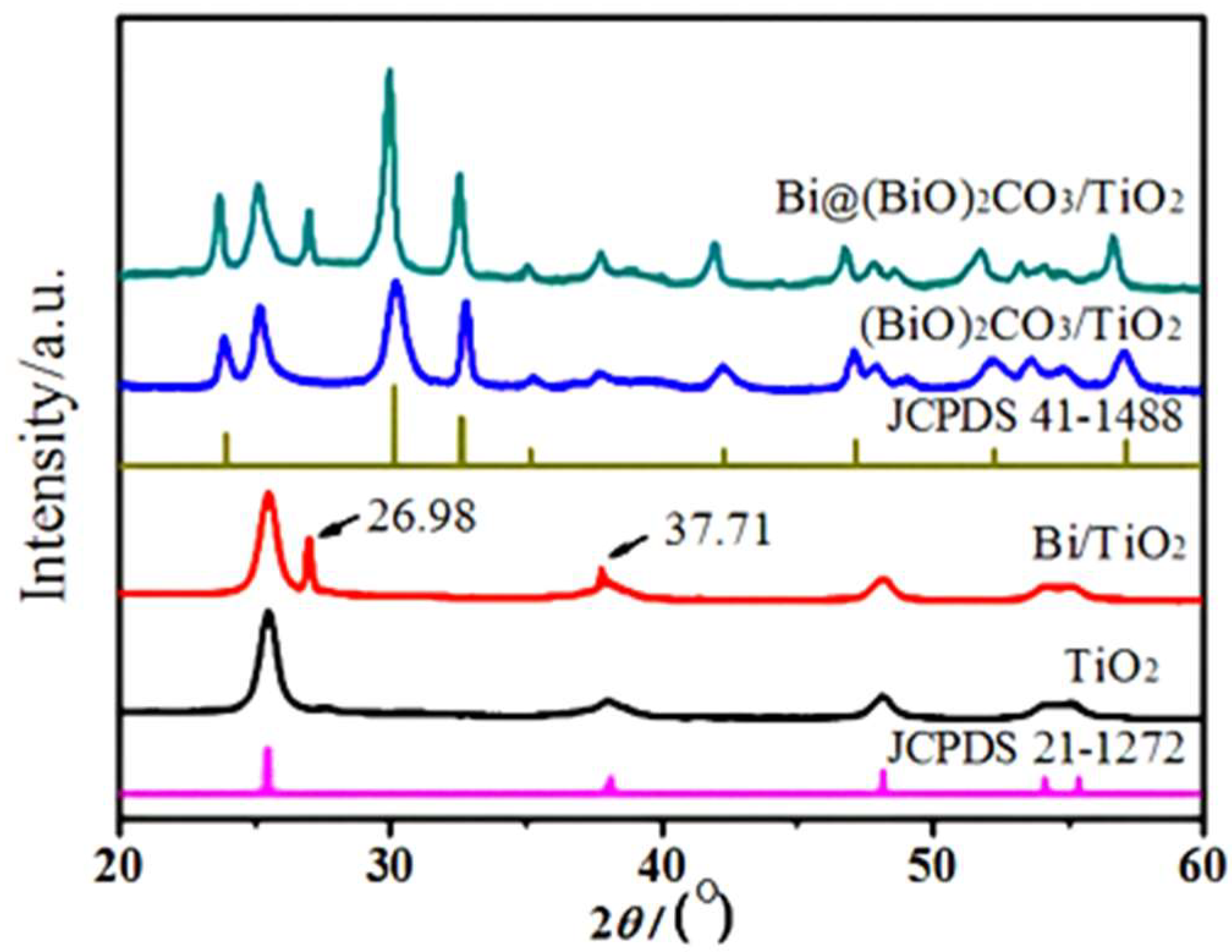
2.1. XRD Analysis

Figure 1 shows the X-ray diffraction (XRD) patterns of various samples. As shown in Figure 1, the XRD diffraction peaks of the TiO2 sample match the characteristic peaks of anatase TiO2 (JCPDS 21-1272) [18]. The peaks are sharp, and no impurity peaks are observed, confirming that the sample is pure anatase TiO2. In addition to the TiO2 diffraction peaks, two new peaks at 26.98° and 37.71° in the Bi/TiO2 sample correspond to the (012) and (104) crystal planes of metallic Bi (JCPDS 05-0519) [19], indicating that the sample contains two phases: metallic Bi and TiO2. Compared with the JCPDS 41-1488 standard card, the new diffraction peaks in the XRD pattern of the (BiO)2CO3/TiO2 sample correspond to the characteristic peaks of the tetragonal phase of (BiO)2CO3 [20,21], confirming that the sample contains (BiO)2CO3 and TiO2 phases. The XRD pattern of Bi@(BiO)2CO3/TiO2 shows diffraction peaks corresponding to TiO2, Bi, and (BiO)2CO3, confirming that the Bi@(BiO)2CO3/TiO2 composite was successfully synthesized. Using Jade software and the Scherrer formula D = Kλ/(βcos θ)D = Kλ/(βcos θ), where K is a constant, λ is the X-ray wavelength, β is the full width at half maximum of the diffraction peak, and θ is the diffraction angle, the particle sizes of metal Bi and (BiO)2CO3 on the surface of the Bi@(BiO)2CO3/TiO2 composite fiber were calculated to be approximately 28 nm and 80 nm, respectively.

2.2. XPS Analysis

Figure 2 presents the X-ray photoelectron spectroscopy (XPS) analysis of the Bi@(BiO)2CO3/TiO2 sample. Figure 2a illustrates the XPS spectrum of the sample, revealing the presence of four elements: Bi, Ti, C, and O. Figure 2b displays the high-resolution XPS spectrum of Bi4f. After peak fitting, two sets of peaks with varying intensities are observed between 155 and 170 eV, corresponding to Bi4f7/2 and Bi4f5/2, respectively, indicating the presence of two valence states of Bi in the sample. Notably, strong peaks at binding energies of 164.4 and 159.2 eV are attributed to Ti3+ ions [22], while weaker peaks at 162.9 and 157.7 eV are associated with elemental Bi [23]. Figure 2c depicts the high-resolution XPS spectrum of Ti2p. Peak fitting reveals asymmetric photoelectron peaks at 458.6 and 464.3 eV, corresponding to Ti2p3/2 and Ti2p1/2, respectively, confirming that Ti exists in the +4 oxidation state in the sample [24]. Figure 2d reveals a high-resolution XPS photoelectron peak for C1s at 284.3 eV. Figure 2e illustrates the high-resolution XPS spectrum of O1s. Two photoelectron peaks at 531.7 and 529.6 eV correspond to lattice oxygen (Olatt) and surface-adsorbed oxygen (OADS), respectively [25].

2.3. SEM and TEM Analysis

Figure 3 presents the SEM and TEM images of various samples. As shown in Figure 3a, TiO2 nanofibers exhibit a fibrous morphology with diameters of approximately 250–300 nm. The fibers have good uniformity, dispersibility, and a smooth surface, with no other species attached. Figure 3b shows that the fibrous morphology of the Bi/TiO2 sample is well preserved. However, the surface is no longer smooth, as numerous Bi nanoparticles with irregular spherical shapes are deposited, with particle sizes ranging from approximately 25–30 nm. In Figure 3c, petal-shaped (BiO)2CO3 nanosheets are uniformly distributed on the surface of the (BiO)2CO3/TiO2 fibers. These nanosheets have sizes of approximately 70–100 nm and thicknesses of 10–15 nm. Figure 3d shows the SEM image of the Bi@(BiO)2CO3/TiO2 sample. Irregular nanoparticles and petal-shaped nanosheets are simultaneously observed on the fiber surface. These features are of similar size and are evenly distributed. Figure 3e presents the TEM image of the Bi@(BiO)2CO3/TiO2 sample, where the constructed nanoparticles and nanosheets are also observed on the fiber surface. The intersection of the three components in the Bi@(BiO)2CO3/TiO2 sample was further examined using high-resolution transmission electron microscopy (HRTEM), as shown in Figure 3f. The image revealed lattice fringes with spacings of 0.35, 0.27, and 0.33 nm, corresponding to the (101) plane of TiO2, the (110) plane of (BiO)2CO3, and the (012) plane of metallic Bi, respectively [26]. This confirms that the Bi@(BiO)2CO3/TiO2 sample consists of three phases: metallic Bi, (BiO)2CO3, and TiO2.
Based on the microstructure and composition analysis of the samples, it can be inferred that the formation of metallic Bi and (BiO)2CO3 on the surface of TiO2 nanofibers is primarily governed by the initial acid-base strength of the reaction system. The reaction equations are as follows:
Bi3+ + C5H11O5CHO + H2O → Bi↓ + C5H11O5COO + H+
Bi3+ + 3OH = BiO(OH) + H2O
CO(NH2)2 + 2H2O → 2NH3 + CO2
2Bi3+ + CO2 + 3H2O = (BiO)2CO3↓ +6H+
2BiO(OH) + CO2 = (BiO)2CO3↓ +H2O
Based on the reactions described above, the formation mechanism of Bi@(BiO)2CO3/TiO2 is explained. Most redox reactions in solution occur as ionic processes. Consequently, H+ and OH in the medium influence the concentration and activity of certain ions to varying degrees, thereby affecting the reaction’s extent and feasibility. In an acidic environment (pH = 4.5), Bi3+ acts as a metal ion with oxidizing properties, while glucose functions as a polyhydroxyaldehyde with reducing properties. Under high-temperature hydrothermal conditions, a redox reaction takes place, reducing Bi3+ to metallic Bi (Equation (1)). In a strongly alkaline environment (pH = 11.5), Bi3+ reacts with OH to form BiO(OH) (Equation (2)), significantly decreasing the Bi3+ concentration and weakening its oxidizing ability. Consequently, Bi3+ can no longer be reduced to metallic Bi by C5H11O5CHO under these conditions. At this stage, CO(NH2)2 in the solution undergoes hydrolysis to produce CO2 and NH3 (Equation (3)). BiO(OH) then reacts with CO2 to form (BiO)2CO3 (Equation (4)).
In a weakly alkaline environment (pH = 8.0), only part of Bi3+ reacts with OH to form BiO(OH) (Equation (2)), while the rest is reduced to metallic Bi by C5H11O5CHO (Equation (1)). Simultaneously, CO(NH2)2 undergoes partial hydrolysis to generate CO2 and NH3 (Equation (3)). Finally, BiO(OH) reacts with CO2 to form (BiO)2CO3 (Equation (5)).

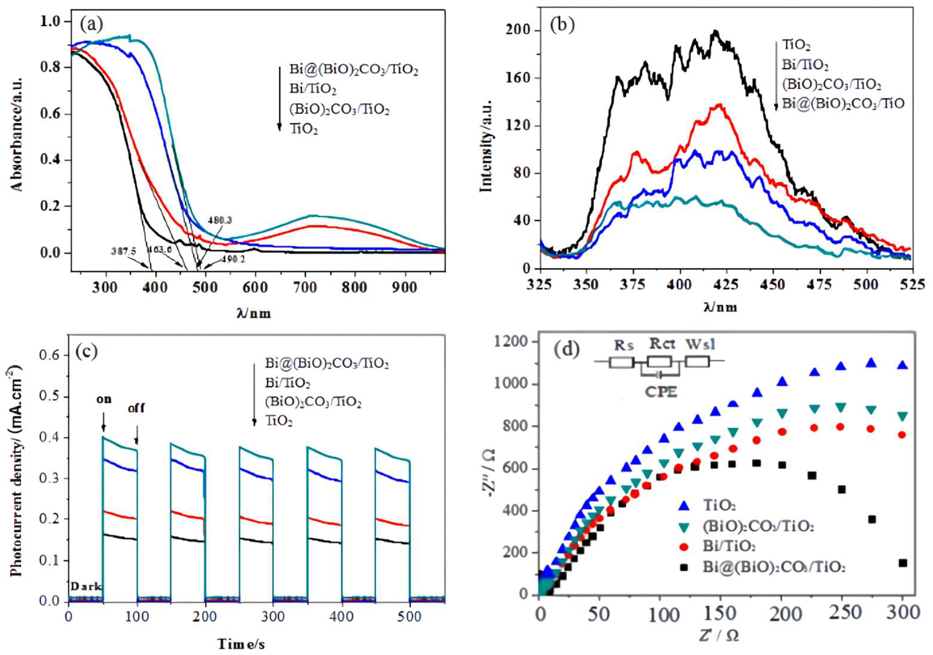
2.4. Photoelectric Performance Analysis

Figure 4a presents the UV-vis diffuse reflectance spectra (DRS) of various photocatalyst samples. As shown in the figure, anatase-phase TiO2 exhibits strong absorption in the ultraviolet region (λ ≤ 387.5 nm). The absorption edge of the (BiO)2CO3/TiO2 sample shifts to 480.3 nm, expanding the spectral response range. The absorption band edge of the Bi/TiO2 sample exhibits a red shift and significant absorption in the visible region. This phenomenon is attributed to the surface plasmon resonance (SPR) effect of metallic Bi [27,28]. Additionally, the absorption band edge of Bi@(BiO)2CO3/TiO2 further shifts to 490.2 nm, with an enhanced light absorption intensity in the visible region. This suggests that the synergistic modification by metallic Bi and (BiO)2CO3 significantly improves the absorption and utilization of visible light by TiO2.
Figure 4b presents the photoluminescence (PL) spectra of various catalyst samples. The test results in the range of 300–550 nm, shown in Figure 5, are used to evaluate the charge separation efficiency of modified TiO2. As shown in the figure, TiO2 nanofibers exhibit the highest fluorescence intensity, indicating the highest recombination probability of photogenerated electron–hole pairs. Compared with TiO2, the fluorescence intensities of Bi/TiO2 and (BiO)2CO3/TiO2 are reduced. This suggests that the combination of metallic Bi or (BiO)2CO3 with TiO2 forms Schottky or heterojunctions, facilitating effective separation of photogenerated electrons and holes. Additionally, the fluorescence intensity of Bi/TiO2 is slightly higher than that of (BiO)2CO3/TiO2, indicating that the heterojunction formed by (BiO)2CO3 and TiO2 is more effective in facilitating the separation of photogenerated electrons and holes. However, the Bi@(BiO)2CO3/TiO2 sample exhibits the lowest fluorescence intensity, suggesting that the synergistic effect of metallic Bi and (BiO)2CO3 further alters the transport pathway of photogenerated carriers, prolonging their lifetime and enhancing the separation efficiency of photogenerated electrons and holes.
Figure 4c depicts the transient photocurrent responses of photocatalysts from different samples. When the light source was switched on and off at 50 s intervals, all the tested samples generated distinct photocurrent signals. This indicates that the xenon lamp excited the samples to produce photogenerated charges, which were subsequently transmitted to the electrodes to generate photocurrent. The Bi@(BiO)2CO3/TiO2 sample exhibited the highest photocurrent intensity, followed by Bi/TiO2. The photocurrent intensity of (BiO)2CO3/TiO2 was similar to that of TiO2. This demonstrates that metallic Bi plays a crucial role in the photocurrent generation of composite nanofiber samples. The curves in Figure 4d depict the electrochemical impedance spectra (EIS) of the prepared samples, measured using a simulated circuit and fitted for analysis. The radius of the impedance arc shows an inverse correlation with the transient photocurrent values. In the inset of Figure 4d, Rs, Rct, CPE, and Ws1 denote the electrolyte solution resistance, charge transfer resistance, constant phase element, and Warburg impedance, respectively. Fitting calculations showed that the impedance values of PANI, ZnO/PANI, ZnWO4/PANI, and ZnO@ZnWO4/PANI were 14.8, 6.5, 4.9, and 2.3 kΩ, respectively. These results indicate that ZnO@ZnWO4/PANI exhibits the lowest charge transfer resistance, promoting the migration of photogenerated carriers and enhancing their participation in photocatalytic reactions.

2.5. Photocatalytic Performance Analysis

Figure 5 presents the photocatalytic degradation of antibiotics and the reaction rate constants of various samples under simulated sunlight. Figure 5a shows that the concentration of lomefloxacin remains unchanged under conditions without light or a catalyst. After 60 min of simulated sunlight irradiation, the photodegradation efficiencies of TiO2, Bi/TiO2, (BiO)2CO3/TiO2, and Bi@(BiO)2CO3/TiO2 were 71.6%, 84.9%, 92.4%, and 97.5%, respectively. The results indicate that (BiO)2CO3 or the combination of metallic Bi and TiO2 enhances the photocatalytic activity of TiO2. This enhancement is primarily attributed to the formation of an effective heterostructure between (BiO)2CO3 and TiO2, which modifies the transport paths of photogenerated carriers, extends the lifetime of photogenerated electrons and holes, and facilitates their effective separation. Additionally, metallic Bi nanoparticles exhibit an SPR effect similar to that of precious metals. These nanoparticles can be excited by visible light to generate high-energy hot electrons, thereby enhancing the photocatalytic activity of TiO2. Notably, Bi@(BiO)2CO3/TiO2 demonstrates the highest degradation efficiency for lomefloxacin. This indicates that the synergistic interaction between (BiO)2CO3 and Bi significantly enhances the photocatalytic activity of TiO2. Figure 5b illustrates the degradation kinetics of lomefloxacin under sunlight, following the quasi-first-order reaction kinetic equation ln(Ct/C0) = −k t. The reaction rate constants kk for TiO2, Bi/TiO2, (BiO)2CO3/TiO2, and Bi@(BiO)2CO3/TiO2 are 0.0205, 0.0308, and 0.002, respectively. Compared to TiO2, the photocatalytic rate of Bi@(BiO)2CO3/TiO2 is approximately three times higher.
Figure 5c,d show that ciprofloxacin and norfloxacin are highly stable without light or a catalyst, with negligible changes in their concentrations. TiO2, Bi/TiO2, (BiO)2CO3/TiO2, and Bi@(BiO)2CO3/TiO2 also exhibit significant photocatalytic activity for ciprofloxacin and norfloxacin. Among them, Bi@(BiO)2CO3/TiO2 achieves the highest photocatalytic activity, with photodegradation efficiencies of 93.2% and 100% for ciprofloxacin and norfloxacin, respectively, under 60 min of simulated sunlight. This indicates that among quinolones, norfloxacin is the easiest to degrade, followed by lomefloxacin, whereas ciprofloxacin is relatively more difficult to degrade.
Stability and reusability are critical prerequisites for the practical application of catalysts. The fibrous structure of the prepared catalyst allows it to be reused after photocatalytic degradation through simple filtration, separation, recovery, and drying. Figure 6a shows that the photodegradation efficiency of lomefloxacin remains above 90% after five photocatalytic cycles using Bi@(BiO)2CO3/TiO2. Figure 6b indicates that the position and morphology of the XRD diffraction peaks of Bi@(BiO)2CO3/TiO2 remain essentially unchanged before and after the cyclic reaction. These results demonstrate that Bi@(BiO)2CO3/TiO2 exhibits excellent stability and reusability.

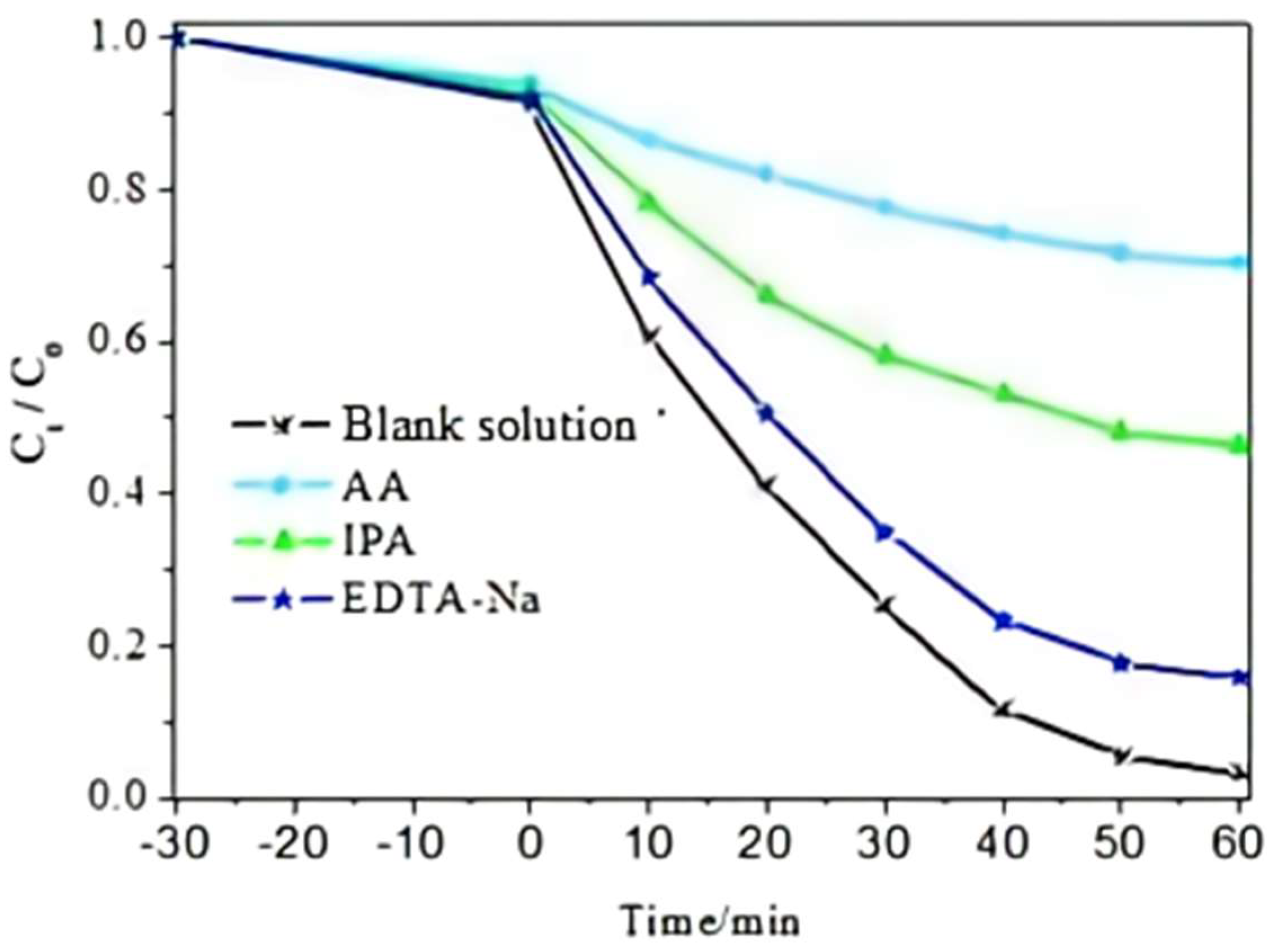
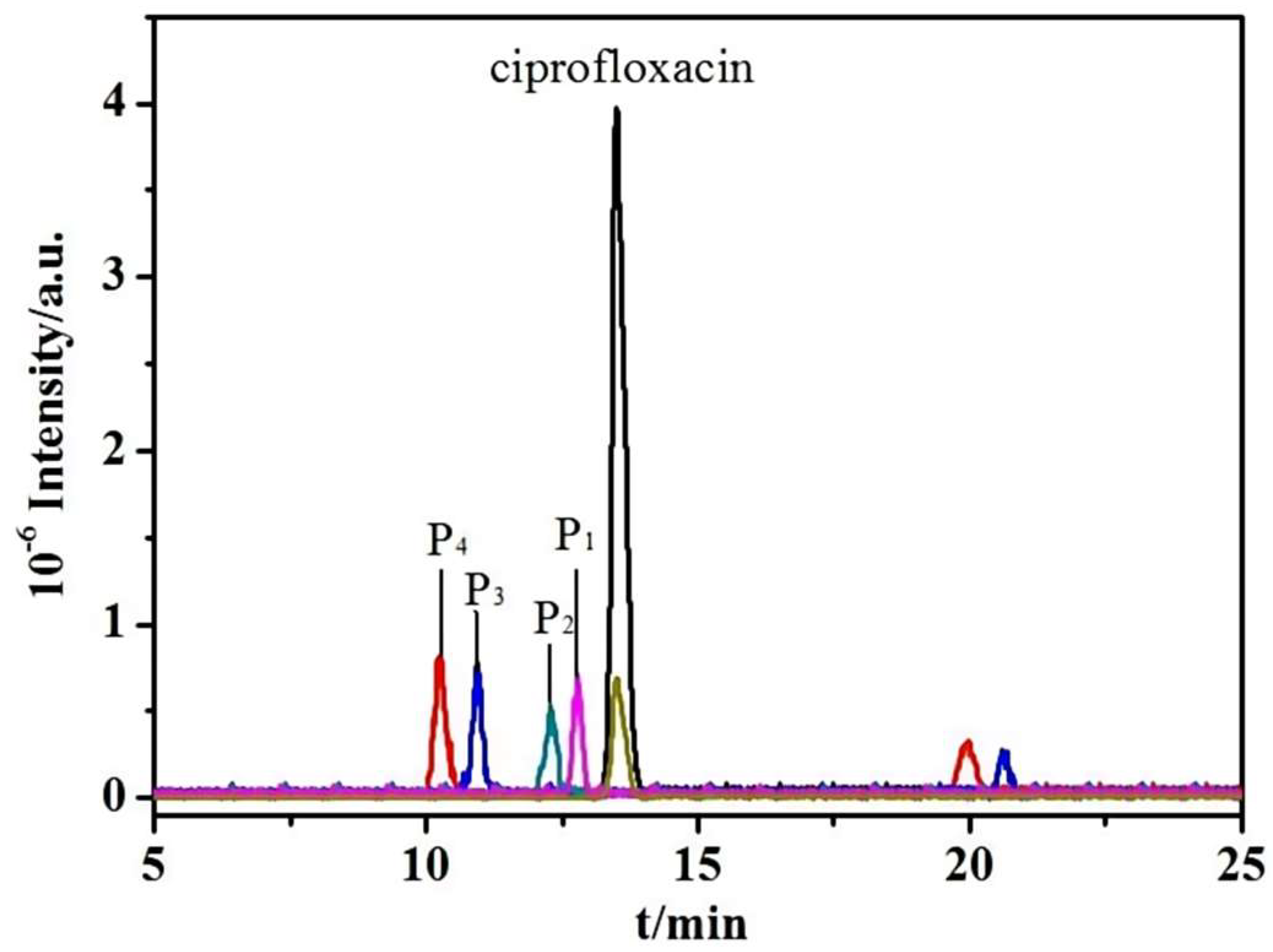
2.6. Analysis of Photocatalytic Mechanism

To investigate the photocatalytic degradation mechanism of (BiO)2CO3/TiO2, the active species involved in the photocatalytic degradation of lomefloxacin were identified through free radical capture experiments. Sodium EDTA-Na, isopropanol (IPA), and ascorbic acid (AA) were added to the reaction system to selectively capture holes (h+), hydroxyl radicals (⋅OH), and superoxide radicals (O2), respectively. As shown in Figure 7, compared to the blank sample (without scavengers), the degradation rate of lomefloxacin significantly decreased with the addition of AA and IPA but showed minimal change with EDTA-Na. Among the tested scavengers, the degradation rate of lomefloxacin was lowest with AA, indicating that O2 is the most active species in the photocatalytic degradation process, followed by ⋅OH, while h+ has minimal direct impact on the degradation.
Based on the characterization results and the semiconductor energy band structure, the plasma effect and heterojunction photocatalytic reaction mechanism for the degradation of antibiotics by Bi@(BiO)2CO3/TiO2 are proposed. Both TiO2 and (BiO)2CO3 are N-type semiconductors. The valence band (VB) and conduction band (CB) of TiO2 are positioned at −0.3 and 2.9 eV, respectively, while those of (BiO)2CO3 are located at 0.5 and 3.2 eV. The VB and CB energy levels are staggered, forming a Type-II heterojunction, as shown in Figure 8. Under illumination, VB electrons of TiO2 and (BiO)2CO3 are simultaneously excited to the CB, while holes remain in the VB. Since the CB potential of TiO2 is lower than that of (BiO)2CO3, CB electrons in TiO2 migrate to the CB of (BiO)2CO3 through the heterojunction interface driven by the potential difference. Conversely, holes in the VB of (BiO)2CO3 migrate to the VB of TiO2 through the heterojunction interface. The migration of photogenerated electrons and holes alters their paths, prolongs their lifetimes, reduces recombination probabilities, and significantly enhances photocatalytic degradation activity. Under visible light irradiation, the electric field induced by the SPR effect of Bi nanoparticles significantly enhances the separation efficiency of photogenerated carriers in (BiO)2CO3. Additionally, Bi nanoparticles rapidly transfer photogenerated electrons from the CB of (BiO)2CO3, generating superoxide radicals (O2−) and suppressing the recombination of electrons and holes. Holes on the surface of TiO2 are captured by adsorbed water, producing hydroxyl radicals (OH). Under the constant attack of active oxygen species, OH and O2−, the pollutants are ultimately oxidized and degraded into small molecules like CO2 and H2O. This study offers a novel approach to sensitize non-noble metals in wide band gap semiconductors, enhance their photocatalytic performance, and address environmental challenges.
The photodegradation pathways of antibiotics by Bi@(BiO)2CO3/TiO2 under visible light were investigated using the HPLC-ESI-MS analysis method. Figure 9 illustrates the total ion chromatogram of ciprofloxacin during its photocatalytic degradation. Ciprofloxacin (m/z 331.34) degraded rapidly within 20 min of simulated sunlight exposure, producing a new product, P1 (m/z 333.17). After 30 min of light exposure, additional degradation products, P2 (m/z 315.31), P3 (m/z 305.38), and P4 (m/z 277.21), were formed. Figure 10 provides mass spectrometric data for the major degradation products and proposes the photodegradation pathways of ciprofloxacin. With the progression of the photocatalytic degradation reaction, these intermediates undergo further oxidation through ring-opening reactions and defunctionalization, ultimately breaking down ciprofloxacin and other antibiotics into CO2, H2O, and other small molecules.

3. Experiment

3.1. Materials and Methods

Polyvinylpyrrolidone (PVP, Ms = 1,300,000, AR) and tetrabutyl titanate (TBT) were obtained from Sigma-Aldrich (St. Louis, MO, USA). Bismuth nitrate pentahydrate (AR) [Bi(NO3)3·5H2O] was obtained from Sigma-Aldrich (St. Louis, MO, USA); while glucose (AR) (C6H12O6), urea (AR) [CO(NH2)2], anhydrous ethanol (AR) (C2H6O), and glacial acetic acid (AR) (C2H4O2) were obtained from Sigma-Aldrich (St. Louis, MO, USA). Self-prepared secondary distilled water was used.

3.2. Sample Preparation

Weigh 0.485 g of Bi(NO3)3·5H2O and dissolve it in 20 mL of 1 mol/L diluted HNO3. Dissolve 0.198 g of glucose and 0.3 g of CO(NH2)2 in 15 mL of deionized water. Mix the two solutions thoroughly and add 10 mg of TiO2 nanofibers. Stir the mixture magnetically for 30 min. Adjust the pH of the system to 8.5 using a 1 mol/L NaOH or HNO3 solution. Transfer the suspension into a 50 mL Teflon-lined stainless steel autoclave. React at 160 °C for 24 h at a constant temperature. After cooling to room temperature, wash the product three times with distilled water and ethanol, and then dry it in an oven at 12 h to obtain Bi@(BiO)2CO3/TiO2 composite fibers. Using the same conditions, adjust the system pH to 4 and 12 to synthesize Bi/TiO2 and (BiO)2CO3/TiO2 composite fibers, respectively.
0.485 g of bismuth nitrate pentahydrate was dissolved in 20 mL of 1 mol/L dilute HNO3. Separately, 0.198 g of glucose was dissolved in 15 mL of deionized water, and 10 mg of TiO2 nanofibers was added. The mixture was stirred magnetically for 30 min, and the pH of the solution was adjusted to 8.5 using 1 mol/L NaOH or HNO3. The suspension was transferred to a 50 mL PTFE-lined stainless steel autoclave and reacted at 160 °C for 24 h. After cooling to room temperature, the product was washed three times each with distilled water and ethanol, and then dried in an oven for 12 h to yield Bi@(BiO)2CO3/TiO2 composite fibers. Bi/TiO2 and (BiO)2CO3/TiO2 composite fibers were prepared under the same conditions by adjusting the system pH to 4 and 12, respectively.

3.3. Sample Characterization

The crystal phase and composition of the samples were analyzed using a Panalytical X’Pert3 powder X-ray diffractometer (XRD). In the experiment, a Cu Kα target (λ = 0.154056 nm) was employed with a tube current of 40 mA, a tube voltage of 40 kV, and a scanning range of 20–80°. The morphology of the samples was observed using a Hitachi SU8010 field-emission scanning electron microscope (SEM) with a working voltage of 5 kV. The microstructure of the samples was examined using a JEOL JSM-2010 high-resolution transmission electron microscope (HRTEM) at a working voltage of 100 kV. The chemical composition and valence states of the sample were analyzed using PHI-5000 VersaProbe X-ray photoelectron spectroscopy (XPS), with charge shift correction performed using the C1s peak of adventitious carbon at 284.6 eV. The ultraviolet–visible diffuse reflectance spectrum (UV-vis DRS) was recorded using a PerkinElmer Lambda 35 ultraviolet–visible spectrophotometer, with BaSO4 as the standard reflectance material. The photoluminescence spectrum (PL) was analyzed using a Hitachi F-4500 fluorescence spectrophotometer with an excitation wavelength of 350 nm. The photocurrent of the sample was measured using a CHI660B electrochemical workstation. The absorbance of target pollutants in the solution was measured using a UV2600 ultraviolet spectrophotometer (Shunyu Group Co., Ltd. ShangDong, China). The LC/MS-2010EV high-performance liquid chromatography–mass spectrometry system, manufactured by Shimadzu Corporation (Shimadzu Co., Ltd., Kyoto, Japan), was used to investigate the pathways of antibiotic photodegradation catalyzed by the catalyst.

3.4. Photocatalytic Degradation of Antibiotics

A PLS-SXE 300 W xenon lamp (Beijing Bofeilai Technology Co., Ltd. BeiJing, China) was used as the external light source with an average light intensity of 5 mW/cm2, a spot diameter of 63 mm, and an emission spectrum simulating sunlight (320–680 nm) for the photocatalytic degradation experiment. Then, 20 mg of photocatalyst was dispersed in 50 mL of a 20 mg/L solution containing three quinolone antibiotics: lomefloxacin, ofloxacin, and ciprofloxacin. The solution was allowed to equilibrate in the dark for 30 min to achieve adsorption–desorption equilibrium between the degradation product and the catalyst. After illumination began, samples were collected every 10 min, and the maximum absorbance of the supernatant at 356 nm was measured using an ultraviolet spectrophotometer. The photodegradation efficiency was calculated as
(1 − Ct/C0) × 100%
where C0 and Ct represent the concentrations of the solution at 0 min and t minutes of illumination, respectively.

3.5. Photoelectric Performance Test

The photocurrent of the sample was measured using a CHI 660D electrochemical workstation equipped with a standard three-electrode system. The sample was coated onto FTO conductive glass to prepare a working electrode with an effective area of approximately 1 cm2. A Pt electrode and a saturated calomel electrode were used as the counter and reference electrodes, respectively. The working electrode was self-fabricated, and a 0.1 mol/L Na2SO4 solution was used as the electrolyte.
Preparation method for the working electrode: Dissolve 10 mg of the catalyst sample in 1 mL of distilled water. Subject the mixture to ultrasonic treatment for 10 min, then evenly drop it onto a 1 cm2 fluorine-doped tin oxide (FTO) conductive glass substrate. Dry the coated glass under an infrared lamp to form a film and obtain the working electrode. The photocurrent curve was measured using a 300 W xenon lamp as the light source, while the electrochemical impedance was recorded at a bias voltage of 0.8 V.

4. Conclusions

The Bi@(BiO)2CO3/TiO2 composite fiber was synthesized via an in situ hydrothermal method, using TiO2 nanofibers as the matrix. Metal Bi nanoparticles and (BiO)2CO3 nanosheets were simultaneously deposited on the surface of the TiO2 fibers. Bi@(BiO)2CO3/TiO2 exhibited excellent photocatalytic degradation activity. The photodegradation efficiencies for ciprofloxacin, lomefloxacin, and norfloxacin were 93.2%, 97.5%, and 100%, respectively, significantly surpassing those of pure TiO2 nanofibers. (BiO)2CO3 and TiO2 form a heterojunction, altering the migration path of photogenerated carriers, extending charge lifetimes, and enhancing the separation efficiency of photogenerated electrons and holes. The SPR effect of metallic Bi not only extends the light absorption range into the visible region but also synergizes with the (BiO)2CO3/TiO2 heterojunction to effectively reduce interfacial electron transfer resistance. This synergy enhances the interfacial transport of photogenerated carriers and significantly improves their separation efficiency. Bi@(BiO)2CO3/TiO2 demonstrates excellent stability and recyclability. After five cycles, its photodegradation efficiency remains above 90%. This work offers a novel approach for developing low-cost, efficient, and stable bismuth-based plasmonic heterojunction photocatalysts with solar spectral responsiveness.

Author Contributions

Conceptualization, Y.G. and T.C.; methodology, J.D.; software, X.Q.; validation, H.Y. formal analysis, X.X. All authors have read and agreed to the published version of the manuscript.

Funding

The study was supported by the university-level team research project of Hainan Vocational University of Science and Technology in 2024 “Research on Polyionic Liquid Materials and Their Catalytic Properties (HKKY2024-TD-17)”; Hainan Vocational University of Science and Technology university-level Teaching Reform Project 2025: Research on Ideological and Political Teaching Model of ‘Chemical Engineering Principles’ Based on Goal and Problem Orientation (HIKJG2024-012); Research on Teaching Methods of Mechanical Manufacturing Courses under Informationization Conditions (HKJG2024-35).

Data Availability Statement

No new data were created.

Conflicts of Interest

The authors declare no conflicts of interest.

References

  1. Huyan, J.; Tian, Z.; Zhang, Y.; Zhang, H.; Shi, Y.; Gillings, M.R.; Yang, M. Dynamics of class 1 integrons in aerobic biofilm reactors spiked with antibiotics. Environ. Int. 2020, 140, 105816. [Google Scholar] [CrossRef] [PubMed]
  2. Ovis-Sánchez, J.O.; Perera-Pérez, V.D.; Buitrón, G.; Quintela-Baluja, M.; Graham, D.W.; Morales-Espinosa, R.; Carrillo-Reyes, J. Exploring resistomes and microbiomes in pilot-scale microalgae-bacteria wastewater treatment systems for use in low-resource settings. Sci. Total Environ. 2023, 882, 163545. [Google Scholar] [CrossRef] [PubMed]
  3. Zhang, S.; Chen, J.; Fang, C.; Zhang, Y.; Xu, Z.; Yan, Z.; Yao, K. Enhanced photocatalytic removal of antibiotics over graphitic carbon nitride induced by acetic acid post-treatment. Colloids Surf. A Physicochem. Eng. Asp. 2023, 664, 131165. [Google Scholar] [CrossRef]
  4. Xu, Z.; Jia, Y.; Huang, B.; Zhao, D.; Long, X.; Hu, S.; Li, C.; Dao, G.; Chen, B.; Pan, X. Spatial distribution, pollution characteristics, and health risks of antibiotic resistance genes in China: A review. Environ. Chem. Lett. 2023, 21, 2285–2309. [Google Scholar] [CrossRef]
  5. Huang, Z.; Liu, H. Insights into the pathways, intermediates, influence factors and toxicological properties in the degradation of tetracycline by TiO2-based photocatalysts. J. Environ. Chem. Eng. 2023, 11, 110587. [Google Scholar] [CrossRef]
  6. Bian, C.; Zhou, B.; Mo, F.; Liu, X.; Sun, P.; Dong, X. Post-synthetically covalent modification of g-C3N4 to regulate electronic structure and investigation of photocatalytic activity for eliminating antibiotics. Sep. Purif. Technol. 2023, 325, 124556. [Google Scholar] [CrossRef]
  7. Kakhki, S.; Khiadani, M.; Naderi, K.; Forough, M.; Kholghi, V.; Yektay, S. Effect of ozonation-based disinfection methods on the removal of antibiotic resistant bacteria and resistance genes (ARB/ARGs) in water and wastewater treatment: A systematic review. Sci. Total Environ. 2022, 811, 151404. [Google Scholar] [CrossRef]
  8. Molaei, M.J. Recent advances in hydrogen production through photocatalytic water splitting: A review. Fuel 2024, 365, 131159. [Google Scholar] [CrossRef]
  9. Ren, C.; Li, Q.; Ling, C.; Wang, J. Mechaniam-Guided Design of Photocatalysts for CO2 Reduction toward Multiarbon Products. J. Am. Chem. Soc. 2023, 145, 28276. [Google Scholar] [CrossRef]
  10. Si, S.; Shou, H.; Mao, Y.; Bao, X.; Zhai, G.; Song, K.; Wang, Z.; Wang, P.; Liu, Y.; Zheng, Z.; et al. Low-Coordination Single Au Atoms on Ultrathin ZnIn2S4 Nanosheets for Selective Photocatalytic CO2 Reduction towards CH4. Angew. Chem. Int. Ed. 2022, 61, e202209446. [Google Scholar] [CrossRef]
  11. Dong, F.; Xiong, T.; Sun, Y.; Zhao, Z.; Zhou, Y.; Feng, X.; Wu, Z. A semimetal bismuth element as a direct plasmonic photocatalyst. Chem. Commun. 2014, 50, 10386–10389. [Google Scholar] [CrossRef] [PubMed]
  12. Dong, F.; Xiong, T.; Yan, S.; Wang, H.; Sun, Y.; Zhang, Y.; Huang, H.; Wu, Z. Facets and defects cooperatively promote visible light plasmonic photocatalysis with Bi nanowires@BiOCl nanosheets. J. Catal. 2016, 344, 401–410. [Google Scholar] [CrossRef]
  13. Qu, L.L.; Luo, Z.J.; Tang, C. One Step Synthesis of Bi@Bi2O3@Carboxylate-rich Carbon Spheres with Enhanced Photocatalytic Performance. Mater. Res. Bull. 2013, 48, 4601–4605. [Google Scholar] [CrossRef]
  14. Li, L.; Gao, H.; Yi, Z.; Wang, S.; Wu, X.; Li, R.; Yang, H. Comparative investigation on synthesis, morphological tailoring and photocatalytic activities of Bi2O2CO3 nanostructures. Colloids Surf. A Physicochem. Eng. Asp. 2022, 644, 128758. [Google Scholar] [CrossRef]
  15. Liu, Y.; Wang, Z.; Huang, B.; Yang, K.; Zhang, X.; Qin, X.; Dai, Y. Preparation, electronic structure, and photocatalytic properties of Bi2O2CO3 nanosheet. Appl. Surf. Sci. 2010, 257, 172–175. [Google Scholar] [CrossRef]
  16. Cen, W.; Xiong, T.; Tang, C.; Yuan, S.; Dong, F. Effects of morphology and crystallinity on the photocatalytic activity of (BiO)2CO3 nano/microstructures. Ind. Eng. Chem. Res. 2014, 53, 15002–15011. [Google Scholar] [CrossRef]
  17. Nie, J.; Gao, J.; Shen, Q.; Zhang, W.; Rao, F.; Hojamberdiev, M.; Zhu, G.Q. Flower-like Bi0/CeO2−δ plasmonic photocatalysts with enhanced visible-light-induced photocatalytic activity for NO removal. Sci. China Mater. 2020, 63, 2272–2280. [Google Scholar] [CrossRef]
  18. Li, Y.; Cao, T.; Mei, Z.; Li, X.; Sun, D. Separating type I heterojunction of NaBi(MoO4)2/Bi2MoO6 by TiO2 nanofibers for enhanced visible-photocatalysis. Chem. Phys. 2020, 533, 110696. [Google Scholar] [CrossRef]
  19. Qin, F.; Li, G.; Xiao, H.; Lu, Z.; Sun, H.; Chen, R. Large-scale synthesis of bismuth hollow nanospheres for highly efficient Cr(VI) removal. Dalton Trans. 2012, 41, 11263–11266. [Google Scholar] [CrossRef] [PubMed]
  20. Zhang, Y.; Duang, F.; Chen, M.Q.; Xie, Y. Synthetic Bi2O2CO3 nanostructures: Novel photocatalyst with controlled special surface exposed. J. Mol. Catal. A Chem. 2010, 317, 34–40. [Google Scholar] [CrossRef]
  21. Huang, H.; Tian, N.; Jin, S.; Zhang, Y.; Wang, S. Syntheses, characterization and nonlinear optical properties of a bismuth subcarbonate Bi2O2CO3. Solid State Sci. 2014, 30, 1–5. [Google Scholar] [CrossRef]
  22. Pang, B.; Miao, J.; Wang, H.; Wu, C.; Wu, L.; Yuan, G.; Wang, X. Construction of fast charge-transferred 0D/2D BiOBr/Bi2WO6 S-scheme heterojunction with enhanced photocatalytic performance. Appl. Surf. Sci. 2024, 649, 159104. [Google Scholar] [CrossRef]
  23. Song, D.; Li, M.; Yang, F.; Yu, M.; Li, Z.; Chen, J.; Zhang, X.; Zhou, W. Plasmon Bi in-situ anchored on BiOCl nanosheets assembled microspheres towards optimized photothermal-photocatalytic performance. Chin. Chem. Lett. 2024, 35, 108591. [Google Scholar] [CrossRef]
  24. Giovannini, T.; Bonatti, L.; Lafiosca, P.; Nicoli, L.; Castagnola, M.; Illobre, P.G.; Corni, S.; Cappelli, C. Do we really need quantum mechanics to describe plasmonic properties of metal nanostructures? ACS Photonics 2022, 9, 3025–3034. [Google Scholar] [CrossRef] [PubMed]
  25. Zhang, Y.; Shao, Q.; Chen, C.; Jiang, H.; Su, F.; Hu, Q.; Guo, Z. Microwave-hydrothermal synthesis of beta-bismuth (III) oxide nanopowders and their enhanced photocatalytic properties. Powder Technol. 2020, 370, 226–236. [Google Scholar] [CrossRef]
  26. Dong, F.; Li, Q.; Sun, Y.; Ho, W.K. Noble metal-like behavior of plasmonic Bi particles as a cocatalyst deposited on (BiO)2CO3 microspheres for efficient visible light photocatalysis. ACS Catal. 2014, 4, 4341–4350. [Google Scholar] [CrossRef]
  27. McMahon, J.M.; Schatz, G.C.; Gray, S.K. Plasmonics in the ultraviolet with the poor metals Al, Ga, In, Sn, Tl, Pb, and Bi. Phys. Chem. Chem. Phys. 2013, 15, 5415–5423. [Google Scholar] [CrossRef] [PubMed]
  28. Wang, Z.; Jiang, C.; Huang, R.; Peng, H.; Tang, X. Investigation of optical and photocatalytic properties of bismuth nanospheres prepared by a facile thermolysis method. J. Phys. Chem. C 2014, 118, 1155–1160. [Google Scholar] [CrossRef]
Figure 1. XRD patterns of different photocatalyst samples.
Catalysts 15 00056 g001
Figure 2. XPS spectrum of the Bi@(BiO)2CO3/TiO2 sample. (a) Survey Scan, (b) Bi4f, (c) Ti2P, (d) C1s, (e) O1s.
Catalysts 15 00056 g002
Figure 3. SEM and TEM images of TiO2 nanofibers and composite fibers: (a) TiO2 SEM, (b) Bi/TiO2 SEM, (c) (BiO)2CO3/TiO2 SEM, (d) Bi@(BiO)2CO3/TiO2 SEM, (e) Bi@(BiO)2CO3/TiO2 TEM, (f) Bi@(BiO)2CO3/TiO2 HRTEM.
Catalysts 15 00056 g003
Figure 4. Photoelectric properties of different samples: (a) UV-vis DRS, (b) PL, (c) transient photocurrent, (d) EIS.
Catalysts 15 00056 g004
Figure 5. Photodegradation efficiency and reaction rate constant curves of various catalyst samples for antibiotics under simulated sunlight: (a) lomefloxacin, (b) lomefloxacin, (c) ciprofloxacin, (d) norfloxacin.
Catalysts 15 00056 g005
Figure 6. (a) Comparison of the cyclic photocatalytic degradation of antibiotics by Bi@(BiO)2CO3/TiO2, (b) XRD before and after the reaction.
Catalysts 15 00056 g006
Figure 7. Degradation curves of (BiO)2CO3/TiO2 with different quenchers.
Catalysts 15 00056 g007
Figure 8. Photocatalytic mechanism and photogenerated electron–hole migration in Bi@(BiO)2CO3/TiO2.
Catalysts 15 00056 g008
Figure 9. Total ion chromatogram of ciprofloxacin after 20, 30, and 40 min of irradiation in the presence of the prepared Bi@(BiO)2CO3/TiO2 photocatalyst.
Catalysts 15 00056 g009
Figure 10. Possible degradation pathways of ciprofloxacin on Bi@(BiO)2CO3/TiO2 photocatalysts.
Catalysts 15 00056 g010
Disclaimer/Publisher’s Note: The statements, opinions and data contained in all publications are solely those of the individual author(s) and contributor(s) and not of MDPI and/or the editor(s). MDPI and/or the editor(s) disclaim responsibility for any injury to people or property resulting from any ideas, methods, instructions or products referred to in the content.

