Article

Thermal Inactivation Mechanism and Structural Features Providing Enhanced Thermal Stability of Hyperthermophilic Thermococcus sibiricus L-Asparaginase in Comparison with Mesophilic and Thermophilic L-Asparaginases

by
Natalia Dobryakova
1,2,
Dmitry Zhdanov
1,3,
Maria Dumina
1,
Svetlana Aleksandrova
3,
Marina Pokrovskaya
3,
Alexander Genin
2,
Anastasia Shishparenok
1,
Alexander Zhgun
1 and
Elena Vadimovna Kudryashova
1,2,*
1
Group of Fungal Genetic Engineering, Federal Research Center “Fundamentals of Biotechnology of the Russian Academy of Sciences”, 117312 Moscow, Russia
2
Chemical Faculty, Lomonosov Moscow State University, Leninskie Gory St. 1, 119991 Moscow, Russia
3
Laboratory of Medical Biotechnology, Institute of Biomedical Chemistry, 119121 Moscow, Russia
*
Author to whom correspondence should be addressed.
Catalysts 2023, 13(5), 832; https://doi.org/10.3390/catal13050832
Submission received: 25 March 2023 / Revised: 28 April 2023 / Accepted: 29 April 2023 / Published: 2 May 2023
(This article belongs to the Section Biocatalysis)

Abstract

:
This work aimed to study the structural features and mechanisms of thermoinactivation of hyperthermophilic L-asparaginase (L-ASNase) from archaea Thermococcus sibiricus (TsA) in comparison with bacterial L-ASNases from Melioribacter roseus (MrA) and Rhodospirillum rubrum (RrA). The catalytic parameters of L-asparagine hydrolysis under optimal conditions (pH 9) were determined for these enzymes by circular dichroism (CD) spectroscopy. TsA showed the highest activity among the studied L-ASNases (640 IU/mg at 90 °C). Thermo-inactivation kinetics were studied at temperatures close to the enzyme optimum: the first-order inactivation constants were 0.065 min−1 (TsA), 0.011 min−1 (MrA), and 0.026 min−1 (RrA). In contrast to RrA and MrA, aggregation was detected as one of the thermoinactivation mechanisms for TsA. From the analysis of thermograms obtained with CD spectroscopy, the melting temperatures (Tm) for RrA, MrA, and TsA were determined as 50, 69, and 89 °C, respectively. A significant increase in the percentage of β-structures for TsA during heating (from 8 to 16%) indicating aggregation was observed in the interval from 70 to 100 °C. For RrA and MrA this value did not increase. Changes in the tertiary structure of the enzymes during heating were monitored by fluorescence spectroscopy. Thermal inactivation of RrA and MrA were accompanied by changes in the tertiary structure. For TsA, the observed denaturation enthalpy (ΔH) was 346 kJ/mol, which was 1.5–2 times higher than the same values for RrA and MrA. The study of the specific thermoinactivation mechanisms and structural- features in hyperthermophilic enzymes in comparison with mesophilic ones allows us to shed light on the molecular adaptation variants of the enzyme to function at high temperatures.

1. Introduction

L-asparaginases are amidohydrolases (E.C. 3.5.1.1) that catalyze the hydrolysis of L-asparagine to L-aspartic acid and ammonia (Figure 1). These enzymes are applied in biomedicine to treat various types of leukemia and in the food industry to reduce the amount of carcinogenic acrylamide during the production of starch-rich foods [1,2]. In both cases, the main mechanism of action is based on a decrease in the concentration of L-asparagine, which is the main substrate of L-ASNases. This hydrolysis takes place either in the processing of carbohydrate-rich foods or in the blood of a leukemia patient when administered intravenously.
L-ASNases from the mesophilic bacteria Escherichia coli (EcA) и Erwinia chrysanthemi (ErA) (Elspar®, Medac®, Oncaspar®, Vero-asparaginase® и Erwinase®) are used in medicine [3,4,5]. These type II L-ASNases are excessively active and highly affinitive to L-asparagine. However, they are not very stable at elevated temperatures. The industry typically uses a blanching method for potato products to reduce the formation of acrylamide [6]. Such pretreatment is carried out at temperatures of 65–80 °C in hot water for 10–30 min. Therefore, more thermostable enzymes are required for this application. Approved by the FDA, commercial enzyme preparations from the mold fungi Aspergillus oryzae and Aspergillus niger (Acrylaway® and PreventAse®) are currently used in the industry [7,8]. Acrylaway® was used to treat and reduce acrylamide formation in coffee beans [9]. However, these enzymes have temperature optimums no higher than 37 and 50 °C [10]. The application of these methods requires either an additional processing step at lower temperatures or processing only at low temperatures, which can reduce the overall efficiency of reduction of L-asparagine concentration in products. Thus, an important problem is searching for new sources of thermophilic L-ASNases with high activity and specificity to L-asparagine or search for ways to increase the thermal stability of enzymes.
In our laboratory, we study L-ASNases from various thermophilic and hyperthermophilic microorganisms. Previously, we worked on the expression and study of the specific activity of several extremophilic L-ASNases. The most active enzymes from the hyperthermophilic archaea Thermococcus sibiricus and the thermophilic bacterium Melioribacter roseus were selected [11,12]. These enzymes have optimal activities at approximately 85–90 °C and 65–70 °C, respectively. Their thermostability and high activity at elevated temperatures make them excellent research models. In this study, an enzyme from the mesophilic bacterium Rhodospirillum rubrum with an activity optimum of 45–50 °C was also used to compare the structure and thermostability of different L-ASNases previously characterized in our laboratory [13,14,15].
The aim of this work is to identify the structural features and mechanism of inactivation of L-ASNase from the hyperthermophilic archaea T. sibiricus that provides thermal stability in comparison with bacterial L-ASNases from the thermophile M. roseus and mesophile R. rubrum.

2. Results

2.1. Comparison of L-Asparaginase Sequences

To elucidate the molecular reasons for the different thermostability of L-ASNases from mesophilic, thermophilic, and hyperthermophilic organisms (RrA, MrA, TsA), it is necessary to analyze their structural differences. It was shown earlier that bacterial L-ASNases are not identical in their protein sequence to enzymes from archaea, and similarities did not exceed 37% [16].
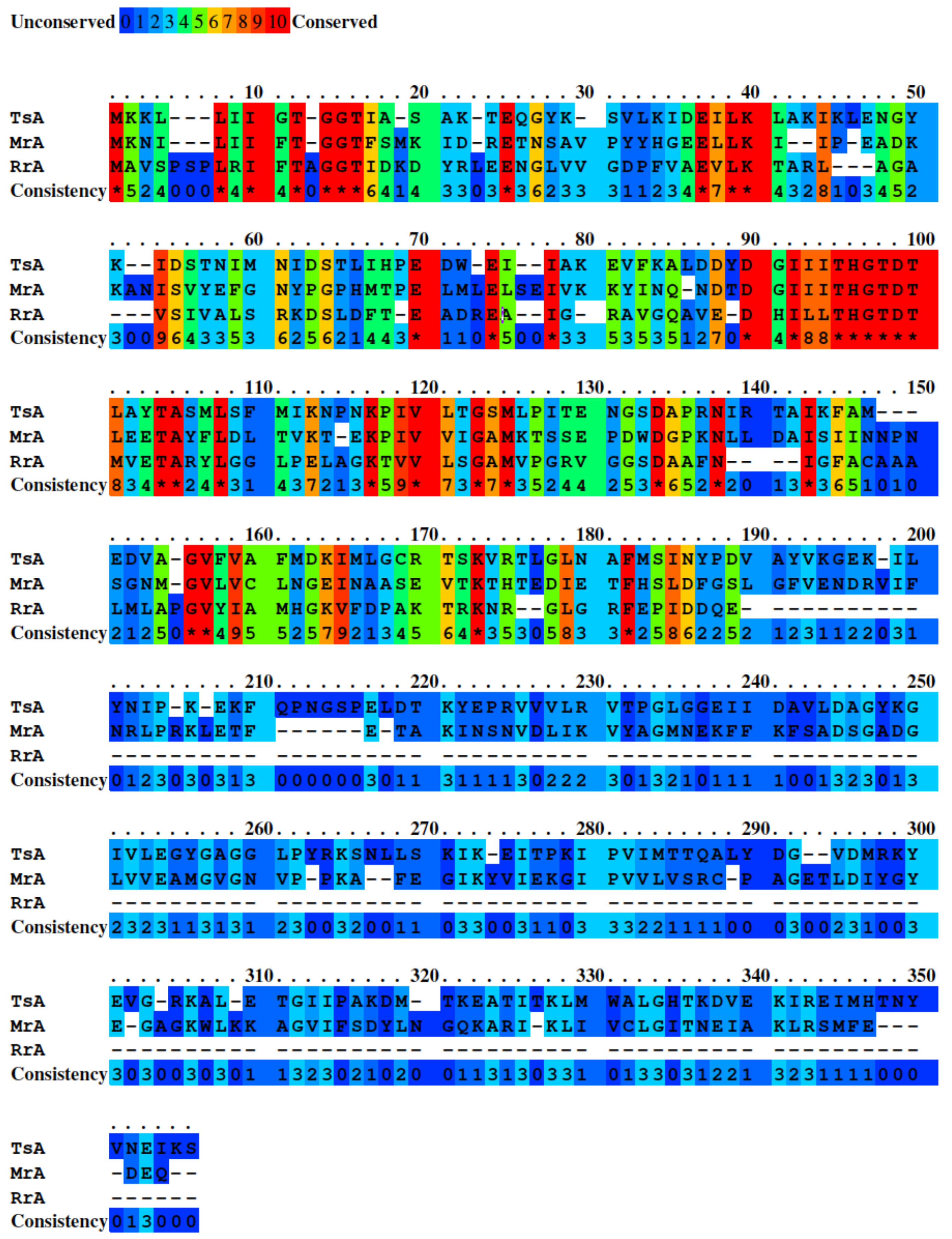
The PRALINE multiple sequence alignment tool (https://www.ibi.vu.nl, accessed on 16 January 2023, Amsterdam, The Netherlands) was used to align the sequences of the three L-ASNases [17]. The results of the alignment are shown in Figure 2. Due to their different origin, the studied L-ASNases have rather strong differences in the protein sequence. RrA has the most significant differences with MrA and TsA: the percentage of equivalence was 19 and 20%, respectively. Between MrA and TsA, the percentage of equivalence was 31%. Despite the weak similarities in the amino acid sequences, these L-ASNases have several highly conserved sites. Some residues (Thr12, Gly15, Thr16, and Asp90) in the active center of these three enzymes are conserved [14,18]. It is also noted that although L-ASNases from archaea have sequence differences from bacterial enzymes, they have a similar tertiary structure [18].
The characteristics of the three enzymes TsA, MrA, and RrA are compared in Table 1. RrA is presented as a homotetramer, and TsA is presented as a homodimer, which is typical for thermophilic enzymes [16,19,20]. It is assumed that MrA also exists primarily as a dimer. It cannot be excluded that the quaternary structure may affect the stability of L-ASNases. For EcA and ErA, the tetrameric form is the most catalytically active [21]. In addition, an equilibrium between monomer and dimer or dimer and tetramer can be observed in the solution. It has recently been shown that the dimeric form of L-ASNase can also have activity similar to the tetrameric form [22]. This is important because, in the case of thermal dissociation of the enzyme to dimers, the activity can be partially preserved. It is also worth noting that RrA has a 2-fold shorter amino acid sequence compared to thermophiles. This may affect the packaging of the protein globule, which is also related to the stability at high temperatures.
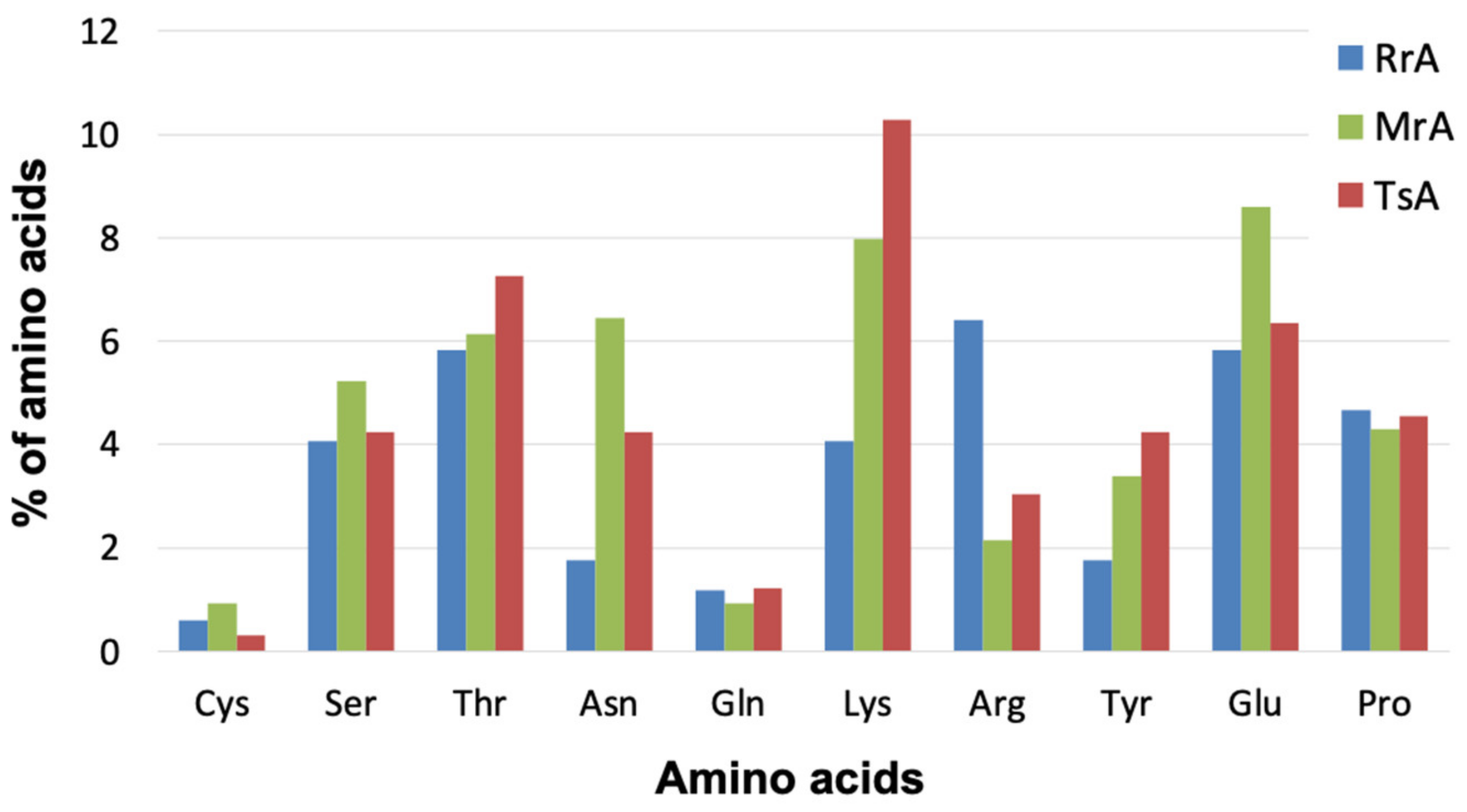
Sequence analysis of RrA, MrA, and TsA revealed no significant differences in the content of certain amino acids. Nevertheless, the total amount of hydrophobic amino acids was 3–4% higher in RrA than in MrA and TsA, the number of hydrophilic residues was 4–5% lower, and the same value for charged residues was 1–2% lower. TsA had the highest Lys content of approximately 10%, which is more than 2 times higher than that in RrA (Figure 3). Furthermore, TsA has a higher Tyr content (4.2%), and MrA has more than 6% Arg compared with 3 and 2% for TsA and RrA, respectively. The contents of Cys, Ser, and Pro differ by less than 1%. Thus, due to the increased content of charged amino acids, the more thermophilic enzymes MrA and TsA have increased stability at higher temperatures.

2.2. Determination of L-ASNase Activity

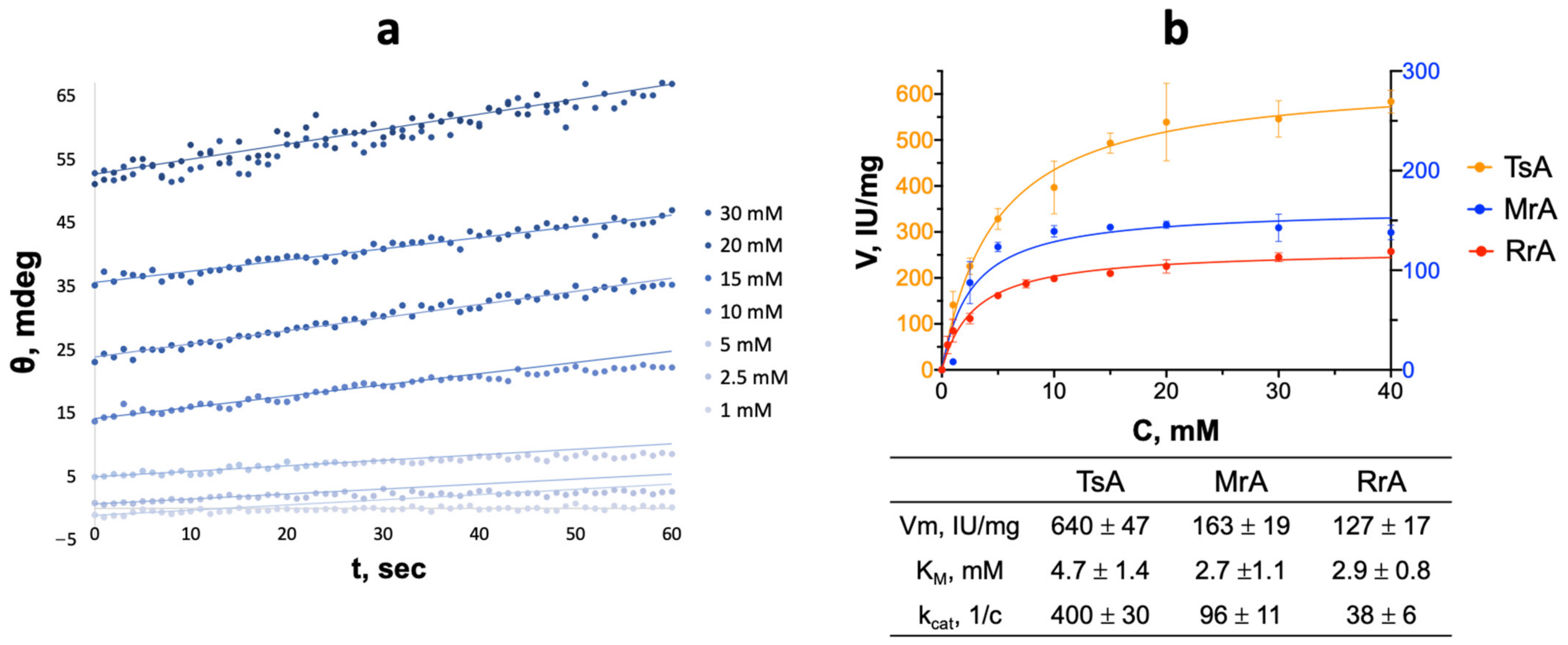
The main method to determine the activity of enzymes in this work was CD spectroscopy [23]. This method provides continuous registration of kinetic curves and does not require additional reagents, as in the Nessler method [24,25]. Figure 4 shows the initial reaction rate dependencies on the substrate concentration (L-asparagine) and the resulting kinetic parameters (Vmax and KM) of L-ASNases under optimal conditions: pH 9.3 and the temperature optimum of the enzymes. The KM was similar for all enzymes and had values in the range of 2–5 mM. Indeed, these L-ASNases belong to the first type (type I) of L-ASNases. This type of L-ASNases usually has a high KM and cytoplasmic localization [26]. TsA had the highest activity (640 IU/mg) under optimal conditions (pH 9.3, 90 °C).
To compare the temperature optimum for the activity of L-ASNase from different sources, the dependencies of enzyme activity on the temperature of L-asparagine hydrolysis were obtained. The temperature optimum was determined under standardized conditions for all the studied enzymes for correct comparison [27,28]. Catalytic activity optimums are observed at 50 °C, 60 °C, and 90 °C for RrA, MrA, and TsA correspondingly (Figure 5). At temperatures above the optimum value, the activity drops sharply due to the thermodenaturation of the enzyme.

2.3. Thermoinactivation of L-ASNases

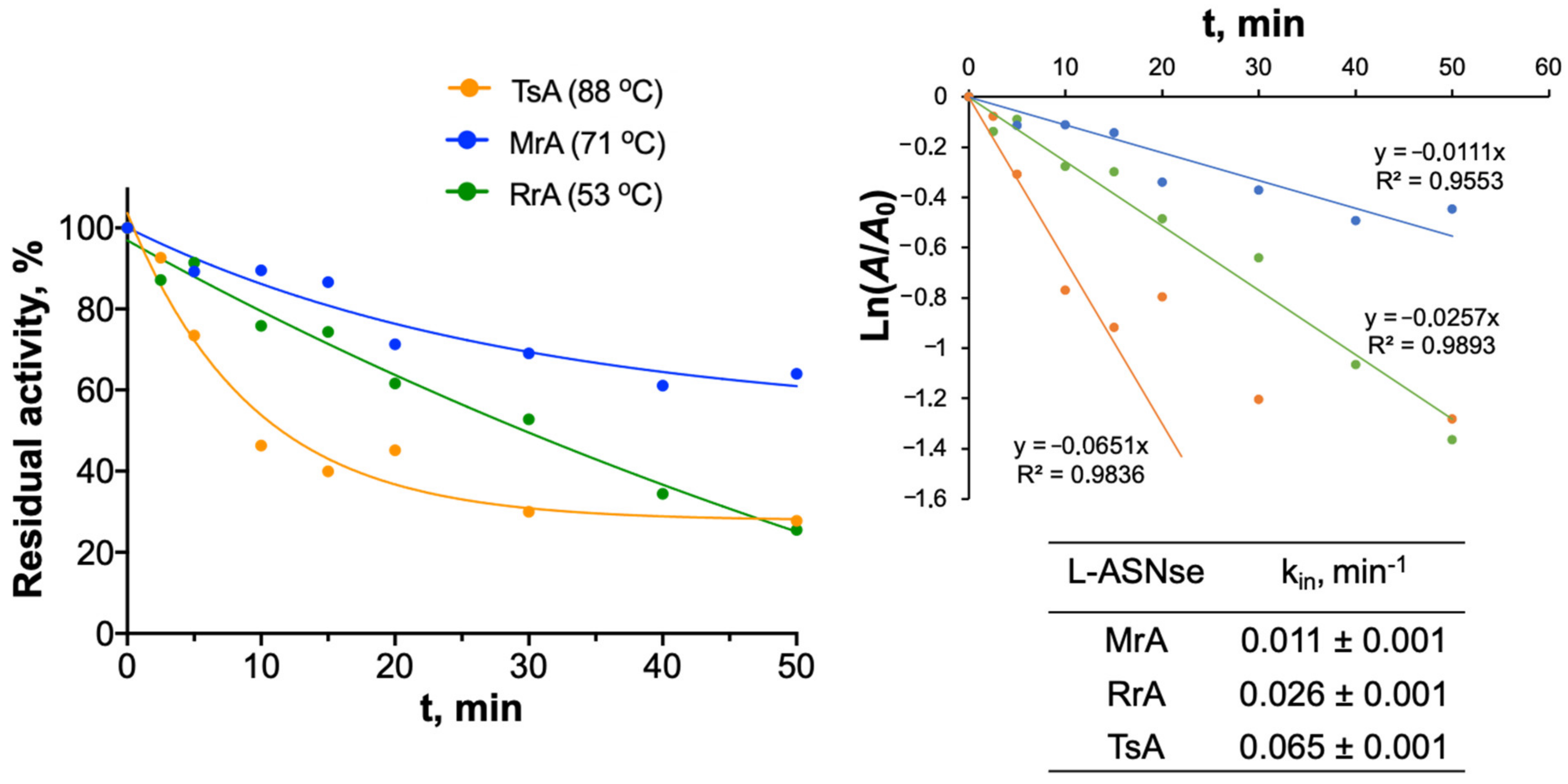
To reveal the mechanisms of thermoinactivation of the studied L-ASNases, we measured the activity dependencies on the incubation time at temperatures close to the enzyme optimum (Figure 6). To determine the inactivation reaction order, the obtained enzyme’s thermal inactivation curves were analyzed in semilogarithmic coordinates (ln(A/A0) = −kint). The thermal inactivation curves over 50 min for RrA and MrA straightened in these coordinates, which confirms that thermal inactivation for these enzymes is of the first order. For TsA, straightening was observed only for the initial part of the curve (15 min). The first-order inactivation constant is 0.065 min−1. At higher incubation times for TsA, a higher-order thermoinactivation reaction was observed (Figure S1), which is characteristic of aggregation (multimolecular process). The time interval data for 10–30 min were straightened in second-order coordinates. The relative inactivation constant was 0.38.

2.4. Secondary Structure Changes and Thermodynamic Parameters of L-ASNases during Thermodenaturation Studied by CD Spectrometry

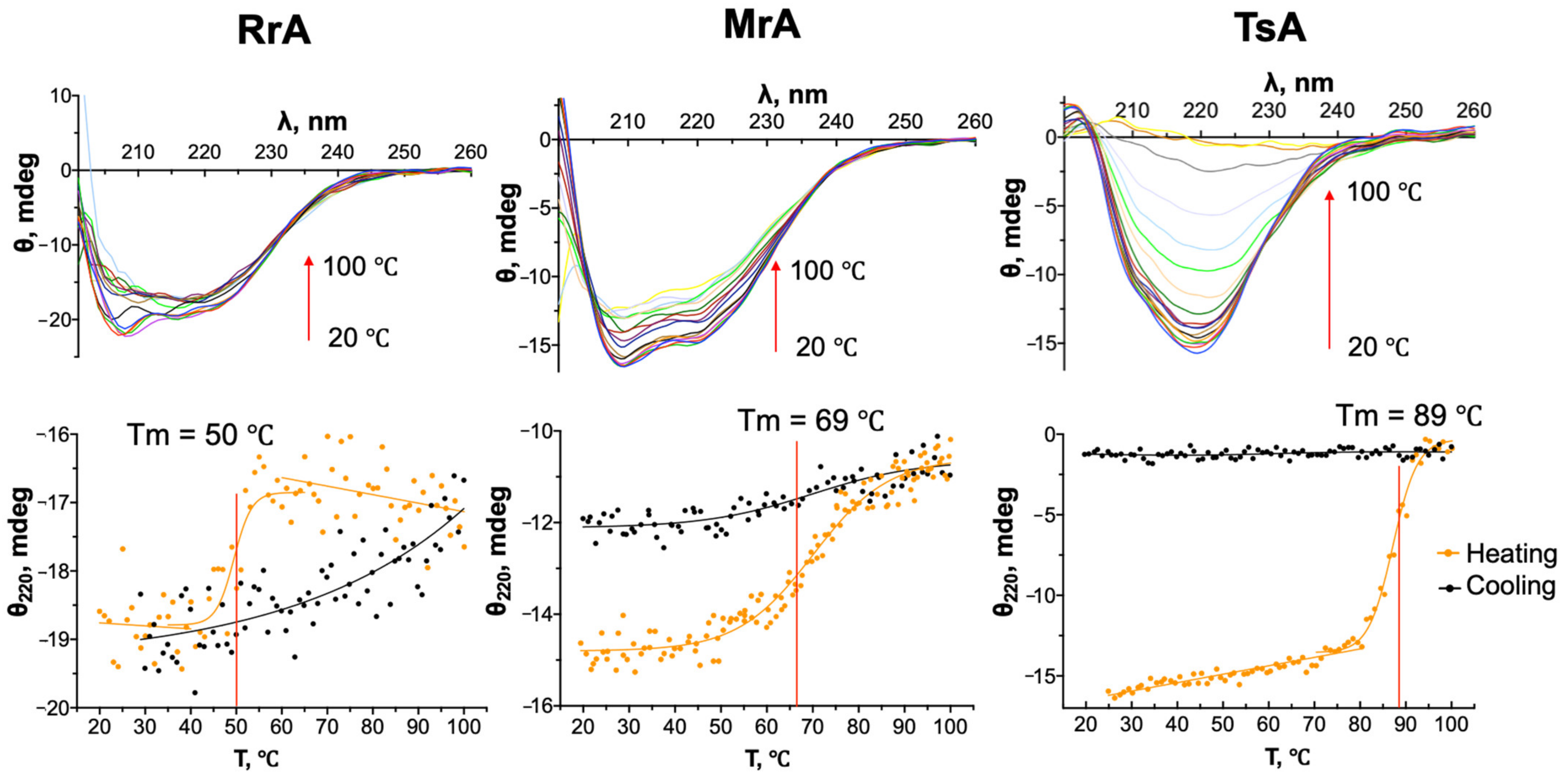
To obtain information on the conformational changes of L-ASNases during heating, we compared the parameters of conformational stability of the enzymes: melting temperatures and thermodynamic parameters of the phase transition. For this purpose, we recorded CD spectra and thermograms when the enzymes were heated from 20 to 100 °C under the same conditions (Figure 7). The melting temperatures (Tm) of the enzymes were determined from the thermograms obtained. For RrA, the melting temperature was 50 °C lower than for the thermostable L-ASNases: for MrA and TsA, Tm were 69 and 89 °C, respectively. The observed phase transition was cooperative for all enzymes, but it was less expressed for RrA.
To analyze the ratio of secondary structure elements and their changes upon heating, as well as the susceptibility of L-ASNases to aggregation, deconvolution of the obtained CD spectra was performed (Figure 8). The most significant changes in the secondary structure were observed for TsA: the percentage of α-helices decreased from approximately 36 to 9%, and the percentage of disordered structures increased from 32 to 43% (Table 2). The percentage of β-structures considerably increased: the content of antiparallel and parallel β-sheets rose from 8 to 15–16%. In RrA and MrA, the changes in the secondary structure were less significant. The percentage of α-helices decreased by 3–5%, and the percentage of β-structures practically did not increase. It can be noted that in RrA, the changes in the secondary structure were gradual, without sharp transitions, as was observed in MrA and TsA.
For TsA at elevated temperatures (85–95 °C), there was visual aggregation associated with an increase in solution turbidity (registered by absorbance at A600 from 0.05 to 0.2–0.3). RrA and MrA solutions remained transparent without precipitating when heated even to 100 °C. To examine the reversibility of the denaturation process, after heating the enzyme solutions to 100 °C, they were cooled again to 20 °C under the same conditions. Upon reverse cooling for RrA, a “hysteresis” was observed in the thermograms: the secondary structure was restored when the enzyme mixture was cooled to 20 °C. In MrA, the secondary structure was only partially restored (by 35%). At the same time, none of the enzymes recovered activity, which may indicate incorrect folding of the tertiary structure.
To analyze in more detail the process of transition of L-ASNases from the native to the denatured state, the effective (observable) thermodynamic parameters were calculated using the Thermal Denaturation Analysis program (Table 3). These parameters were determined from thermograms recorded under the same conditions.
ΔG(eff) is highest for TsA, which indicates that this enzyme is more thermostable than RrA and TsA. The TsA ΔH(eff) value was higher than that for MrA ΔH(eff) and RrA ΔH(eff) and was approximately 346 kJ/mol. This value is explained by the presence of numerous hydrogen bonds and salt bridges.

2.5. Changes in the Tertiary Structure of L-ASNases during Thermal Denaturation

Generally, the first step in the thermal inactivation of enzymes is the unfolding of the tertiary structure. Then, irreversible changes in the secondary structure can occur, including aggregation, which we observed for TsA by CD spectra. Fluorescence spectroscopy was used to trace the features of changes in the tertiary structure of L-ASNases from different sources during thermal inactivation. Proteins have fluorescence spectra at the excitation wavelength of 280 nm due to the aromatic residues Trp and Tyr [29]. MrA and TsA each have two Trp residues, which make the main contribution to the fluorescence. RrA lacks Trp, and thus, only Tyr residues have a spectral contribution.
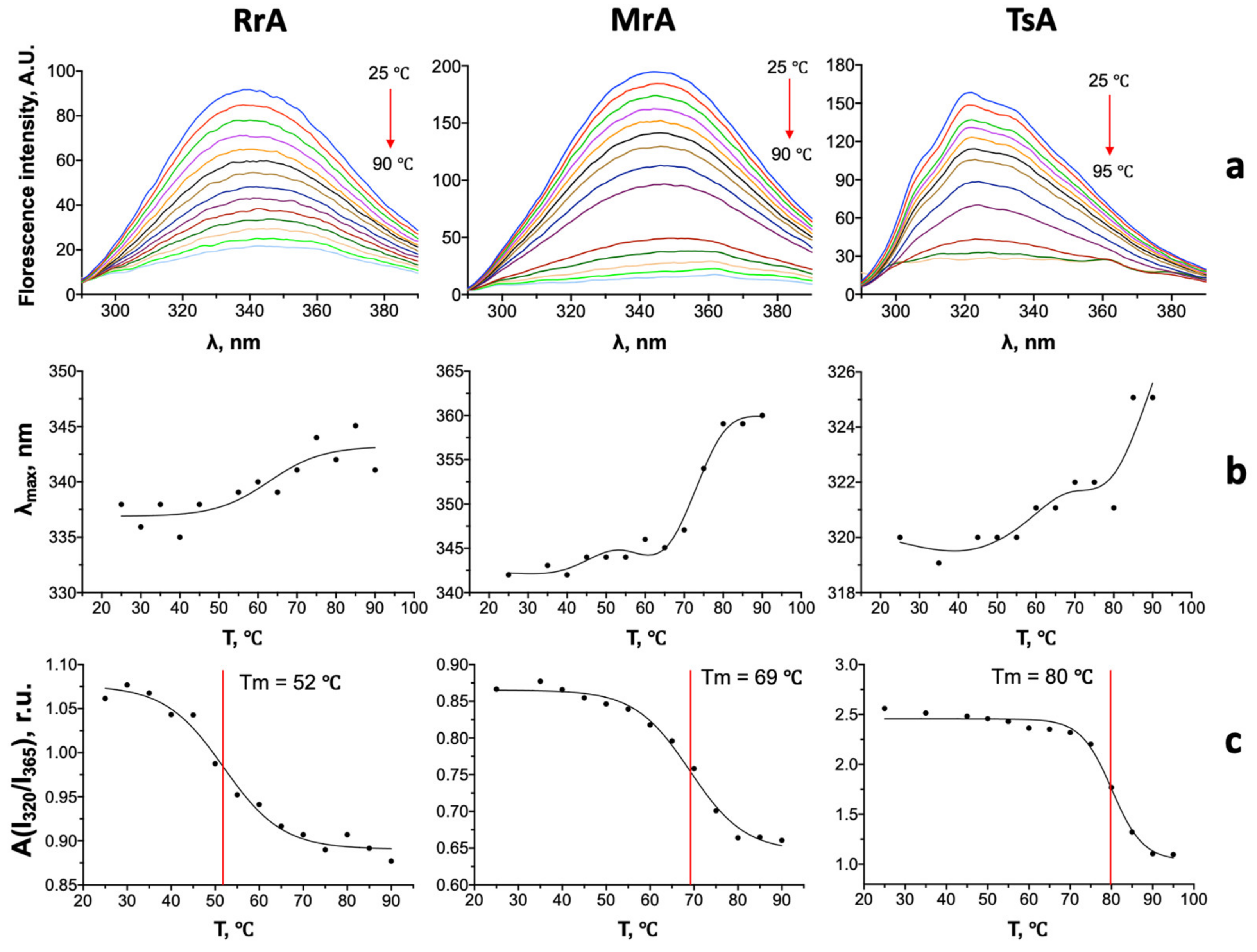
When the enzymes are heated, the fluorescence intensity decreases due to protein unfolding and the transition of fluorophores (Trp, Tyr) from nonpolar to polar environments (Figure 9a).
For all L-ASNases, we observed a shift λmax to a longer wavelength region (Figure 9b). For RrA, the shift was approximately 6 nm (from 337 to 343 nm). The decrease in the intensity of the fluorescence spectra for RrA was more pronounced than the changes in the CD spectra. Denaturation in the case of this enzyme occurs primarily due to changes in the tertiary structure of the protein. For MrA, there was first a gradual shift (approximately 5 nm) to 70 °C, followed by a sharp shift of another 13 nm (from 342 to 360 nm). For TsA, there was also first a slight shift of 2 nm and then a sharp shift at 80–90 °C from 322 to 325 nm, which corresponds to the change and microenvironment of aromatic residues during the unfolding of the protein globule.
To analyze changes in the tertiary structure of L-ASNases, parameter A equal to the ratio I320/I365 at an excitation wavelength of 280–290 nm was used. This value is used to represent changes in the microenvironment of Trp or Tyr residues [30]. The phase transition temperatures (Tm) of the enzymes were determined from the fluorescence thermograms (Figure 9a). For RrA Tm = 52 °C, MrA—69 °C, TsA—80 °C. It is shown that Tm for RrA and MrA are close to Tm obtained from thermograms by CD spectrometry. For TsA the temperature of conformational transition related to the partial unfolding of tertiary structure, it is lower than that obtained by CD by 9 °C. As we showed earlier, the temperature optimum for TsA is approximately 90 °C. Most likely, to adopt the optimal conformation in terms of higher activity TsA needs to acquire a more expanded conformation to exhibit high activity.

3. Discussion

As we noted earlier, to compare the features of the conformational properties of enzymes from different sources at elevated temperatures, it is necessary to analyze their amino acid sequence as well as to understand their structural features at the level of secondary and tertiary structures and their oligomeric composition. It is known that the thermodynamic stability of proteins depends on the number of weak interactions, such as hydrogen bonds, salt bridges, and Van der Waals interactions [31]. In addition, the stability is influenced by the environment (ionic strength, polarity of the solvent).In studies related to the thermostability of proteins, it has been shown that certain amino acids can increase stability, while others can decrease it. For example, polar long-chain amino acids such as Lys, Arg, and Tyr contribute to the formation of hydrogen bonds and salt bridges [32]. These amino acids are more often found in enzymes from thermophilic microorganisms [33]. Thr, Gln, Asn, and Ser, the other polar amino acids, are less common in thermostable enzymes. Thr and Ser can interact with water molecules at high temperatures and reduce the stability of the protein globule. In addition, Ser can disrupt the interactions between the beta strands of the protein. It was found that L-ASNase from the thermophilic archaea Thermococcus kodakarensis has an increased content of salt bridges and Arg amino acid residues compared to mesophilic enzymes [19]. At the same time, the enzyme has fewer thermolabile Cys and Ser residues. A recent review comparing the sequences of L-ASNases of mesophilic and thermophilic origin also revealed that thermophiles contain more charged (Glu, Lys) as well as hydrophobic (Ile, Val) residues and fewer uncharged (Asn, Gln, His, Cys) residues [16]. The difference in amino acid content, which can affect the thermostability of the enzymes, was not significantly noticeable. However, unlike RrA and MrA, a feature of TsA is the susceptibility of the enzyme to aggregation at elevated temperatures.
Aggregation of TsA during thermal denaturation was demonstrated by an increase in the content of antiparallel β-structures, which we observed from the CD spectrum as well as from the kinetic data of thermoinactivation of the enzyme. It is known that the formation of the antiparallel β-sheets is caused by protein aggregation [34,35,36]. In addition, some proteins can aggregate into dense and more stable amyloid structures during thermodenaturation [37]. RrA and MrA did not show such significant changes in secondary structures upon thermodenaturation. However, we observed significant changes in the tertiary structure of these L-ASNases (Figure 7). Most likely, RrA and MrA lose activity mainly due to the destruction of the tertiary structure, and the elements of the secondary structure remain partially undamaged even when heated to 90–100 °C. Enzyme activity was not restored when the enzymes were cooled. Thus, no reverse folding to the native tertiary conformation occurs during cooling, or it occurs abnormally under the given experimental conditions. In TsA, thermodenaturation (at temperatures of 90–100 °C) leads to a significant change in both tertiary and secondary structures. The unfolding of the tertiary structure (transition of Trp and Tyr into solution) is observed before the temperature optimum is reached (90 °C). This may indicate that for TsA, the protein globule needs to change its primal conformation by slightly unfolding to exhibit more activity. Thus, for RrA and MrA a cooperative change in the secondary and tertiary structures is observed upon thermodenaturation. For TsA, denaturation of the tertiary structure is observed first. Then the secondary structure is destroyed, which already causes the loss of enzyme activity.
It is known that when a protein structure undergoes thermoinactvation it can undergo conformational changes. Partially denatured protein molecules can collide with each other to form larger particles. In this case, di- and oligomerization reactions may be reversible, and the aggregates stay soluble. Over time, the particle size grows more and more and the aggregates irreversibly change from small and soluble to large insoluble forms [38]. These forms can be either ordered fibrils or disordered amorphous aggregates [39]. We showed from the thermal inactivation curves (Figure 6) that for RrA and MrA the inactivation is a first-order process. For TsA a more complex mechanism of inactivation is observed. In the first minutes of thermal inactivation (10–15 min) TsA can dissociate the dimer into monomers. Then, the denaturation of protein monomers and their subsequent aggregation between each other occur. Data on the aggregation of particles at approximately 15 to 30 min already follows the second-order dependence, which confirms the aggregation of TsA.
This allows a correct comparison of the conformational stability of enzymes among themselves and with the literature data for conformationally more and less stable enzymes [28]. The obtained enthalpy values are similar to the enthalpy for ovalbumin denaturation (ΔH = 514 kJ/mol) [40]. This protein is also characterized by increased thermostability and has a denaturation temperature of approximately 78 °C. The higher enthalpy value for TsA, which indicates the presence of more weak bonds, is consistent with the higher Tm value for this enzyme. These weak bonds increase the energy barrier that must be overcome to move the enzyme to the denatured state. These bonds allow the native conformation of the protein to be maintained at higher temperatures, which is what we observed for TsA. However, when a large number of hydrogen bonds are broken, the number of conformational states of the enzyme molecule increases significantly. Therefore, denaturation is also characterized by a larger standard change in entropy (ΔS(eff)).

4. Materials and Methods

4.1. Enzymes and Chemicals

L-ASNase enzyme preparations from the microorganisms Melioribacter roseus and Thermococus sibiricus were obtained by the method described earlier [11,12]. The L-ASNase genes of Melioribacter roseus (sequence 1199322–1200302 https://www.ncbi.nlm.nih.gov/nuccore/397689003, accessed on 27 February 2023, protein GenBank accession no. WP_014855710.1) and Thermococus sibiricus (sequence 1510265–1511260 https://www.ncbi.nlm.nih.gov/nuccore/NC_012883.1, accessed on 27 February 2023, protein GenBank accession no. WP_015849943.1) were synthesized by TWIST Bioscience (Twist Bioscience HQ, San Francisco, CA, USA). The synthesized genes were cloned into the pET-28a(+) vector controlled by the T7 promoter. The constructed vectors were transformed and expressed in E. coli BL21 (DE3). The RrA gene was isolated from the bacterium Rhodospirillum rubrum (collection of the Department of Microbiology, Lomonosov Moscow State University, Moscow, Russia) using the pET-23a vector (Novagen, Madison, WI, USA) [13].
Selected recombinant E. coli clones were cultivated as described earlier [14]. For the cultivation of cells containing plasmids, 0.05 mg/mL kanamycin was added to the medium. Target protein expression was induced by lactose added to the expressed culture at an OD600 density of 1.9 to a final concentration of 0.2%. Cells were cultivated for another 17–20 h and then centrifuged at 4000× g for 15 min.
All stages of enzyme purification were performed at +4 °C. Five grams of biomass was suspended in 50 mL of buffer (20 mM sodium phosphate buffer pH 7.2, 1 mM glycine, 1 mM EDTA) and sonicated. Cellular debris and unbroken cells were removed by centrifugation (35,000× g, 30 min). The supernatant containing the target enzyme was applied to SP-Sepharose (MrA and TsA) or Q-Sepharose (RrA) columns. Protein was eluted with a linear gradient of 0–1.0 M NaCl. Fractions were tested for protein content by absorbance at 280 nm and by measuring enzyme activity. Ultrafiltration, desalting, and buffer exchange were performed using Amicon membranes (Millipore, Burlington, MA, USA). Samples were frozen and stored at −20 °C.
For the experiments, enzyme preparations were diluted in 10 mM PBS until a concentration of 1 mg/mL was reached.
The following reagents were used in this work: L-asparagine (BioChemica, UK), phosphate-buffered saline (PBS; Eco-Service, St. Petersburg, Russia), sodium tetraborate (Na2B4O7–10H2O, Reahim, Moscow, Russia), and NaOH (Sigma-Aldrich, St. Louis, MO, USA).

4.2. Determination of L-ASNase Catalytic Activity

The enzymatic activity of L-ASNases was measured on a Jasco J-815 CD spectrometer (Jasco, Tokyo, Japan) according to the procedure described earlier [41]. The reaction was performed by mixing L-asparagine and L-ASNase solutions in 50 mM borate buffer (pH 9.3) to final concentrations of 20 mM and 0.030–0.035 mg/mL, respectively. Changes in ellipticity were recorded at 210 nm in 3 repeats. The reaction was carried out in a 300 μL quartz cuvette with an optical path length of 1 mm in a thermostatically controlled cell.
To determine the temperature dependencies of L-ASNase activity, the Peltier element PMH-428s/15 was used to heat the CD spectrometer cell. In a typical experiment, 300 μL of substrate solution (L-asparagine 20 mM, 10 mM PBS pH 7.3) was preheated in the cuvette to the desired temperature, and then 10 μL of enzyme solution was added. Activity was measured at temperatures of 20–65 °C (RrA), 35–75 °C (MrA), and 35–95 °C (TsA). Product accumulation curves were recorded for 100–200 s.
Thermoinactivation curves of L-ASNases were obtained according to the procedure described earlier [42]. Each enzyme was preincubated at 53 °C (RrA), 71 °C (MrA), or 88 °C (TsA). In a typical experiment, the enzyme sample solution (1 mg/mL) was incubated in 10 mM PBS pH 7.3 at the desired temperature. Every 2.5–10 min of incubation, aliquots were taken and cooled for 4–5 min to room temperature. The enzymatic activity of the samples was measured at 37 °C. The data obtained were linearized in first-order reaction coordinates.

4.3. Registration and Analysis of CD Spectra

CD spectra were recorded at 200–260 nm in the range of 20–100 °C. The concentration of enzymes was adjusted to 0.5 mg/mL with a 10 mM PBS solution. Deconvolution of spectra to analyze the content of secondary structures was performed using CDNN 2.1 software (Applied Photophysics Ltd., Surrey, UK).
Thermal denaturation curves were recorded at 220 nm when the sample was heated from 20 to 100 °C in steps of 1 °C at a rate of 5 °C/min. The data were processed using Prism 8 software (GraphPad Software, San Diego, CA, USA). The melting temperature (Tm), defined as the inflection point and slope of the sigmoidal curve, was calculated using the sigmoidal Boltzmann equation. Thermodynamic parameters of enthalpy and entropy (ΔH(eff) and ΔS(eff)) of the thermodenaturation process were determined using Thermal Denaturation Analysis software (https://jascoinc.com, accessed on 27 October 2022, Jasco, Tokyo, Japan).

4.4. Registration of Fluorescence Spectra

Fluorescence spectra in the 290–390 nm range were obtained on a Cary Eclipse Fluorescence spectrometer (Agilent Technologies, CA, USA) with an excitation wavelength of 280 nm (for Trp and Tyr excitation). L-ASNase solutions were prepared in 10 mM PBS to a final concentration of 1 mg/mL. The obtained solutions were heated to the required temperatures in a thermostatically controlled cell for 1–2 min. Then, measurements were performed. From the obtained spectra, the value A = I320/I365, equal to the ratio of the emission intensity at wavelengths of 320 and 365 nm, was calculated [43]. The data were used to plot the dependence of A on the heating temperature for each L-ASNases.

5. Conclusions

The findings of the study revealed that L-ASNases from different sources have specific features in the structure and mechanism of thermoinactivation. Among the main structural features of the enzyme from hyperthermophilic archaea (TsA), the increased content of charged and other polar amino acids (Lys and Tyr) was highlighted. The greater number of weak bonds also increases the enthalpy value of the denaturation process. Another feature was the aggregation of TsA during denaturation, as shown by the increase in antiparallel beta structures during heating. In the bacterial enzymes RrA and MrA, denaturation was associated primarily with the loss of tertiary structure. The increased entropy of the denaturation process for TsA may indicate an insufficiently stable intermediate state of the enzyme during denaturation. Thus, a higher phase transition temperature for a more thermostable enzyme does not provide aggregation protection, which is important to consider when selecting an enzyme for biotechnological applications.

Supplementary Materials

The following supporting information can be downloaded at: https://www.mdpi.com/article/10.3390/catal13050832/s1, Figure S1: Straightening of the thermal inactivation curve for TsA in (a) first order coordinates, (b) second order coordinates.

Author Contributions

N.D. and E.V.K. contributed to the conception and design of the study. M.D.—design of expression systems of the enzymes, molecular cloning, project administration. M.P. and S.A. performed purification of the enzymes. N.D. and A.G. conducted the experiments. E.V.K., A.S. and D.Z. contributed to data analysis and interpretation of the data. A.Z.—discussion of experimental results. N.D. and E.V.K. drafted the article. E.V.K., M.D, A.S. and D.Z. critically reviewed and commented on the manuscript. All authors have read and agreed to the published version of the manuscript.

Funding

The research was supported by the Russian Science Foundation (project No. 22-74-10100, https://rscf.ru/project/22-74-10100) and carried out at the Federal Research Center “Fundamentals of Biotechnology” of the Russian Academy of Sciences.

Data Availability Statement

The data presented in this study are available on request from the corresponding author.

Acknowledgments

The work was performed using equipment (FTIR spectrometer Bruker Tensor 27 and Jasco J-815 CD Spectrometer) of the program for the development of Moscow State University.

Conflicts of Interest

The authors declare no conflict of interest.

References

  1. Egler, R.A.; Ahuja, S.P.; Matloub, Y. L-asparaginase in the treatment of patients with acute lymphoblastic leukemia. J. Pharmacol. Pharmacother. 2016, 7, 62–71. [Google Scholar] [CrossRef] [PubMed]
  2. Jia, R.; Wan, X.; Geng, X.; Xue, D.; Xie, Z.; Chen, C. Microbial L-asparaginase for application in acrylamide mitigation from food: Current research status and future perspectives. Microorganisms 2021, 9, 1659. [Google Scholar] [CrossRef] [PubMed]
  3. Juluri, K.R.; Siu, C.; Cassaday, R.D. Asparaginase in the Treatment of Acute Lymphoblastic Leukemia in Adults: Current Evidence and Place in Therapy. Blood Lymphat. Cancer 2022, 12, 55–79. [Google Scholar] [CrossRef] [PubMed]
  4. Medawar, C.V.; Mosegui, G.B.G.; Vianna, C.M.d.M.; da Costa, T.M.A. PEG-asparaginase and native Escherichia coli L-asparaginase in acute lymphoblastic leukemia in children and adolescents: A systematic review. Hematol. Transfus. Cell Ther. 2020, 42, 54–61. [Google Scholar] [CrossRef] [PubMed]
  5. Kotzia, G.A.; Labrou, N.E. Cloning, expression and characterisation of Erwinia carotovora L-asparaginase. J. Biotechnol. 2005, 119, 309–323. [Google Scholar] [CrossRef]
  6. Zuo, S.; Zhang, T.; Jiang, B.; Mu, W. Reduction of acrylamide level through blanching with treatment by an extremely thermostable l-asparaginase during French fries processing. Extremophiles 2015, 19, 841–851. [Google Scholar] [CrossRef]
  7. Kornbrust, B.A.; Stringer, M.A.; Lange, N.E.K.; Hendriksen, H.V. Asparaginase—An Enzyme for Acrylamide Reduction in Food Products. In Enzymes in Food Technology, 2nd ed.; Wiley-Blackwell: Hoboken, NJ, USA, 2009; pp. 59–87. ISBN 978-1-444-30993-5. [Google Scholar]
  8. da Cunha, M.C.; Aguilar, J.G.d.S.; Orrillo Lindo, S.M.D.R.; de Castro, R.J.S.; Sato, H.H. L-asparaginase from Aspergillus oryzae spp.: Effects of production process and biochemical parameters. Prep. Biochem. Biotechnol. 2022, 52, 253–263. [Google Scholar] [CrossRef]
  9. Hong, S.-J.; Lee, Y.-H.; Khan, A.R.; Ullah, I.; Lee, C.; Park, C.K.; Shin, J.-H. Cloning, expression, and characterization of thermophilic L-asparaginase from Thermococcus kodakarensis KOD1. J. Basic Microbiol. 2014, 54, 500–508. [Google Scholar] [CrossRef]
  10. Xu, F.; Oruna-Concha, M.J.; Elmore, J.S. The use of asparaginase to reduce acrylamide levels in cooked food. Food Chem. 2016, 210, 163–171. [Google Scholar] [CrossRef]
  11. Dumina, M.; Zhgun, A.; Pokrovskaya, M.; Aleksandrova, S.; Zhdanov, D.; Sokolov, N.; El’darov, M. Highly active thermophilic l-asparaginase from melioribacter roseus represents a novel large group of type II bacterial L-asparaginases from chlorobi-ignavibacteriae-bacteroidetes clade. Int. J. Mol. Sci. 2021, 22, 13632. [Google Scholar] [CrossRef]
  12. Dumina, M.; Zhgun, A.; Pokrovskaya, M.; Aleksandrova, S.; Zhdanov, D.; Sokolov, N.; El’darov, M. A Novel L-Asparaginase from Hyperthermophilic Archaeon Thermococcus sibiricus: Heterologous Expression and Characterization for Biotechnology Application. Int. J. Mol. Sci. 2021, 22, 9894. [Google Scholar] [CrossRef] [PubMed]
  13. Pokrovskaya, M.V.; Pokrovskiy, V.S.; Aleksandrova, S.S.; Anisimova, N.Y.; Andrianov, R.M.; Treschalina, E.M.; Ponomarev, G.V.; Sokolov, N.N. Recombinant intracellular Rhodospirillum rubrum L-asparaginase with low L-glutaminase activity and antiproliferative effect. Biochem. Suppl. Ser. B Biomed. Chem. 2012, 6, 123–131. [Google Scholar] [CrossRef]
  14. Pokrovskaya, M.V.; Aleksandrova, S.S.; Pokrovsky, V.S.; Veselovsky, A.V.; Grishin, D.V.; Abakumova, O.Y.; Podobed, O.V.; Mishin, A.A.; Zhdanov, D.D.; Sokolov, N.N. Identification of Functional Regions in the Rhodospirillum rubrum l-Asparaginase by Site-Directed Mutagenesis. Mol. Biotechnol. 2015, 57, 251–264. [Google Scholar] [CrossRef]
  15. Dobryakova, N.V.; Zhdanov, D.D.; Sokolov, N.N.; Aleksandrova, S.S.; Pokrovskaya, M.V.; Kudryashova, E.V. Improvement of Biocatalytic Properties and Cytotoxic Activity of L-Asparaginase from Rhodospirillum rubrum by Conjugation with Chitosan-Based Cationic Polyelectrolytes. Pharmaceuticals 2022, 15, 406. [Google Scholar] [CrossRef]
  16. Dumina, M.; Zhgun, A. Thermo-L-Asparaginases: From the Role in the Viability of Thermophiles and Hyperthermophiles at High Temperatures to a Molecular Understanding of Their Thermoactivity and Thermostability. Int. J. Mol. Sci. 2023, 24, 2674. [Google Scholar] [CrossRef]
  17. Simossis, V.A.; Heringa, J. PRALINE: A multiple sequence alignment toolbox that integrates homology-extended and secondary structure information. Nucleic Acids Res. 2005, 33, W289–W294. [Google Scholar] [CrossRef] [PubMed]
  18. Lubkowski, J.; Wlodawer, A. Structural and biochemical properties of L-asparaginase. FEBS J. 2021, 288, 4183–4209. [Google Scholar] [CrossRef] [PubMed]
  19. Guo, J.; Coker, A.R.; Wood, S.P.; Cooper, J.B.; Chohan, S.M.; Rashid, N.; Akhtar, M. Structure and function of the thermostable L-asparaginase from Thermococcus kodakarensis. Acta Crystallogr. Sect. D Struct. Biol. 2017, 73, 889–895. [Google Scholar] [CrossRef]
  20. Zuo, S.; Xue, D.; Zhang, T.; Jiang, B.; Mu, W. Biochemical characterization of an extremely thermostable l-asparaginase from Thermococcus gammatolerans EJ3. J. Mol. Catal. B Enzym. 2014, 109, 122–129. [Google Scholar] [CrossRef]
  21. Papageorgiou, A.C.; Posypanova, G.A.; Andersson, C.S.; Sokolov, N.N.; Krasotkina, J. Structural and functional insights into Erwinia carotovora L-asparaginase. FEBS J. 2008, 275, 4306–4316. [Google Scholar] [CrossRef]
  22. Strzelczyk, P.; Zhang, D.; Alexandratos, J.; Piszczek, G.; Wlodawer, A.; Lubkowski, J. The dimeric form of bacterial l-asparaginase YpAI is fully active. FEBS J. 2022, 290, 780–795. [Google Scholar] [CrossRef] [PubMed]
  23. Micsonai, A.; Wien, F.; Kernya, L.; Lee, Y.H.; Goto, Y.; Réfrégiers, M.; Kardos, J. Accurate secondary structure prediction and fold recognition for circular dichroism spectroscopy. Proc. Natl. Acad. Sci. USA 2015, 112, E3095–E3103. [Google Scholar] [CrossRef]
  24. Magri, A.; Soler, M.F.; Lopes, A.M.; Cilli, E.M.; Barber, P.S.; Pessoa, A.; Pereira, J.F.B. A critical analysis of L-asparaginase activity quantification methods—Colorimetric methods versus high-performance liquid chromatography. Anal. Bioanal. Chem. 2018, 410, 6985–6990. [Google Scholar] [CrossRef] [PubMed]
  25. Wriston, J.C. Asparaginase. Methods Enzymol. 1970, 17, 732–742. [Google Scholar]
  26. Pokrovskaya, M.V.; Pokrovsky, V.S.; Aleksandrova, S.S.; Sokolov, N.N.; Zhdanov, D.D. Molecular Analysis of L-Asparaginases for Clarification of the Mechanism of Action and Optimization of Pharmacological Functions. Pharmaceutics 2022, 14, 599. [Google Scholar] [CrossRef]
  27. Wojcik, M.; Miłek, J. A new method to determine optimum temperature and activation energies for enzymatic reactions. Bioprocess Biosyst. Eng. 2016, 39, 1319–1323. [Google Scholar] [CrossRef]
  28. Cornish-Bowden, A. Fundamentals of Enzyme Kinetics; Elsevier: Amsterdam, The Netherlands, 1979. [Google Scholar]
  29. Pain, R.H. Determining the Fluorescence Spectrum of a Protein. Curr. Protoc. Protein Sci. 2004, 38, 1–20. [Google Scholar] [CrossRef]
  30. Gao, Y.S.; Su, J.T.; Yan, Y. Bin Sequential events in the irreversible thermal denaturation of human brain-type creatine kinase by spectroscopic methods. Int. J. Mol. Sci. 2010, 11, 2584–2596. [Google Scholar] [CrossRef]
  31. Szilágyi, A.; Závodszky, P. Structural differences between mesophilic, moderately thermophilic and extremely thermophilic protein subunits: Results of a comprehensive survey. Structure 2000, 8, 493–504. [Google Scholar] [CrossRef]
  32. Ahmed, Z.; Zulfiqar, H.; Tang, L.; Lin, H. A Statistical Analysis of the Sequence and Structure of Thermophilic and Non-Thermophilic Proteins. Int. J. Mol. Sci. 2022, 23, 10166. [Google Scholar] [CrossRef]
  33. Taylor, T.J.; Vaisman, I.I. Discrimination of thermophilic and mesophilic proteins. BMC Struct. Biol. 2010, 10, S5. [Google Scholar] [CrossRef]
  34. Kudryashova, E.V. Reversible self-association of ovalbumin at air-water interfaces and the consequences for the exerted surface pressure. Protein Sci. 2005, 14, 483–493. [Google Scholar] [CrossRef]
  35. Khan, T.A.; Amani, S.; Naeem, A. Glycation promotes the formation of genotoxic aggregates in glucose oxidase. Amino Acids 2012, 43, 1311–1322. [Google Scholar] [CrossRef]
  36. Gao, Y.; Guo, C.; Watzlawik, J.O.; Randolph, P.S.; Lee, E.J.; Huang, D.; Stagg, S.M.; Zhou, H.X.; Rosenberry, T.L.; Paravastu, A.K. Out-of-Register Parallel β-Sheets and Antiparallel β-Sheets Coexist in 150-kDa Oligomers Formed by Amyloid-β(1–42). J. Mol. Biol. 2020, 432, 4388–4407. [Google Scholar] [CrossRef]
  37. Buell, A.K.; Dobson, C.M.; Knowles, T.P.J. The physical chemistry of the amyloid phenomenon: Thermodynamics and kinetics of fi lamentous protein aggregation. Essays Biochem. 2014, 56, 11–39. [Google Scholar] [CrossRef] [PubMed]
  38. Mahler, H.-C.; Friess, W.; Grauschopf, U.; Kiese, S. Protein aggregation: Pathways, induction factors and analysis. J. Pharm. Sci. 2009, 98, 2909–2934. [Google Scholar] [CrossRef] [PubMed]
  39. Cohen, S.I.A.; Vendruscolo, M.; Dobson, C.M.; Knowles, T.P.J. From macroscopic measurements to microscopic mechanisms of protein aggregation. J. Mol. Biol. 2012, 421, 160–171. [Google Scholar] [CrossRef]
  40. Kosters, H.A.; Broersen, K.; de Groot, J.; Simons, J.-W.F.A.; Wierenga, P.; de Jongh, H.H.J. Chemical processing as a tool to generate ovalbumin variants with changed stability. Biotechnol. Bioeng. 2003, 84, 61–70. [Google Scholar] [CrossRef]
  41. Kudryashova, E.V.; Sukhoverkov, K.V. “Reagent-free” l-asparaginase activity assay based on CD spectroscopy and conductometry. Anal. Bioanal. Chem. 2016, 408, 1183–1189. [Google Scholar] [CrossRef] [PubMed]
  42. Dobryakova, N.V.; Kudryashova, E.V. Stabilization of Erwinia carotovora and Rhodospirillum rubrum L-Asparaginases in Complexes with Polycations. Biotekhnologiya 2022, 38, 34–43. [Google Scholar] [CrossRef]
  43. Turoverov, K.K.; Kuznetsova, I.M. Intrinsic Fluorescence of Actin. J. Fluoresc. 2003, 13, 41–57. [Google Scholar] [CrossRef]
Figure 1. Reaction scheme for the hydrolysis of L-Asn (L-Asparagine) to L-Asp (L-Aspartate) and ammonia (NH3) by L-ASNases (L-Asparaginases).
Catalysts 13 00832 g001
Figure 2. Multiple sequence alignment and conserved residues of the L-ASNases studied in this work. Conservative residues are marked as indicated in the legend. RrA, Rhodospirillum rubrum L-ASNase; MrA, Melioribacter roseus L-ASNase; TsA, Thermocococus sibiricus L-ASNase.
Catalysts 13 00832 g002
Figure 3. The percentage of amino acids in RrA, MrA, and TsA.
Catalysts 13 00832 g003
Figure 4. (a) Example of determining the initial slope of the hydrolysis kinetic curves of 1–30 mM L-asparagine by Thermocococus sibiricus L-ASNase; (b) Dependences of initial rates on substrate concentration for TsA (Thermocococus sibiricus L-ASNase), MrA (Melioribacter roseus L-ASNase), RrA (Rhodospirillum rubrum L-ASNase) and kinetic parameters of L-ASNases (Maximum velocity, Vm; Michaelis constant, KM). Reaction conditions: borate buffer 50 mM pH 9.3, reaction temperature: RrA—50 °C, MrA—60 °C, TsA—90 °C. The kinetic parameters were determined in the Prism 8 software.
Catalysts 13 00832 g004
Figure 5. Dependencies of L-ASNase activity on temperature. Reaction conditions: 50 mM borate buffer pH 9.3; enzyme concentration 0.030–0.035 mg/mL.
Catalysts 13 00832 g005
Figure 6. Thermal inactivation curves and semilogarithmic dependencies of RrA, MrA, and TsA. The table shows the first-order inactivation constants. The equation for the first-order thermoinactivation reaction is ln(A/A0) = −kint.
Catalysts 13 00832 g006
Figure 7. The upper graphs are the CD spectra of RrA, MrA, and TsA upon heating from 20 to 100 °C. The lower graphs are thermograms (ellipticity at 220 nm as a function of temperature). Thermograms show both processes: heating and cooling of the enzyme. Thermograms were recorded when heating the sample from 20 to 100 °C in steps of 1 °C at a rate of 5 °C/min and when cooling from 100 to 20 °C. Reaction conditions: 10 mM PBS pH 7.3; Tm is the melting temperature.
Catalysts 13 00832 g007
Figure 8. Secondary structure percent change of L-ASNases at temperatures of 25–100 °C.
Catalysts 13 00832 g008
Figure 9. (a) Fluorescence spectra for L-ASNases RrA, MrA, and TsA when heated from 20 to 90–95 °C; (b) wavelength dependencies at the maximum intensity on the heating temperature; (c) the dependence of parameter A(I320/I365) on heating temperature.
Catalysts 13 00832 g009
Table 1. Comparative characterization of the L-ASNases studied in the work [11,12,13].
ParameterRrAMrATsA
Quaternary structure 1tetramerNDdimer
MW (monomer), kDa1835.337.5
Number of amino acids172326331
optimum pH9.29.39.0
T optimum, °C45–5065–7085–90
1 The prevailing and the most active form of the enzyme in solution.
Table 2. Comparative characterization of the L-ASNases studied in the work.
RrAMrATsA
T, °C251002510025100
α-helix33.228.533.530.635.68.8
Antiparallel β-sheet8.410.38.28.67.515.2
Parallel β-sheet8.79.58.79.18.415.7
β-turn16.917.916.817.316.217.4
Random coil32.233.432.633.332.243.2
Table 3. The effective values of thermodynamic parameters (enthalpy, entropy) and melting temperature (Tm) of the L-ASNases transition from the native state to the denatured state when heated. Thermodynamic parameters were calculated in the Thermal Denaturation Analysis program.
RrAMrATsA
ΔG(eff), kJ/mol11 ± 228 ± 459 ± 6
ΔH(eff), kJ/mol148 ± 25237 ± 52346 ± 35
ΔS(eff), kJ/(mol∙K)0.5 ± 0.10.7 ± 0.20.9 ± 0.1
Tm, °C50 ± 169 ± 189 ± 1
Disclaimer/Publisher’s Note: The statements, opinions and data contained in all publications are solely those of the individual author(s) and contributor(s) and not of MDPI and/or the editor(s). MDPI and/or the editor(s) disclaim responsibility for any injury to people or property resulting from any ideas, methods, instructions or products referred to in the content.