Article

Amalgamated Titanium Oxide-Carbon Hollow Sphere/Nickel-Layered Double Hydroxide as an Efficient Photocatalyst for the Degradation of Methyl Orange

by
Auhood S. Al-Soihi
,
Qana A. Alsulami
and
Mohamed Mokhtar M. Mostafa
*
Department of Chemistry, Faculty of Science, King Abdulaziz University, P.O. Box 80203, Jeddah 21589, Saudi Arabia
*
Author to whom correspondence should be addressed.
Catalysts 2022, 12(10), 1200; https://doi.org/10.3390/catal12101200
Submission received: 19 August 2022 / Revised: 27 September 2022 / Accepted: 28 September 2022 / Published: 9 October 2022
(This article belongs to the Special Issue The Role of Catalysts in Functionalization of C-H and C-C Bonds)

Abstract

:
Investigating efficient and selective photocatalysts for water treatment can help address the energy crisis and numerous environmental issues associated with the use of current fossil fuels. As a shell, we used nickel-layered double hydroxide nanosheets on top of an anatase TiO2-carbon core to create an integrated photocatalyst. Materials were characterized using FTIR, XRD, SEM, HRTEM, and XPS methods for their physical-chemical properties. Using N2 adsorption/desorption at −196 °C, BET-surface area and pore structure were determined. Diffuse reflectance UV–vis was used to determine the photocatalysts band gap. For the TiO2-C/NiLDH amalgam, showed the lowest band gap (3.1 eV) with an exceptional ability to degrade methyl orange as an organic pollutant. Core–shell symmetry in the TiO2-C/NiLDH amalgam provides a larger surface area (72 m2/g) for interfacial interaction and a wider base for efficient charge transfer. In subsequent tests, this photocatalyst showed a remarkable level of stability and water treatment efficacy. That the TiO2-C/NiLDH amalgam can be used to alter solar energy and protect the environment has been demonstrated by these promising results.

Graphical Abstract

1. Introduction

Finding alternate energy sources to reduce fossil fuel consumption and the resultant emissions is indispensable to alleviating energy shortages and environmental crises [1,2,3]. In the 21st century, industrialization and rapid population growth have been topics of discussion around the world, because of the rising demand for energy and incipient environmental problems [4]. As the world’s population grows, so does its need for energy, and as a result, the highly efficient utilization of solar energy has evolved into a huge endeavor. As a potential source of new energy for this century’s sustainable development, the sun’s never-ending supply of light and heat could be harnessed [5]. Photocatalysis has been regarded as a viable technique for the efficient exploitation of solar energy, to attain pollutant dilapidation, hydrogen creation, carbon dioxide lessening, and nitrogen fascination, since the Honda–Fujishima result was discovered. Precisely, light energy is used to create electron–hole sets in the materials, the photoexcited charge carriers are then separated and transferred to catalytic positions for reduction and redox reactions [6].
Many efforts have been ended to adventure supplies with advanced photocatalytic properties [7]. Sequences of photoresponsive materials, counting noble metals, organic molecules, metal complexes, and semiconductors, were used in photo-induced catalytic methods. Among them, semiconductors offer protuberant returns in engineering cost, material noxiousness, and constancy, associated with other cases [8]. Recently, it has been reported that the catalytic performance of the immobilization of hybrid gold–cesium nanoparticles (Au–Cs NPs) onto magnesium ferrites (MgFe2O4) surface reached 99% [9].
Traditional waste treatment processes are insufficient for purifying a significant amount of wastewater generated by various fabric dyeing and laundry operations. Recently, various methods such as combined thickening, electrochemical oxidation, and active mud, were studied and established to be tolerable to challenge this issue. Other methods, including flocculation, inverse osmosis, and adsorbents were also explored. However, these methods create a more concentrated pollutant phase. Modern wastewater treatment has increased the oxidative breakdown of organic molecules dissolved or distributed in water [10].
Photocatalysis can completely mineralize organic contaminants to harmless products using only air oxygen. It is thus a nontoxic, non-energy overwhelming, and non-polluting tactic. Photocatalysis is recurrently used to conquer organic pollutants due to its high effectiveness, ease, duplicability, and ease of handling. Organic contaminants are treated with strong oxidizing and reducing agents (h+ and e) on photocatalyst surfaces, using UV or visible light [9,10]. It has been reported that AuSe QDs/Cs2Fe2O4 NCs show superior stability and high photocatalytic performance as promising photocatalysts for water environmental treatment [11]. Additionally, another study stated that the immobilization of inorganic metal quantum dots on organic polymers to produce nanometal-polymer composites (NMPCs) reduces the band gap and enhances the synergistic effect, resulting in high photocatalytic activity which has potential applications in organic pollutant purification area [12]. Recently, the assembly and interfacial coupling of BiVO4 nanoparticles with high surface mesoporous silica was evaluated and showed an improved crystallinity with 2.4 nm and heightened photocatalytic activity to 90.8% [13].
Titanium dioxide (TiO2) was effusively considered in environmental protection due to its high photocatalytic efficacy in water and air refinement methods. Among numerous candidates for photocatalysts, TiO2 is practically the only material appropriate for this application because it has to exist the greatest effectual photoactivity, the uppermost constancy, and the lowermost cost and nontoxicity [14].
TiO2 can be used in various applications such as hydrogen generation, electricity production through dye-stimulated solar panels, photokilling and self-cleaning effects, organic contaminants photoxidizing, wastewater management, and organic synthesis [10,12,13]. It possesses high photocatalytic efficiency for water and air purification processes and has thus been extensively studied for environmental protection. Among the many candidates available for photocatalysis, TiO2 is virtually the solitary solid appropriate for this application [15].
TiO2 exists in three crystallographic phases: brookite, rutile, and anatase. Between these three mineral assemblies, anatase and rutile are frequently used as photocatalysts [16]. As a result, it can captivate ultraviolet light with an energy gap of 3.2 eV. However, just 5% of the sun’s rays are in the UV range, thus TiO2’s applicability in the visible spectrum is limited [17]. The TiO2 surface’s photogenerated electron–hole recombination rate can be reduced by improving the TiO2’s visible light usage through a series of adjustments [18]. An effective way to enhance water splitting performance is to build elaborate amalgam semiconductors with high solar light utilization [19].
Layered double hydroxide (LDH) materials are gaining popularity as supports for TiO2 particles in LDH/TiO2 nanocomposites. The nanosized TiO2 on the LDH substrate is photoactive and poses no hazard to the environment or human health [20]. These nanoparticles are placed in a porous LDH, preventing the formation of macroscopic aggregates that reduce efficiency [16]. Additionally, LDHs are harmless and affordable supports that protect TiO2 particles from washing or attrition [15,16,17]. The high visible light response, controlled particle size, and programmable metal cation composition of layered double hydroxides (LDHs) have attracted much attention [21]. Based on earlier research, researchers are now focusing on employing pure LDHs or pairing them with other species such as metal oxide precursors [22].
The main objective of this study was to synthesize and characterize TiO2-C hollow spheres/Ni-layered double hydroxide amalgam by participating TiO2-C hollow spheres and Ni-LDH for superior photocatalytic water treatment. We characterized the synthesized coatings in terms of their physical, mineralogical, and morphological aspects. We further tested the decomposition efficiency of methyl orange (MO) which was elected as the trial reaction to investigate the photocatalytic activity of the manufactured catalysts.

2. Results

2.1. Characterization of Catalysts

The prepared TiO2-Carbon hollow sphere / layered double hydroxide (TiO2-C/NiLDH) amalgams were fully characterized, using FTIR, XRD, SEM-EDS, TEM, N2 physisorption measurement UV-DRS, and XPS techniques.

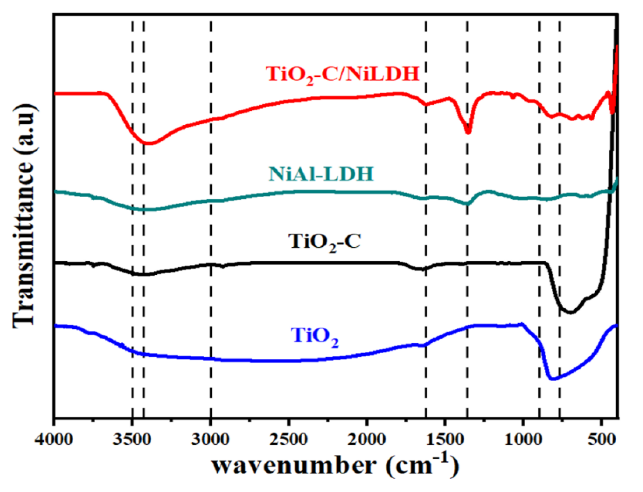
2.1.1. Fourier Transform Infrared Spectroscopy (FTIR) Analysis

Figure 1 shows the FT-IR spectra of TiO2-C, NiAl-LDH, and TiO2-C/NiLDH amalgams. TiO2-C sample displayed peaks ranging between 3000 and 3500 cm−1, which links to substantially physically bonded water to the surface of the metal oxide. The peaks with wavenumbers at ~1000–400 cm−1 were credited to the Ti-O-Ti and Ti-O elongating vibration modes [18,20]. The sharp peak at 1617 cm−1 is associated with O-H stretching and bending vibrations of adsorbed water molecules [23]. The very intense band situated at 1701 cm−1 belongs to the C=O stretching vibrations, which indicated that TiO2-C is formed [24]. The sharp peak at 1617 cm−1 is caused by the O-H stretching and bending vibrations of adsorbed water molecules [25]. The band at 3452 cm−1 in the FT-IR spectrum of NiAl-LDH is associated with the symmetric vibration of the O-H groups between the LDH layers. The interlayer CO32− group is represented by the spectra at 1376 cm−1, while the lattice vibration modes of M-O, O-M-O, and M-O-M are represented by the peaks at 400–800 cm−1. While the peak at 601 cm−1 is associated with the M-O metal oxide stretching vibration spectra in the LDH sheets, the peak at 1634 cm−1 corresponds to the H-O-H bending vibration [26]. The FT-IR spectrum of TiO2-C/NiLDH amalgam has distinctive bands associated with both TiO2-C and NiAl-LDH; the nickel layered double hydroxide bands dominate, perhaps owing to the ultrathin NiAl-LDH nanosheets amalgamated with TiO2-C with close contact [21].

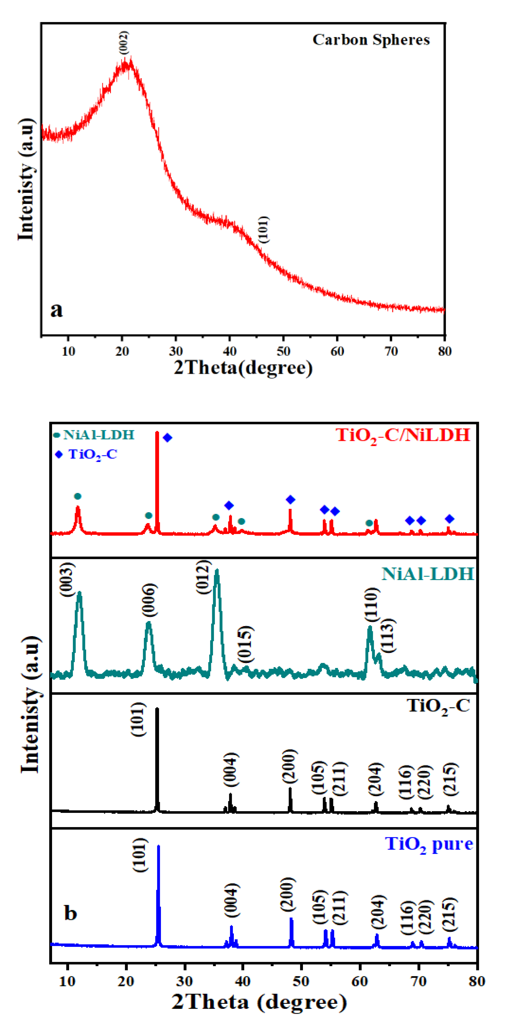
2.1.2. X-ray Diffraction (XRD)

The XRD patterns of the carbon spheres (panel a), and TiO2, TiO2-C, NiAl-LDH, and TiO2-C/NiLDH amalgam samples (panel b) are shown in Figure 2. The two distinctive peaks in Figure 2a at 2θ = 20° and 45°, respectively, correspond to the (002) and (101) planes of the carbon spheres. Figure 2b the peaks at 2θ = 25.3°, 37.8°, 48°, 54°, 55.2°, and 62.7°, respectively, correspond to the (101), (004), (220), (105), (211), and (204) planes of the anatase phase of TiO2 and TiO2-C [16,18,19,20,21]. The sharpness of the diffraction peaks indicates the extreme crystallinity of anatase TiO2-C [27]. The XRD shape of the original NiAl-LDH unveiled diffraction heights at 2θ values of 11.58°, 23.28°, 34.98°, 39.46°, 46.83°, 61.02°, and 62.29°, corresponding to the hexagonal NiAl-LDH phase with (003), (006), (012), (015), (018), (110), and (113) planes, respectively (JCPDS No. 22-0452) [28]. All the diffraction peaks observed in the XRD pattern of TiO2-C/NiLDH heterostructure are in excellent correspondence with the reflections observed for TiO2-C and NiAl-LDH [21]. The pronounced intensity suppression of the XRD peaks of the fabricated TiO2-C/NiLDH sample is credited to its amalgamated geometric fashion. The average crystallite size of TiO2, TiO2-C, NiAl-LDH, and TiO2-C/NiLDH were calculated from the Scherrer equation and was found to be 24.7 nm, 25.4 nm, 2.4 nm, and 25.6 nm, respectively. The relatively small increase in the crystallite size of the TiO2-C/NiLDH amalgam sample was attributed to the grafting of ultrathin NiAl-LDH nanoflakes into the TiO2-C hollow sphere cores [21].

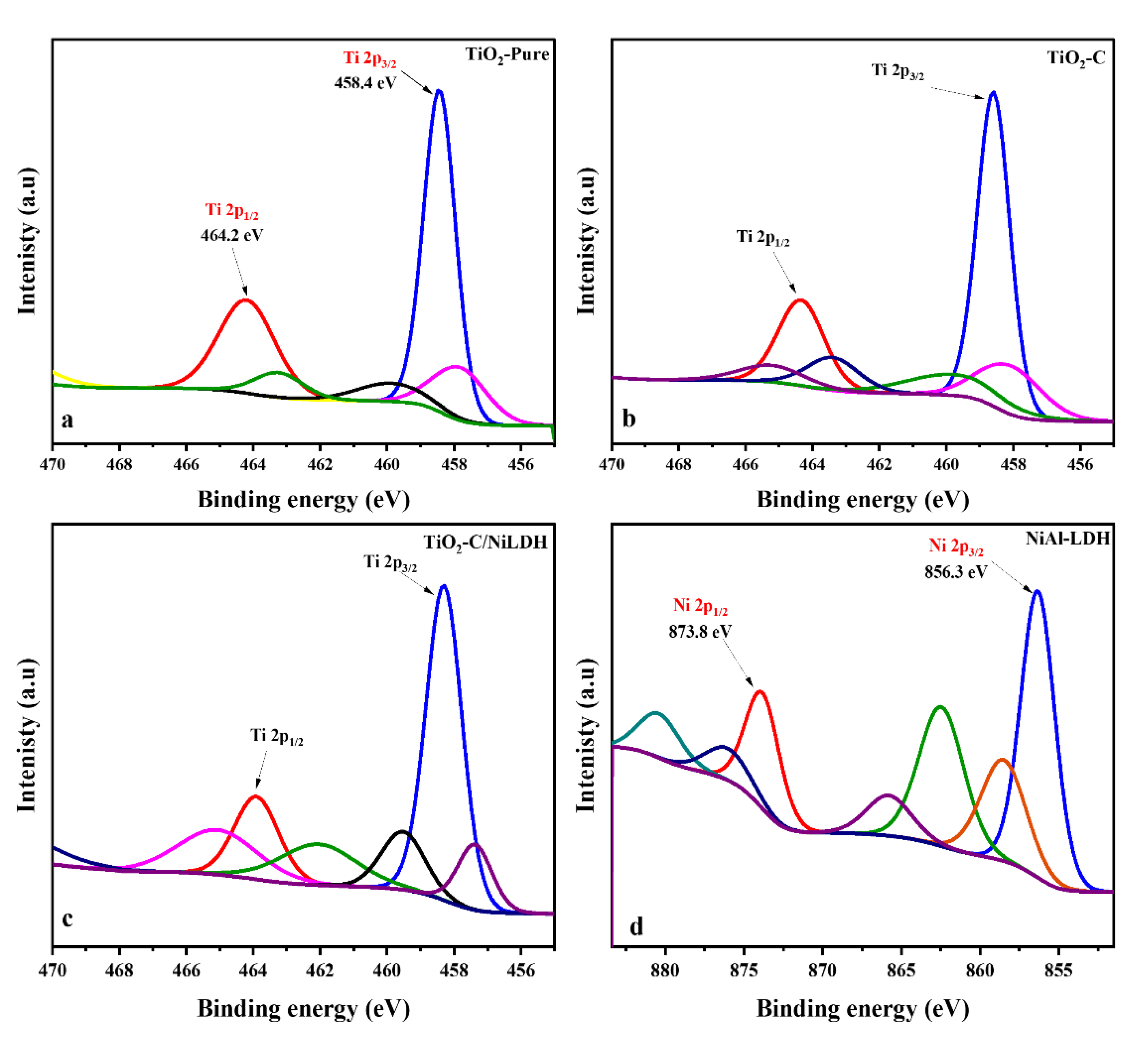
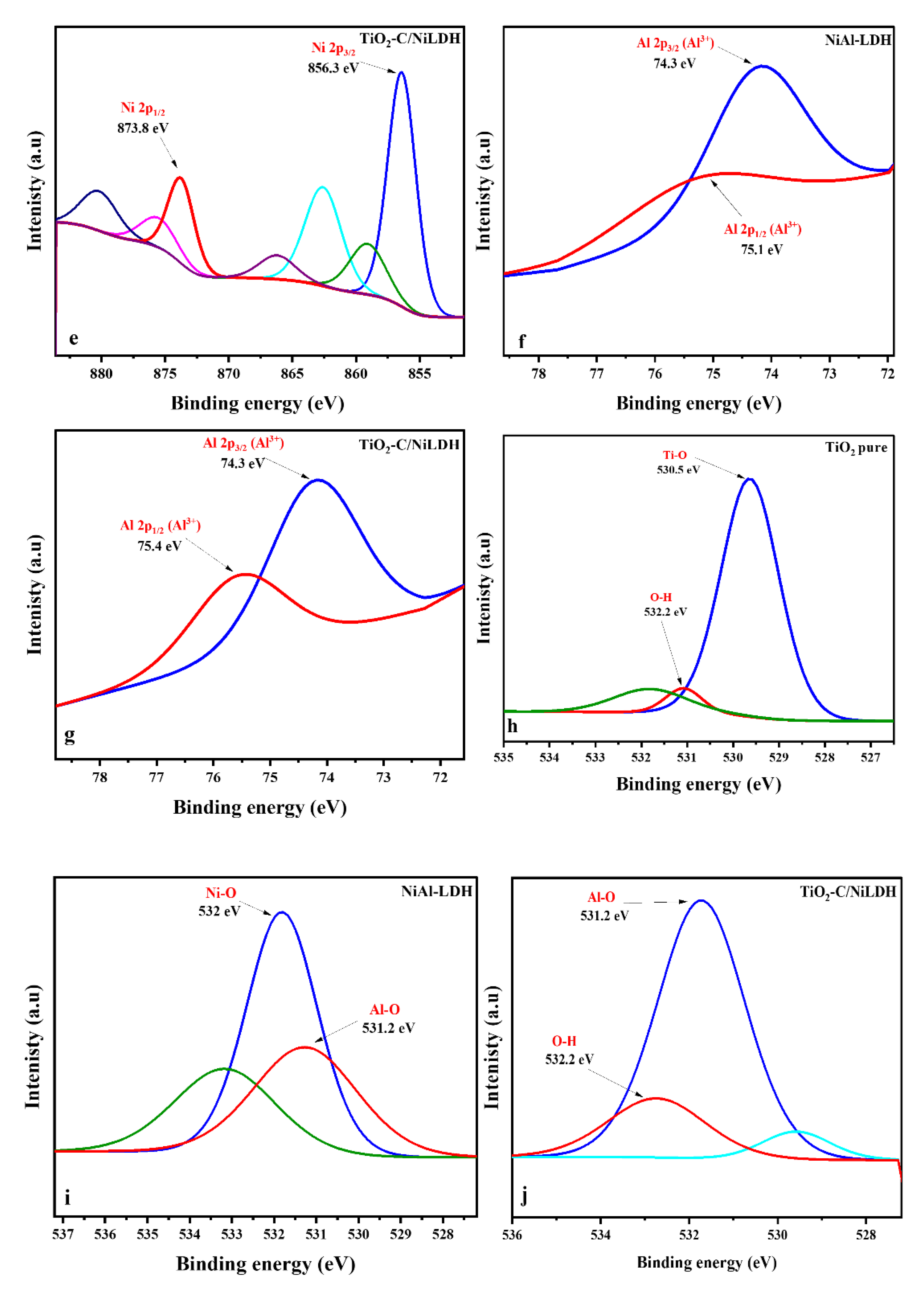
2.1.3. X-ray Photoelectron Microscopy (XPS)

Elemental composition, empirical formula, chemical state, and electronic state can all be determined by XPS, which can identify a material’s electronic state. (Figure 3a–c) illustrates an envelope for the Ti 2p band at 459.1 and 464.2 eV in TiO2 pure, TiO2-C, TiO2-C/NiLDH amalgam which refers to the presence of Ti4+ corresponding to Ti 2p3/2 and Ti 2p1/2, respectively, indicating that Ti exists in the tetravalent valence state [16,21,22,23,24]. Figure 3d shows two distinct peaks at 857.3 and 873.9 eV, which matches Ni 2p3/2 and Ni 2p1/2 because of the spin–orbit coupling. This suggests that Ni is in a high-spin divalent state based on the attendance of concurrent shake-up satellite peaks in the NiAl-LDH. For Ni 2p3/2 and Ni 2p1/2 BEs, the TiO2-C/NiLDH amalgam showed a largely positive shift in comparison to NiAl-LDH associated with the Ti 2pBE (Figure 3e). As shown in (Figure 3f,g), Al 2p spectra of both NiAl-LDH and TiO2-C/NiLDH core–shell amalgams show a distinctive peak at 74.3 eV, demonstrating the trivalent oxidation state of Al [21]. TiO2 has two peaks in its XPS band at 530.5 and 532.2 eV, which link to lattice O (OL; Ti–OL) species and surface-adsorbed water, separately (Figure 3h). NiAl-LDH has two peaks at 531 and 532 eV, which resemble lattice oxygen species (M–OL; M = Ni, Al) and hydroxide groups (O–H) that show in (Figure 3i). The O 1s spectra of the TiO2-C/NiLDH amalgam show the presence of typical TiO2 and NiAl-LDH O 1s components (Figure 3j) [29]. TiO2-C’s and NiAl-BEs LDH’s altered in a positive and negative route, respectively, representing that the two components cooperated powerfully [21]. Based on this result, the XPS shape of the TiO2-C/NiLDH amalgamation has confirmed the presence of elements such as Ti, Ni, Al, and O.

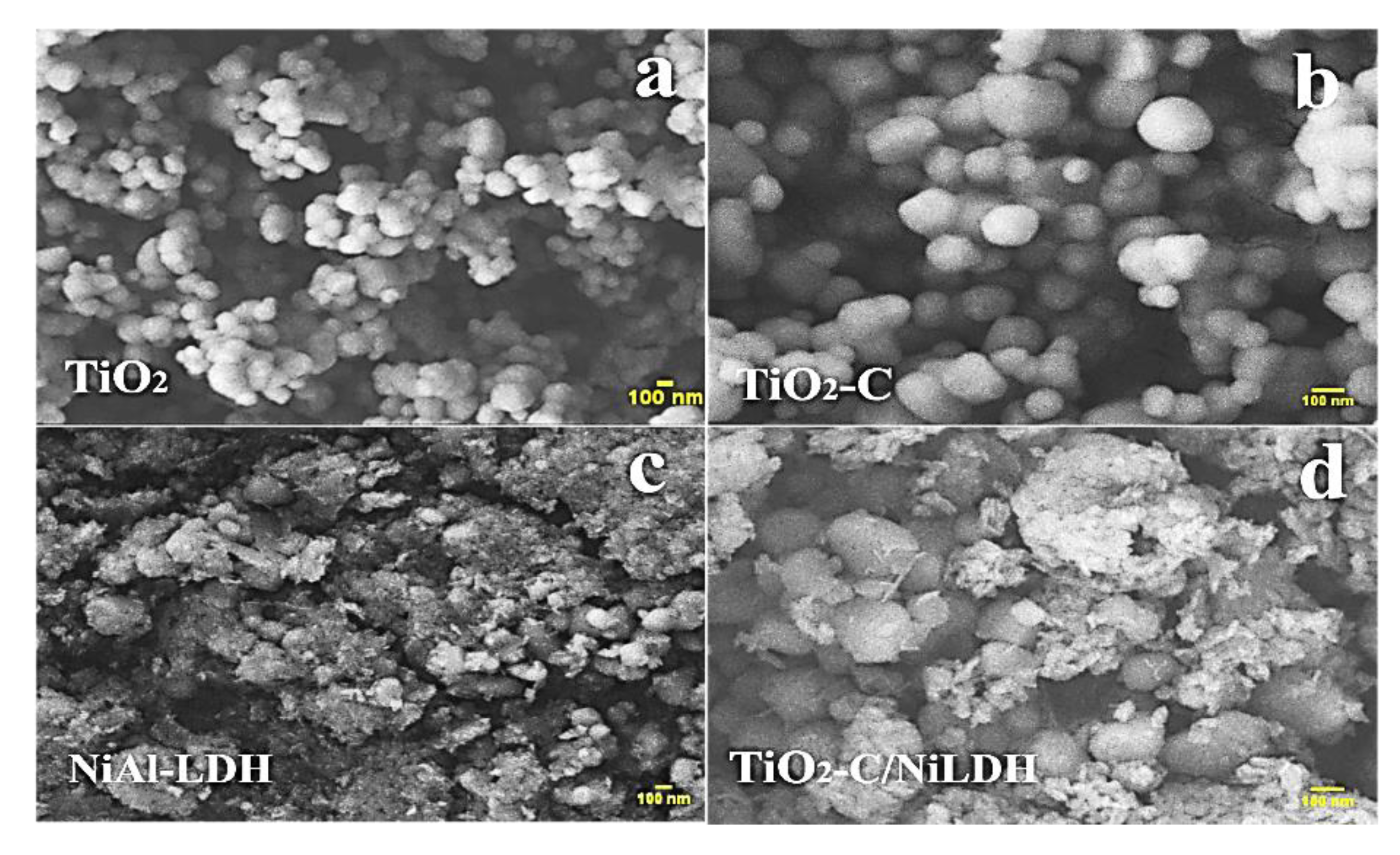
2.1.4. Scanning Electron Microscopy (SEM)

SEM was used to portray the shape and size of the prepared nanohybrid [30]. The SEM pictures (Figure 4a,b) displayed a spherical form, of TiO2 and the TiO2-C samples. It could be possible that the TiO2 particles (found in plenty), are consistently deposited on top of the external carbon spheres. However, the present micrographs could not confirm this speculation. Accordingly, HRTEM images will be discussed extensively in the next section. The SEM pictures of NiAl-LDH bare a typical lamellar assembly, as demonstrated in (Figure 4c,d). The intimate incorporation of the TiO2-C core and NiAl-LDH shell was confirmed by the TiO2-C/NiLDH amalgam enlarged SEM image (Figure 4d). These outcomes infer that ultrathin NiAl-LDH nano-sheets in the reaction blend were securely joined on top of the surface of the TiO2-C hollow spheres throughout the hydrothermal handling, resulting in the creation of amalgamated TiO2-C/NiLDH photocatalyst.

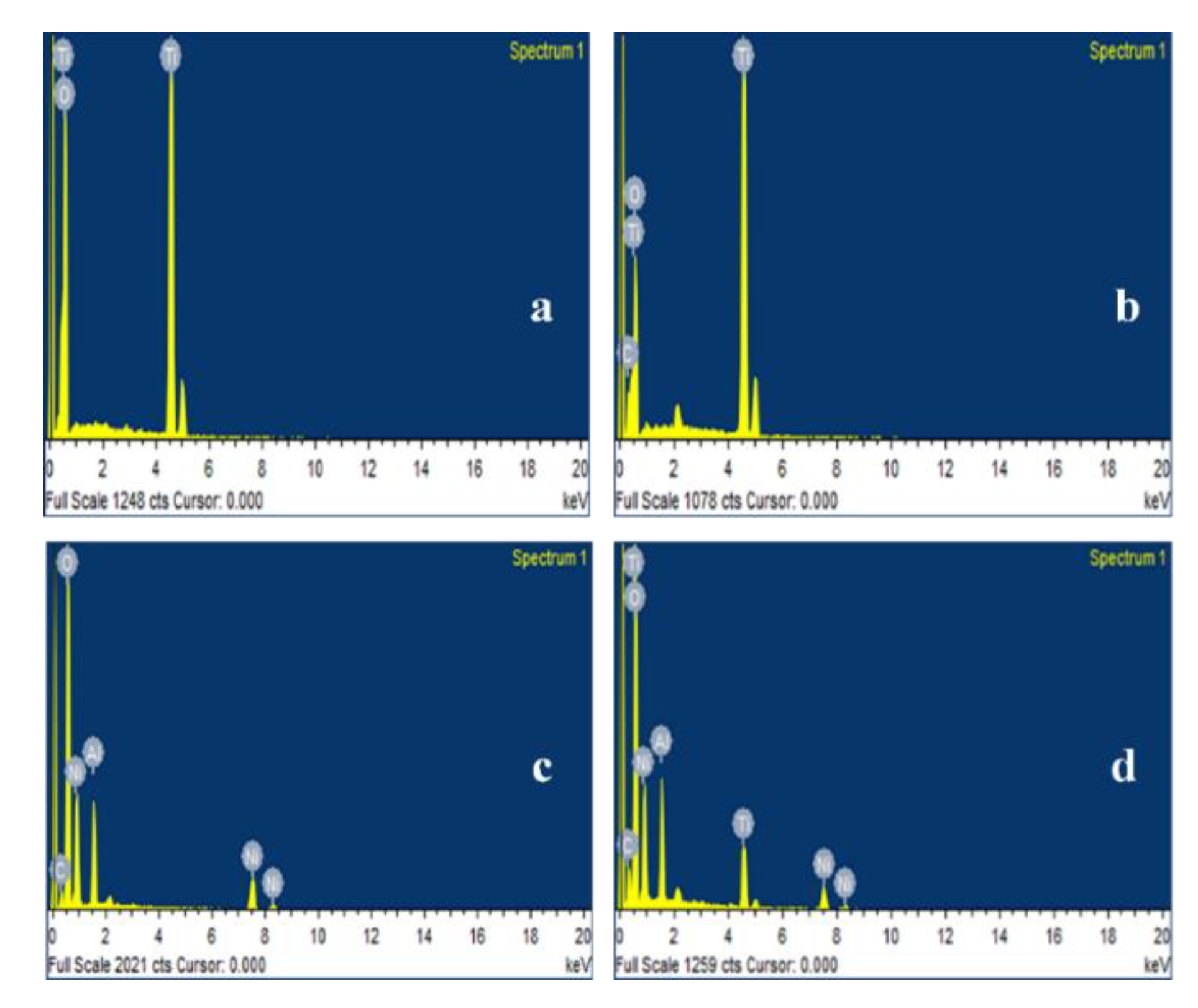
2.1.5. Energy Dispersive Spectroscopy (EDS)

EDS was used to categorize the various components of the nanocomposites [30]. Figure 4 shows the EDS spectra obtained. The EDS spectrum of TiO2 exhibit a peak indicative of the Ti metal at 4.6 keV (Figure 5a). Table 1 lists the estimated atomic weight percentages of the components detected in TiO2, TiO2-C, NiAl-LDH, and TiO2-C/NiLDH amalgams. Our findings confirmed the existence of Ti and O, which are constituents of TiO2, in the samples. An extra C was observed in the TiO2-C EDS spectrum (Figure 5b) [31]. The intercalation of Ni and Al into the brucite-like layer was verified by their molar ratio of 3:1, as shown in the figure (Figure 5c). This reveals that the coprecipitation synthesis effectively enriched the LDH layer with Ni. The presence of carbon and oxygen in TiO2-C/NiLDH confirmed by the EDS measurements can be attributed to the oxygen in the CO32− anions, H2O molecules, and OH groups, which represent a brucite-like layer (Figure 5d) [32].

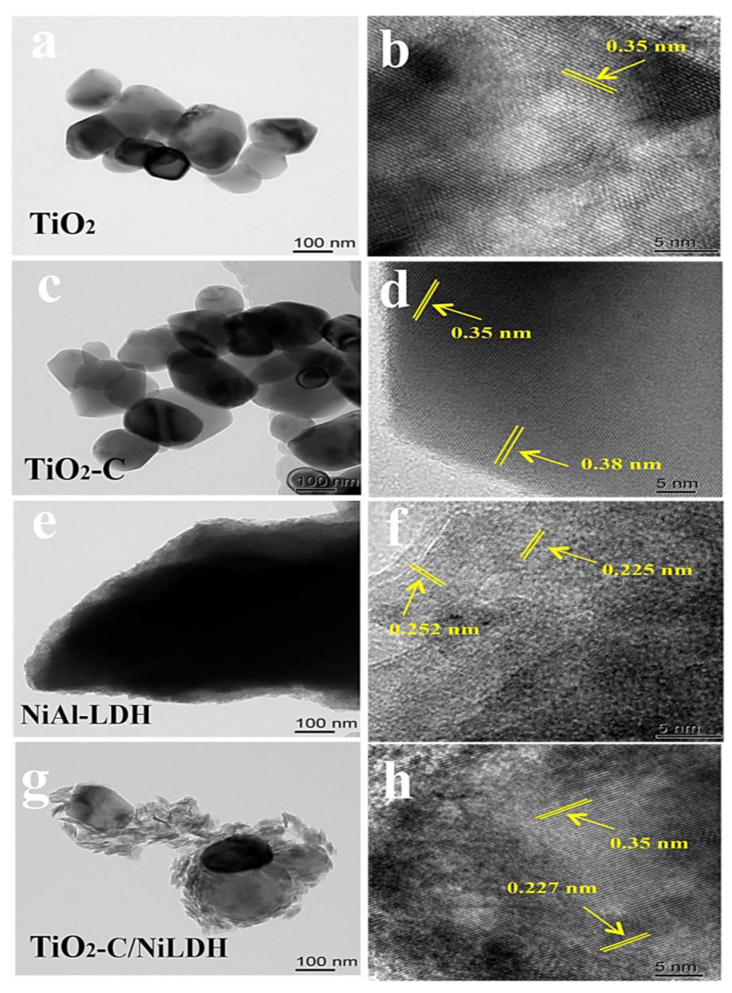
2.1.6. Transmission Electron Microscopy (TEM)

As is obvious in Figure 6b, the lattice fringe spacing of TiO2 nanoparticles was measured to be 0.384 nm. The lattice fringes with an interlayer distance of 0.384 nm are close to the 0.352 nm lattice spacing of the (101) planes in anatase TiO2, which is in accordance with XRD results. The mechanochemical agitation of TiO2 with the carbon hollow sphere did not affect the phase structure as evidenced by the unchanged lattice spacing at 0.352 nm (Figure 6d). Stacked nanoflakes of NiLDH displayed in Figure 6e,f showed interlayer distance lattice fringes at 0.252 nm which is close to 0.26 of the (012) LDH plane. The HRTEM image (Figure 6h) illustrates lattice fringes with d-spacings of 0.35 nm and 0.28 nm, which are allocated to the (101) plane of anatase TiO2 and the (012) plane of NiLDH, respectively [25,33]. A possible NiLDH nanoflakes definitely grafted onto the surface of TiO2-C during the hydrothermal treatment, which results in the formation of an amalgamated TiO2 C/NiLDH photocatalyst.

2.1.7. Diffuse Reflectance Spectroscopy (UV-DRS)

Diffuse reflectance UV–vis (DR-UVvis) was used to determine the absorption band edges of the synthesized nanocomposites. The observed DR-UV–vis spectra demonstrated that the nanocomposite was active under visible light. These data were used to generate Tauc plots, which were then used to calculate the bandgap of the composite photocatalysts [34]. Ultraviolet–visible diffuse reflectance spectroscopy studies stayed used to examine the light-harvesting features of the TiO2-C, NiAl-LDH, and TiO2-C/NiLDH samples (Figure S1).
From the Tauc plots, we estimated the bandgap of TiO2, TiO2-C/NiLDH, and TiO2-C to be 3.2 eV, 2.9 eV, and 3.1 eV, respectively. Evidently, TiO2-C/NiLDH has the smallest bandgap among them all. Additionally, the bandgap of TiO2 was consistent with previously reported values [35]. The data indicate that NiAl-LDH incorporated with the TiO2 lattice structure is synergistically enabled to absorb photons of longer wavelengths. With its broad bandgap and inherent UV-light reaction, TiO2-C, like TiO2, exhibits a sturdy absorption edge at around 400 nm. In the short term, NiAl-LDH has photosensitive captivation bands in the full spectra range that can be stimulated by light. There is d–d transitions in visible light, but the UV absorption band is linked with ligand-to-metal charge transmission (LMCT). TiO2-C/NiLDH amalgam DRS profile shows a high synergy between TiO2 and NiAl-LDH in the manufactured amalgam’s visible-light absorption bands as well as TiO2 UV-light absorption bands. Integrating both UV-stimulated TiO2-C and visible-light-stimulated NiAl-LDH into the TiO2-C/NiLDH amalgam caused by frequent photoinduced charge carriers is crucial for photocatalytic processes [21].

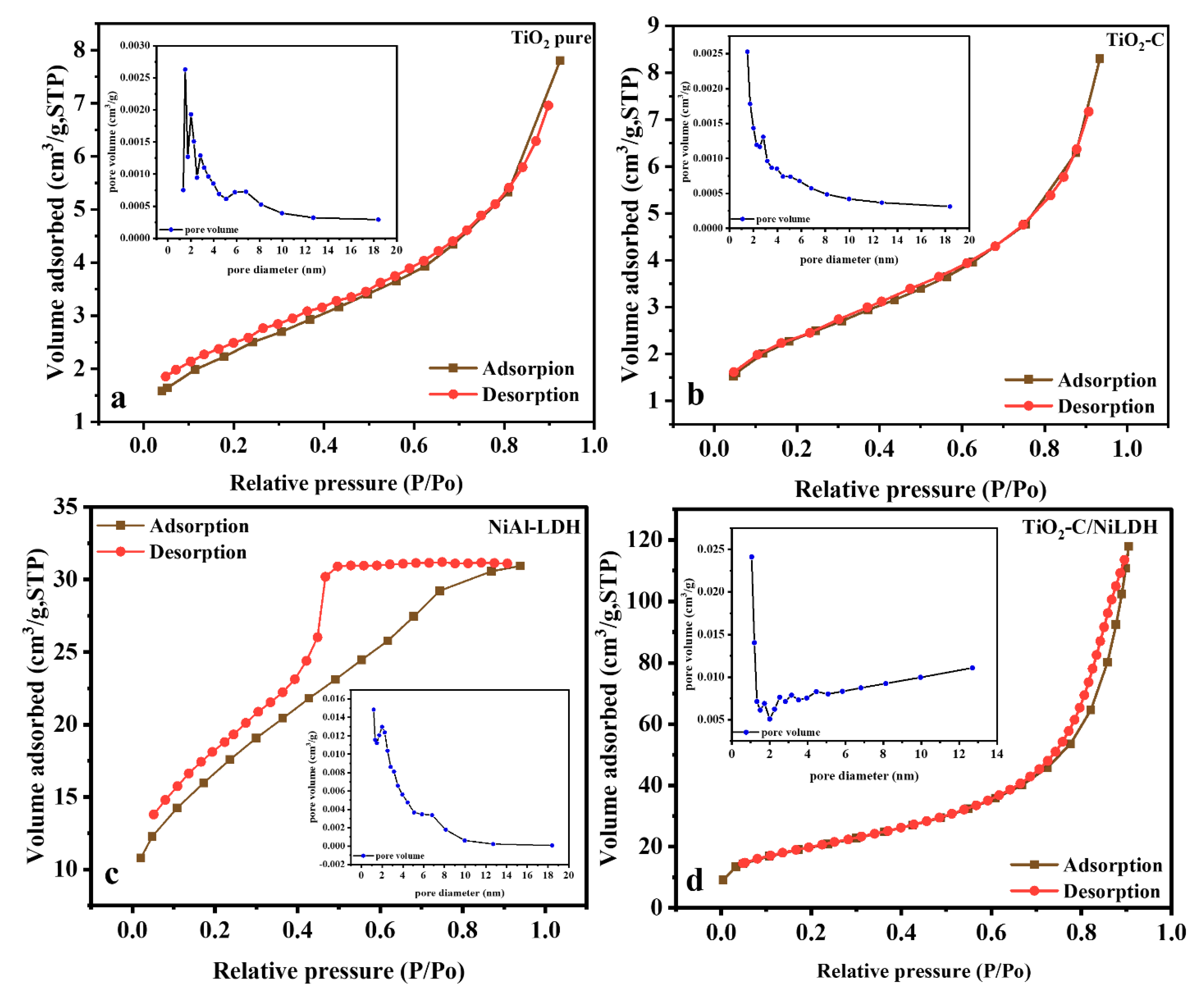
2.1.8. N2 Physisorption Measurement

The N2 adsorption–desorption isotherms are displayed in Figure 7. TiO2 and TiO2-C samples were found to exhibit type II isotherms, without hysteresis referring to a non-porous material as per the IUPAC classification [28]. NiAl-LDH exhibited a typical type IV isotherm with an H2-type hysteresis loop indicating that the pore cavity size is wider than the neck size, leading to a steep desorption step in the isotherm [29]. The pore size distribution analysis of the samples, using pore diameter, revealed that they exhibited bimodal distribution curves in the mesopore ranges. Table 2 summarizes the textural properties of NiAl-LDH and TiO2-C/NiLDH catalysts. The BET surface area of TiO2 in the TiO2-C hollow spheres was found to be 9 m2g1, whereas that of NiAl-LDH nanoflakes was 61 m2g1. However, the TiO2-C/NiLDH amalgam, synthesized by the inclusion of TiO2-C hollow spheres in NiAl-LDH nanoflakes, exhibited the highest BET surface area (72 m2g1). The pronounced increase in surface area is attributed to the good dispersion of NiAl-LDH in the TiO2-C core–shell with a small micropore volume, as shown in Table 2.

2.2. Evaluation of Photocatalytic Activity

2.2.1. Photocatalytic Studies

Photocatalytic tests were performed on TiO2, TiO2-C, NiAl-LDH, and amalgamated TiO2-C/NiLDH for 90 min. The photodegradation profiles and the effectiveness of MO removal of different nanocomposites under UV and visible light irradiation are presented in Figure S2. We investigated the removal efficiencies of the photocatalysts under visible and UV irradiation and the results are displayed in (Figure S2a,b). An aqueous solution of MO was used as a target pollutant to investigate the photocatalytic efficacy of TiO2-C/NiLDH under both visible and UV irradiation, along with TiO2, TiO2-C, and NiAl-LDH for comparison. The amalgamated TiO2-C/NiLDH amalgam exhibited a removal efficiency of approximately 100% in 90 min at pH 4 under visible light irradiation, whereas TiO2, TiO2-C, and NiAl-LDH catalysts achieved removal efficiencies of 9%, 18%, and 98%, respectively (Figure S2a). The TiO2-C/NiLDH amalgam exhibited a removal efficiency of approximately 93% under UV irradiation, while TiO2, TiO2-C, and NiAl-LDH had removal efficiencies of 51%, 80%, and 88%, respectively (Figure S2a). Overall, these nanocomposites with an anatase crystal phase supported on a NiAl-LDH-type matrix showed strong photocatalytic activity under both UV and visible light irradiation. Furthermore, the removal efficiency of the catalyst may be readily modified by changing the nature of the cations inside the stacked sheets. TiO2 has a band gap energy of 3.2 eV, which implies that it can absorb only UV radiation. The photocatalytic capabilities of the TiO2-C/NiLDH amalgam under UV and visible irradiation were better than those of TiO2 and TiO2-C. Moreover, the catalytic efficacy of TiO2-C/NiLDH was found to be higher than that of NiAl-LDH.
Methyl orange has a strong characteristic peak in its UV–vis absorption spectrum at 465 nm and a weak absorption peak at 274 nm [36]. As the reaction time is prolonged, these absorption peaks weaken and disappear. According to the UV–vis data, the reaction resulted in the degradation of methyl orange. The homo- and hetero-polyaromatic rings in the dye molecules were broken, which resulted in a decrease in the solution’s absorbance. Under UV and VIS light, it was discovered that the TiO2-C/NiLDH amalgam was an effective catalyst. Figure S3 showed degradation efficiency of methyl orange aqueous solution at 25°C in the presence of the TiO2-C/NiLDH amalgam under UV and VIS light where photocatalyst showed 98.6% degradation of methyl orange in 90 min of reaction, and 100% degradation in the same reaction time, respectively.
A literature review on the comparison of catalytic activity of as-prepared samples with some typical titania-based catalysts for degradation of MO displayed in Table 3. As it can be noted, pure TiO2 (anatase) showed 51% photocatayic degradation of MO (current study). However, amalgamation of TiO2 with different supports improved its catalytic performance as shown in Table 3. Fe3O4@SiO2@TiO2, showed 91% catalytic degradation efficiency under UV irradiation [37]. While in our current study, TiO2-C/NiLDH amalgam showed 100% photocatalytic degradation efficiency of MO.

2.2.2. Recycling Test

The reusability of the catalysts was tested over many reaction cycles, and under optimal reaction conditions, after which the catalyst was removed by filtering, washed with deionized water, and dried under a vacuum. We re-used the recovered catalyst three times under identical reaction conditions. To assess the stability of the TiO2-C/NiLDH amalgam, we used it to catalyze three different runs of MO removal. Our experiments demonstrated the stability of the amalgam photocatalyst (see Figure S4). However, we observed a slight decrease in the catalytic activity of the amalgam after the third run, which is presumably due to the weight loss of the catalyst throughout each work-up. It is important to know that stability and reusability are the main critical indicators of photocatalysts in real-world applications and for the assessment of the catalysts [40,41].
It was testified that photocatalytic degradation primarily happens on the surface of the photocatalyst, consequently, the surface characteristics of the photocatalyst play a considerable influence over its degradation efficiency [42]. The amalgamatedTiO2-C/NiLDH exhibited reasonably more surface area and pore volume among the synthesized nanohybrids. It seems that the surface area of this particular nanohybrid is inclined by the size of crystallite size of the synthesized sample; a drop in the crystallite size of NiLDH led to an increase in the surface area. The amalgamated nanocomposite sample with a high surface area owns further active sites to adsorb the methyl orange molecules and permits the passage and mobility of MO fragments and reactive OH species. Additionally, the nanohybrid assistances transportation of e and h+ to the surface from the crystal lattice more and more after regeneration of the sample.
Furthermore, the stability of the catalyst was investigated using XRD analysis. Figure S5 shows no change in the XRD pattern of TiO2-C/NiLDH amalgam photocatalyst confirming its excellent reproducibility and stability. However, a possible change in morphology was detected from the change in the degree of crystallinity of the phase structure.

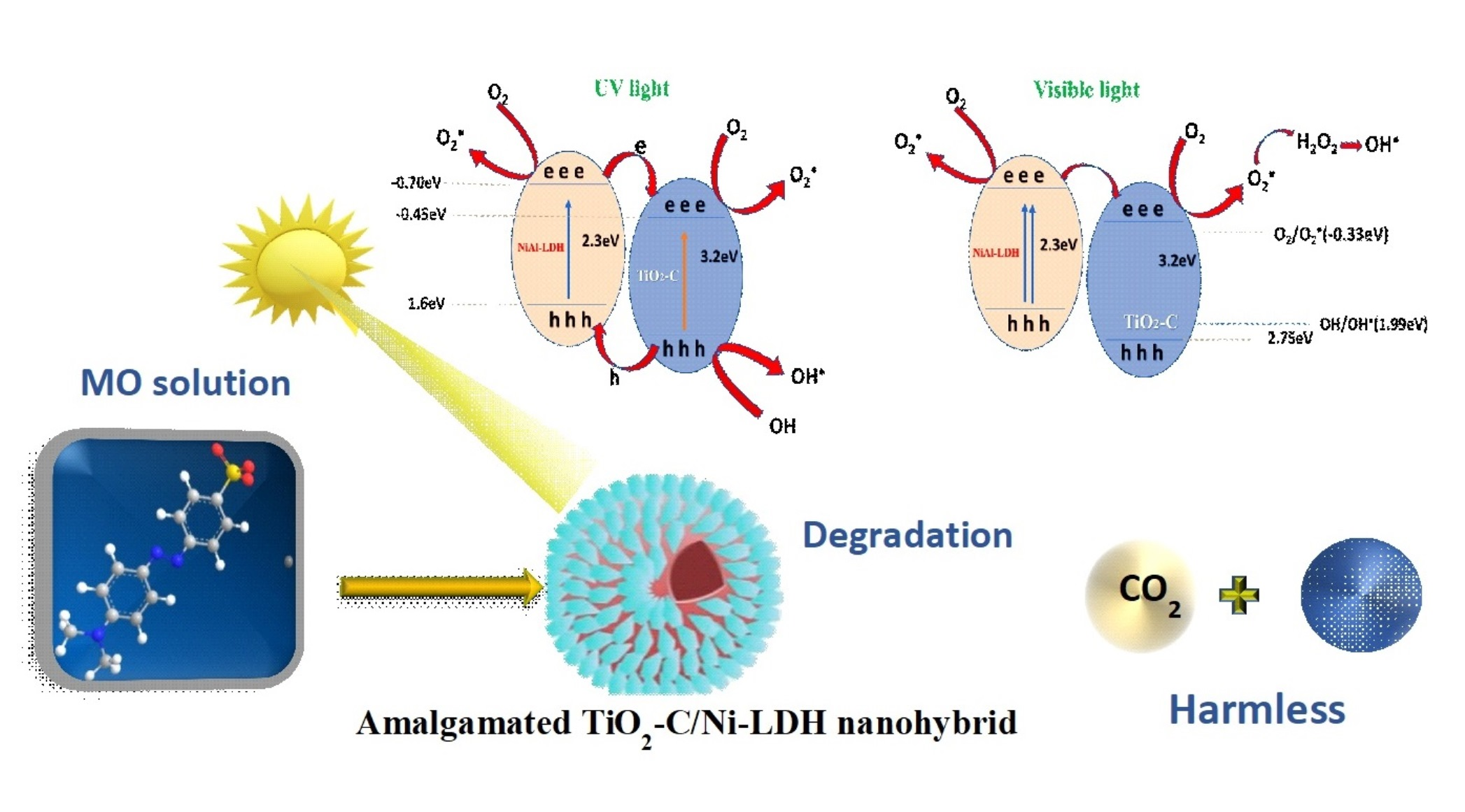
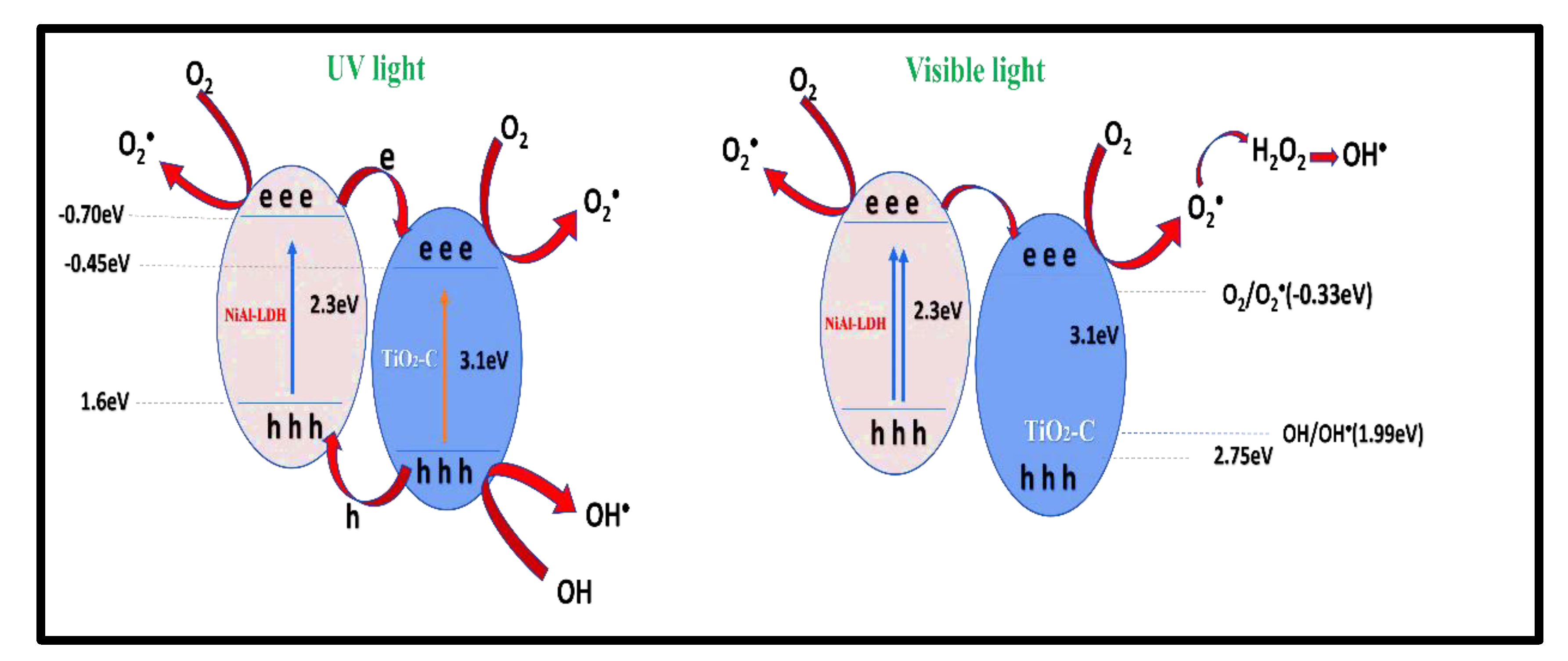
2.2.3. Photocatalytic Mechanism

By uniting light energy with semiconductors, photocatalysis can jump or speed up certain reduction and oxidation reactions. There are three elementary steps in a photocatalytic reaction: electron–hole pairs are thrilled by photoexcitation. Photogenerated electron–hole pairs are detached and diffused to the semiconductor’s surface. Reactions at the semiconductor surface’s active site catalyze the reduction of some species and the oxidation of others [43]. Since MO is an anionic dye, it is conceivable that at lower pHs, its adsorption is favored on a positively charged surface [10].
UV–vis DRS in the preceding section revealed that the amalgamation of TiO2-C (3.1 eV) and NiLDH (2.3 eV) resulted in a TiO2-C/NiLDH amalgam with a boosted photoabsorption capacity. Photoexcited charges may consequence of this wide-spectrum light absorption. To maximize photocatalytic degradation, one of the most important factors is the rate at which photo-excited electrons and holes separate and recombine, which is determined by the rate at which electrons and holes are separated and recombined [44]. The charge departure and carriage mechanisms of amalgamated TiO2-C/NiLDH must be thoroughly studied as a result. Z-scheme transfer or double transfer may occur at the interface between TiO2-C and NiLDH because TiO2-C has higher CB and VB potentials than NiLDH. If the current amalgamated system follows the Z-scheme transmission pathway, the reduction and oxidation processes take place on the conduction band of NiLDH and the valance band of TiO2-C, respectively. Both TiO2-C and NiLDH in the amalgamated TiO2-C/NiLDH are photoexcited by light, subsequent in electrons (e) in the CB of one and holes (h+) in the VB of another when the amalgam is exposed to light. Excited electrons transmission from the CB of NiLDH to the CB of TiO2-C, while holes transmission from the VB of TiO2-C to the VB of NiLDH as a consequence of the band alignment and inner electrical field fashioned between the two materials. The straight recombination of photoexcited electrons and holes in both components was significantly delayed by this process of charge separation. Thus, the VB of NiLDH and the CB of TiO2-C comprise additional holes than electrons. H2O molecules trapped the holes in the VB of NiLDH, discharging protons (H+) and oxygen (O2). As a result of this reaction, harmless chemicals such as carbon dioxide and water can be formed by the TiO2-C CB’s electrons reacting with organic pollutant molecules adsorbing on the catalyst surface. There are a lot of electrons in the TiO2-C CB as a consequence of the two-fold transference process. The slight photodeterioration observed and schematically depicted in Scheme 1 may be caused by electrons remaining in the CB of NiLDH after charge transfer. The following equations review the reactions that take place throughout photodegradation:
TiO2-C/NiLDH + ℎν TiO2-C/NiLDH (hVB+ + eCB)
NiLDH (hVB+) + OH   OH
NiLDH (hVB+) + H2O H+ + OH
TiO2-C (eCB) + O2  O2
O2 + H2O + H+  H2O2 + OH
H2O2 + e  OH + OH
OH + MO CO2 + H2

3. Discussion

We prepared TiO2-C, NiAl-LDH, and TiO2-C/NiLDH and characterized them by various techniques such as FTIR, XRD, SEM-EDS, TEM, N2 physisorption measurement UV-DRS, and XPS techniques. FTIR spectroscopy was used to identify the various functional groups present in TiO2-C, NiAl-LDH, and TiO2-C/NiLDH amalgams. The crystal structure of TiO2 in the catalysts evidenced from XRD was found to be mostly anatase, which has been proven to have higher photocatalytic activity than other phases [28]. SEM images of the TiO2-C/NiLDH amalgam indicated that ultrathin NiLDH nanoflakes in the reaction mixture were securely grafted onto the surface of the TiO2-C hollow spheres, resulting in the creation of a TiO2-C/NiLDH core–shell amalgam photocatalyst. EDS elemental analysis revealed the presence of key elements in the produced amalgam. The performance of the TiO2-C/NiLDH photocatalyst in the photodegradation of MO dye, which was validated by UV-DRS data, revealed that the TiO2-C/NiLDH amalgam had the lowest bandgap of 2.9 eV, among all the catalysts studied in this work. The synthesized amalgam catalyst also exhibited the largest surface area, which improves the surface reactant contact area for better photocatalytic exposure and degradation. TiO2 and TiO2-C are known to introduce mid-band states, allowing the utilization of low-energy light in photocatalytic processes [29]. The TiO2-C/NiLDH photocatalyst’s ability to utilize visible light results in a high quantum yield as visible light constitutes a significantly larger portion of the solar energy spectrum (approximately 40%). This increases the likelihood of photons of proper wavelength impacting the photocatalyst surface and activating them [21]. The performance of the TiO2-C/NiLDH photocatalyst was also examined under both sunlight and stringent UV light. Photodegradation of MO under visible light proceeded with extremely high efficiency compared to that under UV light irradiation [25,26,27,28,29]. Future studies could examine the potential of TiO2-C/NiLDH as a filtering medium to develop an effective water treatment technique and for the photoreductive removal of heavy metal contaminants from water and wastewater.

4. Materials and Methods

4.1. Materials

Agarose, titanium (P25), nickel (II) nitrate hexahydrate, aluminum nitrate nonahydrate, sodium hydroxide, sodium carbonate, nitric acid (69%), and sodium chloride were purchased from Sigma-Aldrich, Bengaluru, India. All other chemicals used in this study were of analytical reagent grade and were used as received without further purification. The deionized water used in this study was obtained from a Wellix Plus water purification system.

4.2. Synthesis of Carbon Spheres

In a typical synthesis, 4.58 g agarose powder was dissolved in 70 mL de-ionized water (preheated to 90 °C) with vigorous stirring. To this, 0.1169 g NaCl was added, and the reaction mixture was stirred for 10 min. Following this, the mixture was placed in a 100 mL Teflon-lined autoclave reactor and cooked at 180 °C for 12 h. The reaction mixture was centrifuged to recover the final product as a dark brown precipitate, which was washed three times with deionized water and ethanol, and subsequently dried at 80 °C to yield carbon spheres [27].

4.3. Preparation of TiO2-Carbon Hollow Spheres

To prepare the TiO2-C hollow spheres, we ground 1 g of the as-prepared carbon spheres with 0.1 g of titanium (P25) for 15 min. This ensures adequate diffusion of titanium ions into the carbon surface layer to obtain TiO2-C powder.

4.4. Preparation of Amalgamated TiO2-C/Ni-LDH

Pure Ni-LDH was synthesized by vigorously stirring a mixture of Ni(NO3)2.6H2O (0.3 M), Al(NO3)3.9H2O (0.2 M), NaOH (1 M), and Na2CO3 (0.5 M) at 25 °C while maintaining a pH of ~10. The resultant precipitate was centrifuged, washed several times until the pH reached ~7, and dried at 80 °C for 18 h. The TiO2-C/NiLDH core–shell amalgam was created in the same way as the NiAl-LDH reference sample, except that TiO2-C powder was employed in the synthesis technique [21]. TiO2-C/Ni-LDH core–shell amalgam was created using a hydrothermal method. A precise quantity of TiO2-C powder was dispersed in 100 mL of water using ultrasonication. After adding Ni(NO3)2.6H2O (0.3 M) and Al(NO3)3.9H2O (0.2 M), the dispersion was magnetically agitated for 30 min. Following this, the reaction mixture was mixed with NaOH (1 M) and Na2CO3 (0.5 M) and stirred while maintaining a constant pH of ~10. The mixture was then transferred to a 150 mL Teflon-lined autoclave reactor and heated at 120 °C for 6 h. The reaction mixture was then centrifuged, and the resultant precipitate was washed with water until the pH of the wash solution was neutral. We then dried the precipitate at 80 °C for 18 h.

4.5. Experimental Techniques

A PerkinElmer UATR Two FT-IR spectrometer was used for the Fourier transform infrared (FT-IR) spectroscopy. The measurements were performed in the transmission mode in the range 4000–500 cm−1 with a resolution of 4 cm−1; the measurements were averaged over 32 scans. The samples were prepared using KBr pellets.
Powder X-ray diffraction (XRD) analysis was performed using a Bruker diffractometer (Bruker D8 advance target, Karlsruhe, Germany). The measurements were performed at room temperature using a Cu K1 source operating at 40 kV and 40 mA, and a monochromator (λ = 1.5405 Å). The patterns generated with 2θ vary between 2° and 80°.
To investigate the chemistry of the synthesized materials at the sub-micron scale, a high-resolution field emission (JEOL FESEM 7600F) scanning electron microscope (SEM) was connected to an energy dispersive X-ray (EDX) detector.
Transmission electron microscopy (TEM) was performed using an FEI Tecnai G2 F30 TEM microscope in bright field (BF) mode. The samples were prepared by dispersing the catalyst powder in alcohol by sonication, and then drop-casting the dispersion onto an ultrathin carbon layer.
The ultraviolet–visible diffuse reflectance spectra (UV–vis DRS) of the samples were collected using a Thermo Scientific TM spectrophotometer UV–Visible Evolution TM 260 Bio. The data were recorded in the range of 220–1100 nm and the optical band gaps of the samples were estimated via the Tauc plots technique.
X-ray photoelectron spectroscopy (XPS) measurements were performed using a K-Alpha (Thermo Fisher Scientific, USA) instrument with a monochromatic X-ray Al K-alpha source (−10 to 1350 eV). The instrument was operated at a pressure of 109 mbar, spot size 400 microns, broad spectrum pass energy of 200 eV, and narrow spectrum pass energy of 50 eV. The data obtained was calibrated with the binding energy of the adventitious carbon (C 1s) line at 284.6 eV.
N2 adsorption/desorption experiments were performed using a Belsorp Max II analyzer. The specific surface area was calculated using the Brunauer–Emmett–Teller (BET) method and the pore size distribution was determined using the Barrett–Joyner–Halenda (BJH) method for the desorption branch.
The photocatalytic activity of the catalyst was investigated by using methyl orange (MO) as a model organic pollutant and irradiating it with ultraviolet (UV) and visible irradiation (VIS) at a pH of ~4. The pH of the reaction mixtures was adjusted by using a 0.1 M HNO3 solution. In a typical experiment, an appropriate amount of the catalyst was suspended (0.1 g) in MO solutions (solid/liquid) and stirred in the dark for 30 min to establish adsorption/desorption equilibrium between the pollutant molecules and the catalyst surface. Subsequently, the suspensions were stirred under exposure to UV and visible light. A 100 W Hg lamp (Toshiba BLB 20 W) was used as the UV light source, and a 150 W halogen lamp (Philips Lamps, TL-D,18W) [45] was used as the VIS light source. Beer’s law was used to correlate the absorbance of the solution (which is indicative of the dye concentration) with the evolution of photocatalytic degradation. Following the degradation of the organic pollutants in the aqueous solution, the absorption profiles in the range of 190–700 nm for the MO removal tests were measured using a Thermo-Electron Evolution 300 UV–Vis spectrometer.

5. Conclusions

In this study, we fabricated carbon hollow spheres, TiO2-C, and TiO2-C/NiLDH core–shell amalgams using an ecologically safe and cost-effective synthesis. Physicochemical characterization using different techniques elucidated the functional groups (FTIR), crystallographic structure (XRD), oxidation states (XPS), and BET surface area of the material. The morphologies of the synthesized materials were investigated by SEM and TEM. The band gap energy, calculated from the UV–VIS DSR data, demonstrated that the TiO2-C/NiLDH amalgam catalyst had a lower bandgap (2.9 eV) than the pure TiO2 (3.2 eV) and TiO2-C catalysts (3.1 eV). The TiO2-C/NiLDH core–shell amalgam exhibited considerably higher photocatalytic activity than TiO2, TiO2-C, and NiAl-LDH, for aiding the photodegradation of organic pollutants under both visible and UV irradiation. This new material shows great potential as a solar-powered catalyst for depolluting wastewater containing textile dyes. As textile dyes deteriorate water quality and may cause several serious health effects if left untreated, the water treatment technology mentioned here can help avoid such issues by successfully depolluting wastewater.

Supplementary Materials

The following supporting information can be downloaded at: https://www.mdpi.com/article/10.3390/catal12101200/s1, Figure S1: (a) Tauc plots to determine the optical band gaps and (b) UV-VIS DRS spectra of TiO2, TiO2-C, NiAl-LDH and TiO2-C/NiLDH catalysts; Figure S2: (a and b) removal efficiency of all the investigated catalysts under UV and VIS irradiation, respectively; Figure S3: UV-visible absorption changes of methyl orange aqueous solution at 25°C in the presence of the TiO2-C/NiLDH amalgam samples; Figure S4: Recycling tests of the photocatalytic degradation of MO over TiO2-C/NiLDH nanohybrid; Figure S5: XRD of TiO2-C/Ni-LDH core–shell amalgam before and after the degradation [46,47,48].

Author Contributions

Original draft preparation, A.S.A.-S. and Q.A.A.; Writing—review and editing, A.S.A.-S., Q.A.A. and M.M.M.M.; Supervision, M.M.M.M. and Q.A.A. All authors have read and agreed to the published version of the manuscript.

Funding

This research was funded by the Institutional Fund Projects under Grant No. (IFPIP: 1000-130-1442). Ministry of Education and King Abdulaziz University, DSR, Jeddah, Saudi Arabia.

Data Availability Statement

Not applicable.

Acknowledgments

This research was funded by the Institutional Fund Projects under Grant No. (IFPIP: 1000-130-1442). The authors gratefully acknowledge the technical and financial support from the Ministry of Education and King Abdulaziz University, DSR, Jeddah, Saudi Arabia.

Conflicts of Interest

The authors declare no conflict of interest.

References

  1. Luo, B.; Liu, G.; Wang, L. Recent advances in 2D materials for photocatalysis. Nanoscale 2016, 8, 6904–6920. [Google Scholar] [CrossRef] [PubMed]
  2. Lu, Z.; Prouty, M.D.; Guo, Z.; Golub, V.; Kumar, C.S.S.R.; Lvov, Y.M. Magnetic switch of permeability for polyelectrolyte microcapsules embedded with Co@Aunanoparticles. Langmuir 2005, 21, 2042–2050. [Google Scholar] [CrossRef] [PubMed]
  3. Liu, C.; Mao, S.; Shi, M.; Hong, X.; Wang, D.; Wang, F.; Xia, M.; Chen, Q. Enhanced photocatalytic degradation performance of BiVO4/BiOBr through combining Fermi level alteration and oxygen defect engineering. Chem. Eng. J. 2022, 449, 137757. [Google Scholar] [CrossRef]
  4. Li, C.; Xu, Y.; Tu, W.; Chen, G.; Xu, R. Metal-free photocatalysts for various applications in energy conversion and environmental purification. Green Chem. 2016, 19, 882–899. [Google Scholar] [CrossRef]
  5. Lai, C.; An, N.; Li, B.; Zhang, M.; Yi, H.; Liu, S.; Qin, L.; Liu, X.; Li, L.; Fu, Y.; et al. Future roadmap on nonmetal-based 2D ultrathin nanomaterials for photocatalysis. Chem. Eng. J. 2020, 406, 126780. [Google Scholar] [CrossRef]
  6. Li, Y.; Gao, C.; Long, R.; Xiong, Y. Photocatalyst design based on two-dimensional materials. Mater. Today Chem. 2019, 11, 197–216. [Google Scholar] [CrossRef]
  7. Yang, X.; Singh, D.; Ahuja, R. Recent advancements and future prospects in ultrathin 2d semiconductor-based photocatalysts for water splitting. Catalysts 2020, 10, 1111. [Google Scholar] [CrossRef]
  8. Wang, H.; Zhang, X.; Xie, Y. Recent progress in ultrathin two-dimensional semiconductors for photocatalysis. Mater. Sci. Eng. R Rep. 2018, 130, 1–39. [Google Scholar] [CrossRef]
  9. Alshorifi, F.T.; Ali, S.L.; Salama, R.S. Promotional Synergistic Effect of Cs–Au NPs on the Performance of Cs–Au/MgFe2O4 Catalysts in Catalysis 3,4-Dihydropyrimidin-2(1H)-Ones and Degradation of RhB Dye. J. Inorg. Organomet. Polym. Mater. 2022, 2, 1–12. [Google Scholar] [CrossRef]
  10. Lachheb, H.; Puzenat, E.; Houas, A.; Ksibi, M.; Elaloui, E.; Guillard, C.; Herrmann, J.-M. Photocatalytic degradation of various types of dyes (Alizarin S, Crocein Orange G, Methyl Red, Congo Red, Methylene Blue) in water by UV-irradiated titania. Appl. Catal. B Environ. 2002, 39, 75–90. [Google Scholar] [CrossRef]
  11. Alshorifi, F.T.; Alswat, A.A.; Salama, R.S. Gold-selenide quantum dots supported onto cesium ferrite nanocomposites for the efficient degradation of rhodamine B. Heliyon 2022, 8, 4–10. [Google Scholar] [CrossRef] [PubMed]
  12. Alshorifi, F.T.; Alswat, A.A.; Mannaa, M.A.; Alotaibi, M.T.; El-Bahy, S.M.; Salama, R.S. Facile and Green Synthesis of Silver Quantum Dots Immobilized onto a Polymeric CTS–PEO Blend for the Photocatalytic Degradation of p-Nitrophenol. ACS Omega 2021, 6, 30432–30441. [Google Scholar] [CrossRef] [PubMed]
  13. El-Hakam, S.A.; Alshorifi, F.T.; Salama, R.S.; Gamal, S.; El-Yazeed, W.A.; Ibrahim, A.A.; Ahmed, A.I. Application of nanostructured mesoporous silica/ bismuth vanadate composite catalysts for the degradation of methylene blue and brilliant green. J. Mater. Res. Technol. 2022, 18, 1963–1976. [Google Scholar] [CrossRef]
  14. Jovanov, V.; Rudic, O.; Ranogajec, J.; Fidanchevska, E. Synthesis of nanocomposite coating based on TiO2/ZnAl layer double hydroxides. Mater. Constr. 2017, 67, 112. [Google Scholar] [CrossRef] [Green Version]
  15. Balu, S.; Chen, Y.-L.; Yang, T.C.-K.; Chen, J.-N.; Chen, S.-W. Effect of ultrasound-induced hydroxylation and exfoliation on P90–TiO2/g-C3N4 hybrids with enhanced optoelectronic properties for visible-light photocatalysis and electrochemical sensing. Ceram. Int. 2020, 46, 18002–18018. [Google Scholar] [CrossRef]
  16. Seftel, E.M.; Popovici, E.; Beyers, E.; Mertens, M.; Zhu, H.Y.; Vansant, E.F.; Cool, P. New TiO 2/MgAl-LDH nanocomposites for the photocatalytic degradation of dyes. J. Nanosci. Nanotechnol. 2010, 10, 8227–8233. [Google Scholar] [CrossRef]
  17. Korošec, R.C.; Miljević, B.; Umek, P.; van der Bergh, J.M.; Vučetić, S.; Ranogajec, J. Photocatalytic self-cleaning properties of Mo:TiO2 loaded Zn–Al layered double hydroxide synthesised at optimised pH value for the application on mineral substrates. Ceram. Int. 2019, 46, 6756–6766. [Google Scholar] [CrossRef]
  18. Al-Ghafri, B.; Lau, W.-J.; Al-Abri, M.; Goh, P.-S.; Ismail, A.F. Titanium dioxide-modified polyetherimide nanofiber membrane for water treatment. J. Water Process Eng. 2019, 32, 100970. [Google Scholar] [CrossRef]
  19. Dou, Y.; Zhang, S.; Pan, T.; Xu, S.; Zhou, A.; Pu, M.; Yan, H.; Han, J.; Wei, M.; Evans, D.G.; et al. TiO2@layered double hydroxide core-shell nanospheres with largely enhanced photocatalytic activity toward O2 generation. Adv. Funct. Mater. 2015, 25, 2243–2249. [Google Scholar] [CrossRef]
  20. Pai, S.; Kini, S.M.; Selvaraj, R.; Pugazhendhi, A. A review on the synthesis of hydroxyapatite, its composites and adsorptive removal of pollutants from wastewater. J. Water Process Eng. 2020, 38, 101574. [Google Scholar] [CrossRef]
  21. Jo, W.-K.; Moru, S.; Tonda, S. A green approach to the fabrication of a TiO2/NiAl-LDH core–shell hybrid photocatalyst for efficient and selective solar-powered reduction of CO2 into value-added fuels. J. Mater. Chem. A 2020, 8, 8020–8032. [Google Scholar] [CrossRef]
  22. Saber, O.; Kotb, H.M. Designing dual-function nanostructures for water purification in sunlight. Appl. Sci. 2020, 10, 1786. [Google Scholar] [CrossRef] [Green Version]
  23. Serga, V.; Burve, R.; Krumina, A.; Romanova, M.; Kotomin, E.; Popov, A. Extraction–pyrolytic method for TiO2 polymorphs production. Crystals 2021, 11, 431. [Google Scholar] [CrossRef]
  24. Singh, T.; Pal, D.B.; Bhatiya, A.K.; Mishra, P.K.; Hashem, A.; Alqarawi, A.A.; AbdAllah, E.F.; Gupta, V.K.; Srivastava, N. Integrated process approach for degradation of p-cresol pollutant under photocatalytic reactor using activated carbon/TiO2 nanocomposite: Application in wastewater treatment. Environ. Sci. Pollut. Res. 2021, 29. [Google Scholar] [CrossRef] [PubMed]
  25. Tonda, S.; Kumar, S.; Bhardwaj, M.; Yadav, P.; Ogale, S. g-C3N4/NiAl-LDH 2D/2D Hybrid Heterojunction for High-Performance Photocatalytic Reduction of CO2 into Renewable Fuels. ACS Appl. Mater. Interfaces 2018, 10, 2667–2678. [Google Scholar] [CrossRef] [Green Version]
  26. Zhang, H.; Du, G.; Lu, W.; Cheng, L.; Zhu, X.; Jiao, Z. Porous TiO2 hollow nanospheres: Synthesis, characterization and enhanced photocatalytic properties. CrystEngComm 2012, 14, 3793–3801. [Google Scholar] [CrossRef]
  27. Yang, H.; Wang, G.; Ding, N.; Yin, C.; Chen, Y. Size-controllable synthesis of carbon spheres with assistance of metal ions. Synth. Met. 2016, 214, 1–4. [Google Scholar] [CrossRef]
  28. Thommes, M.; Kaneko, K.; Neimark, A.V.; Olivier, J.P.; Rodriguez-Reinoso, F.; Rouquerol, J.; Sing, K.S.W. Physisorption of gases, with special reference to the evaluation of surface area and pore size distribution (IUPAC Technical Report). Pure Appl. Chem. 2015, 87, 1051–1069. [Google Scholar] [CrossRef] [Green Version]
  29. Li, X.; Xiong, J.; Xu, Y.; Feng, Z.; Huang, J. Defect-assisted surface modification enhances the visible light photocatalytic performance of g-C3N4@C-TiO2 direct Z-scheme heterojunctions. Chin. J. Catal. 2019, 40, 424–433. [Google Scholar] [CrossRef]
  30. Cychosz, K.A.; Thommes, M. Progress in the Physisorption Characterization of Nanoporous Gas Storage Materials. Engineering 2018, 4, 559–566. [Google Scholar] [CrossRef]
  31. Lai, C.W.; Sreekantan, S. Single step formation of C-TiO2 nanotubes: Influence of applied voltage and their photocatalytic activity under solar illumination. Int. J. Photoenergy 2013, 2013, 11–15. [Google Scholar] [CrossRef] [Green Version]
  32. Claydon, R.; Wood, J. A Mechanistic Study of Layered-Double Hydroxide (LDH)-Derived Nickel-Enriched Mixed Oxide (Ni-MMO) in Ultradispersed Catalytic Pyrolysis of Heavy Oil and Related Petroleum Coke Formation. Energy Fuels 2019, 33, 10820–10832. [Google Scholar] [CrossRef] [PubMed] [Green Version]
  33. Cao, S.; Xue, Z.; Yang, C.; Qin, J.; Zhang, L.; Yu, P.; Wang, S.; Zhao, Y.; Zhang, X.; Liu, R. Insights into the Li+ storage mechanism of TiC@C-TiO2 core-shell nanostructures as high-performance anodes. Nano Energy 2018, 50, 25–34. [Google Scholar] [CrossRef]
  34. Jeong, I.R.; Lee, J.H.; Song, J.; Oh, Y.S.; Cho, S. Control of structural disorder in spinel ceramics derived from layered double hydroxides. Ceram. Int. 2019, 46, 6594–6599. [Google Scholar] [CrossRef]
  35. Kumar, S.; Isaacs, M.A.; Trofimovaite, R.; Durndell, L.; Parlett, C.M.; Douthwaite, R.E.; Coulson, B.; Cockett, M.C.; Wilson, K.; Lee, A.F. P25@CoAl layered double hydroxide heterojunction nanocomposites for CO2 photocatalytic reduction. Appl. Catal. B Environ. 2017, 209, 394–404. [Google Scholar] [CrossRef]
  36. Basahel, S.N.; Ali, T.T.; Mokhtar, M.; Narasimharao, K. Influence of crystal structure of nanosized ZrO2 on photocatalytic degradation of methyl orange. Nanoscale Res. Lett. 2015, 10, 73. [Google Scholar] [CrossRef] [Green Version]
  37. Wang, Z.; Shen, L.; Zhu, S. Synthesis of core-shell Fe 3O 4@SiO 2@TiO 2 microspheres and their application as recyclable photocatalysts. Int. J. Photoenergy 2012, 2012, 202519. [Google Scholar] [CrossRef] [Green Version]
  38. Nguyen, T.H.; Vu, A.T.; Dang, V.H.; Wu, J.C.-S.; Le, M.T. Photocatalytic Degradation of Phenol and Methyl Orange with Titania-Based Photocatalysts Synthesized by Various Methods in Comparison with ZnO–Graphene Oxide Composite. Top. Catal. 2020, 63, 1215–1226. [Google Scholar] [CrossRef]
  39. Baig, M.M.; Pervaiz, E.P.E.; Afzal, M.J.A.M.J. Catalytic Activity and Kinetic Studies of Core@Shell Nanostructure NiFe2O4@TiO2 for Photocatalytic Degradation of Methyl Orange Dye. J. Chem. Soc. Pak. 2020, 42, 531. [Google Scholar] [CrossRef]
  40. Liu, C.; Mao, S.; Shi, M.; Wang, F.; Xia, M.; Chen, Q.; Ju, X. Peroxymonosulfate activation through 2D/2D Z-scheme CoAl-LDH/BiOBr photocatalyst under visible light for ciprofloxacin degradation. J. Hazard. Mater. 2021, 420, 126613. [Google Scholar] [CrossRef]
  41. Liu, C.; Mao, S.; Wang, H.; Wu, Y.; Wang, F.; Xia, M.; Chen, Q. Peroxymonosulfate-assisted for facilitating photocatalytic degradation performance of 2D/2D WO3/BiOBr S-scheme heterojunction. Chem. Eng. J. 2021, 430, 132806. [Google Scholar] [CrossRef]
  42. Ali, T.T.; Narasimharao, K.; Basahel, S.N.; Mokhtar, M.; Alsharaeh, E.H.; Mahmoud, H.A. Template Assisted Microwave Synthesis of rGO-ZrO2 Composites: Efficient Photocatalysts Under Visible Light. J. Nanosci. Nanotechnol. 2019, 19, 5177–5188. [Google Scholar] [CrossRef]
  43. Kowsari, E. Carbon-Based Nanocomposites for Visible Light-Induced Photocatalysis. In Nanocomposites for Visible Light-Induced Photocatalysis; Springer: Berlin/Heidelberg, Germany, 2017; pp. 203–249. [Google Scholar] [CrossRef]
  44. Zhu, D.; Zhou, Q. Action and mechanism of semiconductor photocatalysis on degradation of organic pollutants in water treatment: A review. Environ. Nanotechnol. Monit. Manag. 2019, 12, 100255. [Google Scholar] [CrossRef]
  45. Seftel, E.; Niarchos, M.; Mitropoulos, C.; Mertens, M.; Vansant, E.; Cool, P. Photocatalytic removal of phenol and methylene-blue in aqueous media using TiO2@LDH clay nanocomposites. Catal. Today 2015, 252, 120–127. [Google Scholar] [CrossRef]
  46. Makuła, P.; Pacia, M.; Macyk, W. How To Correctly Determine the Band Gap Energy of Modified Semiconductor Photocatalysts Based on UV-Vis Spectra. J. Phys. Chem. Lett. 2018, 9, 6814–6817. [Google Scholar] [CrossRef] [PubMed] [Green Version]
  47. Basu, P.K. Theory of Optical Processes in Semiconductors. Theory Opt. Process. Semicond. 2010. [Google Scholar] [CrossRef]
  48. López, R.; Gómez, R. Band-gap energy estimation from diffuse reflectance measurements on sol-gel and commercial TiO 2: A comparative study. J. Sol-Gel Sci. Technol. 2012, 61, 1–7. [Google Scholar] [CrossRef]
Figure 1. FT-IR spectra of TiO2, TiO2-C, NiAl-LDH, and TiO2-C/NiLDH amalgam samples.
Catalysts 12 01200 g001
Figure 2. X-ray diffraction (XRD) patterns of (a) carbon spheres and (b) TiO2, TiO2-C, NiAl-LDH, and TiO2-C/NiLDH amalgam.
Catalysts 12 01200 g002
Figure 3. Deconvoluted XPS spectra of TiO2, TiO2-C, NiAl-LDH, and amalgamated TiO2-C/NiLDH samples. (a,f) Ti 2p, (d,g) Ni 2p, (c,h) Al 2p, and (b,e,i,j) O 1s.
Catalysts 12 01200 g003aCatalysts 12 01200 g003b
Figure 4. SEM images of (a) TiO2, (b) TiO2-C, (c) NiAl-LDH and (d) TiO2-C/NiLDH amalgam.
Catalysts 12 01200 g004
Figure 5. EDS spectra for (a) TiO2, (b) TiO2-C, (c) NiAl-LDH and (d) TiO2-C/NiLDH amalgam.
Catalysts 12 01200 g005
Figure 6. TEM and HRTEM images for TiO2 (a,b), TiO2-C (c,d), NiAl-LDH (e,f) and TiO2-C/NiLDH amalgam (g,h).
Catalysts 12 01200 g006
Figure 7. N2 adsorption/desorption isotherms and pore size distribution inset the figures for all the investigated solids: (a) TiO2 pure sample, (b) TiO2-C sample, (c) NiAl-LDH sample, (d) TiO2-C/NiLDH sample.
Catalysts 12 01200 g007
Scheme 1. The degradation mechanism of MO over TiO2-C/NiLDH amalgam.
Catalysts 12 01200 sch001
Table 1. Atomic weight percentages of the elements present in TiO2, TiO2-C, NiAl-LDH, and TiO2-C/NiLDH amalgam catalysts.
Element (Atomic %)CTiONiAl
TiO2-42.1257.88--
TiO2-C5.2650.3044.44--
NiAl-LDH8.48-52.1929.779.56
CTL-Ni10.2111.9756.0214.787.02
Table 2. The textural properties for all the investigated catalysts.
CatalystSBET
m2 g−1
Vp
cm3 g−1
Vmic.
cm3 g−1
Av. Pore
Diameter
nm
Cconstant
NiAl-LDH610.04613.9432.859185.449
TiO2-C/NiLDH720.0125916.5364.91990.725
Table 3. Literature review on comparison of the catalytic activity of as-prepared samples with some typical titania-based catalysts for degradation of MO.
CatalystProcess Operating
Condition
% Photocatalytic DegradationReference
TiO2 (anatase)UV irradiation, A 100 W Hg lamp (Toshiba BLB 20 W), pH = 4about 51%In this study
GO–TiO2UV irradiation,
(λ = 254 nm), 100 W
about 18%[38]
NiFe2O4@TiO2UV lamp, (λ = 400 nm),
300 W Xenon
about 90.06%[39]
Fe3O4@SiO2@TiO2UV light
300 W, pH = 3
about 91%[37]
TiO2-C/NiLDH amalgamUV irradiation, A 100 W Hg lamp (Toshiba BLB 20 W), pH = 4100%In this study
Publisher’s Note: MDPI stays neutral with regard to jurisdictional claims in published maps and institutional affiliations.

