Article

Effects of Site-Directed Mutations on the Communicability between Local Segments and Binding Pocket Distortion of Engineered GH11 Xylanases Visualized through Network Topology Analysis

1
Theoretical and Computational Physics Group, Department of Physics, King Mongkut’s University of Technology Thonburi (KMUTT), Bangkok 10140, Thailand
2
Department of Physics, Faculty of Science, Kano University of Science and Technology (KUST), Wudil 700006, Nigeria
3
Department of Microbiology, Science Laboratory Building, Faculty of Science, King Mongkut’s University of Technology Thonburi, Bangkok 10140, Thailand
*
Author to whom correspondence should be addressed.
Catalysts 2022, 12(10), 1165; https://doi.org/10.3390/catal12101165
Submission received: 1 September 2022 / Revised: 16 September 2022 / Accepted: 22 September 2022 / Published: 3 October 2022

Abstract

:
Mutations occurred within the binding pocket of enzymes directly modified the interaction network between an enzyme and its substrate. However, some mutations affecting the catalytic efficiency occurred far from the binding pocket and the explanation regarding mechanisms underlying the transmission of the mechanical signal from the mutated site to the binding pocket was lacking. In this study, network topology analysis was used to characterize and visualize the changes of interaction networks caused by site-directed mutations on a GH11 xylanase from our previous study. For each structure, coordinates from molecular dynamics (MD) trajectory were obtained to create networks of representative atoms from all protein and xylooligosaccharide substrate residues, in which edges were defined between pairs of residues within a cutoff distance. Then, communicability matrices were extracted from the network to provide information on the mechanical signal transmission from the number of possible paths between any residue pairs or local protein segments. The analysis of subgraph centrality and communicability clearly showed that site-direct mutagenesis at non-reducing or reducing ends caused binding pocket distortion close to the opposite ends and created denser interaction networks. However, site-direct mutagenesis at both ends cancelled the binding pocket distortion, while enhancing the thermostability. Therefore, the network topology analysis tool on the atomistic simulations of engineered proteins could play some roles in protein design for the minimization to the correction of binding pocket tilting, which could affect the functionality and efficacy of enzymes.

1. Introduction

A great number of protein engineering studies have demonstrated the effects of replacing one or more amino acids to either the catalytic efficiency [1] or the stability of enzymes under high temperature [2] or highly acidic environments [3]. One enzyme family that has been extensively studied for the structures, functions, along with the enhancement of enzymatic efficacy through protein engineering is the glycoside hydrolase of the family 11 (GH11) [4]. The GH11 xylanase is a family of enzymes that can hydrolyze xylan polymer contained within the hemicellulose fiber and play important roles in numerous biotechnological processes, such as those which require digestions of lignocellulose complex in plant cell walls by its synergistic interactions with cellulase enzymes [5,6,7]. In addition, xylooligosaccharide products of some GH11 enzymes are with prebiotic properties [8].
The activity and thermostability of GH11 xylanases have been improved using protein engineering techniques, namely, directed evolution [9,10] and site-directed mutagenesis [11]. Here, we focused more on the site-directed mutagenesis in order to elucidate the impacts of an individual or a pair of mutated sites. Several studies reported the enhanced properties of GH11 by mutations on either the N- or C-terminal flexible loops and the substitution of amino acid residues in the outer-helix or thumb domain, along with the synergistic effects which arose from more than one mutated site. As an example, a site-saturation mutagenesis was performed to modify the GH11 xylanase from Bacillus sp. strain (XynJ) to boost its thermostability through the elimination of unfavorable van der Waals interactions [12]. Three GH11 xylanases variances from Neocallimastix patriciarum displayed the difference in catalytic activity and product profiles, probably because the network of hydrogen bonds between substrates and catalytic residues in the active site influenced their binding ability [13]. Han et al. uncovered the conserved C-terminal residues responsible for this enhanced thermostability by combining the experiments with atomistic molecular dynamics (MD) simulations [14]. Synergistic effects at the residue level of different GH11 mutated sites were observed by Wu et al. on two N-terminal residues of an XynB from Aspergillus niger, which both increased the activity and enhanced the thermostability [15], as well as another study by Zhang et al. [16], where synergistic effects of seven N-terminal residues helped TfxA from Thermomonospora fusca to achieve hyperthermostability.
In our previous GH11 engineering study [17], synergistic effects between two distal mutated sites were examined both in vitro and in silico by considering four GH11 structures: (i) a recombinant xyn11A from Bacillus firmus K-1 defined as the wildtype (WT; Figure 1a) (ii) the K40L mutant with an N-terminal modification to expand the hydrophobic network (Figure 1b), (iii) the S100C/N147C (CC; Figure 1c) mutant with two additional cysteines forming a disulfide bond at an alpha helix connected to the ‘Thumb’ region or the upper gate of the binding pocket, and (iv) the CC/K40L combining the mutagenesis in (ii) and (iii) (Figure 1d). Figure 1e displayed specific activities of the wildtype xyn11A and its mutants at temperatures ranged from 30 °C to 80 °C. The addition of a disulfide bond for the CC mutant resulted in a positive shift of the temperature profile of specific activity that increased the peak temperature from 50 °C to 60 °C, while maintaining maximum specific activity within the 3000–3500 U/(mg protein) range. For the K40L mutant, a significant drop of the maximum specific activity was measured along with the distorted or ‘tilted’ binding pocket observed in the atomistic molecular dynamics simulation. Further analysis on the modes of action for K40L suggested that enzymatic activity of the mutant left an amount of xylotetraose (X4) byproduct with relatively slow further cleavage into shorter oligomers [18]. However, combining both CC and K40L mutations surprisingly resulted in the synergistic effect between these two distal sites that both the positive shift of the peak temperature from 50 °C to 60 °C and the increase in maximum specific activity to ~4500 U/(mg protein) were observed. This study, along with several previous attempts provides some information on the methods of GH11 activity and thermostability enhancement, but the underlying mechanisms on the synergistic effects between distal amino acid residues and other residues within the interaction network remain unclear.
In this study, we presented a use of network topology analysis on the residue interaction networks (RINs) [19,20] to elucidate the effects of site-directed mutations on the communicability between local protein segments and the xylohexaose ligand, which was the approach previously used in addressing allosteric effects and communication between distal regions of proteins [21]. Residue Interaction Networks (RINs), along with the adjacency matrix, communicability matrix, and other topological parameters were defined and calculated from all systems to address the synergistic effects of CC and K40L mutations observed from the previous experimental study [17] in terms of changes in network topology.

2. Results and Discussion

2.1. Characterization of Structural Network of Wildtype Xyn11A through the Adjacency Matrix

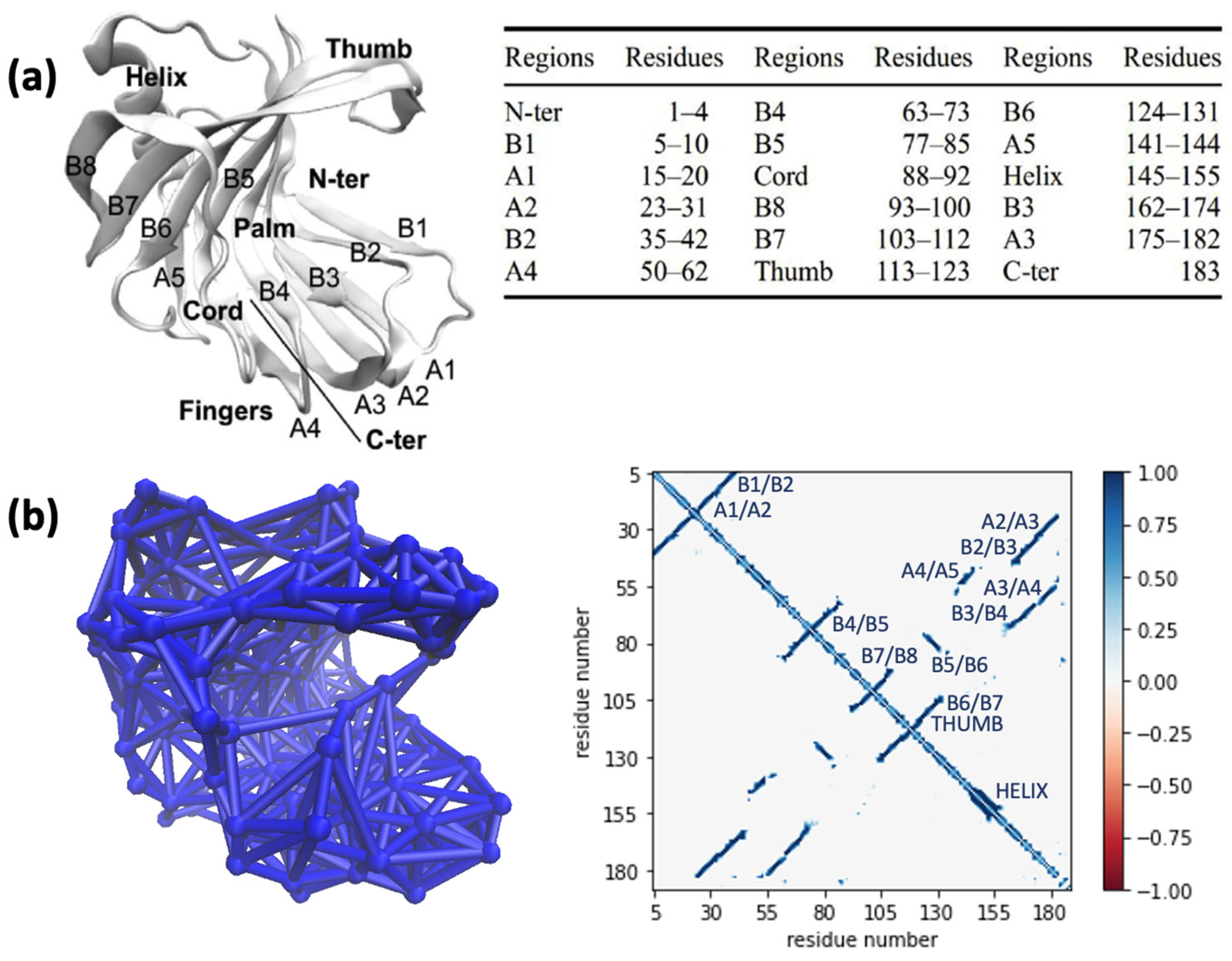
Figure 2a displayed the 3D structural representation of a GH11 xylanase. As the shape of a GH11 xylanase resembled a right hand that grabbed the substrate, local regions were labelled as ‘Palm’ and ‘Thumb’. The Palm region was a large beta sheet folded as a cleft for substrate binding and contained catalytic residues Glu78 and Glu171. Beta strands within the Palm region were labelled by BX, where B1 was located near the N-terminus or the non-reducing end. The Thumb region represented a long loop of two connecting beta strands, which could either open or close the binding cleft for substrate binding. Meanwhile, another beta sheet or the Fingers region was folded underneath the Palm region and formed a hydrophobic core to help stabilizing the Palm region. The beta strands comprised the Fingers region were labelled as AX. Finally, the enzyme structure was further stabilized by an alpha helix connecting the beta strand A5 of the Fingers region and the beta strand B3 of the Palm region and the ‘Cord’ that further connect the Palm and the Fingers regions, which reduced the conformational entropy of the structure.
The 3D network representation of the wildtype xyn11A shown in Figure 2b was created from the information within the time-averaged adjacency matrix A ^ , whose elements represented the probability of amino acid node pairwise connections, defined by the 7.4 Å cutoff distance between C-alpha atoms of the protein and glycosidic oxygen atoms for the xylohexaose substrate. From the 3D visual inspection, the alpha helix and beta sheets could form dense subnetworks. For example, two connected beta strands within the Thumb region could form a subnetwork through a hydrogen bond between backbone atoms from the residue (i, j), and the hydrogen bond could bring (i, j − 1) and (i, j + 1) pairs into proximity. Therefore, several non-zero cross diagonal elements could be seen from the time-averaged adjacency matrix A ^ .
The first kind of off-diagonal elements were the element A ^ i , i 1 and A ^ i , i + 1 nearest to the diagonal elements, which represented the nearest neighbors along the polypeptide chain connected through the peptide bonds. Local folding of some regions could be interpreted through the A ^ i , i ±   m elements where m > 1, where an alpha helix could be seen in the mapping of adjacency matrix with up to nonzero A ^ i , i ±   4 elements. Further protein folding that determine the tertiary structure of the enzyme was depicted by several far off-diagonal regions within the adjacency matrix, corresponding to the hydrogen bonding between beta strands within beta sheets of both Palm and Fingers regions. Connections between beta strands were mostly classified as ‘antiparallel’ with non-zero probability found between nodes i + n and j − n. Only the B5/B6 pairs of beta strands was classified as ‘parallel’ with non-zero probability found between nodes i + n and j + n.

2.2. Characterization of Structural Network of Wildtype Xyn11A through the Communicability Matrix

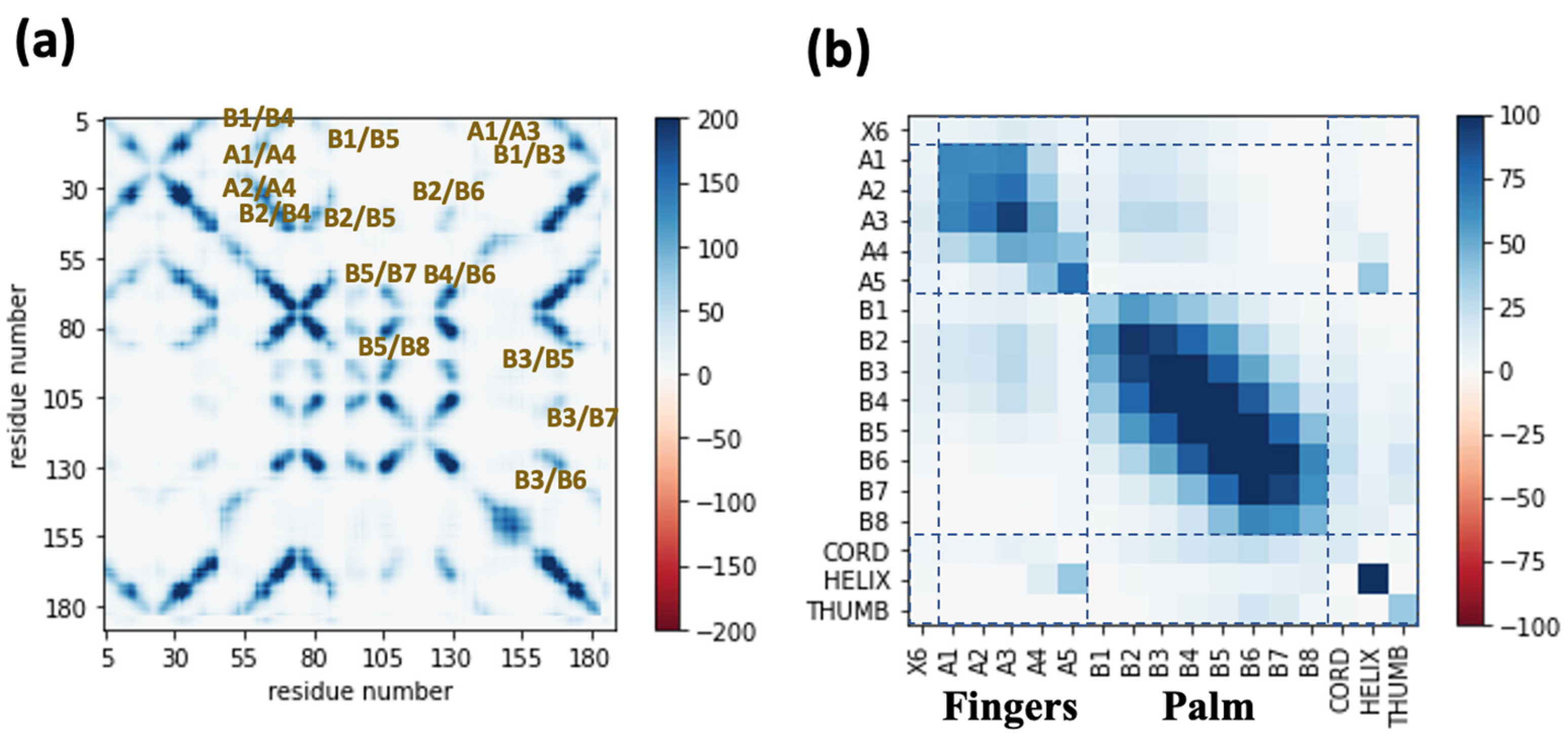
Figure 3a displayed the time-averaged communicability matrix G ^ calculated from the last 50 ns of MD trajectory of the wildtype xyn11A form Bacillus firmus K-1. The communicability G ^ i j between the C-alpha atoms of amino acid residues i and j referred to sum of possible paths of any length from a node i to another node j divided by the permutation of signal along the path length as described in Equation (2). Unlike the adjacency matrix, non-zero elements within the G ^ matrix contained the communicability between the non-adjacent amino acids, e.g., B1/B3, B1/B4, …, B2/B4, B2/B5, … pairs shown in Figure 3a. For the case of the jellyroll folded GH11 structure, the communicability matrix demonstrated the ability of amino acids or protein regions located at further sites to communicate mechanical signals through the network of large beta sheets.
To better illustrate the communicability between far beta strands within the beta sheets of a GH11 structure, the residue-wise communicability matrix in Figure 3a was mapped into the region-wise communicability matrix in Figure 3b by averaging the communicability between all residues in the first region and all residues in the second region. Then, submatrices were defined to illustrate the Fingers region with AX beta strands and the Palm region with BX beta strands (see dashed lines in Figure 3b). For the Fingers region, beta strands A1 to A3 formed a network with high communicability, while the ‘flapping’ A5 strand at the reducing end of the Fingers region was relatively isolated from other beta structures with low communicability with other regions. Interestingly, the Palm region containing the larger beta sheets with eight beta strands formed a rather symmetric communicability profile. It was clearly seen that the middle beta strands, e.g., strands B4 and B5, were with non-zero communicability with all beta strands within the Palm regions.

2.3. Effects of Mutation to Binding Pocket Distortion Visualized through the Communicability Matrix

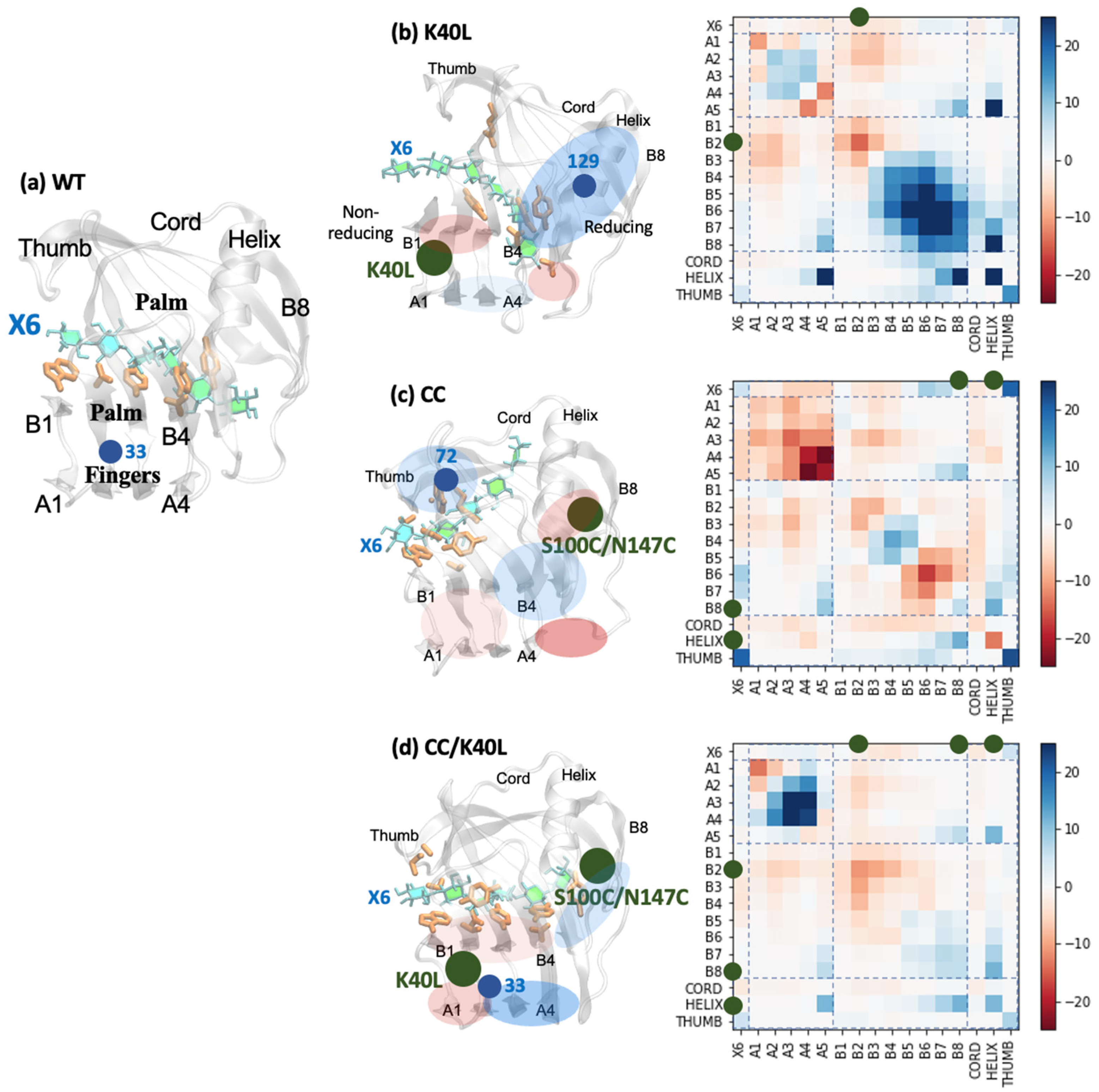
The diagonal elements of a communicability matrix were referred as the ‘subgraph centrality’ or the ‘self-communicability’, defined by the weighted sum of possible closed paths of any node. Regions along the diagonal elements with zero subgraph centrality represented the random coils without contacts from folded tertiary structures. On the other hand, a region along the diagonal elements with high value of subgraph centrality represented a beta strand within a beta sheet, surrounded by neighboring beta strands. The concept that referred subgraph centrality as the participation of the corresponding node in any subgraph or signal circuit [10] was illustrated by the maximum subgraph centrality within the wildtype xyn11A found at the residue 33, located at a short loop between A2 and B2 beta strands adjacent to one end of four other strands: A1, B1, A3, and B3 (see Figure 4a). Effects of site-directed mutations K40L and CC near the non-reducing and reducing ends of xyn11A on the subgraph centrality and communicability were further shown in Table 1 and Figure 4. The shifting of subgraph centrality maxima seen in Table 1 due to the site-directed mutation corresponded to the previously reported binding cleft distortion [6], which could either increase or decrease the density of local network at both ends of the binding pocket.
For the K40L mutant near the non-reducing end (Figure 4a), the amino acid position with maximum subgraph centrality was shifted to the residue 129 near the reducing end of the binding cleft. This shifting of subgraph centrality maxima was in concurrence with the previously reported binding pocket distortion of the K40L mutant, in which the non-reducing end became opened, but the reducing end became closed [6]. Therefore, local communicability within the B1 and B2 strands near the N-terminus and the non-reducing end of K40L was decreased from that of WT (see Figure 4b), but the communicability became significantly increased within the B4 to B8 regions near non-reducing end. Moreover, the communicability of the amino acid residues within the alpha helix, along with that between the helix and A5/B8 beta strands near the non-reducing end were highly increased and confirmed the denser network caused by binding pocket distortion. According to Table 1, the K40L mutant possessed both the highest average subgraph centrality and communicability within the protein structure. This could be explained through the strengthening of the hydrophobic clusters formed between the Fingers and the Palm regions and extended towards the alpha helix region by replacing a positively charge lysine residue by a hydrophobic leucine. The pocket distortion caused by this K40L mutation left the non-reducing end opened and resulted in the loss of substrate binding, which corresponded to the lowest communicability between the protein structure and the xylohexaose substrate.
On the other hand, for the CC mutant near the reducing end (Figure 4b), the amino acid position with maximum subgraph centrality was shifted to the residue 72 near the non-reducing end instead. This shifting of subgraph centrality maxima was in concurrence with the previously reported binding pocket distortion of the CC mutant, in which the non-reducing end became closed, but the reducing end became opened [6]. Local communicability within the A4 and A5 strands near the reducing end of CC was significantly decreased from that of WT and the ‘flapping’ A5 strand became detached from A4 (see Figure 4b). However, the communicability of the amino acid residues within the Thumb region and the xylohexaose substrate was greatly enhanced, corresponded to the closing of Thumb loop that caused a denser subnetwork around the residue 72 with maximum subgraph centrality near the non-reducing end. According to Table 1, the CC mutant possessed the lowest average subgraph centrality and communicability within the protein structure. This could be explained through the disruption of the hydrophobic clusters between the Fingers and the Palm regions by the newly formed disulfide bonds that pull the loop containing the A5 strand from the Fingers region and caused a significant loss of communicability. However, the pocket distortion caused by this CC mutation connected the upper parts of B3 to B5 strands, the Thumb loop, and the substrate, which corresponded to the highest communicability between the protein structure and the xylohexaose substrate.
Interestingly, for the CC/K40L mutant where site-directed mutations were performed both at reducing and non-reducing ends (Figure 4c), the maximum subgraph centrality was shifted back to the residue 33 with relatively small value of maximum subgraph centrality. However, the CC/K40L mutant possessed higher average subgraph centrality than the wildtype, along with the lower standard deviation, which indicated a higher uniformity of the network. From the communicability changes within the Palm region, CC/K40L mutation caused a slight distortion with an opened non-reducing end. However, communicability between beta strands at the center of the Fingers region was significantly improved. According to Table 1, average subgraph centrality within the catalytic residues and the whole protein structure was improved, despite of a slight loss of communicability between the protein structure and the xylohexaose substrate for the CC/K40L mutant. This network topology analysis confirmed that the CC and K40L site-directed mutations performed near both ends of the binding pocket could cancel the binding pocket distortion effects caused by each other, so that the enzyme could preserve the function of the wildtype while the thermostability was enhanced through both hydrophobic and disulfide bond addition.

3. Materials and Methods

Sample atomistic MD trajectory files of the protein/xylohexaose (X6) complexes consisting of the recombinant xyn11A from Bacillus firmus K-1 and its mutants shown in Figure 1 were available at https://github.com/jamesreddeviltna/Xylanase-Network-Analysis (accessed on 25 August 2022). The trajectory data was generated by explicitly solvated MD simulations in an NPT ensemble at 300 K reference temperature and 1 atm reference pressure. The full detail of MD simulation protocol could be found from our previous work [17].
All resulting trajectory files were then processed through an in-house python script also available at https://github.com/jamesreddeviltna/Xylanase-Network-Analysis (accessed on 25 August 2022), utilizing the commands from ‘MDAnalysis1.5’ library [22] to extract the coordinates of C-alpha atoms of proteins and oxygen atoms of the beta (1, 4) glycosidic bonds connecting xylose rings for all timesteps. Those selected atoms were defined as ‘nodes’ for the residue interaction network (RIN). Then, the time-dependent formation of edges within the RINs of all simulated trajectories was encoded within a 3D array A ^ ( t ) that contained a 2D adjacency matrix for each timestep. An element A i j , k of the adjacency matrix A ^ k of the timestep k that represented the edge between residue pairs ( i , j ) was defined through the Euclidian distance r i j , k as
A i j , k = H ( r 0 r i j , k ) = { 1 ,     r i j , k < r 0     0 ,     r i j , k r 0  
where the cutoff distance r 0 was set as 7.4 Å, which was the distance between pairs of C-alpha atoms of the greatest pair correlation, excluding the pairs covalently bonded. Then, the time average of A ^ k or A ^ ( t ) A ^ was calculated over the last 50 ns of each MD trajectory where the system conformation was equilibrated and the element A ^ i j ( t ) A ^ i j represented the probability of edge formation between nodes i and j. An adjacency defined by Equation (1) was visualized through a 2D colormap by matplotlib and a 3D network by the NetworkView [23] plugin embedded within the VMD 1.9.4 software [24].
Both the time-dependent and time-averaged adjacency matrices provided the information about the local connectivity between nodes of adjacent amino acid residues while were unable to describe the transmission of mechanical signals between far residues. Therefore, the secondary category of measurement for mechanical signal transmission between protein residues was defined in a previous study [25] through the communicability matrix G ^ k for each timestep k, by
G ^ k = exp ( A ^ k ) = n = 0 A ^ k n n ! = I ^ + A ^ k + A ^ k 2 2 ! + A ^ k 3 3 ! +
Each element G i j , k of G ^ k represented the communicability between nodes i and j. From Taylor series expansion of the exponential of adjacency matrix in Equation (2), the n t h term represented the number of possible walks from a node i to another node j that required n steps and was weighted by the inverse of n ! . Similar to adjacency matrices, the time average G ^ ( t ) G ^ of communicability matrices from all simulations were calculated from the last 50 ns of each MD trajectory and the element G ^ i j ( t ) G i j represented the time average of dynamic communicability between amino acids i and j.

4. Conclusions

In this study, network topology analysis of the adjacency and communicability between amino acid nodes was employed both to characterize the system of interaction networks for an enzyme and to visualize the effects of site-directed mutation on the simulated mutants. The analysis could depict the effects of binding pocket distortion due to the disruption of interaction network caused by the mutagenesis, which could be crucial to the enzyme function. Therefore, the prediction of binding pocket distortion that combined atomistic simulations with network topology analysis could serve as an additional tool or protocol for protein engineering, as proper mutation design could minimize or correct the pocket distortion effects and preserve the protein function, while enhancing the thermostability.

Author Contributions

Conceptualization, T.S. and P.K.; methodology, T.S.; software, N.S.; validation, T.S., A.M. and P.K.; formal analysis, T.S.; investigation, P.K.; resources, T.S.; writing—original draft preparation, T.S.; writing—review and editing, A.M.; visualization, N.S.; supervision, T.S. and P.K. All authors have read and agreed to the published version of the manuscript.

Funding

This research project is supported by Thailand Science Research and Innovation (TSRI) Basic Research Fund: Fiscal year 2022 under project number FRB650048/0164.

Data Availability Statement

MD trajectories and python source code for network topology analysis available at https://github.com/jamesreddeviltna/Xylanase-Network-Analysis (accessed on 25 August 2022).

Acknowledgments

The authors acknowledge the support provided by the Center of Excellence in Theoretical and Computational Science (TaCS-CoE), KMUTT. Authors also thank Jirasak Wong-Ekkabut and Saree Phongpanpanee for their conceptual discussion on the protein network topology and Harit Boonyaputtikul for providing the experimental data.

Conflicts of Interest

The authors declare no conflict of interest.

References

  1. Li, C.; Zhang, R.; Wang, J.; Wilson, L.M.; Yan, Y. Protein Engineering for Improving and Diversifying Natural Product Biosynthesis. Trends Biotechnol. 2020, 38, 729–744. [Google Scholar] [CrossRef] [PubMed]
  2. Modarres, H.P.; Mofrad, M.R.; Sanati-Nezhad, A. Protein Thermostability Engineering. RSC Adv. 2016, 6, 115252–115270. [Google Scholar] [CrossRef]
  3. Mukhopadhyay, A. Tolerance Engineering in Bacteria for the Production of Advanced Biofuels and Chemicals. Trends Microbiol. 2015, 23, 498–508. [Google Scholar] [CrossRef] [PubMed] [Green Version]
  4. Paës, G.; Berrin, J.-G.; Beaugrand, J. GH11 Xylanases: Structure/Function/Properties Relationships and Applications. Biotechnol. Adv. 2012, 30, 564–592. [Google Scholar] [CrossRef] [PubMed]
  5. Hu, J.; Arantes, V.; Saddler, J.N. The Enhancement of Enzymatic Hydrolysis of Lignocellulosic Substrates by the Addition of Accessory Enzymes Such as Xylanase: Is It an Additive or Synergistic Effect? Biotechnol. Biofuels 2011, 4, 36. [Google Scholar] [CrossRef] [PubMed] [Green Version]
  6. Bhardwaj, N.; Kumar, B.; Verma, P. A Detailed Overview of Xylanases: An Emerging Biomolecule for Current and Future Prospective. Bioresour. Bioprocess. 2019, 6, 40. [Google Scholar] [CrossRef] [Green Version]
  7. Zhang, S.; Zhao, S.; Shang, W.; Yan, Z.; Wu, X.; Li, Y.; Chen, G.; Liu, X.; Wang, L. Synergistic Mechanism of GH11 Xylanases with Different Action Modes from Aspergillus Niger An76. Biotechnol. Biofuels 2021, 14, 118. [Google Scholar] [CrossRef]
  8. Nordberg Karlsson, E.; Schmitz, E.; Linares-Pastén, J.A.; Adlercreutz, P. Endo-Xylanases as Tools for Production of Substituted Xylooligosaccharides with Prebiotic Properties. Appl. Microbiol. Biotechnol. 2018, 102, 9081–9088. [Google Scholar] [CrossRef] [Green Version]
  9. Stephens, D.E.; Rumbold, K.; Permaul, K.; Prior, B.A.; Singh, S. Directed Evolution of the Thermostable Xylanase from Thermomyces Lanuginosus. J. Biotechnol. 2007, 127, 348–354. [Google Scholar] [CrossRef]
  10. Xu, X.; Liu, M.; Huo, W.; Dai, X. Obtaining a Mutant of Bacillus Amyloliquefaciens Xylanase A with Improved Catalytic Activity by Directed Evolution. Enzyme Microb. Technol. 2016, 86, 59–66. [Google Scholar] [CrossRef]
  11. Prajapati, A.S.; Panchal, K.J.; Pawar, V.A.; Noronha, M.J.; Patel, D.H.; Subramanian, R.B. Review on Cellulase and Xylanase Engineering for Biofuel Production. Ind. Biotechnol. 2018, 14, 38–44. [Google Scholar] [CrossRef]
  12. Takita, T.; Nakatani, K.; Katano, Y.; Suzuki, M.; Kojima, K.; Saka, N.; Mikami, B.; Yatsunami, R.; Nakamura, S.; Yasukawa, K. Increase in the Thermostability of GH11 Xylanase XynJ from Bacillus Sp. Strain 41M-1 Using Site Saturation Mutagenesis. Enzyme Microb. Technol. 2019, 130, 109363. [Google Scholar] [CrossRef] [PubMed]
  13. Cheng, Y.-S.; Chen, C.-C.; Huang, C.-H.; Ko, T.-P.; Luo, W.; Huang, J.-W.; Liu, J.-R.; Guo, R.-T. Structural Analysis of a Glycoside Hydrolase Family 11 Xylanase from Neocallimastix Patriciarum. J. Biol. Chem. 2014, 289, 11020–11028. [Google Scholar] [CrossRef] [PubMed] [Green Version]
  14. Han, N.; Miao, H.; Ding, J.; Li, J.; Mu, Y.; Zhou, J.; Huang, Z. Improving the Thermostability of a Fungal GH11 Xylanase via Site-Directed Mutagenesis Guided by Sequence and Structural Analysis. Biotechnol. Biofuels 2017, 10, 133. [Google Scholar] [CrossRef] [Green Version]
  15. Wu, X.; Tian, Z.; Jiang, X.; Zhang, Q.; Wang, L. Enhancement in Catalytic Activity of Aspergillus Niger XynB by Selective Site-Directed Mutagenesis of Active Site Amino Acids. Appl. Microbiol. Biotechnol. 2018, 102, 249–260. [Google Scholar] [CrossRef]
  16. Zhang, S.; He, Y.; Yu, H.; Dong, Z. Seven N-Terminal Residues of a Thermophilic Xylanase Are Sufficient to Confer Hyperthermostability on Its Mesophilic Counterpart. PLoS ONE 2014, 9, e87632. [Google Scholar] [CrossRef]
  17. Boonyaputthikul, H.; Muhammad, A.; Roekring, S.; Rattanarojpong, T.; Khunrae, P.; Sutthibutpong, T. Synergistic Effects between the Additions of a Disulphide Bridge and an N-Terminal Hydrophobic Sidechain on the Binding Pocket Tilting and Enhanced Xyn11A Activity. Arch. Biochem. Biophys. 2019, 672, 108068. [Google Scholar] [CrossRef]
  18. Ngenyoung, A.; Muhammad, A.; Rattanarojpong, T.; Sutthibutpong, T.; Khunrae, P. Effect of N-Terminal Modification on the Mode of Action between the Xyn11A and Xylotetraose. Int. J. Biol. Macromol. 2021, 170, 240–247. [Google Scholar] [CrossRef]
  19. Grewal, R.; Roy, S. Modeling Proteins as Residue Interaction Networks. Protein Pept. Lett. 2015, 22, 923–933. [Google Scholar] [CrossRef]
  20. O’Rourke, K.F.; Gorman, S.D.; Boehr, D.D. Biophysical and Computational Methods to Analyze Amino Acid Interaction Networks in Proteins. Comput. Struct. Biotechnol. J. 2016, 14, 245–251. [Google Scholar] [CrossRef]
  21. Stetz, G.; Verkhivker, G.M. Computational Analysis of Residue Interaction Networks and Coevolutionary Relationships in the Hsp70 Chaperones: A Community-Hopping Model of Allosteric Regulation and Communication. PLoS Comput. Biol. 2017, 13, e1005299. [Google Scholar] [CrossRef] [PubMed] [Green Version]
  22. Michaud-Agrawal, N.; Denning, E.J.; Woolf, T.B.; Beckstein, O. MDAnalysis: A Toolkit for the Analysis of Molecular Dynamics Simulations. J. Comput. Chem. 2011, 32, 2319–2327. [Google Scholar] [CrossRef] [PubMed] [Green Version]
  23. Eargle, J.; Luthey-Schulten, Z. NetworkView: 3D Display and Analysis of Protein{middle Dot}RNA Interaction Networks. Bioinformatics 2012, 28, 3000–3001. [Google Scholar] [CrossRef] [PubMed] [Green Version]
  24. Humphrey, W.; Dalke, A.; Schulten, K. VMD: Visual Molecular Dynamics. J. Mol. Graph. 1996, 14, 33–38. [Google Scholar] [CrossRef]
  25. Estrada, E. Topological Analysis of SARS CoV-2 Main Protease. Chaos Interdiscip. J. Nonlinear Sci. 2020, 30, 061102. [Google Scholar] [CrossRef]
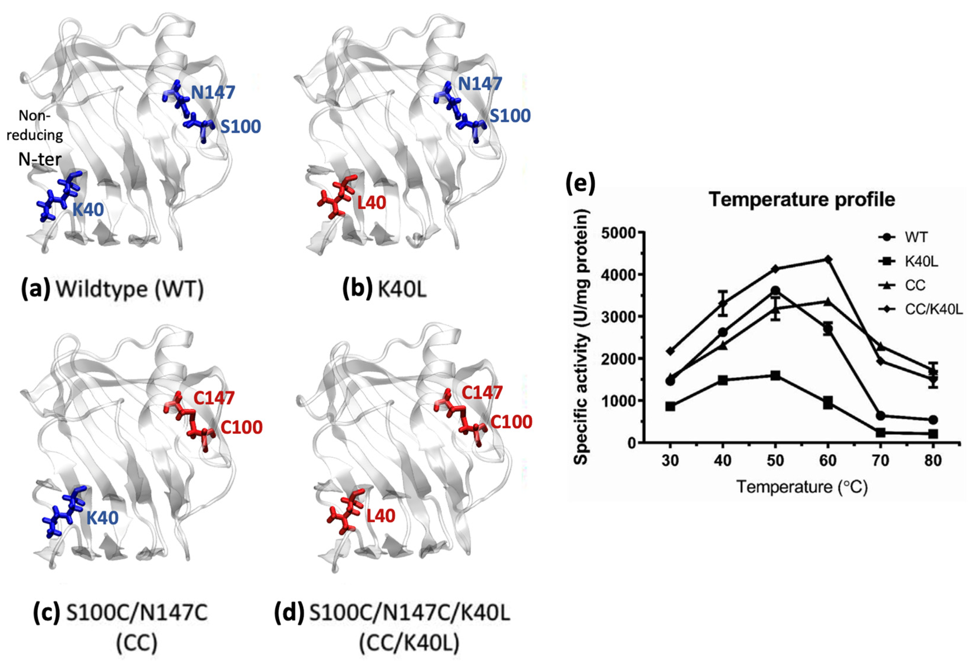
Figure 1. Three-dimensional structures of the recombinant xyn11A from Bacillus firmus K-1 and its mutants designed by Boonyaputtikul et al. [17] (a) wildtype (WT), (b) K40L, (c) S100C/N147C or CC, and (d) S100C/N147C/K40L (CC/K40L). Unmutated amino acid residues at positions 40, 100, and 147 were shown in blue, while the mutated residues were shown in red. (e) Temperature profiles of specific activity of enzymes in (ad) measured by the Nelson-Somogyi method.
Catalysts 12 01165 g001
Figure 2. (a) The map of local segments with defined secondary structures according to the residue numbers. Beta strands within the Finger regions were named by AX, while those within the Palm regions were named by BX. (b) (Left)—a residue interaction network (RIN) representation of the wildtype xyn11A in this study, and (Right)—adjacency matrix defining edges of the RIN by the 7.4 Å distance cutoff between pairs of C-alpha atoms. Regions of non-zero off diagonal elements were labelled as the connection between beta strands within the beta sheet and the ‘Thumb’ regions.
Catalysts 12 01165 g002
Figure 3. (a) Communicability matrix of the WT-X6 complex showing the non-adjacent regions, and (b) region-wise communicability of the WT-X6 complex displaying the communicability between pairs of local segments with defined secondary structure and the X6 substrate.
Catalysts 12 01165 g003
Figure 4. Final structures of the xyn11A mutants-xylohexaose (X6) complexes of (a) Wildtype (WT), (b) K40L, (c) S100C/N147C or CC, and (d) S100C/N147C/K40L (CC/K40L) after 100 ns MD simulations. Sidechains of X6 binding residues were shown in orange. Regions with increased communicability relative to the wildtype were highlighted in blue, while regions with decreased communicability were shown in red. Mutated residues were highlighted in green and the residues with maximum subgraph centrality were highlighted in dark blue. (Right) the region-wise matrix of communicability changes relative to the wildtype complex was also provided for each mutant.
Catalysts 12 01165 g004
Table 1. Parameters calculated from communicability matrices of four simulations.
WTK40LCCCC/K40L
max. subgraph centrality 458.59399.14419.09366.75
residue with max. subgraph centrality 331297233
region with max. subgraph centrality FingersreducingnonreducingFingers
average subgraph centrality [protein]128.90139.97122.81133.11
average subgraph centrality [catalytic]177.30184.02213.05186.32
communicability [enzyme-enzyme]22.6624.9921.2523.15
communicability [enzyme-X6]5.995.355.885.86
Publisher’s Note: MDPI stays neutral with regard to jurisdictional claims in published maps and institutional affiliations.