Article

Regioselective Hydroxylation of Oleanolic Acid Catalyzed by Human CYP3A4 to Produce Hederagenenin, a Chiral Metabolite

1
School of Biological Sciences and Technology, Chonnam National University, 77 Yongbongro, Gwangju 61186, Korea
2
Namhae Garlic Research Institute, 2465-8 Namhaedaero, Gyeongsangnamdo 52430, Korea
3
Gwangju Center, Korea Basic Science Institute, Gwangju 61186, Korea
*
Author to whom correspondence should be addressed.
Catalysts 2021, 11(2), 267; https://doi.org/10.3390/catal11020267
Submission received: 28 January 2021 / Revised: 9 February 2021 / Accepted: 12 February 2021 / Published: 17 February 2021
(This article belongs to the Special Issue Recent Advances in Biocatalysis and Metabolic Engineering)

Abstract

:
Oleanolic acid (OA) is a pentacyclic triterpenoid widely found in plants and foods as an aglycone of triterpenoid saponins or as a free acid. OA exhibits beneficial activities for humans, including antitumor, antivirus, and hepatoprotection properties without apparent toxicity. The metabolites produced by the cytochrome P450 (P450) enzymes are critical for the evaluation of the efficacy and safety of drugs. In this study, the potential metabolites of OA were investigated by P450-catalyzed oxidation reactions. Among the various tested human P450s, only human CYP3A4 was active for the hydroxylation of OA. The major metabolite was characterized by a set of analyses using HPLC, LC–MS, and NMR. It was found to be 4-epi-hederagenenin, a chiral product, by regioselective hydroxylation of the methyl group at the C-23 position. These results indicated that CYP3A4 can hydroxylate an OA substrate to make 4-epi-hederagenenin. Possible drug–food interactions are discussed.

1. Introduction

The use of natural products to treat chronic conditions such as hepatitis, liver disorder, or cancer has attracted the attention of researchers [1]. Native triterpenoids are of interest due to their availability and multiple biological activities, including antibacterial, anti-inflammatory, antitumor, and hepatoprotection properties. However, triterpenoids are highly hydrophobic, which significantly limits their uses as effective therapeutic agents. One of the most common ways to increase the bioavailability of these triterpenoid compounds is a hydroxylation reaction to add a hydrophilic hydroxyl group to carbon [2].
Oleanolic acid (OA) is a pentacylic triterpenoid (Figure 1). It is part of a group of natural compounds with diverse structures, including steroids, sterols, and triterpenoid saponins [3]. OA has been found in nearly 2000 types of plants [4]. In addition to its ability to exist as a free acid, OA is also used as a precursor to triterpenoid saponins [5]. It exhibits both pro- and anti-inflammatory properties based on the dose and chemical structure. OA has several beneficial effects, including hepatoprotective, antiviral, and antitumor properties [6,7,8,9,10]. Currently, OA is considered as an important ingredient in alternative therapy in the research, management, and treatment of chronic diseases [11].
Hederagenin is also a pentacyclic triterpenoid (Figure 1). It is found in the Hedera helix and exists as sapogenins or saponins in many plants. Hederacoside C and alpha-hederin are the most popular glycosides of hederagenin [12]. Hederagenin has several beneficial effects, including antiapoptotic, anti-inflammatory, anticoagulation, antiatherosclerosis, and antidiabetic activities [13].
Cytochrome P450 (CYP or P450) proteins are a large enzyme family found almost everywhere, including bacteria, plants, animals, and humans [14,15]. P450s play important roles in the metabolism of drugs, carcinogens, and steroids [16]. It has been reported that P450 (moxA) from Nonomuraea recticatena, coexpressed with camAB for pseudomonad redox partners in Escherichia coli, hydroxylated OA to produce queretaroic acid, which is a monohydroxylated OA at the C-30 position [17]. Of the human P450 enzymes, CYP3A4 is known as one of the most active in terms of the metabolism of xenobiotics in the human body.
The purpose of this work was to find an enzymatic strategy for an efficient synthesis of hydroxylated OA derivatives with a high potential of biological activity from OA, an inexpensive substrate, using P450. We found that OA is regioselectively hydroxylated by CYP3A4 to produce hederagenin (Figure 1).

2. Results and Discussion

2.1. OA Metabolism by Human Liver Microsomes and Identification of the Major Metabolite

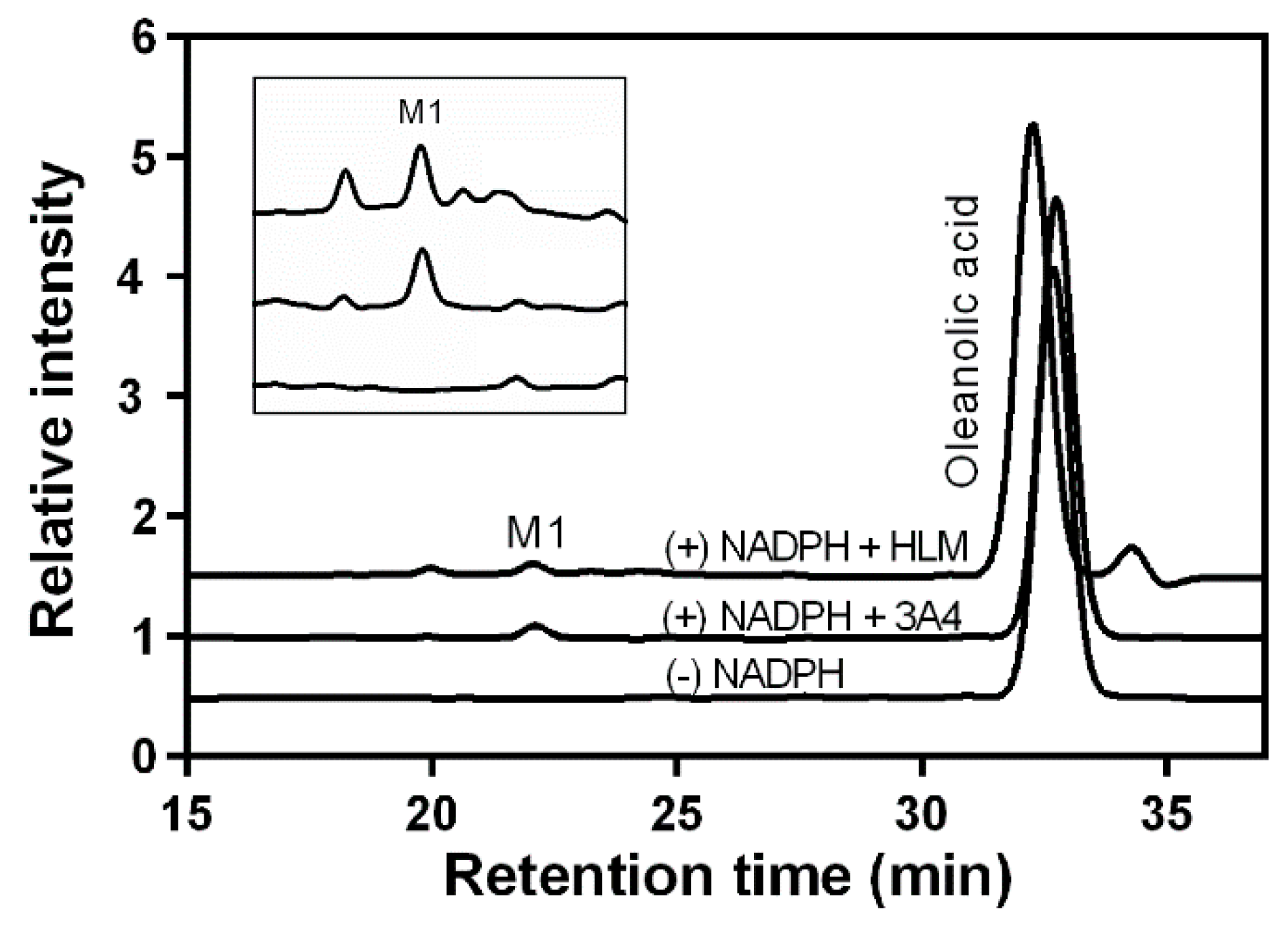
The initial investigations to determine the catalytic activity of P450 enzymes in OA hydroxylation were performed using high-performance liquid chromatography (HPLC). First, to verify the ability of human liver microsomes (HLMs) to hydroxylate OA, the reaction was performed with 500 µM of OA and HLMs at 37 °C for 60 min. Figure 2 shows the HPLC results of OA and its metabolites by HLMs. The retention time of OA was 32.3 min, and a major metabolite of HLMs in the presence of β-nicotinamide adenine dinucleotide phosphate (reduced form) (NADPH) showed a retention time of 22.4 min. When the OA metabolites were formed with the CYP3A4 enzyme in the mixture in the presence of NADPH, the same peak of the metabolite was observed.
Next, LC–MS was used to investigate the molecular weight of the major metabolite generated by CYP3A4. A full-scan chromatogram of the mixture reaction at A210 (Figure 3A) and the total ion current (TIC) profile (Figure 3B) are shown. The metabolite (M1) and substrate peaks were determined at 49 min and 72 min, respectively. The protonated molecular ions ([M-H]+) of OA (m/z 439) and the metabolite (M1) (m/z 455) were observed. The result confirmed the hydroxylation ability of CYP3A4 toward OA.
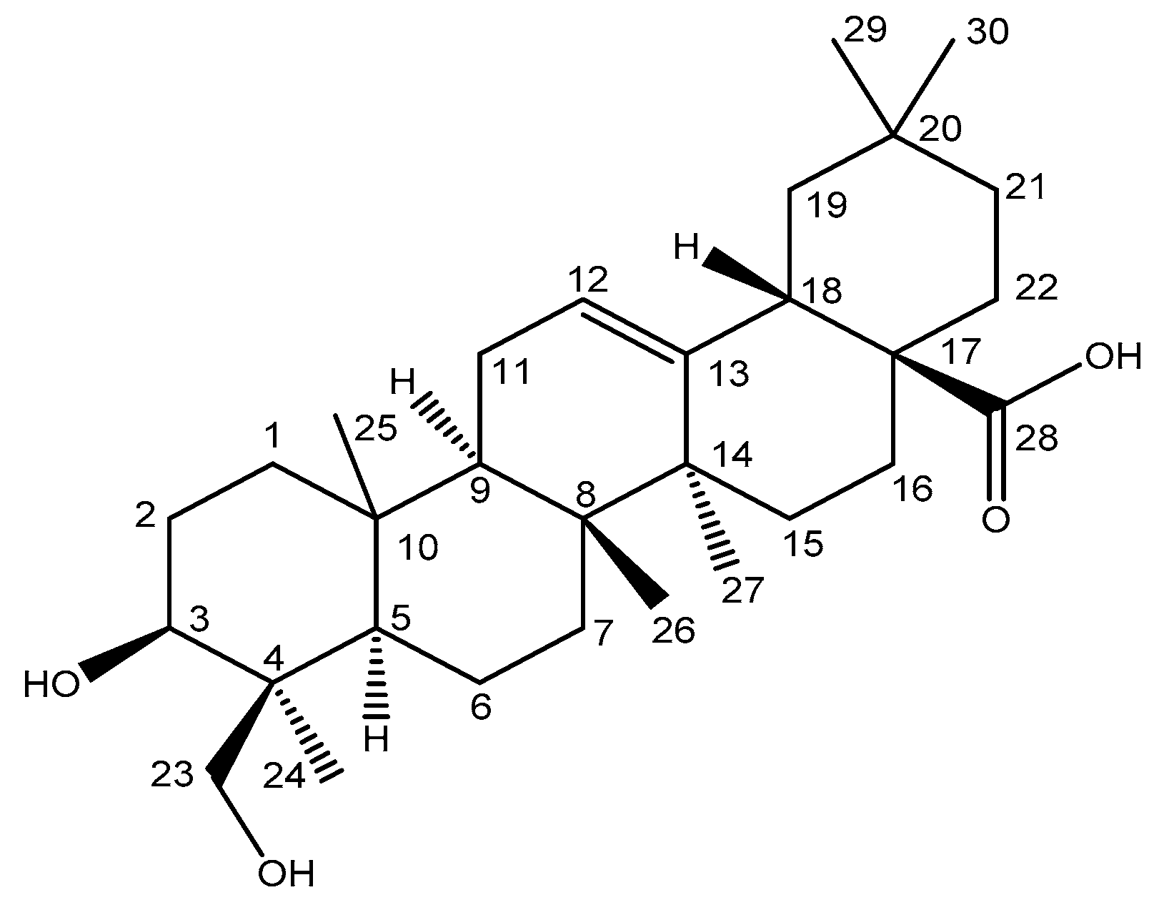
NMR experiments were performed with the major metabolite obtained from the reaction to determine the structure based on the chemical changes of the 13C NMR spectrum. The result of the chemical shifts of 13C NMR of the metabolite (M1) matched the reported NMR results of 4-epi-hederagenin (Figure 4, Table S1, Figures S1 and S2) [18,19]. The results showed the assigned 13C NMR chemical shifts of the metabolite (M1) with atomic positions (Figure 4 and Table S1).
When the pH-dependence of 4-epi-hederagenin formation by CYP3A4 was examined, the pH range of 7.4–9 was optimal with the maximal activity at pH 8 (Figure S3). All of the experiments in this study were done at pH 7.4, a neutral pH.

2.2. Kinetics Parameters and Total Turnover Numbers of OA Hydroxylation by CYP3A4 and HLMs

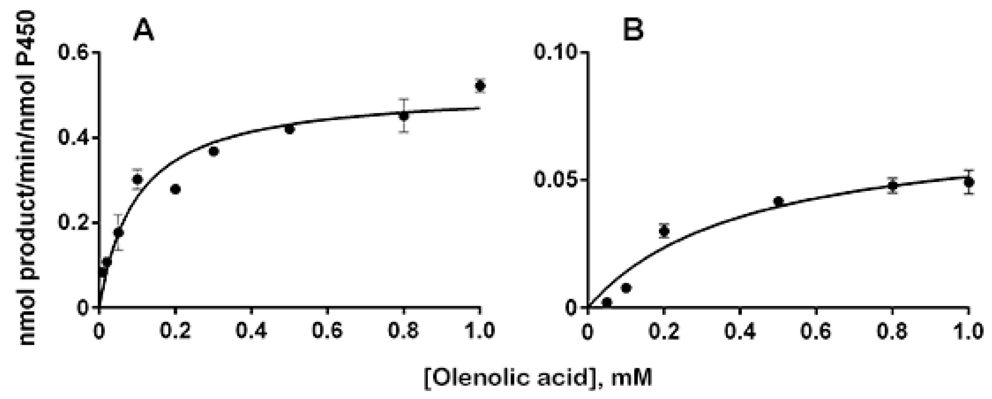
We examined several human CYPs, such as CYP1A2, CYP1B1, CYP2C9, CYP2C19, CYP2D6, CYP2E1, and CYP3A4, to find which CYPs are involved in OA hydroxylation activities. Among them, only CYP3A4 was found to have an apparent hydroxylation activity of 0.42 min−1 at 500 μM of OA (Figure 5). Other CYPs did not show any apparent activity (less than 0.05 min−1) (Figure S4).
Both CYP3A4 and HLMs were used to determine the kinetic parameters of OA hydroxylation. The steady-state kinetics of the 4-epi-hederagenin formation by HLMs and CYP3A4 are shown in Table 1 and Figure 5. HLMs exhibited kcat values of 0.072 min−1 and Km values of 0.42 mM compared to CYP3A4. CYP3A4 showed the highest kcat value of 0.51 min−1 and Km value of 98 μM. The results showed that the catalyzed efficiency of CYP3A4 is 31-fold higher than that of HLMs. These results suggested that CYP3A4 may be an important CYP in the catalytic conversion of OA to 4-epi-hederagenin.
When a time profile of the reactions was determined by prolonging the incubation time to 240 min, the formation of 4-epi-hederagenin increased with the reaction time. The total turnover number (TTN, nmol product/nmol enzyme) reached 40 at 210 min into the reaction (Figure 6).

2.3. Inhibition of OA Hydroxylation Activity by Antibodies in HLMs

Immunoinhibition experiments with anti-CYP3A4 were performed to investigate the role of CYP3A4 in OA hydroxylation. The results showed that increasing the concentrations of anti-CYP3A4 inhibited OA hydroxylation (Figure 7). The immunoinhibitory effect of anti-CYP3A4 on OA hydroxylation in HLMs was fairly high, at 42 and 51% inhibition when 5 and 10 mg IgG per nmol of P450 were treated, respectively. The results indicated that CYP3A4 plays an important role in the hydroxylation of OA in HLMs. This also suggested that other CYPs in addition to CYP3A4 may be involved in OA hydroxylation.
When 1–10 mg IgG of anti-CYP2C19 per nmol of P450 was added to the reaction mixtures, the anti-CYP2C19 did not show an apparent inhibition of OA hydroxylation up to 5 mg IgG per nmol of P450. When 10 mg IgG was used, only a 9% inhibition was shown; the inhibition by the anti-CYP2C9 seemed to be nonspecific. When the preimmune IgG was treated with up to 10 mg per nmol of P450, no apparent inhibition was found.
Regarding the OA oxidation reactions, only a few results have been reported. OA could be hydroxylated at C-30 to produce queretaroic acid, a metabolite of OA produced in several plants, via bacterial P450 (moxA) from Nonomuraea recticatena [17]. Escherichia coli whole-cell and in vitro reactions were performed using the coexpression of P450 (moxA) and Pseudomonas redox partners. However, in this study, we found that human CYP3A4 can hydroxylate at C-23 to produce 4-epi-hederagenin, showing a distinct regioselectivity of the CYP3A4 toward OA. The hydroxide positions for CYP3A4 and P450 (moxA) are the farthest within the OA molecule. This result showed the diverse catalytic activities of P450 from different sources.
Hederagenin has been shown to be easily digested and absorbed. It also has many pharmacological benefits such as anti-inflammatory, antidegenerative (for nerves), and antidiabetic properties [13]. In vitro and in vivo experiments have shown that hederagenin induces apoptosis effectively in cisplatin-resistant HNC cells by targeting the Nrf2-ARE antioxidant pathway [20]. These results suggested that hederagenin, a metabolite from OA, may show new and different effects than OA. Further studies with 4-epi-hederagenin need to be done to better understand its role in humans, especially in the treatment of conditions such as cancer and inflammation. Furthermore, other metabolites of OA in addition to hederagenin and queretaroic acid should be studied to find their beneficial pharmacological effects using engineered P450s.
Human P450 plays an important role in the metabolism of drugs and other compounds. Previous studies have shown that CYP3A4 plays a role in the metabolism of many anti-inflammatory and anticancer drugs [21]. In this study, we found a catalytic ability of CYP3A4 to metabolize OA to 4-epi-hederagenin. Notably, OA was found to inhibit the CYP3A4-catalyzed midazolam 1-hydroxylation reaction by 60% at 100 μM [22]. Therefore, drug–food interactions should be considered when consuming foods or vegetables containing OA. The treatment of clinical drugs with OA will inhibit CYP3A4′s ability to function in the body or may cause significant side effects. More research is needed to study the effects of OA on other common foods and supplements that are also likely to be metabolized by CYP3A4, as it is the most common drug-metabolizing enzyme in the small intestine and liver.

3. Materials and Methods

3.1. Materials

The OA hydrate was purchased from Tokyo Chemical Industry (Tokyo, Japan). The betulinic acid (BA), β-nicotinamide adenine dinucleotide phosphate (reduced form) (NADPH), glucose-6-phosphate dehydrogenase, and glucose-6-phosphate were bought from Sigma-Aldrich (St. Louis, MO, USA). The methanol, formic acid, acetonitrile, and other solvents were purchased from Fisher Scientific (Houston, TX, USA). Other chemicals and solvents used in the research were of high-quality analytical grade and were bought from other companies with no further purification.
The HLMs were purchased from Thermo Fisher Scientific (Waltham, MA, USA). The antibodies against CYP3A4 and CYP2C19 have been studied and implemented in previous reports [23]. The CYPs of CYP1A2, CYP1B1, CYP2C9, CYP2C19, CYP2D6, CYP2E1, and CYP3A4 were expressed in Escherichia coli with a pCW vector containing human P450 cDNA and rat NADPH-P450 reductase (CPR). Human P450 cDNA and pCW vector expression was performed as described in previous studies [24].

3.2. Hydroxylation of OA by CYP3A4 and HLMs

The reaction mixtures contained 0.20 µM of CYP3A4 or 0.80 µM of HLMs in 0.25 mL of potassium phosphate buffer (100 mM, pH 7.4) with a final concentration of 0.5 mM of OA. An NADPH-generating system was used to start the reaction, with final concentrations of 10 mM of glucose-6-phosphate, 1 IU of yeast glucose-6-phosphate dehydrogenase per mL, and 0.5 mM of NADP+.
The samples were incubated for 30 min for CYP3A4 and 60 min for HLMs with or without NADPH at 37 °C, and the reaction was stopped with 0.60 mL of ice-cold ethyl acetate. The BA (50 µM) was added as an internal standard to this solution, followed by centrifugation (1000× g, 20 min). The metabolites were analyzed by HPLC and quantified via comparison to the internal standard, BA (Figures S4 and S5).
Mobile phase A was water containing 0.1% of formic acid and methanol (20:80, v/v), and mobile phase B was acetonitrile; mobile phase A/B (95:5, v/v) had a flow rate of 0.7 mL/min through a gradient pump (LC-20AD; Shimadzu, Kyoto, Japan). The UV wavelength was 210 nm [25].
To study the TTN for CYP3A4, 500 µM of OA was used. The reaction was started by the addition of an NADPH-generating system and incubated at 10, 20, 30, 60, 90, 120, 150, 180, 210, and 240 min at 37 °C. The formation rate of the OA was determined by HPLC as described above.
The kinetic parameters of the OA hydroxylation by CYP3A4 were determined by the reactions, including 10–1000 μM of OA in 100 mM of potassium phosphate buffer (pH 7.4) and 0.20 μM of enzymes. The NADPH-generating systems were added to the initial reaction, and the reaction mixtures were incubated for 60 min at 37 °C. The kinetic parameter results were analyzed using GraphPad Prism software (Graph, San Diego, CA, USA).

3.3. LC–MS Analysis of OA Metabolite

To detect OA metabolites by CYP3A4, an LC–MS analysis of the metabolites was executed to compare the fragmentation patterns and LC profiles with authentic compounds. The CYP3A4 was incubated with 500 µM of OA at 37 °C for 2 h with an NADPH-generating system, and the injection samples were prepared as described above. An aliquot (7 µL) of this solution was injected into the LC column. The LC–MS analysis was carried out in electrospray ionization (negative) mode on a Shimadzu LC–MS-2010 EV system (Shimadzu Corporation, Japan) with the LC–MS solution software. The separation was performed on a Shim-pack VP-ODS column (2.0 mm × 250 mm, Shimadzu Corporation, Japan). Mobile phase A was water containing 0.1% of phosphoric acid and methanol (20:80, v/v), and mobile phase B was methanol and acetonitrile (80:20, v/v); mobile phase A/B (77:23, v/v) was delivered at a flow rate of 0.16 mL/min. The retention times for the major metabolite and OA were 48.85 and 71.133 min, respectively. The interface and detector voltages were 4.4 kV and 1.7 kV, respectively. The nebulization gas flow was set at 1.5 L/min. The interface, curve desolvation line, and heat block temperatures were 250, 250, and 200 °C, respectively.

3.4. NMR Spectroscopy Analysis of OA Metabolite

The NMR experiments were performed at an ambient temperature on a Varian VNMRS 600 MHz NMR spectrometer equipped with a carbon-enhanced cryogenic probe. Pyridine-d5 was used as a solvent, and the chemical shifts for proton NMR spectra were measured in parts per million (ppm) relative to tetramethylsilane. All of the NMR experiments were performed with standard pulse sequences in the VNMRJ (v. 3.2) library and processed with the same software.

3.5. Immunoinhibition of Antibodies on OA Hydroxylation Activity

The experiment was performed by incubating a mixture of anti-P450 IgG (anti-CYP3A4 and anti-CYP2C19) at different concentrations at room temperature for 30 min, then adding ingredients such as OA and the NADPH required for the reaction. Controlled experiments with different concentrations of preimmune IgG have been performed as described previously [26]. The substrates and products were tested with HPLC; mobile phase A was water containing 0.1% of formic acid and methanol (20:80, v/v), and mobile phase B was acetonitrile; mobile phase A/B (95:5, v/v) had a flow rate of 0.7 mL/min through a gradient pump (LC-20AD; Shimadzu, Kyoto, Japan). The UV wavelength was 210 nm.

4. Conclusions

In this study, the potential metabolites of OA were investigated via human P450-catalyzed oxidation reactions. Among the tested human P450s, only CYP3A4 was active in the hydroxylation of OA. The major metabolite was characterized by a set of analyses using HPLC, LC–MS, and NMR and was found to be 4-epi-hederagenenin, a chiral product of regioselective hydroxylation of the methyl group at the C-23 position. These results indicate that CYP3A4 can hydroxylate an OA substrate to make 4-epi-hederagenenin. Possible drug–food interactions should be considered when consuming food or vegetables containing OA.

Supplementary Materials

The following are available online at https://www.mdpi.com/2073-4344/11/2/267/s1: Table S1. 13C NMR chemical shifts of metabolite, M1; Figure S1. 13C NMR spectra of metabolite, M1; Figure S2. Expanded regions of 13C NMR spectra of metabolite, M1. (A) 15–37 ppm. (B) 37–190 ppm; Figure S3. pH-dependence of 4-epi-hederagenin formation by CYP3A4; Figure S4. HPLC chromatograms 268 of OA and its metabolite produced by human P450s with (+NGS) and without NADPH (-NGS). Figure S5. Standard curves of internal standard (BA), and OA; and Figure S6. HPLC chromatograms of OA and its metabolite produced by CYP3A4 with and without NADPH.

Author Contributions

Conceptualization, C.-H.Y.; investigation, N.T.C., N.A.N., T.-K.L., G.S.C., and K.D.P.; writing—original draft preparation, N.T.C. and C.H.Y.; supervision, C.H.Y.; funding acquisition, C.-H.Y. All authors have read and agreed to the published version of the manuscript.

Funding

This research was funded by the Next-Generation BioGreen 21 program (SSAC, grant no.: PJ01105801), Rural Development Administration and the National Research Foundation of Korea (NRF-2018R1A4A1023882), Republic of Korea.

Conflicts of Interest

The authors declare no conflict of interest.

References

  1. Ramana, K.V.; Singhal, S.S.; Reddy, A.B. Therapeutic Potential of Natural Pharmacological Agents in the Treatment of Human Diseases. Available online: https://www.hindawi.com/journals/bmri/2014/573452/ (accessed on 30 November 2020).
  2. Luchnikova, N.A.; Grishko, V.V.; Ivshina, I.B. Biotransformation of Oleanane and Ursane Triterpenic Acids. Molecules 2020, 25, 5526. [Google Scholar] [CrossRef] [PubMed]
  3. Phillips, D.R.; Rasbery, J.M.; Bartel, B.; Matsuda, S.P. Biosynthetic Diversity in Plant Triterpene Cyclization. Curr. Opin. Plant Biol. 2006, 9, 305–314. [Google Scholar] [CrossRef]
  4. Yumpu.com A Review of Presence of Oleanolic Acid in—Natura Proda Medica. Available online: https://www.yumpu.com/en/document/read/4492852/a-review-of-presence-of-oleanolic-acid-in-natura-proda-medica (accessed on 25 November 2020).
  5. Liu, J. Pharmacology of Oleanolic Acid and Ursolic Acid. J. Ethnopharmacol. 1995, 49, 57–68. [Google Scholar] [CrossRef]
  6. Liu, J. Oleanolic Acid and Ursolic Acid: Research Perspectives. J. Ethnopharmacol. 2005, 100, 92–94. [Google Scholar] [CrossRef] [PubMed]
  7. Jesus, J.A.; Lago, J.H.G.; Laurenti, M.D.; Yamamoto, E.S.; Passero, L.F.D. Antimicrobial Activity of Oleanolic and Ursolic Acids: An Update. Evid. Based Complement. Altern. Med. ECAM 2015, 2015. [Google Scholar] [CrossRef] [PubMed]
  8. Yu, F.; Wang, Q.; Zhang, Z.; Peng, Y.; Qiu, Y.; Shi, Y.; Zheng, Y.; Xiao, S.; Wang, H.; Huang, X.; et al. Development of Oleanane-Type Triterpenes as a New Class of HCV Entry Inhibitors. J. Med. Chem. 2013, 56, 4300–4319. [Google Scholar] [CrossRef] [PubMed]
  9. Zou, L.-W.; Dou, T.-Y.; Wang, P.; Lei, W.; Weng, Z.-M.; Hou, J.; Wang, D.-D.; Fan, Y.-M.; Zhang, W.-D.; Ge, G.-B.; et al. Structure-Activity Relationships of Pentacyclic Triterpenoids as Potent and Selective Inhibitors against Human Carboxylesterase 1. Front. Pharmacol. 2017, 8, 435. [Google Scholar] [CrossRef]
  10. Villar, V.H.; Vögler, O.; Barceló, F.; Gómez-Florit, M.; Martínez-Serra, J.; Obrador-Hevia, A.; Martín-Broto, J.; Ruiz-Gutiérrez, V.; Alemany, R. Oleanolic and Maslinic Acid Sensitize Soft Tissue Sarcoma Cells to Doxorubicin by Inhibiting the Multidrug Resistance Protein MRP-1, but Not P-Glycoprotein. J. Nutr. Biochem. 2014, 25, 429–438. [Google Scholar] [CrossRef] [PubMed] [Green Version]
  11. Ayeleso, T.B.; Matumba, M.G.; Mukwevho, E. Oleanolic Acid and Its Derivatives: Biological Activities and Therapeutic Potential in Chronic Diseases. Molecules 2017, 22, 1915. [Google Scholar] [CrossRef] [Green Version]
  12. Sieben, A.; Prenner, L.; Sorkalla, T.; Wolf, A.; Jakobs, D.; Runkel, F.; Häberlein, H. α-Hederin, but Not Hederacoside C and Hederagenin from Hedera Helix, Affects the Binding Behavior, Dynamics, and Regulation of Β2-Adrenergic Receptors. Biochemistry 2009, 48, 3477–3482. [Google Scholar] [CrossRef]
  13. Zeng, J.; Huang, T.; Xue, M.; Chen, J.; Feng, L.; Du, R.; Feng, Y. Current Knowledge and Development of Hederagenin as a Promising Medicinal Agent: A Comprehensive Review. RSC Adv. 2018, 8, 24188–24202. [Google Scholar] [CrossRef] [Green Version]
  14. Hasler, J.A.; Estabrook, R.; Murray, M.; Pikuleva, I.; Waterman, M.; Capdevila, J.; Holla, V.; Helvig, C.; Falck, J.R.; Farrell, G.; et al. Human Cytochromes P450. Mol. Aspects Med. 1999, 20, 1–137. [Google Scholar] [CrossRef]
  15. Guengerich, F.P. Human Cytochrome P450 Enzymes. In Cytochrome P450: Structure, Mechanism, and Biochemistry; de Montellano, P.R.O., Ed.; Springer International Publishing: Cham, Switzerland, 2015; pp. 523–785. ISBN 978-3-319-12108-6. [Google Scholar]
  16. Nelson, D.R.; Kamataki, T.; Waxman, D.J.; Guengerich, F.P.; Estabrook, R.W.; Feyereisen, R.; Gonzalez, F.J.; Coon, M.J.; Gunsalus, I.C.; Gotoh, O.; et al. The P450 Superfamily: Update on New Sequences, Gene Mapping, Accession Numbers, Early Trivial Names of Enzymes, and Nomenclature. DNA Cell Biol. 1993, 12, 1–51. [Google Scholar] [CrossRef] [PubMed]
  17. Fujii, Y.; Hirosue, S.; Fujii, T.; Matsumoto, N.; Agematu, H.; Arisawa, A. Hydroxylation of Oleanolic Acid to Queretaroic Acid by Cytochrome P450 from Nonomuraea Recticatena. Biosci. Biotechnol. Biochem. 2006, 70, 2299–2302. [Google Scholar] [CrossRef] [Green Version]
  18. Molina-García, L.; Martínez-Expósito, R.; Fernández-de Córdova, M.L.; Llorent-Martínez, E.J. Determination of the Phenolic Profile and Antioxidant Activity of Leaves and Fruits of Spanish Quercus Coccifera. Available online: https://www.hindawi.com/journals/jchem/2018/2573270/ (accessed on 25 January 2021).
  19. Kizu, H.; Tomimori, T. Studies on the Constituents of Clematis Species. V. On the Saponins of the Root of Clematis Chinensis OSBECK (5). Chem. Pharm. Bull. 1982, 30, 3340–3346. [Google Scholar] [CrossRef] [Green Version]
  20. Kim, E.H.; Baek, S.; Shin, D.; Lee, J.; Roh, J.-L. Hederagenin Induces Apoptosis in Cisplatin-Resistant Head and Neck Cancer Cells by Inhibiting the Nrf2-ARE Antioxidant Pathway. Oxid. Med. Cell. Longev. 2017, 2017. [Google Scholar] [CrossRef] [PubMed]
  21. Kivistö, K.T.; Kroemer, H.K.; Eichelbaum, M. The Role of Human Cytochrome P450 Enzymes in the Metabolism of Anticancer Agents: Implications for Drug Interactions. Br. J. Clin. Pharmacol. 1995, 40, 523–530. [Google Scholar] [CrossRef] [PubMed]
  22. Kim, K.-A.; Lee, J.-S.; Park, H.-J.; Kim, J.-W.; Kim, C.-J.; Shim, I.-S.; Kim, N.-J.; Han, S.-M.; Lim, S. Inhibition of Cytochrome P450 Activities by Oleanolic Acid and Ursolic Acid in Human Liver Microsomes. Life Sci. 2004, 74, 2769–2779. [Google Scholar] [CrossRef] [PubMed]
  23. Yun, C.-H.; Ahn, T.; Guengerich, F.P.; Yamazaki, H.; Shimada, T. Phospholipase D Activity of Cytochrome P450 in Human Liver Endoplasmic Reticulum. Arch. Biochem. Biophys. 1999, 367, 81–88. [Google Scholar] [CrossRef]
  24. Yun, C.-H.; Yim, S.-K.; Kim, D.-H.; Ahn, T. Functional Expression of Human Cytochrome P450 Enzymes in Escherichia Coli. Curr. Drug Metab. 2006, 7, 411–429. [Google Scholar] [CrossRef] [PubMed]
  25. Le, T.-K.; Jang, H.-H.; Nguyen, H.T.H.; Doan, T.T.M.; Lee, G.-Y.; Park, K.D.; Ahn, T.; Joung, Y.H.; Kang, H.-S.; Yun, C.-H. Highly Regioselective Hydroxylation of Polydatin, a Resveratrol Glucoside, for One-Step Synthesis of Astringin, a Piceatannol Glucoside, by P450 BM3. Enzyme Microb. Technol. 2017, 97, 34–42. [Google Scholar] [CrossRef] [PubMed]
  26. Yun, C.-H.; Shimada, T.; Guengerich, F.P. Roles of Human Liver Cytochrome P4502C and 3A Enzymes in the 3-Hydroxylation of Benzo(a)Pyrene. Cancer Res. 1992, 52, 1868–1874. [Google Scholar] [PubMed]
Figure 1. Chemical structures of oleanolic acid (OA) and 4-epi-hederagenin. CYP3A4 catalyzes the regioselective hydroxylation of OA to produce 4-epi-hederagenin in the presence of ß-nicotinaminde adenine dinucleotide phosphate (NADPH). The location of the alteration marked with a star is a chiral carbon generated by cytochrome P450 (CYP)-catalyzed hydroxylation.
Catalysts 11 00267 g001
Figure 2. HPLC chromatograms of OA and its metabolites produced by human liver microsomes (HLMs). HPLC analyses of oxidation of OA by HLMs and CYP3A4. Peaks were identified by comparing the retention times of metabolite (M1): 4-epi-hederagenin (tR = 22.4 min) and substrate OA (tR = 32.3 min).
Catalysts 11 00267 g002
Figure 3. LC–MS spectra of a major metabolite of OA produced by CYP3A4. The LC–MS chromatogram of OA and its major metabolite (A) was observed at A210, and the total ion current (TIC) profile of the metabolites produced by CYP3A4 (B) is shown. The MS spectra showing the protonated molecular ions of OA (C) and its major metabolite (D) were 439 and 455, respectively.
Catalysts 11 00267 g003
Figure 4. Chemical structure of the metabolite (M1) from OA. The OH group at position C-23 shows the structure of the metabolite as 4-epi-hederagenin.
Catalysts 11 00267 g004
Figure 5. Steady-state kinetics of OA hydroxylation by CYP3A4 (A) and HLMs (B). The kinetic parameters of HLMs and CYP3A4 were estimated in a potassium phosphate buffer (100 mM, pH 7.4) of 0.25 mL final mixture volume, including an NADPH regeneration system, OA (20–1000 µM), and 0.2 µM of CYP3A4 or 0.8 µM of HLMs. The mixtures were incubated at 37 °C for 30 min for CYP3A4 and 60 min for HLMs.
Catalysts 11 00267 g005
Figure 6. Total turnover numbers (TTNs) for OA hydroxylation by CYP3A4. The TTNs by CYP3A4 were measured by HPLC after the reaction at 37 °C during the indicated time of 10–240 min. The reaction mixture included 0.2 µM of CYP3A4, an NADPH regeneration system, and 0.5 mM of substrate in final volume of 0.25 mL in potassium phosphate buffer (100 mM, pH 7.4).
Catalysts 11 00267 g006
Figure 7. Immunoinhibition of antibodies against CYP3A4 on OA hydroxylation activity in HLMs. The effects of anti-CYP3A4 (●) and anti-CYP2C19 IgG (○) on OA hydroxylation in HLMs were examined. Values represent the mean ± SD of the three determinations.
Catalysts 11 00267 g007
Table 1. Kinetic parameters of OA hydroxylation by CYP3A4.
Enzymeskcat (min−1)Km (μM)kcat/Km (min−1μM−1)
HLMs0.072 ± 0.010420 ± 1400.00017 ± 0.00006
CYP3A40.51 ± 0.0698 ± 190.0052 ± 0.0012
Publisher’s Note: MDPI stays neutral with regard to jurisdictional claims in published maps and institutional affiliations.