Review

Epstein-Barr Virus-Encoded RNAs: Key Molecules in Viral Pathogenesis

Institute for Genetic Medicine, Hokkaido University, N15 W7 Kita-Ku, Sapporo 060-0815, Japan
Cancers 2014, 6(3), 1615-1630; https://doi.org/10.3390/cancers6031615
Submission received: 20 June 2014 / Revised: 18 July 2014 / Accepted: 21 July 2014 / Published: 6 August 2014
(This article belongs to the Special Issue DNA Viruses in Human Cancer)

Abstract

:
The Epstein-Barr virus (EBV) is known as an oncogenic herpesvirus that has been implicated in the pathogenesis of various malignancies. EBV-encoded RNAs (EBERs) are non-coding RNAs expressed abundantly in latently EBV-infected cells. Herein, I summarize the current understanding of the functions of EBERs, including the interactions with cellular factors through which EBERs contribute to EBV-mediated pathogenesis. Previous studies have demonstrated that EBERs are responsible for malignant phenotypes in lymphoid cells, and can induce several cytokines that can promote the growth of various EBV-infected cancer cells. EBERs were also found to bind retinoic acid-inducible gene I (RIG-I) and thus activate its downstream signaling. Furthermore, EBERs induce interleukin-10, an autocrine growth factor for Burkitt’s lymphoma cells, by activating RIG-I/interferon regulatory factor 3 pathway, suggesting that EBER-mediated innate immune signaling modulation contributes to EBV-mediated oncogenesis. Recently, EBV-infected cells were reported to secret EBERs, which were then recognized by toll-like receptor 3 (TLR3), leading to the induction of type I interferon and inflammatory cytokines, and subsequent immune activation. Furthermore, EBER1 was detected in the sera of patients with active EBV-infectious diseases, suggesting that EBER1-meidated TLR3 signaling activation could account for the pathogenesis of active EBV-infectious diseases.

1. Introduction

Epstein-Barr virus (EBV), a ubiquitous human herpes virus that has infected more than 90% of the global populations, is associated with a variety of malignancies, including endemic Burkitt’s lymphoma (BL), Hodgkin’s lymphoma, nasopharyngeal carcinoma (NPC), gastric carcinoma (GC), and opportunistic lymphoma. These “EBV-associated” cancers are characterized by the proliferation of monoclonal EBV-infected cells with restricted latent viral gene expression [1].
EBV encodes small nonpolyadenylated, non-coding (nc) RNAs called EBV-encoded RNA (EBER) [1,2]; these are the most abundant viral transcripts in latently EBV-infected cells [3]. EBER1 and EBER2, which are respectively 167 and 172 nucleotides long, are transcribed by RNA polymerase III (pol III) [4]. EBERs can be used as target molecules for the in situ hybridization (ISH) detection of EBV-infected cells in tissues [5] and are considered a reliable marker of the presence of EBV, as they are abundantly expressed in latently EBV-infected cells. Previous studies have demonstrated the roles of EBERs in EBV oncogenicity. Expression of EBER in B lymphocytes induces colony formation in soft agar and tumorigenesis in nude mice [6,7]. In addition, EBERs confer resistance to RNA-dependent protein kinase (PKR)-induced apoptosis upon BL cells [8]. Previous studies have also demonstrated that EBERs induce the transcription of various cytokines depending on cell type, such as interleukin-10 (IL-10) in BL cells, insulin-like growth factor-1 (IGF1) in epithelial cells and IL-9 in T cells; these cytokines subsequently act as autocrine growth factors for the EBV-infected cancer cells [9,10,11,12]. More recent studies have shown that EBER1 is sufficient to elicit these phenotypes [13,14]. Regarding the molecular action mechanisms of EBREs, these ncRNAs were reported to contribute to the EBV infection pathogenesis by modulating of innate immune signals [15,16,17].

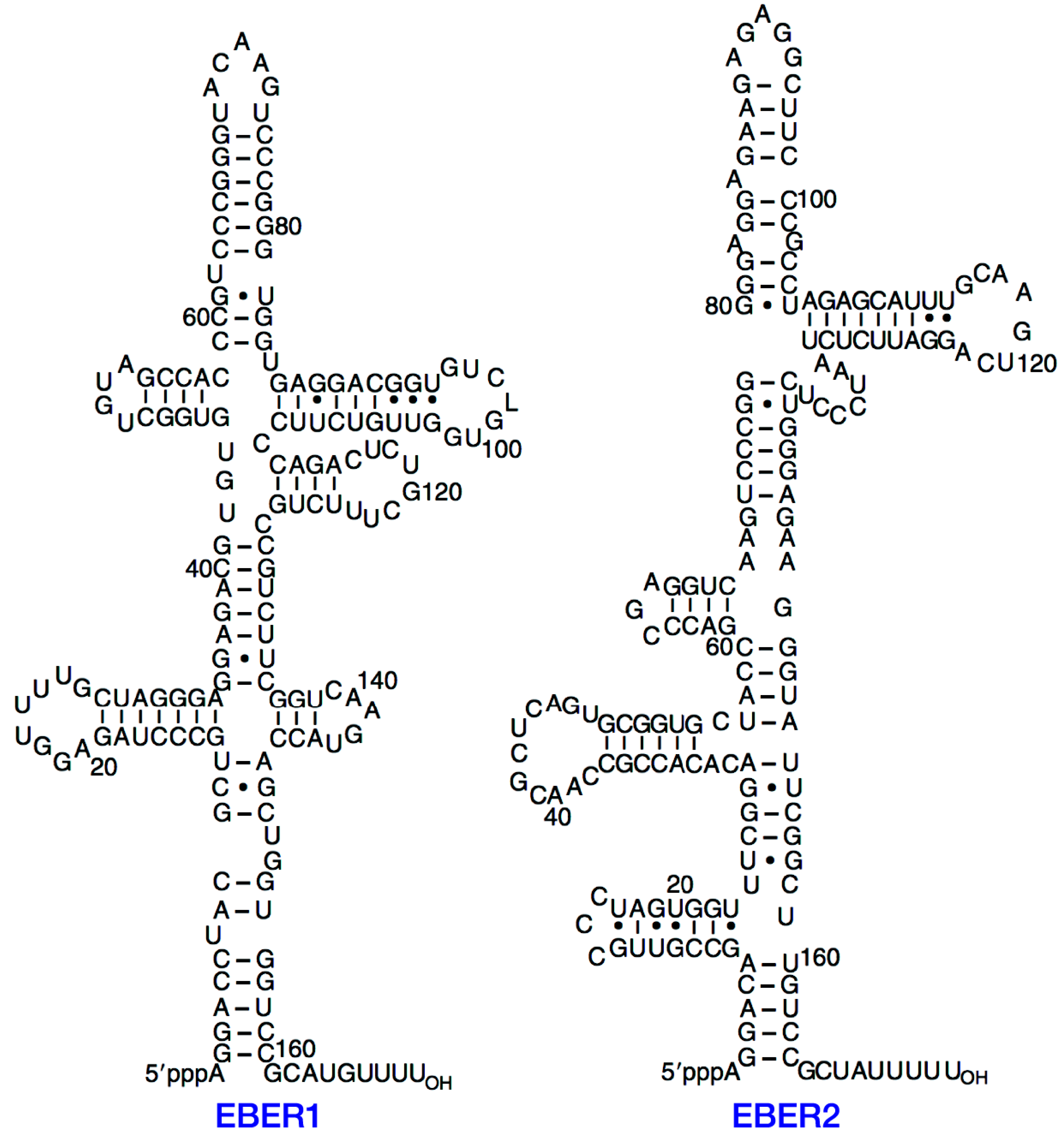
2. Structure of EBERs

EBER1 and EBER2 are 166 and 172 nucleotides long, respectively [4], and only 54% sequence homology exists between these two RNAs. The EBER genes are separated by 161 base pairs and are transcribed from left to right on the EBV map. Both EBER genes carry intragenic transcription control regions for RNA polymerase (pol) III, and can be transcribed by this molecule. Despite the low (54%) primary sequence similarity, both EBER1 and EBER2 exhibit striking similarities in their secondary structures and feature extensively base-paired structures with several short stem loops (Figure 1, [4]). The secondary structure of EBERs is similar to that of adenovirus-associated RNAs (VAs), which are also small non-polyadenylated RNAs. In addition, the function of VA1 can be partially replaced by EBERs [4]. It has also been reported the similarity between the primary RNA structure of EBER1 and herpesvirus papio type 1 [18]. The differences in the secondary structures suggest that EBER1 and EBER2 have potentially distinct functions, although secondary structure conservation seems to be crucial for the functions of EBERs. The replacement of guanine residues with inosine in EBER1 was reported to disrupt the secondary structure of EBER1, leading to lost ability to form complexes with PKR [19].
The primary sequences of EBERs are highly conserved among a number of EBV strains, with only two reported changes within EBER2 and none in EBER1. A total of eight changes have been observed outside of the coding regions [19]. Family 1 and family 2 parallel type A and type B EBVs (also called type I and type II EBVs) are defined by variations in EBV-nuclear antigen 2 (EBNA2) and EBNA3; however, some isolates appear to be intertypic recombinants, which are often observed in isolates from HIV patients [20]. The high level of sequence conservation among EBERs suggests the importance of EBERs in the virus life cycle.
Figure 1. Secondary structures of EBERs. Reproduced from Rosa et al. [4]. EBER, Epstein-Barr virus-encoded RNA.
Cancers 06 01615 g001

3. Synthesis and Expression of EBERs

EBERs are primarily transcribed by RNA pol III [3]. Class III promoters are characterized by their intragenic location and, in the case of EBERs, have sequences that are nearly identical to the consensus sequences derived from boxes A and B. In addition, EBERs contain three upstream elements that together stimulated in vivo expression 50-fold and contain a TATA box and ATF- and Sp1-like promoter elements, which resemble sites associated with typical class II promoters [21], thus indicating the possibility that EBERs can also be transcribed by RNA pol II [21,22].
The EBER copy number appears to be related to the DNA molecule copy number in each cell type [23]. Following the EBV infection of primary B lymphocytes, EBV-determined nuclear antigen 2 (EBNA2) appears first at 6 h post infection, followed by other viral genes, latent membrane proteins (LMPs) and EBERs. On the other hand, the non-transforming P3HR-1 strain, from which the EBNA2 gene is deleted, expresses only the EBNA-leader protein (EBNA-LP) and trace amounts of EBER1 in primary B lymphocytes, whereas the same virus can express EBNA1, EBNA3, EBNA-LP and EBERs in EBV-genome-negative BL cell lines [24]. These findings suggest that EBER expression depends on the host cell, likely through products specific to the cell cycle or the state of B-cell differentiation.
EBER expression appears to be affected by the state of viral life cycle status. A previous study demonstrated downregulated EBER transcription during the switch from latent infection to lytic viral replication [25]. In contrast, the amounts of EBERs remain unaltered within 72 h after the induction of lytic replication. Although both EBERs are transcribed at approximately equal rates, EBER1 has been reported to be present at 10-fold higher levels relative to EBER2, presumably because of the longer half-life of EBER1. In the presence of actinomycin D, the half-lives of EBER1 and EBER2 are 8 to 9 h and 45 min, respectively [26].
EBERs are not expressed in the tissues of oral hairy leukoplakia, Sjögren’s syndrome, salivary gland lymphoma or oral papilloma, in which EBV actively replicates [27,28,29,30] expressed in latently infected cells. Therefore, an ISH targeted EBER1 has been extensively used as a reliable marker of EBV infection in tissue specimens [5]. A previous study reported the heterogeneous EBER expression in individual NPC tumor cells; specifically, some tumor cells expressed high levels of EBER whereas adjacent tumor cells expressed very little or no EBER [31], although it was unclear whether EBER-negative cells were positive for the EBV genome.

4. Localization of EBERs

Although EBERs are located primarily in the nucleus, as was demonstrated via intense nuclear EBER ISH staining [5,32], high-resolution ISH using confocal laser scanning microscopy has demonstrated that EBERs are found in both the cytoplasm and nuclei of interphase cells [33], showing the close association of EBER with the perinuclear region corresponding to the location of the rough endoplasmic reticulum (ER) and Golgi apparatus [33]. More recently, EBERs were shown to be confined to the nucleus [34]. On the other hand, Iwakiri et al. [17] reported a new finding that suggested that EBERs are positively secreted in a complex with the lupus antigen (La) protein. Given that EBERs can bind various proteins that are not only restricted to the nucleus but are also located in the cytoplasm, EBERs would be able to localize in the cytoplasm [15,35,36,37].

5. Interactions with Cellular Proteins

5.1. La Protein

EBERs exist in nuclear ribonucleoprotein (RNP) complexes that can be precipitated using anti-La antibodies, which are associated with systemic lupus erythematosus (SLE) [2]. In these complexes, La binds the oligouridylate stretch at the 3'-termini of all mammalian RNA pol III transcripts; this binding is transient for most RNAs but stable in the case of the EBERs [18]. Although the significance of this interaction is unknown, it is expected to affect the interaction between La and RNA pol III in EBV-infected cells. La is primarily localized in the nucleus; however, it can localize in the cytoplasm under certain condition [37,38,39]. The poliovirus-mediated cleavage of the La nuclear localization signals was reported to result in the shuttling of La from the nucleus to the cytoplasm [39]. A recent study suggested that the EBER-La interaction is significant in terms of the secretion of EBER from EBV-infected cells, as since EBER was found to be mainly released in complex with La [17].

5.2. PKR

The adenovirus-associated RNAs, VA1 and VA2, are small RNAs that are transcribed by RNA pol III [40]. Although no striking nucleotide sequence homology exists between EBERs and VAs, similarities exist with respect to size, degree of secondary structure and genomic organization [41]. Similar to VA RNAs, EBERs bind double-stranded RNA-dependent protein kinase (PKR), an interferon (IFN)-inducible serine/threonine kinase that acts as a key mediator of the antiviral effects of IFN [34,42,43,44]. PKR has been reported to bind to the stem-loop IV of EBER1 [45]. IFNs activates PKR, thus leading to phosphorylation of the α-subunit of the protein synthesis initiation factor eIF2 and causing translational inhibition at the level of initiation. In vitro assays have demonstrated that EBERs can inhibit PKR activation and block the phosphorylation of eIF2α, thus blocking the eiF2α-mediated inhibition of protein synthesis [19,44,46]. In BL cells, EBERs were also reported to confer resistance to IFN α induced apoptosis by directly binding to PKR and inhibiting its phosphorylation [8]. McKenna et al. [47] reported that EBERs/VAs bind preferentially to the latent dephosphorylated form of PKR with a similar affinity to that of dsRNA activators. However, EBERs/VAs prevent PKR dimerization, which is required for efficient PKR trans-autophosphorylation. Consequently, PKR substrate phosphorylation is blocked, allowing protein synthesis to proceed.

5.3. L22

A second highly abundant protein designated EBER-associated protein (EAP), was identified in La-containing RNP complexes [48]. EBER1 was reported to primarily bind to EAP [49], and EAP was subsequently shown to be the ribosomal protein L22 [50]. Although its functions are not well understood, L22 was identified as the target of chromosomal translocation in certain leukemia-associated proteins [51,52], suggesting that L22 levels might be a determinant in cell transformation. A previous study suggested that the cellular functions of L22 might involve its association with the human telomerase [53], and L22 has also been shown to interact with a number of small viral RNAs other than EBERs [54,55].
L22 localizes to the nucleoli and cytoplasm in uninfected human B lymphocytes, however, in EBV-infected cells, in which approximately 30%–50% of the L22 has been found to associate with EBER1, L22 relocalizes to the nucleoplasm [50], suggesting that the cellular distribution of L22 in EBV-infected cells results from the L22-EBER1 interaction. A recent study reported that the distribution of L22 was predominately cytoplasmic in EBV-immortalized lymphoblastoid cell lines (LCLs), but independent of EBER1 [56]. Previous studies have shown that EBER1 contains multiple L22-binding domains, including stem-loop III [49], stem-loop IV [57], and stem-loop I [58]. These multiple L22 binding domains suggest the possibility that most EBERs form complexes with L22 in vivo and thereby EBERs might modulate protein translation [58]. Elia et al. [59] reported that L22 and PKR compete for a common binding site on EBER1. L22 hampers the ability of EBERs to inhibit PKR activation by dsRNA through this competition. Transient EBER1 expression in murine embryonic fibroblasts results in reporter gene upregulation and partially blocks the inhibitory effects of PKR. However, EBER1 is also stimulatory when transfected into PKR-null cells, suggesting a PKR-independent manner of EBER function. L22 expression prevents both the PKR-dependent and -independent effects of EBER1 in vivo. These findings suggest that the L22-EBER1 interaction can reduce the biological effect of viral ncRNA, including PKR inhibition and another mechanism by which EBER1 induces gene expression.

5.4. Other Interacting Proteins

Recent studies have reported the other cellular proteins that interact with EBERs. The host evokes innate immune responses that eliminate invading pathogens by detecting the presence of infection. Cells express a limited number of germ line-encoded pattern-recognition receptors (PRR) that specifically recognize pathogen-associated molecular patterns within microbes. The retinoic acid-inducible gene I (RIG-I) like receptor (RLR) family, which includes RIG-I [60], melanoma differentiation-associated gene (Mda)-5 [61], and LGP2 [62], comprises cytoplasmic proteins that recognize viral RNA. RLRs are known to play as a key role in IFN-inducible antiviral effects [63]. When RIG-I is activated via an interaction with viral dsRNA, it initiates signaling pathways that lead to the induction of protective cellular genes, including type I IFNs and inflammatory cytokines. RIG-I contains a C-terminal DExD/H-box RNA helicase domain and an N-terminal caspase recruitment domain (CARD). The helicase domain is responsible for dsRNA recognition, and the CARD domain activates downstream signaling cascades via the mitochondrial adaptor IFN-β promoter stimulator (IPS)-1, leading to activating the transcription factors, nuclear factor (NF)-κB and interferon regulatory factor 3 (IRF3) [61,64]. RIG-I is known to recognize 5'-triphosphate RNAs [65], and therefore the EBERs as 5'-triphosphate RNA molecules could also interact with RIG-I [15]. A recent study suggested that EBER-mediated RIG-I activation contributes to EBV oncogenesis [15,16], see below for further details.
The AU-rich element binding factor 1 (AUF1) has the ability to bind to AU-rich elements present in the 3'-untranslated regions of precursor RNA [66]. The association between AUF1 and pre-mRNAs in the nucleus was reported to influence pre-mRNA processing, metabolism, and transport [67], whereas AUF1 alone might play a role in stabilizing certain transcripts [68]. A recent study reported that AUF1 is a novel EBER1 binding protein. The EBER1/AUF1 interaction prevents AUF1 from binding to short-lived mRNAs [36]. How this interaction might affect the EBV-mediated regulation of various genes remains to be clarified.

6. Oncogenic Roles of EBERs

EBERs have been linked to the malignant phenotypes of BL cells. The transfection of EBER genes into EBV-negative BL-derived Akata cells restored the capacity for cell growth in soft agar, tumor formation in severe combined immunodeficiency mice, resistance to apoptotic inducers, and the upregulation of bcl-2 that could protect Akata cells from c-Myc induced apoptosis [6,7,69,70]. A recent study of transgenic mice that expressed EBERs in the lymphoid compartment demonstrated the development of lymphoid hyperplasia and, in some cases, lymphoma [14].
Previous studies have demonstrated that EBERs can induce the expression of various cytokines. EBERs induce human IL-10 expression in BL cells [9]; moreover, EBV-positive BL biopsies consistently expressed IL-10, whereas EBV-negative BL biopsies did not. Further analysis revealed that IL-10 acts as an autocrine growth factor for BL cells, suggesting that EBERs contribute to BL development via IL-10 induction [9]. Additionally, EBERs were reported to be responsible for the induction of IL-9, which acts as an autocrine growth factor for T cell proliferation, suggesting that EBERs affect the development of EBV-associated T cell lymphoma [12]. On the other hand, EBERs have been shown to promote the growth and proliferation of epithelial cell lines derived from NPC and GC [10,11,72,73,74]. Iwakiri et al. [10,11] demonstrated that EBERs induce IGF1, which acts as an autocrine growth factor for NPC and GC cells. Further, high levels of IGF1 expression were observed in EBV-positive but not in EBV -negative NPC or GC biopsies, suggesting that EBERs contribute to epithelial carcinogenesis via the induction of IGF1 expression [10,11]. A more recent study also demonstrated EBER-mediated IGF1 induction [56].
Regarding the role of EBERs in the process of EBV-induced B cell transformation, an early study reported that EBERs were not essential for primary infection, viral replication, or the B lymphocytes transformation [75]. Other study reported that the 50% transforming dose of an EBER-deleted virus was approximately 100-fold less than that of the EBER-positive EBV; this difference was due to the significantly reduced growth potential of LCLs established with an EBER-deleted virus [76]. Subsequently, EBER2 was found to contribute to the efficient growth transformation of B lymphocytes via the induction of IL-6, which is required for LCL growth [77]. More recently, EBER deletion was reported to have no effect on LCL transformation efficiency or growth [56]. This discrepancy might be due to differences in the EBV strain backgrounds used in these two experiments.

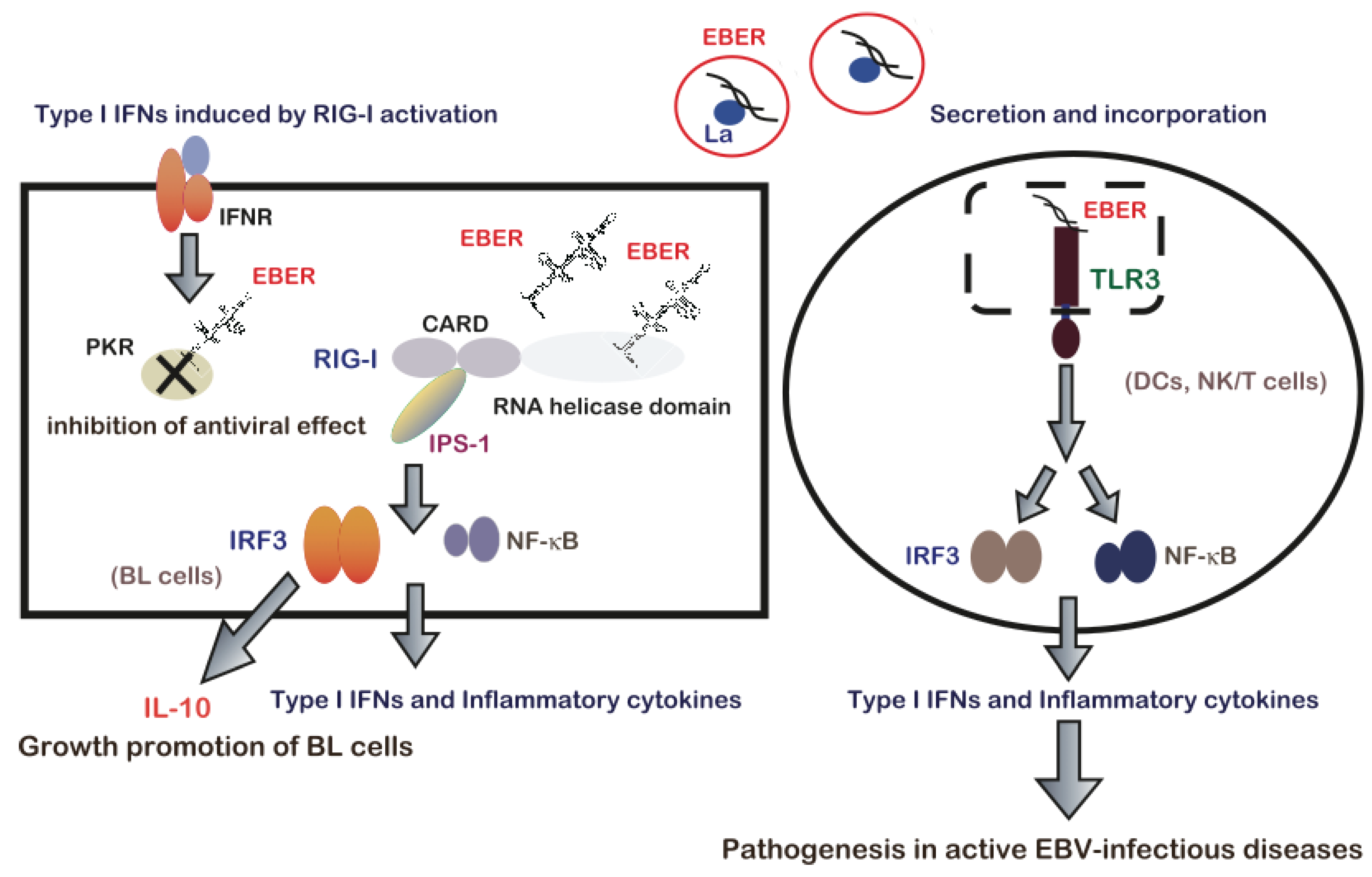
7. EBERs-Mediated Pathogenesis via Modulation of Innate Immune Signals

Interactions between EBERs and host dsRNA sensors have been reported to play a significant role in EBV-mediated pathogenesis (Figure 2). Samanta et al. [15] reported that EBER, which is expected to form dsRNA structures, activates RIG-mediated signaling. The results suggest that in BL cells, RIG-I is constitutively activated by EBERs, leading to the activation of NF-κB and IRF-3, and subsequent induction of type-I IFN. Although IFN induction appears to be disadvantageous for the virus, EBV can maintain a latent infection state because of resistance to IFN, such as that provided by EBER-mediated PKR inhibition [8]. Furthermore, a subsequent study demonstrated the significant role EBER-induced RIG-I activation in oncogenesis. EBER promotes the BL cell growth by inducing expression of anti-inflammatory and growth-promoting cytokine IL-10, in a manner dependent on RIG-I-mediated IRF3 signaling but independent of NF-κB [16].
Figure 2. EBER-mediated modulation of innate immune signaling contributes to EBV-mediated pathogenesis. Reproduced from Iwakiri et al. [71]. Left, in BL cells, EBERs are recognized by RIG-I via the RNA helicase domain of RIG-I and following recognition, RIG-I associates with the adaptor IPS-1 via its CARD. IPS-1 initiates signaling leading to the activation of IRF3 and NF-κB to induce type I IFNs and inflammatory cytokine expression. EBERs induce the expression of the growth-promoting cytokine IL-10 via RIG-I-mediated IRF3 activation and might support BL development. EBERs also bind to IFN-inducible PKR and block its activity, which is required for the IFN-mediated antiviral effect; therefore, EBV might maintain a latent infection state. Right, activation of innate immunity via TLR3 signaling in response to secreted EBER. During an active EBV-infection, EBER1 is released from EBV-infected lymphocytes primarily in a complex with La. Circulating EBER induces DC maturation via TLR3 signaling and induces type I IFN and inflammatory cytokine production by activating IRF3 and NF-κB. DC activation leads to T cell activation and systemic cytokine release. Furthermore, TLR3-expressing T and NK cells including EBV-infected T or NK cells could be activated by EBER1 through TLR3, thus leading to inflammatory cytokine production. Therefore, immunopathologic diseases caused by active EBV infections including T or NK cell activaiton and hypercytokinemia, could be attributed to EBER1-induced TLR3-mediated T cell activation and cytokinemia. EBER, Epstein-Barr virus encoded RNA; EBV, Epstein-Barr virus; BL, Burkitt’s lymphoma; IPS-1, interferon-β promoter stimulator-1; CARD, caspase recruitment domain; RIG-I, retinoic acid-inducible gene I; DC, dendritic cell; IFN, interferon; NK cell, natural killer cell; IL, interleukin; TLR, Toll-like receptor; PKR, RNA-dependent protein kinase; IRF 3, interferon regulatory factor 3.
Cancers 06 01615 g002
Toll-like receptors (TLRs) comprise a distinct family of PRRs that sense virus-derived nucleic acids and trigger antiviral innate immune responses by activating signaling cascades via Toll/IL-1 receptor (TIR) domain-containing adaptors [78]. The role of TLR3 in the dsRNA recognition was demonstrated in a study of TLR3-deficient mice [79]. dsRNA-induced signal transduction via TLR3 leads to the recruitment of TIR domain-containing adaptor inducing IFN-β (TRIF) and the subsequent phosphorylation of downstream molecules such as IRF3 and NF-κB [63]. Iwakiri et al. [17] demonstrated that EBERs are released extracellular environment and are recognized by TLR3, leading to the induction of type I IFN and inflammatory cytokines. The majority of the released EBER1 exists as a complex with La, suggesting that EBER1 is released from the cells via the active secretion of La. EBV has been known to cause active infectious diseases such as infectious mononucleosis (IM), chronic active EBV infection (CAEBV) and EBV-associated hemophagocytic lymphohistiocytosis (EBV-HLH). IM is characterized by the expansion of reactive T-cells and is most likely an immunopathologic disease, in which the general symptoms are caused by inflammatory cytokines [1]. CAEBV and EBV-HLH are also active EBV infections with persistent or recurrent IM-like symptoms. EBV-HLH is characterized by an EBV infection in CD4-positive T cells or naural killer (NK) cells and the systemic release of inflammatory cytokines, leading to blood cell hemophagocytosis via the activation of macrophages [1,80,81]. On the other hand, in CAEBV, CD8+-T cells are primary EBV infection targets [81]. Iwakiri et al. [17] demonstrated that sera from patients with IM, CAEBV and EBV-HLH contained EBER1. A further analysis revealed that serum EBER1 activates TLR3 signaling in immune cells, including dendritic cells, suggesting that EBER1, which is released from EBV-infected cells is responsible for EBV-mediated immune activation, and induction of type I IFN and inflammatory cytokines [17]. Because CD8+-T cells and NK cells express TLR3 and can be activated by TLR3 signaling [82,83], TLR3-expressing T and NK cells could potentially be activated by EBER1 through TLR3 to produce inflammatory cytokines. Therefore, EBER1-induced activation of innate immunity would account for the immunopathologic diseases caused by active EBV infection. A more recent study of a humanized mice model of fetal EBV-infectious disease observed the EBER1 in the serum, thus suggesting that EBER1 contribute to the disease pathology [84]. In summary, EBERs contribute to EBV infection-related pathogenesis, including cancer and active infectious diseases, through interactions with RIG-I and TLR3 [71].

8. Conclusions and Future Direction

Recently, the bioactive extracellular small membrane vesicles also known as exosomes were reported to contribute to cancer development. Exosomes has been shown to contain miRNA, DNA, proteins, and even virus particles [85,86]. Additionally, EBV-infected cells were also reported to produce exosomes that contained viral proteins and miRNA [87]. Previous studies have demonstrated that the La protein can be excreted in exosomes, thus supporting the possibility that EBER is also secreted and transferred to neighboring cells via exosomes [88]. The interactions between EBERs and cellular factors are involved in EBV-mediated pathogenesis, including cancer. A further study of the detailed molecular mechanism is required in order to develop a new therapeutic approach that targets these ncRNAs in EBV-associated cancers.

Acknowledgements

This work was supported by Grant-in-Aid for Scientific Research (KAKENHI).

Conflicts of Interest

The authors declare no conflict of interest.

References

  1. Rickinson, A.B.; Kieff, E. Epstein-Barr virus. In Fields Virol; Knipe, D.M., Howley, P.M., Eds.; Walters Kluwer/Lippincott: Philadelphia, PA, USA, 2007; pp. 2655–2700. [Google Scholar]
  2. Lerner, M.R.; Andrews, N.C.; Miller, G.; Steitz, J.A. Two small RNAs encoded by Epstein-Barr virus and complexed with protein are precipitated by antibodies from patients with systemic lupus erythematosus. Proc. Natl. Acad. Sci. USA 1981, 78, 805–809. [Google Scholar] [CrossRef] [PubMed]
  3. Rymo, L. Identification of transcribed regions of Epstein-Barr virus DNA in Burkitt lymphoma-derived cells. J. Virol. 1979, 32, 8–18. [Google Scholar] [PubMed]
  4. Rosa, M.D.; Gottlieb, E.; Lerner, M.R.; Steitz, J.A. Striking similarities are exhibited by two small Epstein-Barr virus-encoded ribonucleic acids and the adenovirus-associated ribonucleic acids VAI and VAII. Mol. Cell. Biol. 1981, 9, 785–796. [Google Scholar]
  5. Chang, K.L.; Chen, Y.Y.; Shibata, D.; Weiss, L.M. Description of an in situ hybridization methodology for detection of Epstein-Barr virus RNA in paraffin-embedded tissues, with a survey of normal and neoplastic tissues. Diagn. Mol. Pahol. 1992, 1, 246–255. [Google Scholar] [CrossRef]
  6. Komano, J.; Maruo, S.; Kurozumi, K.; Oda, T.; Takada, K. Oncogenic role of Epstein-Barr virus-encoded RNAs in Burkitt’s lymphoma cell line Akata. J. Virol. 1999, 73, 9827–9831. [Google Scholar] [PubMed]
  7. Ruf, I.K.; Rhyne, P.W.; Yang, C.; Cleveland, J.L.; Sample, J.T. Epstein-Barr virus small RNAs potentiate tumorigenicity of Burkitt lymphoma cells independently of an effect on apoptosis. J. Virol. 2000, 74, 10223–10228. [Google Scholar] [CrossRef] [PubMed]
  8. Nanbo, A.; Inoue, K.; Adachi-Takasawa, K.; Takada, K. Epstein-Barr virus RNA confers resistance to interferon-alpha-induced apoptosis in Burkitt’s lymphoma. EMBO J. 2002, 21, 954–965. [Google Scholar] [CrossRef] [PubMed]
  9. Kitagawa, N.; Goto, M.; Kurozumi, K.; Maruo, S.; Fukayama, M.; Naoe, T.; Yasukaswa, M.; Hino, K.; Suzuki, T.; Todo, S.; et al. Epstein-Barr virus-encoded poly(A)(-) RNA supports Burkitt’s lymphoma growth through interleukin-10 induction. EMBO J. 2000, 19, 6742–6750. [Google Scholar] [CrossRef] [PubMed]
  10. Iwakiri, D.; Eizuru, Y.; Tokunaga, M.; Takada, K. Autocrine growth of Epstein-Barr virus-positive gastric carcinoma cells mediated by an Epstein-Barr virus-encoded small RNA. Cancer Res. 2003, 63, 7062–7067. [Google Scholar] [PubMed]
  11. Iwakiri, D.; Sheen, T.S.; Chen, J.Y.; Huang, D.P.; Takada, K. Epstein-Barr virus-encoded small RNA induces insulin-like growth factor 1 and supports growth of nasopharyngeal carcinoma-derived cell lines. Oncogene 2005, 24, 1767–1773. [Google Scholar] [CrossRef] [PubMed]
  12. Yang, L.; Aozasa, K.; Oshimi, K.; Takada, K. Epstein-Barr virus (EBV)-encoded RNA promotes growth of EBV-infected T cells through interleukin-9 induction. Cancer Res. 2004, 64, 5332–5337. [Google Scholar] [CrossRef] [PubMed]
  13. Houmani, J.L.; Davis, C.I.; Ruf, I.K. Growth-promoting properties of Epstein-Barr virus EBER-1 RNA correlate with ribosomal protein L22 binding. J. Virol. 2009, 83, 9844–9853. [Google Scholar] [CrossRef]
  14. Repellin, C.E.; Tsimbouri, P.M.; Philbey, A.W.; Wilson, J.B. Lymphoid hyperplasia and lymphoma in transgenic mice expressing the small non-coding RNA, EBER1 of Epstein-Barr virus. PLoS One 2010, 5, e9092. [Google Scholar]
  15. Samanta, M.; Iwakiri, D.; Kanda, T.; Imaizumi, T.; Takada, K. EB virus-encoded RNAs are recognized by RIG-I and activate signaling to induce type I IFN. EMBO J. 2006, 25, 4207–4214. [Google Scholar] [CrossRef] [PubMed]
  16. Samanta, M.; Iwakiri, D.; Takada, K. Epstein-Barr virus-encoded small RNA induces IL-10 through RIG-I-mediated IRF-3 signaling. Oncogene 2008, 27, 4150–4160. [Google Scholar] [CrossRef]
  17. Iwakiri, D.; Zhou, L.; Samanta, M.; Matsumoto, M.; Ebihara, T.; Seya, T.; Imai, S.; Fujieda, M.; Kawa, K.; Takada, K. Epstein-Barr virus (EBV)-encoded small RNA is released from EBV-infected cells and activates signaling from Toll-like receptor 3. J. Exp. Med. 2009, 206, 2091–2099. [Google Scholar] [CrossRef] [PubMed]
  18. Howe, J.G.; Shu, M.D. Isolation and characterization of the genes for two small RNAs of herpesvirus papio and their comparison with Epstein-Barr virus-encoded EBER RNAs. J. Virol. 1988, 62, 2790–2798. [Google Scholar] [PubMed]
  19. Katze, M.G.; Wambach, M.; Wong, M.L.; Garfinkel, M.; Meurs, E.; Chong, K.; Williams, B.R.; Hovanessian, A.G.; Barber, G.N. Functional expression and RNA binding analysis of the interferon-induced, double-stranded RNA-activated, 68,000-Mr protein kinase in a cell-free system. Mol. Cell. Biol. 1991, 11, 5497–5505. [Google Scholar] [PubMed]
  20. Arrand, J.R.; Young, L.S.; Tugwood, J.D. Two families of sequences in the small RNA-encoding region of Epstein-Barr virus (EBV) correlate with EBV types A and B. J. Virol. 1989, 63, 983–986. [Google Scholar] [PubMed]
  21. Yao, Q.Y.; Tierney, R.J.; Croom-Carter, D.; Cooper, G.M.; Ellis, C.J.; Rowe, M.; Rickinson, A.B. Isolation of intertypic recombinants of Epstein-Barr virus from T-cell-immunocompromised individuals. J. Virol. 1996, 70, 4895–4903. [Google Scholar] [PubMed]
  22. Howe, J.G.; Shu, M.D. Epstein-Barr virus small RNA (EBER) genes: Unique transcription units that combine RNA polymerase II and III promoter elements. Cell 1989, 57, 825–834. [Google Scholar] [CrossRef] [PubMed]
  23. Arraud, J.R.; Ryomo, L. Characterization of the major Epstein-Barr virus-specific RNA in Burkitt lymphoma-derived cells. J. Virol. 1982, 41, 376–389. [Google Scholar] [PubMed]
  24. Rooney, C.; Howe, J.G.; Speck, S.H.; Miller, G. Influence of Burkitt’s lymphoma and primary B cells on latent gene expression by the nonimmortalizing P3J-HR-1 strain of Epstein-Barr virus. J. Virol. 1989, 63, 1531–1539. [Google Scholar]
  25. Greifenegger, N.; Jäger, M.; Kunz-Schughart, L.A.; Wolf, H.; Schwarzmann, F. Epstein-Barr virus small RNA (EBER) genes: Differential regulation during lytic viral replication. J. Virol. 1998, 72, 9323–9328. [Google Scholar] [PubMed]
  26. Clarke, P.A.; Sharp, N.A.; Clemens, M.J. Expression of genes for the Epstein-Barr virus small RNAs EBER-1 and EBER-2 in Daudi Burkitt’s lymphoma cells: Effects of interferon treatment. J. Gen. Virol. 1992, 73, 3169–3175. [Google Scholar] [CrossRef]
  27. Gilligan, K.; Rajadurai, P.; Resnick, L.; Raab-Traub, N. Epstein-Barr virus small nuclear RNAs are not expressed in permissively infected cells in AIDS-associated leukoplakia. Proc. Natl. Acad. Sci. USA 1990, 87, 8790–8794. [Google Scholar] [CrossRef] [PubMed]
  28. Wen, S.; Mizugaki, Y.; Shinozaki, F.; Takada, K. Epstein-Barr virus (EBV) infection in salivary gland tumors: Lytic EBV infection in nonmalignant epithelial cells surrounded by EBV-positive T-lymphoma cells. Virology 1997, 227, 484–487. [Google Scholar] [CrossRef] [PubMed]
  29. Wen, S.; Shimizu, N.; Yoshiyama, H.; Mizugaki, Y.; Shinozaki, F.; Takada, K. Association of Epstein-Barr virus (EBV) with Sjögren’s syndrome: Differential EBV expression between epithelial cells and lymphocytes in salivary glands. Am. J. Pathol. 1996, 149, 1511–1517. [Google Scholar] [PubMed]
  30. Mizugaki, Y.; Sugawara, Y.; Shinozaki, F.; Takada, K. Detection of Epstein-Barr virus in oral papilloma. Jpn. J. Cancer Res. 1998, 89, 604–607. [Google Scholar] [CrossRef] [PubMed]
  31. Yao, Y.; Minter, H.A.; Chen, X.; Reynolds, G.M.; Bromley, M.; Arrand, J.R. Heterogeneity of HLA and EBER expression in Epstein-Barr virus-associated nasopharyngeal carcinoma. Int. J. Cancer. 2000, 88, 949–955. [Google Scholar] [CrossRef] [PubMed]
  32. Howe, J.G.; Steitz, J.A. Localization of Epstein-Barr virus-encoded small RNAs by in situ hybridization. Proc. Natl. Acad. Sci. USA 1986, 83, 9006–9010. [Google Scholar] [CrossRef]
  33. Schwemmle, M.; Clemens, M.J.; Hilse, K.; Pfeifer, K.; Tröster, H.; Müller, W.E.; Bachmann, M. Localization of Epstein-Barr virus-encoded RNAs EBER-1 and EBER-2 in interphase and mitotic Burkitt lymphoma cells. Proc. Natl. Acad. Sci. USA 1992, 89, 10292–10296. [Google Scholar] [CrossRef] [PubMed]
  34. Fok, V.; Friend, K.; Steitz, J.A. Epstein-Barr virus noncoding RNAs are confined to the nucleus, whereas their partner, the human La protein, undergoes nuclecytoplasmic shuttling. J. Cell. Biol. 2006, 173, 319–325. [Google Scholar] [CrossRef] [PubMed]
  35. Clarke, P.A.; Schwemmle, M.; Schickinger, J.; Hilse, K.; Clemens, M.J. Binding of Epstein-Barr virus small RNA EBER-1 to the double-stranded RNA-activated protein kinase DAI. Nucleic Acids Res. 1991, 19, 243–248. [Google Scholar] [CrossRef]
  36. Lee, N.; Pimienta, G.; Steitz, J.A. AUF1/hnRNP D is a novel protein partner of the EBER1 noncoding RNA of Epstein-Barr virus. RNA 2012, 18, 2073–2082. [Google Scholar]
  37. Bachmann, M.; Pfeifer, K.; Schröder, H.C.; Müller, W.E. The La antigen shuttles between the nucleus and the cytoplasm in CV-1 cells. Mol. Cell. Biochem. 1989, 85, 103–114. [Google Scholar] [CrossRef] [PubMed]
  38. Huang, M.; Ida, H.; Arima, K.; Nakamura, H.; Aramaki, T.; Fujikawa, K.; Tamai, M.; Kamachi, M.; Kawakami, A.; Yamasaki, H.; et al. La autoantigen translocates to cytoplasm after cleavage during granzyme B-mediated cytotoxicity. Life Sci. 2007, 81, 1461–1466. [Google Scholar]
  39. Meerovitch, K.; Svitkin, Y.V.; Lee, H.S.; Lejbkowicz, F.; Kenan, D.J.; Chan, E.K.; Agol, V.I.; Keene, J.D.; Sonenberg, N. La autoantigen enhances and corrects aberrant translation of poliovirus RNA in reticulocyte lysate. J. Virol. 1993, 67, 3798–3807. [Google Scholar] [PubMed]
  40. Shiroki, K.; Isoyama, T.; Kuge, S.; Ishii, T.; Ohmi, S.; Hata, S.; Suzuki, K.; Takasaki, Y.; Nomoto, A. Intracellular redistribution of truncated La protein produced by poliovirus 3Cpro-mediated cleavage. J. Virol. 1999, 73, 2193–2200. [Google Scholar] [PubMed]
  41. Akusjärvi, G.; Mathews, M.B.; Andersson, P.; Vennström, B.; Pettersson, U. Structure of genes for virus-associated RNAI and RNAII of adenovirus type 2. Proc. Natl. Acad. Sci. USA 1980, 77, 2424–2428. [Google Scholar] [CrossRef] [PubMed]
  42. Bhat, R.A.; Thimmappaya, B. Two small RNAs encoded by Epstein-Barr virus can functionally substitute for the virus-associated RNAs in the lytic growth of adenovirus 5. Proc. Natl. Acad. Sci. USA 1983, 80, 4789–4793. [Google Scholar] [CrossRef] [PubMed]
  43. Meurs, E.; Chong, K.; Galabru, J.; Thomas, N.S.; Kerr, I.M.; Williams, B.R.; Hovanessian, A.G. Molecular cloning and characterization of the human double-stranded RNA-activated protein kinase induced by interferon. Cell 1990, 62, 379–390. [Google Scholar] [CrossRef] [PubMed]
  44. Sharp, T.V.; Schwemmle, M.; Jeffrey, I.; Laing, K.; Mellor, H.; Proud, C.G.; Hilse, K.; Clemens, M.J. Comparative analysis of the regulation of the interferon-inducible protein kinase PKR by Epstein-Barr virus RNAs EBER-1 and EBER-2 and adenovirus VAI RNA. Nucleic Acids Res. 1993, 21, 4483–4490. [Google Scholar] [CrossRef] [PubMed]
  45. Vuyisich, M.; Spanggord, R.J.; Beal, P.A. The binding site of the RNA-dependent protein kinase (PKR) on EBER1 RNA from Epstein-Barr virus. EMBO Rep. 2000, 3, 622–627. [Google Scholar] [CrossRef]
  46. Clarke, P.A.; Sharp, N.A.; Clemens, M.J. Translational control by the Epstein-Barr virus small RNA EBER-1. Reversal of the double-stranded RNA-induced inhibition of protein synthesis in reticulocyte lysates. Eur. J. Biochem. 1990, 193, 635–641. [Google Scholar]
  47. McKenna, S.A.; Lindhout, D.A.; Shimoike, T.; Aitken, C.E.; Puglisis, J.D. Viral dsRNA inhibitors prevent self-association and autophosphorylation of PKR. J. Mol. Biol. 2007, 372, 103–113. [Google Scholar] [CrossRef] [PubMed]
  48. Toczyski, D.P.; Steitz, J.A. EAP, a highly conserved cellular protein associated with Epstein-Barr virus small RNAs (EBERs). EMBO J. 1991, 10, 459–466. [Google Scholar]
  49. Toczyski, D.P.; Steitz, J.A. The cellular RNA-binding protein EAP recognizes a conserved stem-loop in the Epstein-Barr virus small RNA EBER 1. Mol. Cell. Biol. 1993, 1, 703–710. [Google Scholar]
  50. Toczyski, D.P.; Matera, A.G.; Ward, D.C.; Steitz, J.A. The Epstein-Barr virus (EBV) small RNA EBER1 binds and relocalizes ribosomal protein L22 in EBV-infected human B lymphocytes. Proc. Natl. Acad. Sci. USA 1994, 91, 3463–3467. [Google Scholar] [CrossRef] [PubMed]
  51. Liu, P.; Tarlé, S.A.; Hajra, A.; Claxton, D.F.; Marlton, P.; Freedman, M.; Siciliano, M.J.; Collins, F.S. Fusion between transcription factor CBF beta/PEBP2 beta and a myosin heavy chain in acute myeloid leukemia. Science 1993, 261, 1041–1044. [Google Scholar] [CrossRef] [PubMed]
  52. Nucifora, G.; Begy, C.R.; Erickson, P.; Drabkin, H.A.; Rowley, J.D. The 3;21 translocation in myelodysplasia results in a fusion transcript between the AML1 gene and the gene for EAP, a highly conserved protein associated with the Epstein-Barr virus small RNA EBER 1. Proc. Natl. Acad. Sci. USA 1993, 90, 7784–7788. [Google Scholar] [CrossRef] [PubMed]
  53. Le, S.; Sternglanz, R.; Greider, C.W. Identification of two RNA-binding proteins associated with human telomerase RNA. Mol. Biol. Cell 2000, 11, 999–1010. [Google Scholar] [CrossRef] [PubMed]
  54. Leopardi, R.; Ward, P.L.; Ogle, W.O.; Roizman, B. Association of herpes simplex virus regulatory protein ICP22 with transcrip-tional complexes containing EAP, ICP4, RNA polymerase II, and viral DNA requires posttranslational modification by the U(L)13 proteinkinase. J. Virol. 1997, 71, 1133–1139. [Google Scholar] [PubMed]
  55. Wood, J.; Frederickson, R.M.; Fields, S.; Patel, A.H. Hepatitis C virus 3'X region interacts with human ribosomal proteins. J. Virol. 2001, 75, 1348–1358. [Google Scholar] [CrossRef] [PubMed]
  56. Gregorovic, G.; Bosshard, R.; Karstegl, C.E.; White, R.E.; Pattle, S.; Chiang, A.K.; Dittrich-Breiholz, O.; Kracht, M.; Russ, R.; Farrell, P.J. Cellular gene expression that correlates with EBER expression in Epstein-Barr virus-infected lymphoblastoid cell lines. J. Virol. 2011, 85, 3535–3545. [Google Scholar]
  57. Dobbelstein, M.; Shenk, T. In vitro selection of RNA ligands for the ribosomal L22 protein associated with Epstein-Barr virus-expressed RNA by using randomized and cDNA-derived RNA libraries. J. Virol. 1995, 69, 8027–8034. [Google Scholar] [PubMed]
  58. Fok, V.; Mitton-Fry, R.M.; Grech, A.; Steitz, J.A. Multiple domains of EBER 1, an Epstein-Barr virus noncoding RNA, recruit human ribosomal protein L22. RNA 2006, 12, 872–882. [Google Scholar] [CrossRef] [PubMed]
  59. Elia, A.; Vyas, J.; Laing, K.G.; Clemens, M.J. Ribosomal protein L22 inhibits regulation of cellular activities by the Epstein-Barr virus small RNA EBER-1. Eur. J. Biochem. 2004, 27, 1895–1905. [Google Scholar] [CrossRef]
  60. Yoneyama, M.; Kikuchi, M.; Natsukawa, T.; Shinobu, N.; Imaizumi, T.; Miyagishi, M.; Taira, K.; Akira, S.; Fujita, T. The RNA helicase RIG-I has an essential function in double-stranded RNA-induced innate antiviral responses. Nat. Immunol. 2004, 5, 730–737. [Google Scholar] [CrossRef] [PubMed]
  61. Kang, D.C.; Gopalkrishnan, R.V.; Wu, Q.; Jankowsky, E.; Pyle, A.M.; Fisher, P.B. mda-5: An interferon-inducible putative RNA helicase with double-stranded RNA-dependent ATPase activity and melanoma growth-suppressive properties. Proc. Natl. Acad. Sci. USA 2002, 99, 637–642. [Google Scholar]
  62. Yoneyama, M.; Fujita, T. Function of RIG-I-like receptors in antiviral innate immunity. J. Biol. Chem. 2007, 282, 15315–15318. [Google Scholar] [CrossRef] [PubMed]
  63. Meylan, E.; Tschopp, J. Toll-like receptors and RNA helicases: Two parallel ways to trigger antiviral responses. Mol. Cell 2006, 22, 561–569. [Google Scholar] [CrossRef] [PubMed]
  64. Kawai, T.; Takahashi, K.; Sato, S.; Coban, C.; Kumar, H.; Kato, H.; Ishii, K.J.; Takeuchi, O.; Akira, S. IPS-1, an adaptor triggering RIG-I- and Mda5-mediated type I interferon induction. Nat. Immunol. 2005, 11, 1074–1076. [Google Scholar]
  65. Hornung, V.; Ellegast, J.; Kim, S.; Brzózka, K.; Jung, A.; Kato, H.; Poeck, H.; Akira, S.; Conzelmann, K.K.; Schlee, M.; et al. 5'-Triphosphate RNA is the ligand for RIG-I. Science 2006, 314, 994–997. [Google Scholar] [CrossRef] [PubMed]
  66. Lu, J.-Y.; Sadri, N.; Schneider, R.J. Endotoxic shock in AUF1 knockout mice mediated by failure to degrade proinflammatory cytokine mRNAs. Gen. Dev. 2006, 20, 3174–3184. [Google Scholar] [CrossRef]
  67. Gratacós, F.M.; Brewer, G. The role of AUF1 in regulated mRNA decay. Wiley Interdiscip. Rev. RNA 2010, 1, 457–473. [Google Scholar] [CrossRef] [PubMed]
  68. Lal, A.; Mazan-Mamczarz, K.; Kawai, T.; Yang, X.; Martindale, J.L.; Gorospe, M. Concurrent versus individual binding of HuR and AUF1 to common labile target mRNAs. EMBO J. 2004, 23, 3092–3102. [Google Scholar] [CrossRef] [PubMed]
  69. Shimizu, N.; Tanabe-Tochikura, A.; Kuroiwa, Y.; Takada, K. Isolation of Epstein-Barr virus (EBV)-negative cell clones from the EBV- positive Burkitt’s lymphoma (BL) line Akata: Malignant phenotypes of BL cells are dependent on EBV. J. Virol. 1994, 68, 6069–6073. [Google Scholar]
  70. Yamamoto, N.; Takizawa, T.; Iwanaga, Y.; Shimizu, N.; Yamamoto, N. Malignant transformation of B lymphoma cell line BJAB by Epstein-Barr virus-encoded small RNAs. FEBS Lett. 2000, 484, 153–158. [Google Scholar] [CrossRef] [PubMed]
  71. Iwakiri, D.; Takada, K. Role of EBERs in the pathogenesis of EBV infection. Adv. Cancer Res. 2010, 107, 119–136. [Google Scholar]
  72. Wong, H.L.; Wang, X.; Chang, R.C.; Jin, D.Y.; Feng, H.; Wang, Q.; Lo, K.W.; Huang, D.P.; Yuen, P.W.; Takada, K.; et al. Stable expression of EBERs in immortalized nasopharyngeal epithelial cells confers resistance to apoptotic stress. Mol. Carccing. 2005, 44, 92–101. [Google Scholar] [CrossRef]
  73. Yoshizaki, T.; Endo, K.; Ren, Q.; Wakisaka, N.; Murono, S.; Kondo, S.; Sato, H.; Furukawa, M. Oncogenic role of Epstein-Barr virus-encoded small RNAs (EBERs) in nasopharyngeal carcinoma. Auris. Nasus. Larynx 2007, 34, 73–78. [Google Scholar] [CrossRef] [PubMed]
  74. Tsang, C.M.; Zhang, G.; Seto, E.; Takada, K.; Deng, W.; Yip, Y.L.; Man, C.; Hau, P.M.; Chen, H.; Cao, Y.; et al. Epstein-Barr virus infection in immortalized nasopharyngeal epithelial cells: Regulation of infection and phenotypic characterization. Int. J. Cancer 2010, 127, 1570–1583. [Google Scholar] [CrossRef] [PubMed]
  75. Swaminathan, S.; Tomkinson, B.; Kieff, E. Recombinant Epstein-Barr virus with small RNA (EBER) genes deleted transforms lymphocytes and replicates in vitro. J. Virol. 1991, 66, 5133–5136. [Google Scholar]
  76. Yajima, M.; Kanda, T.; Takada, K. Critical role of Epstein-Barr Virus (EBV)-encoded RNA in efficient EBV-induced B-lymphocyte growth transformation. J. Virol. 2005, 79, 4298–4307. [Google Scholar]
  77. Wu, Y.; Maruo, S.; Yajima, M.; Kanda, T.; Takada, K. Epstein-Barr virus (EBV)-encoded RNA 2 (EBER2) but not EBER1 plays a critical role in EBV-induced B-cell growth transformation. J. Virol. 2007, 80, 11236–11245. [Google Scholar] [CrossRef]
  78. Akira, S.; Takeda, K. Toll-like receptor signaling. Nat. Rev. Immunol. 2004, 4, 499–511. [Google Scholar] [CrossRef] [PubMed]
  79. Alexopoulou, L.; Holt, A.C.; Medzhitov, R.; Flavell, R.A. Recognition of double-stranded RNA and activation of NF-kappaB by Toll-like receptor 3. Nature 2001, 413, 732–738. [Google Scholar] [CrossRef] [PubMed]
  80. Kikuta, H.; Sakiyama, Y.; Matsumoto, S.; Oh-Ishi, T.; Nakano, T.; Nagashima, T.; Oka, T.; Hironaka, T.; Hirai, K. Fatal Epstein-Barr virus-associated hemophagocytic syndrome. Blood 1993, 82, 3259–3264. [Google Scholar] [PubMed]
  81. Kasahara, Y.; Yachie, A.; Takei, K.; Kanegane, C.; Okada, K.; Ohta, K.; Seki, H.; Igarashi, N.; Maruhashi, K.; Katayama, K.; et al. Differential cellular targets of Epstein-Barr virus (EBV) infection between acute EBV-associated hemophagocytic lymphohistiocytosis and chronic active EBV infection. Blood 2001, 98, 1882–1888. [Google Scholar] [CrossRef] [PubMed]
  82. Tabiasco, J.; Devêvre, E.; Rufer, N.; Salaun, B.; Cerottini, J.C.; Speiser, D.; Romero, D. Human effector CD8+ T lymphocytes express TLR3 as a functional coreceptor. J. Immunol. 2006, 177, 8708–8713. [Google Scholar] [CrossRef] [PubMed]
  83. Schmidt, K.N.; Leung, B.; Kwong, M.; Zarember, K.A.; Satyal, S.; Navas, T.A.; Wang, F.; Godowski, P.J. APC-independent activation of NK cells by the Toll-like receptor 3 agonist double-stranded RNA. J. Immunol. 2004, 172, 138–143. [Google Scholar] [CrossRef] [PubMed]
  84. Sato, K.; Misawa, N.; Nie, C.; Satou, Y.; Iwakiri, D.; Matsuoka, M.; Takahashi, R.; Kuzushima, K.; Ito, M.; Takada, K.; et al. A novel animal model of Epstein-Barr virus-associated hemophagocytic lymphohistiocytosis in humanized mice. Blood 2011, 117, 5663–5673. [Google Scholar] [CrossRef] [PubMed]
  85. Raposo, G.; Stoorvogel, W. Extracellular vesicles: Exosomes, microvesicles, and friends. J. Cell. Biol. 2013, 200, 373–383. [Google Scholar] [CrossRef] [PubMed]
  86. Pant, S.; Hilton, H.; Burczynski, M.E. The multi-faceted exosome: Biogenesis, role in normal and aberrant cellular function, and frontiers for pharmacological and biomarker opportunities. Biochem. Pharmacol. 2012, 83, 1484–1494. [Google Scholar] [CrossRef] [PubMed]
  87. Pegtel, D.M.; Cosmopoulos, K.; Thorley-Lawson, D.A.; van Eijndhoven, M.A.; Hopmans, E.S.; Lindenberg, J.L.; de Gruijl, T.D.; Würdinger, T.; Middeldorp, J.M. Functional delivery of viral miRNAs via exosomes. Proc. Natl. Acad. Sci. USA 2010, 107, 6328–6333. [Google Scholar] [CrossRef] [PubMed]
  88. Kapsogeorgou, E.K.; Abu-Helu, R.F.; Moutsopoulos, H.M.; Manoussakis, M.N. Salivary gland epithelial cell exosomes: A source of autoantigenic ribonucleoproteins. Arthtitis Rheum. 2005, 52, 1517–1521. [Google Scholar] [CrossRef]

