Review

The Molecular Basis of Pediatric Brain Tumors: A Review with Clinical Implications

by
Elias Antoniades
1,*,
Nikolaos Keffes
1,
Stamatia Vorri
2,
Vassilios Tsitouras
1,
Nikolaos Gkantsinikoudis
1,
Parmenion Tsitsopoulos
1 and
John Magras
1
1
Second Department of Neurosurgery, Aristotle University School of Medicine, 546 36 Thessaloniki, Greece
2
New York City Health and Hospital—Jacobi Medical Center Department of Pediatrics, Albert Einstein College of Medicine, Bronx, NY 10461, USA
*
Author to whom correspondence should be addressed.
Cancers 2025, 17(9), 1566; https://doi.org/10.3390/cancers17091566
Submission received: 9 March 2025 / Revised: 27 April 2025 / Accepted: 29 April 2025 / Published: 4 May 2025
(This article belongs to the Special Issue New Advances in the Treatment of Pediatric Solid Tumors)

Simple Summary

Central nervous system (CNS) tumors in the pediatric population are the second most common among other malignancies of this age spectrum. Carcinogenesis of these lesions is the result of the aberrant cell signaling process. The previous classification based on histologic characteristics cannot appropriately define the prognosis. According to the current classification, each tumor relates to specific identity mutations in one or more of these signaling steps. Lesions with deficient supervision of transcription, uncontrollable cell cycle initiation and apoptosis, or combined mutations are considered more aggressive. Consequently, treating physicians have to be acquainted with novel nomenclature. In this review, we review the recent literature and attempt to present the major tumor groups with their associated alterations.

Abstract

Central nervous system (CNS) tumors are the most common solid malignancy in the pediatric population. These lesions are the result of the aberrant cell signaling step proteins, which normally regulate cell proliferation. Mitogen-activated protein kinase (MAPK) pathways and tyrosine kinase receptors are involved in tumorigenesis of low-grade gliomas. High-grade gliomas may carry similar mutations, but loss of epigenetic control is the dominant molecular event; it can occur either due to histone mutations or inappropriate binding or unbinding of DNA on histones. Therefore, despite the absence of genetic alteration in the classic oncogenes or tumor suppressor genes, uncontrolled transcription results in tumorigenesis. Isocitric dehydrogenase (IDH) mutations do not predominate compared to their adult counterpart. Embryonic tumors include medulloblastomas, which bear mutations of transcription-regulating pathways, such as wingless-related integration sites or sonic hedgehog pathways. They may also relate to high expression of Myc family genes. Atypical teratoid rhabdoid tumors harbor alterations of molecules that contribute to ATP hydrolysis of chromatin. Embryonic tumors with multilayered rosettes are associated with microRNA mutations and impaired translation. Ependymomas exhibit great variability. As far as supratentorial lesions are concerned, the major events are mutations either of NFkB or Hippo pathways. Posterior fossa tumors are further divided into two types with different prognoses. Type A group is associated with mutations of DNA damage repair molecules. Lastly, germ cell tumors are a heterogeneous group. Among them, germinomas manifest KIT receptor mutations, a subgroup of the tyrosine kinase receptor family.

1. Introduction

Central nervous system malignancies are the most frequent space-occupying lesions in children. They are second in incidence, only after leukemia in the etiological ranking. From an anatomical perspective, they can be divided into supratentorial and posterior fossa malignancies. Based on the time of diagnosis, these tumors are also categorized into congenital neoplasms (detected postnatally within the first two months postpartum), neoplasms of infancy (<1 year), and tumors of older children (>1 year) [1]. Their incidence is 5.4–5.6 cases per 100,000 individuals [2].
The current classification from the World Health Organization (WHO) highlights the molecular profile of these tumors. Specifically, the referred molecules may be present at either the cell signaling process, DNA transcription or throughout cell cycle [2].
Herewith, we performed a narrative review, aiming to present succinctly the new molecular identity of pediatric brain tumors. A novel nomenclature has entered the daily clinical praxis, which includes knowledge of cell signaling, growth, and duplication. We also intend to provide treating physicians with an easily approachable body of literature.

2. Oncologic Terms

The basic steps of carcinogenesis include non-ceasing proliferative signaling, evasiveness of tumor suppression, recalcitrance to apoptosis, novel angiogenesis, multiplicative deathlessness, infiltration, and dissemination [3].
Initiation is the establishment of a firm, mutated cell. This is an irreversible phenomenon. Initiation by itself does not appear to be sufficient for neoplastic growth. Only when these changes have not undergone repair and the cell completes its DNA synthesis are these aberrations considered constant [4].
The promotion phase entails the distinct proliferation of the initiated cell to shape a class of preneoplastic cells. A distinctive feature of the promotion is also the decrease in cell death. Tumor promoters do not induce either DNA mutations or tumors by themselves; rather, they act by sustaining proliferation of preneoplastic lesions [5]. Specifically, molecular alterations in tumor-suppressive genes or telomerase subunit alterations provoke uncontrolled cell proliferation and elongate this phase.
The progression stage includes enhanced DNA synthesis in the preneoplastic lesions, additional DNA aberrations, and translocations. Hence, independent proliferation is achieved [6].
Proliferation denotes cell number increment in a population due to exertion of the cell cycle and high cell mitosis [7]. The basic steps of the cell signaling sequence are schematically presented in Figure 1.

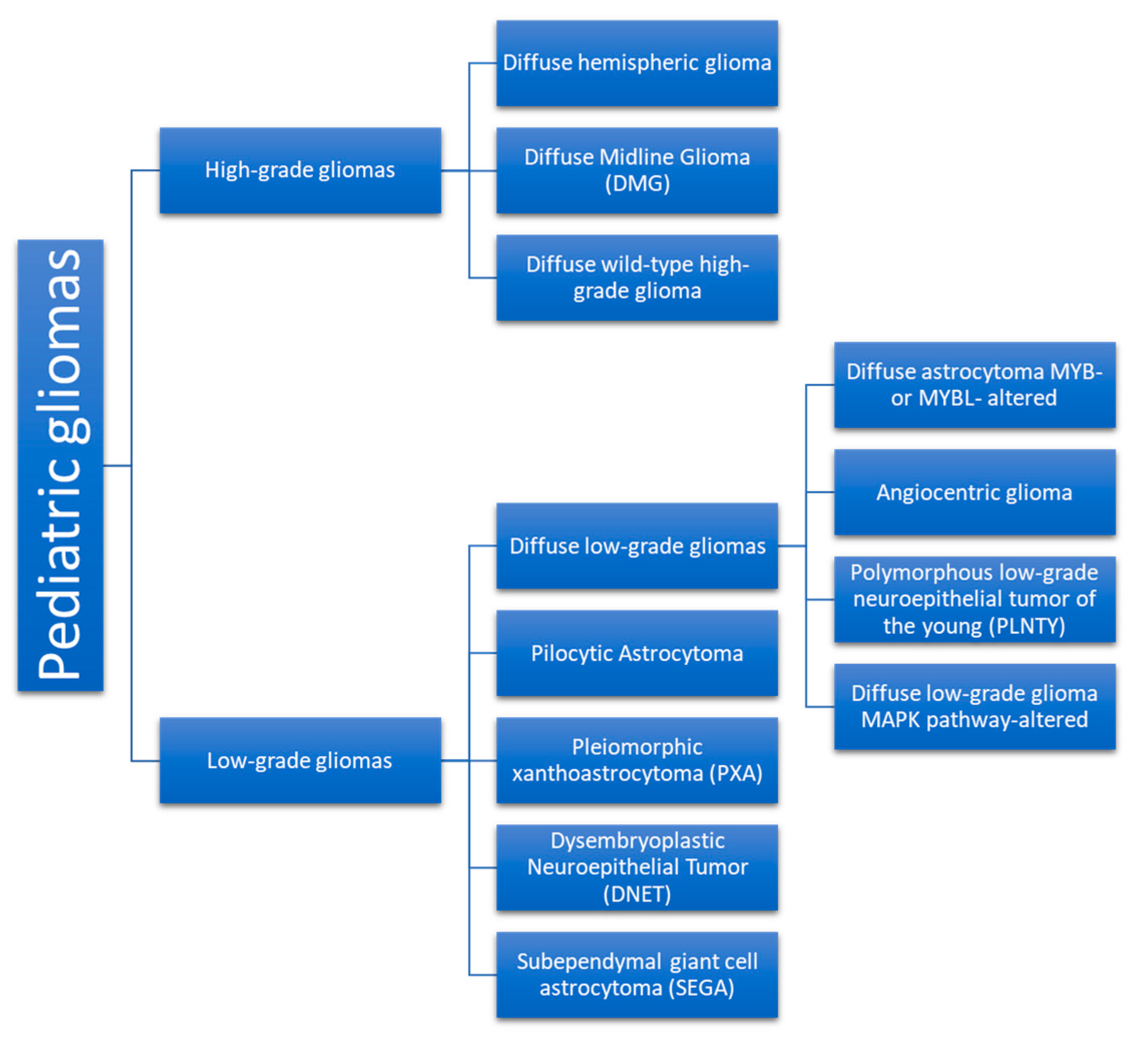
3. Neoplasm Categories

Gliomas are the most common tumors of childhood. They are further divided in low-grade and high-grade lesions [1], (Scheme 1).

3.1. Low-Grade Gliomas

3.1.1. Molecular Events

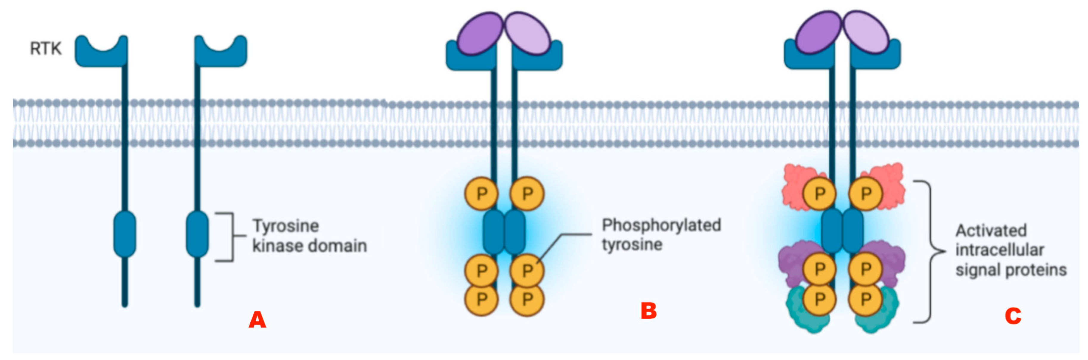
Alterations of Tyrosine Kinase Receptors

The attachment of a growth factor (GF) to a receptor unit ignites a signaling cascade. Receptor tyrosine kinases (RTKs) are highly specified cell surface receptors for polypeptide growth factors, hormones, and cytokines. Overall, 20 separate RTK groups exist [8]. Their structure comprises an outer membrane ligand-attaching segment, a single transmembrane helix, and a cytoplasmic segment, which contains a proximal-to-membrane modificatory area, a tyrosine kinase domain (TKD), and a carboxyl (C-) appendage [9] (Figure 2).
FGFR (Fibroblast Growth Factor receptor) Alterations
FGFR1 alterations respond to three scenarios: FGFR1 point alterations, FGFR1-TACC1 conjunction, and FGFR1-TKD duplications. FGFR1 alteration occurs in 5–10% of patients. Duplication refers to the region that encodes the tyrosine kinase domain (TKD). These alterations are relevant with either low-grade extracerebellar astrocytomas of the cortex or dysembryoplastic neuroepithelial tumors [10]. Focal alterations of FGFR1 are the most frequent alteration of the FGFR1 gene in pediatric gliomas, affecting almost 30% of the patients [11] (Table 1).

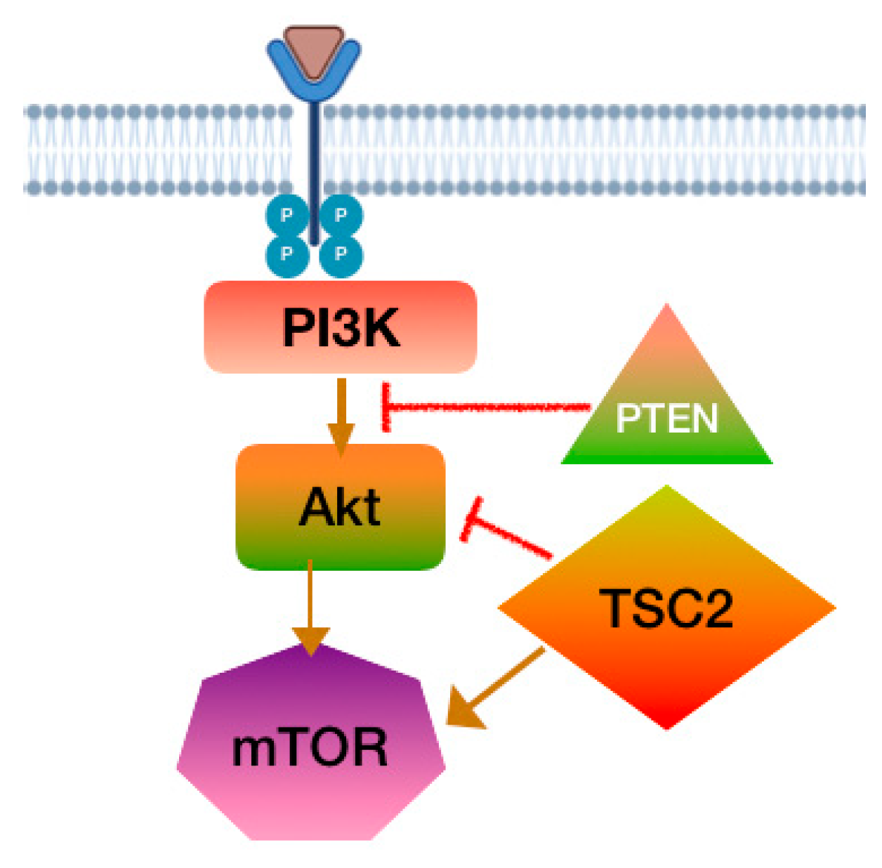
PI3K-Akt-mTOR Pathway
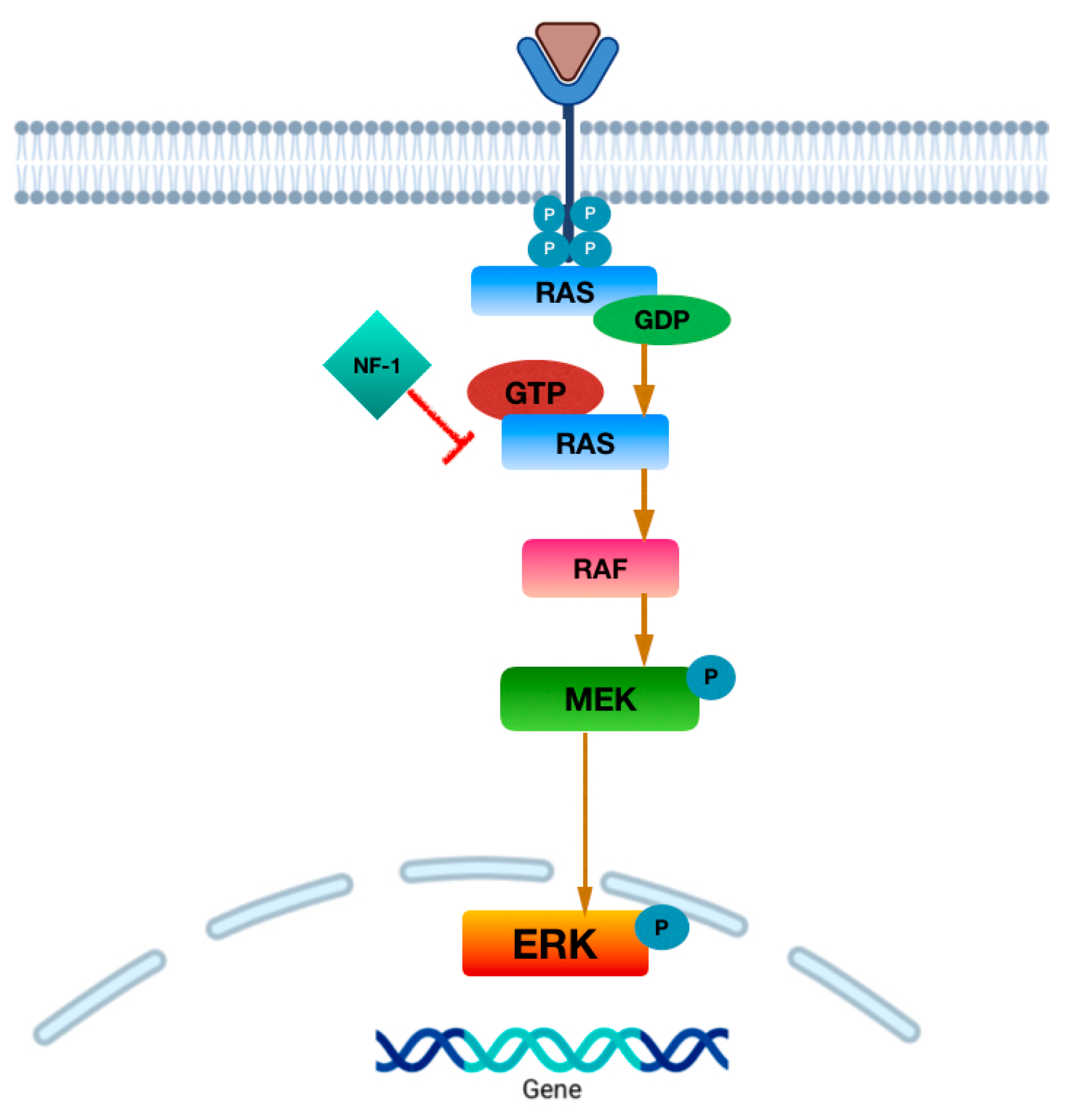
The phosphoinositide 3-kinase (PI3K) cascade is implicated in crucial cellular stages like proliferation, apoptosis, motion, novel vascularization, and stem cell revitalization. The PI3K pathway is activated by growth factor receptors, such as epidermal growth factor (EGFR), EGFRvIII, platelet-derived growth factor (PDGFR), and RAS. It induces the production of phosphatidylinositol-3,4,5 trisphosphate (PIP3). Protein kinase B and Akt become operative by PIP3, activating a cascade that contributes to cell growth and debilitated cell death [12].
Phosphatase and tensin homolog (PTEN) dephosphorylates PIP3 and negatively modulates PI3K. Following Akt’s activation, the mammalian target of rapamycin (mTOR) exerts its metabolic impact on cellular growth. Aberrant and uncontrolled function of the PI3K pathway, due to mutations, occurs in neoplastic cells. Deletions of PTEN are observed in approximately 40% of gliomas [13] (Figure 3).

MAPK Pathway

Mitogen-activated protein kinases (MAPKs) are serine/threonine kinases that phosphorylate their own dual serine and threonine remainder to activate or deactivate their target [14]. MAPKs modulate cellular functions like metabolism, cell death, and immune responses [15]. The activation of the MAPK pathway takes place in the context of sequential phosphorylation. This setting includes a MAP3K (MAP kinase kinase kinase), which activates a MAP2K (MAP kinase kinase), which consequently activates a MAPK (MAP kinase) [14]. MAPK phosphorylation events can be inactivated by MAPK protein phosphatases (MKPs) [14]. There are three well-known MAPK pathways in mammalian cells. One of them is the ERK1/2, which responds to growth factors, hormones, and proinflammatory stimuli [16]. The function of both ERK1 and ERK2 variants commences after the attachment of a binding molecule to an RTK at the cell membrane. Accordingly, Ras protein, a member of G proteins, is activated. Ras incites the serine/threonine protein kinase, Raf, a MAP3K, which establishes the function of MAP2K or MEK, which finally adds phosphate molecules at threonine and tyrosine remnants of MAPK or ERK1/2 [16] (Figure 4). B-raf kinase is encoded by the BRAF gene located in 7q34. Mutations of this gene are frequently encountered in less malignant glial tumors and rarely in more malignant ones.
The Ras and PI3K cascades can be interchangeably modulated by various interrelated processes in different cellular phases. Therefore, Ras reacts immediately to the p110 fermentation segment of PI3K and induces the initiation of the PI3K cascade [17]. Furthermore, lengthened Ras activation decreases the encoding of PTEN, a phosphatase that dampens the PI3K activation [18]. Conversely, the PI3K-Akt cascade impedes the Ras cascade via the addition of a phosphate group on Raf1 [19].

MYB/MYBL1 Alterations

MYB/MYBL1 mutations are regarded as discrete in the relevant studies. MYB protein production is detected in the nervous system [20]. MYB, along with the ubiquitous variant of the group MYBL2, modulates cyclin-dependent kinases (CDKs) production and function [20]. The MYB gene self-modulates its expression [20]. Excessive production of wild-type MYB protein is a promoter of tumorigenesis, and additional genetic alterations are needed for progression [21,22]. MYB adjuncts to the RNA-ligand protein QKI. It functions as a repositioned super enhancer of an active transcription factor [20,23]. MYBL1 resettling is characteristic of low-malignancy glial cell tumors. 8q13.1 (which includes MYBL1) additions have been detected remarkably in diffuse astrocytoma grade 2 [24]. Lastly, transcription factor c-Myb binds and ignites the MKP-3 gene transcription and downregulates the Ras-Raf-MEK-ERK cascade [25]. Gliomas harboring MYB/MYBL1 mutations are overall infrequent compared to other astrocytic tumors and recently included in the WHO grading [6]. Despite the variability in histopathology and clinical manifestations, they bear a good prognosis after adequate excision. Of note, all types of MYB or MYBL1 mutations relate to the presence of supratentorial lesions resembling angiocentric gliomas (AG) [22,26].

3.1.2. Types of LGGs

Pediatric-type low-grade gliomas (pLGGs) represent almost 10–20% of all cerebral neoplasias [26]. These malignancies are considered WHO grade 1 or 2 and develop anywhere along the neural axis. Common signs and symptoms include headaches, cognitive impairment, focal neurologic deficits, and personality or behavioral changes [26].

Diffuse Astrocytoma and MYB- or MYBL1-Altered

Diffuse astrocytoma with alterations of MYB/MYBL1 constitutes a separate category of tumors with indolent course and a very good prognosis [27,28]. This neoplasia is seen in the cerebral hemispheres. Patients are primarily young children, approximately 5 years old. Epilepsy, pyramidal deficits, and behavioral alterations are the main signs and symptoms [29]. Ten-year overall survival (OS) is 95.2%. The non-surgical treatment is recommended [28].

Angiocentric Glioma (AG)

AG is an infrequent cortical neoplasia (WHO grade 1) that occurs in adolescents and is usually manifested with seizures. Its molecular identity is MYB-QKI gene fusion [30]. This mutation responds to almost 90% of AGs and in less than half of pediatric DLGGs. The result of this alteration is the deficient onco-suppression of QKI accompanied by the intense expression of MYB. The tumor demonstrates indistinct boundaries [31].

Polymorphous Low-Grade Neuroepithelial Tumor in Youth (PLNTY)

PLNTY is a seizure-relevant neoplasia with alterations in the MAPK pathway [32]. Molecular profiling reveals a BRAF V600E focal alteration (≈40%) or an FGFR 2/3 fusion (≈50%) [33].

Diffuse Low-Grade Glioma—MAPK Pathway-Altered

This is a recently described lesion that harbors both BRAF V600E mutation and FGFR1. This glioma is classified into the intermediate risk group [34].

Pilocytic Astrocytoma (PA) (Figure 5)

Pilocytic astrocytoma is the most frequent cerebral neoplasia of childhood and adolescence, and its incidence rate is approximately 15%. It usually develops in the first two decades of life [35]. The term pilocytic originates from the histologic features, which include thin, elongated astrocytic projections stained with GFAP or cells with hair-like, bipolar processes [36]. The cerebellum is the most frequent site of development (43%), followed by the convexity areas (36%), the visual tract and hypothalamus (9%), and the brainstem and spinal cord [36]. Three histologically distinct patterns exist: (1) The two-compartment type is very frequently detected in cerebellar PA; it involves cellular areas with saccules, glassy vascular networks, and scarce aggregations of endoplasmic reticulum with rosettes. (2) The compact pattern is mainly composed of bipolar cells with GFAP-positive fibers. (3) The not firmly arranged pattern contains cells carrying one axon and many dendrites. Microscopic infiltration of the leptomeninges is frequent. In the case of the optic nerve PA, the invasion occurs in the subarachnoid space, and the optic nerve is significantly enlarged. In the genetic field, PA bears typical molecular alterations of the MAPK pathway [36].
Figure 5. Optic pathway glioma. (A) T1 Sequence with contrast medium enhancement of Dumbbell-like glioma of left optic nerve. (B) tumor exhibits moderate diffusion of water molecules in the ADC sequence. (C) Hematoxylin–eosine: biphasic pattern with eosinoplic granular bodies, lack of Rosenthal fibers and myxoid cystic background. (D)Epithelial membrane stain: Optic nerve infiltration, remarkable subarachnoid space thickening and meningothelial excessive proliferation.
Cancers 17 01566 g005
PA strongly correlates with Neurofibromatosis type 1 disease in such a way that one-fifth of NF1 patients develop PA regarding the visual tract [37]. The most frequently encountered mutation, though, is the KIAA1549-BRAF gene fusion [38]. This mutation is an internal tandem copy in the 7q34 locus. The incidence of the mutation varies overall from 50–70% and responds to cerebellar lesions (90%) [38,39]. In addition, a common somatic alteration of the BRAF gene is the V600E substitution, which is strongly associated with extracerebellar sites of development. FGFR1 mutations are majorly confined to midline areas. Finally, combined BRAF V600E and NTRK group gene hybridization are frequent in supratentorial neoplasms [36].
The NF1 gene on 17q11.2 expresses neurofibromin, an onco-suppressor protein, which also downregulates the MAPK cascade (Figure 4). Fifty percent (50%) of the patients with NF1 harbor a pathogenic germline mutation from a parent, while the other half represent a sporadic novel mutation [40]. NF1-associated tumors occur when the NF1 gene is bi-allelically inactivated [41]. This inactivation results in the excessive function of the RAS/MAPK signaling cascade. BRAF mutations are rare in PAs, but when they are present, they relate to cerebellar location [41]. Clinical manifestations depend on the location of origin [35]. In the field of imaging, two broad patterns exist: (1) expansive solid lesions with scant saccular regions of variable size. The lesion usually exhibits negligible surrounding edema. The lesions usually exhibit negligible surrounding edema and may emerge in the cerebellum or the hemispheres. (2) Infiltrative PA is usually located in the optic-diencephalic region or brainstem [42] (Figure 5). PAs, therefore, constitute a neoplasia with common clinical and histological characteristics, rather than a distinct tumor type. Molecular profiling aims to prompt diagnosis of these tumors. Despite their assumed variability, their treatment and strongest prognosticator is surgical excision.
The pilomyxoid variant (PMA) is contained in the body of the pilocytic astrocytoma and is considered a WHO grade II malignancy. PMAs arise mostly in the hypothalamic/chiasmatic region in very young children. Some pilomyxoid neoplasias relapse as classical PA, which may constitute the primary phase of PA evolution [36]. PMA is also called the “infantile form” of PA because it is often recognized at the early stages of life. Radiologically it has similar features to PA, but without the cystic part, and it has a higher incidence of leptomeningeal dissemination. In the molecular field, PMA is characterized by BRAF gene fusion and chromosomal losses in contrast to PA [43]. Treatment of PA is surgical resection, targeting the solid and contrast-enhanced part of it [35,42]. There is a controversy about removing the cystic wall. It has been suggested that when the cyst wall is enhanced in MRI imaging, the surgeon should proceed to its removal if possible. When tumor recurrence is encountered, the preferred method of management is re-operation. However, when this is not a feasible option, then chemotherapy is applied. Radiotherapy is also an option in recurrent episodes when surgery is not a plausible option. This is the least alternative, due to its consequences (endocrinological derangement, vasculopathy, cognitive decline, and the occurrence of post-irradiation malignancy) [35]. In the case of brainstem gliomas, stereotactic biopsy is the preferred method for histologic identification, while radiotherapy is the first line of treatment [35]. When complete excision is not feasible, there are several reports that PA remnants may stop growing. This phenomenon is probably attributed to vascular interruption and cellular apoptosis during surgical manipulations. Therefore, in those cases a “watch and wait” policy is recommended with a surveillance imaging plan at 12, 18, 30, 42, and 66 postoperative months [42].

Pleiomorphic Xanthoastrocytoma (PXA)

Almost half of individuals with either PXAs (WHO grade 2) or anaplastic PXAs (WHO grade 3) carry the BRAF V600E mutation (leucine substitutes valine at the aforementioned position within the gene) [44]. It has been suggested that PXA derives from astrocytes under leptomeninges, justifying their cortical emergence [45]. PXA is very often encountered in adolescence. Temporal location is the most frequent scenario, accounting for <1% of all astrocytomas [46]. BRAF V600E is seen in other brain malignancies combined with other mutations, though [47]. BRAF V600E seems to be the most frequent genetic alteration [48]. The overall 10-year survival is 67%. PXAs occur mostly in teenagers and adolescents. The frequency of their emergence is sequential in the temporal, parietal, and occipital lobes. Seizures are the main clinical sign [49]. Despite the fact that PXA is a non-aggressive lesion, it tends usually to relapse and to deteriorate into a high-grade tumor. Relapse rate is approximately 30% and progression rate 10–20% [45]. Infratentorial location of the lesions seems to have higher rates of recurrence, approximately 47% [50]. Histologically, PXA seems to bear features of moderate cellularity without any necrosis and rare mitosis [51]. A histologic hallmark of PXA is the superficial location of the lesion with leptomeningeal extension. It engulfs cortical vessels rather frequently [45]. On CT and MRI, the scalloping of the inner table of the skull bone is a frequent finding [52]. Clinical manifestations of PXA most frequently include long-standing headaches and focal neurological deficits, with seizures occurring in 58.8% of the cases. They are attributed to the temporal location [53]. The gold standard of care for PXA is excision and post-procedural chemotherapy in cases of remaining disease or relapse. Complete resection seems to achieve a decade overall survival rate of 82% of cases [45].

Dysembryoblastic Neuroepithelial Tumor (DNET)

FGFR1 point alterations or duplications are isolated in most DNETs. These are classified as dysplastic low-grade tumors. The clinical stability of DNET is being reappraised. True neoplastic development appears in cases of residual lesions. In cases of refractory epilepsy, a re-operation is warranted. Dysplasias are usually situated in the temporal and frontal regions. They appear in older children and adolescents [54].

Subependymal Giant Cell Astrocytoma (SEGA)

SEGA harbors either TSC1 or TSC2 homozygous inactivation in about 80% of cases. Therefore, the tuberin–hamartin protein complex is deactivated, whereas the PI3K cascade is hyperactivated [55]. Miscellaneous genetic aberrations, such as partial loss of Chr22 and BRAF V600E mutations, have been reported [56]. These tumors evolve within the first twenty years of life. They exhibit rather slow progression and arise intraventricularly. Approximately 10% of the patients suffer from tuberous sclerosis [56].

3.2. High-Grade Gliomas (HGGs)

HGGs are grade III and IV gliomas. They comprise 15–20% of all pediatric cerebral neoplasms. Their 24-month survival rate is confined between 10 and 30%. These tumors do not undergo malignant evolution from LGG, in contrast to adult HGGs. They carry mutations in the genes expressing histone H3.3 and H3.1 [57] (Figure 6). Histones are protein complexes that are a part of the nucleosomes and regulate chromatin wrapping. Oncogenic mutations in histones, now termed “oncohistones”, are the molecular hallmarks of pediatric HGGs and appear in histone H3 [58]. There is a characteristic spatial association between specific mutations and the anatomic site of development. The outcome of these mutations is the derangement of the methylation process, which usually results in global hypomethylation states [59].

3.2.1. Molecular Substrate

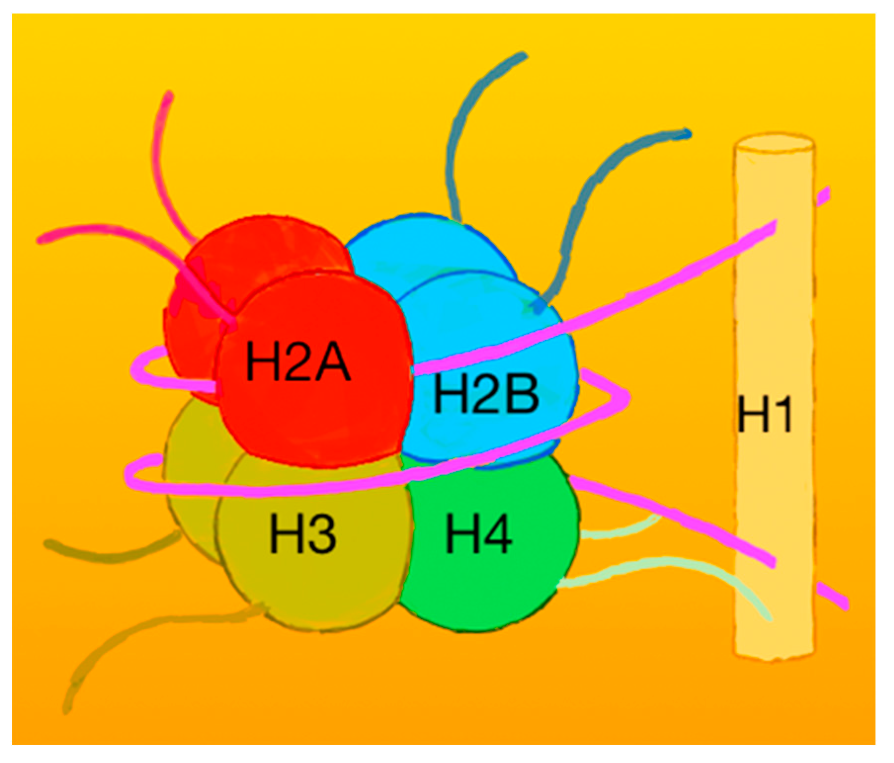
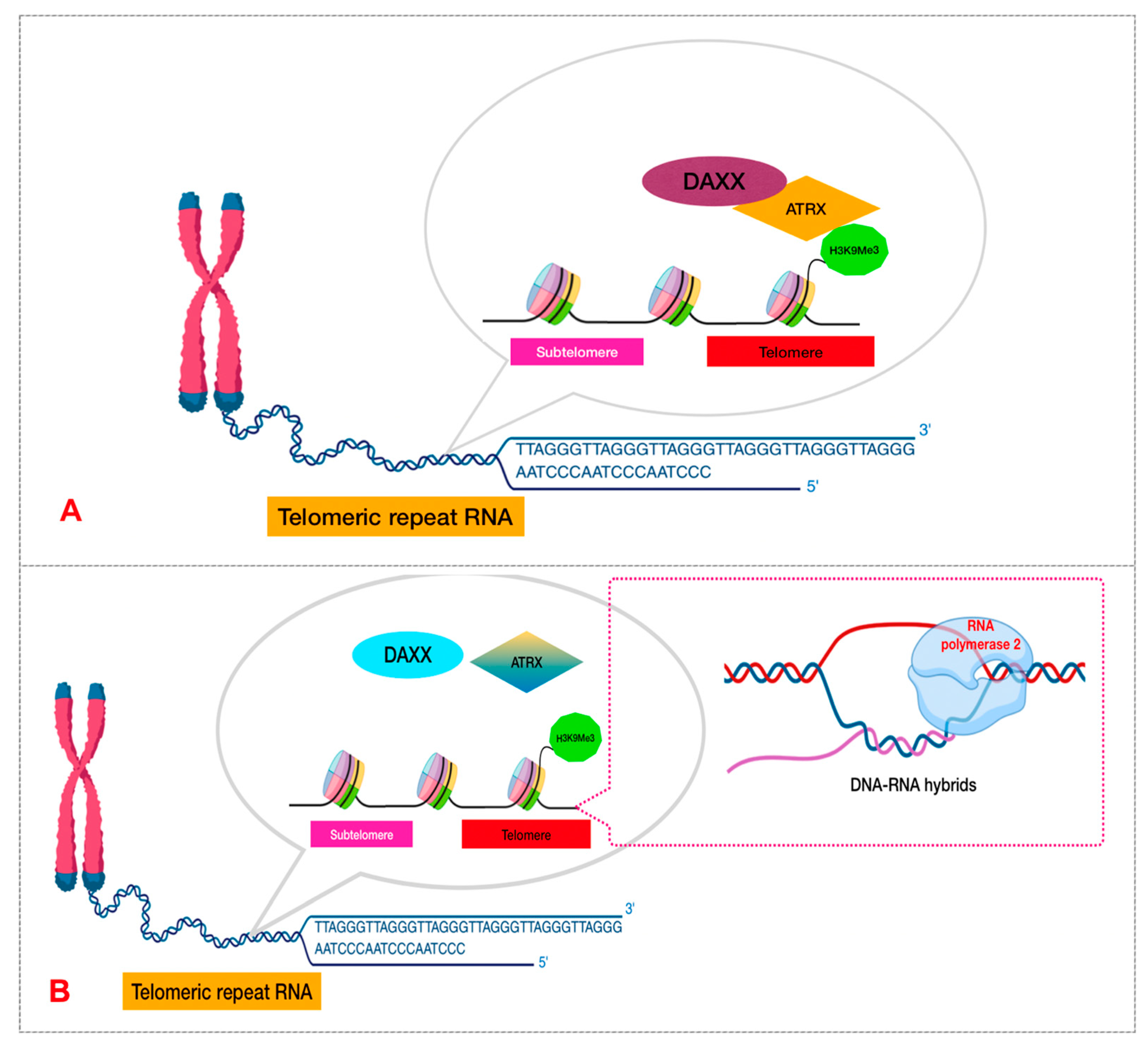
Histones and Nucleosomes

DNA of higher eukaryotes is found tightly swathed around an eight-histone complex (each of H2A, H2B, H3, and H4 are duplicated) comprising a nucleosome (Figure 7). Actually, half of the mass of a chromosome is formed by these proteins. This configuration does not only provide packaging, but also it participates in the epigenetic regulation of gene expression as well [60]. Two phases of chromatin interchange: the unwrapped euchromatin, which is liable to transcription, and heterochromatin. The latter corresponds to a dense DNA–histones complex not amenable to transcription [61].
Three major categories of H3 exist: (1) duplicated H3.1 and H3.2 conjuncts, which are named “canonical histones”, whose zenith production occurs throughout the S-phase. (2) The replacement variant H3.3. Its expression is irrelevant to the cell cycle phase and regulates the action status and maintenance of chromatin. The DAXX-ATRX complex integrates H3.3 into heterochromatic regions. Furthermore, H3.3 and ATRX participate in telomere perseverance throughout cell lineages differentiation [58]. H3.3 deposition relates to enhancers, promoters, and gene loci as well. Lastly, (3) the third category is the centromere protein [62].

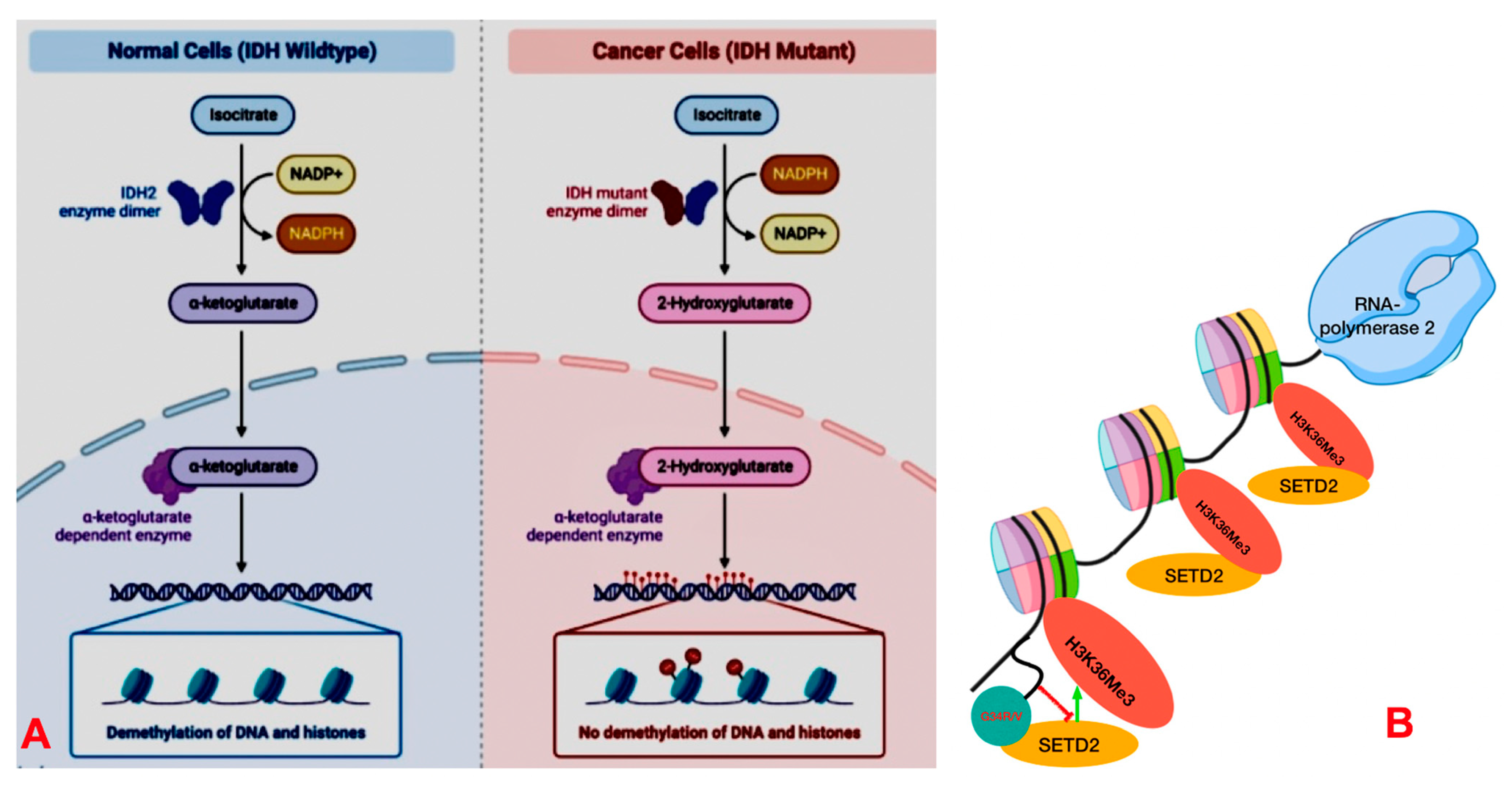
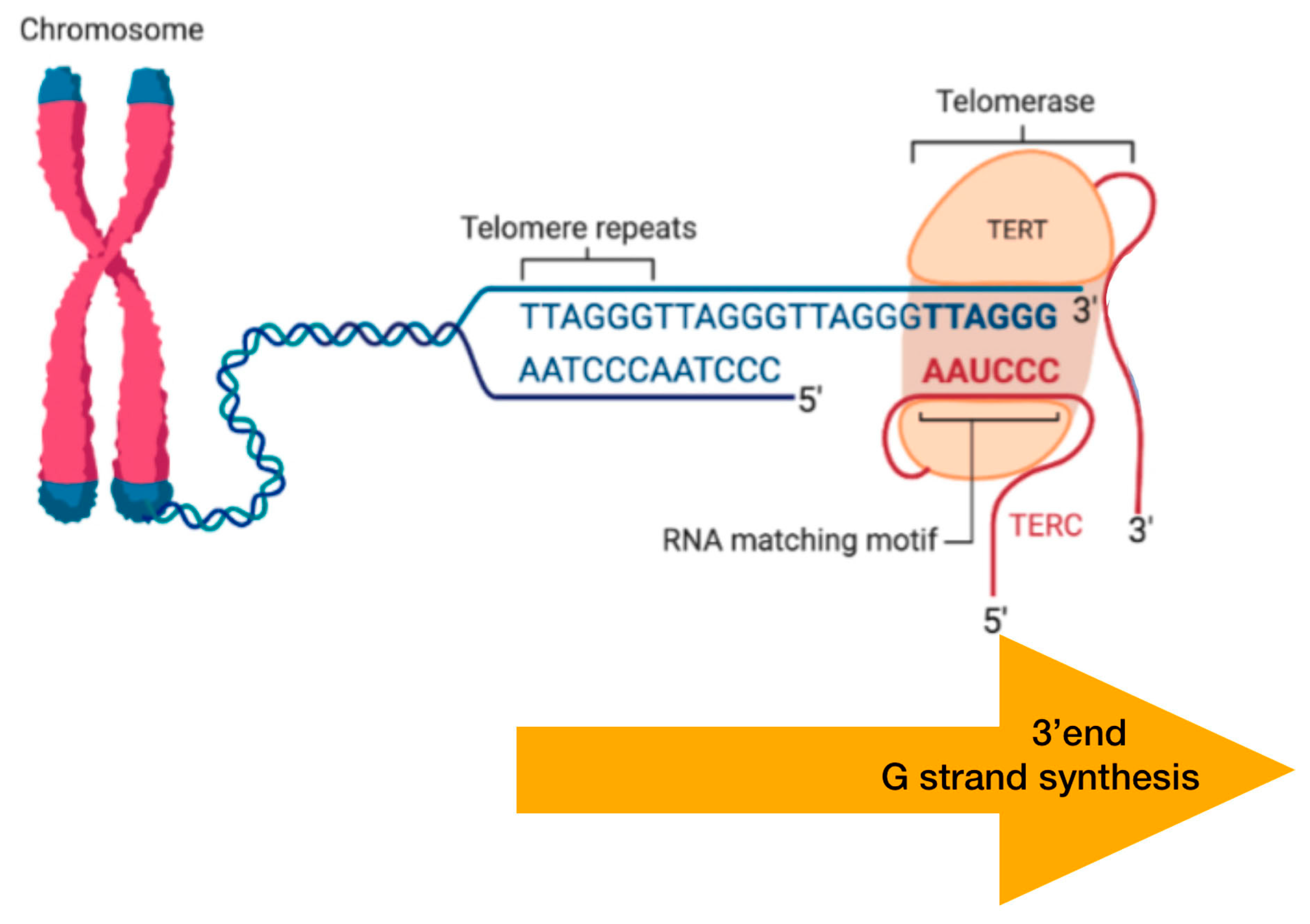
DNA Packaging and Methylation Control
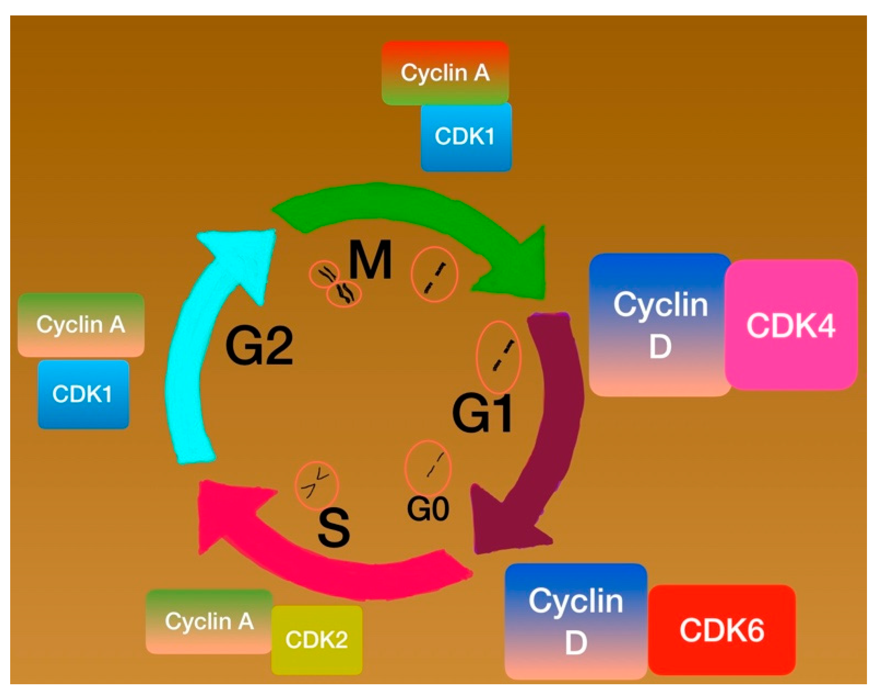
DNA methylation sustains the distinct genes’ expression throughout the cell cycle [63]. It actually confers a chromatin “silent” condition together with the proteins that configure nucleosomes [64,65]. Proteins responsible for DNA methylation act on specific CpG sites of the genome.
At this level, cancer initiation interrelates with mutations impairing the action of enhancer of zeste homolog 2 (EZH2), which participates in H3K27 methylation and is a part of polycomb repressive complex 2 (PRC2). Therefore, EZH2 fusions lead to hypermethylation of H3K27 [66]. On the other hand, deletions of EZH2 relate to hypomethylation of H3K27 target genes [67]. On the contrary, the switch/sucrose non-fermentable (SWI/SNF) protein group, otherwise encountered as the BRG1/BRM-related factor (BAF) complex, modulates transcription by reshaping chromatin using ATP [68]. Two other variant complexes also exist: the polybromo-associated BAF (PBAF) and the non-canonical BAF (ncBAF). This group of proteins facilitates DNA restoration, transcription, and nucleosome arrangement [69] (Figure 8). The transcription profile of genes that contribute to human hindbrain formation (e.g., HOX genes) is epigenetically regulated by the trimethylation of H3K27M. Deficient trimethylation of H3K27M, either due to histone alterations or EZH2 mutations, leads to a loss of the aforementioned transcription factor silencing. This molecular procedure occurs, especially in progenitor cells, and, thus, the tumorigenesis may be explained in areas like the brainstem or posterior fossa [64,66,69].
IDH1/2 mutations have an impact on a Krebs cycle. These mutations suppress the demethylating activity of these enzymes [70]. These alterations are rather infrequent (~6%) in children’s high-grade gliomas, and they emerge in the telencephalon at late adolescence. IDH and ATRX alterations coexist frequently in the same way there is an overlap between IDH and H3.3 alterations [70] (Figure 9A,B).
Telomerase is a transcriptase that identifies and elongates the telomeres (TTAGGG sequences) [71]. The TERT gene produces the functional enzymatic group of telomerases [71]. Telomerase activity ceases in non-neoplastic cell groups, and telomeres decrease in length throughout the consecutive cell cycles. In the end, upon a crucial telomere extent, cells start senescing. The vast majority of neoplasms encode this transcriptase [72] (Figure 10).
Alpha-thalassemia X-linked intellectual disability (ATRX) is a chromatin remodeler. ATRX acts in the setting of the DAXX/ATRX complex, which deposits the H3.3 histone isoform into the compact chromatin areas. Its inactivation leads to the alternative lengthening of the telomere (ALT) that is routinely performed by the telomerase reverse transcriptase (TERT) expression. Therefore, ATRX inactivation allows the perpetual division of cancer cells and, thus, the accumulation of additional mutations [12]. It belongs to the SWI/SNF group [73]. The deposition of H3.3 facilitates the trimethylating at the K9 position (H3K9me3) and inhibits transcription [74]. This deposition is situated at the telomere point and participates in DNA damage repair. DAXX acts as an ATP-unrelated satellite that deters pathological protein accumulation [75] (Figure 11A,B).
SETD2 restores chromatin and maintains transcription by methylating histone H3 on lysine 36 (H3K36). Mutations on H3G34 (arginine or valine instead of glycine) modify the transcriptional and epigenetic status [76] (Figure 9B).
The enzyme O-6-methylguanine-DNA methyltransferase (MGMT) inhibits the addition of alkyl groups at the O6 location of guanine [77]. The encoding of MGMT is inversely related to the (CpG) islands’ hypermethylation along the MGMT gene promoter. This fact confers liability to the chemotherapeutics, such as temozolomide (TMZ), which methylates this location and deters the DNA replication [78].

Cell Phases Control

The cell’s sequential phases secure the equal allocation of the genome into two descendant cells. The consecutive phases are actually four: G1 phase (gap1 phase), S phase (synthesis phase), G2 phase (gap2 phase), and M phase (mitosis) [79]. After cycle completion, cells may transit to an inert phase recognized as G0. At the G1 phase, D-type cyclins (D1, D2, and D3) are encoded and activate the Cyclin D-CDK4/6 complex. The latter deactivates, through phosphorylation, the retinoblastoma protein (pRb), which detaches from E2F transcription factor. This factor, together with Rb, previously constituted a complex that hindered the passage from the G1 phase to the S phase [79] (Figure 12).

Cyclins and Cyclin-Dependent Kinases (CDKs)

Cyclins are supervisory protein substrates with high specificity for their fermentation partner CDKs. The latter are proline-directed serine/threonine-protein kinases. They have a two-lobed configuration and modulate the shift during the cell phases [82]. The alterations undergo the supervision of transcription factors (TFs) or regulatory proteins like pRb, which suppress transcription [83]. The retinoblastoma 1 (RB1) gene is located at 13q14.p16. It is also encountered as either p16INK4 or CDKN2A. Loss of function of pRb leads to the disinhibition of proteins necessary for either the S period or mitosis [83] (Figure 13).

Check Point Proteins

The cyclin-dependent kinase inhibitors constitute a group of cell phase supervisors as well. The major activity is the configuration of secure complexes, along with the impedence of the cell phase transition. Prominent proteins, such as p15, p16, p18, and p19, attach firmly to CDK4/6 and hinder the CDK4/6–Cyclin D interaction [84].
p15 and p16 hinder the addition of phosphate molecules to Rb and passage to the S-period [80]. p16 is an onco-suppressor protein expressed by the INK4a/ARF locus of chromosome 9p21 [85]. p16 modifies Rb, and their expression is inversely correlated [86]. Rb1 modifies E2F transcription factors. When Rb1 is not impaired, D-type cyclins combined with CDK4/6 add phosphates to Rb1, and the latter detaches the inhibitory E2Fs [87] (Figure 12).
The p53 gene lies at 17p13.1 and encodes TP53 protein. All normal cells express this, with its normal half-life protein being minimal. The TP53 protein‘s function concerns the sequence-based transcription. Alterations of the p53 gene are usual in high-grade malignancies. The p53 mutations respond to patients with more brief disease-free phases and poorer outcomes [86,88].
Almost 40% of HGGs in children relate to alterations in the p53, and in contrast to high-grade gliomas in adults, those HGGs in childhood have been correlated with improved 5-year progression-free survival (PFS) [89].

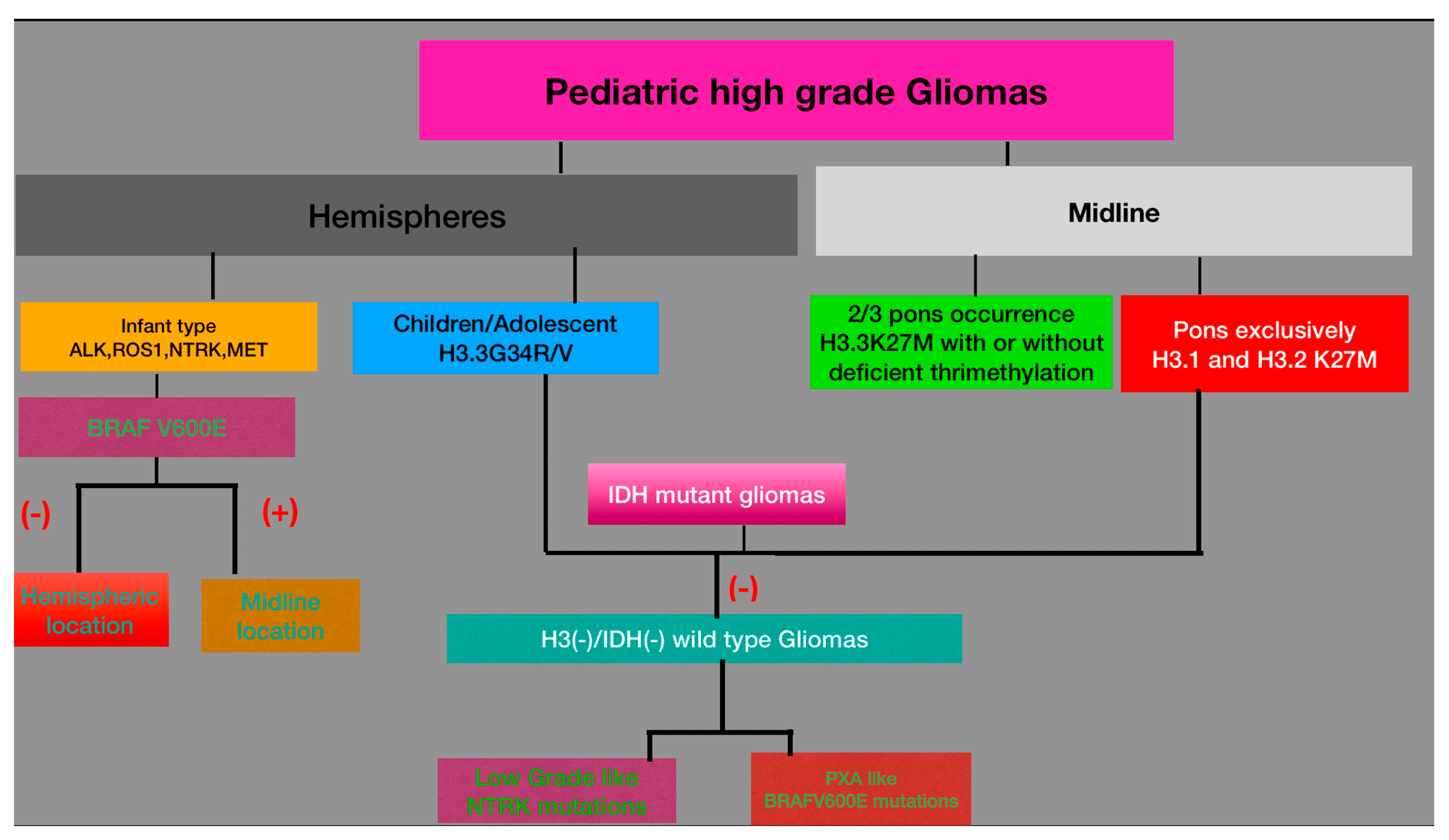
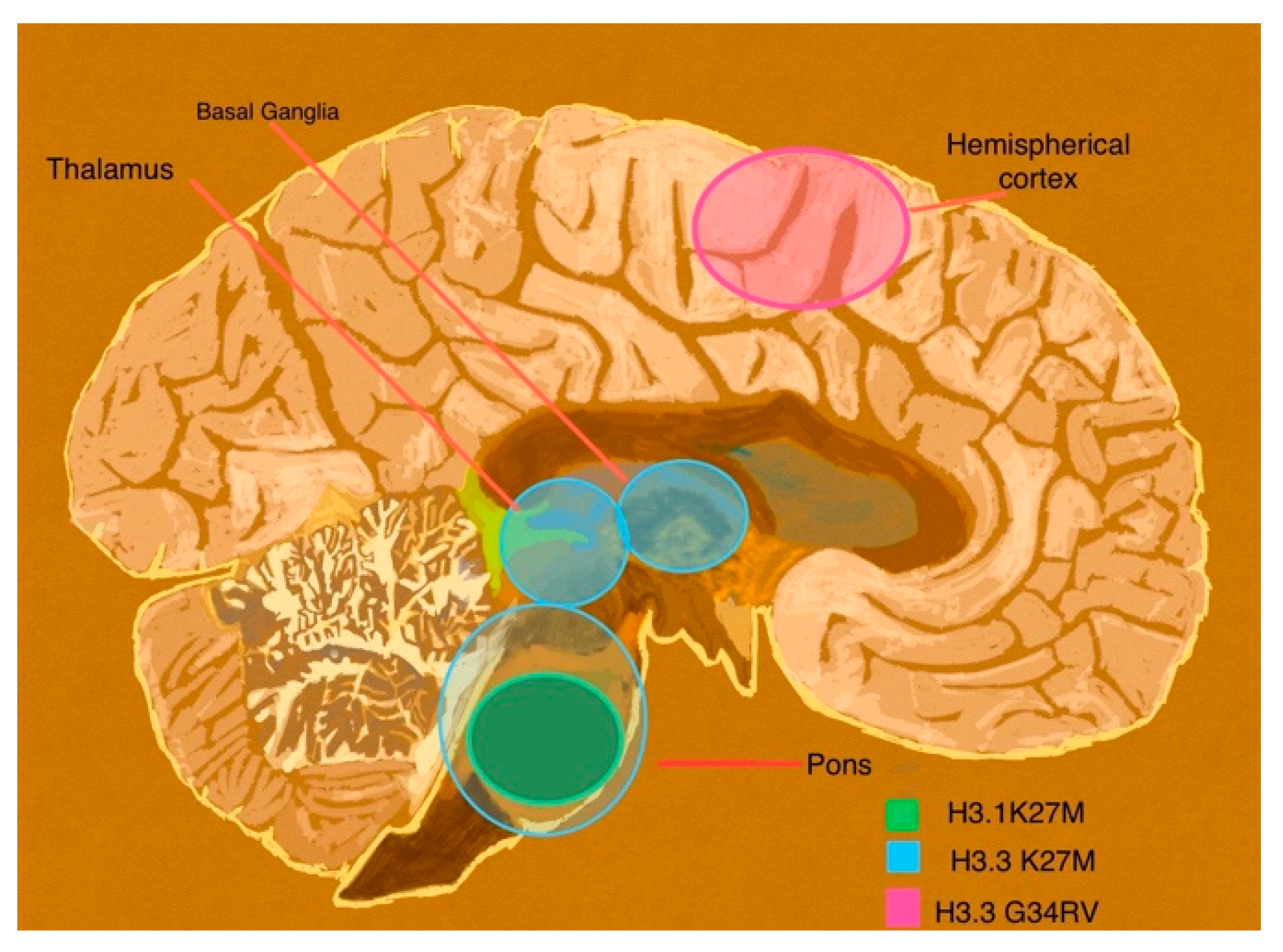
3.2.2. Diffuse Pediatric Midline Glioma (DMG)

This tumor is a WHO grade 4 astrocytic lesion of the brainstem or the midline. These pediatric tumors are typically located in the pons (diffuse intrinsic pontine glioma—DIPG), or they may extend to both thalami [90]. In adolescents and adults, they invade only one thalamus or the spinal cord, respectively [91]. DIPG bears the worst prognosis, with a 2-year survival of less than 20% [92]. The hallmark is the loss of trimethylation at H3K27 and is subclassified into three types: (1) DMG, with H3K27 alterations of either H3.3, H3.1, or H3.2-variant; (2) DMG, H3-wild-type without K27 alterations, but excessive function of enhancer of zeste homologue inhibitory protein (EZHIP) and hypomethylation at K27 location; and (3) DMG, with EGFR mutations [93]. The DMG H3.3 K27-mutant exists in children 7–8 years old and lies in the midline or the pons; its median OS counts 10 months. A total of 25–50% of DIPG carry K27M aberration of the H3.1 isoform [59].
H3.1 and H3.2 K27–altered DMG appear exclusively in the pons of children aged 5 years old, and its OS rate is approximately 15 months [94]. The DMG H3K27-mutant, compared to the DMG H3-wild-type, characteristically exhibits both hypomethylation and hyperacetylation at the H3K27 location [95]. H3 K27M mutations restrict PRC2 activity by sequestration of its catalytic subunit (EZH2) [96]. PRC2 target genes are involved in developmental processes, and, as a result of hypomethylation, they are all upregulated, and its tumor-suppression action is diminished [95]. Generalized deficiency of H3K27me3 overlaps with a reciprocal overall excess of H3K27 acetylation that is copious in the proximity of bromodomain-containing proteins BRD 2/4. The latter are supervisors of RNA Polymerase II throughout the transcription. Therefore, H3 K27M acetylation presumably enhances transcription [58,97]. The loss of H3K27 trimethylation coexists with BRAF V600E substitution and, less commonly, with IDH1 alterations [93]. The H3-wild-type DMG is associated with EZHIP excessive production. It is not so frequently encountered and emerges in common cerebral areas and within the same time spectrum as the H3.3-altered type [93,98]. EGFR-mutant DMG invades the thalami bilaterally. Its target group is school-aged children [99].

3.2.3. Diffuse Pediatric Hemispheric Glioma

Prognosis is generally ominous. Median survival ranges from 9 to 15 months, and hardly one-fifth of the patients survive up to 60 months [12]. Other than ionizing radiation, no other environmental risk factor has been identified so far [100]. Genetic inheritance is implicated in pediatric high-grade gliomas, such as NF1, Turcot syndrome, and Li–Fraumeni syndrome [92].
This tumor carries the H3 G34 mutation. There are two subtypes, according to the amino acid interchange during H3 expression: the G34R (arginine substitutes glycine) or the less frequent G34V (valine substitutes glycine) [101]. It is a highly infiltrative WHO grade 4 lesion that originates in the forebrain structures, especially in the temporal and parietal lobes. The median age of occurrence is the middle of adolescence [94].
TP53 coexists in about 40% of cases, while ATRX and (DAXX) alterations usually co-occur; the MGMT promoter (O6-methylguanine-DNA methyltransferase) is frequently methylated [100,101]. Furthermore, 80% of these tumors disclose excessive function of the PI3-Kinase/Akt/mTOR cascade. This is attributed to PTEN promoter methylation [92]. Approximately 60% of this type carry PDGFRA alterations [102]. Finally, H3.3 G34R/V has been somehow related to MYC/MYCN robustness, and when that happens, MYC upregulation occurs through differential H3K36me3 binding. The MYCN upregulation is considered a driver of tumor development [94]. In hemispheric pediatric HGGs, K36 trimethylation (K36me3) is decreased. In general, K36 can only be methylated with three methylate groups by SETD2 (SET domain containing 2 protein) methyltransferase. SETD2’s normal function is the junction of histones’ flexible appendix to a protein’s furrow. This process fails in the case of H3.3 G34R/V mutation due to the fact that protein’s furrow does not engulf an amino acid greater than glycine [12]. The glioblastoma-like type exhibits massive expression of glial fibrillary acidic protein (GFAP) [102]. The median survival is hardly two years. The MGMT promoter methylation portends a better outcome, whereas amplification of oncogenes (e.g., EGFR, CDK4, and MDM2) relates to an ominous prognosis [101]. The isolations of PDGFRA mutations in half of patients confer innovative treatment chances [102]. Molecular profile correlates strongly with location [103] (Figure 14).

3.2.4. Diffuse Wild Type High-Grade Gliomas

These gliomas exhibit methylation variations and are classified as pHGG (receptor tyrosine kinase) RTK1 (with copious PDGFRa amplification) or pHGG RTK2 (with copious EGFR amplification). Finally, a subgroup with n-Myc amplification has been observed [104].
The great proportion of these tumors arise supratentorially. A small part of pHGG MYCN (responding to 15%) appears in the brainstem [101]. pHGG RTK1 emerges both infratentorially and in structures within the brainstem [101]. The outcome has been considered dismal, and median OS counts for almost one and a half years. On the contrary, RTK2 pHGG interrelates with a median OS of 44 months and pHGG MYCN 14 months [105]. Pontine tumors are the most malignant of HGG-MYCN, and OS rates are 1.5 months [106]. PDGFRA amplification commonly results in activation of the PI3K/mTOR or MAPK cascades. They relate to worse prognosis [107].
pHGG RTK1 type encompasses the majority of irradiation-associated gliomas [106]. The occurrences of TP53 and ATRX mutations are associated with adverse outcomes [108]. pHGG BRAFV600E mutants (WHO-grade 3 PXAs) have a 24-month OS in 67% of the cases [94]. They coexist, however, with alterations like CDKN2A/B deletion or TERT promoter mutations. The aforementioned alterations result in benign tumors with ominous outcomes [109].

3.2.5. Infant-Type Hemispheric Gliomas

These gliomas include three types: (1) hemispheric (RTK)-promoted malignancies, including anaplastic lymphoma kinase (ALK), c-ros oncogene (ROS1), neurotrophic receptor kinase 1 NTRK, and mesenchymal epithelial transition (MET) fusions. This group exhibits a moderately poor outcome, (2) hemispheric RAS/MAPK-related lesions that demonstrate prolonged OS under short-term adjuvant therapy, and (3) midline RAS/MAPK-related neoplasms with a rather dismal prognosis in the setting of novel chemotherapeutic protocols [110].
ALK mutations align with LGGs (10-year OS 53.8%), whereas ROS1/MET (25%) and NTRK fusions (42.9%) align with HGG. The second group does not include BRAF mutations and has the best 10-year OS among the three groups (93%). The dismal prognosis of BRAF-fused midline neoplasms up to the first year of life is remarkable, and their behavior is completely different than in older age groups (5-year OS rates 23%). This discrepancy is imputed to both age-related genetic and microenvironment agents [110]. BRAF V600E alterations in PXAs have been extensively reported. PXAs tend to upgrade to more malignant lesions, especially when they combine with CDKN2A deletions [111]. Interestingly, the frequency of BRAF alterations in pHGG varies from 10% to 25% [112]. Therefore, BRAF alterations in the high-grade lesions probably exist in the setting of secondary pHGG [113].

3.3. Current Treatment Approaches
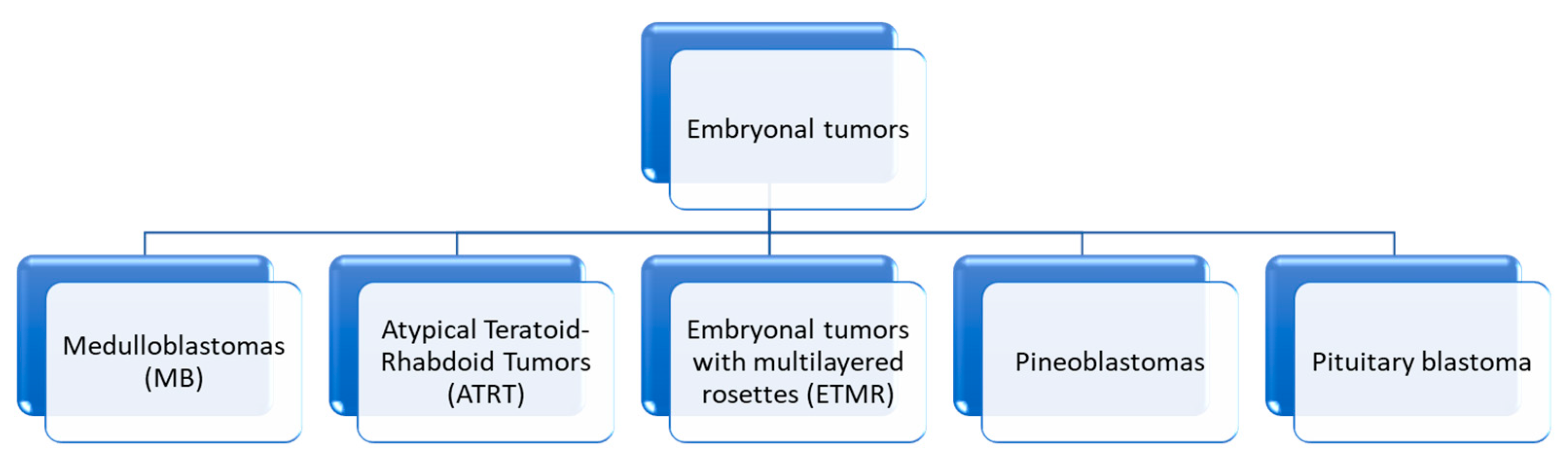
Pathology of pLGG constitutes an independent prognosticator of outcome. Therefore, the well-demarcated PAs are amenable to complete excision [114]. Extent of surgical excision is the predictor for prolonged progression-free and OS outmatching pathology [115,116]. The 60-month progression-free survival (PFS) varies from 75% to 100% of cases [117]. Regular clinical and radiological reevaluations are necessary. Chemotherapy is administered in case of recurrence [118]. Postsurgical irradiation following subtotal excision provides longer PFS, but not OS [119]. The precise involvement of BRAFs in PA is not clear; however, its inhibitors participate in the therapeutic planning [120].
High-grade glial-originating neoplasms constitute a hugely inhomogeneous category with specific alterations and prognoses, which warrant tailored targeted therapy. Surgery for pediatric HGGs has three primary objectives: (i) obtain samples for identification, (ii) relieve elevated intracranial pressure, and (iii) tumor cytoreduction. Stereotactic needle biopsy is indicated for deep or ineloquent areas seated malignancies. Surgery is followed by radiotherapy and temozolomide administration. This schema does not markedly contribute in the pediatric population, though [121]. The 60-month OS is confined to 20%, and the median OS is between 12 and 42 months [122].
Children with GBMs and MGMT promoter methylation exhibit 13.7 months OS in contrast to hardly three months for those that do not harbor the methylation [123]. Excessive activation of Akt shortens 12-month failure-free survival compared with gliomas with none [124].
Both location and consequent prognosis correlate with molecular profile. PDGFRa usually undergoes amplification [125]. Non-mutated PDGFRa lesions occur in the brainstem, whereas PDGFRa somatic alterations predilect tumor emergence far from the brainstem [126]. Frequently, pHGGs are erroneously characterized as pLGGs. This had been the rule with pLGGs according to the previous WHO classifications, which did not evaluate H3.3K27M mutations. Another reason is the limited and insufficient tissue sampling, which excludes malignant areas, in cases of non-homogeneous lesions. Overall PLGGs carrying either H3.3 K27M or TP53 alterations constitute high-grade-behaving gliomas [113].
In case of recurrent pediatric HGGs, besides the fact that prognosis remains unfavorable, a palliative treatment plan has to be established. Another schema of radiotherapy with 30–54 Gy confers clinical stabilization and survival enhancement even in DIPG cases [127].

4. Embryonal Tumors

This group involves tumors whose microscopic characteristic is the presence of small round cell populations. This tumor group is, in fact, genetically diverse [128]. Medulloblastomas, atypical teratoid/rhabdoids, and embryonal tumors with multilayered rosettes are the main representatives (Scheme 2).

4.1. Molecular History

4.1.1. E-Cadherin/Beta-Catenin Complex

The wingless (Wnt) signaling hinders the addition of phosphates and decomposition of the beta-catenin protein. The latter aggregates in the cytoplasm and translocates to the nucleus. It acts as a transcription cofactor and enhances cell proliferation [60]. Decreased activation of the E-cadherin/beta-catenin complex implies high malignancy [60].
Alterations of the CTNNB1 lead to the accumulation of b-catenin and bear resemblance to the excessively functioning Wnt pathway [60] (Figure 15).

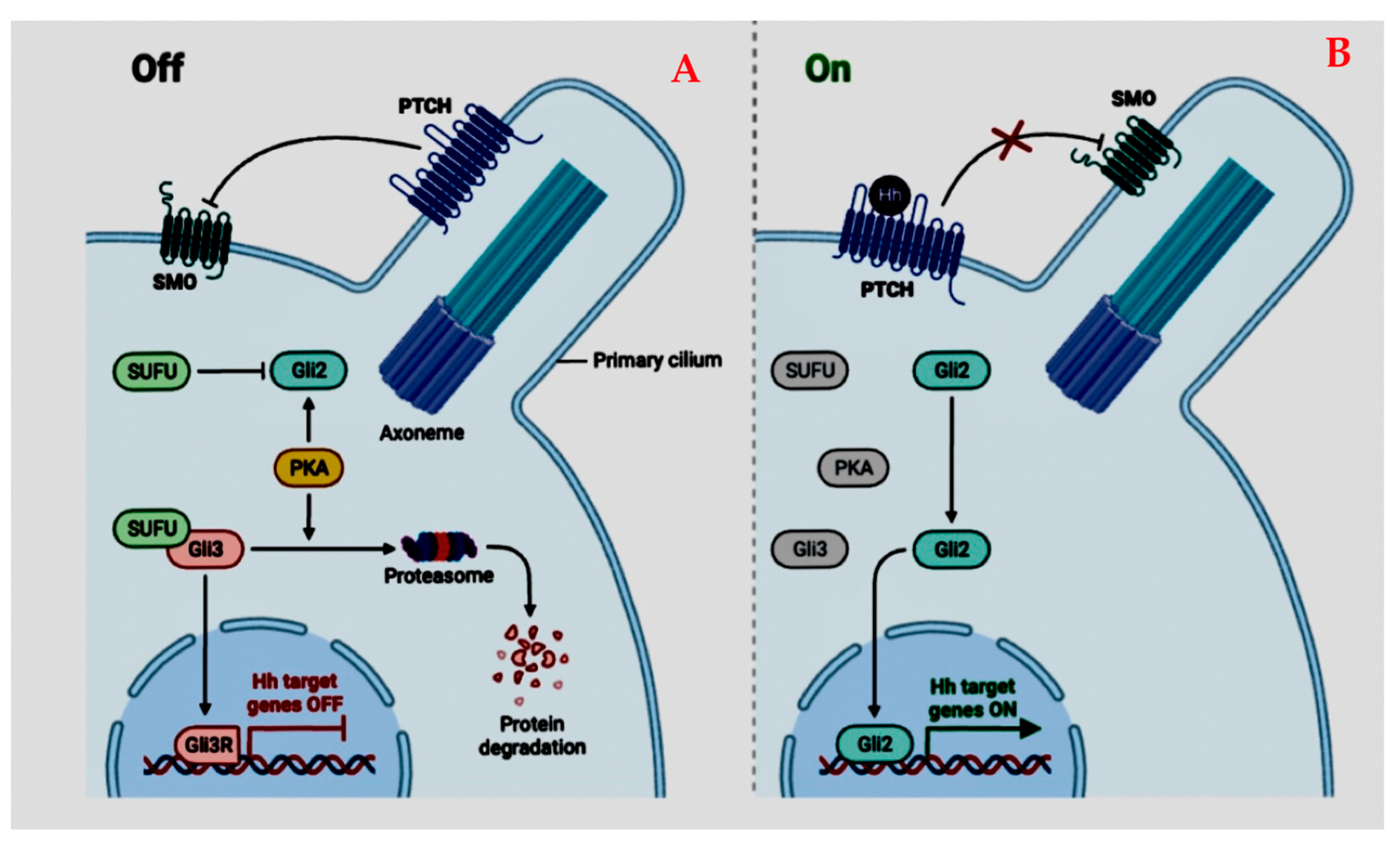
4.1.2. Sonic Hedgehog Pathway (SHH)

The sonic hedgehog (SHH) canonical pathway initiates when the glycoprotein SHH attaches to and impairs the functioning of the transmembrane protein, Patched 1 (PTCH1). When the SHH-binding molecule is absent, the transmembrane protein smoothened (SMO) is inhibited by PTCH1. Hence, the joined molecule of SHH/PTCH1 modulates SMO function [129]. SMO is a GPCR-like (G protein-coupled receptor) protein, and its translocation into the cilia membrane activates Gli [129]. In response to SHH signaling, PTCH1 inhibition of SMO is abolished [130]. When PTCH1 is decomposed, SMO aggregates and incites the downregulation of the SHH pathway (Figure 16).
This pathway enables Gli proteins to enter the nucleus. Expression of the Ptch1 gene enhances transcription, whereas encoding of Gli1 hinders it [131]. Vice versa, GLI entrance to the nucleus regulates the expression of WNT and Noggin [131]. The SHH cascade is also regulated by Suppressor of Fused Protein (SUFU). When the SHH ligand is absent, SUFU attaches immediately to GLI proteins and impedes their presence at the nucleus [131].

4.1.3. Medulloblastomas (MB) (Figure 17)

MBs represent a diverse category of neoplasms. Normal development and tumorigenesis share changes in cell growth, migration, and death [132]. MBs are assumed to emanate from precursor cells of the external granular layer (EGL) of the hindbrain [133]. They are initially located around the rhombic crest during organogenesis. Granular neural cell growth is driven by the SHH pathway [134]. One out of two MBs reveals the aberrant initiation of the SHH or Wnt cascades [135]. A good interaction between them regulates the neural crest formation [136]. These pathways induce expression of MYCN. Excessive MYCN encoding incites development and transposition of neuroblasts, while an attenuated encoding leads to terminal differentiation [137]. According to a 2012 international consensus study on molecular analysis of medulloblastomas, four distinct types with different prognoses were introduced. These involved MBs with aberrancies of WNT cascade, SHH pathway, Group 3, and Group 4 [138] (Table 2). More recently, WHO advocates the usage of WNT, SHH with and without TP53 mutation, and non-WNT/SHH molecular groups [139].
Figure 17. (A) MRI. T1 sequence. Midline vermian intraventricular tumor, which enhances after contrast medium administration. (B) Increased diffusion in the DWI sequence indicative of high cellularity. (C) Hematoxylin–eosin stain: Areas of high cellular concentration. Cells are majorly spherical, with an increased nuclear–cytoplasmic ratio.
Cancers 17 01566 g017
WNT-activated MBs are the most infrequent (10%) and are observed in late adolescence. They are seldom encountered in ages below one year [140]. These tumors emanate from the inferior rhombic crest and dorsal rhombencephalon. They tend to occupy the cerebellopontine angle and lie along the lateral foramen, cisterna magna, or fourth ventricle [141]. Molecular detection of CTNNB1 mutation is recommended. The great proportion of these lesions coexist with monosomy 6. Additional ignition of the SHH cascade is sometimes documented [142]. It can coexist in the context of Turcot syndrome (10%) when APC mutations exist [143]. They reveal excellent prognosis in children, and metastatic disease is rare. They hardly recur, and their 5-year OS is greater than 90% [142].
SHH-activated MBs (SHH MBs) comprise 30% of all MBs. They emanate from cerebellar granule neuron precursors and are located in the cerebellum [142]. Apart from the SHH pathway activation, this group may carry the TP53 mutation, which has repercussions for prognoses. MBSHH has a great variance of forms [144]. SHH MB wild-type TP53 has a two-peak age distribution. It concerns children younger than one year of life and the period shortly after adolescence. No differences in sex distribution exist [145]. Extensive nodularity in histology is characteristic and concerns infants. PTCH1 or SUFU alterations are usually responsible for the SHH cascade ignition [143]. Patients with Gorlin syndrome, who carry SUFU mutations, are liable to MB occurrence at a young age [145]. SHH MB with altered TP53 is infrequent and manifests in school-age children and teens. They have a worse prognosis with a poorer response to therapy. On the contrary, TP53 wild-type tumors during infancy are low-risk [142]. Germline TP53 mutation detection is included in the diagnostic workup when Li–Fraumeni syndrome is suspected [145].
Group 3 MB concerns almost one-fifth of MBs. Infants constitute one-half of the affected population, with a preponderance for the male sex. In almost half of the cases, metastases exist [138]. This group is hardly observed in adults [145]. These tumors originate from neural crest cells and are ordinarily situated in the cerebellar vermis near the fourth ventricle [146].
A variant of MB3 reveals increased copies of c-Myc, which signifies an ominous outcome [142] (Figure 18). Additional molecular alterations involve SMARC4, a subunit of the SWI/SNNF complex [142,143]. This group is linked with the worst prognosis because of the tendency to metastasize and c-Myc excessive expression [142].
Group 4 comprises 40% of all MBs. These tumors appear predominantly in school-aged children and young adolescents. Males have a threefold higher risk of developing these tumors [145]. They emanate from the superior rhombic crest and extend to the cerebellar vermis [97]. MB4 coexists with isodicentric chromosome 17 and manifests n-Myc amplification [143] (Figure 18). They tend to metastasize, and this is the main prognosticator [146].

4.1.4. Treatment Status

Radiation Therapy

Adjunct to surgical excision RT led to advances in the survival of patients. The 5-year OS counts are 50–65% with RT alone [147]. No general agreement exists about the cut-off age regarding the delay of employment of craniospinal RT. Ranges vary from 3, and in some studies, to 4 or 5 years old [147]. Treatment with proton beams may confer reduced long-period toxicity. In the short-term, though, demarcated necrosis, especially in the brainstem, exists [148].

Chemotherapy in Standard-Risk Patients

The long-term sequelae of this treatment are remarkable, especially in preschoolers and school-aged children. Combined chemotherapy and radiotherapy result in significantly lower doses of craniospinal radiation [148]. The results revealed a favorable 60-month OS in 79% of the patients, compared to studies without chemotherapy up to that time. Furthermore, long-period neuropsychological assessments may improve intellectual outcomes [147].

4.2. Atypical Teratoid/Rhabdoid Tumors

This group is considered a highly aggressive neoplasm. Its hallmark mutation regards the chromatin modifier group B member 1 (SMARCB1) that constitutes a subunit of the SWI/SNF molecule (Figure 8). These tumors appear in infancy and early childhood. They exhibit very poor survival [149]. Recently mutations in SMARCA4 have also been described [150].
Four subtypes are described based on the methylation profile [151].
The TYR subtype relates to the monosomy of chromosome 22 and point alterations of the Smarcb1 gene. This group is predominantly located at the posterior fossa and demonstrates hypermethylation of their DNA.
The SHH subtype, which is imputed to either focal deletions or point mutations of the Smarcb1 gene, has the greatest incidence among all groups. These lesions are frequently situated supratentorially and present hypermethylation in their DNA as well. They carry abnormal SHH signaling.
The MYC subtype is characterized by broad deletions of the Smarcb1 gene. The Myc genes are overexpressed. It has almost equivalent presence in both supra- and infra-tentorial areas.
The SMARCA 4 subtype is the least common. Its features are the point mutations of Smarca 4.
Reported therapeutic protocols involve combined treatment with resection, irradiation, and chemotherapy. The latter involves either the conventional protocols or high-dose chemotherapy (HDC). In this case autologous hematopoietic progenitor cell rescue (AHPCR) should precede, and intrathecal (IT) administration should be a part of the protocol. Significant presupposition remains, though, about the degree of the malignancy’s excision [150].

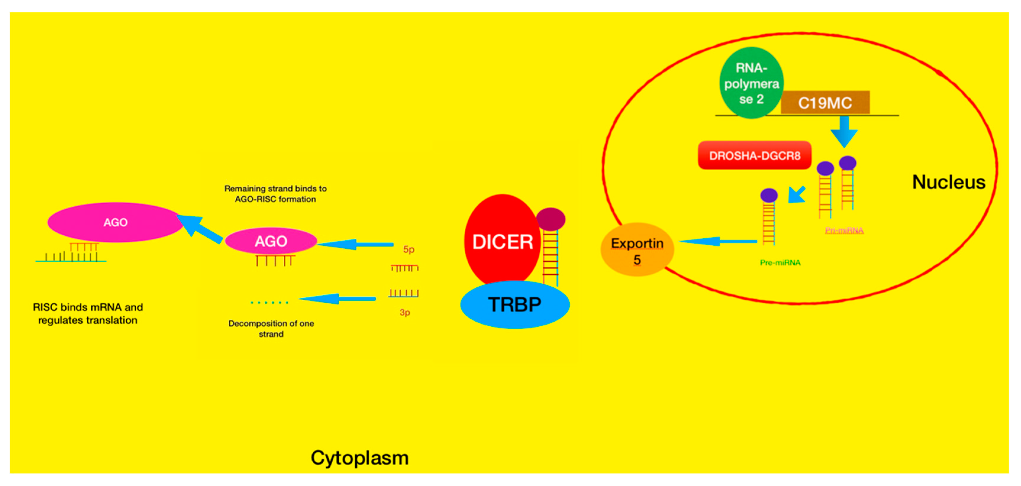
4.3. Embryonal Tumor with Multilayered Rosettes (ETMR)—C19MC-Altered) and Embryonal Tumor with Multilayered Rosettes—Not Otherwise Specified

4.3.1. ETMR Tumors

ETMR is a group of tumors whose origin is attributed to the presence of chromosome 19 microRNA clusters (C19 MC). The group manifests either excessive copies of the respective gene or annexation with the TTYH1 gene. This novel group engulfs the older entities of embryonal tumor with abundant neuropil and true rosettes (ETANTR), ependymoblastoma, and medulloepithelioma [152]. They occur in toddlers or even in younger groups [142]. They appear predominantly in cerebral hemispheres and occasionally extend to both of them. They may carry cystic regions and calcifications. Their histological hallmark is the true rosettes accompanied by neuroblasts [142]. ETANTR contains areas of embryonal minuscule stratified spherical blue cells and true rosettes, combined with scant cytoplasmic neuropil domains [153]. Ependymoblastomas also contain multiple stratified rosettes, primitive cells, and sometimes fibrillary processes. They lack neuropil. Medulloepitheliomas do not exhibit a significant amount of neuropil. Mature neurons and astrocytes intermingle with embryonal cells [142].
Except for C19 MC mutations, the gaining of chromosome 2 is usually encountered [142]. The lin-28 homolog A antibody is an immunological substitute for C19 MC alterations; however, it is not specific [154]. Further molecular profiling showed biallelic alterations of the DICER1 gene (Figure 19). The outcome is poor. They relapse and often metastasize [155]. The gold standard of treatment involves the maximum safe resection and chemotherapy; albeit, the median OS remains one year [155]. Radiotherapy, in particular proton therapy, has recently been shown to prolong survival [156].

4.3.2. Pineoblastomas (PBs)

These are excessively invasive pineal neoplasms with poorly delineated tumor margins. They appear within the first twenty years of life. They exhibit embryonal tumor morphology with true rosettes and sometimes variable differentiation [158]. Epigenetically, pineoblastoma is inhomogeneous and is composed of five separate fundamental molecular subgroups with distinct courses [159]. These subgroups demonstrate aberrant microRNA transcription. Succinctly, they are the DICER1, the DROSHA (DGCR8), and the KBTBD4 subgroups [160]. Older children are usually affected, and the outcome is good after irradiation (5-year OS is 60–100%) [161]. The remaining two subgroups are PB-MYC/FOXR2 and PB-RB1. Their distinctive feature is the oncogenic MYC-miR-17/92-RB1 circuit. They concern infants and young children. Irradiation is, thus, deterred and associated with bad outcomes, with 5-year OS varying from 0% to 25% [161,162].

5. Pediatric Ependymomas (pEPNs)

Pediatric ependymomas constitute the third most frequent intracranial tumor. This group emerges in all compartments of the CNS. The most frequent locus is the posterior fossa [163,164]. A slight overall male predominance has been documented [164]. The greatest proportion (90%) of pEPNs arise intracranially. A total of 66% of them are situated in the posterior fossa (PF) and the rest of them are positioned supratentorially [165].

5.1. Identity Molecules

Overall, ependymomas originate from radial glial cells and mutations of genes that regulate the proliferation and specialization of neural precursor cells. A copy of the chromosome arm 1q constitutes a predictor of a dismal outcome, especially in the PF ependymomas during childhood [165].

5.1.1. NF-kB Pathway

Canonical NF-kB signaling reacts to tumor necrosis factor-a (TNFa) and interleukin-1 (IL-1) signaling inflammation cytokines. The NF-kB activation relates to RelA- or cRel-containing complexes. The suppression of NF-kB when the inflammation is supposed to cease prolongs the inflammatory response and deters apoptosis [166]. Interestingly, NF-kB constitutes an important inhibitor of pathogen-induced apoptosis of macrophages, at least in vitro. Thereby, NF-kB promotes inflammation via macrophage activation [166] (Figure 20). Initiating genes in the majority of supratentorial ependymomas are fusions between RELA of the NF-kB pathway and the gene C11orf95 caused by a breakage of chromosome 11. This fusion of stem cells can independently promote neoplastic cell proliferation [167].

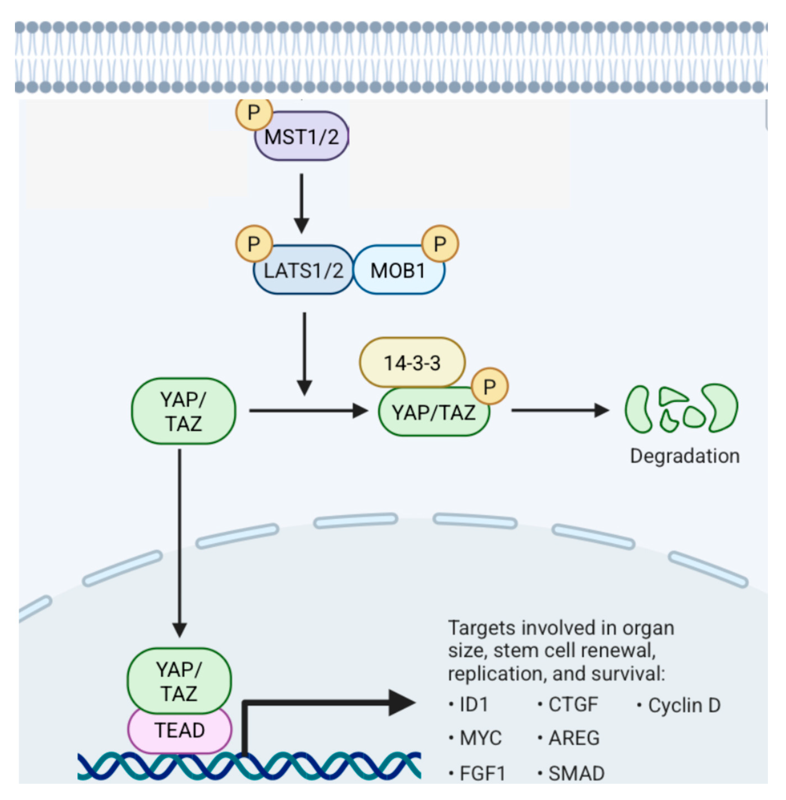
5.1.2. Hippo Pathway (Figure 21)

The mammalian onco-suppressor Hippo pathway is composed of mammalian sterile-like kinase 1/2 (MST 1/2), Salvador homolog protein 1 (SAV1), large tumor suppressor kinases 1/2 (LATS1/2), monopolar spindle-one-binder protein 1 (MOB1), and yes-associated protein/tafazzin family protein (YAP/TAZ). When the Hippo signaling is active, YAP/TAZ receives phosphate molecules and is either restricted in the cytoplasm or spotted for disintegration. When signaling is inactive, YAP/TAZ enters the nucleus to ignite the transcriptional enhancer factor domain family (TEAD) [168]. Mechanical traits are significant stimuli by which cells apprehend their microenvironment. Hippo’s signaling is promoted by these mechanical stimuli [168]. The frequency of the cells multiplication is inversely proportional to the cell compactness [169]. LATS becomes functional compared to YAP, which remains inert. Furthermore, the overexpression of YAP could reverse the inhibition of growth induced by cell density. Overall, YAP/TAZ is a significant suppressor of Hippo signaling [168].
Figure 21. Hippo pathway. MST1/2, SAV1, LATS1/2, YAP, and TAZ are significant components of the signaling. When the Hippo pathway functions, MAP4Ks, MST1/2, and its docking protein, SAV1, add phosphate molecules to LATS1/2 and MOB1A/B [170]. Functional LATS1/2 adds phosphates and suppresses YAP and TAZ, obstructing them from entering the nucleus. MST1 and MST2 are serine/threonine kinases whose activity can be enhanced after their binding with the docking protein SAV1 [168].
Cancers 17 01566 g021
For PF EPNs, two distinct molecular subgroups have been consistently identified [171]. These were the PF Group A and Group B, or else ascribed as PFA and PFB [172].

5.2. Supratentorial Ependymomas (ST EPNs)

RELA fusions have been solely detected in the supratentorial-RELA subgroup. More than two-thirds of ST EPNs bear a combination of C11orf95–RELA fusions [167]. C11orf95–RELA fusion proteins enter and incite the canonical NF-κB cascade [165]. These ependymomas manifest a copious capillary network [173]. RelA protein is detected through immunohistochemistry, which is a cost-effective method [174]. The tumor has been traditionally associated with low survival rates [165]. However, recent findings in an exclusively pediatric population have revealed no prognostic disadvantage compared to other subtypes [175].
The other type of ST EPNs is interrelated with amplifications on chromosome 11 at the YAP1 locus [172]. The most well-studied mutation is the YAP1-MAMLD1 fusion. The latter appears exclusively in school-aged children and has an excellent outcome after complete excision, even without adjunct treatment [175,176]. There is another group of supratentorial ependymomas in children that bear the homozygous deletion of CDKN2A [172]. In pediatric ependymomas, CDKN2A deletion coexists with Rela fusion [165]. Lastly, ST EPNs, without the aforementioned molecular alterations, are rare and warrant further observation [177].

5.3. Posterior Fossa Ependymomas (PF Ependymomas)

Apart from histological grading in classic and anaplastic ependymomas, prognosis relies on the distance from the midline [165]. Furthermore, malignancies with chromosomal mutations carry an ominous prognosis, especially those with an addition of the q-arm of chromosome 1. Ependymomas of the posterior fossa can be divided into two defined groups [165], which are discussed in detail in the following paragraphs.
PF-A EPNs appear majorly in school-aged children. Boys are usually afflicted with worse survival [13]. PF-A tumors do not harbor alterations at a specific genetic location but rather gains on the long branch of the 1st chromosome in approximately 20% of the cases [171]. Furthermore, excessive additions of methyl groups at CpG-island are present in PF-A tumors [171]. This is attributed to excessive encoding of EZHIP (CXorf67) (enhancer of Zeste inhibitory protein) [165]. EZHIP dampens the action of the PRC 2 complex, which suppresses the expression of oncogenes by trimethylating the H3K27 [178]. Diminished trimethylation at the H3K27 location is the hallmark of PF-A EPNs [179]. The deficient methylation pattern is estimated by immunohistochemistry. PF-B ependymoma is found primarily in older children and adults younger than thirty years old. H3K27me3 is detected in normal levels. Girls are afflicted slightly more [165]. They emerge from the obex, and the prognosis is considered good.

5.4. Spinal Ependymomas

Spinal ependymoma is infrequent in children. It generally afflicts older patients compared to intracranial malignancies [164]. WHO grade 1 neoplasia is the myxopapillary group. WHO grade 2 tumors are the classic ependymomas. Finally, the anaplastic ependymoma corresponds to WHO grade III tumors [180]. n-MYC amplification relates to dismal prognosis [181]. Spinal ependymomas manifest a methylation pattern differentiated from subependymomas, myxopapillary ependymomas, and MYCN-amplified (anaplastic) ependymomas [182].

5.5. Current Treatment Status

Maximal safe excision accompanied by focal radiation is the hallmark of therapy. The degree of resection is the most significant predictor of prognosis [147]. We observed 60-month OS after resection rates of 67–80% compared to 22–47% after incomplete excision and irradiation. Unfortunately, gross total resection is possible in 42–62% of the cases [147]. Proton-beam radiation therapy (proton-RT) belongs to the armamentarium of treatment. Patients with local disease demonstrate a 3-year progression-free survival percentage similar to patients who had undergone intensity-modulated RT. An amount of 59.4 Gy is usually applied [183].
To shun the toxicity of radiation in preschooler children, protocols with precedent chemotherapy were introduced. Younger children without metastases and no precedent irradiation had a 60-month OS in 63.4% of the cases [147]. Commencement of RT 10 months after chemotherapy did not bear risks for young children with complete resection. Response to chemotherapy is remarkable, and OS rates are 81.1% [147]. Ependymomas respond well to certain chemotherapeutic regimens [184]. Overall, recurrence portends a dire outcome. The incidence of tumor relapse is moderately less than half of the cases and usually appears within 19 months; some subtypes can relapse 20 years after the initial treatment [185].

6. Germ Cell (GC) Tumors

GC neoplasms arise from embryonic cells that have eluded regular neural tube formation. The origin of these tumors is hypothesized to be the result of the deviant translocation of primordial germ cells (PGCs) towards the growing fetal gonadal fold [186]. These tumors reveal overall DNA hypomethylation, and this observation corroborates the former hypothesis [187]. The downregulation of the KIT/KITLG signaling cascade (subtype of tyrosine kinase receptors) caused by BAK1 deletions hinders primordial germ cell death [188] (Figure 22). A gain of 2q and 8q and deficiencies in 5q, 9p/q, 13q, and 15q portend to worse outcomes.
Non-germinomatous germ cell tumors (NGGCTs) manifest neuronal differentiation and increased expression of the Wnt/β-catenin cascade [189]. Chromosome 12p additions are associated with reduced OS and appear in one-third of CNS GCTs and in half of NGGCT cases [190].
KIT/RAS and PI3K/AKT1 alterations link to germinoma formation [168]. MAPK signaling mutations portend thalamic lesions [191]. NGGCT had significantly higher immune cell infiltration, implying an immune-suppression phenotype [192]. Epigenetically, germinoma/seminoma and non-germinoma/non-seminoma are studied distinctively, according to histology and MAPK cascade mutations. Fundamental structural aberrations of sex chromosomes, such as Klinefelter’s syndrome (47, XXY) and Down’s syndrome (Trisomy 21), herald increased chances of CNS GCT [186].

6.1. Diagnostic Work-Up

Germ cell tumors produce specific biochemical traits involving alpha-fetoprotein (AFP) and human chorionic gonadotropin (HCG). AFP is secreted, apart from yolk sac, in the liver as well. Embryonal carcinomas also secrete this marker. Levels > 25 ng/mL in serum and/or CSF are regarded as diagnostic, and treatment may commence without biopsy [193].
HCG is produced by the placenta and by non-germinomatous germ cell tumors. Levels more than 50 IU/L in serum and CSF are pathognomonic. Hence, treatment may follow without biopsy [194].
Germinomas exhibit lymphocytic invasion; hence, diagnosis may be laborious due to the scant presence of malignant cells. Therefore, an armamentarium of immunohistochemical markers is used. These involve molecules, such as KIT, OCT3/4, or NANOG (Figure 20). When syncytial cell populations are present, then HCG staining is positive in serum and CSF [193].
Embryonal carcinoma is composed of cells with great nucleoli, organized in niduses with surrounding septa. They present increased mitoses. The immunohistochemical marker is the CD30 molecule. Yolk sac neoplasms are formed by primordial-resembling epithelial cells arranged in a loose network. Immunohistochemical identity includes AFP, along with SALL4 and glypican 3. Choriocarcinomas are markedly identified by cytotrophoblastic tissue and positivity for HCG.
Teratomas also include ectodermal, mesodermal, and endodermal tissue. They may also be classified as mature or immature. Mixed malignant germ cell tumors include the teratoma component and anything of the aforementioned germ cell tumors [194].
When AFP and HCG are negative in both serum and CSF, these molecular markers should be used. OCT3/4 and NANOG are found in germinomas and EC. SOX2 is expressed exclusively in EC; therefore, they can be differentiated. CD30 is also positive in EC [194].

6.2. Treatment

In cases of elevated intracranial pressure and clinically stable patients, MRI of the neuraxis should be employed together with serum markers. Cytology of CSF follows during the shunt procedure, which may be combined with biopsy. The endoscopic third ventriculostomy is the preferred procedure. Otherwise, external ventricular drainage may be sufficient, as long as chemotherapy and radiotherapy are very effective, and obstruction may recede [195].
When patients manifest no signs of hydrocephalus, CSF should be extracted as the first step of the diagnosis [196].
Diabetes insipidus is a common comorbidity in the setting of pituitary or hypothalamic infiltration. Hydrocortisone is administered in the perioperative period. Desmopressin should precede surgery [194].
Disease is considered metastatic when ventricles seem dotted intraoperatively or when dissemination is noticed in imaging. Finally, positive immunohistochemistry of ventricular CSF signifies dissemination, even when lumbar CSF is negative [194].
Germinomas are responsive to chemo- and radiotherapy. Traditionally, neuraxis radiotherapy was considered the mainstay of treatment. The accumulation of irradiation and its long-term sequels brought chemotherapy to the foreground [193]. On the other hand, chemotherapy solely cannot attain significant treatment benefits compared to irradiation (50% versus 90%, respectively) [197]. Initially, the radiation dosage was 30.4 Gray (Gy) [198].
Radiation dosage varies from 24 to 40 Gy according to the dissemination of the tumor, where the tumor bed may receive the first fraction followed by a boost dose [199]. Paradoxically, residual lesions do not constitute an inauspicious predictor [200]. Apart from this, approximately one-fifth of the patients with germinoma have metastatic disease. Even in this scenario, irradiation provides thirty-six months of PFS in 95% of the patients [201]. Surgery is discussed in some cases when the lesion’s diameter exceeds two centimeters after neoadjuvant chemotherapy or when teratoma is suspected.
The non-germinomatous GCTs are overall regarded as more malignant than germinomas. Therein, neoadjuvant chemotherapy is administered initially, and then higher doses of radiotherapy follow. The dose varies from 50 to 54 Gy. In cases of teratomas, which are recalcitrant to chemotherapy and irradiation, surgical resection is warranted. Overall, any case should be individualized and discussed [194].

7. Future and Experimental Treatments for Pediatric Brain Cancer

Pediatric brain neoplasias present unique challenges for innovative treatment due to the developing nature of children’s brains. Both the severity of the disease and the restrictively permeable blood–brain barrier warrant a special approach. Recent advancements have led to the development of new and experimental treatments that hold promise for improving outcomes in young individuals (Table 3). Novel biological agents, such as monoclonal antibodies, intrathecal or intravenous administration of adoptive cells, cancer vaccines, and cancer cell apoptosis-inducing viruses, are applicable. They target high-grade malignancies, like DIPG and medulloblastomas, or recurrences. Apart from these, cellular pathway inhibitors are used. Thus, aberrant function of these altered proteins is selectively hindered. Finally, non-organic molecules or ultrasound devices modify the permeability of the peritumoral blood–brain barrier, thus enhancing the effect of chemotherapy.

8. Conclusions

Based on the current WHO classification, older borderlines have been substantially revised. Tumor behavior and potential cannot be defined exclusively by descriptive histology, since its molecular profile has become a major player. The latter can affect tumor location, safety of resection, and finally the overall prognosis.
A vast spectrum of genetic alterations, including DNA point mutations, fusion of genes’ sequences, and amplifications or deletions of genetic loci, participate in tumorigenesis. DNA methylation status and epigenetic control are of paramount importance. Apart from methylation profiles, histone alterations, microRNAs, and chromatin remodeling complexes contribute to epigenetic control. Tailored diagnostic methods, though, are not available to every institution at any time.
Pediatric CNS malignancies have a different molecular profile compared to adult tumors. The presence of IDH-altered malignancies is uncommon. Histone alterations and NTRK mutations predominate in infancy and childhood. The first pattern is associated with high-grade lesions. On the contrary, the second one responds to both low- and high-grade lesions. Based on the methylation profile, they do not constitute, though, a distinct subtype. Furthermore, pediatric high-grade gliomas do not derive from low-grade lesions, which is the rule in the adult population. As far as pLGGs are concerned, detection of BRAF mutations renders these tumors liable to novel treatment approaches, such as BRAF inhibitors.
Embryonal tumors, despite their common morphology under a microscope, exhibit molecular diversion. This further corresponds to significant variation in the prognosis, even within the subgroups. Epigenetic dysregulation is also present herein.
Ependymomas are now studied as separate groups, mainly according to their location. This is remarkable in PF EPNs, whose occurrence, surgical outcomes, and clinical course are completely different. They share common epigenetic dysregulation patterns and anatomical locations with DIPGs.
Treating physicians, including neurosurgeons, should be familiar with the basic aspects of the WHO classification, since the therapeutic strategy has become multimodal.
In summary, the current WHO classification that includes complex molecular features for pediatric neoplasms tends to define more explicitly the distinction of malignant from benign tumors, providing clinicians with time-saving diagnoses and better knowledge for prognosis and overall survival for each group. In addition, it paves the way for the development of new and targeted therapeutic strategies that are less toxic and less harmful for the patient compared to traditional strategies.

Author Contributions

Conceptualization, E.A., N.K. and P.T.; methodology, E.A. and P.T.; software, P.T.; validation, P.T. and J.M.; resources, S.V. and N.G.; data curation, V.T.; writing—original draft preparation, E.A. and N.K.; writing—review and editing, P.T. and V.T.; visualization, E.A.; supervision, J.M. and P.T.; project administration, E.A. All authors have read and agreed to the published version of the manuscript.

Funding

This research received no external funding.

Institutional Review Board Statement

Not applicable.

Informed Consent Statement

Informed consent was obtained from all subjects involved in the study.

Acknowledgments

We would like to thank Nikolaos Flaris for the provision of the histological images.

Conflicts of Interest

The authors declare no conflicts of interest.

Abbreviations

ACTHAdrenocorticotropic hormone
AFPAlpha-fetoprotein
AGAngiocentric glioma
AHPCRAutologous hematopietic progenitor cell rescue
Akt1v-akt murine thymoma viral oncogene homolog 1
ALKAnaplastic lymphoma kinase
APCAdenomatous polyposis coli
ARFAlternating reading frame (tumor suppressor)
ATRTAtypical teratoid/rhabdoid tumors
ATRXalpha-thalassemia intellectual disability syndrome protein
BAFBRG1/BRM-related factor
BakBCL2-antagonist/killer 1
BaxBCL2-associated X protein
BCLB-cell leukemia/lymphoma
BMPBone morophogenetic protein
BokBCL2 ovarian killer/BCL2 family apoptosis regulator BOK
BRAFv-raf murine sarcoma viral oncogene homolog B1
BRG1hSWI/SNF brahma-related gene
BRMBrahma
C19MCChromosome 19q microRNA cluster
CD30/TNFRSF8TNF receptor superfamily member 8
CDKCyclin-dependent kinases
ChrChromosome
CNSCentral nervous system
CPACerebellopontine angle
CSFCerebrospinal fluid
CTNNB1Catenin, beta-1
DAXXDeath-associated protein 6
DIPGDiffuse intrinsic pontine glioma
DLGGsDiffuse low-grade gliomas
DMG Diffuse midline glioma
DNADeoxyribonucleic acid
DNETDysembryoplastic neuroepithelial tumor
DROSHADrosha ribonuclease III
DshDisheveled
DWIDiffusion-weighted imaging
EGFREpidermal growth factor receptor
EGLExternal granular layer
EMTEpithelial-to-mesenchymal transition
ERK 1/2Extracellular signal-regulated kinases 1/2
ETANTREmbryonal tumor with abundant neuropil and true rosettes
ETMREmbryonal tumor with multilayered rosettes
EZH2Enhancer of zeste homologue 2
EZHIPEnhancer of zeste homologue inhibitory protein
FADDFas-associated via death domain
FGFRFibroblast growth factor receptor
FOXR2Forkhead box R2
GBMGlioblastoma multiforme
GCTGerm cell tumors
GFGrowth factor
GFAPGlial fibrillary acidic protein
GliGlioma-associated oncogene homolog 1
GPCRG protein-coupled receptor
GyGray (unit of ionizing radiation)
HCGHuman chorionic gonadotrophin
HDCHigh-dose chemotherapy
HGGsHigh-grade gliomas
IDHIsocitric dehydrogenase
INK4Inhibitors of CDK4
IL-1Interleukin-1
ITIntrathecal
IkBIkappa B protein
KBTBD4Kelch repeat and BTB domain containing 4
KITv-kit Hardy–Zuckerman 4 feline sarcoma viral oncogene homolog
KITLGKIT ligand
LATS 1/2Large tumor suppressor kinases 1/2
LIN28lin-28 homolog A
MAPKMitogen-activated protein kinases
MB Medulloblastoma
MCL1Myeloid cell leukemia 1
MDM2Mouse double minute 2 homolog
MEKMitogen-activated proteins kinase
MGMTO-6-methylguanine-DNA methyltransferase
MKPMAPK protein phosphatase
MOBMonopolar spindle-one-binder protein 1
MRIMagnetic resonance imaging
MST 1/2Mammalian sterile-like kinase 1/2
mTORMammalian target of rapamycin or FK506 binding protein 12-rapamycin associated protein 1
MYBMyeloblastosis or v-myb myeloblastosis viral oncogene homolog (avian)
MYBL1v-myb myeloblastosis viral oncogene homolog (avian)-like 1
MYCMYC proto-oncogene, bHLH transcription factor
MYCNv-myc myelocytomatosis viral-related oncogene, neuroblastoma-derived (avian)
NF1Neurofibromatosis type 1
NF-κBNuclear factor kappa-light-chain-enhancer of activated B-cells
NGGCTNon-germinomatous germ cell tumors
NTRKNeurotrophic tyrosine receptor kinase
NOXA/PMAIP1Phorbol-12-myristate-13-acetate-induced protein 1
OCTOctamer binding factor
OSOverall survival
OTX2Orthodenticle homeobox 2
PAPilocytic astrocytoma
PBsPineoblastomas
PBAFPolybromo-associated BAF
PDGFRPlatelet-derived growth factor receptor
pEPNsPediatric ependymomas
PFPosterior fossa
PFSProgression-free survival
PGCPrimordial germ cell
PI3K Phosphoinositide 3-kinase
PIP3Phosphatidylinositol-3,4,5 trisphosphate
pLGGsPediatric low-grade gliomas
PLNTYPolymorphus low-grade neuroepithelial tumor of the young
PMAPilomyxoid variant of pilocytic astrocytoma
pRbProtein retinoblastoma
PRC2Polycomb repressive complex 2
PTCH1Patched homolog 1
PTENPhosphatase and tensin homolog deleted on chromosome ten
PUMAp53 upregulated modulator of apoptosis
PXAPleiomorphic xanthoastrocytoma
RAFRapidly accelerated fibrosarcoma
RASRat sarcoma
RelAv-rel reticuloendotheliosis viral oncogene homolog A (avian)
RNARibonucleic acid
ROSROS proto-oncogene 1, receptor tyrosine kinase
RTRadiation therapy
RTKReceptor tyrosine kinase
SALL4Spalt-like transcription factor 4
SAV1Salvador homolog protein 1
SHHSonic hedgehog
SEGASubependymal giant-cell astrocytoma
SETSuppressor of variegation, enhancer of zeste and trithorax
SETD2SET domain containing 2, histone lysine methyltransferase
SMARCA4SWI/SNF-related, matrix-associated, actin-dependent regulator of chromatin, subfamily a, member 4
SMARCB1SWI/SNF-related, matrix-associated, actin-dependent regulator of chromatin, subfamily b, member 1
SMOSmoothened, frizzled class receptor
SNCAIPSynuclein alppha interacting protein
SOX2SRY (sex-determining region Y)-box 2
SUFUSupressor of fused protein
SWI/SNFSwitch/sucrose non-fermentable protein group
TACCTransforming, acidic coiled–coil-containing protein
TEADTranscriptional enhancer factor domain
TERTTelomerase reverse transcriptase
TKDTyrosine kinase domain
TMZTemozolamide
TNFaTumor necrosis factor alpha
TP53Tumor protein 53 (Li–Fraumeni syndrome)
TSC 1/2Tuberous sclerosis 1/2
TTYH1Tweety family member 1
QKIQKI, KH domain containing RNA binding
WHOWorld Health Organization
WntWingless-type MMTV integration site family member
YAP/TAZYes-associated protein/PDZ-binding motif
ZFTA/C11orf95Zinc finger translocation associated

References

  1. El-Ayadi, M.; Ansari, M.; Sturm, D.; Gielen, G.H.; Warmuth-Metz, M.; Kramm, C.M.; von Bueren, A.O. High-grade glioma in very young children: A rare and particular patient population. Oncotarget 2017, 8, 64564–64578. [Google Scholar] [CrossRef] [PubMed]
  2. Slegers, R.J.; Blumcke, I. Low-grade developmental and epilepsy associated brain tumors: A critical update 2020. Acta Neuropathol. Commun. 2020, 8, 27. [Google Scholar] [CrossRef] [PubMed]
  3. Abolhassani, H.; Wang, Y.; Hammarström, L.; Pan-Hammarström, Q. Hallmarks of cancers: Primary antibody de-ficiency versus other inborn errors of immunity. Front. Immunol. 2021, 12, 720025. [Google Scholar] [CrossRef] [PubMed]
  4. Yu, X.; Li, C.; Wang, Z.; Xu, Y.; Shao, S.; Shao, F.; Wang, H.; Liu, J. Neutrophils in cancer: Dual roles through intercellular interactions. Oncogene 2024, 43, 1163–1177. [Google Scholar] [CrossRef]
  5. Zhuravleva, E.; O’rourke, C.J.; Andersen, J.B. Mutational signatures and processes in hepatobiliary cancers. Nat. Rev. Gastroenterol. Hepatol. 2022, 19, 367–382. [Google Scholar] [CrossRef]
  6. Trosko, J.E. The Concept of “Cancer Stem Cells” in the Context of Classic Carcinogenesis Hypotheses and Experimental Findings. Life 2021, 11, 1308. [Google Scholar] [CrossRef]
  7. DeBerardinis, R.J.; Thompson, C.B. Metabolism of cell growth and proliferation. In The Molecular Ba-sis of Cancer, 3rd ed.; Mendelson, J., Howley, P.M., Israel, M.A., Gray, J.W., Tompson, C.B., Eds.; Elsevier Inc.: Philadelphia, PA, USA, 2008; pp. 189–203. [Google Scholar]
  8. Zhao, M.; Jung, Y.; Jiang, Z.; Svensson, K.J. Regulation of Energy Metabolism by Receptor Tyrosine Kinase Ligands. Front. Physiol. 2020, 11, 354. [Google Scholar] [CrossRef]
  9. Du, Z.; Lovly, C.M. Mechanisms of receptor tyrosine kinase activation in cancer. Mol. Cancer 2018, 17, 58. [Google Scholar] [CrossRef]
  10. Gorodezki, D.; Schuhmann, M.U.; Ebinger, M.; Schittenhelm, J. Dissecting the Natural Patterns of Progression and Senescence in Pediatric Low-Grade Glioma: From Cellular Mechanisms to Clinical Implications. Cells 2024, 13, 1215. [Google Scholar] [CrossRef]
  11. Schwark, K.; Messinger, D.; Cummings, J.R.; Bradin, J.; Kawakibi, A.; Babila, C.M.; Lyons, S.; Ji, S.; Cartaxo, R.T.; Kong, S.; et al. Receptor tyrosine kinase (RTK) targeting in pediatric high-grade glioma and diffuse midline glioma: Pre-clinical models and precision medicine. Front. Oncol. 2022, 12, 922928. [Google Scholar] [CrossRef]
  12. Haase, S.; Nuñez, F.M.; Gauss, J.C.; Thompsonnn, S.; Brumley, E.; Lowenstein, P.; Castro, M.G. Hemispherical pediatric high-grade glioma: Molecular basis and therapeutic op-portunities. Int. J. Mol. Sci. 2020, 21, 9654. [Google Scholar] [CrossRef] [PubMed]
  13. DeBerardinis, R.J.; Lum, J.J.; Hatzivassiliou, G.; Thompson, C.B. The Biology of Cancer: Metabolic Reprogramming Fuels Cell Growth and Proliferation. Cell Metab. 2008, 7, 11–20. [Google Scholar] [CrossRef] [PubMed]
  14. Wu, L.; Huang, S.; Tian, W.; Liu, P.; Xie, Y.; Qiu, Y.; Li, X.; Tang, Y.; Zheng, S.; Sun, Y.; et al. PIWI-interacting RNA-YBX1 inhibits proliferation and metastasis by the MAPK signaling pathway via YBX1 in triple-negative breast cancer. Cell Death Discov. 2024, 10, 7. [Google Scholar] [CrossRef]
  15. Ma, Y.; Flückiger, I.; Nicolet, J.; Pang, J.; Dickinson, J.B.; De Bellis, D.; Emonet, A.; Fujita, S.; Geldner, N. Comparisons of two receptor-MAPK pathways in a single cell-type reveal mechanisms of signalling specificity. Nat. Plants 2024, 10, 1343–1362. [Google Scholar] [CrossRef]
  16. Kyriakis, J.M.; Avruch, J. Mammalian MAPK Signal Transduction Pathways Activated by Stress and Inflammation: A 10-Year Update. Physiol. Rev. 2012, 92, 689–737. [Google Scholar] [CrossRef]
  17. Hua, H.; Kong, Q.; Zhang, H.; Wang, J.; Luo, T.; Jiang, Y. Targeting mTOR for cancer therapy. J. Hematol. Oncol. 2019, 12, 71. [Google Scholar] [CrossRef]
  18. Moses, C.; Nugent, F.; Waryah, C.B.; Garcia-Bloj, B.; Harvey, A.R.; Blancafort, P. Activating PTEN Tumor Suppressor Expression with the CRISPR/dCas9 System. Mol. Ther.—Nucleic Acids 2019, 14, 287–300. [Google Scholar] [CrossRef]
  19. Shorning, B.Y.; Dass, M.S.; Smalley, M.J.; Pearson, H.B. The PI3K-AKT-mTOR pathway and prostate cancer: At the crossroads of AR, MAPK, and WNT signaling. Int. J. Mol. Sci. 2020, 21, 4507. [Google Scholar] [CrossRef]
  20. Cicirò, Y.; Sala, A. MYB oncoproteins: Emerging players and potential therapeutic targets in human cancer. Oncogenesis 2021, 10, 19. [Google Scholar] [CrossRef]
  21. Andersson, M.K.; Mangiapane, G.; Nevado, P.T.; Tsakaneli, A.; Carlsson, T.; Corda, G.; Nieddu, V.; Abrahamian, C.; Chayka, O.; Rai, L.; et al. ATR is a MYB regulated gene and potential therapeutic target in adenoid cystic carcinoma. Oncogenesis 2020, 9, 5. [Google Scholar] [CrossRef]
  22. Mitra, P. Transcription regulation of MYB: A potential and novel therapeutic target in cancer. Ann. Transl. Med. 2018, 6, 443. [Google Scholar] [CrossRef] [PubMed]
  23. Hermann, B.P.; Cheng, K.; Singh, A.; Roa-De La Cruz, L.; Mutoji, K.N.; Chen, I.-C.; Gildersleeve, H.; Lehle, J.D.; Mayo, M.; Westernströer, B.; et al. The mammalian spermatogenesis single-cell transcriptome, from spermatogonial stem cells to spermatids. Cell Rep. 2018, 25, 1650–1667. [Google Scholar] [CrossRef] [PubMed]
  24. Park, Y.J.; Lee, J.M.; Lee, M.S.; Kim, Y.H.; Shin, S.Y. c-Myb negatively regulates Ras signaling through induction of dual phosphatase MKP-3 in NIH3T3 cells. Biochem. Biophys. Res. Commun. 2014, 450, 1032–1037. [Google Scholar] [CrossRef]
  25. Pizzimenti, C.; Fiorentino, V.; Germanò, A.; Martini, M.; Ieni, A.; Tuccari, G. Pilocytic astrocytoma: The paradigmatic entity in low grade gliomas. Oncol. Lett. 2024, 27, 146. [Google Scholar] [CrossRef]
  26. Ellison, D.W.; Hawkins, C.; Jones, D.T.; Onar-Thomas, A.; Pfister, S.M.; Reifenberger, G.; Louis, D.N. cIMPACT-NOW update 4: Diffuse gliomas characterized by MYB, MYBL1, or FGFR1 alterations or BRAF V600E mutation. Acta Neuropathol. 2019, 137, 683–687. [Google Scholar] [CrossRef]
  27. Kalelioglu, T.; Rama, B.; Cho, B.B.; Lopes, B.M.; Patel, S.H. Pediatric-type diffuse low-grade glioma with MYB/MYBL1 alteration: Report of 2 cases. Neuroradiol. J. 2022, 36, 232–235. [Google Scholar] [CrossRef]
  28. Quiroz Tejada, A.R.; Miranda-Lloret, P.; Llavador Ros, M.; Ramirez, E.P.; Pancucci, G.; Barber, A.R.; Simal-Julián, J.A.; Botella-Asunción, C. Gangliogliomas in the pediatric population. Childs Nerv. Syst. 2021, 37, 831–837. [Google Scholar] [CrossRef]
  29. Kurokawa, R.; Baba, A.; Emile, P.; Kurokawa, M.; Ota, Y.; Kim, J.; Capizzano, A.; Srinivasan, A.; Moritani, T. Neuroimaging features of angiocentric glioma: A case series and sys-tematic review. J. Neuroimaging 2022, 32, 389–399. [Google Scholar] [CrossRef]
  30. Bale, T.A.; Rosenblum, M.K. The 2021 WHO Classification of Tumors of the Central Nervous System: An update on pediatric low-grade gliomas and glioneuronal tumors. Brain Pathol. 2022, 32, e13060. [Google Scholar] [CrossRef]
  31. Huse, J.T.; Snuderl, M.; Jones, D.T.; Brathwaite, C.D.; Altman, N.; Lavi, E.; Saffery, R.; Sexton-Oates, A.; Blumcke, I.; Capper, D.; et al. Polymorphous low-grade neuroepithelial tumor of the young (PLNTY): An epilep-togenic neoplasm with oligodendroglioma-like components, aberrant CD34 expression, and genetic altera-tions involving the MAP kinase pathway. Acta Neuropathol. 2017, 133, 417–429. [Google Scholar] [CrossRef]
  32. Purkait, S.; Mahajan, S.; Sharma, M.C.; Sarkar, C.; Suri, V. Pediatric-type diffuse low grade gliomas: Histomolecular profile and practical approach to their integrated diagnosis according to the WHO CNS5 classification. Indian J. Pathol. Microbiol. 2022, 65, 42–49. [Google Scholar] [CrossRef]
  33. Chen, J.; Qi, X.; Zhang, M.; Zhang, J.; Han, T.; Wang, C.; Cai, C. Review on neuroimaging in pediatric-type diffuse low-grade gliomas. Front. Pediatr. 2023, 11, 1149646. [Google Scholar] [CrossRef] [PubMed]
  34. Salles, D.; Laviola, G.A.; Malinverni, C.d.M.; Stávale, J.N. Pilocytic astrocytoma: A review of general, clinical, and molecular characteristics. J. Child Neurol. 2020, 35, 852–858. [Google Scholar] [CrossRef] [PubMed]
  35. Collins, V.P.; Jones, D.T.; Giannini, C. Pilocytic astrocytoma: Pathology, molecular mechanisms and markers. Acta Neuropathol. 2015, 129, 775–788. [Google Scholar] [CrossRef]
  36. Gutmann, D.H.; McLellan, M.D.; Hussain, I.; Wallis, J.W.; Fulton, L.L.; Fulton, R.S.; Magrini, V.; Demeter, R.; Wylie, T.; Kandoth, C.; et al. Somatic neurofibromatosis type 1 (NF1) inactivation characterizes NF1-associated pilocytic astrocytoma. Genome Res. 2012, 23, 431–439. [Google Scholar] [CrossRef]
  37. AlShail, E.; Alahmari, A.N.; Dababo, A.A.; Alsagob, M.; Al-Hindi, H.; Khalil, H.; Al Masseri, Z.; AlSalamah, R.; Almohseny, E.; Alduhaish, A.; et al. A molecular study of pediatric pilomyxoid and pilocytic astro-cytomas: Genome-wide copy number screening, retrospective analysis of clinicopathological features and long-term clinical outcome. Front. Oncol. 2023, 13, 1034292. [Google Scholar] [CrossRef]
  38. Kong, X.; Mao, Y.; Xi, F.; Li, Y.; Luo, Y.; Ma, J. Development of a nomogram based on radiomics and semantic features for pre-dicting chro-mosome 7 gain/chromosome 10 loss in IDH wild-type histologically low-grade gliomas. Front. Oncol. 2023, 13, 1196614. [Google Scholar] [CrossRef]
  39. Tamura, R. Current understanding of neurofibromatosis type 1, 2, and schwannomatosis. Int. J. Mol. Sci. 2021, 22, 5850. [Google Scholar] [CrossRef]
  40. Lemberg, K.M.; Wang, J.; Pratilas, C.A. From Genes to -Omics: The Evolving Molecular Landscape of Malignant Peripheral Nerve Sheath Tumor. Genes 2020, 11, 691. [Google Scholar] [CrossRef]
  41. Gaudino, S.; Martucci, M.; Russo, R.; Visconti, E.; Gangemi, E.; D’argento, F.; Verdolotti, T.; Lauriola, L.; Colosimo, C. MR imaging of brain pilocytic astrocytoma: Beyond the stereotype of benign astrocytoma. Childs Nerv. Syst. 2016, 33, 35–54. [Google Scholar] [CrossRef]
  42. Kulac, I.; Tihan, T. Pilomyxoid astrocytomas: A short review. Brain Tumor Pathol. 2019, 36, 52–55. [Google Scholar] [CrossRef] [PubMed]
  43. Huang, W.; Cai, J.; Lin, N.; Xu, Y.; Wang, H.; Wu, Z.; Kang, D. Identification of BRAF p. V600E-Mutant and Wild-Type by MR Imaging in Pleomorphic Xanthoastrocytoma and Anaplastic Pleomorphic Xanthoastrocytoma. Am. J. Neuroradiol. 2021, 42, 2152–2159. [Google Scholar] [CrossRef] [PubMed]
  44. Mahajan, S.; Dandapath, I.; Garg, A.; Sharma, M.C.; Suri, V.; Sarkar, C. The evolution of pleomorphic xanthoastrocytoma: From genesis to molecular al-terations and mimics. Lab. Investig. 2022, 102, 670–681. [Google Scholar] [CrossRef]
  45. Detti, B.; Scoccianti, S.; Maragna, V.; Lucidi, S.; Ganovelli, M.; Teriaca, M.A.; Caini, S.; Desideri, I.; Agresti, B.; Greto, D.; et al. Pleomorphic Xanthoastrocytoma: A single institution retrospective analysis and a review of the literature. La Radiol. Medica 2022, 127, 1134–1141. [Google Scholar] [CrossRef]
  46. Alves de Castro, J.V.; D’Almeida Costa, F. Pleomorphic xanthoastrocytoma in the multiverse of epigenomics: Is it time to recognize the variants? Acta Neuropathol. Commun. 2022, 10, 85. [Google Scholar] [CrossRef]
  47. Vaubel, R.; Zschernack, V.; Tran, Q.T.; Jenkins, S.; Caron, A.; Milosevic, D.; Smadbeck, J.; Vasmatzis, G.; Kandels, D.; Gnekow, A.; et al. Biology and grading of pleomorphic xanthoastrocytoma—What have we learned about it? Brain Pathol. 2021, 31, 20–32. [Google Scholar] [CrossRef]
  48. Phillips, J.J.; Gong, H.; Chen, K.; Joseph, N.M.; van Ziffle, J.; Bastian, B.C.; Grenert, J.P.; Kline, C.N.; Mueller, S.; Banerjee, A.; et al. The genetic landscape of anaplastic pleomorphic xanthoastrocytoma. Brain Pathol. 2018, 29, 85–96. [Google Scholar] [CrossRef]
  49. Kim, S.H.; Hwang, K.; Lee, K.S.; Choe, G.; Kim, C.-Y. Cerebellar Pleomorphic Xanthoastrocytoma with BRAF V600E Mutation. World Neurosurg. 2020, 139, 577–581. [Google Scholar] [CrossRef]
  50. Shaikh, N.; Brahmbhatt, N.; Kruser, T.J.; Kam, K.L.; Appin, C.L.; Wadhwani, N.R.; Chandler, J.; Kumthekar, P.; Lukas, R.V. Pleomorphic xanthoastrocytoma: A brief review. CNS Oncol. 2019, 8, CNS39. [Google Scholar] [CrossRef]
  51. Moore, W.; Mathis, D.; Gargan, L.; Bowers, D.C.; Klesse, L.J.; Margraf, L.; Koral, K. Pleomorphic xanthoastrocytoma of childhood: MR imaging and dif-fusion MR imaging features. Am. J. Neuroradiol. 2014, 35, 2192–2196. [Google Scholar] [CrossRef]
  52. Wu, W.; Zuo, P.; Li, C.; Gong, J. Clinical Features and Surgical Results of Pediatric Pleomorphic Xanthoastrocytoma: Analysis of 17 Cases with a Literature Review. World Neurosurg. 2021, 151, e778–e785. [Google Scholar] [CrossRef] [PubMed]
  53. Ataseven, E.; Özcan, M.; Ölçülü, C.B.; Bolat, E.; Ertan, Y.; Kitis, Ö.; Tekgül, H.; Kantar, M. Dysembryoplastic neuroepithelial tumors of childhood: Ege Univer-sity experi-ence. Childs Nerv. Syst. 2022, 38, 1699–1706. [Google Scholar] [CrossRef] [PubMed]
  54. d’Amati, A.; Bargiacchi, L.; Rossi, S.; Carai, A.; Bertero, L.; Barresi, V.; Errico, M.E.; Buccoliero, A.M.; Asioli, S.; Marucci, G.; et al. Pediatric CNS tumors and 2021 WHO classification: What do oncol-ogists need from pathologists? Front. Mol. Neurosci. 2024, 17, 1268038. [Google Scholar] [CrossRef] [PubMed]
  55. Giannikou, K.; Zhu, Z.; Kim, J.; Winden, K.D.; Tyburczy, M.E.; Marron, D.; Parker, J.S.; Hebert, Z.; Bongaarts, A.; Taing, L.; et al. Subependymal giant cell astrocytomas are characterized by mTORC1 hyperactivation, a very low somatic mutation rate, and a unique gene expression profile. Mod. Pathol. 2021, 34, 264–279. [Google Scholar] [CrossRef]
  56. Davalos, V.; Esteller, M. Cancer epigenetics in clinical practice. CA Cancer J. 2023, 73, 376–424. [Google Scholar] [CrossRef]
  57. Ocasio, J.K.; Budd, K.M.; Roach, J.T.; Andrews, J.M.; Baker, S.J. Oncohistones and disrupted development in pediatric-type diffuse high-grade glioma. Cancer Metastasis Rev. 2023, 42, 367–388. [Google Scholar] [CrossRef]
  58. Baker, S.J.; Ellison, D.W.; Gutmann, D.H. Pediatric gliomas as neurodevelopmental disorders. Glia 2015, 64, 879–895. [Google Scholar] [CrossRef]
  59. Gogou, P.; Pakos, E.; Batistatou, A.; Panelos, I.; Briasoulis, E.; Stefanou, D.; Apostolikas, N.; Tsekeris, P. Clinicopathologic study of E-cadherin/beta-catenin complex, and topoisomerase-II in a series of 71 liposarcoma cases. World J. Surg. Oncol. 2012, 10, 28. [Google Scholar] [CrossRef]
  60. Sharpless, N.E. INK4a/ARF: A multifunctional tumor suppressor locus. Mut. Res. 2005, 576, 22–38. [Google Scholar] [CrossRef]
  61. Martire, S.; Banaszynski, L.A. The roles of histone variants in fine-tuning chromatin organization and func-tion. Nat. Rev. Mol. Cell Biol. 2020, 21, 522–541. [Google Scholar] [CrossRef]
  62. Sakurai, R.; Kaira, K.; Miura, Y.; Sunaga, N.; Saito, R.; Oyama, T.; Hisada, T.; Yamada, M. Clinical significance of topoisomerase-II expression in patients with advanced non-small cell lung cancer treated with amrubicin. Thorac. Cancer 2020, 11, 426–435. [Google Scholar] [CrossRef] [PubMed]
  63. Xavier, M.J.; Roman, S.D.; Aitken, R.J.; Nixon, B. Transgenerational inheritance: How impacts to the epigenetic and genetic information of parents affect offspring health. Hum. Reprod. Updat. 2019, 25, 519–541. [Google Scholar] [CrossRef] [PubMed]
  64. Nagy, G.; Bojcsuk, D.; Tzerpos, P.; Cseh, T.; Nagy, L. Lineage-determining transcription factor-driven promoters regulate cell type-specific macrophage gene expression. Nucleic Acids Res. 2024, 52, 4234–4256. [Google Scholar] [CrossRef]
  65. Mortimer, T.; Wainwright, E.N.; Patel, H.; Siow, B.M.; Jaunmuktane, Z.; Brandner, S.; Scaffidi, P. Redistribution of EZH 2 promotes malignant phenotypes by rewiring de-velopmental programmes. EMBO Rep. 2019, 20, e48155. [Google Scholar] [CrossRef]
  66. Liu, L.; Xiao, B.; Hirukawa, A.; Smith, H.W.; Zuo, D.; Sanguin-Gendreau, V.; McCaffrey, L.; Nam, A.J.; Muller, W.J. Ezh2 promotes mammary tumor initiation through epigenetic regulation of the Wnt and mTORC1 signaling pathways. Proc. Natl. Acad. Sci. USA 2023, 120, e2303010120. [Google Scholar] [CrossRef]
  67. Pang, Y.; Chen, X.; Ji, T.; Cheng, M.; Wang, R.; Zhang, C.; Liu, M.; Zhang, J.; Zhong, C. The Chromatin Remodeler ATRX: Role and Mechanism in Biology and Cancer. Cancers 2023, 15, 2228. [Google Scholar] [CrossRef]
  68. Mittal, P.; Roberts, C.W. The SWI/SNF complex in cancer—Biology, biomarkers and therapy. Nat. Rev. Clin. Oncol. 2020, 17, 435–448. [Google Scholar] [CrossRef]
  69. Técher, H.; Koundrioukoff, S.; Nicolas, A.; Debatisse, M. The impact of replication stress on replication dynamics and DNA damage in vertebrate cells. Nat. Rev. Genet. 2017, 18, 535–550. [Google Scholar] [CrossRef]
  70. Shay, J.W.; Wright, W.E. Telomeres and telomerase: Three decades of progress. Nat. Rev. Genet. 2019, 20, 299–309. [Google Scholar] [CrossRef]
  71. Dratwa, M.; Wysoczańska, B.; Łacina, P.; Kubik, T.; Bogunia-Kubik, K. TERT—Regulation and roles in cancer formation. Front. Immunol. 2020, 11, 589929. [Google Scholar] [CrossRef]
  72. Cullen, M.M.; Floyd, W.; Dow, B.; Schleupner, B.; Brigman, B.E.; Visgauss, J.D.; Cardona, D.M.; Somarelli, J.A.; Eward, W.C. ATRX and Its Prognostic Significance in Soft Tissue Sarcoma. Sarcoma 2024, 2024, 4001796. [Google Scholar] [CrossRef] [PubMed]
  73. Raghunandan, M.; Yeo, J.E.; Walter, R.; Saito, K.; Harvey, A.J.; Ittershagen, S.; Lee, E.-A.; Yang, J.; Hoatlin, M.E.; Bielinsky, A.K.; et al. Functional cross talk between the Fanconi anemia and ATRX/DAXX histone chaperone pathways promotes replication fork recovery. Hum. Mol. Genet. 2020, 29, 1083–1095. [Google Scholar] [CrossRef] [PubMed]
  74. Mahmud, I.; Tian, G.; Wang, J.; Hutchinson, T.E.; Kim, B.J.; Awasthee, N.; Hale, S.; Meng, C.; Moore, A.; Zhao, L.; et al. DAXX drives de novo lipogenesis and contributes to tumorigenesis. Nat. Commun. 2023, 14, 1927. [Google Scholar] [CrossRef]
  75. Molenaar, T.M.; van Leeuwen, F. SETD2: From chromatin modifier to multipronged regulator of the genome and beyond. Cell. Mol. Life Sci. 2022, 79, 346. [Google Scholar] [CrossRef]
  76. Singh, N.; Miner, A.; Hennis, L.; Mittal, S. Mechanisms of temozolomide resistance in glioblastoma-a compre-hensive review. Cancer Drug Resist. 2021, 4, 17. [Google Scholar]
  77. Ashkan, K.; Mirza, A.B.; Soumpasis, C.; Syrris, C.; Kalaitzoglou, D.; Sharma, C.; James, Z.J.; Khoja, A.K.; Ahmed, R.; Vastani, A.; et al. MGMT Promoter Methylation: Prognostication beyond Treatment Response. J. Pers. Med. 2023, 13, 999. [Google Scholar] [CrossRef]
  78. Xiao, Y.; Dong, J. The Hippo Signaling Pathway in Cancer: A Cell Cycle Perspective. Cancers 2021, 13, 6214. [Google Scholar] [CrossRef]
  79. Ahmadi, S.E.; Rahimi, S.; Zarandi, B.; Chegeni, R.; Safa, M. MYC: A multipurpose oncogene with prognostic and therapeutic implications in blood malignancies. J. Hematol. Oncol. 2021, 14, 1–49. [Google Scholar] [CrossRef]
  80. Kuang, Y.; Kang, J.; Li, H.; Liu, B.; Zhao, X.; Li, L.; Jin, X.; Li, Q. Multiple functions of p21 in cancer radiotherapy. J. Cancer Res. Clin. Oncol. 2021, 147, 987–1006. [Google Scholar] [CrossRef]
  81. Pluta, A.J.; Studniarek, C.; Murphy, S.; Norbury, C.J. Cyclin-dependent kinases: Masters of the eukaryotic universe. Wiley Interdiscip. Rev. RNA 2023, 15, e1816. [Google Scholar] [CrossRef]
  82. Magge, T.; Rajendran, S.; Brufsky, A.M.; Foldi, J. CDK4/6 inhibitors: The Devil is in the Detail. Cur. Oncol. Rep. 2024, 26, 665–678. [Google Scholar] [CrossRef] [PubMed]
  83. Ziada, S.; Diharce, J.; Serillon, D.; Bonnet, P.; Aci-Sèche, S. Highlighting the Major Role of Cyclin C in Cyclin-Dependent Kinase 8 Activity through Molecular Dynamics Simulations. Int. J. Mol. Sci. 2024, 25, 5411. [Google Scholar] [CrossRef] [PubMed]
  84. Chin, L.; Pomerantz, J.; DePinho, R.A. The INK4a/ARF: The INK4a/ARF tumor suppressor: One gene—Two products—Two pathways. Trends. Biochem. Sci. 1998, 23, 291–296. [Google Scholar] [CrossRef] [PubMed]
  85. Weidner, N.; Cote, R.J.; Suster, S.; Weiss, L. Modern Surgical Pathology E-Book, 2nd ed.; Elsevier Health Sciences: Philadelphia, PA, USA, 2009; pp. 1984–2038. [Google Scholar]
  86. Goel, S.; Bergholz, J.S.; Zhao, J.J. Targeting CDK4 and CDK6 in cancer. Nat. Rev. Cancer 2022, 22, 356–372. [Google Scholar] [CrossRef]
  87. Meel, M.H.; Schaper, S.A.; Kaspers, G.J.L.; Hulleman, E. Signaling pathways and mesenchymal transition in pediatric high-grade glioma. Cell. Mol. Life Sci. 2017, 75, 871–887. [Google Scholar] [CrossRef]
  88. Sturm, D.; Pfister, S.M.; Jones, D.T. Pediatric gliomas: Current concepts on diagnosis, biology, and clinical man-agement. J. Clin. Oncol. 2017, 35, 2370–2377. [Google Scholar] [CrossRef]
  89. Ostrom, Q.T.; Cioffi, G.; Waite, K.; Kruchko, C.; Barnholtz-Sloan, J.S. CBTRUS Statistical Report: Primary Brain and Other Central Nervous System Tumors Diagnosed in the United States in 2014–2018. Neuro-Oncology 2021, 23, iii1–iii105. [Google Scholar] [CrossRef]
  90. Chai, R.-C.; Zhang, Y.-W.; Liu, Y.-Q.; Chang, Y.-Z.; Pang, B.; Jiang, T.; Jia, W.-Q.; Wang, Y.-Z. The molecular characteristics of spinal cord gliomas with or without H3 K27M mutation. Acta Neuropathol. Commun. 2020, 8, 40. [Google Scholar] [CrossRef]
  91. Braunstein, S.; Raleigh, D.; Bindra, R.; Mueller, S.; Haas-Kogan, D. Pediatric high-grade glioma: Current molecular landscape and therapeutic ap-proaches. J. Neuro-Oncol. 2017, 134, 541–549. [Google Scholar] [CrossRef]
  92. Gianno, F.; Giovannoni, I.; Cafferata, B.; Diomedi-Camassei, F.; Minasi, S.; Barresi, S.; Buttarelli, F.R.; Alesi, V.; Cardoni, A.; Antonelli, M.; et al. Paediatric-type diffuse high-grade gliomas in the 5th CNS WHO Classification. Pathologica 2022, 114, 422–435. [Google Scholar] [CrossRef]
  93. Mackay, A.; Burford, A.; Carvalho, D.; Izquierdo, E.; Fazal-Salom, J.; Taylor, K.R.; Bjerke, L.; Clarke, M.; Vinci, M.; Nandhabalan, M.; et al. Integrated Molecular Meta-Analysis of 1,000 Pediatric High-Grade and Diffuse Intrinsic Pontine Glioma. Cancer Cell 2017, 32, 520–537.e5. [Google Scholar] [CrossRef] [PubMed]
  94. Ross, J.L.; Vega, J.V.; Plant, A.; MacDonald, T.J.; Becher, O.J.; Hambardzumyan, D. Tumour immune landscape of paediatric high-grade gliomas. Brain 2021, 144, 2594–2609. [Google Scholar] [CrossRef] [PubMed]
  95. Buccoliero, A.M.; Giunti, L.; Moscardi, S.; Castiglione, F.; Provenzano, A.; Sardi, I.; Scagnet, M.; Genitori, L.; Caporalini, C. Pediatric High-Grade Glioma Classification Criteria and Molecular Features of a Case Series. Genes 2022, 13, 624. [Google Scholar] [CrossRef] [PubMed]
  96. Luo, W.; Lin, G.N.; Song, W.; Zhang, Y.; Lai, H.; Zhang, M.; Miao, J.; Cheng, X.; Wang, Y.; Li, W.; et al. Single-cell spatial transcriptomic analysis reveals common and divergent features of developing postnatal granule cerebellar cells and medulloblastoma. BMC Biol. 2021, 19, 135. [Google Scholar] [CrossRef]
  97. Vladoiu, M.C.; El-Hamamy, I.; Donovan, L.K.; Farooq, H.; Holgado, B.L.; Sundaravadanam, Y.; Ramaswamy, V.; Hendrikse, L.D.; Kumar, S.; Mack, S.C.; et al. Childhood cerebellar tumours mirror conserved fetal transcriptional programs. Nature 2019, 572, 67–73. [Google Scholar] [CrossRef]
  98. Mondal, G.; Lee, J.C.; Ravindranathan, A.; Villanueva-Meyer, J.E.; Tran, Q.T.; Allen, S.J.; Barreto, J.; Gupta, R.; Doo, P.; Van Ziffle, J.; et al. Pediatric bithalamic gliomas have a distinct epigenetic signature and frequent EGFR exon 20 insertions resulting in potential sensitivity to targeted kinase inhibition. Acta Neuropathol. 2020, 139, 1071–1088. [Google Scholar] [CrossRef]
  99. Coleman, C.; Stoller, S.; Grotzer, M.; Stucklin, A.G.; Nazarian, J.; Mueller, S. Pediatric hemispheric high-grade glioma: Targeting the future. Cancer Metastasis Rev. 2020, 39, 245–260. [Google Scholar] [CrossRef]
  100. Korshunov, A.; Capper, D.; Reuss, D.; Schrimpf, D.; Ryzhova, M.; Hovestadt, V.; Sturm, D.; Meyer, J.; Jones, C.; Zheludkova, O.; et al. Histologically distinct neuroepithelial tumors with histone 3 G34 mutation are molecularly similar and comprise a single nosologic entity. Acta Neuropathol. 2015, 131, 137–146. [Google Scholar] [CrossRef]
  101. Gianno, F.; Antonelli, M.; Di Dio, T.; Minasi, S.; Donofrio, V.; Buccoliero, A.M.; Gardiman, M.P.; Pollo, B.; Camassei, F.M.; Rossi, S.; et al. Correlation between immunohistochemistry and sequencing in H3G34-mutant gli-omas. Am. J. Surg. Pathol. 2021, 45, 200–204. [Google Scholar] [CrossRef]
  102. Lulla, R.R.; Saratsis, A.M.; Hashizume, R. Mutations in chromatin machinery and pediatric high-grade glioma. Sci. Adv. 2016, 2, e1501354. [Google Scholar] [CrossRef]
  103. Louis, D.N.; Perry, A.; Wesseling, P.; Brat, D.J.; Cree, I.A.; Fingarella-Branger, D.; Hawkins, C.; Ng, H.K.; Pfister, S.M.; Reifenberger, G.; et al. The 2021 WHO Classification of Tumours of the Central Nervous System: A sum-mary. Neuro Oncol. 2021, 23, 1231–1251. [Google Scholar] [CrossRef] [PubMed]
  104. Bender, K.; Kahn, J.; Perez, E.; Ehret, F.; Roohani, S.; Capper, D.; Schmid, S.; Kaul, D. Diffuse paediatric-type high-grade glioma, H3-wildtype and IDH-wildtype: Case series of a new entity. Brain Tumor Pathol. 2023, 40, 204–214. [Google Scholar] [CrossRef] [PubMed]
  105. DeSisto, J.; Lucas, J.T., Jr.; Xu, K.; Donson, A.; Lin, T.; Sanford, B.; Wu, G.; Tran, Q.T.; Hedges, D.; Hsu, C.-Y.; et al. Comprehensive molecular characterization of pediatric radia-tion-induced high-grade glioma. Nat. Commun. 2021, 12, 5531. [Google Scholar] [CrossRef] [PubMed]
  106. Nafe, R.; Hattingen, E. The Spectrum of Molecular Pathways in Gliomas-An Up-to-Date Review. Biomedicines 2023, 11, 2281. [Google Scholar] [CrossRef]
  107. Hong, L.; Shi, Z.-F.; Li, K.K.-W.; Wang, W.-W.; Yang, R.R.; Kwan, J.S.-H.; Chen, H.; Li, F.-C.; Liu, X.-Z.; Chan, D.T.-M.; et al. Molecular landscape of pediatric type IDH wildtype, H3 wildtype hem-ispheric glio-blastomas. Lab. Investig. 2022, 102, 731–740. [Google Scholar] [CrossRef]
  108. Lehmann, R.; Rayner, B.S.; Ziegler, D.S. Resistance mechanisms in BRAFV600E paediatric high-grade glioma and current therapeutic approaches. Front. Oncol. 2022, 12, 1031378. [Google Scholar] [CrossRef]
  109. Guerreiro Stucklin, A.S.; Ryall, S.; Fukuoka, K.; Zapotocky, M.; Lassaletta, A.; Li, C.; Bridge, T.; Kim, B.; Arnoldo, A.; Kowalski, P.E.; et al. Alterations in ALK/ROS1/NTRK/MET drive a group of infantile hemi-spheric gliomas. Nat. Commun. 2019, 10, 4343. [Google Scholar] [CrossRef]
  110. Lau, L.M.S.; Dagg, R.A.; Henson, J.D.; Au, A.Y.M.; Royds, J.A.; Reddel, R.R. Detection of alternative lengthening of telomeres by telomere quantitative PCR. Nucleic Acids Res. 2012, 41, e34. [Google Scholar] [CrossRef]
  111. Martínez-Jiménez, F.; Muiños, F.; Sentís, I.; Deu-Pons, J.; Reyes-Salazar, I.; Arnedo-Pac, C.; Mularoni, L.; Pich, O.; Bonet, J.; Kranas, H.; et al. A compendium of mutational cancer driver genes. Nat. Rev. Cancer 2020, 20, 273. [Google Scholar] [CrossRef]
  112. Mistry, M.; Zhukova, N.; Merico, D.; Rakopoulos, P.; Krishnatry, R.; Shago, M.; Stavropoulos, J.; Alon, N.; Pole, J.D.; Ray, P.N.; et al. BRAF Mutation and CDKN2A Deletion Define a Clinically Distinct Subgroup of Childhood Secondary High-Grade Glioma. J. Clin. Oncol. 2015, 33, 1015–1022. [Google Scholar] [CrossRef]
  113. Cohen, J.; Bannykh, S.; Breunig, J.; Danielpour, M. Cerebral Gliomas. In Textbook of Pediatric Neurosurgery; Di Rocco, C., Pang, D., Rutka, J., Eds.; Springer Nature: Cham, Switzerland, 2020; pp. 1853–1875. [Google Scholar]
  114. Wisoff, J.H.; Sanford, R.A.; Heier, L.A.; Sposto, R.; Burger, P.C.; Yates, A.J.; Holmes, E.J.; Kun, L.E. Primary neurosurgery for pediatric low-grade gliomas: A pro-spective mul-ti-institutional study from the Childrens Oncology Group. Neurosurgery 2011, 68, 1548–1555. [Google Scholar] [CrossRef] [PubMed]
  115. Fahmy, S.A.; Dawoud, A.; Zeinelabdeen, Y.A.; Kiriacos, C.J.; Daniel, K.A.; Eltahtawy, O.; Abdelhalim, M.M.; Braoudaki, M.; Youness, R.A. Molecular Engines, Therapeutic Targets, and Challenges in Pediatric Brain Tumors: A Special Emphasis on Hydrogen Sulfide and RNA-Based Nano-Delivery. Cancers 2022, 14, 5244. [Google Scholar] [CrossRef] [PubMed]
  116. Metu, C.L.N.; Sutihar, S.K.; Sohel; Zohora, F.; Hasan, A.; Miah, T.; Kar, T.R.; Hossain, A.; Rahman, H. Unraveling the signaling mechanism behind astrocytoma and possible therapeutics strategies: A comprehensive review. Cancer Rep. 2023, 6, e1889. [Google Scholar] [CrossRef] [PubMed]
  117. Kudus, K.; Wagner, M.W.; Namdar, K.; Bennett, J.; Nobre, L.; Tabori, U.; Hawkins, C.; Ertl-Wagner, B.B.; Khalvati, F. Beyond hand-crafted features for pretherapeutic molecular status identification of pediatric low-grade gliomas. Sci. Rep. 2024, 14, 19102. [Google Scholar] [CrossRef]
  118. Sait, S.F.; Giantini-Larsen, A.M.; Tringale, K.R.; Souweidane, M.M.; Karajannis, M.A. Treatment of Pediatric Low-Grade Gliomas. Curr. Neurol. Neurosci. Rep. 2023, 23, 185–199. [Google Scholar] [CrossRef]
  119. Maraka, S.; Janku, F. BRAF Alterations in Primary Brain Tumors. Discov. Med. 2018, 26, 51–60. [Google Scholar]
  120. Arafah, O.; Maher, E.; Mosaab, A.; Naguib, E.; Refaat, A.; Ahmed, S.; Taha, H.; El-Beltagy, M.; El-Ayadi, M. High-grade glioma in infants and very young children: Characteris-tics, treatment, and outcomes. Child’s Nerv. Syst. 2024, 40, 2667–2675. [Google Scholar] [CrossRef]
  121. Rizzo, D.; Ruggiero, A.; Martini, M.; Rizzo, V.; Maurizi, P.; Riccardi, R. Molecular biology in pediatric high-grade glioma: Impact on prog-nosis and treatment. BioMed Res. Int. 2015, 1, 215135. [Google Scholar]
  122. Ruffle, J.K.; Mohinta, S.; Pombo, G.; Gray, R.; Kopanitsa, V.; Lee, F.; Brandner, S.; Hyare, H.; Nachev, P. Brain tumour genetic network signatures of survival. Brain 2023, 146, 4736–4754. [Google Scholar] [CrossRef]
  123. The Children’s Oncology Group; Pollack, I.F.; Hamilton, R.L.; Burger, P.C.; Brat, D.J.; Rosenblum, M.K.; Murdoch, G.H.; Nikiforova, M.N.; Holmes, E.J.; Zhou, T.; et al. Akt activation is a common event in pediatric malignant gliomas and a potential adverse prognostic marker: A report from the Children’s Oncology Group. J. Neuro-Oncol. 2010, 99, 155–163. [Google Scholar] [CrossRef]
  124. Diaz, A.K.; Wu, G.; Paugh, B.S.; Li, Y.; Zhu, X.; Rankin, S.; Qu, C.; Chen, X.; Zhang, J.; Easton, J.; et al. Abstract PR03: The genomic landscape of diffuse intrinsic pontine glioma and pediatric non-brainstem high-grade glioma. In Proceedings of the Abstracts: AACR Special Conference: Pediatric Cancer at the Crossroads: Translating Discovery into Improved Outcomes, San Diego, CA, USA, 3–6 November 2013; p. PR03. [Google Scholar]
  125. Russo, R.; Verdolotti, T.; Perna, A.; Ruscelli, L.; D’abronzo, R.; Romano, A.; Ferrara, G.; Parisi, D.; Infante, A.; Chiesa, S.; et al. Central nervous system pediatric multi-disciplinary tumor board: A single center experience. BMC Cancer 2024, 24, 1146. [Google Scholar] [CrossRef] [PubMed]
  126. Perwein, T.; Giese, B.; Nussbaumer, G.; von Bueren, A.O.; van Buiren, M.; Benesch, M.; Kramm, C.M. How I treat recurrent pediatric high-grade glioma (pHGG): A Europe-wide survey study. J. Neuro-Oncol. 2023, 161, 525–538. [Google Scholar] [CrossRef] [PubMed]
  127. Kulubya, E.S.; Kercher, M.J.; Phillips, H.W.; Antony, R.; Edwards, M.S.B. Advances in the Treatment of Pediatric Brain Tumors. Children 2022, 10, 62. [Google Scholar] [CrossRef]
  128. Kacker, S.; Parsad, V.; Singh, N.; Hordiichuk, D.; Alvarez, S.; Gohar, M.; Kacker, A.; Rai, S.K. Planar Cell Polarity Signaling: Coordinated Crosstalk for Cell Orientation. J. Dev. Biol. 2024, 12, 12. [Google Scholar] [CrossRef]
  129. Wang, X.; Wang, T.; Song, X.; Gao, J.; Xu, G.; Ma, Y.; Song, G. Current Status of Hedgehog Signaling Inhibitors. Curr. Top. Med. Chem. 2024, 24, 243–258. [Google Scholar] [CrossRef]
  130. Chen, L.; Li, Y.; Song, Z.; Xue, S.; Liu, F.; Chang, X.; Wu, Y.; Duan, X.; Wu, H. O-GlcNAcylation promotes cerebellum development and medulloblastoma on-cogenesis via SHH signaling. Proc. Natl. Acad. Sci. USA 2022, 119, e2202821119. [Google Scholar] [CrossRef]
  131. Falsini, A.; Giuntini, G.; Mori, M.; Ghirga, F.; Quaglio, D.; Cucinotta, A.; Coppola, F.; Filippi, I.; Naldini, A.; Botta, B.; et al. Hedgehog Pathway Inhibition by Novel Small Molecules Impairs Melanoma Cell Migration and Invasion under Hypoxia. Pharmaceuticals 2024, 17, 227. [Google Scholar] [CrossRef]
  132. Schilling, K. Specification and development of GABAergic interneurons. In Handbook of the Cerebellum and Cerebellar Dis-Orders; Manto, M.U., Gruol, D.L., Schmamann, J.D., Koibuchi, N., Sillitoe, R.V., Eds.; Springer Nature: Cham, Switzerland, 2021; pp. 235–264. [Google Scholar]
  133. ten Donkelaar, H.J.; Lammens, M.; Wesseling, P.; Hori, A. Development and developmental disorders of the human cerebel-lum. In Clinical Neuroembryology: Development and Developmental Disorders of the Human Central Nervous System, 2nd ed.; ten Donkelaar, H.J., Lammens, M., Hori, A., Eds.; Springer: Berlin/Heidelberg, Germany, 2014; pp. 371–412. [Google Scholar]
  134. Zhou, Q.; Xiang, J.; Qiu, N.; Wang, Y.; Piao, Y.; Shao, S.; Tang, J.; Zhou, Z.; Shen, Y. Tumor Abnormality-Oriented Nanomedicine Design. Chem. Rev. 2023, 123, 10920–10989. [Google Scholar] [CrossRef]
  135. Tomecka, P.; Kunachowicz, D.; Górczyńska, J.; Gebuza, M.; Kuźnicki, J.; Skinderowicz, K.; Choromańska, A. Factors Determining Epithelial-Mesenchymal Transition in Cancer Pro-gression. Int. J. Mol. Sci. 2024, 25, 8972. [Google Scholar] [CrossRef]
  136. Fernando, D.; Ahmed, A.U.; Williams, B.R.G. Therapeutically targeting the unique disease landscape of pediatric high-grade gliomas. Front. Oncol. 2024, 14, 1347694. [Google Scholar] [CrossRef]
  137. Rees, D.; Gianferante, D.M.; Kim, J.; Stavrou, T.; Reaman, G.; Sapkota, Y.; Gramatges, M.M.; Morton, L.M.; Hudson, M.M.; Armstrong, G.T.; et al. Frequency of pathogenic germline variants in pediatric medullo-blastoma survivors. Front. Oncol. 2024, 14, 1441958. [Google Scholar] [CrossRef] [PubMed]
  138. Dakal, T.C.; Dhabhai, B.; Pant, A.; Moar, K.; Chaudary, K.; Yadav, V.; Ranga, V.; Sarma, N.K.; Kumar, A.; Maurya, P.K.; et al. Oncogenes and tumor suppressor genes: Functions and roles in can-cers. Med. Comm. 2024, 5, e582. [Google Scholar]
  139. AlRayahi, J.; Zapotocky, M.; Ramaswamy, V.; Hanagandi, P.; Branson, H.; Mubarak, W.; Raybaud, C.; Laughlin, S. Pediatric Brain Tumor Genetics: What Radiologists Need to Know. RadioGraphics 2018, 38, 2102–2122. [Google Scholar] [CrossRef]
  140. Aldape, K.; Brindle, K.M.; Chesler, L.; Chopra, R.; Gajjar, A.; Gilbert, M.R.; Gottardo, N.; Gutmann, D.H.; Hargrave, D.; Holland, E.C.; et al. Challenges to curing primary brain tumours. Nat. Rev. Clin. Oncol. 2019, 16, 509–520. [Google Scholar] [CrossRef]
  141. Blessing, M.M.; Alexandrescu, S. Embryonal tumors of the central nervous system: An update. Surg. Pathol. Clin. 2020, 13, 235–247. [Google Scholar] [CrossRef]
  142. Northcott, P.A.; Buchhalter, I.; Morrissy, A.S.; Hovestadt, V.; Weiscenfeldt, J.; Ehrenberger, T.; Gröbner, S.; Segura-Wang, M.; Zichner, T.; Rudneva, V.A.; et al. The whole-genome landscape of medulloblastoma sub-types. Nature 2017, 547, 311–317. [Google Scholar] [CrossRef]
  143. Colafati, G.S.; Voicu, I.P.; Carducci, C.; Miele, E.; Carai, A.; Di Loreto, S.; Marrazzo, A.; Cacchione, A.; Cecinati, V.; Tornesello, A.; et al. MRI features as a helpful tool to predict the molecular sub-groups of medullo-blastoma: State of the art. Ther. Adv. Neurol. Disord. 2018, 11, 1756286418775375. [Google Scholar] [CrossRef]
  144. Ostrom, Q.T.; Patil, N.; Cioffi, G.; Waite, K.; Kruchko, C.; Barnholtz-Sloan, J.S. CBTRUS Statistical Report: Primary Brain and Other Central Nervous System Tumors Diagnosed in the United States in 2013–2017. Neuro-Oncology 2020, 22, iv1–iv96. [Google Scholar] [CrossRef]
  145. Phuong, C.; Qiu, B.; Mueller, S.; Braunstein, S.E. Precision based approach to tailoring radiotherapy in the multidisciplinary management of pediatric central nervous system tumors. J. Natl. Cancer Cent. 2023, 3, 141–149. [Google Scholar] [CrossRef]
  146. Kilburn, L.B.; Packer, R.J. Chemoterapy for Medulloblastoma-Childhood. In Handbook of Brain Tumor Chemotherapy, Molecular Therapeutics, and Immunotherapy, 2nd ed.; Newton, H.B., Ed.; Elsevier Academic Press: Amsterdam, The Netherlands, 2018; pp. 569–583. [Google Scholar]
  147. Eaton, B.R.; Esiashvili, N.; Kim, S.; Patterson, B.; Weymann, E.A.; Thornton, L.T.; Mazewski, C.; MacDonald, T.J.; Ebb, D.; MacDonald, S.M.; et al. Endocrine outcomes with proton and photon radiotherapy for standard risk medul-loblastoma. Neuro-Oncology 2016, 18, 881–887. [Google Scholar] [CrossRef]
  148. Fangusaro, J.; Bandopadhayay, P. Advances in the classification and treatment of pediatric brain tumors. Curr. Opin. Pediatr. 2020, 33, 26–32. [Google Scholar] [CrossRef] [PubMed]
  149. Reddy, A.T.; Strother, D.R.; Judkins, A.R.; Burger, P.C.; Pollack, I.F.; Krailo, M.D.; Buxton, A.B.; Williams-Hughes, C.; Fouladi, M.; Mahajan, A.; et al. Efficacy of High-Dose Chemotherapy and Three-Dimensional Conformal Radiation for Atypical Teratoid/Rhabdoid Tumor: A Report From the Children’s Oncology Group Trial ACNS0333. J. Clin. Oncol. 2020, 38, 1175–1185. [Google Scholar] [CrossRef] [PubMed]
  150. Holdhof, D.; Johann, P.D.; Spohn, M.; Bbockmayr, M.; Safaei, S.; Joshi, P.; Masliah-Planchon, J.; Ho, B.; Andrianteranagna, M.; Bourdeaut, F.; et al. Atypical teratoid/rhabdoid tumors (ATRTs) with SMARCA4 muta-tion are mo-lecularly distinct from SMARCB1-deficient cases. Acta Neuropathol. 2021, 141, 291–301. [Google Scholar] [PubMed]
  151. Korshunov, A.; Sturm, D.; Ryzhova, M.; Hovestadt, V.; Gessi, M.; Jones, D.T.W.; Remke, M.; Northcott, P.; Perry, A.; Picard, D.; et al. Embryonal tumor with abundant neuropil and true rosettes (ETANTR), epen-dymoblastoma, and medulloepithelioma share molecular similarity and comprise a single clinico-pathological entity. Acta Neuropathol. 2014, 128, 279–289. [Google Scholar] [CrossRef]
  152. Loosen, A.M.; Kato, A.; Gu, X. Revisiting the role of computational neuroimaging in the era of integrative neuroscience. Neuropsychopharmacology 2024, 50, 103–113. [Google Scholar] [CrossRef]
  153. Uro-Coste, E.; Masliah-Planchon, J.; Siegfried, A.; Blanluet, M.; Lambo, S.; Kool, M.; Roujeau, T.; Boetto, S.; Palenzuela, G.; Bertozzi, A.-I.; et al. ETMR-like infantile cerebellar embryonal tumors in the extended mor-phologic spectrum of DICER1-related tumors. Acta Neuropathol. 2019, 137, 175–177. [Google Scholar] [CrossRef]
  154. Schmidt, C.; A Schubert, N.; Brabetz, S.; Mack, N.; Schwalm, B.; A Chan, J.; Selt, F.; Herold-Mende, C.; Witt, O.; Milde, T.; et al. Preclinical drug screen reveals topotecan, actinomycin D, and volasertib as potential new therapeutic candidates for ETMR brain tumor patients. Neuro-Oncology 2017, 19, 1607–1617. [Google Scholar] [CrossRef]
  155. Jaramillo, S.; Grosshans, D.R.; Philip, N.; Varan, A.; Akyüz, C.; McAleer, M.F.; Mahajan, A.; McGovern, S.L. Radiation for ETMR: Literature review and case series of patients treated with proton therapy. Clin. Transl. Radiat. Oncol. 2019, 15, 31–37. [Google Scholar] [CrossRef]
  156. Kommoss, F.K.; Chong, A.-S.; Chong, A.-L.; Pfaff, E.; Jones, D.T.W.; Hiemcke-Jiwa, L.S.; Kester, L.A.; Flucke, U.; Gessler, M.; Schrimpf, D.; et al. Genomic characterization of DICER1-associated neoplasms uncovers mo-lecular classes. Nat. Commun. 2023, 14, 1677. [Google Scholar] [CrossRef]
  157. Vasiljevic, A. Histopathology and molecular pathology of pediatric pineal parenchymal tumors. Childs Nerv. Syst. 2022, 39, 2273–2284. [Google Scholar] [CrossRef]
  158. Liu, A.P.; Gudenas, B.; Lin, T.; Orr, B.A.; Klimo Jr, P.; Kumar, R.; Bouffet, E.; Sridharan, G.; Crawford, J.R.; Kellie, S.J.; et al. Risk-adapted therapy and biological heterogeneity in pineoblastoma: Inte-grated clini-co-pathological analysis from the prospective, multi-center SJMB03 and SJYC07 trials. Acta Neuropathol. 2020, 139, 259–271. [Google Scholar] [CrossRef] [PubMed]
  159. Schultz, K.A.P.; Williams, G.M.; Kamihara, J.; Stewart, D.R.; Harris, A.K.; Bauer, A.J.; Turner, J.; Shah, R.; Schneider, K.; Schneider, K.W.; et al. DICER1 and Associated Conditions: Identification of At-risk Individuals and Recommended Surveillance Strategies. Clin. Cancer Res. 2018, 24, 2251–2261. [Google Scholar] [CrossRef] [PubMed]
  160. Chung, P.E.D.; Gendoo, D.M.A.; Ghanbari-Azarnier, R.; Liu, J.C.; Jiang, Z.; Tsui, J.; Wang, D.-Y.; Xiao, X.; Li, B.; Dubuc, A.; et al. Modeling germline mutations in pineoblastoma uncovers lysosome disruption-based therapy. Nat. Commun. 2020, 11, 1825. [Google Scholar] [CrossRef]
  161. de Kock, L.; Priest, J.R.; Foulkes, W.D.; Alexandrescu, S. An update on the central nervous system manifestations of DICER1 syndrome. Acta Neuropathol. 2019, 139, 689–701. [Google Scholar] [CrossRef] [PubMed]
  162. Foulkes, W.D.; Priest, J.R.; Duchaine, T.F. DICER1: Mutations, microRNAs and mechanisms. Nat. Rev. Cancer 2014, 14, 662–672. [Google Scholar] [CrossRef]
  163. A Elsamadicy, A.; Koo, A.B.; David, W.B.; Lee, V.; Zogg, C.K.; Kundishora, A.J.; Hong, C.S.; DeSpenza, T.; Reeves, B.C.; Kahle, K.T.; et al. Comparison of epidemiology, treatments, and outcomes in pediatric versus adult ependymoma. Neuro-Oncol. Adv. 2020, 2, vdaa019. [Google Scholar] [CrossRef]
  164. Jünger, S.T.; Timmermann, B.; Pietsch, T. Pediatric ependymoma: An overview of a complex disease. Childs Nerv. Syst. 2021, 37, 2451–2463. [Google Scholar] [CrossRef]
  165. Rogovskii, V. Tumor-produced immune regulatory factors as a therapeutic target in cancer treatment. Front. Immunol. 2024, 15, 1416458. [Google Scholar] [CrossRef]
  166. Perrod, V.; Levy, R.; Tauziède-Espariat, A.; Roux, C.J.; Beccaria, K.; Blauwblomme, T.; Grill, J.; Dufour, C.; Guerrini-Rousseau, L.; Abbbou, S.; et al. Supra-tentorial Ependymomas with ZFTA Fusion, YAP1 Fu-sion, and As-troblastomas, MN1-altered: Characteristic Imaging Features. Clin. Neuroradiol. 2024, 34, 939–950. [Google Scholar] [CrossRef]
  167. Fu, M.; Hu, Y.; Lan, T.; Guan, K.L.; Luo, T.; Luo, M. The Hippo signalling pathway and its implications in human health and diseas-es. Signal Transduct. Target. Ther. 2022, 7, 376. [Google Scholar] [CrossRef]
  168. Apollonio, M.; Bellazzo, A.; Franco, N.; Lombardi, S.; Senigagliesi, B.; Casalis, L.; Parisse, P.; Thalhammer, A.; Baj, G.; Fania, R.D.F.; et al. The Tumor Suppressor DAB2IP Is Regulated by Cell Contact and Contributes to YAP/TAZ Inhibition in Confluent Cells. Cancers 2023, 15, 3379. [Google Scholar] [CrossRef] [PubMed]
  169. Yin, F.; Yu, J.; Zheng, Y.; Chen, Q.; Zhang, N.; Pan, D. Spatial Organization of Hippo Signaling at the Plasma Membrane Mediated by the Tumor Suppressor Merlin/NF2. Cell 2013, 154, 1342–1355. [Google Scholar] [CrossRef] [PubMed]
  170. Jünger, S.T.; Zschernack, V.; Messing-Jünger, M.; Timmermann, B.; Pietsch, T. Ependymoma from Benign to Highly Aggressive Diseases: A Review. Adv. Tech. Stand. Neurosurg. 2024, 50, 31–62. [Google Scholar] [PubMed]
  171. Pajtler, K.W.; Witt, H.; Sill, M.; Jones, D.T.W.; Hovestadt, V.; Kratochwil, F.; Wani, K.; Tatevossian, R.; Punchihewa, C.; Johann, P.; et al. Molecular classification of ependymal tumors across all CNS compartments, histo-pathological grades, and age groups. Cancer Cell 2015, 27, 728–743. [Google Scholar] [CrossRef]
  172. Neumann, J.E.; Spohn, M.; Obrecht, D.; Mynarek, M.; Thomas, C.; Hasselblatt, M.; Dorostkar, M.M.; Wefers, A.K.; Frank, S.; Monoranu, C.-M.; et al. Molecular characterization of histopathological ependymoma variants. Acta Neuropathol. 2019, 139, 305–318. [Google Scholar] [CrossRef]
  173. Gessi, M.; Giagnacovo, M.; Modena, P.; Elefante, G.; Gianno, F.; Buttarelli, F.R.; Arcella, A.; Donofrio, V.; Camassei, F.D.; Nozza, P.; et al. Role of Immunohistochemistry in the Identification of Supratentorial C11ORF95-RELA Fused Ependymoma in Routine Neuropathology. Am. J. Surg. Pathol. 2019, 43, 56–63. [Google Scholar] [CrossRef]
  174. A Upadhyaya, S.; Robinson, G.W.; Onar-Thomas, A.; A Orr, B.; A Billups, C.; Bowers, D.C.; E Bendel, A.; Hassall, T.; Crawford, J.R.; Partap, S.; et al. Molecular grouping and outcomes of young children with newly diagnosed ependymoma treated on the multi-institutional SJYC07 trial. Neuro-Oncology 2019, 21, 1319–1330. [Google Scholar] [CrossRef]
  175. Andreiuolo, F.; Varlet, P.; Tauziède-Espariat, A.; Jünger, S.T.; Dörner, E.; Dreschmann, V.; Kuchelmeister, K.; Waha, A.; Haberler, C.; Slavc, I.; et al. Childhood supratentorial ependymomas with YAP1-MAMLD1 fusion: An entity with characteristic clinical, radiological, cytogenetic and histopathological features. Brain Pathol. 2019, 29, 205–216. [Google Scholar] [CrossRef]
  176. Pagès, M.; Pajtler, K.W.; Puget, S.; Castel, D.; Boddaert, N.; Tauziède-Espariat, A.; Picot, S.; Debily, M.A.; Kool, M.; Capper, D.; et al. Diagnostics of pediatric supratentorial RELA ependymomas: Integra-tion of infor-mation from histopathology, genetics, DNA methylation and imaging. Brain Pathol. 2019, 29, 325–335. [Google Scholar] [CrossRef]
  177. Mack, S.C.; Witt, H.; Piro, R.M.; Gu, L.; Zuyderduyn, S.D.; Stutz, A.M.; Wang, X.; Gallo, M.; Garzia, L.; Zayne, K.; et al. Epigenomic alterations define lethal CIMP-positive ependymomas of infancy. Nature 2014, 506, 445–450. [Google Scholar] [CrossRef]
  178. Panwalkar, P.; Clark, J.; Ramaswamy, V.; Hawes, D.; Yang, F.; Dunham, C.; Yip, S.; Hukin, J.; Sun, Y.; Schipper, M.J.; et al. Immunohistochemical analysis of H3K27me3 demonstrates global reduction in group-A childhood posterior fossa ependymoma and is a powerful predictor of outcome. Acta Neuropathol. 2017, 134, 705–714. [Google Scholar] [CrossRef] [PubMed]
  179. Celano, E.; Salehani, A.; Malcolm, J.G.; Reinertsen, E.; Hadjipanayis, K.G. Spinal cord ependymoma: A review of the literature and case se-ries of ten pa-tients. J. Neuro-Oncol. 2016, 128, 377–386. [Google Scholar] [CrossRef] [PubMed]
  180. Ghasemi, D.R.; Sill, M.; Okonechnikov, K.; Korshunov, A.; Yip, S.; Schutz, P.W.; Scheie, D.; Kruse, A.; Harter, P.N.; Kastelan, M.; et al. MYCN amplification drives an aggressive form of spinal ep-endymoma. Acta Neuropathol. 2019, 138, 1075–1089. [Google Scholar] [CrossRef]
  181. A Swanson, A.; Raghunathan, A.; Jenkins, R.B.; Messing-Jünger, M.; Pietsch, T.; Clarke, M.J.; Kaufmann, T.J.; Giannini, C. Spinal Cord Ependymomas With MYCN Amplification Show Aggressive Clinical Behavior. J. Neuropathol. Exp. Neurol. 2019, 78, 791–797. [Google Scholar] [CrossRef]
  182. Sato, M.; Gunther, J.R.; Mahajan, A.; Jo, E.; Paulino, A.C.; Adesina, A.M.; Jones, J.Y.; Ketonen, L.M.; Su, J.M.; Okcu, M.F.; et al. Progression-free survival of children with localized ependymoma treated with intensity-modulated radiation therapy or proton-beam radiation therapy. Cancer 2017, 123, 2570–2578. [Google Scholar] [CrossRef]
  183. Merchant, T.E. Current Clinical Challenges in Childhood Ependymoma: A Focused Review. J. Clin. Oncol. 2017, 35, 2364–2369. [Google Scholar] [CrossRef]
  184. Ellison, D.W.; Aldape, K.D.; Capper, D.; Fouladi, M.; Gilbert, M.R.; Gilbertson, R.J.; Hawkins, C.; Merchant, T.E.; Pajtler, K.; Venneti, S.; et al. cIMPACT-NOW update 7: Advancing the molecular classification of ependymal tumors. Brain Pathol. 2020, 30, 863–866. [Google Scholar] [CrossRef]
  185. Yeo, K.K.; Nagabushan, S.; Dhall, G.; Abdelbaki, M.S. Primary central nervous system germ cell tumors in children and young adults: A review of controversies in diagnostic and treatment approach. Neoplasia 2022, 36, 100860. [Google Scholar] [CrossRef]
  186. On behalf of The Intracranial Germ Cell Tumor Genome Analysis Consortium (The iGCTConsortium); Fukushima, S.; Yamashita, S.; Kobayashi, H.; Takami, H.; Fukuoka, K.; Nakamura, T.; Yamasaki, K.; Matsushita, Y.; Nakamura, H.; et al. Genome-wide methylation profiles in primary intracranial germ cell tumors indicate a primordial germ cell origin for germinomas. Acta Neuropathol. 2017, 133, 445–462. [Google Scholar] [CrossRef]
  187. Sonehara, K.; Kimura, Y.; Nakano, Y.; Ozawa, T.; Takahashi, M.; Suzuki, K.; Fujii, T.; Matsushita, Y.; Tomiyama, A.; Kishikawa, T.; et al. A common deletion at BAK1 reduces enhancer activity and con-fers risk of in-tracranial germ cell tumors. Nat. Commun. 2022, 13, 4478. [Google Scholar] [CrossRef]
  188. Atlasi, Y.; van Dorsten, R.T.; Sacchetti, A.; Joosten, R.; Oosterhuis, J.W.; Looijenga, L.H.J.; Fodde, R. Ectopic activation of WNT signaling in human embryonal carcinoma cells and its effects in short- and long-term in vitro culture. Sci. Rep. 2019, 9, 11928. [Google Scholar] [CrossRef] [PubMed]
  189. Satomi, K.; Takami, H.; Fukushima, S.; Yamashita, S.; Matsushita, Y.; Nakazato, Y.; Suzuki, T.; Tanaka, S.; Mukasa, A.; Saito, N.; et al. 12p gain is predominantly observed in non-germinomatous germ cell tumors and identifies an unfavorable subgroup of central nervous system germ cell tumors. Neuro-Oncology 2021, 24, 834–846. [Google Scholar] [CrossRef] [PubMed]
  190. Takami, H.; Fukuoka, K.; Fukushima, S.; Nakamura, T.; Mukasa, A.; Saito, N.; Yanagisawa, T.; Nakamura, H.; Sugiyama, K.; Kanamori, M.; et al. Integrated clinical, histopathological, and molecular data anal-ysis of 190 central nervous system germ cell tumors from the iGCT Consortium. Neuro Oncol. 2019, 21, 1565–1577. [Google Scholar] [CrossRef]
  191. Takami, H.; Elzawahry, A.; Mamatjan, Y.; Fukushima, S.; Fukuoka, K.; Suzuki, T.; Yanagisawa, T.; Matsushita, Y.; Nakamura, T.; Satomi, K.; et al. Transcriptome and methylome analysis of CNS germ cell tumor finds its cell-of-origin in embryogenesis and reveals shared similarities with testicular counterparts. Neuro-Oncology 2022, 24, 1246–1258. [Google Scholar] [CrossRef]
  192. Calaminus, G.; Frappaz, D.; Kortmann, R.D.; Krefeld, B.; Saran, F.; Pietsch, T.; Vasiljevic, A.; Garre, M.L.; Ricardi, U.; Mann, J.R.; et al. Outcome of patients with intracranial non-germinomatous germ cell tumors—Lessons from the SIOP-CNS-GCT-96 trial. Neuro-Oncology 2017, 19, 1661–1672. [Google Scholar] [CrossRef]
  193. Diezi, M.; Pizer, B.; Murray, M.J. Overview of current European practice for the management of patients with intracranial germ cell tumours. EJC Paediatr. Oncol. 2024, 3, 100146. [Google Scholar] [CrossRef]
  194. Murray, M.J.; Bartels, U.; Nishikawa, R.; Fangusaro, J.; Matsutani, M.; Nicholson, J.C. Consensus on the management of intracranial germ-cell tumours. Lancet Oncol. 2015, 16, e470–e477. [Google Scholar] [CrossRef]
  195. Ronsley, R.; Bouffet, E.; Dirks, P.; Drakes, J.; Kulkarni, A.; Bartels, U. Successful management of symptomatic hydrocephalus using a tempo-rary external ventricular drain with or without endoscopic third ventriculostomy in pediatric patients with ger-minoma. J. Neurosurg. 2021, 137, 807–812. [Google Scholar] [CrossRef]
  196. Kellie, S.J.; Boyce, H.; Dunkel, I.J.; Diez, B.; Rosenblum, M.; Brualdi, L.; Finlay, J.L. Intensive cisplatin and cyclophosphamide-based chemotherapy without radiotherapy for intracranial germinomas: Failure of a primary chemotherapy approach. Pediatr. Blood Cancer 2004, 43, 126–133. [Google Scholar] [CrossRef]
  197. Kretschmar, C.; Kleinberg, L.; Greenberg, M.; Burger, P.; Holmes, E.; Wharam, M. Pre-radiation chemotherapy with response-based radiation therapy in children with central nervous system germ cell tumors: A report from the Childrens Oncology Group. Pediatr. Blood Cancer 2007, 48, 285–291. [Google Scholar] [CrossRef]
  198. Morana, G.; Shaw, D.; Macdonald, S.M.; Alapetite, C.; Ajitkumar, T.; Bhatia, A.; Brisse, H.; Jaimes, C.; Czech, T.; Dhall, G.; et al. Imaging response assessment for CNS germ cell tumours: Consensus recom-mendations from the European Society for Paediatric Oncology Brain Tumour Group and North American Children’s Oncology Group. Lancet Oncol. 2022, 23, e218–e228. [Google Scholar] [CrossRef] [PubMed]
  199. Calaminus, G.; Kortmann, R.; Worch, J.; Nicholson, J.C.; Alapetite, C.; Garré, M.L.; Patte, C.; Ricardi, U.; Saran, F.; Frappaz, D.; et al. SIOP CNS GCT96: Final report of outcome of a prospective, multinational nonrandomized trial for children and adults with intracranial germinoma, comparing craniospinal irra-diation alone with chemotherapy followed by focal primary site irradiation for patients with localized disease. Neuro Oncol. 2013, 15, 788–796. [Google Scholar] [PubMed]
  200. Calaminus, G.; Bison, B.; Conter, C.F.; Frappaz, D.; Peyrl, A.; Gerber, N.U.; Müller, J.-E.; Ajithkumar, T.; Morana, G.; Cross, J.; et al. GCT-11. 24 Gy whole ventricular radiotherapy alone is sufficient for disease control in localised germinoma in CR after initial chemotherapy—Final of the SIOP CNS GCT II study. Neuro-Oncology 2022, 24, i56. [Google Scholar] [CrossRef]
  201. Dunkel, I.J.; Doz, F.; Foreman, N.K.; Hargrave, D.; Lassaletta, A.; André, N.; Hansford, J.R.; Hassall, T.; Eyrich, M.; Gururangan, S.; et al. Nivolumab with or without ipilimumab in pediatric patients with high-grade CNS malignancies: Safety, efficacy, biomarker, and pharmacokinetics—CheckMate 908. Neuro-Oncology 2023, 25, 1530–1545. [Google Scholar] [CrossRef]
  202. Cupit-Link, M.; Federico, S.M. Treatment of High-Risk Neuroblastoma with Dinutuximab and Chemotherapy Administered in all Cycles of Induction. Cancers 2023, 15, 4609. [Google Scholar] [CrossRef]
  203. Markham, A. Naxitamab: First Approval. Drugs 2021, 81, 291–296. [Google Scholar] [CrossRef]
  204. Filbert, E.L.; Björck, P.K.; Srivastava, M.K.; Bahjat, F.R.; Yang, X. APX005M, a CD40 agonist antibody with unique epitope specificity and Fc receptor binding profile for optimal therapeutic application. Cancer Immunol. Immunother. 2021, 70, 1853–1865. [Google Scholar] [CrossRef]
  205. Rosenberg, S.A.; Restifo, N.P.; Yang, J.C.; Morgan, R.A.; Dudley, M.E. Adoptive cell transfer: A clinical path to effective cancer immunotherapy. Nat. Rev. Cancer 2008, 8, 299–308. [Google Scholar] [CrossRef]
  206. Sagnella, S.M.; White, A.L.; Yeo, D.; Saxena, P.; van Zandwijk, N.; Rasko, J.E. Locoregional delivery of CAR-T cells in the clinic. Pharmacol. Res. 2022, 182, 106329. [Google Scholar] [CrossRef]
  207. Brown, C.E.; Alizadeh, D.; Starr, R.; Weng, L.; Wagner, J.R.; Naranjo, A.; Ostberg, J.R.; Blanchard, M.S.; Kilpatrick, J.; Simpson, J.; et al. Regression of Glioblastoma after Chimeric Antigen Receptor T-Cell Therapy. N. Engl. J. Med. 2016, 375, 2561–2569. [Google Scholar] [CrossRef]
  208. Thompson, E.; Landi, D.; Archer, G.; Lipp, E.; Walter, A.; Archambault, B.; Balajonda, B.; Flahiff, C.; Jaggers, D.; Herndon, J.; et al. EPCT-01. A novel peptide vaccine directed to cmv pp65 for treatment of recurrent malignant glioma and medulloblastoma in children and young adults: Preliminary results of a phase i trial. Neuro-Oncology 2021, 23, i46. [Google Scholar] [CrossRef]
  209. Ahluwalia, M.S.; Reardon, D.A.; Abad, A.P.; Curry, W.T.; Wong, E.T.; Figel, S.A.; Mechtler, L.L.; Peereboom, D.M.; Hutson, A.D.; Withers, H.G.; et al. Phase IIa Study of SurVaxM Plus Adjuvant Temozolomide for Newly Diagnosed Glioblastoma. J. Clin. Oncol. 2023, 41, 1453–1465. [Google Scholar] [CrossRef] [PubMed]
  210. Abraham, R.S.; Mitchell, D.A. Gene-modified dendritic cell vaccines for cancer. Cytotherapy 2016, 18, 1446–1455. [Google Scholar] [CrossRef] [PubMed]
  211. Asija, S.; Chatterjee, A.; Goda, J.S.; Yadav, S.; Chekuri, G.; Purwar, R. Oncolytic immunovirotherapy for high-grade gliomas: A novel and an evolving therapeutic option. Front. Immunol. 2023, 14, 1118246. [Google Scholar] [CrossRef]
  212. Friedman, G.K.; Johnston, J.M.; Bag, A.K.; Bernstock, J.D.; Li, R.; Aban, I.; Kachurak, K.; Nan, L.; Kang, K.-D.; Totsch, S.; et al. Oncolytic HSV-1 G207 Immunovirotherapy for Pediatric High-Grade Gliomas. N. Engl. J. Med. 2021, 384, 1613–1622. [Google Scholar] [CrossRef]
  213. Nassiri, F.; Patil, V.; Yefet, L.S.; Singh, O.; Liu, J.; Dang, R.M.A.; Yamaguchi, T.N.; Daras, M.; Cloughesy, T.F.; Colman, H.; et al. Oncolytic DNX-2401 virotherapy plus pembrolizumab in recurrent glioblastoma: A phase 1/2 trial. Nat. Med. 2023, 29, 1370–1378. [Google Scholar] [CrossRef]
  214. Mochizuki, A.Y.; Frost, I.M.; Mastrodimos, M.B.; Plant, A.S.; Wang, A.C.; Moore, T.B.; Prins, R.M.; Weiss, P.S.; Jonas, S.J. Precision Medicine in Pediatric Neurooncology: A Review. ACS Chem. Neurosci. 2017, 9, 11–28. [Google Scholar] [CrossRef]
  215. D’amico, R.S.; Aghi, M.K.; Vogelbaum, M.A.; Bruce, J.N. Convection-enhanced drug delivery for glioblastoma: A review. J. Neuro-Oncol. 2021, 151, 415–427. [Google Scholar] [CrossRef]
  216. Wang, W.; I Marín-Ramos, N.; He, H.; Zeng, S.; Cho, H.-Y.; Swenson, S.D.; Zheng, L.; Epstein, A.L.; Schönthal, A.H.; Hofman, F.M.; et al. NEO100 enables brain delivery of blood–brain barrier impermeable therapeutics. Neuro-Oncology 2020, 23, 63–75. [Google Scholar] [CrossRef]
  217. Puccetti, D.; Otto, M.; Morgenstern, D.; DeSantes, K.; Cho, S.; Hall, L.; Oliver, K.; Longcor, J. EPCT-09. CLR 131 in patients with relapsed or refractory pediatric malignancies. Neuro-Oncology 2020, 22, iii305. [Google Scholar] [CrossRef]
  218. Rao, R.; Patel, A.; Hanchate, K.; Robinson, E.; Edwards, A.; Shah, S.; Higgins, D.; Haworth, K.J.; Lucke-Wold, B.; Krummel, D.P.; et al. Advances in Focused Ultrasound for the Treatment of Brain Tumors. Tomography 2023, 9, 1094–1109. [Google Scholar] [CrossRef]
  219. Goldman, S.; Margol, A.; I Hwang, E.; Tanaka, K.; Suchorska, B.; Crawford, J.R.; Kesari, S. Safety of Tumor Treating Fields (TTFields) therapy in pediatric patients with malignant brain tumors: Post-marketing surveil-lance data. Front. Oncol. 2022, 29, 958637. [Google Scholar]
Figure 1. Cell signaling transduction and potential points of oncogenesis. Binding of a growth factor (GF) at cell membrane (intermittent ellipsoid) culminates in regulatory gene expression within the nucleus (unintermittent ellipsoid). Aberrations in all steps may induce carcinogenesis.
Cancers 17 01566 g001
Scheme 1. Gross Classification of Pediatric Gliomas.
Cancers 17 01566 sch001
Figure 2. (A) Inactivated tyrosine kinase receptor, and (B) dimerized and (C) activated receptor with phosphorylation of the non-kinase domains leads to initiation of signaling cascades.
Cancers 17 01566 g002
Figure 3. Phosphoinositide 3 kinase cascade ignites protein kinase B or Akt. Akt inhibits normally TSC2. Mutations of TSC1 and TSC2, which express hamartin and tuberin, respectively, disinhibit the action of the cascade. Arrows respond to activation, whereas the red inverse symbol signifies inhibition.
Cancers 17 01566 g003
Figure 4. The MAPK pathway. The NF1 gene protein, neurofibromin, negatively regulates RAS protein. Potential NF1 deficiency results in excessive pathway function. Ras proteins dissociate from GDP and attach to a GTP molecule, thus activating the intrinsic GTPase activity. Their function resembles a molecular switch, which initiates intracellular signaling. Their role is crucial for both cell viability and multiplication. Raf kinases (B- and C-) are responsible for endothelial apoptosis and consequently vasculogenesis. MEK and ERK1/2 contribute to cell viability, multiplication, and phenotypical specialization, according to their phosphorylation targets. Every step has a critical role in cell sustainability, and hence altered molecules may have a significant impact. Arrows respond to activation, whereas the inverse red T symbol signifies inhibition.
Cancers 17 01566 g004
Figure 6. Schematic classification of pediatric high-grade gliomas. Each tumor category responds to a different color.
Cancers 17 01566 g006
Figure 7. The histone family involves the core and linker members. Core ones form an octamer, which is the main part of a nucleosome. A total of 147 base pairs wrap around it. The H1 subgroup includes the linker histones. It is situated on both end points of the DNA and sustains DNA’s wrapping.
Cancers 17 01566 g007
Figure 8. SWI/SNF molecules (CBAF, PBAF, and NcBAF) are situated at loci of acetylated histones H3K27ac. Acetylation of histones provides relaxation and enhancement of transcription. At these sites, transcription is vivid and chromatin is “open”. Promoter activity is related to the methylation status of chromatin, which regulates the elevation or suppression of the genes. These actions conflict with PCR2, which trimethylates H3 at location K27 and stabilizes chromatin through its unit EZH2.
Cancers 17 01566 g008
Figure 9. (A) DNA methylation associated with mitochondrial metabolism. Hypermethylated chromatin results in hypermethylation of the MGMT promoter. Hence, it is liable to the temozolomide effect (B). SETD2 is a tumor suppressor that stabilizes chromatin at the K36 location. Point mutations of H3 proximal to this site deprive H3 of its trimethylation. Therefore, SETD2 is not detached from the nucleosome, and the function of RNA polymerase 2 is obstructed. Arrow responds to activation; inverse red T responds to inhibition.
Cancers 17 01566 g009
Figure 10. The telomerase elongates the G-strand of telomeric DNA, adding TTAGGG sequences. TERT is the core component of telomerase. The function of telomerases is important due to the fact that DNA polymerases cannot duplicate the terminals of linear eukaryotic chromosomes, leading, thus, to smaller DNA sequences than the prototype ones. Despite the fact that telomeres shorten as senescence proceeds, RNA-DNA hybrids at chromosome terminals sustain homologous recombination among the ending sequences. Hence, cell apoptosis is hindered and genomic occurs.
Cancers 17 01566 g010
Figure 11. (A) The ATRX/DAXX complex acts as a stabilizer of chromatin at the center of chromatin. Similar is the rule for telomeres and subtelomeres. The presence of H3K9Me3 and the telomeric repeat of RNA are responsible for this. (B) When ATRX is not present, telomeric repeats of RNA increase and DNA–RNA hybrids appear. They inhibit telomere replication and induce recombination processes along with an abnormal alternative lengthening (ALT).
Cancers 17 01566 g011
Figure 12. At the beginning of the G1 phase, Rb remains unphosphorylated and attached to the E2F transcription factor. G1/S passage depends on the CDK2 ignition by Cyclin E. The beginning of the S period warrants the transcription of E2F target genes, which are associated with Rb phosphorylation. The latter is the result of Cyclin E1/2-CDK2 when the G1 period culminates [80]. Cyclin E decomposes, and Cyclin A, necessary for DNA replication, substitutes it. Therewith, the passage from the S to the M period occurs [81]. CDK1-Cyclin promotes transition into M phase. Hypophosphorylated Rb remnants form a complex with E2F1, which inhibits the transcription factor’s action associated with S-phase [80].
Cancers 17 01566 g012
Figure 13. In the G1 phase, Cyclin D expression is more intense. The latter is combined with CDK4/6, and the cell cycle initiates. Transition from G1 to S phase occurs. At this phase, the Cyclin A and CDK2 combination enhances DNA replication. Cyclin A is active throughout the G2 phase, whose main regulator is CDK1. At the G1 phase cell growth takes place; some cells, though, may exit the cell cycle—G0 phase. During the M phase, mitosis and cytokinesis take place.
Cancers 17 01566 g013
Figure 14. Molecular profile of gliomas and their relation to brain location.
Cancers 17 01566 g014
Scheme 2. Gross classification of embryonal tumors.
Cancers 17 01566 sch002
Figure 15. The Wnt pathway is situated in adherence junctions, which remain stable by E-cadherin. (A) Absence of Wnt. Wnt pathway glycoproteins bind to the frizzled receptor. Afterwards the disheveled (Dsh) protein is activated. Afterwards, the complex of glycogen synthase kinase 3, adenomatosis polyposis coli (APC), and axin-degrading b-catenin are also activated. (B) Presence of Wnt. When Wnt attaches to the frizzled receptor, this degradation is impeded and b-catenin aggregates into the nucleus. Therein, the LEF-TCF (lymphoid enhancer-binding factor 1-T cell specific transcription factor 7) complex of transcription factors joins. The aforementioned complex exerts its positive action on the Myc gene, which amplifies transcription. LRP: lipoprotein receptor-related protein.
Cancers 17 01566 g015
Figure 16. The schematic presentation of the SHH cascade pathway. (A) SHH-free state. PTCH1 suppresses SMO activity. The SHH receptor is encoded by PTCH. SUFU prevents nuclear accumulation of GLI transcriptional factors. Binding of SHH to PTCH regulates activity of SMO (B) SHH-bound state. The SMO and PTCH1 receptor complex induces a downstream signaling cascade. PTCH1 protein is inhibited by SHH ligation. SMO is activated, and transcription is enhanced. GLI2 and GLI3 bear both a suppressive area and an activating region. On the contrary, GLI1 functions as an activator. The three factors exhibit a balanced operation. When SHH is inert, GLI2/3 exhibit their suppressive properties, and GLI1 does not undergo transcription. Canonical operation of the SHH hinders GLI slicing, and target genes are transcribed. However, GLI operational status does not rely exclusively on the canonical SHH function.
Cancers 17 01566 g016
Figure 18. MYC pathways. Intrinsic pathway (mitochondrial). ARF (alternative reading frame) disinhibits the cell cycle at the G1 and G2 phases. ARF separates MDM2 from p53, and p53-dependent apoptosis occurs. The BCL-2 protein group controls mitochondrial membrane transparency, along with the release of cytochrome C. The BCL-2 group involves (1) anti-apoptotic factors (BCL-2, BCL-XL, MCL-1, etc.); (2) pro-apoptotic factors, PUMA, NOXA, etc.; and (3) pore-formers (pro-apoptotic) factors (Bax, Bak, and Bok). Bax and Bak promote the release of cytochrome C. The equilibrium between promoting and inhibiting factors of apoptosis regulates the release of cytochrome C. Caspase 9, which is activated by ATP, activates caspase 3 and 7, maintaining apoptosis. Caspase 8 directly incites caspase 3 and 7. Caspase 8 drives the permeabilization of the membrane. Thereby, MYC induces a p53-independent apoptosis. Extrinsic pathway. FADD, an adaptor molecule, oligomerizes the Fas receptor (a member of the tumor necrosis factor receptors family). The latter is activated after the attachment of the respective binding molecules. Caspase 8 is activated by death receptors. Vice versa, FADD is self-activated by recruiting procaspase 8. MYC enhances the function of death receptors/ligands complexes [80]. Arrows respond to activation, and the inverse red T responds to inhibition.
Cancers 17 01566 g018
Figure 19. The DROSHA protein produces pre-miRNA from pri-miRNA. Pre-miRNA exits the nucleus via Exportin 5 and then binds to DICER. DICER1 is an endoribonuclease that separates the double-stranded miRNA to the final single-stranded forms [157]. The mature miRNA is then docked onto an Argonaut (AGO) protein to form an RNA-driven silencing structure. Arrows indicate the course of the events.
Cancers 17 01566 g019
Figure 20. Canonical NF-kB pathway activation. NF-kB is a dimer of proteins. It is composed of a RelA subunit joined with a p50 complex and an Ikappa B protein (IkB). IkB binds and restricts the RelA subunit. Ikappa B kinase (IKK) is phosphorylated and activated. The activated IKK phosphorylates IkB. Afterwards, the separated RelA and p50 subunits can freely enter the nucleus. The IkB is then degraded. Arrows indicate the course of the events.
Cancers 17 01566 g020
Figure 22. Epiphysis germinomatous germ cell tumor. In immunohistochemistry it is positive for (A). Octamer binding factor (OCT) 3/4 and (B) podoplanin that are present in rapidly developing germ cells. (C) The tumor is also positive for c-Kit.
Cancers 17 01566 g022
Table 1. FGFR1 mutations and their association with particular tumor groups [10,11].
Type of MutationIncidenceMajor Tumor Groups
FGFR1-TKD Duplications7.4–24%Diffuse gliomas or
dysembryoblastic neuroepithelial tumor
FGFR1-TACC1 fusions3%High-grade gliomas
extra ventricular neurocytomas
FGFR point alterations9.6%Low-grade gliomas
FGFR: fibroblast growth factor receptor; TKD: tyrosine kinase domain; TACC1: transforming acidic coiled containing protein 1.
Table 2. Molecular subtypes of medulloblastomas. Despite the most recent classifications, the current table was considered fundamental for the study of MBs. CPA = cerebellopontine, CTNNB1 = catenin beta 1 gene, GLI = zinc finger protein gene, OTX2 = orthodenticle homeobox 2 gene, PTCHD1 = patched-1 gene, SNCAIP = synuclein alpha interacting protein gene, SUFU = suppressor of fused gene [140].
WntSHHGroup 3Group 4
AgeOlder childrenInfants, older children, adultsYounger childrenAll age groups
LocationCPACerebellar hemisphereMidline or 4th ventricleMidline or 4th ventricle
SurvivalVery good. Independent of p53Intermediate. It
depends on p53
PoorIntermediate
Incidence10%30%20%40%
M/F1:11:12:13:1
Tumorigenesis pathwayCTNNB1 mutation, 6 monosomyPTCHD1, SMO, SUFU, GLI 1, 2 MYC amplification LI2, MYCAmplifications of MYC, OTX2, i17qI17q, MYCN and OTX2 amplifications, duplication of SNCAIP
Table 3. Novel treatment approaches for pediatric brain tumors.
Modalities Name of MedicationMechanismTumor Group
BiologyMonoclonal antibodiesPembrolizumab
Nivolumab [201]
PD-1/PD-L1 inhibitorHigh-grade neoplasias of CNS
Ipilimumab [201]CTLA-4PD-1/PD-L1 inhibitor
Dinutuximab [202]Anti-GD2
Naxitimab [203]Humanized IgG1 anti-GD2
APX005M [204]CD40 signaling initiationDIPG
Adoptive cell transferAutologous or allogenic NK or T-cell [205]
Locoregional placement of chimeric antigen T-cells intraventricularly or in tumor bed [206]
IL13Ra2 allogenic T-cells with chimeric antigen receptors [207]
Chimeric antigen receptors specific to cancer cell antigens (EGFR, GD2, HER2, and IL13Ra2)Glioblastomas and high-grade gliomas
Cancer vaccinesPEP-CMV [208]
Survivin [209]
Tumor antigens induce epitopes which are presented by APCs to cytotoxic T-lympocytesRecurrent malignant gliomas or medulloblastomas
Dendritic cell vaccines [210]
Oncolytic virus therapy [211]HSV-1 G207 [212]
DNX-2401 [213] (tasadenoturev or Delta-24-RGD)
PVSRIPO
Cytokine transgenes (IL-12, IL-15)
Immune-activating molecules (OX40, GM-CSF) [212]
Direct oncolysis (apoptosis) or immune-mediated cell deathRecurrent high-grade gliomas
Precision MedicineCellular pathways inhibitors [214]Emurafenib
Selumetinib, Trametinib
Olutasidenib, Ivosidenib
Lenvatinib
Tovorafeib
Avapritinib
BRAF inhibitors
MEK inhibitors
VEGF inhibitors
Anti-VEGF receptor
RAF kinase inhibitor
KIT or PDGFRA inhibitors
Gliomas
Gliomas
IDH mutant gliomas
IDH mutant gliomas
Germinomas and high-grade gliomas
Novel Drug Delivery Systems for CNS TumorsAlteration of tumor microenvironment [215]NEO100 Intranasally [216]
LAM-561 oral treatment
CLR 131 [217]
Ultrasound device ‘Exablate’ + doxorubicin [218]
Sonocloud 9 device + carboplatin
Tumor-treating fields [219]
It increases BBB permeability
Modifies BBB permeability
Targeted delivery of radioisotope I131
Targeted disruption of BBB
Locoregional, noninvasive electric fields
Primary CNS neoplasias
Gliomas
Refractory pediatric malignancies
DIPG
High-grade gliomas
embryonal tumors
High-grade gliomas
ependymomas
Anti-GD2: antidisialoganglioside 2; APCs: antigens presenting cells; BBB: blood–brain permeability; CMV: cytomegalovirus; CTLA-4: cytotoxic T-lymphocyte-associated protein; GD2: disialoganglioside; GM-CSF: granulocyte–macrophage colony-stimulating factor; HER2: human epidermal growth factor; IL13Ra2: interleukin 13 receptor subunit alpa-2; MEK: mitogen-activated protein kinase; PD-1/PD-L1: programmed cell death protein/programmed cell death protein–ligand; PVSRIPO: recombinant polio/rhinovirus and wild-type reovirus; PDGFRA: platelet-derived growth factor receptor A; RAF: rapidly accelerated fibrosarcomas; VEGF: vascular endothelial growth factor.
Disclaimer/Publisher’s Note: The statements, opinions and data contained in all publications are solely those of the individual author(s) and contributor(s) and not of MDPI and/or the editor(s). MDPI and/or the editor(s) disclaim responsibility for any injury to people or property resulting from any ideas, methods, instructions or products referred to in the content.

