Article

Molecular Profiling of Nasopharyngeal Carcinoma Using the AACR Project GENIE Repository

1
School of Medicine, Creighton University, Phoenix, AZ 85012, USA
2
School of Medicine, Boston University, Boston, MA 02118, USA
3
School of Engineering Medicine, Texas A&M University, Houston, TX 77030, USA
4
Herbert Wertheim College of Medicine, Florida International University, Miami, FL 33199, USA
5
Rutgers New Jersey Medical School, Newark, NJ 07103, USA
6
Department of Otolaryngology-Head and Neck Surgery, Naval Medical Center San Diego, San Diego, CA 92134, USA
7
Department of Otolaryngology-Head and Neck Surgery, Mayo Clinic Florida, Jacksonville, FL 32224, USA
8
Department of Neurosurgery, University of California San Diego-Rady Children’s Hospital, San Diego, CA 92123, USA
9
Department of Pediatric Oncology, Children’s Hospital of Orange County, Orange, CA 92868, USA
10
Department of Pediatrics and Neurology, Children’s Hospital Orange County, University of California Irvine, Orange, CA 92868, USA
11
Department of Neurosurgery, University of Chicago, Chicago, IL 60637, USA
12
Department of Otolaryngology-Head and Neck Surgery, University of California San Diego, San Diego, CA 92093, USA
13
Division of Pediatric Otolaryngology, Rady Children’s Hospital, San Diego, CA 92123, USA
*
Authors to whom correspondence should be addressed.
Cancers 2025, 17(9), 1544; https://doi.org/10.3390/cancers17091544
Submission received: 26 March 2025 / Revised: 28 April 2025 / Accepted: 30 April 2025 / Published: 1 May 2025
(This article belongs to the Section Molecular Cancer Biology)

Simple Summary

Nasopharyngeal carcinoma (NPC) originates from the epithelial cells of the nasopharyngeal mucosa. This study investigates the genomic landscape of NPC using a large, national, patient-level dataset from the American Association for Cancer Research (AACR) Project Genomics, Evidence, Neoplasia, Information, Exchange (GENIE). To advance the understanding of NPC biology, this research characterized the profile of somatic alterations and investigated their influence on tumor characteristics, therapeutic efficacy, and clinical prognosis. These findings suggest potential biomarkers and targets for future therapeutic strategies.

Abstract

Background: Nasopharyngeal carcinoma (NPC) is a rare head and neck cancer arising from the mucosal lining of the nasopharynx, for which systemic therapeutic options remain scarce, reflecting the limited characterization of its genomic profile. This study utilized a large patient-level genomic repository to characterize genetic alterations, identify potential therapeutic targets, and improve disease modeling in NPC. Methods: A retrospective analysis of NPC samples was conducted using the AACR Project GENIE database. Targeted sequencing data were analyzed for recurrent somatic mutations, tumor mutational burden, and chromosomal copy number variations, with significance set at p < 0.05. Results: Frequent mutations were identified in KMT2D (20%), TP53 (16%), CYLD (9.6%), NFKBIA (6.4%), and PIK3CA (5.6%), implicating the p53, NF-κB, and PI3K pathways in NPC development. Notably, significantly distinct mutational profiles were observed based on both sex and race, with female patients exhibiting higher frequencies of PIK3C2G, ETV6, and CDKN1B mutations and non-Asian patients showing enrichment in KDM5A, CCND2, and TP53 mutations. Conclusions: This study presents a detailed genomic profile of NPC, identifying key mutations within established cancer-associated pathways. The identification of frequently mutated pathways (p53, NF-κB, and PI3K) suggests potential targets for novel therapies. Furthermore, distinct mutational landscapes in female and Asian NPC patients offer possibilities for precision therapeutic interventions.

1. Introduction

Nasopharyngeal carcinoma (NPC) is a malignant epithelial neoplasm arising from the mucosal lining of the nasopharynx, most commonly within the fossa of Rosenmüller [1]. Histopathologically, it is classified into keratinizing, non-keratinizing, and basaloid squamous cell carcinoma [2]. Clinically, presentations are diverse and stage-dependent, ranging from asymptomatic neck masses to epistaxis, nasal obstruction, dysphagia, hoarseness, otalgia, and rhinorrhea [3]. Prognosis is variable, with an overall 5-year survival rate of approximately 63%, but this is significantly influenced by histological subtype and disease stage at diagnosis [4].
Although NPC is relatively rare globally, with an age-standardized rate (ASR) of 1.5 per 100,000 in 2020, its incidence exhibits marked geographic variation [5]. Over 80% of cases occur in Asia, with particularly high ASRs observed in China (2.4 per 100,000) compared to predominantly Caucasian populations (less than 0.2 per 100,000) [5]. Established risk factors include Epstein–Barr virus (EBV) infection, occupational exposures, tobacco and alcohol use, diet, and genetic predisposition [6]. Notably, incidence is two to three times higher in males. In low-risk populations, a bimodal age distribution is observed, with peaks in young adulthood (15–25 years) and later life (65–79 years) [7].
The initial workup for suspected NPC requires endoscopic examination of the nasopharynx with biopsy of any suspicious lesions and is essential for definitive diagnosis [8]. Imaging studies, such as MRI or CT scans, are crucial for evaluating the extent of the primary tumor and assessing for regional or distant metastases [9]. As seen in Figure 1, imaging can reveal the primary tumor mass and its relationship to surrounding structures.
Due to the complex anatomy of the nasopharynx, surgical resection is typically not feasible, and treatment primarily relies on platinum-based chemotherapy, radiotherapy, or concurrent chemoradiotherapy [8,9]. Emerging evidence supports the use of neoadjuvant chemotherapy followed by chemoradiotherapy, with one randomized controlled trial demonstrating a significant improvement in 5-year overall survival (87.9% vs. 78.8% with standard therapy) [10]. Further investigation into the molecular mechanisms driving NPC is crucial for developing novel therapeutic strategies, particularly targeted neoadjuvant approaches, to improve patient outcomes.
While EBV DNA has long been recognized as a valuable biomarker, recent studies have highlighted the potential of TP53 mutations and aberrant expression of microRNAs such as miR-29 and miR-375 for molecular classification and risk stratification [11,12]. Given that the majority of NPCs present at advanced stages, and that 5-year survival rates decline from 82% in Stage I to 49% in Stage IV, the development of effective screening strategies is paramount [4,13]. Preliminary studies suggest that targeted screening programs in high-risk populations may lead to improved survival [14].
Despite these advances, a comprehensive understanding of the genetic alterations underlying NPC pathogenesis remains incomplete. Identifying additional genetic drivers of tumor progression, treatment resistance, and metastasis is essential for developing more effective diagnostic and therapeutic interventions. This study leverages a publicly available repository to provide a deeper characterization of the somatic genomic landscape of NPC, with the goal of informing the development of future therapeutics and screening methodologies.

2. Materials and Methods

Creighton University (Phoenix, AZ, USA) granted this study exemption from institutional review board approval due to the utilization of the de-identified and publicly accessible American Association for Cancer Research (AACR) Project Genomics Evidence Neoplasia Information Exchange (GENIE)® database. Data retrieval was performed via the cBioPortal (v17.0-public) online software on 6 February 2025, encompassing clinical and genomic data archived from 2017 onwards. The AACR GENIE® repository aggregates genomic sequencing data contributed by 19 international cancer centers. This dataset exhibits heterogeneity in sequencing platforms, incorporating whole-genome sequencing (WGS), whole-exome sequencing (WES), and targeted gene panels (spanning 50 to 555 genes). The distribution of sequencing platforms across the dataset is as follows: approximately 80% of samples underwent targeted panel sequencing, 15% underwent WES, and 5% underwent WGS. Variations in sequencing depth were observed across platforms: targeted panels achieved coverage exceeding 500×, WES yielded approximately 150× coverage, and WGS generated approximately 30× coverage. The sample cohort consisted of 65% tumor-only sequencing specimens and 35% matched tumor-normal pairs; the latter allowed for germline variant filtering.
While participating institutions employ institution-specific pipelines for mutation calling and annotation, adherence to the GENIE harmonization protocols is maintained according to the Genome NEXUS (e.g., utilizing GATK for variant detection and ANNOVAR for annotation, although each participating institution uses their own version of these software applications). Therapeutic response and clinical outcome data are available for a subset of cancer types within the database; however, treatment regimens were not recorded for NPC. Furthermore, it should be noted that variations in bioinformatic pipelines may exist both between and within participating institutions. Genomic sequencing is performed using either unbiased whole-genome/exome sequencing or targeted panels encompassing up to 555 genes.
The study cohort comprised patients with a pathologic diagnosis of NPC, identified from a larger group of head and neck tumor cases. Samples were designated as primary (originating from the initial tumor site) or metastatic (obtained from distant disease sites). Differences in mutation frequencies per gene between primary and metastatic tumors were evaluated using a chi-squared test based on the proportion of mutated samples in each group. The dataset comprised genomic data (e.g., somatic mutations), histological subtype, and clinical demographics (e.g., race, sex, and age). While targeted panel designs differed between institutions, key cancer-associated genes (e.g., KMT2D, TP53, PIK3CA) were covered by the majority. Non-actionable genes were generally absent from panels, and structural variants were not included in the current analysis. We assessed copy number alterations (CNAs), focusing on homozygous deletions and amplifications, and calculated the frequencies of recurrent events. Tumor mutational burden (TMB) was quantified as somatic mutations (synonymous and nonsynonymous) per megabase sequenced. Panel-derived TMB was normalized by panel size (e.g., total mutations/1.5 for a 1.5 Mb panel). These normalized values were subsequently adjusted using linear regression models developed by the AACR Project GENIE consortium to estimate whole-exome sequencing (WES)-equivalent TMB, and are available upon request to GENIE [15]. These models incorporate panel size and potentially other features to correct for panel size heterogeneity and enhance comparability across diverse sequencing platforms. Samples exhibiting missing data were excluded from the analysis. Statistical analyses were performed using R/R Studio (R Foundation for Statistical Computing, Boston, MA, USA), with statistical significance defined as p < 0.05. Continuous variables are presented as means ± standard deviations (SD), and categorical variables are represented as frequencies and percentages. Categorical variable associations were tested using the chi-squared test. For continuous variables, normality was assessed, and comparisons between two groups were performed using a two-sided Student’s t-test (normal distribution) or a Mann–Whitney U test (non-normal distribution). Adjustments for multiple comparisons were made using the Benjamini–Hochberg false discovery rate (FDR) correction.
Somatic mutations were subjected to filtering criteria that included only nonsynonymous variants (missense, nonsense, frameshift, and splice-site mutations) exhibiting a variant allele frequency (VAF) of ≥5% and sequencing coverage of ≥100×. Synonymous mutations and variants of unknown significance were excluded from the analysis. Mutation calls were derived from the GENIE harmonized mutation annotation format files. These files provide standardized variant annotation, including abbreviated gene and protein alteration, across all participating institutions.

3. Results

3.1. Patient Demographics of Nasopharyngeal Carcinoma

Due to the limited sample size of NPC within genomic cohorts, the initial demographic analysis combined primary and metastatic tumor samples. Patient demographics are detailed in Table 1. This study included 125 samples from 119 patients. Of these, 83 (69.7%) were male and 42 (35.3%) were female. Regarding ethnicity, 71 (59.7%) were non-Spanish/non-Hispanic, 9 (7.6%) were Spanish/Hispanic, and the ethnicity of 22 (18.5%) was unknown. By race, the cohort comprised 51 (42.9%) Asian, 33 (27.7%) White, 15 (12.6%) Black, and 8 (6.7%) Other. The race of 5 (4.2%) patients was unknown. The cohort included 4 (3.4%) pediatric patients and 121 (96.6%) adult patients. The average age of the adult patients was 52.2 ± 13.4 years. Of the samples, 48 (40.3%) were from primary tumors and 67 (56.3%) were from metastatic tumors.

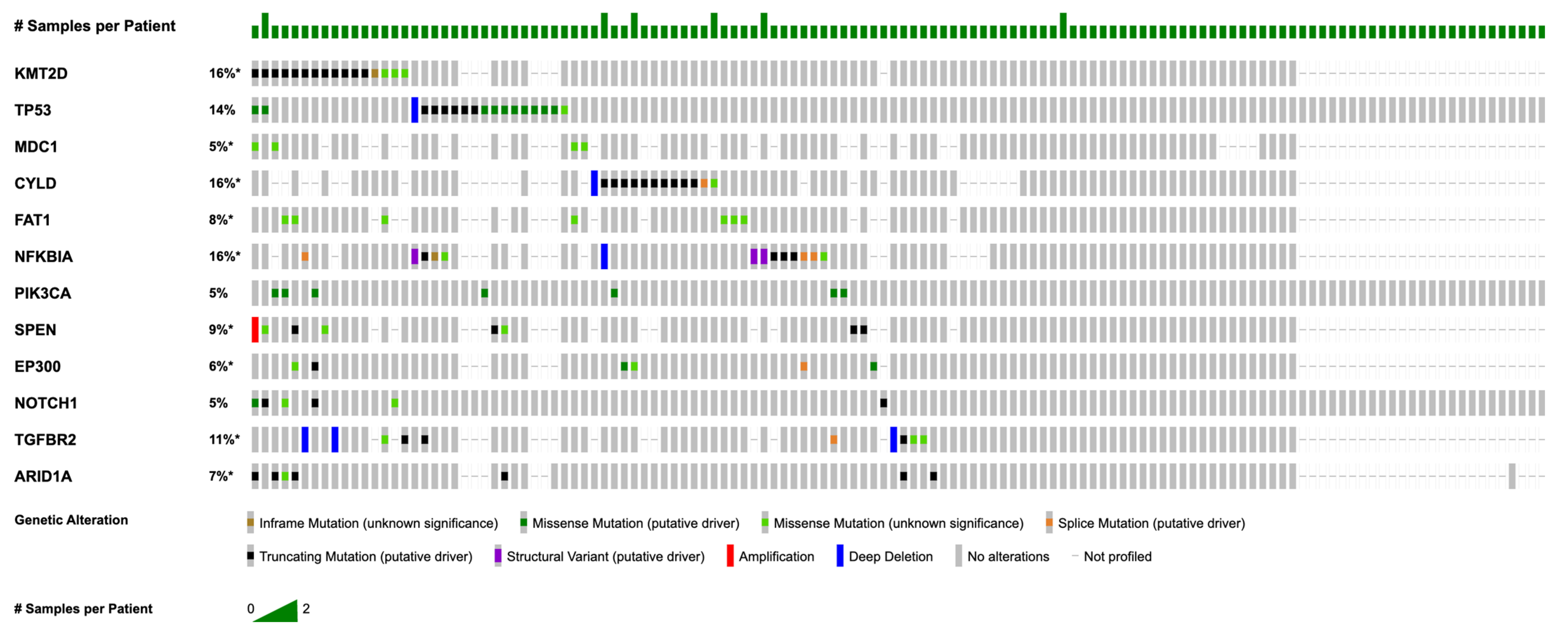
3.2. Most Common Somatic Mutations and Copy Number Alterations

Figure 2 summarizes somatic mutations that were most frequent in this NPC cohort. The most common mutations were identified in KMT2D (n = 25; 20.0%), TP53 (n = 20; 16.0%), CYLD (n = 12; 9.6%), FAT1 (n = 8; 6.4%), NFKBIA (n = 8; 6.4%), PIK3CA (n = 7; 5.6%), SPEN (n = 7; 5.6%), MDC1 (n = 6; 4.8%), EP300 (n = 6; 4.8%), ROS1 (n = 6; 4.8%), NOTCH1 (n = 6; 4.8%), TGFBR2 (n = 6; 4.8%), and AR (n = 5; 4.0%). KMT2D mutations were the most prevalent, with mutations in CYLD, FAT1, and NFKBIA also observed at notable frequencies. Specific mutations for select genes are listed in Table A1. In addition to somatic mutations, we identified recurrent copy number alterations (CNAs) in 74 samples. Loss of heterozygosity (LOH) events was prevalent, particularly affecting tumor suppressor genes such as CDKN2A (n = 19; 25.7%) and CDKN2B (n = 19; 25.7%). Less prevalent were amplifications, which occurred in genes such as FGF19, FGF4, FGF3, and CCND1 (n = 7; 10.4% for all).

3.3. Genetic Differences by Race and Sex

In this cohort, several mutations were uniquely observed in non-Asian patients (p = 0.0192), each occurring once (n = 1): ASNS, CD40, CHD7, and ITPKB. TP53 mutations were also significantly enriched in non-Asian patients (n = 15 vs. n = 3; p = 0.0361). The differences in recurrent mutations between Asian and Non-Asian patients are highlighted in Table 2.
When stratified by sex, female patients exhibited a significant enrichment of specific mutations. Mutations in PIK3C2G (n = 5; p = 0.0018), ETV6 (n = 4; p = 0.0093), and CDKN1B (n = 4; p = 0.0099) were exclusively found in females. Similarly, ASNS, CD40, and CHD7 were detected only in females, each with a single occurrence (n = 1). KDM5A mutations occurred at higher frequencies in females compared to males (n = 5 vs. n = 1; p = 0.0137), as did mutations in CCND2 (n = 5 vs. n = 1; p = 0.0149).

3.4. Co-Occurrence and Mutual Exclusivity of Mutations

Significant co-occurrence patterns were observed among frequently mutated genes. KMT2D mutations frequently co-occurred with NOTCH1 mutations (n = 4/13; p < 0.001), TGFBR2 (n = 4/11; p = 0.008), and PIK3CA (n = 3/13; p = 0.02). CYLD and EP300 mutations co-occurred in two samples (n = 2/12; p = 0.056), though this was not statistically significant. KMT2D and FAT1 mutations demonstrated significant co-occurrence in two cases (n = 2/10; p = 0.025). No significant mutual exclusivity patterns were identified (all comparisons showed p > 0.100).

3.5. Primary vs. Metastatic Mutations

The overall study cohort comprised 48 primary and 67 metastatic NPC cases. For the comparative genomic analysis, samples from 67 primary and 54 metastatic tumors were included. These analysis group sizes (n = 67 vs. n = 54) were of comparable size, minimizing potential bias. Among the 48 primary tumor samples, ASNS, CD40, CHD7, and ITPKB mutations were exclusively identified (n = 1 each; p = 0.0147) and were absent in the 67 metastatic samples. Conversely, NFKB1 (n = 1) and RNF213 (n = 1) mutations were exclusively observed in the metastatic samples (p = 0.04) and not detected in primary tumors. Although there were genes that were exclusively identified in primary or metastatic samples as demonstrated above, key characteristics of the mutational landscape, including tumor mutational burden (TMB) and frequencies of recurrent alterations in genes like KMT2D, TP53, and NFKBIA, showed substantial overlap and no significant differences between the groups.

4. Discussion

4.1. Subgroups and Mutational Landscape

This study leveraged the AACR Project GENIE repository to examine the somatic mutational landscape of nasopharyngeal carcinoma (NPC). Analysis revealed notable variations in mutation patterns across different patient subgroups. Reflecting global incidence patterns, the largest racial group in this cohort was Asian (n = 51), consistent with the established higher incidence of NPC among Asian/Pacific Islanders [16].
Comparative analysis identified distinct mutational profiles between Asian and non-Asian patients. KDM5A and CCND2 mutations were exclusively observed in six non-Asian patients (n = 6). Mutations in ASNS, CD40, CHD7, and ITPKB were observed exclusively in one non-Asian patient each (n = 1 for each gene). Furthermore, TP53 mutations were significantly more frequent in the non-Asian subgroup (Asian: n = 3, non-Asian: n = 15, p = 0.0361). While previous research has documented disparities in genetic susceptibility between Asian and non-Asian populations, including variations in HLA haplotypes and risk factors such as Epstein–Barr virus (EBV) infection [16,17], the observed enrichment of KDM5A and CCND2 mutations and the exclusive presence of ASNS, CD40, CHD7, and ITPKB mutations, along with the significantly higher TP53 mutation frequency, represent a novel finding. Notably, EBV infection rates are reportedly higher in Asian populations [18]. These findings suggest that demographic factors, including ethnicity and EBV-associated risks, contribute significantly to the distinct genetic landscapes of NPC [5,6,17].
This cohort was predominantly male (n = 83; 69.7%), consistent with the established 2–3-fold higher incidence of NPC in males compared to females (n = 42; 35.5%) [7]. Notably, female patients exhibited a higher frequency of mutations in PIK3C2G (5/22; 22.7%), ETV6 (4/29; 13.8%), and CDKN1B (4/31; 12.9%), which were not observed in male patients. These findings highlight sex-based differences in the mutational landscape of NPC. While previous literature has acknowledged the sex disparity in NPC incidence [5,6,7], the genetic basis for these differences remains largely unexplored. This study is among the first to report these significant mutational differences between genders, underscoring the need for further research into the role of sex-specific genetic and biological factors in NPC pathogenesis. Understanding these differences may provide valuable insights into disease mechanisms and inform personalized therapeutic approaches.

4.2. Commonly-Mutated Genes and Known Pathways

Consistent with previous literature, our NPC cohort exhibited substantial genetic heterogeneity, with diverse mutations observed across various loci of genes implicated in multiple pathways, including cell cycle regulation, epigenetic modification, inflammation, and growth factor signaling [19]. This study identified mutations in KMT2D (20.0%), TP53 (16.0%), CYLD (9.6%), FAT1 (6.4%), NFKBIA (6.4%), PIK3CA (5.6%), and SPEN (5.6%). These findings align with prior studies identifying KMT2D, TP53, CYLD, NFKBIA, and PIK3CA—and their respective roles in the p53, NF-κB, and PI3K pathways—as frequently altered in NPC, further supporting their critical roles as potential driver mutations [20,21].
The efficacy of standard chemotherapies, such as platinum-based agents and 5-FU, can be, at least partially, attributed to their interaction with these commonly mutated pathways [22,23]. These agents induce significant DNA damage, which robustly activates the p53 pathway (when functional) to trigger cell death, and even in cases of p53 mutation, the overwhelming damage can surpass the cell’s repair capacity and bypass the pro-survival effects from aberrant NF-κB and PI3k signaling [24].
Interestingly, this analysis did not reveal distinct mutation types or hotspots specific to NPC, consistent with reports suggesting that NPC is a unique cancer type with few clearly defined driver events [25]. This observation underscores the role of EBV infection in NPC tumorigenesis; however, the high prevalence of EBV infection coupled with the relatively low incidence of NPC suggests a contributing role for genetic susceptibility [26]. The heterogeneous mutation patterns observed in these studies [24,25] emphasize the absence of a single unifying driver mutation, suggesting a polygenic contribution to NPC development. While this genetic diversity complicates therapeutic targeting, it also presents a broad spectrum of potential molecular vulnerabilities for exploration.

4.3. p53 Pathway

The tumor suppressor gene TP53 has been extensively studied in NPC [19,21,27,28,29,30,31,32,33,34,35,36]. Consistent with these reports, we observed TP53 mutations in 16% of our samples, representing the pathway with the greatest mutational burden in this cohort. TP53 mutations typically occur early in tumorigenesis, disrupting G1/S checkpoint surveillance and facilitating the accumulation of subsequent oncogenic alterations [25].
Aberrant epigenetic modification is also recognized as a significant factor in NPC development [19,27,28,29,33,37]. In line with this, KMT2D, a key regulator of chromatin remodeling and implicated in p53 activation, was the most frequently mutated gene in our samples (20%). Mutations affecting KMT2D, or its related isoform KMT2C, have been consistently reported in other NPC studies [19,27,33]. Notably, the frequency of EP300 mutations, a histone acetyltransferase, was similar in our cohort (5%) compared to previous reports (4.8%) [27].
While TP53 mutations are thought to act as an early driver of tumor initiation, mutations in chromatin organization pathways, such as KMT2D, are suggested to emerge later and contribute to phenotypic evolution and adaptability of NPC [25]. These findings collectively suggest that TP53 mutations not only initiate tumorigenesis but also influence the mutational landscape, paving the way for the development of later-stage alterations crucial for tumor progression.
Targeting TP53 pathway alterations also holds promise. Therapies in development include Gendicine, a gene therapy delivering functional TP53, and APR-246, a small molecule aimed at restoring p53 function in p53-deficient cancers [19,38,39,40]. Additionally, MDM2 inhibitors are being explored to inhibit p53 ubiquitination, potentially stabilizing its tumor-suppressor activity [19,38,41]. Ongoing clinical trials [NCT03745716, NCT04214860, NCT03634228, NCT03107780, NCT04485260] are investigating the efficacy of these therapies in TP53-mutated malignancies [38,39,40].

4.4. NF-κB Pathway

The NF-κB pathway, implicated in NPC and other diseases (e.g., inflammatory bowel disease, rheumatoid arthritis, lupus) [42], has a debated role in NPC tumorigenesis, with some studies suggesting it as a primary driver and others as part of a broader mutational landscape. Some studies [28,30] describe NPC as largely defined by NF-κB activation (40–90% of tumors), whereas other studies [18,32,33] report greater mutational diversity and lower NF-κB mutation prevalence (7–12%). The present study supports the significance of NF-κB, with CYLD and NFKBIA mutations identified in 9.6% and 6.4% of tumors, respectively, consistent with prior studies [19,27,28,29,30,31,32,33,35,43]. NFKB1 (n = 1) was exclusively observed in the metastatic samples (p = 0.04). Although no mutations in other NF-κB pathway components (e.g., TRAF2, TRAF3, NLRC5) were detected, mutated NF-κB proteins remain promising druggable targets, particularly for metastatic NPC [19,28,29,31,33,43].

4.5. PI3K Pathway

The PI3K pathway, involved in cellular responses to various growth factors (e.g., IGF), is another well-established hotspot of mutation in NPC [19,21,27,28,29,30,32,33,34,37,44,45]. PIK3CA mutations were identified in 5.6% of analyzed samples. Similarly, another study [34] found 7% PIK3CA mutations and reported that approximately 15% of tumors harbored at least one mutated protein in the PI3K pathway, including PIK3CA, PCG1, EGFR, PTEN, and PRKCZ. Although representing an incomplete portion of total NPC cases, the PI3K pathway is a potentially druggable target warranting further exploration in susceptible NPC. PIK3CA-specific inhibitors, such as BYL719, are already under investigation for treating other PIK3CA-mutated tumors and PIK3CA-related overgrowth syndromes (e.g., CLOVES) [46,47]. Additionally, the efficacy of everolimus for solid PIK3CA-mutated tumors is under investigation [34].

4.6. Co-Occurrence Patterns and Functional Implications

The observed co-occurrence of KMT2D mutations with PIK3CA and FAT1 suggests potential cooperative roles in NPC progression. This aligns with broader findings in other cancers, where PIK3CA mutations are commonly associated with pathways promoting cell survival and proliferation [48,49]. This contrasts with findings in some studies that suggest an association between TP53/HERC1 and KMT2D mutations [25]. In NPC, combination therapies targeting PIK3CA and its co-mutated partners might be an avenue for improved outcomes, as mutations in PIK3CA, particularly hotspot variants like H1047R, are often linked to therapy resistance and poor outcomes in other cancers [50].
While our data did not reveal significant mutual exclusivity patterns, 98% of cases exhibited mutations in only one of the three most frequently mutated genes (KMT2D, TP53, and CYLD). This observation contrasts with findings in some studies that have demonstrated mutual exclusivity between LMP1 expression and NF-κB mutations or have not found any mutually exclusive driver pairs [25,29]. These discrepancies underscore the complexity of NPC’s genetic landscape and suggest that the interplay of mutational patterns may be context-dependent, influenced by cohort characteristics, environmental factors, and tumor heterogeneity, warranting further exploration in diverse populations.

4.7. Limitations

This study has several limitations. First, the AACR Project GENIE database lacks transcriptomic data, which is particularly relevant in NPC given the reported role of gene overexpression, even in the absence of mutations, especially for genes involved in epigenetic modification [27]. The absence of transcriptomic data precluded the correlation of mutational status with downstream pathway activity or gene expression levels. Similarly, the potential of tumor suppressor miRNAs as diagnostic and prognostic tools in NPC could not be explored due to the absence of miRNA data [37]. Second, treatment information is not included within this repository, which would have allowed the analysis of treatment response with mutational status and histologic subtype. GENIE’s lack of treatment data also precludes analysis of therapy-related genomic changes that may confound comparisons between primary and metastatic tumors. Third, because the database aggregates data from multiple centers using diverse sequencing platforms, potential inconsistencies in sequencing protocols could introduce bias in estimated mutation rates. Fourth, this study could not examine the crucial influence of DNA methylation on epigenetic control within NPC, nor its potential implications for tumor behavior and treatment response, as methylation data were not generated. Incorporating such data could offer deeper biological understanding. Fifth, the statistical robustness needed to definitively link specific genetic alterations with patient outcomes or distinct disease features in NPC was constrained by the modest cohort size. Therefore, validating mutations as independent predictors of prognosis necessitates subsequent investigations involving larger patient groups with standardized annotations. Sixth, distinguishing functionally significant driver mutations from passenger alterations accumulated during tumor evolution was impeded by the study design, which lacked serially collected samples (e.g., matched primary/metastatic tissues) from the same individuals over time. Seventh, the potential confounding effect of including a minor subset of related samples within the GENIE cohort (such as multiple tumors from one patient) is acknowledged, although prior evaluations suggest this factor likely exerts minimal influence on the principal results reported. Eighth, an inability to associate the detected mutation patterns with patient survival endpoints (including overall or disease-free survival) stems from the lack of such clinical outcome information within the specific GENIE data utilized for this NPC analysis. Ninth, the database aggregates all NPC histological subtypes (keratinizing, non-keratinizing, and basaloid squamous cell carcinoma) into a single group, preventing analysis of potential subtype-specific mutational profiles and their respective clinical implications. Ascertaining the impact of repeatedly observed mutations on patient prognosis in NPC necessitates future research that pairs genomic characterization with clinical follow-up data collected over time. An additional constraint was the inability to investigate potential links between specific genetic alterations and the protein expression patterns of diverse tumor-cell or immune-related markers via immunohistochemistry. Acknowledging these constraints, the current analysis still yields important information concerning the profile of genomic alterations typically found in NPC, highlighting the importance of several known pathways such as the p53, NF-κB, and PIK3 pathways, and identifying potential new targets for therapeutic intervention.

5. Conclusions

This study provides a comprehensive analysis of the NPC mutational landscape using the AACR Project GENIE database, revealing frequent alterations in KMT2D, TP53, CYLD, NFKBIA, and PIK3CA, key components of the p53, NF-κB, and PI3K pathways. Notably, distinct mutational patterns based on both sex and race were identified, highlighting the potential for personalized therapeutic strategies tailored to specific patient populations. These findings advance understanding of NPC biology and may inform the development of more effective preclinical models, diagnostic testing, and targeted therapies.

Author Contributions

Conceptualization, V.A.P.; methodology, B.H., S.A.A. and G.B.; validation, B.H. and B.A.V.-S.; formal analysis, B.H.; data curation, B.H.; writing—original draft preparation, B.H.; writing—review and editing, B.H., A.S., R.D., N.J., J.K., G.B., S.A.A., J.D.K., B.A.V.-S., M.G.B., M.S., J.R.C., M.L.L., S.P.P. and V.A.P.; visualization, B.H. and S.P.P.; supervision, V.A.P. and S.P.P.; project administration, V.A.P. and S.P.P. All authors have read and agreed to the published version of the manuscript.

Funding

This research received no external funding.

Institutional Review Board Statement

Ethical review and approval were waived for this study because the AACR Project GENIE is a publicly available cancer genomic database containing de-identified patient data, which minimizes potential risks to human subjects and eliminates the need for individual participant consent.

Informed Consent Statement

Patient consent was waived due to the nature of the study, which used only de-identified data from the publicly available AACR Project GENIE database, and this does not require individual informed consent.

Data Availability Statement

The data presented in this study are available from the AACR GENIE Database at https://genie.cbioportal.org/ (accessed on 6 February 2025).

Conflicts of Interest

The author (J.D.K.) is a military service member of the US government. This work was prepared as part of their official duties. Title 17, USC §105, provides that copyright protection under this title is not available for any work of the US government. Title 17, USC §101, defines a US government work as a work prepared by a military service member or employee of the US government as part of that person’s official duties. The views expressed in this article reflect the results of research conducted by the authors and do not necessarily reflect the official policy or position of the Department of the Navy, Department of Defense, or the US government. All other authors declare no conflicts of interest.

Abbreviations

American Association for Cancer Research (AACR), asparagine synthetase (ASNS), cyclin D2 (CCND2), CD40 molecule (CD40), cyclin-dependent kinase inhibitor 1B (CDKN1B), chromodomain helicase DNA binding protein 7 (CHD7), copy number alterations (CNAs), CYLD lysine 63 deubiquitinase (CYLD), deoxyribonucleic acid (DNA), Epstein–Barr Virus (EBV), E1A binding protein P300 (EP300), ETS variant transcription factor 6 (ETV6), FAT atypical cadherin 1 (FAT1), Genomics, Evidence, Neoplasia, Information, Exchange (GENIE), inositol-trisphosphate 3-kinase B (ITPKB), lysine demethylase 5A (KDM5A), lysine methyltransferase 2D (KMT2D), microRNA (miRNA), nuclear factor kappa-light-chain-enhancer of activated B cells (NF-κB), nuclear factor kappa B subunit 1 (NFKB1), NFKB inhibitor alpha (NFKBIA), notch receptor 1 (NOTCH1), nasopharyngeal carcinoma (NPC), tumor protein p53 (p53), phosphoinositide 3-kinase (PI3K), phosphatidylinositol-4-phosphate 3-kinase catalytic subunit type 2 gamma (PIK3C2G), phosphatidylinositol-4,5-bisphosphate 3-kinase catalytic subunit alpha (PIK3CA), standard deviation (SD), spen family transcriptional repressor (SPEN), transforming growth factor beta receptor 2 (TGFBR2), tumor mutational burden (TMB), tumor protein P53 (TP53), and variant allele frequency (VAF).

Appendix A

Table A1. Specific mutations for the select genes in NPC. Asterisk (*) indicates a premature stop codon.
GeneProtein ChangeMutation Type
KMT2DM4159Cfs*2FS del
S2431Lfs*53FS del
A2119Lfs*25FS del
L3542Vfs*13FS del
C5226Afs*16FS del
P648Tfs*2FS ins
L2331Pfs*46FS ins
Q3863delIF del
E2393KMissense
D32NMissense
R4420WMissense
I3065MMissense
K2288NMissense
L894FMissense
Q4249HMissense
D1724YMissense
A2925VMissense
R1615 *Nonsense
Q3441 *Nonsense
W4987 *Nonsense
Q2416 *Nonsense
E4662 *Nonsense
Q4609 *Nonsense
S3239 *Nonsense
X4882_spliceSplice
TP53K382Nfs*40FS del
L257Gfs*6FS del
P71Rfs*87FS ins
Y220CMissense
Y220CMissense
R248QMissense
R273CMissense
Y163CMissense
E285KMissense
P278SMissense
V157FMissense
V157FMissense
E271KMissense
C238GMissense
G245AMissense
E349KMissense
R213 *Nonsense
Q165 *Nonsense
R196 *Nonsense
X307_spliceSplice
CYLDE585Rfs*2FS del
P910Qfs*3FS del
Q443Pfs*4FS ins
I161Efs*43FS ins
D681NMissense
Q898 *Nonsense
Q554 *Nonsense
S371 *Nonsense
S371 *Nonsense
E626 *Nonsense
Q823 *Nonsense
X896_spliceSplice
NFKBIAR264Pfs*21FS ins
D100Rfs*29FS ins
K238Efs*5FS ins
E14Gfs*72FS ins
NFKBIA intragenicFusion
A158_N182delIF del
G161EMissense
X113_spliceSplice
X154_spliceSplice

References

  1. Chen, Y.-P.; Chan, A.T.C.; Le, Q.-T.; Blanchard, P.; Sun, Y.; Ma, J. Nasopharyngeal Carcinoma. Lancet 2019, 394, 64–80. [Google Scholar] [CrossRef] [PubMed]
  2. Badoual, C. Update from the 5th Edition of the World Health Organization Classification of Head and Neck Tumors: Oropharynx and Nasopharynx. Head Neck Pathol. 2022, 16, 19–30. [Google Scholar] [CrossRef]
  3. Sinha, S.; Winters, R.; Gajra, A. Nasopharyngeal Cancer. In StatPearls; StatPearls Publishing: Treasure Island, FL, USA, 2025. [Google Scholar]
  4. Liu, K.; Wang, J. Developing a Nomogram Model and Prognostic Analysis of Nasopharyngeal Squamous Cell Carcinoma Patients: A Population-Based Study. J. Cancer Res. Clin. Oncol. 2023, 149, 12165–12175. [Google Scholar] [CrossRef]
  5. Zhang, Y.; Rumgay, H.; Li, M.; Cao, S.; Chen, W. Nasopharyngeal Cancer Incidence and Mortality in 185 Countries in 2020 and the Projected Burden in 2040: Population-Based Global Epidemiological Profiling. JMIR Public Health Surveill. 2023, 9, e49968. [Google Scholar] [CrossRef]
  6. Jia, W.-H.; Qin, H.-D. Non-Viral Environmental Risk Factors for Nasopharyngeal Carcinoma: A Systematic Review. Semin. Cancer Biol. 2012, 22, 117–126. [Google Scholar] [CrossRef] [PubMed]
  7. Chang, E.T.; Ye, W.; Zeng, Y.-X.; Adami, H.-O. The Evolving Epidemiology of Nasopharyngeal Carcinoma. Cancer Epidemiol. Biomark. Prev. 2021, 30, 1035–1047. [Google Scholar] [CrossRef]
  8. Abdullah, B.; Alias, A.; Hassan, S. Challenges in the Management of Nasopharyngeal Carcinoma: A Review. Malays. J. Med. Sci. MJMS 2009, 16, 50–54. [Google Scholar] [PubMed]
  9. Juarez-Vignon Whaley, J.J.; Afkhami, M.; Onyshchenko, M.; Massarelli, E.; Sampath, S.; Amini, A.; Bell, D.; Villaflor, V.M. Recurrent/Metastatic Nasopharyngeal Carcinoma Treatment from Present to Future: Where Are We and Where Are We Heading? Curr. Treat. Options Oncol. 2023, 24, 1138–1166. [Google Scholar] [CrossRef]
  10. Zhang, Y.; Chen, L.; Hu, G.-Q.; Zhang, N.; Zhu, X.-D.; Yang, K.-Y.; Jin, F.; Shi, M.; Chen, Y.-P.; Hu, W.-H.; et al. Final Overall Survival Analysis of Gemcitabine and Cisplatin Induction Chemotherapy in Nasopharyngeal Carcinoma: A Multicenter, Randomized Phase III Trial. J. Clin. Oncol. 2022, 40, 2420–2425. [Google Scholar] [CrossRef]
  11. Lin, J.-C.; Wang, W.-Y.; Chen, K.Y.; Wei, Y.-H.; Liang, W.-M.; Jan, J.-S.; Jiang, R.-S. Quantification of Plasma Epstein–Barr Virus DNA in Patients with Advanced Nasopharyngeal Carcinoma. N. Engl. J. Med. 2004, 350, 2461–2470. [Google Scholar] [CrossRef]
  12. Krishnan, M.; Babu, S. Biomarkers in Nasopharyngeal Carcinoma (NPC): Clinical Relevance and Prognostic Potential. Oral Oncol. Rep. 2024, 11, 100640. [Google Scholar] [CrossRef]
  13. Tang, L.-Q.; Li, C.-F.; Li, J.; Chen, W.-H.; Chen, Q.-Y.; Yuan, L.-X.; Lai, X.-P.; He, Y.; Xu, Y.-X.-X.; Hu, D.-P.; et al. Establishment and Validation of Prognostic Nomograms for Endemic Nasopharyngeal Carcinoma. J. Natl. Cancer Inst. 2016, 108, djv291. [Google Scholar] [CrossRef] [PubMed]
  14. Sykes, E.A.; Weisbrod, N.; Rival, E.; Haque, A.; Fu, R.; Eskander, A. Methods, Detection Rates, and Survival Outcomes of Screening for Head and Neck Cancers: A Systematic Review. JAMA Otolaryngol. Neck Surg. 2023, 149, 1047. [Google Scholar] [CrossRef]
  15. Arter, Z.; Shieh, K.; Nagasaka, M.; Ou, S.-H. Comprehensive Survey of AACR GENIE Database of Tumor Mutation Burden (TMB) Among All Three Classes (I, II, III) of BRAF Mutated (BRAF+) NSCLC. Lung Cancer Targets Ther. 2025, 16, 1–9. [Google Scholar] [CrossRef]
  16. Lee, A.W.; Sou, A.; Patel, M.; Guzman, S.; Liu, L. Early Onset of Nasopharyngeal Cancer in Asian/Pacific Islander Americans Revealed by Age-Specific Analysis. Ann. Epidemiol. 2023, 80, 25–29. [Google Scholar] [CrossRef]
  17. Zhou, X.; Cui, J.; Macias, V.; Kajdacsy-Balla, A.A.; Ye, H.; Wang, J.; Rao, P.N. The Progress on Genetic Analysis of Nasopharyngeal Carcinoma. Comp. Funct. Genom. 2007, 2007, 57513. [Google Scholar] [CrossRef] [PubMed]
  18. Xiong, G.; Zhang, B.; Huang, M.; Zhou, H.; Chen, L.; Feng, Q.; Luo, X.; Lin, H.; Zeng, Y. Epstein-Barr Virus (EBV) Infection in Chinese Children: A Retrospective Study of Age-Specific Prevalence. PLoS ONE 2014, 9, e99857. [Google Scholar] [CrossRef]
  19. Liu, X.; Deng, Y.; Huang, Y.; Ye, J.; Xie, S.; He, Q.; Chen, Y.; Lin, Y.; Liang, R.; Wei, J.; et al. Nasopharyngeal Carcinoma Progression: Accumulating Genomic Instability and Persistent Epstein–Barr Virus Infection. Curr. Oncol. 2022, 29, 6035–6052. [Google Scholar] [CrossRef]
  20. Zhou, Z.; Li, P.; Zhang, X.; Xu, J.; Xu, J.; Yu, S.; Wang, D.; Dong, W.; Cao, X.; Yan, H.; et al. Mutational Landscape of Nasopharyngeal Carcinoma Based on Targeted Next-Generation Sequencing: Implications for Predicting Clinical Outcomes. Mol. Med. Camb. Mass 2022, 28, 55. [Google Scholar] [CrossRef]
  21. Lin, D.-C.; Meng, X.; Hazawa, M.; Nagata, Y.; Varela, A.M.; Xu, L.; Sato, Y.; Liu, L.-Z.; Ding, L.-W.; Sharma, A.; et al. The Genomic Landscape of Nasopharyngeal Carcinoma. Nat. Genet. 2014, 46, 866–871. [Google Scholar] [CrossRef]
  22. Na, D.; Chae, J.; Cho, S.-Y.; Kang, W.; Lee, A.; Min, S.; Kang, J.; Kim, M.J.; Choi, J.; Lee, W.; et al. Predictive Biomarkers for 5-Fluorouracil and Oxaliplatin-Based Chemotherapy in Gastric Cancers via Profiling of Patient-Derived Xenografts. Nat. Commun. 2021, 12, 4840. [Google Scholar] [CrossRef]
  23. Petitjean, A.; Mathe, E.; Kato, S.; Ishioka, C.; Tavtigian, S.V.; Hainaut, P.; Olivier, M. Impact of Mutant P53 Functional Properties on TP53 Mutation Patterns and Tumor Phenotype: Lessons from Recent Developments in the IARC TP53 Database. Hum. Mutat. 2007, 28, 622–629. [Google Scholar] [CrossRef]
  24. Armstrong, M.B.; Bian, X.; Liu, Y.; Subramanian, C.; Ratanaproeksa, A.B.; Shao, F.; Yu, V.C.; Kwok, R.P.S.; Opipari, A.W.; Castle, V.P. Signaling from P53 to NF-κB Determines the Chemotherapy Responsiveness of Neuroblastoma. Neoplasia 2006, 8, 964–974. [Google Scholar] [CrossRef] [PubMed]
  25. Liu, X.; Li, Y.; Zhou, X.; Zhu, S.; Kaya, N.A.; Chan, Y.S.; Ma, L.; Xu, M.; Zhai, W. An Integrative Analysis of Nasopharyngeal Carcinoma Genomes Unraveled Unique Processes Driving a Viral-Positive Cancer. Cancers 2023, 15, 1243. [Google Scholar] [CrossRef] [PubMed]
  26. Xu, M.; Feng, R.; Liu, Z.; Zhou, X.; Chen, Y.; Cao, Y.; Valeri, L.; Li, Z.; Liu, Z.; Cao, S.-M.; et al. Host Genetic Variants, Epstein-Barr Virus Subtypes, and the Risk of Nasopharyngeal Carcinoma: Assessment of Interaction and Mediation. Cell Genom. 2024, 4, 100474. [Google Scholar] [CrossRef] [PubMed]
  27. Wong, K.C.W.; Hui, E.P.; Lo, K.-W.; Lam, W.K.J.; Johnson, D.; Li, L.; Tao, Q.; Chan, K.C.A.; To, K.-F.; King, A.D.; et al. Nasopharyngeal Carcinoma: An Evolving Paradigm. Nat. Rev. Clin. Oncol. 2021, 18, 679–695. [Google Scholar] [CrossRef]
  28. Dai, W.; Chung, D.L.-S.; Chow, L.K.-Y.; Yu, V.Z.; Lei, L.C.; Leong, M.M.-L.; Chan, C.K.-C.; Ko, J.M.-Y.; Lung, M.L. Clinical Outcome–Related Mutational Signatures Identified by Integrative Genomic Analysis in Nasopharyngeal Carcinoma. Clin. Cancer Res. 2020, 26, 6494–6504. [Google Scholar] [CrossRef]
  29. Tsang, C.M.; Lui, V.W.Y.; Bruce, J.P.; Pugh, T.J.; Lo, K.W. Translational Genomics of Nasopharyngeal Cancer. Semin. Cancer Biol. 2020, 61, 84–100. [Google Scholar] [CrossRef]
  30. Lin, M.; Zhang, X.-L.; You, R.; Liu, Y.-P.; Cai, H.-M.; Liu, L.-Z.; Liu, X.-F.; Zou, X.; Xie, Y.-L.; Zou, R.-H.; et al. Evolutionary Route of Nasopharyngeal Carcinoma Metastasis and Its Clinical Significance. Nat. Commun. 2023, 14, 610. [Google Scholar] [CrossRef]
  31. Bruce, J.P.; To, K.-F.; Lui, V.W.Y.; Chung, G.T.Y.; Chan, Y.-Y.; Tsang, C.M.; Yip, K.Y.; Ma, B.B.Y.; Woo, J.K.S.; Hui, E.P.; et al. Whole-Genome Profiling of Nasopharyngeal Carcinoma Reveals Viral-Host Co-Operation in Inflammatory NF-κB Activation and Immune Escape. Nat. Commun. 2021, 12, 4193. [Google Scholar] [CrossRef]
  32. Chung, A.; OuYang, C.; Liu, H.; Chao, M.; Luo, J.; Lee, C.; Lu, Y.; Chung, I.; Chen, L.; Wu, S.; et al. Targeted Sequencing of Cancer-related Genes in Nasopharyngeal Carcinoma Identifies Mutations in the TGF-β Pathway. Cancer Med. 2019, 8, 5116–5127. [Google Scholar] [CrossRef] [PubMed]
  33. Zheng, H.; Dai, W.; Cheung, A.K.L.; Ko, J.M.Y.; Kan, R.; Wong, B.W.Y.; Leong, M.M.L.; Deng, M.; Kwok, T.C.T.; Chan, J.Y.-W.; et al. Whole-Exome Sequencing Identifies Multiple Loss-of-Function Mutations of NF-κB Pathway Regulators in Nasopharyngeal Carcinoma. Proc. Natl. Acad. Sci. USA 2016, 113, 11283–11288. [Google Scholar] [CrossRef]
  34. Chow, Y.P.; Tan, L.P.; Chai, S.J.; Abdul Aziz, N.; Choo, S.W.; Lim, P.V.H.; Pathmanathan, R.; Mohd Kornain, N.K.; Lum, C.L.; Pua, K.C.; et al. Exome Sequencing Identifies Potentially Druggable Mutations in Nasopharyngeal Carcinoma. Sci. Rep. 2017, 7, 42980. [Google Scholar] [CrossRef] [PubMed]
  35. You, R.; Liu, Y.-P.; Lin, D.-C.; Li, Q.; Yu, T.; Zou, X.; Lin, M.; Zhang, X.-L.; He, G.-P.; Yang, Q.; et al. Clonal Mutations Activate the NF-κB Pathway to Promote Recurrence of Nasopharyngeal Carcinoma. Cancer Res. 2019, 79, 5930–5943. [Google Scholar] [CrossRef]
  36. Li, J.; Guo, M.; Chen, L.; Chen, Z.; Fu, Y.; Chen, Y. Amyloid Aggregates Induced by the P53-R280T Mutation Lead to Loss of P53 Function in Nasopharyngeal Carcinoma. Cell Death Dis. 2024, 15, 35. [Google Scholar] [CrossRef] [PubMed]
  37. EAR, E.N.S.; Irekeola, A.A.; Yean Yean, C. Diagnostic and Prognostic Indications of Nasopharyngeal Carcinoma. Diagnostics 2020, 10, 611. [Google Scholar] [CrossRef]
  38. Wang, S.; Zhao, Y.; Aguilar, A.; Bernard, D.; Yang, C.-Y. Targeting the MDM2–P53 Protein–Protein Interaction for New Cancer Therapy: Progress and Challenges. Cold Spring Harb. Perspect. Med. 2017, 7, a026245. [Google Scholar] [CrossRef] [PubMed]
  39. National Cancer Institute. Phase 1/2 Study of APR-246 in Combination with Pembrolizumab in Subjects with Solid Tumor Malignancies. Available online: https://www.cancer.gov/research/participate/clinical-trials-search/v?id=NCI-2020-05550 (accessed on 15 March 2025).
  40. ClinicalTrials.gov. P53 Gene Combined with Radio- and Chemo-therapy in Treatment of Unresectable Locally Advanced Head and Neck Cancer; NCT02429037. Available online: https://clinicaltrials.gov/study/NCT02429037 (accessed on 15 March 2025).
  41. National Cancer Institute. A Novel MDM2 Inhibitor (APG-115) for the Treatment of p53 Wild-Type Salivary Gland Cancer. Available online: https://www.cancer.gov/research/participate/clinical-trials-search/v?id=NCI-2022-00506 (accessed on 15 March 2025).
  42. Liu, T.; Zhang, L.; Joo, D.; Sun, S.-C. NF-κB Signaling in Inflammation. Signal Transduct. Target. Ther. 2017, 2, 17023. [Google Scholar] [CrossRef]
  43. Li, Y.Y.; Chung, G.T.Y.; Lui, V.W.Y.; To, K.-F.; Ma, B.B.Y.; Chow, C.; Woo, J.K.S.; Yip, K.Y.; Seo, J.; Hui, E.P.; et al. Exome and Genome Sequencing of Nasopharynx Cancer Identifies NF-κB Pathway Activating Mutations. Nat. Commun. 2017, 8, 14121. [Google Scholar] [CrossRef]
  44. Or, Y.Y.; Hui, A.B.; To, K.; Lam, C.N.; Lo, K. PIK3CA Mutations in Nasopharyngeal Carcinoma. Int. J. Cancer 2006, 118, 1065–1067. [Google Scholar] [CrossRef]
  45. Chou, C.-C.; Chou, M.-J.; Tzen, C.-Y. PIK3CA Mutation Occurs in Nasopharyngeal Carcinoma but Does Not Significantly Influence the Disease-Specific Survival. Med. Oncol. 2009, 26, 322–326. [Google Scholar] [CrossRef] [PubMed]
  46. Venot, Q.; Blanc, T.; Rabia, S.H.; Berteloot, L.; Ladraa, S.; Duong, J.-P.; Blanc, E.; Johnson, S.C.; Hoguin, C.; Boccara, O.; et al. Targeted Therapy in Patients with PIK3CA-Related Overgrowth Syndrome. Nature 2018, 558, 540–546. [Google Scholar] [CrossRef] [PubMed]
  47. Xie, S.; Ni, J.; Guo, H.; Luu, V.; Wang, Y.; Zhao, J.J.; Roberts, T.M. The Role of the PIK3CA Gene in the Development and Aging of the Brain. Sci. Rep. 2021, 11, 291. [Google Scholar] [CrossRef] [PubMed]
  48. Alqahtani, A.; Ayesh, H.S.K.; Halawani, H. PIK3CA Gene Mutations in Solid Malignancies: Association with Clinicopathological Parameters and Prognosis. Cancers 2019, 12, 93. [Google Scholar] [CrossRef]
  49. Migliaccio, I.; Paoli, M.; Risi, E.; Biagioni, C.; Biganzoli, L.; Benelli, M.; Malorni, L. PIK3CA Co-Occurring Mutations and Copy-Number Gain in Hormone Receptor Positive and HER2 Negative Breast Cancer. NPJ Breast Cancer 2022, 8, 24. [Google Scholar] [CrossRef]
  50. Rasti, A.R.; Guimaraes-Young, A.; Datko, F.; Borges, V.F.; Aisner, D.L.; Shagisultanova, E. PIK3CA Mutations Drive Therapeutic Resistance in Human Epidermal Growth Factor Receptor 2-Positive Breast Cancer. JCO Precis. Oncol. 2022, 6, e2100370. [Google Scholar] [CrossRef]
Figure 1. Coronal T1-weighted post-contrast MRI demonstrating a large, enhancing mass (indicated by the arrow) in the right nasopharynx, extending into the parapharyngeal space, indicative of nasopharyngeal carcinoma.
Cancers 17 01544 g001
Figure 2. OncoPrint of recurrent mutations in NPC (for genes with n ≥ 5, coverage ≥ 100×, VAF ≥ 5%). Asterisk (*) denotes incomplete sample profiling.
Cancers 17 01544 g002
Table 1. Nasopharyngeal carcinoma patient demographics.
DemographicsCategoryn (%)
SexMale83 (69.7)
Female42 (35.5)
Age categoryAdult121 (96.6)
Pediatric4 (3.4)
EthnicityNon-Hispanic71 (59.7)
Unknown/Not Collected22 (18.5)
Hispanic9 (7.6)
RaceAsian51 (42.9)
White33 (27.7)
Black15 (12.6)
Other8 (6.7)
Unknown5 (4.2)
Sample TypePrimary48 (40.3)
Metastasis67 (56.3)
Not Collected6 (5.0)
Table 2. Race and associated mutations.
Gene (Chi-Squared)Asian, n (%)Non-Asian, n (%)p Value
TP533 (5.9)15 (12.0)p = 0.0361
CCND20 (0.0)6 (4.8)p = 0.0272
KDM5A0 (0.0)6 (4.8)p = 0.0259
ITPKB0 (0.0)1 (0.8)p = 0.0192
CD400 (0.0)1 (0.8)p = 0.0192
CHD70 (0.0)1 (0.8)p = 0.0192
ASNS0 (0.0)1 (0.8)p = 0.0192
Male, n (%)Female, n (%)
KDM5A1 (0.8)5 (4.0)p = 0.0137
CCND21 (0.8)5 (4.0)p = 0.0149
PIK3C2G0 (0.0)5 (4.0)p = 0.0018
ETV60 (0.0)4 (3.2)p = 0.0093
CDKN1B0 (0.0)4 (3.2)p = 0.0099
ASNS0 (0.0)1 (0.8)p = 0.0147
CD400 (0.0)1 (0.8)p = 0.0147
CHD70 (0.0)1 (0.8)p = 0.0147
Disclaimer/Publisher’s Note: The statements, opinions and data contained in all publications are solely those of the individual author(s) and contributor(s) and not of MDPI and/or the editor(s). MDPI and/or the editor(s) disclaim responsibility for any injury to people or property resulting from any ideas, methods, instructions or products referred to in the content.

