Review

Unveiling Primary Cutaneous B-Cell Lymphomas: New Insights into Diagnosis and Treatment Strategies

by
Zachary R. Barbati
1 and
Yann Charli-Joseph
2,*
1
Department of Pathology and Laboratory Medicine, University of California San Francisco, San Francisco, CA 94107, USA
2
Dermatology and Dermatopathology Private Practice, Mexico City 01090, Mexico
*
Author to whom correspondence should be addressed.
Cancers 2025, 17(7), 1202; https://doi.org/10.3390/cancers17071202
Submission received: 20 January 2025 / Revised: 23 March 2025 / Accepted: 26 March 2025 / Published: 1 April 2025
(This article belongs to the Special Issue Cutaneous Lymphomas: From Pathology to Treatment)

Simple Summary

This review discusses the diagnosis and management of B-cell lymphomas of the skin, which can be primary (originating in the skin) or secondary (spreading from other areas, often lymph nodes). The focus is on the three key subtypes of primary cutaneous B-cell lymphomas: follicle center lymphoma and marginal zone lymphoma, which are indolent with excellent prognoses, and diffuse large B-cell lymphoma, leg type, which is aggressive and has a high mortality rate. Each subtype has distinct clinical, microscopic, and molecular features that guide diagnosis and treatment. Indolent subtypes are treated with localized therapies like radiation or skin surgery, while aggressive subtypes require chemotherapy and/or immunotherapy. The authors highlight emerging treatments targeting cancer-specific molecules or enhancing the immune response, and discuss medical knowledge gaps that remain for these rare cancers. They also emphasize the need for further research and emphazise the importance for inclusive studies to improve outcomes and health equity for diverse patient populations.

Abstract

Background/Objectives: Primary cutaneous B-cell lymphomas (PCBCL) are a rare and heterogeneous group of non-Hodgkin lymphomas that are confined to the skin at diagnosis and exhibit a tendency for cutaneous recurrence. The 5th edition of the World Health Organization and the 2022 International Consensus Classification recognize three main subtypes: primary cutaneous follicle center lymphoma (PCFCL), primary cutaneous marginal zone lymphoma/lymphoproliferative disorder (PCMZL/LPD), and primary cutaneous diffuse large B-cell lymphoma, leg type (PCDLBCL,LT). These subtypes differ in clinical behavior, histopathologic features, immunophenotype, and molecular alterations. Diagnosis and management remain challenging for clinicians. This review aims to provide a comprehensive overview of the defining features and current treatment strategies for PCBCL. Methods: This narrative review synthesizes current literature on the clinical, morphologic, immunohistochemical, and molecular characteristics of PCBCL. It also evaluates the diagnostic utility of immunohistochemistry, gene expression profiling, and molecular assays, particularly in distinguishing primary cutaneous disease from secondary cutaneous involvement by systemic lymphomas. Results: PCFCL arises from germinal center B-cells and must be differentiated from nodal follicular lymphoma. PCMZL/LPD is derived from post-germinal center B-cells and is often linked to chronic antigenic stimulation. Both PCFCL and PCMZL/LPD are indolent and associated with favorable outcomes. By contrast, PCDLBCL,LT is an aggressive lymphoma characterized by genetic alterations activating the NF-κB pathway, commonly including mutations to MYD88 and CD79B. Treatment strategies vary by subtype, ranging from localized therapies for indolent lymphomas to systemic chemoimmunotherapy for aggressive PCBCL. Emerging therapies, such as Bruton tyrosine kinase inhibitors and immunoregulatory agents, are being investigated for relapsed/refractory disease. Conclusions: PCBCL encompass distinct clinicopathologic entities with subtype-specific diagnostic and therapeutic considerations. While current management is guided by clinical behavior, significant knowledge gaps remain regarding the molecular mechanisms underlying skin tropism, immune evasion, and disease progression. Future research could focus on improving molecular characterization and developing personalized and immune-based therapies to enhance outcomes. This review consolidates current knowledge and highlights innovations aimed at advancing the diagnosis and treatment of PCBCL in clinical practice.

1. Introduction

B-cell lymphomas (BCLs) of the skin represent a rare and heterogeneous group of extranodal non-Hodgkin lymphomas [1]. These neoplasms, albeit infrequent, are clinically significant in dermatology, oncology, hematology, and pathology, due to their diagnostic and therapeutic complexity [2,3]. Lymphomas of the skin are classified as primary cutaneous (PCL), when confined to the skin at diagnosis, and secondary cutaneous (SCL), when the skin is involved following dissemination from systemic or nodal lymphomas [4,5].
The incidence of PCBCL has increased over recent decades, now affecting approximately 4 per million individuals. PCBCLs account for roughly 25% of PCL and demonstrate a higher prevalence in male, non-Hispanic White individuals, and adults aged over 50 years [6,7]. Data from the Surveillance, Epidemiology and End Results (SEER) program indicate that the incidence rate of PCBCL is 3.5 per 1,000,000 person-years in non-Hispanic White individuals, 2.8 in Hispanic Whites, 1.9 in Asian/Pacific Islanders, and 1.5 in Black people [2].
PCBCL are currently classified into three major subtypes by both the 5th edition of the World Health Organization (5-WHO) Classification of Hematologic Neoplasms [4] and the 2022 International Consensus Classification (2022-ICC) of Mature Lymphoid Neoplasms [7]: primary cutaneous follicle center lymphoma (PCFCL), primary cutaneous marginal zone lymphoma or lymphoproliferative disorder (PCMZL/LPD), and primary cutaneous diffuse large B-cell lymphoma, leg type (PCDLBCL,LT) [4,7]. These subtypes are further categorized based on clinical behavior and prognosis, with PCFCL and PCMZL/LPD being low-grade/indolent, and PCDLBCL,LT classified as a high-grade/intermediate-to-aggressive neoplasm [1,5,6,8]. Notable differences with previous classifications include the categorization of MZL/LPD as a separate entity from the broad group of extranodal marginal zone lymphomas of mucosa-associated lymphoid tissue (MALT lymphoma), its downgrading to an LPD (in the 2022-ICC), and the recognition that PCFCL may be composed of large cells, and regardless, should not be categorized as PCDLBCL (even when occurring on the legs), with the diagnosis relying on its characteristic germinal center B-cell immunophenotype and defining molecular features [4,7,9].
Although not the focus of this review, secondary cutaneous B-cell lymphomas (SCBCLs) share numerous overlapping features with PCBCL, making it essential to differentiate between primary and secondary cutaneous involvement in all suspected PCBCL cases [10,11]. Diagnostic workup for PCBCL follows the tumor, node, metastasis (TNM) staging system endorsed by the International Society for Cutaneous Lymphomas (ISCLs) and the European Organization for the Research and Treatment of Cancer (EORTC) [12]. A thorough evaluation should include clinical assessment for B-symptoms and organ-specific signs. Additionally, blood count with differential, a metabolic panel with lactate dehydrogenase (LDH) levels, and imaging studies (e.g., PET/CT or CT-SCAN with intravenous contrast of the chest, abdomen, pelvis, and neck if applicable) are performed, except in cases of PCMZL/LPD, where they are unnecessary given its growing recognition as an indolent lymphoproliferative disorder. Bone marrow biopsy, peripheral blood flow cytometry, and protein electrophoresis (to rule out monoclonal gammopathy) are optional and of limited value in indolent/low-grade PCBCL (especially in PCMZL/LPD) but are indicated in patients with systemic symptoms, widespread disease, unexplained cytopenias or leukocytosis and are mandatory in patients with PCDLBC,LT, where additional screening for infections (hepatitis B and C, human immunodeficiency virus, latent tuberculosis) is paramount before initiating systemic therapy [7,11,12,13,14].
Herein, we aim to comprehensively examine PCBCLs, detailing their clinical, histopathological, immunohistochemical, and molecular/genetic features [Figure 1], whilst focusing the core of this review on sensible patient management. We also sought to integrate the latest research findings, current treatment approaches, and emerging therapeutic strategies, with the goal of improving diagnostic accuracy and treatment outcomes. Additionally, we identify knowledge gaps and propose future research directions to enhance the management of these complex lymphoid malignancies.

2. Indolent/Low-Grade Primary Cutaneous B-Cell Lymphomas/Lymphoproliferative Disorders

2.1. Primary Cutaneous Follicle Center Lymphoma (PCFCL)

Primary cutaneous follicle center lymphoma is the most common subtype of PCBCL in Caucasians, representing up to 30 to 50% of PCBCL cases, and approximately 10% of all PCL [1,2,5]. By contrast, data from Korea and Japan show that PCFCL represents the least common PCBCL, accounting for less than 4% of all PCL, underscoring the importance of ethnicity in the relative distribution of PCL [15,16]. PCFCL predominantly affects adults, with a median age at diagnosis ranging from 51 to 58 years, and exhibits a slight male predominance [5,6,17]. The prognosis is generally excellent, with a 5-year survival rate exceeding 95%; although remarkably rare, cases located on the legs have a strikingly lower 5-year survival rate of approximately 41% [14,17,18,19].
Clinically, PCFCL typically presents as a solitary erythematous to violaceous nodule (or a cluster of nodules), or as a plaque, commonly located on the head (particularly the scalp), neck, or trunk [Figure 2] [5,14,17,18,19,20,21]. Less common clinical variants include a miliary or agminated form presenting as multiple papules on the face, which may mimic acne, rosacea or folliculitis [22]. Another variant, historically referred to as “Crosti’s lymphoma” or “reticulohistiocytoma of the dorsum” is characterized by figurate annular concentric plaques with peripheral macules or papules on the trunk. Multifocal and/or widespread cutaneous involvement is uncommon (15%), as is ulceration (scarcely reported, chiefly in longstanding cases) [21,22,23]. Although PCFCL is rare in Black American patients, it typically presents at a younger age and is associated with more regional or generalized disease, including the presence of scattered flat plaques [24]. Recurrence occurs in 20–50% of cases, but does not significantly impact long-term outcomes. Extracutaneous involvement is uncommon, occurring in roughly 10% of cases [19,20,21,22,23,25].
PCFCL is a neoplasm of mature germinal center B-cells: predominantly small centrocytes and, to a lesser extent in most cases, centroblasts (which may be medium- or even large-sized) [4,7,9,26]. Tumor cells occupy the reticular dermis, with variable perivascular, periadnexal, and deep-dermal accentuation, with occasional extension into the subcutaneous tissue. The growth pattern may be follicular, follicular and diffuse, or purely diffuse. PCFCLs with a follicular or follicular and diffuse pattern show distorted follicular aggregates (supported by expanded dendritic cell meshworks), that are irregular in shape and size, and that often directly abut adjacent follicles. These follicles are often devoid of mantle zones and typically lack tingible body macrophages [Figure 3]. By contrast, PCFCL with a purely diffuse growth pattern, mostly seen in long-standing or larger lesions, is characterized by sheet-like pandermal proliferations of germinal center neoplastic cells, lacking distinct follicular structures and their supporting follicular dendritic cell (FDC) meshworks [4,5,6,14,21,23,26,27,28].
Immunohistochemically, neoplastic cells express pan-B cell markers, as well as germinal center markers such as B-cell lymphoma-6 (BCL6), stathmin-1, LIM-only transcription factor-2, human germinal center-associated lymphoma and activation-induced cytidine deaminase. The expression of cluster of differentiation (CD) 10 (CD10) is inconsistent. Neoplastic cells typically do not express the anti-apoptotic protein BCL2 (positive in 10–30% of cases). Additionally, lymphoma cells demonstrate minimal staining for interferon regulatory factor-4 (IRF4)/multiple myeloma oncogene-1 (MUM1), with nuclear expression being present in less than 30% of cells. The proliferative index assessed using Ki67/mindbomb homolog-1 (MIB1) is low, with fewer than 50% of neoplastic cells proliferating, compared to greater than 90% in reactive germinal centers. CD21, CD23, and CD35 staining highlight irregular or distorted FDC meshworks [Figure 4] [1,2,5,6,9,17,23,26,27,28].
Molecular analysis reveals clonal immunoglobulin heavy-chain (IgH) rearrangements in roughly 50% of cases through polymerase chain reaction (PCR), and with a higher yield with newer methods, such as tissue flow cytometry and next generation sequencing [1,5,28].
BCL2-IGH rearrangement [t(14:18)], characteristic of nodal follicular lymphoma (FL) is typically absent in PCFCL, even in cases that show BCL2 expression. In PCFCL, such overexpression is likely associated with chromosomal amplification of BCL2 rather than with a translocation. Rarely, point mutations in BCL6, RhoH/TTF, and PAX5, resulting from aberrant somatic hypermutation, have been reported. Gene expression profiling (GEP) has demonstrated upregulation of genes associated with a germinal center B-cell-like pattern, including SLAM, LCK, and SPINK2. Additionally, overexpression of the oncogenic 17–92 microRNA (miR) cluster, particularly miR20a and miR-20b, has been linked to a subset of PCFCL cases and is posited to correlate with a worse prognosis [1,5,28,29,30,31,32,33,34,35].

2.2. Primary Cutaneous Marginal Zone Lymphoma/Lymphoproliferative Disorder (PCMZL/LPD)

Primary cutaneous marginal zone lymphoma/lymphoproliferative disorder accounts for approximately 20–40% of PCBCL and about 7% of all PCL. It prevails in middle-aged adults, with a median age at diagnosis of around 55 years, and demonstrates a male predominance, with a male-to-female ratio of 2:1 [1,2,5]. The prognosis is excellent, with a 5-year disease-specific survival of ≥98%, albeit a high rate of cutaneous recurrence is observed in roughly 50% of cases [12,36]. Extracutaneous spread occurs in fewer than 10% of cases [18,20].
PCMZL/LPD is distinct from extracutaneous mucosa-associated lymphoid tissue (MALT) lymphomas due to differences in its etiological factors, histopathologic features, immunoglobulin and chemokine receptor expression patterns, genetic translocations, and frequency of systemic dissemination [1,4,5,28]. Whilst PCMZL/LPD is still designated as a “lymphoma” in the latest iteration of the WHO Classification of Hematologic Neoplasms [4], it was downgraded to a “lymphoproliferative disorder” in the 2022 International Consensus Classification of Mature Lymphoid Neoplasms [5], owing to its extremely indolent clinical behavior and underscoring the importance of avoiding unnecessary staging studies (such as PET/CT and bone marrow biopsy) and aggressive treatments (such as intravenous rituximab or chemotherapy) in PCMZL/LPD [1,2,5].
The pathogenesis of PCMZL/LPD is thought to be linked to chronic antigenic stimulation, notably to Borrelia burgdorferi infection, although this association has been solely confirmed in a subset of patients from endemic areas in Europe. Furthermore, PCMZL/LPD is significantly associated with systemic conditions, including gastrointestinal disorders, autoimmune diseases, and other malignancies [1,5,28,36,37,38,39].
Clinically, PCMZL/LPD typically presents as multiple (less often solitary), slow-growing, erythematous to violaceous papules, small nodules, or plaques, predominantly occurring on the trunk and/or extremities, with sparing of the head and neck [Figure 5] [1,5,28,37]. Hypopigmented patches have also been reported in Black American patients [24]. While exceedingly rare, atypical variants include anetodermic and agminated forms, the latter resembling the agminated/miliary variant of PCFCL [39].
PCMZL/LPD is a neoplasm of mature post-germinal center B-cells, which may be outnumbered by admixed reactive T-cells. Neoplastic B-cells are small to medium-sized and have been variably referred to as “marginal zone”, “centrocyte-like” or “monocytoid” B-cells. Additionally, lymphoplasmacytoid cells and plasma cells are often present, particularly at the periphery of the infiltrate and within the superficial dermis. The histopathologic pattern in PCMZL/LPD, similarly to PCFCL, is typically nodular or diffuse, centered upon the reticular dermis, with sparing of the papillary dermis and epidermis [1,5,6,26,27,28,40]. Reactive germinal centers may also be observed, either with distinct mantle zones or, in well-established lesions, colonized by neoplastic cells, resulting in a loss of the typical demarcation between germinal centers and their surrounding mantle zones [Figure 6] [1,5,6,27,28].
Immunohistochemically, neoplastic B-cells express pan-B cell markers, with strong and diffuse co-expression of BCL2 [1,5,28]. Plasma cells may be highlighted by CD38, CD138 or MUM1, and exhibit monotypic cytoplasmatic immunoglobulin light-chain restriction (either kappa or lambda), with a kappa-to-lambda ratio of 5:1 to 10:1 [Figure 7] [5,10,27,28,41].
Two subtypes of PCMZL/LPD are currently recognized based largely on IgH expression: the more common class-switched form (approximately 90%), which expresses IgG (with 40% of cases expressing IgG4 in the absence of IgG4-related disease), and, to a lesser extent, IgA or IgE. The rarer non-class switched subtype expresses IgM, akin to non-cutaneous MALT lymphoma, and is associated with a higher rate of extracutaneous spread compared to the class-switched form, which does not appear to portend an unfavorable outcome [42,43,44]. Though still a matter of debate, due to the differences mentioned above, some experts advocate that the class-switched form should be designated as a “lymphoproliferative disorder” (LPD), while the non-class switched subtype may be considered as a bona fide yet completely indolent “lymphoma” [42,43,44].
Aberrant somatic hypermutation has been described in several genes, including PAX5, RhoH/TTF, cMYC, PIM1, SLAMF1, SPEN, and NCOR2. Notably, recent studies have identified FAS mutations in 63–68% of PCMZL/LPD cases, leading to impaired apoptosis of neoplastic cells [1,5,28,45,46,47].

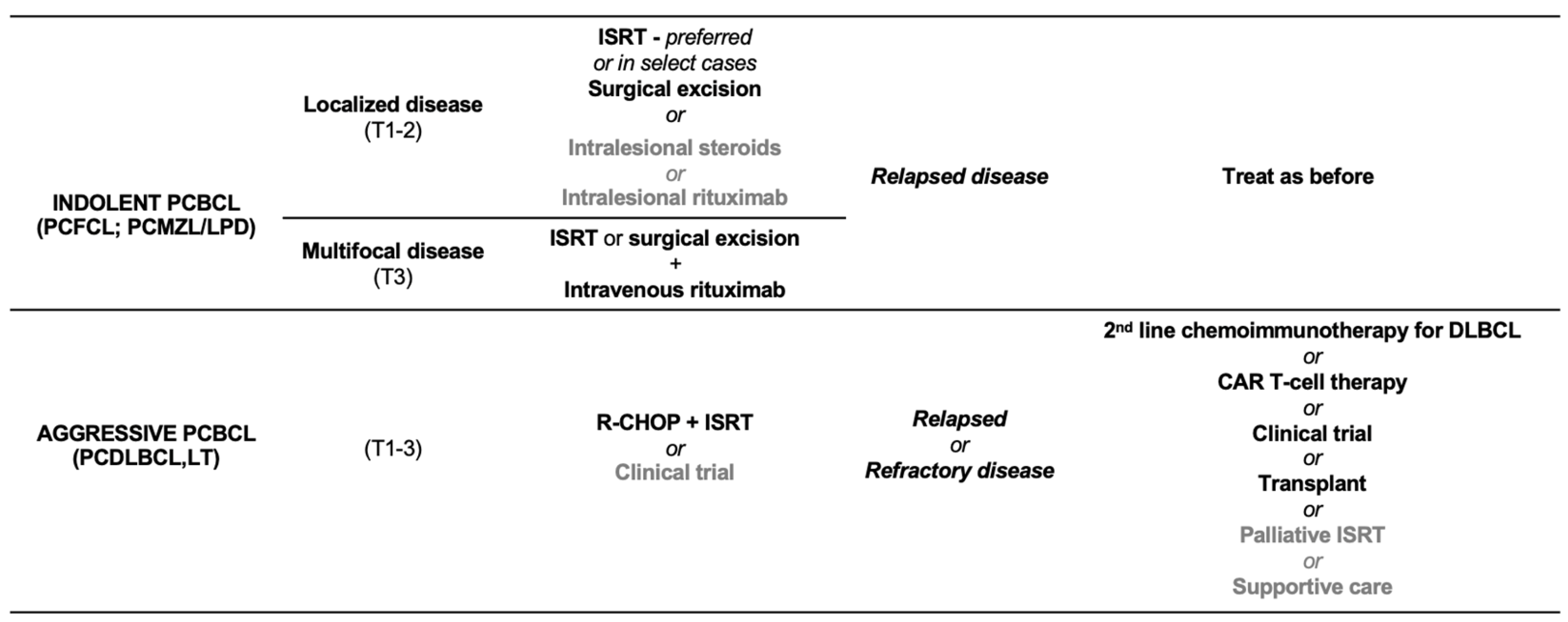
2.3. Treatment of Indolent/Low-Grade Primary Cutaneous B-Cell Lymphomas/Lymphoproliferative Disorders

Given the generally favorable prognosis of indolent/low-grade PCBCL, a conservative therapeutic approach should always be prioritized when clinically appropriate [Figure 8] [1,5,9,17,18,19]. This may include a “wait and see” strategy for solitary PCMZL/LPD, which, while acceptable, is often not preferred by patients and necessitates close surveillance [30].
For both PCMZL/LPD and PCFCL with solitary or limited lesions, the treatment modalities of choice are typically involved-site radiation therapy (ISRT) and, less commonly, surgical excision. ISRT is often favored due to its lower morbidity and excellent outcomes, particularly in older adults, or in anatomic areas where surgical excision may result in suboptimal cosmetic outcomes or healing difficulties, such as the face and lower leg, respectively. While both surgical excision and ISRT can provide complete responses (CRs) in nearly all cases, relapses are common, and optical surgical margins remain ill-defined. Reported surgical margins are generally less than 1 cm, while ISRT typically employs margins ranging from 1 to 5 cm [1,5,30,40,41].
Regarding ISRT, there is no consensus on the optimal radiation doses for indolent PCBCL, with treatment regimens varying widely. Success has been reported from very low-dose ISRT (VLD-IRST; 2–4 Gy in 2 fractions) to standard-dose ISTR (SD-IRST; 24–40 Gy in 12–20 fractions). Both regimens have demonstrated similar efficacy (CRs), but VLD-ISRT is associated with a significantly lower incidence of side effects (15.7% vs. 78.4%, p < 0.0001), making it an attractive option for treating indolent PCBCL. Although some cohorts suggest a higher relapse rate following excisional surgery compared to ISRT, statistically significant differences have not been established [14,36,48,49,50,51,52,53]. Nevertheless, excision is typically only favored in select cases, where small lesions may be removed with minimal non-disfiguring surgery [9].
Alternative treatment strategies for solitary or limited lesions of indolent PCBCL include intralesional corticosteroids, particularly for small lesions of PCMZL/LPD, though the CR rate is suboptimal at approximately 44% and multiple treatment cycles are often required [54,55]. Systemic antibiotics such as cephalosporins and tetracyclines may be effective for lesions associated with Borrelia burgdorferi infection (especially in endemic regions of Europe) [56,57]. Other therapies include intralesional interferon-alpha (though not available in many countries) [58], and intralesional rituximab, a chimeric monoclonal antibody targeting CD20. Intralesional rituximab has shown high CR rates (60–80%) but is often impractical due to the need for multiple injections, associated pain, wheal-like reactions (both at the injection site and distant sites), and mild, albeit bothersome, adverse events, such as urticaria, exanthem, fever, nausea, and malaise. Importantly, rituximab can lead to rare, but potentially serious reductions in B-cell counts, even with low cumulative doses [59,60,61].
In patients with multiple disseminated lesions, or in select cases (i.e., PCFCL with extensive scalp lesions, where IRST may lead to long-standing alopecia), intravenous rituximab (325 mg/m2 weekly for 4–8 infusions) has proven effective, with reported 98% overall response rates (ORRs), 64% CRs, a median progression free survival (PFS) of 58 months, and a median time to next treatment (TTNT) of 60 months in the largest series of 25 cases of PCMZL/LPD. In a similar cohort of 29 patients with PCFCL, systemic rituximab achieved an ORR of 96%, a CR of 72%, a median PFS of 78 months, and a median TTNT of 85 months. In both cohorts, adverse events, mostly grade 1–2, occurred in approximately 32–38% of cases [62,63,64,65,66].
Chemotherapy for indolent/low-grade PCBCL has failed to show reduced relapse rates compared to less aggressive therapies and should only be considered as a last-resort option for patients with extensive cutaneous involvement, or extracutaneous spread, and in PCFCL involving the lower extremities, which carries an unfavorable prognosis [1,5,6,28,38]. For PCMZL/LPD with widespread lesions, a few European studies have evaluated chlorambucil as single-agent chemotherapy, achieving CR rates greater than 60% [5,14,28]. Multi-agent chemotherapy regimens are almost universally discouraged, given the potential for significant side effects [1,5,9,28,67]. However, rare cohorts of multi-agent chemotherapy in PCMZL/LPD have demonstrated CR rates of approximately 85%, though relapses occur at similar rates to those observed with other treatment modalities. Similarly, a systematic review of multi-agent chemotherapy in PCFCLs found CR rates in 85% of patients, with a relapse rate of 44%. The cyclophosphamide, doxorubicin, vincristine, and prednisone (CHOP) regimen has been the most commonly used chemotherapy protocol, often with or without the addition of rituximab and/or ISRT. However, experience with these combined approaches remains limited by the relatively small patient populations treated with such regimens [5,28,67].

2.4. Differential Diagnosis for Indolent/Low-Grade Primary Cutaneous B-Cell Lymphomas/Lymphoproliferative Disorders

PCMZL/LPD and PCFCL are both characterized by infiltrates of small neoplastic B-cells, often accompanied by varying numbers of reactive T-cells. This combination of small cell size and mixed immunophenotype entails a myriad of differential diagnoses, including both reactive and neoplastic conditions, which must be accurately identified due to significant variations in management and prognosis [10,28].
The most common diagnostic challenges involve distinguishing between PCMZL/LPD and PCFCL, as well as differentiating these from cutaneous B-cell hyperplasia, commonly referred to as cutaneous lymphoid hyperplasia (CLH) or “reactive B-cell rich lymphoid proliferation” under the 5-WHO classification [3,27,28].
In the differential diagnosis of neoplastic conditions, one notable entity is primary cutaneous small/medium CD4-positive T-cell lymphoproliferative disorder (PCSMCD4LPD), which shares many similarities, especially with solitary PCMZL/LPD [3,10,27,28,68,69,70,71,72,73]. A critical differential diagnosis for PCFCL is secondary cutaneous involvement by nodal or systemic follicular lymphoma (FL), which shares almost identical histopathological and immunophenotypic features with PCFCL. The primary distinguishing feature is the almost ubiquitous expression of BCL2 in FL due to the characteristic (14;18) (IGH::BCL2) translocation, which is less commonly observed in PCFCL (~17% of cases). Overall, BCL2 expression can be observed in up to 30% of PCFCL cases. While BCL-2 expression in PCFCL has not been shown to confer a more aggressive disease course, it is associated with a higher risk of cutaneous relapse [31,74]. Thus, regardless of BCL2 expression, a thorough staging evaluation is essential to rule out FL in cases of suspected PCFCL [1,3,5,10,27,28].
Other nodal or systemic neoplasms of mature small B-cells that may occasionally involve the skin should be considered the differential diagnosis of indolent/low-grade PCBCL, notably chronic lymphocytic leukemia (CLL) and mantle cell lymphoma (MCL), an exceedingly rare but aggressive B-cell malignancy [3,10,27,28]. An in-depth analysis of other less common secondary cutaneous B-cell neoplasms falls beyond the scope of this review, but such conditions have been previously detailed in a publication by one of the authors herein [10].

3. Intermediate to Aggressive/High-Grade Primary Cutaneous B-Cell Lymphomas

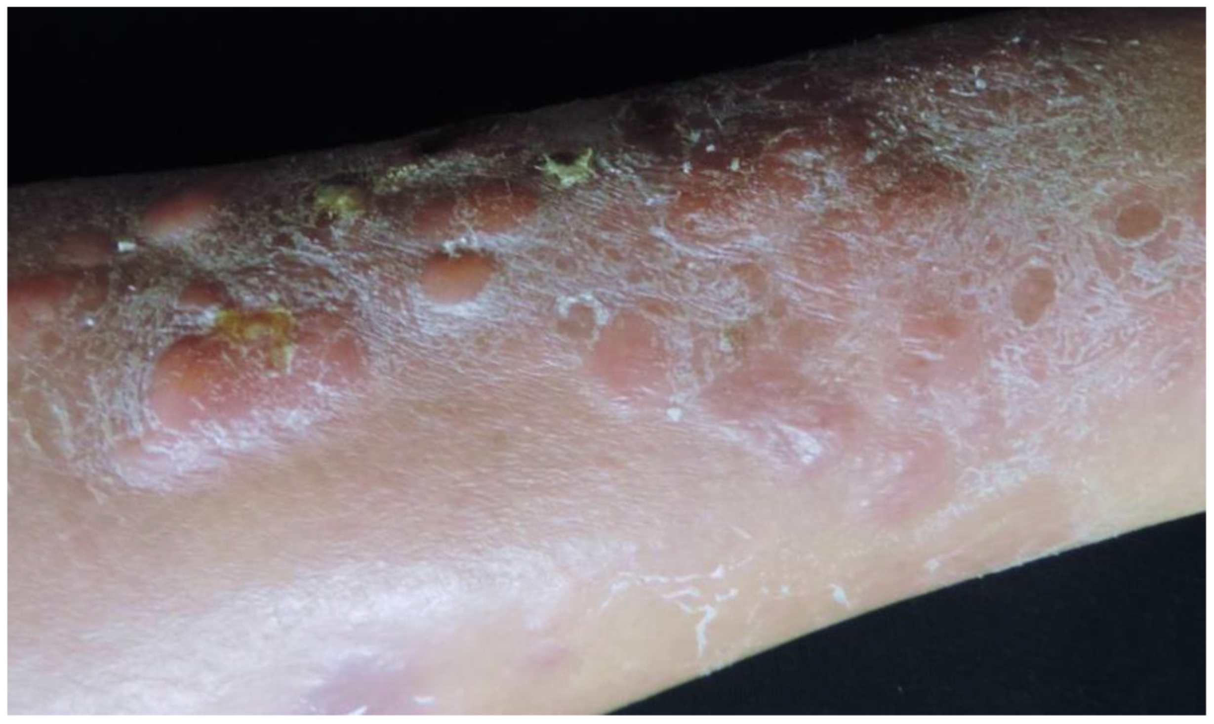
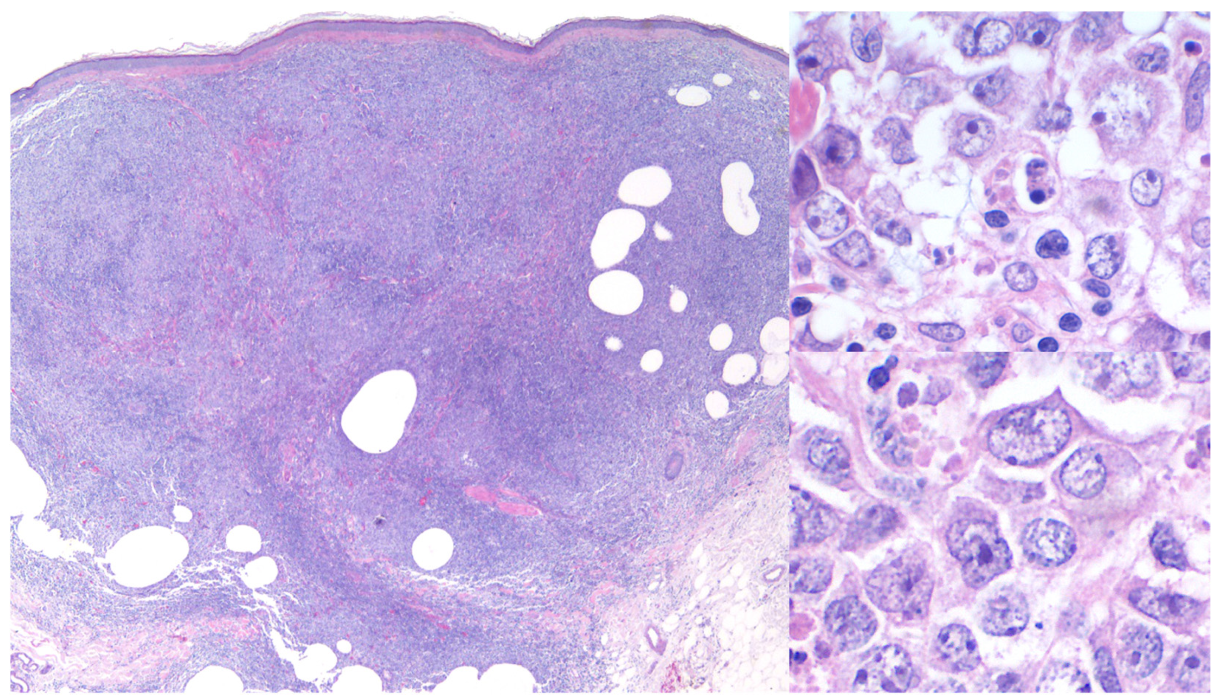
3.1. Primary Cutaneous Diffuse Large B-Cell Lymphoma, Leg Type (PCDLBCL,LT)

Primary cutaneous diffuse large B-cell lymphoma, leg type (PCDLBCL,LT), first described in 1996, is an aggressive subtype of PCBCL that accounts for 1–4% of all PCL [75,76,77]. This entity predominantly affects older adults, with a median age at diagnosis of approximately 70 years, and exhibits a marked female predominance, with a female-to-male ratio ranging from 2:1 to 3:1 [1,2,25]. PCDLBCL,LT has an aggressive clinical course; despite a high rate of initial CR rates following standard treatment, relapses occur in roughly 70% of cases. The 5-year survival rate ranges from 40% to 60%, influenced by factors such as patient age, extent of tumor dissemination, protein expression patterns (particularly loss of P16 and presence of MYC expression exceeding 40%, both associated with a poor prognosis), and underlying molecular characteristics, including mutations in B-cell receptor signaling genes such as CARD11 and CD79A/B [1,28,75,78,79].
Clinically, PCDLBCL,LT typically presents as rapidly enlarging erythematous to violaceous nodules or plaques, primarily located on the lower extremities. Although confined to the legs in most cases, 10–15% of patients may exhibit involvement of other regions, such as trunk or neck. Lesions are often unifocal (approximately one-third of cases), may ulcerate, and cause significant discomfort or pain [Figure 9] [1,5,25,27,28,78,80]. In Black patients, PCDLBCL,LT typically presents with hyperpigmented nodules that may mimic granulomatous lesions, such as those seen in sarcoidosis or infectious granulomas [24]. Constitutional B-symptoms are observed in approximately 10% of patients at diagnosis, with elevated LDH levels in around 12%. Extracutaneous dissemination, most commonly to the lymph nodes, bone marrow, and central nervous system, occurs in about 45% of cases [1,5,25,27,28].
Histopathologically, PCDLBCL,LT is marked by dense, diffuse sheets of large (≥3 times that of reactive lymphocytes), atypical lymphocytes (immunoblasts) infiltrating the dermis and often extending into the subcutis [Figure 10] [1,5,10,25,27,28,75,77].
Immunophenotypically, PCDLBCL,LT cells express pan-B cell markers, and habitually express BCL2, MUM1/IRF4 (unrelated to IRF4 rearrangements), and FOXP1. BCL6 expression is variable and typically weak if present. Immunoglobulin expression is most often restricted to IgM. However, co-expression of IgD is present in approximately 50% of cases. MYC expression, unrelated to MYC rearrangements, is present in more than 55% of cases. A high mitotic rate is typically observed, with Ki-67 indices exceeding 50% and occasionally as high as 90% [Figure 11] [1,5,10,25,27,28].
PCDLBCL,LT demonstrates a post-germinal center B-cell phenotype with monoclonal IgH rearrangements observed in most cases. Lymphoma cells often harbor significant somatic hypermutations and retain superantigen binding sites, suggesting they underwent germinal center processes akin to those in the activated B-cell-like (ABC) subtype of nodal diffuse large B-cell lymphoma. Notably, one study demonstrated that lymphoma cells of PCDLBCL,LT exhibit defects in class switch recombination and express IgM without undergoing isotype switching, indicating a blockage at the transitional stage between germinal center B-cells and plasma cells [80,81].
PCDLBCL,LT has a distinct molecular profile compared to nodal DLBCL and shares similarities with other special-site DLBCL, such as those in the breast, testes, and central nervous system, which frequently harbor MYD88 and CD79b mutations [82]. Gene expression profiling studies have revealed that MYD88 L265P mutations are present in approximately 75% of PCDLBCL,LT cases, contributing to chronic activation of the nuclear factor-kappa B (NF-κB) pathway [81,82,83,84,85,86]. MYD88 L265P mutations are associated with a shorter survival when compared to patients with wild-type MYD88 [87]. Mutations in CD79B are frequently observed, enhancing B-cell receptor signaling and supporting tumor survival and proliferation [82,83]. Deletions or mutations of CDKN2A are present in roughly two-thirds of cases and are associated with a markedly reduced 5-year DSS of 43% vs. 70% when absent [82,83,84]. In PCDLBCL,LT, MYC rearrangements have been associated with shorter DSS and disease-free survival rates (without differences in OS) and occur at a higher incidence than in nodal DLBCL (32 vs. 9–14%) [86]. Per one study, co-expression of MYC and BCL2 (dual expressor status) can be identified in up to 83% of PCDLBCL,LT cases and is associated with a more aggressive disease course [88]. By contrast, double-hit statuses (involving rearrangements of MYC and either BCL2 or BCL6) are occasionally observed but have not been shown to significantly impact prognosis [89].

3.2. Treatment of PCDLBCL,LT

The standard first-line treatment for PCDLBCL,LT involves polychemotherapy with the R-CHOP regimen. The addition of ISRT has been shown to enhance local control and prolong PFS, with a median PFS of 58 months when combined with R-CHOP, compared to 14 months with R-CHOP alone [9,78,79,90,91]. Importantly, localized treatment alone is typically inadequate for effective disease management [90]. In patients who are unable to tolerate the standard R-CHOP regimen due to advanced age or significant comorbidities, alternative reduced-intensity regimens or the combination of rituximab with pegylated liposomal doxorubicin (PLD) may be considered. The use of PLD is advantageous as it has a reduced risk of cardiotoxicity compared to conventional doxorubicin [Figure 8] [76].
Due to limited experience with the treatment of relapsed or refractory PCDLBCL,LT, the National Comprehensive Cancer Network (NCCN) guidelines recommend second-line therapies typically used for systemic DLBCL. These therapies now include chimeric antigen receptor (CAR) T-cell therapy, polatuzumab vedotin ± bendamustine ± rituximab, the CEOP regimen (cyclophosphamide, etoposide, prednisolone, and vincristine) ± rituximab, among others [9,91]. Limited evidence suggests potential benefits of lenalidomide and ibrutinib in the treatment of PCDLBCL,LT, with a single case report indicating the possible efficacy of polatuzumab vedotin [92,93,94,95]. Notably, data presented at the 2024 5th World Congress for Cutaneous Lymphomas showed promising results for patients receiving polatuzumab-based chemotherapy in combination with CAR T-cell therapy [96]. A Phase II clinical trial of patients with refractory or relapsed PCDLBCL,LT demonstrated a 26.3% ORR at 6 months following single-agent treatment with lenalidomide, with dose adjustments correlating with improved response rates [92,97]. Additionally, ibrutinib, a Bruton’s tyrosine kinase inhibitor, has demonstrated efficacy, particularly in cases with MYD88 mutations, where it appears to offer significant therapeutic benefit [93,94]. Several clinical trials, including ZUMA-7 and BELINDA, have established the efficacy of CAR T-cell therapy as a second-line treatment for large B-cell lymphoma, with at least one reported case of PCDLBCL,LT in the ZUMA-7 trial [98,99,100]. These findings suggest a potential shift in the treatment paradigm for large B-cell lymphoma, which may have significant implications for the management of PCDLBCL,LT in the future.
Recent advances in diagnostic and monitoring techniques are critical for the management of PCDLBCL,LT. F-18 fluorodeoxyglucose PET/CT is increasingly used for both staging and assessing response to therapy. However, its long-term prognostic value remains to be fully validated in larger studies [101]. Additionally, circulating tumor DNA (ctDNA) analysis using digital droplet PCR (ddPCR) represents a non-invasive approach to detect minimal residual disease, offering promising utility for monitoring disease status, particularly in cases with MYD88 mutations [102].
Although exceedingly rare, cases of spontaneous regression have been reported, suggesting possible immune-mediated mechanisms that are not yet fully understood [103]. These observations highlight the need for further exploration of immune-based therapies and of the tumor microenvironment in PCDLBCL,LT, potentially leading to novel therapeutic strategies.

3.3. Differential Diagnosis for PCDLBCL,LT

From a clinical and therapeutic standpoint, the primary differential diagnosis for PCDLBCL,LT is PCFCL with a diffuse growth pattern and a prominence of large centroblasts. PCDLBCL,LT typically expresses BCL2, MUM1/IRF4, FOXP1, and IgM and exhibits a high Ki-67 proliferation index. It often lacks CD10 and shows minimal or absent BCL6 expression. By contrast, PCFCL strongly expresses BCL6 (with less frequent CD10 positivity), and BCL2 expression is usually low or absent. Additionally, PCFCL demonstrates a significantly lower proliferation index, an admixture of reactive T-cells, residual follicular dendritic cell meshworks, and an indolent clinical course [1,5,10,27,28]. At the molecular level, PCFCL typically harbors alterations in BCL11A and c-REL genes, whereas PCDLBCL,LT is characterized by frequent chromosomal aberrations, particularly of chromosome 18, which includes amplifications of BCL2 and MALT1 genes [28,29,35,38].
Differentiating PCDLBCL,LT from other large BCL with secondary skin involvement, especially from extranodal DLBCL-NOS (not otherwise specified), the most frequent systemic large BCL to develop secondary skin involvement, is essential for accurate diagnosis, staging, and management [4,104]. While there may be morphologic overlap between PCDLBCL,LT and SC extranodal DLBCL-NOS, these entities display distinct clinical and molecular features [25].
Through GEP, two major subtypes of nodal DLBCL, which can rarely involve the skin, have been identified based on the cell of origin: the germinal center B-cell (GCB) subtype and the activated B-cell (ABC) subtype, each with distinct prognostic implications. Immunohistochemical algorithms, such as the Hans’ (and uncommonly, the Colomos’ and Muris’ algorithms), are used to classify these subtypes. GCB-type nodal DLBCL typically expresses CD10 and BCL-6, with variable BCL-2 positivity, while ABC-type nodal DLBCL consistently expresses MUM1 and FOXP1 [28,81,104,105]. PCDLBCL,LT shares immunophenotypic features with the ABC subtype [10,27,29,81,105]. However, PCDLBCL,LT often harbors CD79B mutations and the gain-of-function MYD88 L265P mutation, which are less common both in nodal and extranodal DLBCL, particularly in the GCB subtype [81]. Additionally, PCDLBCL,LT frequently exhibits BCL-2 gene rearrangements, gains in chromosome 18, and more commonly exhibits MYC rearrangements when compared to nodal DLBCL [30,89,90].
Other exceedingly rare differential diagnoses include intravascular DLBCL, plasmablastic lymphoma, anaplastic lymphoma kinase (ALK)-positive large BCL, and Burkitt lymphoma [3,10,27,28].
Ultimately, the diagnosis of PCDLBCL,LT and its differentiation from systemic or nodal large BCL with SC involvement requires a multidisciplinary approach to achieve accurate diagnosis and develop an appropriate, individualized treatment plan [1,5,6,10,28,38].

4. Conclusions and Future Directions

Primary cutaneous B-cell lymphomas encompass a diverse group of mature B-cell non-Hodgkin lymphomas, characterized by distinct clinical behaviors, histopathological features, and treatment challenges. The indolent subtypes, such as PCFCL and PCMZL/LPD, represent the majority of PCBCL cases and are typically associated with an excellent prognosis, albeit with a notable tendency for recurrence. Specifically, PCFCL is the most prevalent subtype, distinguished from systemic FL by the absence of IGH::BCL2 translocations, which are characteristic of its systemic counterpart. This difference leads to a lack of BCL2 overexpression by immunohistochemistry in the majority of PCFCL cases. Molecular features such as somatic hypermutations and overexpression of oncogenic microRNAs are frequently observed. Despite exceptional survival outcomes, recurrences are common but generally do not affect long-term survival [1,4,5,6,28,38].
PCMZL/LPD, a neoplasm of post-germinal center B-cells, is often associated with chronic antigenic stimulation. Unlike PCFCL, PCMZL/LPD expresses BCL2 and lacks germinal center marker expression. Molecular abnormalities, including somatic hypermutations and mutations in the FAS gene, suggest impaired apoptotic regulation, contributing to disease persistence. Prognosis is excellent; however, cutaneous recurrences are frequent, and extracutaneous dissemination remains rare. Both PCFCL and PCMZL/LPD respond well to localized therapies, such as surgical excision and ISRT. Systemic therapies are typically reserved for disseminated or recurrent disease, with chemotherapy considered a last-line option due to its limited impact on long-term recurrence rates. Despite their indolent course, these lymphomas necessitate careful monitoring due to their recurrent nature, highlighting the need for continued research into optimal treatment strategies and the molecular mechanisms underlying relapse [1,4,5,6,28,38].
By contrast, PCDLBCL,LT represents a more aggressive entity, marked by frequent relapses and limited survival outcomes. Comprehensive diagnostic evaluation is essential for effective management. While the combination of R-CHOP plus ISRT remains the first-line treatment, novel agents such as lenalidomide and targeted therapies like ibrutinib show promise in relapsed and/or refractory cases [1,76,92,93,94].
Ongoing research into genetic markers and advanced monitoring techniques remains critical for improving disease management and patient outcomes in PCBCL. Key areas for further investigation include understanding the molecular mechanisms of treatment resistance, the interactions between the tumor microenvironment and immune evasion, and the molecular drivers of cutaneous homing and imprinting [106,107,108]. Future studies should focus on personalizing treatment regimens based on molecular profiles, optimizing the integration of targeted therapies with existing treatment protocols, and exploring immune-based therapeutic strategies. As with other rare diseases, multidisciplinary collaboration is essential to enhance diagnostic accuracy, disease monitoring, treatment paradigms, and long-term disease control, ultimately improving patient outcomes [28,38].
Furthermore, it is essential to continue including historically marginalized populations and People of the Global Majority (PoGM) in cutaneous lymphoma research. Efforts to ensure equitable representation will promote diversity and inclusion in medical research and clinical care, ultimately contributing to more comprehensive and globally relevant healthcare outcomes.

Author Contributions

Conceptualization, Z.R.B. and Y.C.-J.; methodology, Z.R.B. and Y.C.-J.; validation, NA; formal analysis, Z.R.B. and Y.C.-J.; investigation, Z.R.B. and Y.C.-J.; resources, Z.R.B. and Y.C.-J.; data curation, Z.R.B. and Y.C.-J.; writing—original draft preparation, Z.R.B. and Y.C.-J.; writing—review and editing, Z.R.B. and Y.C-J; supervision, Y.C.-J. All authors have read and agreed to the published version of the manuscript.

Funding

This research received no external funding.

Acknowledgments

Raul Méndez-Flores for providing images of primary cutaneous marginal zone lymphoma/lymphoproliferative disorder (Figure 4, Figure 5 and Figure 6).

Conflicts of Interest

The authors declare no conflicts of interest.

References

  1. Goyal, N.; O’Leary, D.; Carter, J.B.; Comfere, N.; Sokumbi, O.; Goyal, A. A Practical Review of the Presentation, Diagnosis, and Management of Cutaneous B-Cell Lymphomas. Dermatol. Clin. 2023, 41, 187–208. [Google Scholar] [CrossRef] [PubMed]
  2. Bradford, P.T.; Devesa, S.S.; Anderson, W.F.; Toro, J.R. Cutaneous lymphoma incidence patterns in the United States: A population-based study of 3884 cases. Blood 2009, 113, 5064–5073. [Google Scholar] [CrossRef] [PubMed]
  3. Charli-Joseph, Y.V.; Gatica-Torres, M.; Pincus, L.B. Approach to Cutaneous Lymphoid Infiltrates: When to Consider Lymphoma? Indian J. Dermatol. 2016, 61, 351–374. [Google Scholar] [CrossRef] [PubMed]
  4. Alaggio, R.; Amador, C.; Anagnostopoulos, I.; Attygalle, A.D.; Araujo, I.B.O.; Berti, E.; Bhagat, G.; Borges, A.M.; Boyer, D.; Calaminici, M.; et al. The 5th edition of the World Health Organization Classification of Haematolymphoid Tumours: Lymphoid Neoplasms. Leukemia 2022, 36, 1720–1748. [Google Scholar] [CrossRef]
  5. Villasenor-Park, J.; Chung, J.; Kim, E.J. Cutaneous B-Cell Lymphomas. Hematol. Oncol. Clin. N. Am. 2024, 38, 1111–1131. [Google Scholar] [CrossRef]
  6. Suárez, A.L.; Pulitzer, M.; Horwitz, S.; Moskowitz, A.; Querfeld, C.; Myskowski, P.L. Primary cutaneous B-cell lymphomas: Part I. Clinical features, diagnosis, and classification. J. Am. Acad. Dermatol. 2013, 69, 329.e1–329.e13. [Google Scholar] [CrossRef]
  7. Campo, E.; Jaffe, E.S.; Cook, J.R.; Quintanilla-Martinez, L.; Swerdlow, S.H.; Anderson, K.C.; Brousset, P.; Cerroni, L.; de Leval, L.; Dirnhofer, S.; et al. The International Consensus Classification of Mature Lymphoid Neoplasms: A report from the Clinical Advisory Committee. Blood 2022, 140, 1229–1253. [Google Scholar] [CrossRef]
  8. Kempf, W.; Mitteldorf, C.; Cerroni, L.; Willemze, R.; Berti, E.; Guenova, E.; Scarisbrick, J.J.; Battistella, M. Classifications of cutaneous lymphomas and lymphoproliferative disorders: An update from the EORTC cutaneous lymphoma histopathology group. J. Eur. Acad. Dermatol. Venereol. 2024, 38, 1491–1503. [Google Scholar] [CrossRef]
  9. National Comprehensive Cancer Network. Clinical Practice Guidelines in Oncology. Primary Cutaneous Lymphomas (Version 1.25). Available online: https://www.nccn.org/professionals/physician_gls/pdf/primary_cutaneous.pdf (accessed on 16 March 2025).
  10. Charli-Joseph, Y.; Toussaint-Claire, S.; Lome-Maldonado, C.; Montante-Montes de Oca, D.; Ortiz-Hidalgo, C. Approach to dermal-based lymphoid infiltrates and proliferations. Semin. Cutan. Med. Surg. 2018, 37, 61–74. [Google Scholar] [CrossRef]
  11. Leary, D.O.; Goyal, N.; Rubin, N.; Goyal, A. Characterization of Primary and Secondary Cutaneous B-Cell Lymphomas: A Population-Based Study of 4758 Patients. Clin. Lymphoma Myeloma Leuk. 2022, 22, e269–e278. [Google Scholar] [CrossRef]
  12. Kim, Y.H.; Willemze, R.; Pimpinelli, N.; Whittaker, S.; Olsen, E.A.; Ranki, A.; Dummer, R.; Hoppe, R.T.; ISCL and the EORTC. TNM classification system for primary cutaneous lymphomas other than mycosis fungoides and Sezary syndrome: A proposal of the International Society for Cutaneous Lymphomas (ISCL) and the Cutaneous Lymphoma Task Force of the European Organization of Research and Treatment of Cancer (EORTC). Blood 2007, 110, 479–484. [Google Scholar] [CrossRef] [PubMed]
  13. Olsen, E.A.; Whittaker, S.; Willemze, R.; Pinter-Brown, L.; Foss, F.; Geskin, L.; Schwartz, L.; Horwitz, S.; Guitart, J.; Zic, J.; et al. Primary cutaneous lymphoma: Recommendations for clinical trial design and staging update from the ISCL, USCLC, and EORTC. Blood 2022, 140, 419–437. [Google Scholar] [CrossRef] [PubMed]
  14. Senff, N.J.; Noordijk, E.M.; Kim, Y.H.; Bagot, M.; Berti, E.; Cerroni, L.; Dummer, R.; Duvic, M.; Hoppe, R.T.; Pimpinelli, N.; et al. European Organization for Research and Treatment of Cancer and International Society for Cutaneous Lymphoma consensus recommendations for the management of cutaneous B-cell lymphomas. Blood 2008, 112, 1600–1609. [Google Scholar] [CrossRef] [PubMed]
  15. Fujii, K.; Hamada, T.; Shimauchi, T.; Asai, J.; Fujisawa, Y.; Ihn, H.; Katoh, N. Cutaneous lymphoma in Japan, 2012-2017: A nationwide study. J. Dermatol. Sci. 2020, 97, 187–193. [Google Scholar] [CrossRef]
  16. Moon, I.J.; Won, C.H.; Chang, S.E.; Park, C.S.; Yoon, D.H.; Song, S.Y.; Lee, M.W.; Lee, W.J. Prevalence, clinical features, and survival outcome trends of 627 patients with primary cutaneous lymphoma over 29 years: A retrospective review from single tertiary center in Korea. Sci. Rep. 2024, 14, 20118. [Google Scholar] [CrossRef]
  17. Skala, S.L.; Hristov, B.; Hristov, A.C. Primary Cutaneous Follicle Center Lymphoma. Arch. Pathol. Lab. Med. 2018, 142, 1313–1321. [Google Scholar] [CrossRef]
  18. Zinzani, P.L.; Quaglino, P.; Pimpinelli, N.; Berti, E.; Baliva, G.; Rupoli, S.; Martelli, M.; Alaibac, M.; Borroni, G.; Chimenti, S.; et al. Italian Study Group for Cutaneous Lymphomas. Prognostic factors in primary cutaneous B-cell lymphoma: The Italian Study Group for Cutaneous Lymphomas. J. Clin. Oncol. 2006, 24, 1376–1382. [Google Scholar] [CrossRef]
  19. Bessell, E.M.; Humber, C.E.; O’Connor, S.; English, J.S.; Perkins, W.; Dickinson, P.D.; Patel, A.N. Primary cutaneous B-cell lymphoma in Nottinghamshire U.K.: Prognosis of subtypes defined in the WHO-EORTC classification. Br. J. Dermatol. 2012, 167, 1118–1123. [Google Scholar] [CrossRef]
  20. Grange, F.; Bekkenk, M.W.; Wechsler, J.; Meijer, C.J.; Cerroni, L.; Bernengo, M.; Bosq, J.; Hedelin, G.; Fink Puches, R.; van Vloten, W.A.; et al. Prognostic factors in primary cutaneous large B-cell lymphomas: A European multicenter study. J. Clin. Oncol. 2001, 19, 3602–3610. [Google Scholar] [CrossRef]
  21. Bergman, R.; Kurtin, P.J.; Gibson, L.E.; Hull, P.R.; Kimlinger, T.K.; Schroeter, A.L. Clinicopathologic, immunophenotypic, and molecular characterization of primary cutaneous follicular B-cell lymphoma. Arch. Dermatol. 2001, 137, 432–439. [Google Scholar]
  22. Massone, C.; Fink-Puches, R.; Laimer, M.; Rütten, A.; Vale, E.; Cerroni, L. Miliary and agminated-type primary cutaneous follicle center lymphoma: Report of 18 cases. J. Am. Acad. Dermatol. 2011, 65, 749–755. [Google Scholar] [CrossRef] [PubMed]
  23. Gulia, A.; Saggini, A.; Wiesner, T.; Fink-Puches, R.; Argenyi, Z.; Ferrara, G.; Müller, C.S.; Vale, E.; Cerroni, L. Clinicopathologic features of early lesions of primary cutaneous follicle center lymphoma, diffuse type: Implications for early diagnosis and treatment. J. Am. Acad. Dermatol. 2011, 65, 991–1000. [Google Scholar] [CrossRef] [PubMed]
  24. Geller, S.; Pulitzer, M.; Myskowski, P.; Kheterpal, M. Low-Grade Cutaneous B-cell Lymphoma in African American Patients. J. Drugs Dermatol. 2018, 17, 1334–1337. [Google Scholar] [PubMed] [PubMed Central]
  25. Senff, N.J.; Hoefnagel, J.J.; Jansen, P.M.; Vermeer, M.H.; van Baarlen, J.; Blokx, W.A.; Canninga-van Dijk, M.R.; Geerts, M.L.; Hebeda, K.M.; Kluin, P.M.; et al. Reclassification of 300 primary cutaneous B-Cell lymphomas according to the new WHO-EORTC classification for cutaneous lymphomas: Comparison with previous classifications and identification of prognostic markers. J. Clin. Oncol. 2007, 25, 1581–1587. [Google Scholar] [CrossRef]
  26. Charli-Joseph, Y.; Cerroni, L.; LeBoit, P.E. Cutaneous Spindle-Cell B-Cell Lymphomas: Most are Neoplasms of Follicular Center Cell Origin. Am. J. Surg. Pathol. 2015, 39, 737–743. [Google Scholar] [CrossRef]
  27. Cerroni, L. Skin Lymphoma: The Illustrated Guide, 5th ed.; John Wiley & Sons: Hoboken, NJ, USA, 2020; ISBN 978-1-119-48590-2. [Google Scholar]
  28. Gru, A.; Schaffer, A. Hematopathology of the Skin: A Clinical and Practical Approach, 2nd ed.; Lippincott Williams & Wilkins: Philadelphia, PA, USA, 2023; ISBN 978-1-97-515855-2. [Google Scholar]
  29. Hoefnagel, J.J.; Dijkman, R.; Basso, K.; Jansen, P.M.; Hallermann, C.; Willemze, R.; Tensen, C.P.; Vermeer, M.H. Distinct types of primary cutaneous large B-cell lymphoma identified by gene expression profiling. Blood 2005, 105, 3671–3678. [Google Scholar] [CrossRef]
  30. Dijkman, R.; Tensen, C.P.; Jordanova, E.S.; Knijnenburg, J.; Hoefnagel, J.J.; Mulder, A.A.; Rosenberg, C.; Raap, A.K.; Willemze, R.; Szuhai, K.; et al. Array-based comparative genomic hybridization analysis reveals recurrent chromosomal alterations and prognostic parameters in primary cutaneous large B-cell lymphoma. J. Clin. Oncol. 2006, 24, 296–305. [Google Scholar] [CrossRef]
  31. Zhou, X.A.; Yang, J.; Ringbloom, K.G.; Martinez-Escala, M.E.; Stevenson, K.E.; Wenzel, A.T.; Fantini, D.; Martin, H.K.; Moy, A.P.; Morgan, E.A.; et al. Genomic landscape of cutaneous follicular lymphomas reveals 2 subgroups with clinically predictive molecular features. Blood Adv. 2021, 5, 649–661. [Google Scholar] [CrossRef]
  32. Goodlad, J.R.; Krajewski, A.S.; Batstone, P.J.; McKay, P.; White, J.M.; Benton, E.C.; Kavanagh, G.M.; Lucraft, H.H.; Scotland and Newcastle Lymphoma Group. Primary cutaneous follicular lymphoma: A clinicopathologic and molecular study of 16 cases in support of a distinct entity. Am. J. Surg. Pathol. 2002, 26, 733–741. [Google Scholar] [CrossRef]
  33. Child, F.J.; Russell-Jones, R.; Woolford, A.J.; Calonje, E.; Photiou, A.; Orchard, G.; Whittaker, S.J. Absence of the t(14;18) chromosomal translocation in primary cutaneous B-cell lymphoma. Br. J. Dermatol. 2001, 144, 735–744. [Google Scholar] [CrossRef]
  34. Barasch, N.J.K.; Liu, Y.C.; Ho, J.; Bailey, N.; Aggarwal, N.; Cook, J.R.; Swerdlow, S.H. The molecular landscape and other distinctive features of primary cutaneous follicle center lymphoma. Hum. Pathol. 2020, 106, 93–105. [Google Scholar] [CrossRef] [PubMed]
  35. Abdul-Wahab, A.; Tang, S.Y.; Robson, A.; Morris, S.; Agar, N.; Wain, E.M.; Child, F.; Scarisbrick, J.; Neat, M.; Whittaker, S. Chromosomal anomalies in primary cutaneous follicle center cell lymphoma do not portend a poor prognosis. J. Am. Acad. Dermatol. 2014, 70, 1010–1020. [Google Scholar] [CrossRef] [PubMed]
  36. Servitje, O.; Muniesa, C.; Benavente, Y.; Monsálvez, V.; Garcia-Muret, M.P.; Gallardo, F.; Domingo-Domenech, E.; Lucas, A.; Climent, F.; Rodriguez-Peralto, J.L.; et al. Primary cutaneous marginal zone B-cell lymphoma: Response to treatment and disease-free survival in a series of 137 patients. J. Am. Acad. Dermatol. 2013, 69, 357–365. [Google Scholar] [CrossRef] [PubMed]
  37. Hoefnagel, J.J.; Vermeer, M.H.; Jansen, P.M.; Heule, F.; van Voorst Vader, P.C.; Sanders, C.J.; Gerritsen, M.J.; Geerts, M.L.; Meijer, C.J.; Noordijk, E.M.; et al. Primary cutaneous marginal zone B-cell lymphoma: Clinical and therapeutic features in 50 cases. Arch. Dermatol. 2005, 141, 1139–1145. [Google Scholar] [CrossRef]
  38. Kempf, W.; Kazakov, D.V.; Mitteldorf, C. Cutaneous lymphomas: An update. Part 2: B-cell lymphomas and related conditions. Am. J. Dermatopathol. 2014, 36, 197–208, quiz 209-10. [Google Scholar] [CrossRef]
  39. Hodak, E.; Feuerman, H.; Barzilai, A.; David, M.; Cerroni, L.; Feinmesser, M. Anetodermic primary cutaneous B-cell lymphoma: A unique clinicopathological presentation of lymphoma possibly associated with antiphospholipid antibodies. Arch. Dermatol. 2010, 146, 175–182. [Google Scholar] [CrossRef]
  40. Magro, C.M.; Davis, T.L.; Kurtzman, D.J.B. Epidermotropic marginal zone lymphoma: An uncommon cutaneous B-cell lymphoma responsive to rituximab. JAAD Case Rep. 2017, 3, 474–476. [Google Scholar] [CrossRef]
  41. Schafernak, K.T.; Variakojis, D.; Goolsby, C.L.; Tucker, R.M.; Martínez-Escala, M.E.; Smith, F.A.; Dittman, D.; Chenn, A.; Guitart, J. Clonality assessment of cutaneous B-cell lymphoid proliferations: A comparison of flow cytometry immunophenotyping, molecular studies, and immunohistochemistry/in situ hybridization and review of the literature. Am. J. Dermatopathol. 2014, 36, 781–795. [Google Scholar] [CrossRef]
  42. Edinger, J.T.; Kant, J.A.; Swerdlow, S.H. Cutaneous marginal zone lymphomas have distinctive features and include 2 subsets. Am. J. Surg. Pathol. 2010, 34, 1830–1841. [Google Scholar] [CrossRef]
  43. van Maldegem, F.; van Dijk, R.; Wormhoudt, T.A.; Kluin, P.M.; Willemze, R.; Cerroni, L.; van Noesel, C.J.; Bende, R.J. The majority of cutaneous marginal zone B-cell lymphomas expresses class-switched immunoglobulins and develops in a T-helper type 2 inflammatory environment. Blood 2008, 112, 3355–3361. [Google Scholar] [CrossRef]
  44. Carlsen, E.D.; Swerdlow, S.H.; Cook, J.R.; Gibson, S.E. Class-switched Primary Cutaneous Marginal Zone Lymphomas Are Frequently IgG4-positive and Have Features Distinct from IgM-positive Cases. Am. J. Surg. Pathol. 2019, 43, 1403–1412. [Google Scholar] [CrossRef] [PubMed]
  45. Deutsch, A.J.; Frühwirth, M.; Aigelsreiter, A.; Cerroni, L.; Neumeister, P. Primary cutaneous marginal zone B-cell lymphomas are targeted by aberrant somatic hypermutation. J. Investig. Dermatol. 2009, 129, 476–479. [Google Scholar] [CrossRef] [PubMed]
  46. Perez, M.; Pacchiarotti, A.; Frontani, M.; Pescarmona, E.; Caprini, E.; Lombardo, G.A.; Russo, G.; Faraggiana, T. Primary cutaneous B-cell lymphoma is associated with somatically hypermutated immunoglobulin variable genes and frequent use of VH1-69 and VH4-59 segments. Br. J. Dermatol. 2010, 162, 611–618. [Google Scholar] [CrossRef]
  47. Takino, H.; Li, C.; Hu, S.; Kuo, T.T.; Geissinger, E.; Muller-Hermelink, H.K.; Kim, B.; Swerdlow, S.H.; Inagaki, H. Primary cutaneous marginal zone B-cell lymphoma: A molecular and clinicopathological study of cases from Asia, Germany, and the United States. Mod. Pathol. 2008, 21, 1517–1526. [Google Scholar] [CrossRef]
  48. Golling, P.; Cozzio, A.; Dummer, R.; French, L.; Kempf, W. Primary cutaneous B-cell lymphomas—Clinicopathological, prognostic and therapeutic characterization of 54 cases according to the WHO-EORTC classification and the ISCL/EORTC TNM classification system for primary cutaneous lymphomas other than mycosis fungoides and Sezary syndrome. Leuk. Lymphoma 2008, 49, 1094–1103. [Google Scholar] [CrossRef]
  49. Haverkos, B.; Tyler, K.; Gru, A.A.; Winardi, F.K.; Frederickson, J.; Hastings, J.; Elkins, C.; Zhang, X.; Xu-Welliver, M.; Wong, H.K.; et al. Primary Cutaneous B-Cell Lymphoma: Management and Patterns of Recurrence at the Multimodality Cutaneous Lymphoma Clinic of The Ohio State University. Oncologist 2015, 20, 1161–1166. [Google Scholar] [CrossRef]
  50. Goyal, A.; Carter, J.B.; Pashtan, I.; Gallotto, S.; Wang, I.; Isom, S.; Ng, A.; Winkfield, K.M. Very low-dose versus standard dose radiation therapy for indolent primary cutaneous B-cell lymphomas: A retrospective study. J. Am. Acad. Dermatol. 2018, 78, 408–410. [Google Scholar] [CrossRef]
  51. Cozzio, A.; Kempf, W.; Schmid-Meyer, R.; Gilliet, M.; Michaelis, S.; Schärer, L.; Burg, G.; Dummer, R. Intra-lesional low-dose interferon alpha2a therapy for primary cutaneous marginal zone B-cell lymphoma. Leuk. Lymphoma 2006, 47, 865–869. [Google Scholar] [CrossRef]
  52. Specht, L.; Dabaja, B.; Illidge, T.; Wilson, L.D.; Hoppe, R.T.; International Lymphoma Radiation Oncology Group. Modern radiation therapy for primary cutaneous lymphomas: Field and dose guidelines from the International Lymphoma Radiation Oncology Group. Int. J. Radiat. Oncol. Biol. Phys. 2015, 92, 32–39. [Google Scholar] [CrossRef]
  53. Neelis, K.J.; Schimmel, E.C.; Vermeer, M.H.; Senff, N.J.; Willemze, R.; Noordijk, E.M. Low-dose palliative radiotherapy for cutaneous B- and T-cell lymphomas. Int. J. Radiat. Oncol. Biol. Phys. 2009, 74, 154–158. [Google Scholar] [CrossRef]
  54. Perry, A.; Vincent, B.J.; Parker, S.R. Intralesional corticosteroid therapy for primary cutaneous B-cell lymphoma. Br. J. Dermatol. 2010, 163, 223–225. [Google Scholar] [CrossRef] [PubMed]
  55. Kollipara, R.; Hans, A.; Hall, J.; Lisle, A. A case report of primary cutaneous marginal zone lymphoma treated with intralesional steroids. Dermatol. Online J. 2015, 21, 13030/qt9s15929m. [Google Scholar] [CrossRef]
  56. Ponzoni, M.; Ferreri, A.J.; Mappa, S.; Pasini, E.; Govi, S.; Facchetti, F.; Fanoni, D.; Tucci, A.; Vino, A.; Doglioni, C.; et al. Prevalence of Borrelia burgdorferi infection in a series of 98 primary cutaneous lymphomas. Oncologist 2011, 16, 1582–1588. [Google Scholar] [CrossRef] [PubMed]
  57. Wood, G.S.; Kamath, N.V.; Guitart, J.; Heald, P.; Kohler, S.; Smoller, B.R.; Cerroni, L. Absence of Borrelia burgdorferi DNA in cutaneous B-cell lymphomas from the United States. J. Cutan. Pathol. 2001, 28, 502–507. [Google Scholar] [CrossRef]
  58. Trent, J.T.; Romanelli, P.; Kerdel, F.A. Topical targretin and intralesional interferon alfa for cutaneous lymphoma of the scalp. Arch. Dermatol. 2002, 138, 1421–1423. [Google Scholar] [CrossRef]
  59. Kerl, K.; Prins, C.; Saurat, J.H.; French, L.E. Intralesional and intravenous treatment of cutaneous B-cell lymphomas with the monoclonal anti-CD20 antibody rituximab: Report and follow-up of eight cases. Br. J. Dermatol. 2006, 155, 1197–1200. [Google Scholar] [CrossRef]
  60. Heinzerling, L.; Dummer, R.; Kempf, W.; Schmid, M.H.; Burg, G. Intralesional therapy with anti-CD20 monoclonal antibody rituximab in primary cutaneous B-cell lymphoma. Arch. Dermatol. 2000, 136, 374–378. [Google Scholar] [CrossRef]
  61. Kyrtsonis, M.C.; Siakantaris, M.P.; Kalpadakis, C.; Dimopoulou, M.N.; Vassilakopoulos, T.P.; Kontopidou, F.N.; Antoniou, C.; Korkolopoulou, P.; Panayiotidis, P.; Pangalis, G.A. Favorable outcome of primary cutaneous marginal zone lymphoma treated with intralesional rituximab. Eur. J. Haematol. 2006, 77, 300–303. [Google Scholar] [CrossRef]
  62. Heinzerling, L.M.; Urbanek, M.; Funk, J.O.; Peker, S.; Bleck, O.; Neuber, K.; Burg, G.; von Den Driesch, P.; Dummer, R. Reduction of tumor burden and stabilization of disease by systemic therapy with anti-CD20 antibody (rituximab) in patients with primary cutaneous B-cell lymphoma. Cancer 2000, 89, 1835–1844. [Google Scholar] [CrossRef] [PubMed]
  63. Gellrich, S.; Muche, J.M.; Wilks, A.; Jasch, K.C.; Voit, C.; Fischer, T.; Audring, H.; Sterry, W. Systemic eight-cycle anti-CD20 monoclonal antibody (rituximab) therapy in primary cutaneous B-cell lymphomas--an applicational observation. Br. J. Dermatol. 2005, 153, 167–173. [Google Scholar] [CrossRef]
  64. Morales, A.V.; Advani, R.; Horwitz, S.M.; Riaz, N.; Reddy, S.; Hoppe, R.T.; Kim, Y.H. Indolent primary cutaneous B-cell lymphoma: Experience using systemic rituximab. J. Am. Acad. Dermatol. 2008, 59, 953–957. [Google Scholar] [CrossRef] [PubMed]
  65. Valencak, J.; Weihsengruber, F.; Rappersberger, K.; Trautinger, F.; Chott, A.; Streubel, B.; Muellauer, L.; Der-Petrossian, M.; Jonak, C.; Binder, M.; et al. Rituximab monotherapy for primary cutaneous B-cell lymphoma: Response and follow-up in 16 patients. Ann. Oncol. 2009, 20, 326–330. [Google Scholar] [CrossRef] [PubMed]
  66. Brandenburg, A.; Humme, D.; Terhorst, D.; Gellrich, S.; Sterry, W.; Beyer, M. Long-term outcome of intravenous therapy with rituximab in patients with primary cutaneous B-cell lymphomas. Br. J. Dermatol. 2013, 169, 1126–1132. [Google Scholar] [CrossRef]
  67. Vitiello, P.; Sica, A.; Ronchi, A.; Caccavale, S.; Franco, R.; Argenziano, G. Primary Cutaneous B-Cell Lymphomas: An Update. Front. Oncol. 2020, 10, 651. [Google Scholar] [CrossRef]
  68. Grogg, K.L.; Jung, S.; Erickson, L.A.; McClure, R.F.; Dogan, A. Primary cutaneous CD4-positive small/medium-sized pleomorphic T-cell lymphoma: A clonal T-cell lymphoproliferative disorder with indolent behavior. Mod. Pathol. 2008, 21, 708–715. [Google Scholar] [CrossRef]
  69. Rodríguez Pinilla, S.M.; Roncador, G.; Rodríguez-Peralto, J.L.; Mollejo, M.; García, J.F.; Montes-Moreno, S.; Camacho, F.I.; Ortiz, P.; Limeres-González, M.A.; Torres, A.; et al. Primary cutaneous CD4+ small/medium-sized pleomorphic T-cell lymphoma expresses follicular T-cell markers. Am. J. Surg. Pathol. 2009, 33, 81–90. [Google Scholar] [CrossRef]
  70. Beltraminelli, H.; Leinweber, B.; Kerl, H.; Cerroni, L. Primary cutaneous CD4+ small-/medium-sized pleomorphic T-cell lymphoma: A cutaneous nodular proliferation of pleomorphic T lymphocytes of undetermined significance? A study of 136 cases. Am. J. Dermatopathol. 2009, 31, 317–322. [Google Scholar] [CrossRef]
  71. Baum, C.L.; Link, B.K.; Neppalli, V.T.; Swick, B.L.; Liu, V. Reappraisal of the provisional entity primary cutaneous CD4+ small/medium pleomorphic T-cell lymphoma: A series of 10 adult and pediatric patients and review of the literature. J. Am. Acad. Dermatol. 2011, 65, 739–748. [Google Scholar] [CrossRef]
  72. Cetinözman, F.; Jansen, P.M.; Willemze, R. Expression of programmed death-1 in primary cutaneous CD4-positive small/medium-sized pleomorphic T-cell lymphoma, cutaneous pseudo-T-cell lymphoma, and other types of cutaneous T-cell lymphoma. Am. J. Surg. Pathol. 2012, 36, 109–116. [Google Scholar] [CrossRef]
  73. Alberti-Violetti, S.; Torres-Cabala, C.A.; Talpur, R.; Corti, L.; Fanoni, D.; Venegoni, L.; Berti, E.; Duvic, M. Clinicopathological and molecular study of primary cutaneous CD4+ small/medium-sized pleomorphic T-cell lymphoma. J. Cutan. Pathol. 2016, 43, 1121–1130. [Google Scholar] [CrossRef]
  74. Pileri, A.; Grandi, V.; Agostinelli, C.; Santucci, M.; Lastrucci, I.; Guglielmo, A.; Pipitò, C.; Pimpinelli, N. BCL-2 expression in primary cutaneous follicle center lymphoma is associated with a higher risk of cutaneous relapses: A study of 126 cases. J. Eur. Acad. Dermatol. Venereol. 2022, 36, e811–e813. [Google Scholar] [CrossRef] [PubMed]
  75. Ronchi, A.; Vitiello, P.; D’Abbronzo, G.; Caccavale, S.; Argenziano, G.; Sica, A.; Alfan, R.; Savarese, G.; Berretta, M.; Cozzolino, I.; et al. Primary Cutaneous B-Cell Lymphomas with Large Cell Morphology: A Practical Review. Int. J. Mol. Sci. 2023, 24, 6204. [Google Scholar] [CrossRef] [PubMed]
  76. Grange, F.; Beylot-Barry, M.; Courville, P.; Maubec, E.; Bagot, M.; Vergier, B.; Souteyrand, P.; Machet, L.; Dalac, S.; Esteve, E.; et al. Primary cutaneous diffuse large B-cell lymphoma, leg type: Clinicopathologic features and prognostic analysis in 60 cases. Arch. Dermatol. 2007, 143, 1144–1150. [Google Scholar] [CrossRef] [PubMed]
  77. Vermeer, M.H.; Geelen, F.A.; van Haselen, C.W.; van Voorst Vader, P.C.; Geerts, M.L.; van Vloten, W.A.; Willemze, R. Primary cutaneous large B-cell lymphomas of the legs. A distinct type of cutaneous B-cell lymphoma with an intermediate prognosis. Dutch Cutaneous Lymphoma Working Group. Arch. Dermatol. 1996, 132, 1304–1308. [Google Scholar] [PubMed]
  78. Grange, F.; Joly, P.; Barbe, C.; Bagot, M.; Dalle, S.; Ingen-Housz-Oro, S.; Maubec, E.; D’Incan, M.; Ram-Wolff, C.; Dalac, S.; et al. Improvement of survival in patients with primary cutaneous diffuse large B-cell lymphoma, leg type, in France. JAMA Dermatol. 2014, 150, 535–541. [Google Scholar] [CrossRef]
  79. Kraft, R.M.; Ansell, S.M.; Villasboas, J.C.; Bennani, N.N.; Wang, Y.; Habermann, T.M.; Thanarajasingam, G.; Lester, S.C.; Macon, W.; Inwards, D.J.; et al. Outcomes in primary cutaneous diffuse large B-cell lymphoma, leg type. Hematol. Oncol. 2021, 39, 658–663. [Google Scholar] [CrossRef]
  80. Willemze, R.; Meijer, C.J.; Sentis, H.J.; Scheffer, E.; van Vloten, W.A.; Toonstra, J.; van der Putte, S.C. Primary cutaneous large cell lymphomas of follicular center cell origin. A clinical follow-up study of nineteen patients. J. Am. Acad. Dermatol. 1987, 16 Pt 1, 518–526. [Google Scholar] [CrossRef]
  81. Schrader, A.M.R.; de Groen, R.A.L.; Willemze, R.; Jansen, P.M.; Quint, K.D.; van Wezel, T.; van Eijk, R.; Ruano, D.; Tensen, C.P.; Hauben, E.; et al. Cell-of-origin classification using the Hans and Lymph2Cx algorithms in primary cutaneous large B-cell lymphomas. Virchows Arch. 2022, 480, 667–675. [Google Scholar] [CrossRef]
  82. Prieto-Torres, L.; Rodríguez-Pinilla, S.M. New insights and advances in defining primary cutaneous B-cell lymphomas and cutaneous B-cell-rich lymphoid proliferations. Diagn. Histopathol. 2024, 30, 419–429. [Google Scholar] [CrossRef]
  83. Pham-Ledard, A.; Prochazkova-Carlotti, M.; Deveza, M.; Laforet, M.P.; Beylot-Barry, M.; Vergier, B.; Parrens, M.; Feuillard, J.; Merlio, J.P.; Gachard, N. Molecular analysis of immunoglobulin variable genes supports a germinal center experienced normal counterpart in primary cutaneous diffuse large B-cell lymphoma, leg-type. J. Dermatol. Sci. 2017, 88, 238–246. [Google Scholar] [CrossRef]
  84. Schrader, A.M.R.; de Groen, R.A.L.; Willemze, R.; Jansen, P.M.; Quint, K.D.; Cleven, A.H.G.; van Wezel, T.; van Eijk, R.; Ruano, D.; Veelken, J.H.H.; et al. Genetic Stability of Driver Alterations in Primary Cutaneous Diffuse Large B-Cell Lymphoma, Leg Type and Their Relapses: A Rationale for the Use of Molecular-Based Methods for More Effective Disease Monitoring. Cancers 2022, 14, 5152. [Google Scholar] [CrossRef] [PubMed]
  85. Senff, N.J.; Zoutman, W.H.; Vermeer, M.H.; Assaf, C.; Berti, E.; Cerroni, L.; Espinet, B.; de Misa Cabrera, R.F.; Geerts, M.L.; Kempf, W.; et al. Fine-mapping chromosomal loss at 9p21: Correlation with prognosis in primary cutaneous diffuse large B-cell lymphoma, leg type. J. Investig. Dermatol. 2009, 129, 1149–1155. [Google Scholar] [CrossRef] [PubMed]
  86. Schrader, A.M.R.; Jansen, P.M.; Vermeer, M.H.; Kleiverda, J.K.; Vermaat, J.S.P.; Willemze, R. High Incidence and Clinical Significance of MYC Rearrangements in Primary Cutaneous Diffuse Large B-Cell Lymphoma, Leg Type. Am. J. Surg. Pathol. 2018, 42, 1488–1494. [Google Scholar] [CrossRef]
  87. Pham-Ledard, A.; Prochazkova-Carlotti, M.; Andrique, L.; Cappellen, D.; Vergier, B.; Martinez, F.; Grange, F.; Petrella, T.; Beylot-Barry, M.; Merlio, J.P. Multiple genetic alterations in primary cutaneous large B-cell lymphoma, leg type support a common lymphomagenesis with activated B-cell-like diffuse large B-cell lymphoma. Mod. Pathol. 2014, 27, 402–411. [Google Scholar] [CrossRef]
  88. Menguy, S.; Frison, E.; Prochazkova-Carlotti, M.; Dalle, S.; Dereure, O.; Boulinguez, S.; Dalac, S.; Machet, L.; Ram-Wolff, C.; Verneuil, L.; et al. Double-hit or dual expression of MYC and BCL2 in primary cutaneous large B-cell lymphomas. Mod. Pathol. 2018, 31, 1332–1342. [Google Scholar] [CrossRef]
  89. Lucioni, M.; Pescia, C.; Bonometti, A.; Fraticelli, S.; Moltrasio, C.; Ramponi, A.; Riboni, R.; Roccio, S.; Ferrario, G.; Arcaini, L.; et al. Double expressor and double/triple hit status among primary cutaneous diffuse large B-cell lymphoma: A comparison between leg type and not otherwise specified subtypes. Hum. Pathol. 2021, 111, 1–9. [Google Scholar] [CrossRef]
  90. Elsayad, K.; Guenova, E.; Assaf, C.; Nicolay, J.P.; Trautinger, F.; Stadler, R.; Waldstein, C.; Boterberg, T.; Meijnders, P.; Kirova, Y.; et al. Radiotherapy in cutaneous lymphomas: Recommendations from the EORTC cutaneous lymphoma tumour group. Eur. J. Cancer 2024, 212, 115064. [Google Scholar] [CrossRef]
  91. National Comprehensive Cancer Network. Clinical Practice Guidelines in Oncology. B-Cell Lymphomas (Version 2.25). Available online: https://www.nccn.org/professionals/physician_gls/pdf/b-cell.pdf (accessed on 18 February 2025).
  92. Zoli, S.; Pellegrini, C.; Casadei, B.; Broccoli, A.; Argnani, L.; Nanni, L.; Stefoni, V.; Zinzani, P.L. Prolonged Complete Response with Lenalidomide in a Relapsed Diffuse Large B-Cell Lymphoma, Leg-Type: A Case Report. Chemotherapy 2024, 69, 23–26. [Google Scholar] [CrossRef]
  93. Deng, A.L.; Kim, Y.R.; Lichtenstein, E.A.; O’Connor, O.A.; Deng, C. Combination of ibrutinib and chemotherapy produced a durable remission in multiply relapsed diffuse large B-cell lymphoma leg type with mutant MYD88 and wildtype CD79. Haematologica 2017, 102, e275–e277. [Google Scholar] [CrossRef]
  94. Gupta, E.; Accurso, J.; Sluzevich, J.; Menke, D.M.; Tun, H.W. Excellent Outcome of Immunomodulation or Bruton’s Tyrosine Kinase Inhibition in Highly Refractory Primary Cutaneous Diffuse Large B-Cell Lymphoma, Leg Type. Rare Tumors 2015, 7, 6067. [Google Scholar] [CrossRef]
  95. Oka, S.; Ono, K.; Nohgawa, M. Effective treatment with polatuzumab vedotin of relapsed and refractory primary cutaneous diffuse large B-cell lymphoma leg type. Ann. Hematol. 2022, 101, 2353–2354. [Google Scholar] [CrossRef] [PubMed]
  96. Bandali, T. Treatment of primary cutaneous diffuse large B-cell lymphoma, leg-type (PCDLBCL-LT) in the modern era, Abstract. In Proceedings of the 5th World Congress of Cutaneous Lymphomas, Pasadena, CA, USA, 11–13 April 2024. [Google Scholar]
  97. Beylot-Barry, M.; Mermin, D.; Maillard, A.; Bouabdallah, R.; Bonnet, N.; Duval-Modeste, A.B.; Mortier, L.; Ingen-Housz-Oro, S.; Ram-Wolff, C.; Barete, S.; et al. A Single-Arm Phase II Trial of Lenalidomide in Relapsing or Refractory Primary Cutaneous Large B-Cell Lymphoma, Leg Type. J. Investig. Dermatol. 2018, 138, 1982–1989. [Google Scholar] [CrossRef] [PubMed]
  98. Westin, J.; Sehn, L.H. CAR T cells as a second-line therapy for large B-cell lymphoma: A paradigm shift? Blood 2022, 139, 2737–2746, Erratum in Blood 2023, 141, 683. https://doi.org/10.1182/blood.2022018793. [Google Scholar] [CrossRef] [PubMed]
  99. Locke, F.L.; Miklos, D.B.; Jacobson, C.A.; Perales, M.A.; Kersten, M.J.; Oluwole, O.O.; Ghobadi, A.; Rapoport, A.P.; McGuirk, J.; Pagel, J.M.; et al. All ZUMA-7 Investigators and Contributing Kite Members. Axicabtagene Ciloleucel as Second-Line Therapy for Large B-Cell Lymphoma. N. Engl. J. Med. 2022, 386, 640–654. [Google Scholar] [CrossRef]
  100. Bishop, M.R.; Dickinson, M.; Purtill, D.; Barba, P.; Santoro, A.; Hamad, N.; Kato, K.; Sureda, A.; Greil, R.; Thieblemont, C.; et al. Second-Line Tisagenlecleucel or Standard Care in Aggressive B-Cell Lymphoma. N. Engl. J. Med. 2022, 386, 629–639. [Google Scholar] [CrossRef]
  101. Datta, D.; Pandey, R.R.; Kumar, R.; Sharma, R.; Vedant, D. F-18 FDG PET/CT in staging and response assessment of primary cutaneous diffuse large B-cell lymphoma (leg type). Eur. J. Hybrid Imaging 2023, 7, 15. [Google Scholar] [CrossRef]
  102. Schrader, A.M.R.; van Engeland, J.; Willemze, R.; Vermaat, J.S.P.; Ottevanger, R.; Kersten, J.M.; Zoutman, W.H.; Jansen, P.M.; van Eijk, R.; van Egmond, D.; et al. Detection of Circulating Tumor DNA for Disease Monitoring in Patients with Primary Cutaneous Diffuse Large B-Cell Lymphoma, Leg Type. J. Investig. Dermatol. 2024, 145, 440–444. [Google Scholar] [CrossRef]
  103. Winkler, M.; Albrecht, J.D.; Sauer, C.; Kordaß, T.; Guenova, E.; Livingstone, E.; Wobser, M.; Mitteldorf, C.; Géraud, C.; Nicolay, J.P. Spontaneous regression of primary cutaneous diffuse large B-cell lymphoma, leg type: A case series and review of the literature. J. Dermatol. 2024, 51, 1233–1239. [Google Scholar] [CrossRef]
  104. Shi, Y.; Han, Y.; Yang, J.; Liu, P.; He, X.; Zhang, C.; Zhou, S.; Zhou, L.; Qin, Y.; Song, Y.; et al. Clinical features and outcomes of diffuse large B-cell lymphoma based on nodal or extranodal primary sites of origin: Analysis of 1,085 WHO classified cases in a single institution in China. Chin. J. Cancer Res. 2019, 31, 152–161. [Google Scholar] [CrossRef]
  105. Choi, W.W.; Weisenburger, D.D.; Greiner, T.C.; Piris, M.A.; Banham, A.H.; Delabie, J.; Braziel, R.M.; Geng, H.; Iqbal, J.; Lenz, G.; et al. A new immunostain algorithm classifies diffuse large B-cell lymphoma into molecular subtypes with high accuracy. Clin. Cancer Res. 2009, 15, 5494–5502. [Google Scholar] [CrossRef]
  106. Ducharme, O.; Beylot-Barry, M.; Pham-Ledard, A.; Bohers, E.; Viailly, P.J.; Bandres, T.; Faur, N.; Frison, E.; Vergier, B.; Jardin, F.; et al. Mutations of the B-Cell Receptor Pathway Confer Chemoresistance in Primary Cutaneous Diffuse Large B-Cell Lymphoma Leg Type. J. Investig. Dermatol. 2019, 139, 2334–2342.e8. [Google Scholar] [CrossRef] [PubMed]
  107. Zhou, X.A.; Louissaint AJr Wenzel, A.; Yang, J.; Martinez-Escala, M.E.; Moy, A.P.; Morgan, E.A.; Paxton, C.N.; Hong, B.; Andersen, E.F.; Guitart, J.; et al. Genomic Analyses Identify Recurrent Alterations in Immune Evasion Genes in Diffuse Large B-Cell Lymphoma, Leg Type. J. Investig. Dermatol. 2018, 138, 2365–2376. [Google Scholar] [CrossRef] [PubMed]
  108. Egbuniwe, I.U.; Karagiannis, S.N.; Nestle, F.O.; Lacy, K.E. Revisiting the role of B cells in skin immune surveillance. Trends Immunol. 2015, 36, 102–111. [Google Scholar] [CrossRef] [PubMed]
Figure 1. Archetypal features of indolent (green color) and aggressive (red color) primary cutaneous B-cell lymphomas/lymphoproliferative disorders. 400× magnification (neoplastic cells column); 100× for BCL6 and BCL2, and 40× for ILCR (immunohistochemistry column).
Cancers 17 01202 g001
Figure 2. Primary cutaneous follicle center lymphoma presenting on the head. Clinical and dermoscopic (4×) images.
Cancers 17 01202 g002
Figure 3. Primary cutaneous follicle center lymphoma. Hematoxylin and eosin-stained sections 20×, 40×, and 400×.
Cancers 17 01202 g003
Figure 4. Primary cutaneous follicle center lymphoma. Immunohistochemistry, 40×.
Cancers 17 01202 g004
Figure 5. Primary cutaneous marginal zone lymphoma/lymphoproliferative disorder presenting on the trunk: clinical and dermoscopic (4×) images.
Cancers 17 01202 g005
Figure 6. Primary cutaneous marginal zone lymphoma/lymphoproliferative disorder. Hematoxylin and eosin-stained sections 20× and 400×.
Cancers 17 01202 g006
Figure 7. Primary cutaneous marginal zone lymphoma/lymphoproliferative disorder. Immunohistochemistry, 40×.
Cancers 17 01202 g007
Figure 8. Simplified treatment approach for skin-limited indolent and aggressive primary cutaneous B-cell lymphomas/lymphoproliferative disorders.
Cancers 17 01202 g008
Figure 9. Primary cutaneous diffuse large B-cell lymphoma, leg type. Clinical image.
Cancers 17 01202 g009
Figure 10. Primary cutaneous diffuse large B-cell lymphoma, leg type. Hematoxylin and eosin-stained sections 20×, 400× and 600×.
Cancers 17 01202 g010
Figure 11. Primary cutaneous diffuse large B-cell lymphoma, leg type. Immunohistochemistry, 400×.
Cancers 17 01202 g011
Disclaimer/Publisher’s Note: The statements, opinions and data contained in all publications are solely those of the individual author(s) and contributor(s) and not of MDPI and/or the editor(s). MDPI and/or the editor(s) disclaim responsibility for any injury to people or property resulting from any ideas, methods, instructions or products referred to in the content.

