Review

IL-10-Directed Cancer Immunotherapy: Preclinical Advances, Clinical Insights, and Future Perspectives

by
Adel G. El-Shemi
1,2,
Afnan Alqurashi
3,
Jihan Abdullah Abdulrahman
1,
Hanin Dhaifallah Alzahrani
1,
Khawlah Saad Almwalad
1,
Hadeel Hisham Felfilan
1,
Wahaj Saud Alomiri
1,
Jana Ahmed Aloufi
1,
Ghadeer Hassn Madkhali
1,4,
Sarah Adel Maqliyah
1,5,
Jood Bandar Alshahrani
1,
Huda Taj Kamal
1,
Sawsan Hazim Daghistani
1,
Bassem Refaat
1 and
Faisal Minshawi
1,*
1
Department of Clinical Laboratory Sciences, Faculty of Applied Medical Sciences, Umm Al-Qura University, Al Abdeyah, Makkah 21955, Saudi Arabia
2
Department of Pharmacology, Faculty of Medicine, Assiut University, Assiut 71515, Egypt
3
Independent Researcher, Makkah 21955, Saudi Arabia
4
Department of Hematology, Dr. Sulaiman Al-Habib Medical Diagnostic Laboratory, Olaya District, Riyadh 12234-3785, Saudi Arabia
5
Department of Blood Bank and Laboratory, Saudi German Hospital, Makkah 24242, Saudi Arabia
*
Author to whom correspondence should be addressed.
Cancers 2025, 17(6), 1012; https://doi.org/10.3390/cancers17061012
Submission received: 16 February 2025 / Revised: 12 March 2025 / Accepted: 14 March 2025 / Published: 17 March 2025
(This article belongs to the Section Cancer Immunology and Immunotherapy)

Simple Summary

Interleukin-10 (IL-10) is emerging as a promising cancer immunotherapy due to its ability to boost exhausted immune cells (CD8+ T cells), helping them fight tumors more effectively while also reducing harmful inflammation. However, despite encouraging early research, clinical trials of IL-10-based treatments, such as pegilodecakin, have faced challenges like side effects and inconsistent results, highlighting the need for better dosing and delivery methods. To overcome these obstacles, scientists have developed engineered IL-10 variants, fusion proteins, and bispecific constructs, which improve stability and extend IL-10’s activity, enhancing its effects when used with immune checkpoint inhibitors. As research continues to refine IL-10 therapies, innovative delivery systems and combination treatments could unlock its full potential in precision cancer care, making IL-10 a key player in the future of immunotherapy.

Abstract

Cytokine-based immunotherapy has revolutionized cancer treatment; however, its efficacy remains limited by insufficient tumor immune infiltration and resistance mechanisms within the tumor microenvironment. Among these cytokines, interleukin-10 (IL-10) has garnered significant interest due to its dual roles in cancer biology, exhibiting both tumor-suppressive and tumor-promoting properties. Despite these contrasting effects, sustained IL-10 signaling has emerged as a promising strategy for cancer immunotherapy, as it effectively activates and expands tumor-specific cytotoxic CD8+ T cells while concurrently mitigating tumor-associated inflammation. This unique anti-tumor multifunctionality positions IL-10 as a compelling candidate for next-generation cancer immunotherapy, particularly in combination regimens with immune checkpoint inhibitors or other immunomodulatory agents. To harness its anti-tumor therapeutic potential and enhance clinical applicability, various bioengineering strategies are being actively explored, with PEGylation serving as a key advancement in improving IL-10’s stability, half-life, and pharmacokinetics. Furthermore, innovative delivery platforms, including fusion proteins, bispecific constructs, and nanoparticle-based formulations, are being developed to optimize tumor-specific targeting and therapeutic efficacy. This review critically examines recent progress, ongoing challenges, and emerging advancements in IL-10-directed cancer immunotherapy, highlighting the need for refined delivery strategies, enhanced intratumoral targeting, and synergistic combination approaches. Continued research and technological innovation are imperative to unlock the full therapeutic potential of IL-10, paving the way for more effective and durable cancer immunotherapies across diverse malignancies.

Graphical Abstract

1. Introduction

Interleukin-10 (IL-10) is a dimeric cytokine encoded by the IL-10 gene on chromosome 1 [1]. It is produced by a diverse array of cells, including immune cells such as CD4+ T cells (Th1, Th2, and regulatory T (Tregs) cells), myeloid-derived suppressor cells (MDSCs), natural killer (NK) cells, mast cells, B cells, CD8+ T cells, and dendritic cells (DCs) [2,3,4]. Additionally, IL-10 is secreted by tumor cells [5], further emphasizing its broad cellular origin. Mechanistically, IL-10 exerts its effects through a heterodimer receptor complex composed of two IL-10Rα (IL-10R1) subunits and two IL-10Rβ (IL-10R2) subunits [6,7,8]. Upon receptor binding, IL-10 initiates downstream signaling cascades predominantly through Janus kinase 1 (JAK1) and tyrosine kinase 2 (Tyk2). This activation leads to the phosphorylation of signal transducer and activator of transcription (STAT) proteins, thereby modulating the expression of key target genes essential for IL-10’s anti-inflammatory and immunoregulatory functions [9,10,11,12]. IL-10Rs are expressed on various immune-associated hematopoietic cells as well as non-immune cells, such as epithelial cells, endothelial cells, and fibroblasts. However, their expression patterns vary depending on the cell type and specific biological context, and this diversity enables IL-10 to exert a wide range of signaling activities [13,14]. Moreover, the expression of IL-10Rs, particularly IL-10R1, is tightly regulated and can be upregulated in response to inflammatory stimuli or activation of specific signaling pathways [13,14].
Initially identified as a potent anti-inflammatory cytokine [15], IL-10 has since emerged as a pleiotropic molecule with diverse regulatory roles in immune responses [16,17] and cancer biology, encompassing both tumor-promoting and anti-tumor effects [18,19,20]. The key determinants of the paradox tumor-promoting and anti-tumor properties of IL-10 include tumor type, the tumor microenvironment (TME) context, IL-10 sources, concentration, and intratumoral persistence, IL-10R expression levels and binding affinities, activation of JAK/STAT downstream signaling pathways on tumor and immune cells, and the intricate interplay between IL-10 and other immunoregulatory molecules within the TME [21,22,23,24,25,26].
Given its diverse sources, multifaceted functions, and the multiple variables that can modulate its signaling activity, targeting IL-10 for cancer immunotherapy presents a formidable challenge [27]. Notably, IL-10 expression by its heterogeneous cellular subsets can be modulated at multiple levels, including chromatin remodeling, transcriptional control, post-transcriptional processing, and cellular differentiation states. While some regulatory mechanisms are conserved across cell types, others exhibit cell type- or stimulus-specific variability [28,29,30]. For instance, innate immune cells rely on pathogen-derived products, such as those recognized by pattern recognition receptors (PRRs), including Toll-like receptors (TLRs), to induce IL-10 production [31]. Modulation by autocrine or paracrine cytokines further influences IL-10 production in these innate immune cells [28]. In contrast, naïve CD4+ T cells require differentiation into subsets before acquiring the ability to produce IL-10 [32]. Furthermore, several signaling pathways influence IL-10 production, including MAPK/ERK1/2 [33,34], PI3K-AKT [35], NF-κB [36], and mTOR pathways [37]. Similarly, transcription factors such as c-Maf [38], Jun family members (JunB, c-Jun) [39], Foxp3 [40], aryl hydrocarbon receptor (AhR) [41], hypoxia-inducible factor 1-alpha (Hif-1α) [42], as well as Egr2, Gata3, and Batf [30], play critical roles in IL-10 gene regulation and IL-10 production. The integration of these regulatory pathways and transcription factors ultimately determines IL-10 levels and activity. However, disentangling their specific roles in IL-10 production, immune cell differentiation, or both, remains a challenge. For instance, using a novel computational single-cell morphology approach, macrophages have recently been classified into six phenotypes based on their intracellular IL-10 content. This innovative method provides a versatile framework for both cellular phenotyping and estimating IL-10 levels in IL-10-producing cells, contributing to a deeper understanding of cytokine biology [43].

2. Search Strategy

A systematic literature search was conducted using the electronic databases ‘PubMed’, ‘Scopus’, and ‘Web of Science’ in December 2024 to identify relevant studies on IL-10 immunotherapy and cancer. The search terms included ‘interleukin-10’ or ‘IL-10’, ‘Pegilodecakin’ with ‘cancer’, ‘tumour’, ‘neoplasia’, ‘immunotherapy’, ‘biological therapy’, ‘cell proliferation’, ‘apoptosis’, ‘metastasis’, ‘epithelial–mesenchymal transition (EMT)’, ‘tumour microenvironment (TME)’, ‘tumor-associated macrophages (TAMs)’, ‘tumor infiltrated immune cells’, and ‘cancer immunology’. These terms were used in various combinations to retrieve studies published from 2000 to the present. The reference lists of all selected articles were further reviewed to identify additional studies relevant to IL-10 in cancer treatment.

3. IL-10: A Historical Journey as a Tumor-Suppressor Cytokine

Cancer immunotherapy, including cytokine-based therapies, has revolutionized the landscape of cancer treatment over the past few decades. Among the most impactful cytokines, including IFN-α, IFN-γ, IL-2, IL-15, IL-7, IL-10, IL-12, and IL-21, extensive evaluations in preclinical and clinical trials have demonstrated significant cytotoxic T lymphocyte (CTL) responses and antitumor efficacy against various malignancies [44,45,46]. Notably, IFN-α and IL-2 have received FDA approval for cancer treatment, while others remain under investigation in preclinical models and ongoing clinical trials [45,46].
The protective role of IL-10 in preventing endogenous tumor development was initially identified in IL-10-deficient (IL-10−/−) mice, which spontaneously developed colon carcinoma [47]. The deficiency of IL-10R has also been linked to a predisposition to lymphoma, as patients with impaired IL-10 signaling often develop lymphomas within the first decade of life [48]. Experimental models have further demonstrated IL-10’s antitumor properties. When melanoma cells engineered to overexpress IL-10 were implanted into immunocompetent hosts, tumorigenicity was diminished, and tumor eradication was directly correlated with IL-10 secretion levels [49]. Similarly, in transgenic mice overexpressing IL-10 in antigen-presenting cells (IL-10TG), syngeneic tumors were rejected within weeks, a process critically dependent on CD8+ T cells [50]. The role of IL-10 in modulating tumor susceptibility was further demonstrated in squamous skin tumor models. IL-10−/− mice exhibited earlier onset and greater tumor incidence compared to controls, whereas IL-10TG mice were protected from tumor formation [51]. Moreover, systemic administration of pegylated IL-10 (PEG-IL-10), a long-lived form of recombinant IL-10, succeeded to induce CD8+ T-cell–mediated tumor rejection across different murine tumor models, such as transplanted models of PDV6 squamous cell carcinoma, breast carcinoma, and melanoma, as well as chemically induced skin tumors [51,52,53].
As tumor suppressor, IL-10 exerts multiple mechanisms and pathways to eradicate tumor cells and enhance antitumor immune surveillance. It directly activates and expands tumor-resident and intratumoral infiltrating CD8+ T cells while augmenting their tumor-specific cytotoxic activity. Additionally, IL-10 promotes the intratumoral expression of key effector molecules, including IFN-γ, granzymes, and FasL, which are critical for tumor cell elimination [51,52,53,54]. Furthermore, IL-10 has been shown to inhibit CD8+ T cell apoptosis, thereby sustaining their antitumor functionality [55]. Beyond its direct effects on cytotoxic lymphocytes, IL-10 also modulates the TME by suppressing the synthesis of IL-23, downregulating IL-12p40 expression, and inhibiting TGF-β production [56,57]. The latter effect is mediated by limiting macrophage polarization and stromal cell activation, both of which are key drivers of TGF-β generation within the TME [23,24].
Translational research has explored IL-10-based therapies, including clinical trials evaluating PEG-IL-10 as a monotherapy or in combination with immune checkpoint inhibitors (ICIs) [58]. Additionally, emerging bioengineering strategies aim to enhance IL-10’s therapeutic potential by improving its stability, half-life, pharmacokinetics, and targeted-delivery and specificity to tumor sites, showing promising results in the preclinical studies. In this context, this review specifically underscores the progress and challenges in IL-10-directed cancer immunotherapies, highlighting recent advancements in the field.

4. Preclinical Strategies for Bioengineered IL-10 and Innovative Delivery Systems in Cancer Therapy

Various technologies have been employed to enhance the pharmacokinetic profile of IL-10, extending its half-life and improving its delivery and intratumoral accumulation. Pegylated proteins, fusion proteins, nanoparticles, vector delivery systems, and genetically modified IL-10 are revolutionizing cancer research landscape. Pegylation, which is the attachment of polyethylene glycol (PEG) to proteins, enhances protein stability, extends circulation time, and reduces immunogenicity, making it a powerful tool for cancer treatments [59]. Fusion proteins, which combine functional components of different proteins, enable the precise targeting of cancer cells while minimizing adverse effects [60]. Nanoparticles provide unique benefits, including targeted drug delivery and controlled release, which enhance therapeutic efficacy and reduce harm to healthy tissues [61]. Viral and non-viral vector delivery systems have enabled the localized and sustained delivery of therapeutic genes like IL-10 to tumor sites, enhancing its anti-inflammatory and tumor-modulating effects [62]. In our recent advancements, we generated engineered IL-10 proteins with improved stability and biological function, representing a critical step toward incorporating IL-10 into innovative delivery system platforms [63]. This work establishes a foundation for using IL-10 as a building block in cancer immunotherapy, improving its anti-inflammatory and anti-tumorigenic properties potential. Together, these strategies hold promise for developing targeted and effective cancer therapies.
The following section discusses preclinical findings on the most promising bioengineer approaches (Table 1) that have been developed to maximize the anticancer therapeutic potential of IL-10 by improving its tumor specificity and increasing its intratumoral concentrations [64]. These strategies include the development of IL-10-based bispecific fusion proteins, engineered IL-10 variants with increased affinity for IL-10Rβ, incorporation of IL-10 into oncolytic viruses, and conjugation of IL-10 to nanoparticles, excluding pegylated IL-10, as it has already been evaluated in multiple clinical trials, which are addressed later.
Table 1. Preclinical advances for IL-10–based strategies in cancer immunotherapy.
StrategyDesign and Therapeutic Agent(s)Key FindingsRef.
IL-10/Fc
Fusion protein
Cancers 17 01012 i001
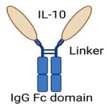
Fusion of IL-10 with IgG Fc domain
  • Prolonged IL-10’s half-life and well-tolerated.
  • Potent antitumor activity, primarily mediated by reinvigorating and expanding exhausted CD8+ TILs.
  • Synergized with ICIs or CAR T-cell therapy.
[65]
Bispecific
CmAb(IL-10)2
Fusion Protein
Cancers 17 01012 i002
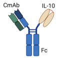
Fusion of Anti-EGFR antibody (Cetuximab “CmAb”) with
IL-10 dimer to engage EGFR and IL-10R in EGFR+ tumors
  • Prolonged IL-10’s half-life and well-tolerated.
  • Targeted EGFR-positive tumors.
  • Improved tumor-specific cytotoxic activity of CD8+ T cells.
  • Hindered DC-mediated apoptosis of CD8+ TILs.
  • Overcame resistance to ICIs therapy.
[66]
BF10 Fusion
Protein
Cancers 17 01012 i003
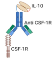
Bispecific Anti-CSF-IR/IL-10 Fusion Protein, combining IL-10 with anti-CSF-1R- antibody
  • Prolonged IL-10’s half-life and well-tolerated.
  • Targeted TAMs-enriched tumors.
  • Simultaneously delivered IL-10 selectively to TME while inhibiting CSF-1R signaling.
  • Depleted TAMs and reprogrammed CD8+ TILs.
[67]
IL-10 variants
for IL-10Rβ
Cancers 17 01012 i004
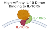
Bioengineered IL-10 variant with increased binding affinity for IL-10Rβ
  • Potent immunomodulatory effects at low doses.
  • Enhanced cytotoxic activity of CAR T cells against acute myeloid leukemia cells.
[68]
Oncolytic
Viruses (OVs) Based Strategy
Cancers 17 01012 i005
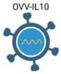
An oncolytic vaccina virus (OVV) armed with armed with IL-10 gene (OVV-IL10)In pancreatic cancer models, OVV-IL-10 showed
  • Sustained tumor suppression.
  • Reduced risk of disease recurrence.
[69]
Nanoparticles
&
Scaffolds
Based Systems
Cancers 17 01012 i006
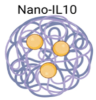
Conjugation of IL-10 into nanoparticles or scaffolds Delivery systems
  • Prolonged IL-10’s half-life.
  • Well tolerated.
  • Enhanced storage stability and antitumor activity.
[70,71]
Abbreviations: CAR T cells: Chimeric antigen receptor T cells, CSF-1R; colony-stimulating factor-1 receptor, EGFR; epidermal growth factor receptor, ICIs: immune checkpoint inhibitors, OVV; oncolytic vaccinia virus, TILs; tumor-infiltrating lymphocytes, TAMs; tumor-associated macrophages, TMEs; tumor microenvironments. Created in BioRender. Minshawi, F. (2025) [63] (https://BioRender.com/o45w353, accessed on 12 November 2024).

4.1. Fusion Proteins and Immunocytokine-Based Strategies

The fusion protein technology employed in the immunocytokine (ICK) approach presents a promising strategy to address the pharmacological limitations, suboptimal pharmacokinetics, and AEs associated with cytokine-based cancer therapies. These fusion proteins are engineered to enhance cytokine delivery specifically to the TME, thereby improving therapeutic efficacy [72,73]. Although multiple ICK strategies have been devised to stimulate antitumor immune responses [74], we herein focused on the most significant ICKs that specifically explore IL-10-based therapies in the preclinical cancer setting.

4.1.1. Il-10/Fc Fusion Protein Approach

Genetic fusion of IL-10 with the fragment crystallizable (Fc) domain of the IgG antibody has led to the development of a therapeutic agent, termed the IL-10/Fc fusion protein. This new approach of IL-10-directed cancer therapy demonstrated low toxicity while exhibiting potent antitumor activity, primarily by reinvigorating and expanding terminally exhausted CD8+ tumor-infiltrating lymphocytes (TILs) [65]. Most importantly, these reinvigorated and expanded cytotoxic CD8+ TILs are crucial for tumor cell elimination. They are typically unresponsive to ICI therapy, representing a significant barrier in cancer immunotherapy [65].
Exhausted CD8+ TILs can be categorized into two subsets with distinct functional roles. Progenitor exhausted TILs (TCF-1+TIM-3−) possess proliferative potential and can differentiate into terminally exhausted TILs (TCF-1−TIM-3+), which directly contribute to tumor killing due to their superior cytotoxic activity [75,76,77]. While progenitor TILs respond to ICI therapies, such as anti-PD-1 blockade and mediate tumor suppression; terminally exhausted TILs lack proliferative capacity and remain largely unresponsive to ICIs and other immunotherapies [77,78]. Consequently, targeting the terminally exhausted CD8+ TILs subset within TME remains a significant challenge but offers immense therapeutic potential. Interestingly, combination therapy involving IL-10/Fc fusion protein and either ICI or chimeric antigen receptor (CAR) T-cell therapy has effectively reinvigorated terminally exhausted CD8+ TILs and demonstrated substantial tumor regression in mice with syngeneic models of melanoma, ovarian, and colorectal carcinoma [65]. These therapies were well-tolerated, with no significant weight loss or adverse toxic effects observed in the treated mice [65]. Moreover, the IL-10/Fc fusion protein has been shown to effectively shift and reprogram T cell metabolism via its mitochondrial pyruvate carrier system [65], and this metabolic shift is critical for reactivating terminally exhausted CD8+ TILs, enabling their functional recovery and antitumor activity [79].

4.1.2. Bispecific CmAb-(IL-10)2 Fusion Protein: Targeting EGFR+ Tumors

To enhance the targeted delivery of IL-10 within the TME and overcome the limitations of its short half-life, Qiao et al. developed a bispecific fusion protein, designed as CmAb-(IL-10)2 [66]. This construction aimed to simultaneously engage an oncogenic receptor and the IL-10R. For proof-of-concept studies, the researchers utilized Cetuximab (CmAb), an FDA-approved monoclonal antibody that selectively blocks ligand binding and intracellular signaling of epidermal growth factor receptor (EGFR) [80]. Notably, EGFR, a type I receptor tyrosine kinase, and its ligands play a pivotal role in regulating multiple cellular pathways involved in cancer cell proliferation, survival, and metastasis, and in tumor angiogenesis [81,82]. Moreover, intratumoral excessive EREG mRNA expression is significantly linked with short OS in several human cancers [82]. Thus, the dual functionality of CmAb-(IL-10)2 is based on Cetuximab in one arm to bind EGFR+ tumor cells and an IL-10 dimer in the other arm to bind and activate IL-10R-mediated signaling pathways [66]. In murine tumor models of transplanted melanoma (B16-cEGFR), colorectal carcinoma (MC38-cEGFR), lung cancer (TC1-cEGFR), and human epidermoid carcinoma (A431-cEGFR)—where „cEGFR” indicates that these cancer cells were engineered to express a chimeric EGFR—CmAb-(IL-10)2 effectively extended the half-life of IL-10 without inducing toxicity. Additionally, it efficiently targeted EGFR+ tumors and specifically inhibited dendritic cell (DC)-mediated apoptosis of CD8+ TILs [66]. Mechanistic studies revealed that the antitumor efficacy of CmAb-(IL-10)2 was highly dependent on the presence of intratumoral CD8+ T cells and required IL-10R signaling on DCs to prevent DC-mediated apoptosis of CD8+ TILs. Furthermore, CmAb-(IL-10)2 demonstrated the capacity to overcome resistance to ICI therapy. When administered either intratumorally or systemically, CmAb-(IL10)2 exhibited a synergistic antitumor effect with ICI therapy, significantly improving therapeutic outcomes. These findings emphasize CmAb-(IL-10)2 as a promising therapeutic agent with potential for clinical translation [66]. In a constant line, a Phase 1/II clinical trial (NCT05396339 (https://clinicaltrials.gov/study/NCT05396339 (accessed on 27 November 2024)) is in recruiting status to investigate the safety, tolerability, dose escalation and pharmacokinetic profile, and the preliminary antitumor activity of IAE0972 (EGFR/IL10) in patients with advanced malignant solid tumors.

4.1.3. Bifunctional Anti-CSF-1R/IL-10 Fusion Protein: Targeting TAMs-Enriched Tumors

Tumor-associated macrophages (TAMs) play a critical role in promoting tumor progression and metastatic colonization through the release of various tumorigenic mediators and growth factors. TAMs are predominant immune cell population within the TME, tumor blood vessels, and stromal regions, and their abundance is linked to poor clinical outcomes and resistance to therapies [83,84]. In addition, TAMs contribute to the induction of exhaustion programs inCD8+ TILs, whereas TAM depletion has been shown to reinvigorate and restore the functional capacity of exhausted CD8+ TILs [85]. Therefore, TAM depletion or repolarization is considered a critical target in cancer immunotherapy, particularly for solid tumors [86]. In this regard, the colony-stimulating factor 1 receptor (CSF-1R) is crucial in managing TAMs and other cellular interactions within the TME [87,88]. CSF-1R overexpression has been observed in various neoplastic conditions, where it is associated with TAMs polarization, tumor immune escaping, and a poorer prognosis [88,89]. Coherently, the inhibition of CSF-IR signaling has been strongly suggested for hindering immune suppression and escape in tumors [90]. In line with this, Chang et al. generated a bifunctional protein, referred to as BF10, which combines IL-10 with a CSF-1R-blocking antibody [67]. This bispecific construct aims to deliver IL-10 selectively to TAM-enriched TMEs while simultaneously inhibiting CSF-1R signaling. As a therapeutic agent, BF10 demonstrated significant antitumor activity across various preclinical cancer models, with particular efficacy against head and neck cancers [67]. Notably, BF10 not only induced TAMs depletion but also triggered the proliferation, activation, and metabolic reprogramming of CD8+ TILs. Moreover, combining BF10 with ICI therapy further amplified its antitumor efficacy. Biodistribution studies of BF10 revealed its accumulation in tumors, tumor-draining lymph nodes, and the spleen, pointing out its systemic and local anti-tumor immunomodulatory effects. These findings underscore the effective engineering strategy for BF10 therapy and its potential for clinical therapeutic development [67].

4.2. Bioengineering of IL-10 Variants with Enhanced Affinity for IL-10Rβ

IL-10 exhibits a high binding affinity for its IL-10Rα receptor subunits and a much lower affinity for its IL-10Rβ subunits [91,92,93]. This differential binding affinity has been proposed to play a crucial role in IL-10’s signaling mechanism and may contribute to the limited in vivo clinical efficacy of wild-type IL-10. Consequently, enhancing IL-10’s binding affinity to IL-10Rβ could improve its therapeutic potential. To investigate this hypothesis, Gorby et al. engineered an IL-10 variant with enhanced affinity for IL-10Rβ [68]. This variant more efficiently recruited IL-10Rβ into active cell surface signaling complexes, leading to stronger activation of STAT1 and STAT3 signaling pathways and increased transcriptional activity in human CD8+ T cells. Notably, these effects were observed even at low ligand concentrations [68]. Moreover, in the presence of this engineered IL-10 variant, chimeric antigen receptor (CAR) T cells expanded and exhibited significantly greater cytotoxicity against acute myeloid leukemia cells compared to those cultured with wild-type IL-10 [68]. Supporting this strategy, recent cryo-electron microscopy structural analysis of the IL-10 receptor complex revealed that the low affinity of IL-10 for IL-10Rβ ensures tightly regulated signaling, occurring only when IL-10 is present at sufficient concentrations [94,95]. Alternatively, the development of IL-10Rβ-specific agonist variants with modified cell-type selectivity may facilitate complete receptor complex formation and enhance IL-10’s clinical efficacy [68,94,95].

4.3. Oncolytic Viruses and Nanoparticle-Based Carriers for Il-10 and Other Biologically Active Il-10 Isoforms

Cancer virotherapy using oncolytic viruses (OVs) has witnessed a resurgence due to its selective replication and tumor-specific lytic effects, sparing normal cells, and its clinically established safety and tolerability profile [96]. Furthermore, we and others have reported that the arming of OVs with genes of pharmacologically active cytokines has ushered in a new generation of OVs combined with cancer immunotherapy, significantly enhancing tumor cell eradication and inducing a robust antitumor immune response [97,98,99,100]. Specifically, Chard et al. investigated the antitumor potential of an oncolytic vaccinia virus armed with IL-10 gene (OVV-IL10) in various pancreatic cancer models [69]. OVV-IL10 proved to be more effective in enhancing anti-tumor efficacy while dampening antiviral immune response compared to its unarmed counterpart. Moreover, it markedly exhibited a sustained tumor suppression and reduced risk of disease recurrence, achieved through modulation of both innate and adaptive immune responses and stimulation of anti-tumor cytotoxic CD8+ T cells [69].
Another promising strategy involves nanoparticle-mediated IL-10 delivery, such as its conjugation with polyvinylpyrrolidone-coated silver-based nanoparticles [70]. This approach has demonstrated the ability to enhance storage stability while preserving the biological functionality of IL-10 [70]. Similarly, scaffold-based delivery systems, including microspheres and hydrogels, have been extensively studied for cytokine delivery. These systems can be injected or implanted directly at the diseased site, enabling controlled and sustained release of cytokines as the scaffold material gradually degrades [101,102]. This controlled release not only prolongs the half-lives of the loaded cytokines but also minimizes their systemic toxicity. Moreover, scaffold-based approaches can allow for the co-delivery of multiple therapeutic agents within a single matrix, and the simultaneous delivery of these agents ensures spatial and temporal coordination, thereby fostering synergistic therapeutic effects and improving treatment efficacy [71,103].
Recently, our research team developed a genetically modified IL-10 variant (stable IL-10) with enhanced stability and biological activity at significantly lower concentrations than natural IL-10 [63]. Furthermore, we are currently designing a targeted delivery system for this stable IL-10 isoform by conjugating it to tumor-specific antibodies (e.g., anti-EGFR) to improve intratumoral accumulation and therapeutic efficacy in preclinical models of colorectal carcinoma. This strategy builds upon our prior success in delivering stable IL-10 to colonic inflammatory sites using anti-VCAM antibodies (unpublished data). However, the safety profile of our genetically engineered IL-10, both with and without antibody conjugation, remains to be evaluated, along with its anticancer efficacy alone and in combination with conventional therapy.
Other approaches to improve the anticancer pharmacological profile of therapeutic IL-10 have also been proposed. In this context, the complement receptors C3aR and C5aR, which are highly expressed in tumor and immune cells, have recently been identified as immune checkpoint receptors with critical roles in tumor development and progression. Their aberrant intratumoral activation fosters chronic inflammation, angiogenesis, tumor cell proliferation, survival, and invasion [104,105,106,107]. Crucially, activation of these receptors suppresses IL-10 production and inhibits IL-10-dependent T-cell-mediated antitumor immunity. Conversely, blocking these signaling pathways reverses the transcriptional suppression of IL-10, stimulates IL-10 production by antitumor T cells, and enhances T-cell expansion and antitumor activity [108]. Accordingly, a combination approach involving IL-10 and C3aR/C5aR antagonists could serve as an effective therapeutic strategy for immune-resistant cancers, particularly when augmented with the addition of IL-2 [IL-10 + IL-2 + C3aR/C5aR signaling blockade] [109]. This proposed strategy is further supported by earlier findings that demonstrated the in vitro synergy between IL-10 and IL-2 in expanding and enhancing the activity of CD8+ T cells derived from lung cancer patients. The IL-10/IL-2 combination not only increased their IFN-γ expression but also upregulated genes associated with cytotoxic antitumor activity [108,109].
In summary, the intratumoral delivery methods and sustainability of therapeutic IL-10 (or its biologically active isoforms), specifically designed to interact with critical targets within the TME, are critical considerations to effectively mediate multimodal antitumor activity within tumor tissues while reducing systemic adverse effects and minimizing inhibitory impacts on non-target cell types.

5. Clinical Trials of Il-10 Directed in Cancer Immunotherapy

The multifaceted role of sustained IL-10 in simultaneously activating and expanding cytotoxic tumor-specific CD8+ T cells while suppressing tumor-associated inflammation has positioned it as a cytokine of significant clinical interest in cancer immunotherapy. To maximize IL-10’s therapeutic potential, various bioengineering strategies are being actively investigated, with PEGylation representing a key advancement in its clinical translation [110].
PEG, an FDA-approved polymer widely used to enhance the pharmacokinetics of therapeutic proteins, improves drug stability by extending circulation half-life, reducing renal clearance, and lowering systemic dosing requirements, thereby mitigating dose-limiting toxicities [74,111]. Specifically, PEGylation extends IL-10’s half-life from only a few hours [112,113] to approximately 24 h, ensuring sustained serum and intratumoral IL-10 concentrations while preserving its dual antitumor and immunoregulatory functions [114]. Within this framework, pegilodecakin (AM0010) has been the most extensively studied PEGylated IL-10 formulation in clinical cancer therapy (Table 2).
Table 2. Summary of published clinical trials on pegilodecakin (PEGylated IL-10) therapy.
Trial (NCT) and [Ref.]Phase, Patients, and DesignTherapeutic Agent(s)Key OutcomesInterpretation
IVY (NCT02009449) [115]Phase 1; patients with various advanced solid tumorsPEG aloneAn acceptable safety and early antitumor activity Warranting further evaluation
IVY (NCT02009449) [116]Phase 1b; patients with various solid tumors, including patients with treatment-refractory NSCLC and RCCPEG + Anti-PD1 therapy (PEMB or NIVO) ORR: 43% (NSCLC) and 40% (RCC)Favorable anti-tumor activity between PEG and Anti-PD1 therapy
IVY (NCT02009449) [117]Phase 1b; mRCC patientsPEG + NIVO vs.
PEG + Pazopanib
ORR: 43% (PEG/NIVO) vs. 33% (PEG/Pazopanib)PEG/NIVO showed better activity and tolerable safety profile
Ovarian Cancer
[118]
Phase II; patients with metastatic Ovarian cancerPEG alone vs.
PEG + platinum-taxane
PFS:2.4 months (PEG) vs.
5.2 months (co-therapy)
Warranting further evaluation
CYPRESS 1 and 2 (NCT03382899 and (NCT03382912)
[119]
Phase II; mNSCLC patients divided into: CYPRESS 1 (PD-L1 TPS ≥ 50%) and CYPRESS 2 (PD-L1 TPS 0–49%)PEG + PEMB (or NIVO)
vs.
PEMB (or NIVO) alone
CYPRESS 1: ORR (47% vs. 44%) and PFS (6.3 vs. 6.1 months) and CYPRESS 2: ORR (15% vs. 12%), PFS (1.9 vs. 1.9 months), and OS (6.7 vs. 10.7 months)PEG + Anti-PD1 therapy (PEMB or NIVO) did not offer benefits over Anti-PD1 alone
SEQUOIA (NCT02923921) [120,121]Phase III; gemcitabine-refractory PDAC patientsPEG + FOLFOX
vs.
FOLFOX alone
ORR: 4.6% vs. 5.6%; PFS: 2.1 vs. 2.1 months; and OS: 5.8 vs. 6.3 monthsPEG did not improve FOLFOX efficacy in advanced gemcitabine-refractory PDAC patients
Abbreviations: AEs; Adverse events, FOLFOX; 5-fluorouracil, leucovorin calcium, oxaliplatin, mNSCLC; metastatic non-small cell lung cancer, mRCC; metastatic renal cell carcinoma; NIVO; nivolumab, ORR; Objective tumor response, OS; overall survival, PEG: pegilodecakin, PEMB; pembrolizumab, PDAC: pancreatic ductal adenocarcinoma; PD-L1 TPS; proportion score of PD-L1–positive tumor cells; PFS; progression-free survival.

5.1. Early-Phase Dose Escalation Study of Pegilodecakin Monotherapy

In Phase 1 of the first-in-human trial, IVY (NCT02009449 (https://clinicaltrials.gov/study/NCT02009449 (accessed on 26 November 2024)), a total of 51 patients with diverse advanced solid tumors who had failed all available prior therapies, including previous ICIs or IL-2 treatments, were enrolled and treated daily with subcutaneous self-injected pegilodecakin monotherapy at escalating doses ranging from 1 μg/kg to 40 μg/kg [115]. Objective tumor response (ORR) was assessed based on immune-related response criteria [122]. Overall, pegilodecakin monotherapy demonstrated encouraging antitumor responses, particularly in patients with uveal melanoma or advanced renal cell carcinoma (RCC). Delayed, prolonged, and mixed antitumor responses were observed in other enrolled patients, including durable disease stability in a patient with microsatellite-stable colorectal cancer [114,115]. Notably, the administered pegilodecakin led to a sustained and clinically meaningful elevation in serum IL-10 concentrations, ranging between 1 and 40 ng/mL. The most frequently observed adverse events (AEs) were mild to moderate (grade 1 or 2) and included injection-site reactions, fever, fatigue, anemia, and thrombocytopenia, with seven patients in the expansion cohort necessitating dose reductions. Grade 3 or 4 anemia, thrombocytopenia, transaminitis, and dermatologic reactions, were reported but were rare and resolved without complications [114,115]. Importantly, hypotension and long-lasting immune-related adverse events (irAEs) were not reported with pegilodecakin monotherapy. Furthermore, consistent with IL-10’s known anti-inflammatory properties, inflammatory irAEs such as hepatitis, colitis, or pneumonitis were also not observed. This contrasts with ICIs or IL-2 treatments, where ICIs frequently induce irAEs [123], and IL-2 is associated with dose- and duration-limiting severe hypotension [124]. Moreover, pharmacodynamic analyses revealed that pegilodecakin-treated patients achieved sustained IL-10 levels and exhibited significant upregulation of IFNγ and IL-18, accompanied by a 42% reduction in serum TGFβ levels [114,115]. These findings support the hypothesis that pharmacologically stabilized IL-10 enhances T-cell activation in cancer patients.

5.2. Evaluation of Pegilodecakin in Combination with Anti-PD-1 Therapy

In Phase 1b of the IVY trial (NCT02009449 (https://clinicaltrials.gov/study/NCT02009449 (accessed on 26 November 2024)), 111 patients were enrolled and received combination therapy with pegilodecakin plus ICIs (pembrolizumab or nivolumab anti-PD-1- monoclonal antibodies). The patient cohorts included those with advanced and treatment-refractory RCC (n = 38), melanoma (n = 37), non-small cell lung cancer (NSCLC, n = 34), bladder cancer (n = 1), and triple-negative breast cancer (n = 1). The achieved ORR was 43% in NSCLC, 40% in RCC, and 10% in melanoma [116]. Moreover, the combination of pegilodecakin and anti-PD-1 therapy resulted in a progression-free survival (PFS) of 9.4 months and an overall survival (OS) of 24.1 months in NSCLC patients, as well as a PFS of 16.7 months and 1-year OS of 93% in patients with RCC [116]. These findings contrast with those observed in earlier studies involving a similar population of RCC patients who were mono-treated with anti-PD-1 (nivolumab) and showed an ORR of 22%, a PFS of 4.2 months and < 80% 1-year OS [125]. Similarly, in patients with advanced NSCLC, who received anti-PD-1 (pembrolizumab) monotherapy, the ORR was 19.4%, with a PFS of 3.7 months and an OS of 12 months [126]. Moreover, in these earlier studies, the response to anti-PD-1 monotherapy was strongly correlated with high PD-L1 expression in tumor cells [126]. In contrast, responses to pegilodecakin–anti-PD-1 combination therapy in the Phase 1b IVY trial were observed even in patients with low PD-L1 expression [116]. The AEs profile across patients’ cohorts was aligned with the prior data reported for single-agent pegilodecakin or anti-PD-L1 therapies. Red blood cell hemophagocytosis and lymphohistiocytosis were identified in three patients (grade 1/2 in two patients, grade 4 in one patient), all of whom recovered uneventfully following resolution of these events [116].
Next, a comprehensive analysis was focused on all metastatic RCC (mRCC) patients in the above IVY trial (NCT02009449 (https://clinicaltrials.gov/study/NCT02009449 (accessed on 26 November 2024)) [117]. Specifically, the combination of pegilodecakin with anti-PD-1 therapy resulted in promising clinical outcomes and achieved an ORR of 43%, compared to 33% for the combination of pegilodecakin with the multi-tyrosine kinase inhibitor “pazopanib”. Additionally, while pegilodecakin monotherapy in these mRCC patients resulted in a PFS of 1.8 months and OS probabilities of 50% at one year and 17% at two years, its combination with anti-PD-1 significantly led to a PFS of 13.9 months and OS probabilities of 76% at one year and 61% at two years. Furthermore, the AEs associated with pegilodecakin, both as monotherapy and in combination with ICIs, were consistent with prior reports [117]. Since most patients in the IVY trial were heavily pretreated and had experienced disease progression prior to their enrollment, these findings may hold clinical significance, particularly in the context of mRCC therapy, emphasizing the need for further investigations [117]. In support, an interim report evaluated the clinical activity of pegilodecakin monotherapy in metastatic epithelial ovarian cancer reported that 44% of the treated patients achieved a disease control rate of 66.7% with PFS exceeding 3.5 months [114,118].

5.3. Evaluation of Pegilodecakin as an Add-On Therapy in CYPRESS and SEQUOIA Clinical Trials

Despite the encouraging outcomes reported above in the IVY study, pegilodecakin failed to demonstrate promising results in the subsequent CYPRESS 1 and 2 clinical trials (NCT03382899(https://clinicaltrials.gov/study/NCT03382912 (accessed on 23 October 2024)); NCT03382912 (https://clinicaltrials.gov/study/NCT03382899 (accessed on 23 October 2024)). These randomized Phase II studies investigated whether the addition of pegilodecakin to immune checkpoint inhibitors (ICIs; anti-PD-1 monoclonal antibodies: pembrolizumab or nivolumab) offered therapeutic benefits for patients with metastatic NSCLC compared to ICIs alone [119]. Specifically, CYPRESS 1 enrolled patients with high PD-L1 expression (tumor proportion score [TPS] ≥ 50%), while CYPRESS 2 included patients with low or negative PD-L1 expression (TPS 0–49%) [119]. In CYPRESS 1, patients received either pembrolizumab monotherapy (n = 50) or a combination of pegilodecakin and pembrolizumab (n = 51), while in CYPRESS 2, treatments included nivolumab alone (n = 25) or combined with pegilodecakin (n = 27). Pegilodecakin was self-administered subcutaneously by the patents at fixed doses of 0.4 mg (for patients ≤ 80 kg) or 0.8 mg (for patients > 80 kg) on days 1–5 and 8–12, with rest periods on days 6–7 and 13–14, as guided by the Phase I IVY study findings. Overall, the pegilodecakin + anti-PD-1 combination therapy did not significantly improve ORR, PFS, or OS compared to anti-PD-1 monotherapy. For CYPRESS 1 (pegilodecakin + pembrolizumab vs. pembrolizumab alone), the results were ORR (47% vs. 44%), PFS (6.3 vs. 6.1 months), and OS (16.3 months vs. not reached). For CYPRESS 2 (pegilodecakin + nivolumab vs. nivolumab alone), the results were ORR (15% vs. 12%), PFS (1.9 vs. 1.9 months), and OS (6.7 vs. 10.7 months). Furthermore, treatment discontinuation rates due to severe hematologic AEs were doubled in the combination treatment arms of both studies [119].
In a constant line, the SEQUOIA study (NCT02923921(https://clinicaltrials.gov/study/NCT02923921 (accessed on 12 November 2024)), a global randomized Phase III trial, assessed the efficacy and safety of adding pegilodecakin to the chemotherapy regimen of folinic acid, fluorouracil, and oxaliplatin (FOLFOX) as a second-line treatment for gemcitabine-refractory metastatic pancreatic ductal adenocarcinoma (PDAC). Patients were randomized to receive pegilodecakin + FOLFOX (n = 283) or FOLFOX alone (n = 284). Alongside the standardized FOLFOX regimen, pegilodecakin was self-administered subcutaneously at fixed doses as described above. Although the pegilodecakin + FOLFOX combination initially demonstrated enhanced antitumor activity compared to FOLFOX alone—indicated by immunostimulatory signals of the IL-10R pathway, including increased levels of IFN-γ and granzyme B and decreased TGF-β levels—its overall results showed no improvement in OS (5.8 vs. 6.3 months), PFS (2.1 vs. 2.1 months), and ORR (4.6% vs. 5.6%) [120,121]. Moreover, the incidence of grade ≥3 hematological AEs, particularly anemia, thrombocytopenia, and neutropenia, was notably higher in the pegilodecakin + FOLFOX arm, necessitating greater use of red blood cell transfusions and erythropoiesis-stimulating agents (5.8% vs. 2.0%). These findings underscore the need for further translational research to identify patients likely to benefit from pegilodecakin-chemotherapy combination in treatment-refractory advanced tumors [120,121].
Taken together, while the PEGylation process of recombinant human IL-10 (pegilodecakin) has notably enhanced its stability, serum concentration, and half-life, all while maintaining its biological activity, its overall benefits in the CYPRESS and SEQUOIA clinical trials has exhibited significant limitations [119,120,121]. The heterogeneity in cancer types, patient populations, and therapeutic combination types and regimens might complicate direct comparisons among these trials [58]. Importantly, although PEGylation is clinically approved for IFNα2a and IL-2 therapy and is currently under evaluation for additional immune-cytokine therapies such as IL-10 and IL-15, it is often associated with unwanted dose-dependent effects on bioactivity, primarily due to its amphiphilic nature [74]. In support, PEG-IL10-induced dose-dependent toxicity has been reported in both preclinical and clinical studies [53]. To overcome these challenges, further investigations, including the development of alternative and reprogrammed strategies optimizing IL-10 delivery and intratumoral localization, are warranted. These efforts are essential to fully harness and realize the mono- or combination-therapeutic potential of IL-10-directed therapy across a range of malignancies.

6. Conclusion

While IL-10 has historically been associated with tumor progression, accumulating evidence underscores its ability to enhance antitumor immunity by reinvigorating exhausted CD8+ TILs and activating their cytotoxic function; inducing IFN-γ, IL-18, granzymes, and FasL expression; reducing tumor-associated inflammation and TGF-β levels; and reprogramming TAMs. These attributes have spurred interest in IL-10-based therapies, both as monotherapy and in combination with ICIs.
Advancements in IL-10 bioengineering are driving its clinical translation. PEGylated IL-10 (pegilodecakin) demonstrated potential in early clinical trials, particularly in patients with RCC [114,116,117]. However, later-stage clinical trials, including CYPRESS and SEQUOIA, revealed challenges such as dose-limiting toxicities and inconsistent efficacy [119,120,121]. These findings underscore the necessity of refining IL-10-based therapeutics through improved delivery mechanisms, optimized dosing strategies, and precise patient selection criteria.
Preclinical advancements offer promising strategies to overcome these limitations [64]. Engineered IL-10 variants with enhanced IL-10Rβ affinity have demonstrated superior stability and antitumor activity in murine models. Additionally, IL-10 fusion proteins, such as IL-10/Fc constructs, have shown potential in prolonging IL-10’s half-life, while minimizing systemic exposure, and in reinvigorating exhausted CD8+ TILs, thereby enhancing tumor-specific immune responses [65]. The development of bispecific fusion proteins, such as CmAb-(IL-10)2 [66] and BF10 [67], further supports the rationale for IL-10-based combination therapies and exhibiting remarkable synergy with ICIs in preclinical studies. Emerging delivery platforms, including nanoparticle-based and scaffold-mediated IL-10 formulations, provide additional avenues to improve IL-10’s therapeutic efficacy. Collectively, such approaches hold promise for overcoming the pharmacokinetic challenges associated with IL-10 therapy, enabling controlled and sustained intratumoral IL-10 release, ensuring localized immune activation with reduced toxicity, and warranting further investigation in translational and clinical settings. Looking ahead, the integration of IL-10 into precision oncology frameworks requires a deeper understanding of its mechanistic role in shaping the TME. Future research should focus on optimizing IL-10-based combination regimens, leveraging predictive biomarkers for patient stratification, and refining delivery strategies to maximize therapeutic efficacy. Additionally, elucidating the interplay between IL-10 and other immunoregulatory pathways will provide valuable insights into its broader applicability across various cancer types.
In conclusion, IL-10 represents a compelling avenue in cancer immunotherapy, with the potential to reshape treatment paradigms. Enhancing tumor specificity while minimizing systemic toxicity remains a critical objective for future clinical applications. Continued innovation in cytokine engineering, targeted delivery, and combination strategies will be instrumental in overcoming existing limitations and unlocking IL-10’s full therapeutic potential. As research progresses, harnessing IL-10’s immunomodulatory capabilities may pave the way for more effective, durable, and personalized cancer treatments.

Author Contributions

Conceptualization: A.G.E.-S., F.M. and B.R.; methodology: J.A.A. (Jihan Abdullah Abdulrahman), H.D.A., K.S.A., H.H.F., W.S.A. and J.A.A. (Jana Ahmed Aloufi); validation: H.T.K., A.A; S.H.D., S.A.M., G.H.M. and J.B.A.; data curation: H.T.K., S.H.D., A.A., S.A.M., G.H.M. and J.B.A.; software: J.A.A. (Jihan Abdullah Abdulrahman), H.D.A., K.S.A., H.H.F., W.S.A. and J.A.A. (Jana Ahmed Aloufi); writing—original draft preparation: A.G.E.-S., F.M. and B.R.; writing—review and editing: A.G.E.-S., A.A., F.M. and B.R.; supervision: A.G.E.-S., F.M. and B.R.; funding acquisition: F.M.; project administration: A.G.E.-S., F.M. and B.R.; All authors have read and agreed to the published version of the manuscript.

Funding

This research work was funded by Umm Al-Qura University, Saudi Arabia under grant number: 25UQU4280261GSSR01.

Data Availability Statement

The data that support the findings of this study are available within the article.

Acknowledgments

The authors extend their appreciation to Umm Al-Qura University, Saudi Arabia for funding this research work through grant number: 25UQU4280261GSSR01.

Conflicts of Interest

The authors declare no conflicts of interest.

References

  1. Lim, Y.Y.; Chin, Y.M.; Tai, M.C.; Fani, S.; Chang, K.M.; Ong, T.C.; Bee, P.C.; Gan, G.G.; Ng, C.C. Analysis of interleukin-10 promoter single nucleotide polymorphisms and risk of non-Hodgkin lymphoma in a Malaysian population. Leuk. Lymphoma 2015, 56, 163–168. [Google Scholar] [CrossRef] [PubMed]
  2. Iyer, S.S.; Cheng, G. Role of interleukin 10 transcriptional regulation in inflammation and autoimmune disease. Crit. Rev. Immunol. 2012, 32, 23–63. [Google Scholar] [CrossRef] [PubMed]
  3. Lin, X.; Wang, X.; Xiao, F.; Ma, K.; Liu, L.; Wang, X.; Xu, D.; Wang, F.; Shi, X.; Liu, D.; et al. IL-10-producing regulatory B cells restrain the T follicular helper cell response in primary Sjogren’s syndrome. Cell Mol. Immunol. 2019, 16, 921–931. [Google Scholar] [CrossRef] [PubMed]
  4. Wei, H.; Li, B.; Sun, A.; Guo, F. Interleukin-10 Family Cytokines Immunobiology and Structure. Adv. Exp. Med. Biol. 2019, 1172, 79–96. [Google Scholar] [CrossRef]
  5. Itakura, E.; Huang, R.R.; Wen, D.R.; Paul, E.; Wunsch, P.H.; Cochran, A.J. IL-10 expression by primary tumor cells correlates with melanoma progression from radial to vertical growth phase and development of metastatic competence. Mod. Pathol. 2011, 24, 801–809. [Google Scholar] [CrossRef]
  6. Ozaki, K.; Leonard, W.J. Cytokine and cytokine receptor pleiotropy and redundancy. J. Biol. Chem. 2002, 277, 29355–29358. [Google Scholar] [CrossRef]
  7. Pestka, S.; Krause, C.D.; Sarkar, D.; Walter, M.R.; Shi, Y.; Fisher, P.B. Interleukin-10 and related cytokines and receptors. Annu. Rev. Immunol. 2004, 22, 929–979. [Google Scholar] [CrossRef]
  8. Yoon, S.I.; Logsdon, N.J.; Sheikh, F.; Donnelly, R.P.; Walter, M.R. Conformational changes mediate interleukin-10 receptor 2 (IL-10R2) binding to IL-10 and assembly of the signaling complex. J. Biol. Chem. 2006, 281, 35088–35096. [Google Scholar] [CrossRef]
  9. Murray, P.J. Understanding and exploiting the endogenous interleukin-10/STAT3-mediated anti-inflammatory response. Curr. Opin. Pharmacol. 2006, 6, 379–386. [Google Scholar] [CrossRef]
  10. Murray, P.J. The JAK-STAT signaling pathway: Input and output integration. J. Immunol. 2007, 178, 2623–2629. [Google Scholar] [CrossRef]
  11. Hutchins, A.P.; Diez, D.; Miranda-Saavedra, D. The IL-10/STAT3-mediated anti-inflammatory response: Recent developments and future challenges. Brief. Funct. Genom. 2013, 12, 489–498. [Google Scholar] [CrossRef]
  12. O’Shea, J.J.; Schwartz, D.M.; Villarino, A.V.; Gadina, M.; McInnes, I.B.; Laurence, A. The JAK-STAT pathway: Impact on human disease and therapeutic intervention. Annu. Rev. Med. 2015, 66, 311–328. [Google Scholar] [CrossRef]
  13. Shouval, D.S.; Ouahed, J.; Biswas, A.; Goettel, J.A.; Horwitz, B.H.; Klein, C.; Muise, A.M.; Snapper, S.B. Interleukin 10 receptor signaling: Master regulator of intestinal mucosal homeostasis in mice and humans. Adv. Immunol. 2014, 122, 177–210. [Google Scholar] [CrossRef]
  14. Walter, M.R. The molecular basis of IL-10 function: From receptor structure to the onset of signaling. In Interleukin-10 in Health and Disease; Springer: Berlin/Heidelberg, Germany, 2014; Volume 380, pp. 191–212. [Google Scholar] [CrossRef]
  15. Fiorentino, D.F.; Zlotnik, A.; Mosmann, T.R.; Howard, M.; O’Garra, A. IL-10 inhibits cytokine production by activated macrophages. J. Immunol. 1991, 147, 3815–3822. [Google Scholar] [CrossRef]
  16. Oft, M. IL-10: Master switch from tumor-promoting inflammation to antitumor immunity. Cancer Immunol. Res. 2014, 2, 194–199. [Google Scholar] [CrossRef]
  17. Bedke, T.; Muscate, F.; Soukou, S.; Gagliani, N.; Huber, S. IL-10-producing T cells and their dual functions. Semin. Immunol. 2019, 44, 101335. [Google Scholar] [CrossRef]
  18. Saraiva, M.; Vieira, P.; O’Garra, A. Biology and therapeutic potential of interleukin-10. J. Exp. Med. 2020, 217, e20190418. [Google Scholar] [CrossRef]
  19. Wang, X.; Wang, X.; Wang, D.; Zhou, C.; Lv, K.; Ma, Y.; Chang, W.; Wang, B.; Hu, J.; Ji, Y.; et al. Interleukin-10 overexpression in 4T1 cells: A gateway to suppressing mammary carcinoma growth. Int. Immunopharmacol. 2024, 142, 113089. [Google Scholar] [CrossRef]
  20. Levin, G.; Gotlieb, W.H. Interleukine-10 in ovarian cancer. Chin. Clin. Oncol. 2024, 13, 48. [Google Scholar] [CrossRef]
  21. Mannino, M.H.; Zhu, Z.; Xiao, H.; Bai, Q.; Wakefield, M.R.; Fang, Y. The paradoxical role of IL-10 in immunity and cancer. Cancer Lett. 2015, 367, 103–107. [Google Scholar] [CrossRef]
  22. Rallis, K.S.; Corrigan, A.E.; Dadah, H.; Stanislovas, J.; Zamani, P.; Makker, S.; Szabados, B.; Sideris, M. IL-10 in cancer: An essential thermostatic regulator between homeostatic immunity and inflammation—A comprehensive review. Future Oncol. 2022, 18, 3349–3365. [Google Scholar] [CrossRef] [PubMed]
  23. Guan, Q.; Han, M.; Guo, Q.; Yan, F.; Wang, M.; Ning, Q.; Xi, D. Strategies to reinvigorate exhausted CD8(+) T cells in tumor microenvironment. Front. Immunol. 2023, 14, 1204363. [Google Scholar] [CrossRef] [PubMed]
  24. Carlini, V.; Noonan, D.M.; Abdalalem, E.; Goletti, D.; Sansone, C.; Calabrone, L.; Albini, A. The multifaceted nature of IL-10: Regulation, role in immunological homeostasis and its relevance to cancer, COVID-19 and post-COVID conditions. Front. Immunol. 2023, 14, 1161067. [Google Scholar] [CrossRef] [PubMed]
  25. Wang, M.; Chen, S.; He, X.; Yuan, Y.; Wei, X. Targeting inflammation as cancer therapy. J. Hematol. Oncol. 2024, 17, 13. [Google Scholar] [CrossRef]
  26. Huang, Y.; Zou, K.; Jiang, H.; Li, Z. The complex role of IL-10 in malignant ascites: A review. Cancer Immunol. Immunother. 2024, 73, 32. [Google Scholar] [CrossRef]
  27. Ni, G.; Zhang, L.; Yang, X.; Li, H.; Ma, B.; Walton, S.; Wu, X.; Yuan, J.; Wang, T.; Liu, X. Targeting interleukin-10 signalling for cancer immunotherapy, a promising and complicated task. Hum. Vaccin. Immunother. 2020, 16, 2328–2332. [Google Scholar] [CrossRef]
  28. Gabryšová, L.; Howes, A.; Saraiva, M.; O’Garra, A. The regulation of IL-10 expression. In Interleukin-10 in Health and Disease; Springer: Berlin/Heidelberg, Germany, 2014; Volume 380, pp. 157–190. [Google Scholar] [CrossRef]
  29. Rutz, S.; Ouyang, W. Regulation of Interleukin-10 Expression. Adv. Exp. Med. Biol. 2016, 941, 89–116. [Google Scholar] [CrossRef]
  30. Fan, L.; Qiu, D.; Huang, G.; Chen, J.; Wu, Q.; Xiong, S.; Wu, C.; Peng, Y.; Zhang, Q. Wogonin Suppresses IL-10 Production in B Cells via STAT3 and ERK Signaling Pathway. J. Immunol. Res. 2020, 2020, 3032425. [Google Scholar] [CrossRef]
  31. Saraiva, M.; O’Garra, A. The regulation of IL-10 production by immune cells. Nat. Rev. Immunol. 2010, 10, 170–181. [Google Scholar] [CrossRef]
  32. Zhu, J.; Yamane, H.; Paul, W.E. Differentiation of effector CD4 T cell populations. Annu. Rev. Immunol. 2010, 28, 445–489. [Google Scholar] [CrossRef]
  33. Yi, A.K.; Yoon, J.G.; Yeo, S.J.; Hong, S.C.; English, B.K.; Krieg, A.M. Role of mitogen-activated protein kinases in CpG DNA-mediated IL-10 and IL-12 production: Central role of extracellular signal-regulated kinase in the negative feedback loop of the CpG DNA-mediated Th1 response. J. Immunol. 2002, 168, 4711–4720. [Google Scholar] [CrossRef] [PubMed]
  34. Kaiser, F.; Cook, D.; Papoutsopoulou, S.; Rajsbaum, R.; Wu, X.; Yang, H.T.; Grant, S.; Ricciardi-Castagnoli, P.; Tsichlis, P.N.; Ley, S.C.; et al. TPL-2 negatively regulates interferon-beta production in macrophages and myeloid dendritic cells. J. Exp. Med. 2009, 206, 1863–1871. [Google Scholar] [CrossRef]
  35. Martin, M.; Schifferle, R.E.; Cuesta, N.; Vogel, S.N.; Katz, J.; Michalek, S.M. Role of the phosphatidylinositol 3 kinase-Akt pathway in the regulation of IL-10 and IL-12 by Porphyromonas gingivalis lipopolysaccharide. J. Immunol. 2003, 171, 717–725. [Google Scholar] [CrossRef]
  36. Kanters, E.; Pasparakis, M.; Gijbels, M.J.; Vergouwe, M.N.; Partouns-Hendriks, I.; Fijneman, R.J.; Clausen, B.E.; Förster, I.; Kockx, M.M.; Rajewsky, K.; et al. Inhibition of NF-kappaB activation in macrophages increases atherosclerosis in LDL receptor-deficient mice. J. Clin. Invest. 2003, 112, 1176–1185. [Google Scholar] [CrossRef]
  37. Weichhart, T.; Costantino, G.; Poglitsch, M.; Rosner, M.; Zeyda, M.; Stuhlmeier, K.M.; Kolbe, T.; Stulnig, T.M.; Hörl, W.H.; Hengstschläger, M.; et al. The TSC-mTOR signaling pathway regulates the innate inflammatory response. Immunity 2008, 29, 565–577. [Google Scholar] [CrossRef]
  38. Cao, S.; Liu, J.; Song, L.; Ma, X. The protooncogene c-Maf is an essential transcription factor for IL-10 gene expression in macrophages. J. Immunol. 2005, 174, 3484–3492. [Google Scholar] [CrossRef]
  39. Wang, Z.Y.; Sato, H.; Kusam, S.; Sehra, S.; Toney, L.M.; Dent, A.L. Regulation of IL-10 gene expression in Th2 cells by Jun proteins. J. Immunol. 2005, 174, 2098–2105. [Google Scholar] [CrossRef]
  40. Jones, M.B.; Alvarez, C.A.; Johnson, J.L.; Zhou, J.Y.; Morris, N.; Cobb, B.A. CD45Rb-low effector T cells require IL-4 to induce IL-10 in Foxp3 Tregs and to protect mice from inflammation. PLoS ONE 2019, 14, e0216893. [Google Scholar] [CrossRef]
  41. Zhu, J.; Luo, L.; Tian, L.; Yin, S.; Ma, X.; Cheng, S.; Tang, W.; Yu, J.; Ma, W.; Zhou, X.; et al. Aryl hydrocarbon receptor promotes IL-10 expression in inflammatory macrophages through Src-STAT3 signaling pathway. Front. Immunol. 2018, 9, 2033. [Google Scholar] [CrossRef]
  42. Meng, X.; Grötsch, B.; Luo, Y.; Knaup, K.X.; Wiesener, M.S.; Chen, X.X.; Jantsch, J.; Fillatreau, S.; Schett, G.; Bozec, A. Hypoxia-inducible factor-1α is a critical transcription factor for IL-10-producing B cells in autoimmune disease. Nat. Commun. 2018, 9, 251. [Google Scholar] [CrossRef]
  43. Selig, M.; Poehlman, L.; Lang, N.C.; Völker, M.; Rolauffs, B.; Hart, M.L. Prediction of six macrophage phenotypes and their IL-10 content based on single-cell morphology using artificial intelligence. Front. Immunol. 2024, 14, 1336393. [Google Scholar] [CrossRef] [PubMed]
  44. Rallis, K.S.; Corrigan, A.E.; Dadah, H.; George, A.M.; Keshwara, S.M.; Sideris, M.; Szabados, B. Cytokine-based cancer immunotherapy: Challenges and opportunities for IL-10. Anticancer Res. 2021, 41, 3247–3252. [Google Scholar] [CrossRef] [PubMed]
  45. Yi, M.; Li, T.; Niu, M.; Zhang, H.; Wu, Y.; Wu, K.; Dai, Z. Targeting cytokine and chemokine signaling pathways for cancer therapy. Signal Transduct. Target. Ther. 2024, 9, 176. [Google Scholar] [CrossRef]
  46. Han, J.; Wang, H. Cytokine-overexpressing dendritic cells for cancer immunotherapy. Exp. Mol. Med. 2024, 56, 2559–2568. [Google Scholar] [CrossRef]
  47. Berg, D.J.; Davidson, N.; Kuhn, R.; Muller, W.; Menon, S.; Holland, G.; Thompson-Snipes, L.; Leach, M.W.; Rennick, D. Enterocolitis and colon cancer in interleukin-10-deficient mice are associated with aberrant cytokine production and CD4(+) TH1-like responses. J. Clin. Invest. 1996, 98, 1010–1020. [Google Scholar] [CrossRef]
  48. Neven, B.; Mamessier, E.; Bruneau, J.; Kaltenbach, S.; Kotlarz, D.; Suarez, F.; Masliah-Planchon, J.; Billot, K.; Canioni, D.; Frange, P.; et al. A Mendelian predisposition to B-cell lymphoma caused by IL-10R deficiency. Blood 2013, 122, 3713–3722. [Google Scholar] [CrossRef]
  49. Gerard, C.M.; Bruyns, C.; Delvaux, A.; Baudson, N.; Dargent, J.L.; Goldman, M.; Velu, T. Loss of tumorigenicity and increased immunogenicity induced by interleukin-10 gene transfer in B16 melanoma cells. Hum. Gene Ther. 1996, 7, 23–31. [Google Scholar] [CrossRef]
  50. Groux, H.; Cottrez, F.; Rouleau, M.; Mauze, S.; Antonenko, S.; Hurst, S.; McNeil, T.; Bigler, M.; Roncarolo, M.G.; Coffman, R.L. A transgenic model to analyze the immunoregulatory role of IL-10 secreted by antigen-presenting cells. J. Immunol. 1999, 162, 1723–1729. [Google Scholar] [CrossRef]
  51. Mumm, J.B.; Emmerich, J.; Zhang, X.; Chan, I.; Wu, L.; Mauze, S.; Blaisdell, S.; Basham, B.; Dai, J.; Grein, J.; et al. IL-10 elicits IFNgamma-dependent tumor immune surveillance. Cancer Cell 2011, 20, 781–796. [Google Scholar] [CrossRef]
  52. Emmerich, J.; Mumm, J.B.; Chan, I.H.; LaFace, D.; Truong, H.; McClanahan, T.; Gorman, D.M.; Oft, M. IL-10 directly activates and expands tumor-resident CD8(+) T cells without de novo infiltration from secondary lymphoid organs. Cancer Res. 2012, 72, 3570–3581. [Google Scholar] [CrossRef]
  53. Qiao, J.; Fu, Y.X. Cytokines that target immune killer cells against tumors. Cell Mol. Immunol. 2020, 17, 722–727. [Google Scholar] [CrossRef] [PubMed]
  54. Oft, M. Immune regulation and cytotoxic T cell activation of IL-10 agonists—Preclinical and clinical experience. Semin. Immunol. 2019, 44, 101325. [Google Scholar] [CrossRef] [PubMed]
  55. Qiu, H.; Hu, X.; Gao, L.; Chen, L.; Chen, J.; Yuan, J.; Huang, C.; Xu, X.; Yang, J. Interleukin 10 enhanced CD8+ T cell activity and reduced CD8(+) T cell apoptosis in patients with diffuse large B cell lymphoma. Exp. Cell Res. 2017, 360, 146–152. [Google Scholar] [CrossRef]
  56. Dennis, K.L.; Wang, Y.; Blatner, N.R.; Wang, S.; Saadalla, A.; Trudeau, E.; Roers, A.; Weaver, C.T.; Lee, J.J.; Gilbert, J.A.; et al. Adenomatous polyps are driven by microbe-instigated focal inflammation and are controlled by IL-10-producing T cells. Cancer Res. 2013, 73, 5905–5913. [Google Scholar] [CrossRef]
  57. Naing, A.; Infante, J.R.; Papadopoulos, K.P.; Chan, I.H.; Shen, C.; Ratti, N.P.; Rojo, B.; Autio, K.A.; Wong, D.J.; Patel, M.R.; et al. PEGylated IL-10 (Pegilodecakin) Induces Systemic Immune Activation, CD8(+) T Cell Invigoration and Polyclonal T Cell Expansion in Cancer Patients. Cancer Cell 2018, 34, 775-791.e3. [Google Scholar] [CrossRef]
  58. Cavallazzi Sebold, B.; Ni, G.; Li, J.; Li, H.; Liu, X.; Wang, T. PEGylated IL-10: Clinical Development in Cancer Immunotherapy, Where to Go? Curr. Oncol. Rep. 2023, 25, 115–122. [Google Scholar] [CrossRef]
  59. Veronese, F.M.; Mero, A. The impact of PEGylation on biological therapies. BioDrugs 2008, 22, 315–329. [Google Scholar] [CrossRef]
  60. Kontermann, R.E. Recombinant bispecific antibodies for cancer therapy. Acta Pharmacol. Sin. 2005, 26, 1–9. [Google Scholar] [CrossRef]
  61. Marradi, M.; Chiodo, F.; Garcia, I.; Penades, S. Glyconanoparticles as multifunctional and multimodal carbohydrate systems. Chem. Soc. Rev. 2013, 42, 4728–4745. [Google Scholar] [CrossRef]
  62. Kay, M.A.; Glorioso, J.C.; Naldini, L. Viral vectors for gene therapy: The art of turning infectious agents into vehicles of therapeutics. Nat. Med. 2001, 7, 33–40. [Google Scholar] [CrossRef]
  63. Minshawi, F.; Lanvermann, S.; McKenzie, E.; Jeffery, R.; Couper, K.; Papoutsopoulou, S.; Roers, A.; Muller, W. The generation of an engineered interleukin-10 protein with improved stability and biological function. Front. Immunol. 2020, 11, 1794. [Google Scholar] [CrossRef] [PubMed]
  64. Deckers, J.; Anbergen, T.; Hokke, A.M.; de Dreu, A.; Schrijver, D.P.; de Bruin, K.; Toner, Y.C.; Beldman, T.J.; Spangler, J.B.; de Greef, T.F.A.; et al. Engineering cytokine therapeutics. Nat. Rev. Bioeng. 2023, 1, 286–303. [Google Scholar] [CrossRef] [PubMed]
  65. Guo, Y.; Xie, Y.Q.; Gao, M.; Zhao, Y.; Franco, F.; Wenes, M.; Siddiqui, I.; Bevilacqua, A.; Wang, H.; Yang, H.; et al. Metabolic reprogramming of terminally exhausted CD8(+) T cells by IL-10 enhances anti-tumor immunity. Nat. Immunol. 2021, 22, 746–756. [Google Scholar] [CrossRef]
  66. Qiao, J.; Liu, Z.; Dong, C.; Luan, Y.; Zhang, A.; Moore, C.; Fu, K.; Peng, J.; Wang, Y.; Ren, Z.; et al. Targeting Tumors with IL-10 Prevents Dendritic Cell-Mediated CD8(+) T Cell Apoptosis. Cancer Cell 2019, 35, 901-915.e4. [Google Scholar] [CrossRef]
  67. Chang, Y.W.; Hsiao, H.W.; Chen, J.P.; Tzeng, S.F.; Tsai, C.H.; Wu, C.Y.; Hsieh, H.H.; Carmona, S.J.; Andreatta, M.; Di Conza, G.; et al. A CSF-1R-blocking antibody/IL-10 fusion protein increases anti-tumor immunity by effectuating tumor-resident CD8(+) T cells. Cell Rep. Med. 2023, 4, 101154. [Google Scholar] [CrossRef]
  68. Gorby, C.; Sotolongo Bellon, J.; Wilmes, S.; Warda, W.; Pohler, E.; Fyfe, P.K.; Cozzani, A.; Ferrand, C.; Walter, M.R.; Mitra, S.; et al. Engineered IL-10 variants elicit potent immunomodulatory effects at low ligand doses. Sci. Signal 2020, 13, eabc0653. [Google Scholar] [CrossRef]
  69. Chard, L.S.; Maniati, E.; Wang, P.; Zhang, Z.; Gao, D.; Wang, J.; Cao, F.; Ahmed, J.; El Khouri, M.; Hughes, J.; et al. A vaccinia virus armed with interleukin-10 is a promising therapeutic agent for treatment of murine pancreatic cancer. Clin. Cancer Res. 2015, 21, 405–416. [Google Scholar] [CrossRef]
  70. Baganizi, D.R.; Nyairo, E.; Duncan, S.A.; Singh, S.R.; Dennis, V.A. Interleukin-10 Conjugation to Carboxylated PVP-Coated Silver Nanoparticles for Improved Stability and Therapeutic Efficacy. Nanomaterials 2017, 7, 165. [Google Scholar] [CrossRef]
  71. Xue, Y.; Che, J.; Ji, X.; Li, Y.; Xie, J.; Chen, X. Recent advances in biomaterial-boosted adoptive cell therapy. Chem. Soc. Rev. 2022, 51, 1766–1794. [Google Scholar] [CrossRef]
  72. Murer, P.; Neri, D. Antibody-cytokine fusion proteins: A novel class of biopharmaceuticals for the therapy of cancer and of chronic inflammation. N. Biotechnol. 2019, 52, 42–53. [Google Scholar] [CrossRef]
  73. Williams, L.; Li, L.; Yazaki, P.J.; Wong, P.; Hong, T.; Poku, E.K.; Hui, S.; Ghimire, H.; Shively, J.E.; Kujawski, M. Comparison of IL-2-antibody to IL-2-Fc with or without stereotactic radiation therapy in CEA immunocompetent mice with CEA positive tumors. Cancer Med. 2024, 13, e6909. [Google Scholar] [CrossRef] [PubMed]
  74. Boersma, B.; Poinot, H.; Pommier, A. Stimulating the Antitumor Immune Response Using Immunocytokines: A Preclinical and Clinical Overview. Pharmaceutics 2024, 16, 974. [Google Scholar] [CrossRef] [PubMed]
  75. Kurtulus, S.; Madi, A.; Escobar, G.; Klapholz, M.; Nyman, J.; Christian, E.; Pawlak, M.; Dionne, D.; Xia, J.; Rozenblatt-Rosen, O.; et al. Checkpoint Blockade Immunotherapy Induces Dynamic Changes in PD-1(-)CD8(+) Tumor-Infiltrating T Cells. Immunity 2019, 50, 181-194.e6. [Google Scholar] [CrossRef] [PubMed]
  76. Miller, B.C.; Sen, D.R.; Al Abosy, R.; Bi, K.; Virkud, Y.V.; LaFleur, M.W.; Yates, K.B.; Lako, A.; Felt, K.; Naik, G.S.; et al. Author Correction: Subsets of exhausted CD8(+) T cells differentially mediate tumor control and respond to checkpoint blockade. Nat. Immunol. 2019, 20, 1556. [Google Scholar] [CrossRef]
  77. Siddiqui, I.; Schaeuble, K.; Chennupati, V.; Fuertes Marraco, S.A.; Calderon-Copete, S.; Pais Ferreira, D.; Carmona, S.J.; Scarpellino, L.; Gfeller, D.; Pradervand, S.; et al. Intratumoral Tcf1(+)PD-1(+)CD8(+) T Cells with Stem-like Properties Promote Tumor Control in Response to Vaccination and Checkpoint Blockade Immunotherapy. Immunity 2019, 50, 195-211.e10. [Google Scholar] [CrossRef]
  78. Im, S.J.; Hashimoto, M.; Gerner, M.Y.; Lee, J.; Kissick, H.T.; Burger, M.C.; Shan, Q.; Hale, J.S.; Lee, J.; Nasti, T.H.; et al. Defining CD8+ T cells that provide the proliferative burst after PD-1 therapy. Nature 2016, 537, 417–421. [Google Scholar] [CrossRef]
  79. Franco, F.; Jaccard, A.; Romero, P.; Yu, Y.R.; Ho, P.C. Metabolic and epigenetic regulation of T-cell exhaustion. Nat. Metab. 2020, 2, 1001–1012. [Google Scholar] [CrossRef]
  80. Vincenzi, B.; Zoccoli, A.; Pantano, F.; Venditti, O.; Galluzzo, S. Cetuximab: From bench to bedside. Curr. Cancer Drug Targets 2010, 10, 80–95. [Google Scholar] [CrossRef]
  81. Rajaram, P.; Chandra, P.; Ticku, S.; Pallavi, B.K.; Rudresh, K.B.; Mansabdar, P. Epidermal growth factor receptor: Role in human cancer. Indian J. Dent. Res. 2017, 28, 687–694. [Google Scholar] [CrossRef]
  82. Cheng, W.L.; Feng, P.H.; Lee, K.Y.; Chen, K.Y.; Sun, W.L.; Van Hiep, N.; Luo, C.S.; Wu, S.M. The Role of EREG/EGFR Pathway in Tumor Progression. Int. J. Mol. Sci. 2021, 22, 12828. [Google Scholar] [CrossRef]
  83. Yuan, X.; Zhang, J.; Li, D.; Mao, Y.; Mo, F.; Du, W.; Ma, X. Prognostic significance of tumor-associated macrophages in ovarian cancer: A meta-analysis. Gynecol. Oncol. 2017, 147, 181–187. [Google Scholar] [CrossRef] [PubMed]
  84. Liang, Z.W.; Ge, X.X.; Xu, M.D.; Qin, H.; Wu, M.Y.; Shen, M.; Zhang, Y.; Liu, X.M.; Chen, K.; Li, W.; et al. Tumor-associated macrophages promote the metastasis and growth of non-small-cell lung cancer cells through NF-kappaB/PP2Ac-positive feedback loop. Cancer Sci. 2021, 112, 2140–2157. [Google Scholar] [CrossRef]
  85. Kersten, K.; Hu, K.H.; Combes, A.J.; Samad, B.; Harwin, T.; Ray, A.; Rao, A.A.; Cai, E.; Marchuk, K.; Artichoker, J.; et al. Spatiotemporal co-dependency between macrophages and exhausted CD8(+) T cells in cancer. Cancer Cell 2022, 40, 624-638.e9. [Google Scholar] [CrossRef]
  86. Chen, Y.; Jin, H.; Song, Y.; Huang, T.; Cao, J.; Tang, Q.; Zou, Z. Targeting tumor-associated macrophages: A potential treatment for solid tumors. J. Cell Physiol. 2021, 236, 3445–3465. [Google Scholar] [CrossRef]
  87. Sun, Y.; Cronin, M.F.; Mendonca, M.C.P.; Guo, J.; O’Driscoll, C.M. Sialic acid-targeted cyclodextrin-based nanoparticles deliver CSF-1R siRNA and reprogram tumour-associated macrophages for immunotherapy of prostate cancer. Eur. J. Pharm. Sci. 2023, 185, 106427. [Google Scholar] [CrossRef]
  88. Tomassetti, C.; Insinga, G.; Gimigliano, F.; Morrione, A.; Giordano, A.; Giurisato, E. Insights into CSF-1R Expression in the Tumor Microenvironment. Biomedicines 2024, 12, 2381. [Google Scholar] [CrossRef]
  89. Cersosimo, F.; Lonardi, S.; Ulivieri, C.; Martini, P.; Morrione, A.; Vermi, W.; Giordano, A.; Giurisato, E. CSF-1R in Cancer: More than a Myeloid Cell Receptor. Cancers 2024, 16, 282. [Google Scholar] [CrossRef]
  90. Xiang, C.; Li, H.; Tang, W. Targeting CSF-1R represents an effective strategy in modulating inflammatory diseases. Pharmacol. Res. 2023, 187, 106566. [Google Scholar] [CrossRef]
  91. Josephson, K.; Logsdon, N.J.; Walter, M.R. Crystal structure of the IL-10/IL-10R1 complex reveals a shared receptor binding site. Immunity 2001, 15, 35–46. [Google Scholar] [CrossRef]
  92. Logsdon, N.J.; Jones, B.C.; Josephson, K.; Cook, J.; Walter, M.R. Comparison of interleukin-22 and interleukin-10 soluble receptor complexes. J. Interferon Cytokine Res. 2002, 22, 1099–1112. [Google Scholar] [CrossRef]
  93. Ouyang, W.; Rutz, S.; Crellin, N.K.; Valdez, P.A.; Hymowitz, S.G. Regulation and functions of the IL-10 family of cytokines in inflammation and disease. Annu. Rev. Immunol. 2011, 29, 71–109. [Google Scholar] [CrossRef] [PubMed]
  94. Saxton, R.A.; Tsutsumi, N.; Su, L.L.; Abhiraman, G.C.; Mohan, K.; Henneberg, L.T.; Aduri, N.G.; Gati, C.; Garcia, K.C. Structure-based decoupling of the pro- and anti-inflammatory functions of interleukin-10. Science 2021, 371, eabc8433. [Google Scholar] [CrossRef]
  95. Saxton, R.A.; Garcia, K.C. Cryo-EM structure of the IL-10 receptor complex provides a blueprint for ligand engineering. FEBS J. 2022, 289, 8032–8036. [Google Scholar] [CrossRef]
  96. Li, M.; Zhang, M.; Ye, Q.; Liu, Y.; Qian, W. Preclinical and clinical trials of oncolytic vaccinia virus in cancer immunotherapy: A comprehensive review. Cancer Biol. Med. 2023, 20, 646–661. [Google Scholar] [CrossRef] [PubMed]
  97. El-Shemi, A.G.; Ashshi, A.M.; Na, Y.; Li, Y.; Basalamah, M.; Al-Allaf, F.A.; Oh, E.; Jung, B.K.; Yun, C.O. Combined therapy with oncolytic adenoviruses encoding TRAIL and IL-12 genes markedly suppressed human hepatocellular carcinoma both in vitro and in an orthotopic transplanted mouse model. J. Exp. Clin. Cancer Res. CR 2016, 35, 74. [Google Scholar] [CrossRef]
  98. Galal El-Shemi, A.; Mohammed Ashshi, A.; Oh, E.; Jung, B.K.; Basalamah, M.; Alsaegh, A.; Yun, C.O. Efficacy of combining ING4 and TRAIL genes in cancer-targeting gene virotherapy strategy: First evidence in preclinical hepatocellular carcinoma. Gene Ther. 2018, 25, 54–65. [Google Scholar] [CrossRef] [PubMed]
  99. Oh, C.M.; Chon, H.J.; Kim, C. Combination Immunotherapy Using Oncolytic Virus for the Treatment of Advanced Solid Tumors. Int. J. Mol. Sci. 2020, 21, 7743. [Google Scholar] [CrossRef]
  100. Shi, T.; Song, X.; Wang, Y.; Liu, F.; Wei, J. Combining Oncolytic Viruses with Cancer Immunotherapy: Establishing a New Generation of Cancer Treatment. Front. Immunol. 2020, 11, 683. [Google Scholar] [CrossRef]
  101. Li, J.; Mooney, D.J. Designing hydrogels for controlled drug delivery. Nat. Rev. Mater. 2016, 1, 16071. [Google Scholar] [CrossRef]
  102. Nguyen, K.G.; Vrabel, M.R.; Mantooth, S.M.; Hopkins, J.J.; Wagner, E.S.; Gabaldon, T.A.; Zaharoff, D.A. Localized Interleukin-12 for Cancer Immunotherapy. Front. Immunol. 2020, 11, 575597. [Google Scholar] [CrossRef]
  103. Lv, Q.; He, C.; Quan, F.; Yu, S.; Chen, X. Corrigendum to “DOX/IL-2/IFN-gamma co-loaded thermo-sensitive polypeptide hydrogel for efficient melanoma treatment” [Bioact. Mater. 3 (2018) 118-128]. Bioact. Mater. 2019, 4, 167. [Google Scholar] [CrossRef]
  104. Ajona, D.; Ortiz-Espinosa, S.; Pio, R. Complement anaphylatoxins C3a and C5a: Emerging roles in cancer progression and treatment. Semin. Cell Dev. Biol. 2019, 85, 153–163. [Google Scholar] [CrossRef]
  105. Hameed, B.H.; Abdulsatar Al-Rayahi, I.; Muhsin, S.S. The preoperative serum levels of the anaphylatoxins C3a and C5a and their association with clinico-pathological factors in breast cancer patients. Arch. Razi Inst. 2022, 77, 1873–1879. [Google Scholar]
  106. Roumenina, L.T.; Daugan, M.V.; Petitprez, F.; Sautès-Fridman, C.; Fridman, W.H. Context-dependent roles of complement in cancer. Nat. Rev. Cancer 2019, 19, 698–715. [Google Scholar] [CrossRef]
  107. Afshar-Kharghan, V. The role of the complement system in cancer. J. Clin. Investig. 2017, 127, 780–789. [Google Scholar] [CrossRef]
  108. Wang, Y.; Sun, S.N.; Liu, Q.; Yu, Y.Y.; Guo, J.; Wang, K.; Xing, B.C.; Zheng, Q.F.; Campa, M.J.; Patz, E.F., Jr.; et al. Autocrine complement inhibits IL-10-dependent T-cell-mediated antitumor immunity to promote tumor progression. Cancer Discov. 2016, 6, 1022–1035. [Google Scholar] [CrossRef]
  109. Wang, Y.; Zhang, H.; He, Y.W. The complement receptors C3aR and C5aR are a new class of immune checkpoint receptor in cancer immunotherapy. Front. Immunol. 2019, 10, 1574. [Google Scholar] [CrossRef]
  110. Berraondo, P.; Sanmamed, M.F.; Ochoa, M.C.; Etxeberria, I.; Aznar, M.A.; Perez-Gracia, J.L.; Rodriguez-Ruiz, M.E.; Ponz-Sarvise, M.; Castanon, E.; Melero, I. Cytokines in clinical cancer immunotherapy. Br. J. Cancer 2019, 120, 6–15. [Google Scholar] [CrossRef]
  111. Bohmer, M.; Xue, Y.; Jankovic, K.; Dong, Y. Advances in engineering and delivery strategies for cytokine immunotherapy. Expert. Opin. Drug Deliv. 2023, 20, 579–595. [Google Scholar] [CrossRef]
  112. Fedorak, R.N.; Gangl, A.; Elson, C.O.; Rutgeerts, P.; Schreiber, S.; Wild, G.; Hanauer, S.B.; Kilian, A.; Cohard, M.; LeBeaut, A.; et al. Recombinant human interleukin 10 in the treatment of patients with mild to moderately active Crohn’s disease. The Interleukin 10 Inflammatory Bowel Disease Cooperative Study Group. Gastroenterology 2000, 119, 1473–1482. [Google Scholar] [CrossRef]
  113. Schreiber, S.; Fedorak, R.N.; Nielsen, O.H.; Wild, G.; Williams, C.N.; Nikolaus, S.; Jacyna, M.; Lashner, B.A.; Gangl, A.; Rutgeerts, P.; et al. Safety and efficacy of recombinant human interleukin 10 in chronic active Crohn’s disease. Crohn’s Disease IL-10 Cooperative Study Group. Gastroenterology 2000, 119, 1461–1472. [Google Scholar] [CrossRef] [PubMed]
  114. Autio, K.; Oft, M. Pegylated Interleukin-10: Clinical Development of an Immunoregulatory Cytokine for Use in Cancer Therapeutics. Curr. Oncol. Rep. 2019, 21, 19. [Google Scholar] [CrossRef]
  115. Naing, A.; Papadopoulos, K.P.; Autio, K.A.; Ott, P.A.; Patel, M.R.; Wong, D.J.; Falchook, G.S.; Pant, S.; Whiteside, M.; Rasco, D.R.; et al. Safety, Antitumor Activity, and Immune Activation of Pegylated Recombinant Human Interleukin-10 (AM0010) in Patients With Advanced Solid Tumors. J. Clin. Oncol. 2016, 34, 3562–3569. [Google Scholar] [CrossRef]
  116. Naing, A.; Wong, D.J.; Infante, J.R.; Korn, W.M.; Aljumaily, R.; Papadopoulos, K.P.; Autio, K.A.; Pant, S.; Bauer, T.M.; Drakaki, A.; et al. Pegilodecakin combined with pembrolizumab or nivolumab for patients with advanced solid tumours (IVY): A multicentre, multicohort, open-label, phase 1b trial. Lancet Oncol. 2019, 20, 1544–1555. [Google Scholar] [CrossRef]
  117. Tannir, N.M.; Papadopoulos, K.P.; Wong, D.J.; Aljumaily, R.; Hung, A.; Afable, M.; Kim, J.S.; Ferry, D.; Drakaki, A.; Bendell, J.; et al. Pegilodecakin as monotherapy or in combination with anti-PD-1 or tyrosine kinase inhibitor in heavily pretreated patients with advanced renal cell carcinoma: Final results of cohorts A, G, H and I of IVY Phase I study. Int. J. Cancer 2021, 149, 403–408. [Google Scholar] [CrossRef]
  118. Autio, K.; Naing, A.; Hung, A.; Oft, M.; Leveque, J.; Falchook, G. Durability of clinical benefit in metastatic epithelial ovarian cancer patients treated with pegilodecakin monotherapy or in combination with platinum plus taxane-based chemotherapy. Ann. Oncol. 2018, 29, viii408. [Google Scholar] [CrossRef]
  119. Spigel, D.; Jotte, R.; Nemunaitis, J.; Shum, M.; Schneider, J.; Goldschmidt, J.; Eisenstein, J.; Berz, D.; Seneviratne, L.; Socoteanu, M.; et al. Randomized Phase 2 Studies of Checkpoint Inhibitors Alone or in Combination with Pegilodecakin in Patients With Metastatic NSCLC (CYPRESS 1 and CYPRESS 2). J. Thorac. Oncol. 2021, 16, 327–333. [Google Scholar] [CrossRef]
  120. Hecht, J.R.; Lonardi, S.; Bendell, J.; Sim, H.W.; Macarulla, T.; Lopez, C.D.; Van Cutsem, E.; Munoz Martin, A.J.; Park, J.O.; Greil, R.; et al. Randomized Phase III Study of FOLFOX Alone or With Pegilodecakin as Second-Line Therapy in Patients With Metastatic Pancreatic Cancer That Progressed After Gemcitabine (SEQUOIA). J. Clin. Oncol. 2021, 39, 1108–1118. [Google Scholar] [CrossRef]
  121. Hecht, J.R.; Papadopoulos, K.P.; Falchook, G.S.; Patel, M.R.; Infante, J.R.; Aljumaily, R.; Wong, D.J.; Autio, K.A.; Wainberg, Z.A.; Bauer, T.M.; et al. Immunologic and tumor responses of pegilodecakin with 5-FU/LV and oxaliplatin (FOLFOX) in pancreatic ductal adenocarcinoma (PDAC). Invest. New Drugs 2021, 39, 182–192. [Google Scholar] [CrossRef]
  122. Wolchok, J.D.; Hoos, A.; O’Day, S.; Weber, J.S.; Hamid, O.; Lebbe, C.; Maio, M.; Binder, M.; Bohnsack, O.; Nichol, G.; et al. Guidelines for the evaluation of immune therapy activity in solid tumors: Immune-related response criteria. Clin. Cancer Res. 2009, 15, 7412–7420. [Google Scholar] [CrossRef]
  123. Weber, J.S.; Hodi, F.S.; Wolchok, J.D.; Topalian, S.L.; Schadendorf, D.; Larkin, J.; Sznol, M.; Long, G.V.; Li, H.; Waxman, I.M.; et al. Safety Profile of Nivolumab Monotherapy: A Pooled Analysis of Patients with Advanced Melanoma. J. Clin. Oncol. 2017, 35, 785–792. [Google Scholar] [CrossRef] [PubMed]
  124. Atkins, M.B.; Lotze, M.T.; Dutcher, J.P.; Fisher, R.I.; Weiss, G.; Margolin, K.; Abrams, J.; Sznol, M.; Parkinson, D.; Hawkins, M.; et al. High-dose recombinant interleukin 2 therapy for patients with metastatic melanoma: Analysis of 270 patients treated between 1985 and 1993. J. Clin. Oncol. 1999, 17, 2105–2116. [Google Scholar] [CrossRef] [PubMed]
  125. Motzer, R.J.; Rini, B.I.; McDermott, D.F.; Redman, B.G.; Kuzel, T.M.; Harrison, M.R.; Vaishampayan, U.N.; Drabkin, H.A.; George, S.; Logan, T.F.; et al. Nivolumab for Metastatic Renal Cell Carcinoma: Results of a Randomized Phase II Trial. J. Clin. Oncol. 2015, 33, 1430–1437. [Google Scholar] [CrossRef]
  126. Garon, E.B.; Rizvi, N.A.; Hui, R.; Leighl, N.; Balmanoukian, A.S.; Eder, J.P.; Patnaik, A.; Aggarwal, C.; Gubens, M.; Horn, L.; et al. Pembrolizumab for the treatment of non-small-cell lung cancer. N. Engl. J. Med. 2015, 372, 2018–2028. [Google Scholar] [CrossRef]
Disclaimer/Publisher’s Note: The statements, opinions and data contained in all publications are solely those of the individual author(s) and contributor(s) and not of MDPI and/or the editor(s). MDPI and/or the editor(s) disclaim responsibility for any injury to people or property resulting from any ideas, methods, instructions or products referred to in the content.

