Article

Evaluation of an IDH1/2 Mutation FastTrack Assay for Patients with Cholangiocarcinoma

1
Dr. Senckenberg Institutes of Pathology and Human Genetics, University Hospital Frankfurt, Goethe University Frankfurt, 60590 Frankfurt, Germany
2
University Hospital Frankfurt MVZ GmbH, 60590 Frankfurt, Germany
3
Medical Clinic 1, University Hospital Frankfurt, Goethe University Frankfurt, 60590 Frankfurt, Germany
*
Author to whom correspondence should be addressed.
Cancers 2025, 17(5), 820; https://doi.org/10.3390/cancers17050820
Submission received: 30 December 2024 / Revised: 14 February 2025 / Accepted: 24 February 2025 / Published: 27 February 2025
(This article belongs to the Special Issue Oncology: State-of-the-Art Research in Germany)

Simple Summary

Cholangiocarcinoma, a malignancy of the bile ducts, is challenging to treat due to late diagnosis and limited options. Advances in molecular genetics enable personalized approaches, including targeting isocitrate dehydrogenase (IDH) mutations, found in some cholangiocarcinoma patients. IDH1 inhibitor Ivosidenib has shown efficacy in clinical trials, offering new hope for patients with IDH1-mutant cholangiocarcinoma. Standardized testing protocols are vital for consistent and accurate mutation detection. In this study we established the Idylla™ IDH1-2 Mutation Assay Kit and evaluated the results compared to Next-Generation Sequencing (NGS), a key tool for molecular characterization. Idylla™ provides a rapid, user-friendly method for IDH1/2 mutation detection, suited to immediate clinical needs. Meanwhile, NGS delivers comprehensive genetic profiles, valuable for personalized medicine and research, albeit with higher cost and longer processing times. Choosing between these methods depends on clinical context, resources, and patient-specific needs.

Abstract

Background: Cholangiocarcinoma, a malignancy originating from the bile ducts, poses significant treatment challenges due to its typically late diagnosis and limited therapeutic options. However, recent advances in molecular genetics enable more personalized treatment approaches. A notable breakthrough in this context is the identification of isocitrate dehydrogenase (IDH) mutations, particularly IDH1 and IDH2, which occur in a subset of cholangiocarcinoma patients. Those with IDH1/2 mutations may benefit from targeted therapies. For instance, Ivosidenib, an IDH1 inhibitor, has shown efficacy in clinical trials, offering a new therapeutic option for patients with IDH1-mutant cholangiocarcinoma. Developing and implementing standardized protocols for testing and reporting mutation status are crucial for consistency and accuracy in clinical practice. Both the Idylla™ IDH1-2 Mutation Assay Kit as a FastTrack method and Next-Generation Sequencing (NGS) panels play critical roles in molecular characterization of cholangiocarcinoma. Methods: Under this aspect, a set of cholangiocarcinomas was tested using the Idylla™ platform regarding the respective recommended guidelines and standards of DIN EN ISO:17020 and DIN EN ISO:15198. Results: Overall, 25 clinically diagnosed intrahepatic cholangiocarcinomas or Adeno-CUPs were analyzed. IDH1/2 mutations were identified in 68% (17/25) of cases using both methods, with high concordance between NGS and Idylla™ results. Discrepancies were observed in two samples, where Idylla™ detected no mutations, but NGS reported IDH1 and IDH2 mutations, respectively. Conclusions: IdyllaTM offers a rapid, user-friendly, and specific method for detecting IDH1/2 mutations, ideal for immediate clinical needs. NGS, while more time-consuming and costly, provides comprehensive genetic profiles valuable for personalized medicine and research. The choice between these methods should be guided by the clinical context, resource availability, and individual patient needs. For routine diagnostics, we recommend an algorithmic approach starting with the FastTrack method followed by NGS for wildtype cases.

1. Introduction

IDH1 and IDH2 mutations are significant genetic alterations found in various tumor entities, particularly in cholangiocarcinomas, gliomas, and hematological malignancies. IDH1 mutations are commonly found in lower-grade gliomas (WHO grades II and III) and secondary glioblastomas. IDH2 mutations occur less frequently than IDH1 ones. IDH1/2 mutations are also found in acute myeloid leukemia (AML), with IDH2 mutations being more prevalent, resulting in specific clinical features and a distinct prognosis.
Cholangiocarcinoma is a rare (2–3 cases per 100,000) but aggressive cancer that develops from the cells of the bile ducts. At around 5–15%, it is the second most common liver malignancy. As people become older, the risk of CCA increases, with men being affected more often than women [1,2,3,4]. A distinction is made between intrahepatic and extrahepatic cholangiocarcinomas [5,6,7,8].
The Identification of IDH mutations in cholangiocarcinomas has clinical and therapeutic implications; patients can benefit from targeted therapy followed by resection through the detection of an IDH mutation [9]. The 5-year survival is approximately 10%, and after R0 resection it is 20–50% [10]. It has also been shown that Ivosidenib was effective in patients with IDH1-mutated cholangiocarcinoma [11,12]. An IDH1/2 mutation is found in approximately 28% of intrahepatic cholangiocarcinomas and approximately 7% in extrahepatic cholangiocarcinomas [13]. IDH1/2 play an important role in cell metabolism and are involved in the conversion of isocitrate to α-ketoglutarate. The most common mutations in the IDH1/2 genes lead to a change in the amino acid sequence at certain positions in the protein. These mutations cause the enzyme to lose its normal function and instead perform an abnormal metabolic reaction that converts α-ketoglutarate to 2-hydroxyglutarate, which is a bona fide oncometabolite [14,15,16]. This oncometabolite may contribute to cancer development and progression by affecting various cellular processes, including cell growth, differentiation, and DNA repair. Mutations in fumarate hydratase (FH), an enzyme of the Tricarboxylic Acid (TCA) cycle, also lead to hereditary and sporadic forms of cancer that establish novel paradigms of oncometabolism [17]. In this article, we briefly review the FastTrack identification of IDH1/2 alteration of cholangiocarcinoma.
Distinct methods exist for identifying IDH1/2 mutations. Each method has unique features, advantages, and limitations, which can impact their clinical utility.
One of the most used methods is, for instance, Sanger sequencing, which allows the identification of specific mutations and is often used to confirm mutations detected by other methods or to analyze specific regions of interest in a targeted manner. Immunohistochemistry does not directly detect IDH1/2 mutations; it can be used to assess the expression of downstream metabolic pathways affected by these mutations. The digital droplet PCR (ddPRC) is useful for detecting low-frequency mutations and assessing mutation burden in a sample. Next-Generation Sequencing is commonly used in comprehensive genomic profiling of tumors, allowing for the detection of IDH1/2 mutations alongside other genetic alterations. Quantitative real-time PCR-based methods are employed for rapid detection of known IDH1/2 mutations, particularly in cases where quick results are needed. The IdyllaTM IDH1-2 Mutation Assay Kit, developed by Biocartis [18], is a fully automated, real-time PCR-based platform designed for rapid and sensitive detection of IDH1 and IDH2 mutations [19,20]. This system is proofed as a cost-effective and rapid prescreening prior to a NGS method for routine diagnostics [21,22]. Under this aspect, a set of FFPE samples was used for the assessment of the IdyllaTM platform regarding the respective recommended guidelines and standards of DIN EN ISO:17020 and DIN EN ISO:15198 [23].

2. Materials and Methods

2.1. Collective of Patients

Formalin-fixed paraffin-embedded (FFPE) samples from 25 patients at the University Hospital Frankfurt/Main with a clinically diagnosed intrahepatic cholangiocarcinoma or Adeno-CUP (cancer of unknown primary) were included. All samples were previously tested using NGS and included 19 positive and 6 negative IDH1/2 cases. A macrodissection was performed to achieve a tumor cell content (TCC) of at least 15%. Both methods were performed on the same material.

2.2. NGS Diagnostics

The DNA was extracted using the QIAamp® DNA Micro Kit (50) (Qiagen N.V., Venlo, The Netherlands) according to the manufacturer’s instructions. For quantitative determination of the DNA, the concentration was measured using the Qubit™ 4.0 system and dsDNA HS Assay Kit according to the protocol. Seraseq® Tumor Mutation DNA Mix v2 AF10 (SeraCare, Milford, MA, USA) was used for quality assurance.
For the library preparation, a panel-specific amount of 20 ng of DNA for each sample was used for the Oncomine Comprehensive Assay v3 (Thermo Fisher Scientific, Waltham, MA, USA). Library preparation was performed according to the manufacturer’s protocol. The Ion ChefTM system (Thermo Fisher) was used for clonal amplification and chip loading. The subsequent sequencing was performed on the Ion GeneStudioTM S5 (Thermo Fisher Scientific, Waltham, MA, USA). Data analysis was performed using the analysis software platforms provided by the manufacturer (Thermo Fisher Scientific, Waltham, MA, USA). The primary analysis of the sequencing data was completed by Torrent Suite™ software (version 5.18.1). Afterward, data were analyzed with the Ion Reporter™ software (version 5.12.0.0); filter chains Oncomine Variants 5.12 and Oncomine Extended 5.12 were used.
Genomic alterations were identified by the alignment on the reference genome hg19 (GRCh37) available at https://www.ncbi.nlm.nih.gov/ (accessed on 20 December 2024). To achieve reliable results, only alterations with fulfilled quality criteria were considered, such as allele frequency (AF) ≥5% and a coverage of at least 500× for the Ion S5™. Classification and interpretation of detected filtered and unfiltered variants of IDH1/2 were evaluated. The variant annotation provided by the respective software was manually reviewed according to the online databases ClinVar [24] and Cosmic [25]. Other databases used for variant interpretation were gnomAD [26], OncoKB [27], dbSNP [28], and cBioPortal [29] (available online). For this study, the annotation of pathogenicity of the detected variants was determined according to the ClinVar classification in “benign”, “likely benign”, “uncertain significance”, “likely pathogenic”, and “pathogenic”. To achieve a consistent approach of naming all variants, sequence variant nomenclature was carried out in concordance with the guidelines by the Human Genome Variation Society (HGVS) [30].

2.3. IdyllaTM IDH1-2 Mutation Testing

The IdyllaTM IDH1-2 Mutation Assay Kit and the Idylla™ instrument (Biocartis N.V., Mechelen, Belgium) as a following automated system were applied. FFPE tissue sections or extracted DNA were placed directly on the vial solution.
Real-time PCR was performed using primers specific for the most common IDH1 and IDH2 mutations:
i.
IDH1: p.Arg132His, p.Arg132Cyc, p.Arg132Gly, p.Arg132Ser, p.Arg132Leu;
ii.
IDH2: p.Arg140Gln, p.Arg140Trp, p.Arg140Leu, p.Arg140Gly, p.Arg172Lys, p.Arg172Met, p.Arg172Gly, p.Arg172Ser, p.Arg172Trp.
Only the above variants could be detected; less common mutations such as IDH1 p.Gly97Asp, which also produce the oncometabolite R-2-hydroxyglutarate, were missing.
The IdyllaTM platform uses built-in software to handle all aspects of mutation detection and reporting. It directly provides a final report, which includes:
i.
Mutation status (positive/negative for IDH1/2 mutations);
ii.
Cycle threshold (Cq) values from the real-time PCR analysis.

3. Results

Overall, 25 clinically diagnosed intrahepatic cholangiocarcinomas (Figure 1) or Adeno-CUPs of six women (24%) and 19 men (76%) at the ages of 40 to 83 years (mean: 64 years, median: 63 years) were analyzed.

3.1. Results Obtained by NGS as a Comparative Method

The average percentage of the tumor cell content was 44% (min = 15%, max = 80%). Among these cases, 76% (19/25) were positive for IDH1 and IDH2, and 24% (6/25) were negative, as shown in Table 1. The average percentage of allele frequency (AF) of the detected mutations was 26.9% (min = 6%, max = 66%). The samples were enriched according to the presence of an IDH1 or IDH2 variant.
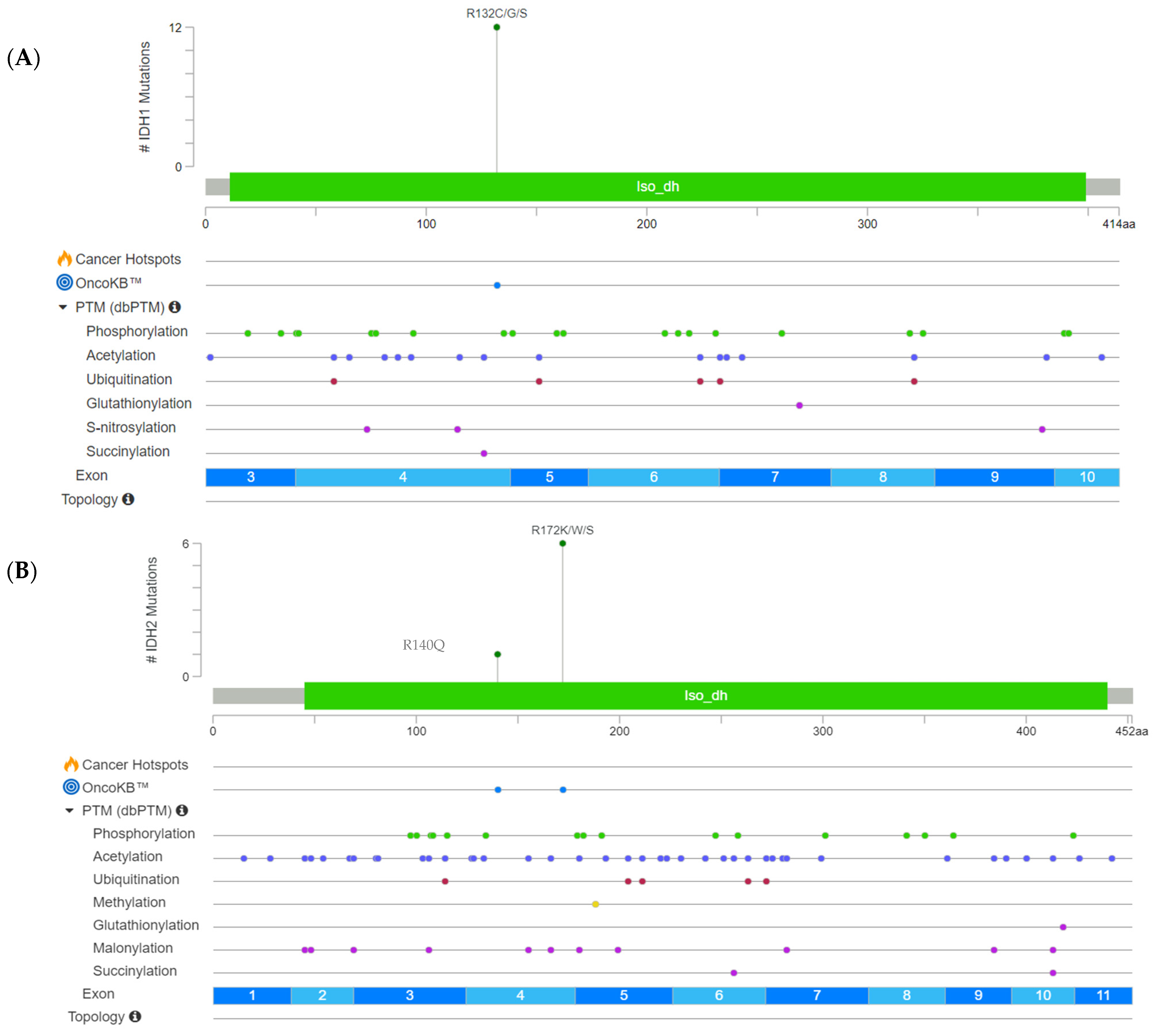
The detected hotspot mutations for IDH1 were located at codon 132 (p.Arg132Cys/Gly/Ser) as visible in the lolliplot below (Figure 2A). IDH2 mutations were detected at codon 140 (p.Arg140Gln) and codon 172 (p.Arg172Lys/Trp/Ser) (Figure 2B).
The following visualized representation, Figure 2, summarizes the IDH1/2 mutation landscape of the set of cholangiocarcinomas and provides a comprehensive overview of the detected genetic hotspot alterations with involved exons, respectively.

3.2. Overall Performance of the IdyllaTM IDH1-2 Mutation Assay Kit

The dataset shown in Table 2 includes 25 samples with DNA concentrations ranging from 0.233 ng/μL to 35.490 ng/μL.
IDH1/2 mutations were identified in 68% (17/25) of cases using both methods, with high concordance between NGS and Idylla™ results. Discrepancies were observed in two samples, 3 and 11, where Idylla™ detected no mutations, but NGS reported IDH1 and IDH2 mutations, respectively.
Median Cq values ranged from 29.7 to 35.2. Samples with lower DNA input (<0.5 ng/μL) had higher Cq values, indicating potential limitations of the Idylla™ platform in cases of low DNA yield.
Additionally, the limit of input amount was tested by a serial dilution of a positive case. A concentration of below 1 ng/µL results in a false negative evaluation.
Table 3 presents the overall concordance and Kappa Correlation Coefficient (r(Phi)) of IdyllaTM Biocartis compared to NGS.
Finally, the sensitivity for IdyllaTM IDH1 and IDH2 detection was 91.7% and 83.3%, respectively. The specificity for both genes was 100%, and the accuracy was 94.4% and 95.7% for IDH1 and IDH2, respectively. The concordance of the IdyllaTM Biocartis system and the reference method (NGS) showed a good overall correlation coefficient of r(Phi) = 0.8186 for IDH1 and r(Phi) = 0.887 for IDH2.
IDH1 variants were detected in 91.7% (11/12) and IDH2 variants in 85.7% (6/7) of previously confirmed positive cases. All IDH1 (13/13) and IDH2 (18/18) variants were accurately classified as wild types in alignment with previously confirmed IDH1- or IDH2-negative cases.

4. Discussion

Cholangiocarcinoma, or bile duct cancer, presents several unique challenges in diagnostics that are important to highlight. One of the primary clinical challenges is the often-late presentation of symptoms, which can lead to a delayed diagnosis. Patients may experience non-specific symptoms such as jaundice, weight loss, and abdominal pain, which can be mistaken for other conditions. Furthermore, the anatomical location of the bile ducts can complicate imaging and biopsy procedures, leading to challenges in obtaining accurate tissue samples for histological and molecular analysis, which underscores the importance of a multidisciplinary approach [31].
Additionally, the mutation patterns in cholangiocarcinoma can differ significantly from other cancers. For instance, mutations in the IDH1 and IDH2 genes, as well as alterations in the FGFR2 gene, are more commonly associated with intrahepatic cholangiocarcinoma. These specific mutation patterns can influence treatment options and prognostic outcomes, making genetic testing a crucial component of the diagnostic process [32].
The identification of cholangiocarcinoma patients with IDH1/IDH2 variants, which are mutually exclusive, may benefit from targeted therapies. Thus Ivosidenib, an IDH1 inhibitor, provides a new therapeutic option for patients with IDH1-mutant cholangiocarcinoma.
The IdyllaTM Biocartis system was evaluated under real-world conditions using FFPE samples or the corresponding extracted DNA. Compared to NGS diagnostics, it offers a significantly shorter turnaround time and utilizes a faster prescreening technology. In this project, the IdyllaTM IDH1-2 Mutation Assay Kit allowed the detection of 89.5% IDH1/2 (17/19) mutations of previously tested positive cases. In a comparative study by James P. Solomon’s Department of Pathology and Laboratory Medicine (Weill Cornell Medicine, New York), 39 samples from glioma patients treated on the New York-Presbyterian Hospital (NYPH) campus between 2018 and 2023 were examined regarding IDH1/2 status.
The prevalence of IDH mutations varies significantly between gliomas and cholangiocarcinomas. In gliomas, particularly in lower-grade gliomas and secondary glioblastomas, IDH mutations are quite common, with prevalence rates ranging from 40% to 80%, depending on the specific subtype. In contrast, cholangiocarcinomas, which are cancers of the bile ducts, have a lower prevalence of IDH mutations, typically around 10–20%.
In terms of performance differences, IDH mutations in gliomas are associated with distinct clinical and pathological features, often correlating with a better prognosis compared to IDH-wildtype tumors. In cholangiocarcinoma, IDH mutations can also influence the tumor’s behavior and response to treatment, but the overall impact on prognosis is less well defined compared to gliomas [33]. The results of the mentioned study of the two assays showed a concordance of 97% (38/39 samples) and an LOD of AF of 2.5–5% [34]. The results of the study largely correspond to the results of this work, which provides an overall concordance of 92% (23/25) regarding negative and positive IDH1/2 samples. After serial dilution, the detection limit with respect to the initial concentration was 1 ng/μL. Poor DNA quality can also interfere with the detection of a mutation, which can cause the detection limit to fluctuate.
The two discordant negative results can also be explained by the limited DNA quantity or quality. For example, in one case, the DNA concentration was finally 0.2 ng/µL, after diluting with water up to the required volume of 50 µL. This sample had the lowest DNA concentration in the entire collective. Unfortunately, it was not possible to confirm these discrepancies by repetition or another method due the limited material available. The quality of the DNA extracted from the samples can significantly impact the results. Generally, other possible reasons for obtaining different results may be due different methodological parameters, for instance, in the detection limits or primer sets. Some mutations may exist in subclonal populations within the tumor. If one method is more sensitive to detecting subclonal mutations, it may report a mutation that another method does not. In summary, discordant mutation status results from two different methods can arise from a combination of methodological differences, sample quality, technical errors, biological factors, and interpretation issues. While IDH mutations are less common in cholangiocarcinoma compared to gliomas, their presence can influence treatment decisions and prognostic assessments. A false negative result may lead to a misleading prognosis underestimating the aggressiveness of the tumor. A false negative result may lead to inadequate follow-up or surveillance strategies, increasing the risk of undetected recurrence. IDH mutations are often associated with specific tumor characteristics and may indicate a different biological behavior. If a patient is incorrectly classified as IDH-wildtype, they may not receive appropriate monitoring or treatment resulting in missed opportunities for targeted therapy, potentially affecting patient outcomes. Additionally, many clinical trials are designed to include patients with specific genetic mutations. A false negative IDH status could exclude patients from potentially beneficial clinical trials that target IDH-mutant cholangiocarcinoma. To resolve these discrepancies and avoid false negative results, we recommend an algorithmic approach for routine diagnostics: prescreening with the FastTrack method, and in the case of a wildtype result, follow with NGS.
As already mentioned, the fully automated analysis takes around 90 min, is quite user-friendly, and is suitable for routine application in molecular pathology. However, mutation coverage is limited to specific hotspot mutations, and it offers less flexibility in terms of customizing or expanding the testing range. With the DNA cartridge and the specific vial for IDH1/2 mutation analysis, five IDH1 mutations in codon R132, four IDH2 mutations in codon R140, and six IDH2 mutations in codon R172 can be detected. A maximum of eight instruments can be controlled with one IdyllaTM console, allowing up to eight analyses to be run simultaneously. The IdyllaTM platform is particularly appropriate for smaller laboratories; the instruments are suitable for stacking and therefore fit on small tables. The risk of contamination is very low even when using several instruments at the same time, as each cartridge represents a closed system. The cost of running a sample is around €100–200. Contrarily, Next-Generation Sequencing is more expensive but allows a comprehensive mutational analysis of several hundreds of genes simultaneously, including IDH1 and IDH2. NGS enables the detection of mutations, copy number variations, and gene fusions in a single test in parallel. The sensitivity is also high, meaning that rare mutations at low allele frequencies can also be detected. The disadvantages of this system are the higher complexity, which requires specialized expertise and extensive data analysis, as well as the longer turnaround time, which takes several days from sample preparation to result. In addition, the costs are higher than with PCR-based methods due to the complexity and the required infrastructure. Furthermore, there is a significantly higher risk of contamination here than with the IdyllaTM platform, due to the many intermediate steps of manual work when preparing several samples at the same time.

5. Conclusions

In conclusion, the IdyllaTM platform is a rapid method and is particularly suitable as a pre-screening method for mutations.
It can be easily implemented in pathology laboratories, because it is a fully automated test, not requiring DNA extraction, and easy to interpret. Table 4 shows the comparison of the Idylla™ and NGS workflow, emphasizing differences in turnaround time, complexity, and costs.
The Idylla™ workflow could be integrated into the diagnostic workflow in case of urgent molecular analysis for cholangiocarcinoma patients who require a rapid therapeutic decision, or when a platform for multiplex DNA analysis is not available. Highly complex tests such as NGS can make it difficult for clinicians to enroll their patients in a clinical trial in a timely manner. The repetition of this specific IdyllaTM assay needs sufficient material, which is why this procedure should be limited to emergency cases or when NGS analysis is not possible within ten working days or fails. The absence of an alteration requires additional testing by an orthogonal NGS-based method. Additionally, in order to provide detailed information about the mutation, a subsequent test using an NGS method would be suitable.
The choice of method for detecting IDH1 and IDH2 mutations in clinical practice depends on various factors, including the specific clinical context, the need for sensitivity and specificity, available resources, and the type of samples being analyzed. Often, a combination of these methods may be employed to ensure accurate and reliable detection of mutations, guiding treatment decisions and patient management.
For routine IDH1/2 diagnostics, we recommend an algorithmic approach starting with the FastTrack method followed by NGS for wildtype cases.

Author Contributions

Conceptualization, M.W. and S.E.; Data curation, M.W., S.E. and N.S.; Methodology, S.E. and N.S.; Project administration, M.W. and P.J.W.; Software, S.E. and N.S.; Supervision, M.W. and P.J.W.; Writing—original draft, M.W. and S.E.; Writing—review and editing, S.E., F.S., M.N.K., D.W. and P.J.W. All authors have read and agreed to the published version of the manuscript.

Funding

This research received no external funding.

Institutional Review Board Statement

Tissue/tumor samples and/or patient data used in this study were provided by the University Cancer Centre Frankfurt (UCT). Patient consent was waived due to the approval by the institutional Review Boards of the UCT and the Ethical Committee at the University Hospital Frankfurt (project-number: SGI-1-2021, 12 April 2021, Amendment 2024).

Informed Consent Statement

Patient consent was waived due to the approval by the institutional Review Boards of the UCT and the Ethical Committee at the University Hospital Frankfurt (project-number: SGI-1-2021).

Data Availability Statement

Data are contained within the article.

Conflicts of Interest

M.W. received honoraria from talks and advisory board roles from Amgen, AstraZeneca, Bayer, Biocartis, Diaceutics, Roche, and Sophia Genetics. P.J.W. has received consulting fees and honoraria for lectures by Bayer, Janssen-Cilag, Novartis, Roche, MSD, Astellas Pharma, Bristol-Myers Squibb, Thermo Fisher Scientific, Molecular Health, Guardant Health, Sophia Genetics, Qiagen, Eli Lilly, Myriad, Hedera Dx, and Astra Zeneca; research support was provided by Astra Zeneca and Roche. The authors declare that there is no relationship relevant to the manuscripts’ subject. All other authors declare no conflicts of interest.

References

  1. Krebs in Deutschland—Gallenblase Gallenwege, Zentrum für Krebsregisterdaten. Available online: https://www.krebsdaten.de/Krebs/DE/Content/Publikationen/Krebs_in_Deutschland/kid_2023/kid_2023_c23_c24_gallenblase.pdf?__blob=publicationFile (accessed on 21 May 2024).
  2. Tyson, G.L.; El-Serag, H.B. Risk factors for cholangiocarcinoma. Hepatology 2011, 54, 173–184. [Google Scholar] [CrossRef] [PubMed]
  3. DeOliveira, M.L.; Cunningham, S.C.; Cameron, J.L.M.; Kamangar, F.; Winter, J.M.; Lillemoe, K.D.M.; Choti, M.A.M.; Yeo, C.J.M.; Schulick, R.D.M. Cholangiocarcinoma: Thirty-one-year experience with 564 patients at a single institution. Ann. Surg. 2007, 245, 755–762. [Google Scholar] [CrossRef]
  4. Walter, D.; Ferstl, P.; Waidmann, O.; Trojan, J.; Hartmann, S.; Schnitzbauer, A.A.; Zeuzem, S.; Kraywinkel, K. Cholangiocarcinoma in Germany: Epidemiologic trends and impact of misclassification. Liver Int. 2018, 39, 316–323. [Google Scholar] [CrossRef] [PubMed]
  5. Kendall, T.; Verheij, J.; Gaudio, E.; Evert, M.; Guido, M.; Goeppert, B.; Carpino, G. Anatomical, histomorphological and molecular classification of cholangiocarcinoma. Liver Int. 2019, 39 (Suppl. S1), 7–18. [Google Scholar] [CrossRef]
  6. Aishima, S.; Oda, Y. Pathogenesis and classification of intrahepatic cholangiocarcinoma: Different characters of perihilar large duct type versus peripheral small duct type. J. Hepato-Biliary-Pancreat. Sci. 2015, 22, 94–100. [Google Scholar] [CrossRef] [PubMed]
  7. Liau, J.-Y.; Tsai, J.-H.; Yuan, R.-H.; Chang, C.-N.; Lee, H.-J.; Jeng, Y.-M. Morphological subclassification of intrahepatic cholangiocarcinoma: Etiological, clinicopathological, and molecular features. Mod. Pathol. 2014, 27, 1163–1173. [Google Scholar] [CrossRef] [PubMed]
  8. Hayashi, A.; Misumi, K.; Shibahara, J.; Arita, J.; Sakamoto, Y.; Hasegawa, K.; Kokudo, N.; Fukayama, M. Distinct Clinicopathologic and Genetic Features of 2 Histologic Subtypes of Intrahepatic Cholangiocarcinoma. Am. J. Surg. Pathol. 2016, 40, 1021–1030. [Google Scholar] [CrossRef]
  9. Vogel, A.; Bridgewater, J.; Edeline, J.; Kelley, R.; Klümpen, H.; Malka, D.; Primrose, J.; Rimassa, L.; Stenzinger, A.; Valle, J.; et al. Biliary tract cancer: ESMO Clinical Practice Guideline for diagnosis, treatment and follow-up. Ann. Oncol. 2023, 34, 127–140. [Google Scholar] [CrossRef] [PubMed]
  10. Koerkamp, B.G.; Fong, Y. Outcomes in biliary malignancy. J. Surg. Oncol. 2014, 110, 585–591. [Google Scholar] [CrossRef]
  11. Zeitler, E. The journal of CardioVascular and Interventional Radiology. Cardiovasc. Interv. Radiol. 1990, 13, 1–2. [Google Scholar] [CrossRef]
  12. Zhu, Y.; Kwong, L.N. IDH1 Inhibition Reawakens the Immune Response against Cholangiocarcinoma. Cancer Discov. 2022, 12, 604–605. [Google Scholar] [CrossRef] [PubMed]
  13. Farshidfar, F.; Zheng, S.; Gingras, M.-C.; Newton, Y.; Shih, J.; Robertson, A.G.; Hinoue, T.; Hoadley, K.A.; Gibb, E.A.; Roszik, J.; et al. Integrative Genomic Analysis of Cholangiocarcinoma Identifies Distinct IDH-Mutant Molecular Profiles. Cell Rep. 2017, 18, 2780–2794. [Google Scholar] [CrossRef] [PubMed]
  14. Dang, L.; White, D.W.; Gross, S.; Bennett, B.D.; Bittinger, M.A.; Driggers, E.M.; Fantin, V.R.; Jang, H.G.; Jin, S.; Keenan, M.C.; et al. Cancer-associated IDH1 mutations produce 2-hydroxyglutarate. Nature 2009, 462, 739–744, Erratum in Nature 2010, 465, 966. [Google Scholar] [CrossRef] [PubMed]
  15. Xiang, X.; Liu, Z.; Zhang, C.; Li, Z.; Gao, J.; Zhang, C.; Cao, Q.; Cheng, J.; Liu, H.; Chen, D.; et al. IDH Mutation Subgroup Status Associates with Intratumor Heterogeneity and the Tumor Microenvironment in Intrahepatic Cholangiocarcinoma. Adv. Sci. 2021, 8, 2101230. [Google Scholar] [CrossRef]
  16. Kinzler, M.N.; Jeroch, J.; Klasen, C.; Himmelsbach, V.; Koch, C.; Finkelmeier, F.; Trojan, J.; Zeuzem, S.; Pession, U.; Reis, H.; et al. Impact of IDH1 mutation on clinical course of patients with intrahepatic cholangiocarcinoma: A retrospective analysis from a German tertiary center. J. Cancer Res. Clin. Oncol. 2023, 149, 6391–6398. [Google Scholar] [CrossRef]
  17. Valcarcel-Jimenez, L.; Frezza, C. Fumarate hydratase (FH) and cancer: A paradigm of oncometabolism. Br. J. Cancer 2023, 129, 1546–1557. [Google Scholar] [CrossRef] [PubMed]
  18. Biocartis—The Idylla Platform. Available online: https://www.biocartis.com/en/meet-idylla (accessed on 26 April 2024).
  19. Delgado-García, M.; Weynand, B.; Gómez-Izquierdo, L.; Hernández, M.J.; Blanco, Á.M.; Varela, M.; Matias-Guiu, X.; Nadal, E.; Márquez-Lobo, B.; Alarcão, A.; et al. Clinical performance evaluation of the Idylla™ EGFR Mutation Test on formalin-fixed paraffin-embedded tissue of non-small cell lung cancer. BMC Cancer 2020, 20, 275. [Google Scholar] [CrossRef]
  20. Ruiz-Villalba, A.; Ruijter, J.M.; Hoff, M.J.B.v.D. Use and Misuse of Cq in qPCR Data Analysis and Reporting. Life 2021, 11, 496. [Google Scholar] [CrossRef]
  21. Ion GeneStudioTM S5 Prime System, ThermoFisher Scientifc. Available online: https://www.thermofisher.com/order/catalog/product/A38196?SID=srch-srp-A38196 (accessed on 24 May 2024).
  22. Ion Chef Instrument, ThermoFisher Scientific. Available online: https://www.thermofisher.com/order/catalog/product/4484177 (accessed on 24 May 2024).
  23. Deutsche Akkreditierungsstelle. Available online: https://www.dakks.de/de/home.html (accessed on 5 December 2024).
  24. Landrum, M.J.; Chitipiralla, S.; Brown, G.R.; Chen, C.; Gu, B.; Hart, J.; Hoffman, D.; Jang, W.; Kaur, K.; Liu, C.; et al. ClinVar: Improvements to accessing data. Nucleic Acids Res. 2020, 48, D835–D844. [Google Scholar] [CrossRef]
  25. Sondka, Z.; Dhir, N.B.; Carvalho-Silva, D.; Jupe, S.; Madhumita; McLaren, K.; Starkey, M.; Ward, S.; Wilding, J.; Ahmed, M.; et al. COSMIC: A curated database of somatic variants and clinical data for cancer. Nucleic Acids Res. 2024, 52, D1210–D1217. [Google Scholar] [CrossRef] [PubMed]
  26. Chen, S.; Francioli, L.C.; Goodrich, J.K.; Collins, R.L.; Kanai, M.; Wang, Q.; Alföldi, J.; Watts, N.A.; Vittal, C.; Gauthier, L.D.; et al. A genomic mutational constraint map using variation in 76,156 human genomes. Nature 2024, 625, 92–100. [Google Scholar] [CrossRef] [PubMed]
  27. Suehnholz, S.P.; Nissan, M.H.; Zhang, H.; Kundra, R.; Nandakumar, S.; Lu, C.; Carrero, S.; Dhaneshwar, A.; Fernandez, N.; Xu, B.W.; et al. Quantifying the Expanding Landscape of Clinical Actionability for Patients with Cancer. Cancer Discov. 2024, 14, 49–65. [Google Scholar] [CrossRef]
  28. Kitts, A.; Sherry, S. The Single Nucleotide Polymorphism Database (dbSNP) of Nucleotide Sequence Variation. In The NCBI Handbook; McEntyre, J., Ostell, J., Eds.; National Center for Biotechnology Information: Bethesda, MD, USA, 2002. Available online: https://www.ncbi.nlm.nih.gov/books/NBK21088/ (accessed on 5 December 2024).
  29. Cerami, E.; Gao, J.; Dogrusoz, U.; Gross, B.E.; Sumer, S.O.; Aksoy, B.A.; Jacobsen, A.; Byrne, C.J.; Heuer, M.L.; Larsson, E.; et al. The cBio cancer genomics portal: An open platform for exploring multidimensional cancer genomics data. Cancer Discov. 2012, 2, 401–404. [Google Scholar] [CrossRef] [PubMed]
  30. den Dunnen, J.T.; Dalgleish, R.; Maglott, D.R.; Hart, R.K.; Greenblatt, M.S.; McGowan-Jordan, J.; Roux, A.-F.; Smith, T.; Antonarakis, S.E.; Taschner, P.E.; et al. HGVS Recommendations for the Description of Sequence Variants: 2016 Update. Hum. Mutat. 2016, 37, 564–569. [Google Scholar] [CrossRef] [PubMed]
  31. Patel, T. Cholangiocarcinoma—Controversies and challenges. Nat. Rev. Gastroenterol. Hepatol. 2011, 8, 189–200. [Google Scholar] [CrossRef] [PubMed]
  32. Lowery, M.A.; Ptashkin, R.; Jordan, E.; Berger, M.F.; Zehir, A.; Capanu, M.; Kemeny, N.E.; O’Reilly, E.M.; El-Dika, I.; Jarnagin, W.R.; et al. Comprehensive Molecular Profiling of Intrahepatic and Extrahepatic Cholangiocarcinomas: Potential Targets for Intervention. Clin. Cancer Res. 2018, 24, 4154–4161. [Google Scholar] [CrossRef] [PubMed]
  33. Boscoe, A.N.; Rolland, C.; Kelley, R.K. Frequency and prognostic significance of isocitrate dehydrogenase 1 mutations in cholangiocarcinoma: A systematic literature review. J. Gastrointest. Oncol. 2019, 10, 751–765. [Google Scholar] [CrossRef]
  34. Solomon, J.P.; Munoz-Zuluaga, C.; Slocum, C.; Dillard, A.; Cong, L.; Wang, J.; Lindeman, N.; Kluk, M.; Liechty, B.; Pisapia, D.; et al. Evaluation of the rapid Idylla IDH1-2 mutation assay in FFPE glioma samples. Diagn. Pathol. 2024, 19, 70. [Google Scholar] [CrossRef]
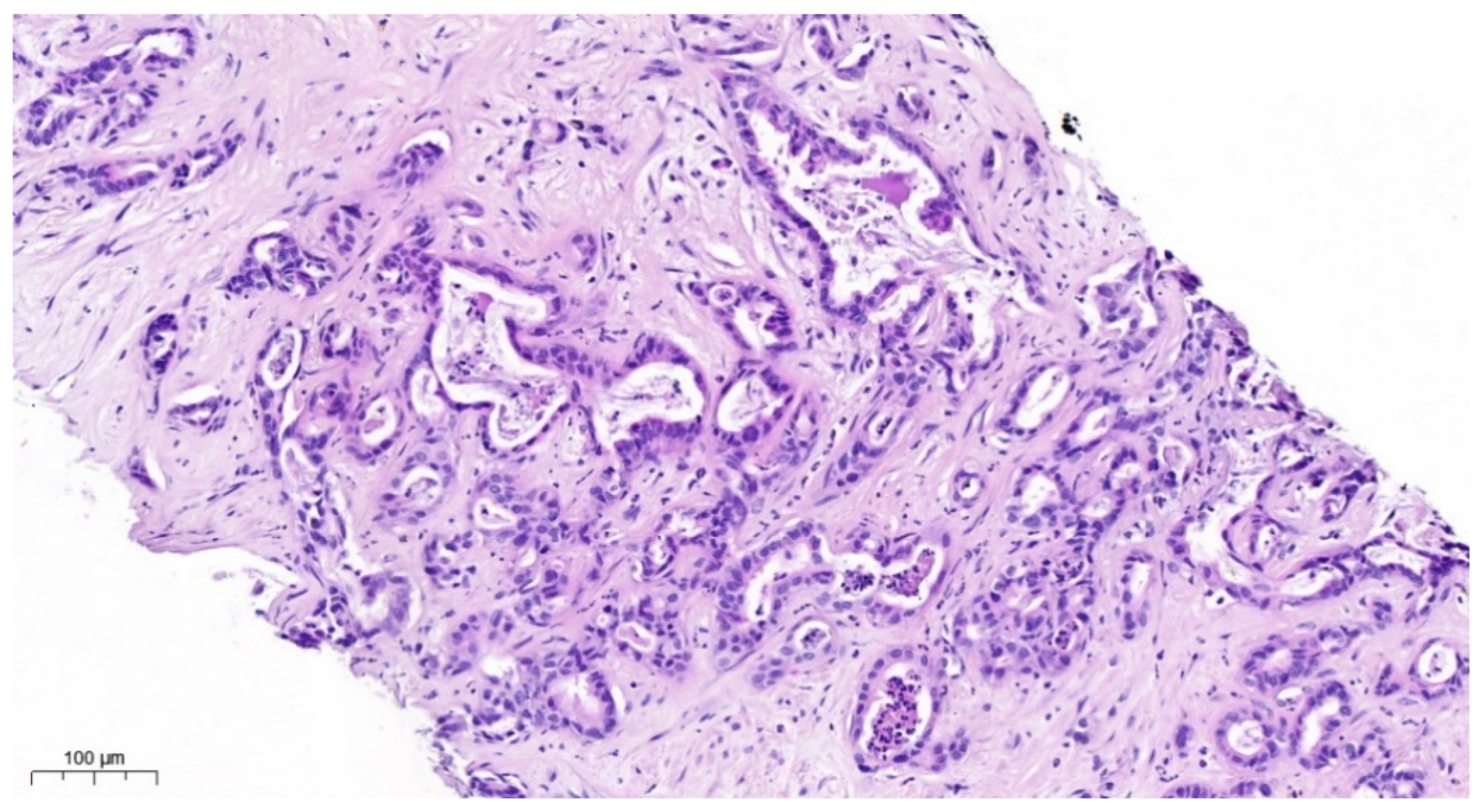
Figure 1. HE staining. iCCA consists of infiltrating glands in an abundant desmoplastic; fibrous stroma glands are lined by atypical cuboidal cells with varying degrees of pleomorphism intraluminal cellular debris.
Cancers 17 00820 g001
Figure 2. NGS-based detected IDH1 (A) and IDH2 (B) missense mutation types with respect to die hotspot region characterized by OncoKB and post-translational modifications (PTMs).
Cancers 17 00820 g002
Table 1. Patient collective with respect to demographic features as well as the IDH1/2 mutation status determined with NGS. F = female, M = male, TCC = tumor cell content, AA = amino acid, AF = allele frequency.
Study-No.GenderAgeTCC [%]IDH1/2
Status
Nucleotide SubstitutionAA SubstitutionAF [%]
1F5715IDH1c.394C>Tp.Arg132Cys15.40
2M7415–20IDH1c.394C>Tp.Arg132Cys15.95
3F5820–25 IDH1c.394C>Tp.Arg132Cys17.70
4F5840IDH1c.394C>Tp.Arg132Cys33.03
5M4030IDH1c.394C>Gp.Arg132Gly6.00
6M6060IDH1c.394C>Tp.Arg132Cys17.68
7M5820IDH1c.394C>Tp.Arg132Cys9.79
8M6970IDH1c.394C>Ap.Arg132Ser54.10
9M5830IDH2c.514A>Tp.Arg172Trp66.00
10M6350IDH2c.514A>Tp.Arg172Trp21.40
11M5360IDH2c.515G>Ap.Arg172Lys32.10
12M7615Negative------
13M8250Negative------
14M6080Negative------
15F5880Negative------
16M7130Negative------
17M8370Negative------
18M6770IDH1c.394C>Tp.Arg132Cys30.50
19M7540IDH1c.394C>Tp.Arg132Cys22.22
20M6320IDH1c.394C>Tp.Arg132Cys19.61
21F5720IDH1c.394C>Tp.Arg132Cys32.99
22M5375IDH2c.515G>Ap.Arg172Lys52.03
23F6345IDH2c.516G>Cp.Arg172Ser20.26
24M7860IDH2c.515G>Ap.Arg172Lys36.85
25M6640IDH2c.419G>Ap.Arg140Gln8.17
Table 2. Patient collective with respect to the IDH1/2 mutation status (NGS vs. IdyllaTM). Median Cq from internal control. * = elevated; median Cq indicates low DNA input or quality.
Study-No.DNA (ng/μL)NGS IdyllaTM
Biocartis
CodonMedian Cq
11.114IDH1IDH113232.4
20.406IDH1IDH113233.9
30.233IDH1negative--35.2 *
41.629IDH1IDH113231.6
51.125IDH1IDH113233.4
60.464IDH1IDH113234.0
70.442IDH1IDH113234.1
88.426IDH1IDH113231.2
91.278IDH2IDH217232.9
1035.490IDH2IDH217230.0
1121.500IDH2negative--31.2
122.145negativenegative--34.8
132.336negativenegative--32.5
141.804negativenegative--34.6
159.804negativenegative--32.1
162.801negativenegative--33.3
1714.586negativenegative--30.5
180.663IDH1IDH113235.0
193.503IDH1IDH113232.2
200.519IDH1IDH113234.1
210.493IDH1IDH113235.2 *
2226.660IDH2IDH2
IDH2
140
172
29.7
230.486IDH2IDH217235.1 *
248.540IDH2IDH217232.0
252.212IDH2IDH214032.7
Table 3. Concordance analysis for IDH1 and IDH2.
IdyllaTM vs. NGSIDH1IDH2
Sensitivity91.7%83.3%
Specificity100%100%
Accuracy94.4%95.7%
Correlation coefficient r(Phi)0.81860.887
Concordance
Right positive 91.7%85.7%
Right negative100%100%
r(Phi): The value of r ranges between −1 and 1. A correlation of −1 shows a perfect negative correlation, while a correlation of 1 shows a perfect positive correlation. A correlation of 0 shows no relationship between the movement of the two variables.
Table 4. Comparison of the Idylla™ and NGS workflow.
IdyllaTM BiocartisNGS
Technologyquantitaive real-time PCRsequencing
Gene coveragelimited 95%
Time required<2 hseveral days
Automationfully automateddepending on the platform
Risk of cross contaminationlow, closed systemmedium to high
Cost/samplelowhigh
User-friendlinesshigh, easy to usetechnical expertise required
Disclaimer/Publisher’s Note: The statements, opinions and data contained in all publications are solely those of the individual author(s) and contributor(s) and not of MDPI and/or the editor(s). MDPI and/or the editor(s) disclaim responsibility for any injury to people or property resulting from any ideas, methods, instructions or products referred to in the content.